Article

Improving Clinical Diagnosis of Transmissible Spongiform Encephalopathies in Sheep: Which Signs Are Important?

Department of Pathology & Animal Sciences, Animal & Plant Health Agency Weybridge, Addlestone KT15 3NB, UK
*
Author to whom correspondence should be addressed.
Animals 2025, 15(9), 1310; https://doi.org/10.3390/ani15091310
Submission received: 25 March 2025 / Revised: 28 April 2025 / Accepted: 29 April 2025 / Published: 1 May 2025
(This article belongs to the Special Issue Prion Diseases in Animals)

Simple Summary

Scrapie is a transmissible spongiform encephalopathy (TSE) in sheep that cannot be diagnosed in live animals, and disease suspicion is based on a clinical examination. An examination protocol was assessed for its suitability as a clinical diagnostic tool and used to examine 1002 sheep that were exposed naturally or experimentally to three scrapie strains, 312 of which had a TSE confirmed, or served as non-exposed controls. The combined occurrence of two or more clinical signs associated with a TSE was compared between TSE-positive and negative sheep, and a classification tree model was also used to determine clinical markers most important for disease suspicion. Incoordination and a response to scratching the sheep’s back were the most important signs for disease suspicion. Both approaches had adequate sensitivities, specificities and predictive values for a clinical test, and could be used to aid in the suspicion of scrapie in both naturally and experimentally infected sheep.

Abstract

Scrapie is a notifiable transmissible spongiform encephalopathy (TSE) in sheep that relies on clinical examinations for reporting suspects. A short examination protocol was used in 1002 sheep to define clinical markers suggestive of scrapie. Sheep were naturally or experimentally exposed to a classical, atypical scrapie or bovine spongiform encephalopathy agent; 312 were positive for a transmissible spongiform encephalopathy (TSE) by brain examination and included non-exposed controls. Assessed signs were posture, behaviour, menace, scratch and blindfolding response, wool loss and skin changes, body condition, incoordination and tremor. First, the combined occurrence of two or more clinical signs was compared between TSE-positive and negative sheep. Second, the importance of clinical markers was determined in a general classification and regression tree model. The main clinical markers to predict TSEs according to the tree model were incoordination and a positive scratch test. Test sensitivities and specificities were 70.8–81.5% and 96.1–93.0%, respectively, and predictive values above 87%. The results suggest that the short clinical protocol, which assesses the presence of certain clinical signs associated with a TSE in sheep and is quick to perform, may be useful to reach a suspect diagnosis in both naturally and experimentally generated TSEs.

1. Introduction

Scrapie is a fatal neurological disease of sheep and goats and the oldest known naturally occurring transmissible spongiform encephalopathy (TSE), compared to Creutzfeldt–Jakob disease (CJD) in humans, chronic wasting disease in deer and bovine spongiform encephalopathy (BSE) in cattle. Scrapie is a notifiable disease, and suspicion of disease must be reported to the relevant authority. Experimental studies have demonstrated that BSE can also be transmitted to sheep [1], which led to the concern that sheep may have become infected through consumption of feed contaminated with the BSE agent, particularly in countries that had cases of BSE in cattle, and may contribute to the occurrence of cases of variant CJD. In 2002, the European Union introduced a scrapie surveillance program to test a proportion of small ruminants that died or were subject to slaughter for human consumption (active TSE surveillance) [2], which identified a goat infected with the BSE agent [3]. Increased surveillance in sheep and cattle also resulted in the discovery of atypical forms: scrapie in sheep, first reported in Norway in clinical suspects [4], then in many other countries, which is believed to be a sporadic disease in older sheep with biological and biochemical features distinct from the “classical” form of scrapie [5]. It also led to the detection of atypical BSE in cattle, which is different from “classical” BSE [6]. Questions remain about the ability of different TSE strains to change properties in the same (atypical scrapie to CH1641 scrapie in sheep [7]) or different (e.g., ovine atypical scrapie to classical BSE in cattle [8,9]) host, at least in experimental models.
In the absence of a reliable ante mortem test, disease suspicion is based on clinical signs, which poses considerable problems: countries that do not use an active surveillance program rely on farmers and veterinarians to report disease suspicion, which requires them to be familiar with the clinical presentation. This is especially challenging in countries that do not have the disease diagnosed or only find sporadic cases of atypical scrapie [10,11]. Furthermore, clinical signs may be different between classical scrapie [12,13], atypical scrapie [14,15,16,17] and other TSEs not normally expected in small ruminants, such as BSE [18,19,20], and—unless accompanied by clear neurological signs like ataxia—may present as unspecific signs (weight loss, pruritus) that can be seen in various other diseases. In addition, the prion protein genotype of the affected sheep may influence the clinical presentation [21,22].
Clinical protocols have been developed to aid in the clinical diagnosis of scrapie, but some were solely based on detection of classical scrapie [23,24]), and some signs may require more clinical expertise (e.g., hyporeflexia, cardiac arrhythmia [24]). By contrast, a short clinical examination protocol was presented that could be used for the clinical diagnosis of both scrapie types, including BSE [25], but it did not include data on clinically affected animals to determine diagnostic sensitivity and specificity. The current study was carried out in sheep that were part of multiple research projects to determine the usefulness of this protocol for the clinical diagnosis of sheep and whether the signs assessed as part of this protocol could be ranked in their importance to potentially concentrate on fewer clinical signs without impacting diagnostic sensitivity and specificity. This may be beneficial if time and space for examinations are limited, such as in an abattoir, or may be applicable to experimental TSE studies in sheep to determine clinical end-point.

2. Materials and Methods

All procedures involving animals were approved under the Animal Scientific Procedures Act 1989 by the British Home Office with the appropriate project licences. These licences are only granted after they have been reviewed first by the institute’s Animal Welfare and Ethical Review Body.
A total of 1002 sheep were examined; they were experimentally or naturally infected with TSE agents, exposed to TSE agents because they were housed with known TSE cases, reported clinical suspects from farms, or sheep that served as non-challenged controls in TSE experiments. Experimental inoculation was by the intracerebral, subcutaneous or oral route, with classical or atypical scrapie or classical BSE brain homogenate as inoculum. There were 594 female, 23 male and 385 castrated male sheep with a mean age of 52 months from birth or inoculation to death (range: 3 months to 141 months; not known in 6 sheep). Multiple breeds were included, and the four most frequent were Cheviot (31%), Romney (21%), Poll Dorset pure and crossbreeds (16%) and Suffolk pure and crossbreeds (13%). The examined sheep population represented all five National Scrapie Plan types of prion protein genotypes (Type I—most resistant to Type V—most susceptible) [26]. All sheep were examined whilst housed in appropriate accommodation. The time from clinical examination to death ranged from 0 to 42 days (mean 6, median 5 days).
TSE status was based on brain examination by postmortem test as gold standard: histopathology, immunohistochemistry, Western immunoblot or rapid test (WOAH Terrestrial manual chapter 3.8.11 scrapie). Table 1 displays TSE status by TSE strain infection or exposure. The full animal details are provided as Supplementary File Table S1: Sheep data.
Percentages in each row represent the proportion of animals compared to the total within each row.
All animals were clinically examined by the same veterinary clinician following the short clinical examination protocol [25], and data from the last clinical examination prior to euthanasia (in case of repeated clinical examinations) were used. If animals underwent a full neurological examination [27], only signs that were assessed for the short clinical assessment protocol were taken into consideration. Assessed sign categories were posture, behaviour, menace response, scratch response, wool and skin changes, body condition score, response to blindfolding, incoordination and tremor. Not all signs were assessed in all sheep, particularly the response to blindfolding, often to reduce the risk of injury to nervous sheep that tend to panic.
Table 2 displays the specific signs exhibited by sheep to be included as abnormalities in each category.
Blinding was generally not possible unless the sheep was a field suspect and not experimentally infected. The inoculation status was generally known because of specific entry and exit procedures in some animal studies (e.g., non-exposed controls were examined first) but rarely prion protein genotype unless the animal was part of a study using only one prion protein genotype, or live classical scrapie status (because the animal was diagnosed with classical scrapie by rectal biopsy examination whilst alive).
Clinical sign analysis was carried out in two parts. In the first part, the numbers of TSE test-positive and -negative sheep displaying abnormalities in each sign category were compared to assess whether the combined occurrence of two or more signs in the categories “behaviour”, “menace response”, “scratch response”, “alopecia or skin lesions”, “blindfolding response”, “incoordination” and “tremor” was more associated with a TSE. For alopecia or skin lesions, only generalised alopecia or skin lesions were counted because local alopecia or skin lesions restricted to one small area (e.g., head) were expected in housed sheep when accessing a hay rack. Data between TSE-positive and -negative sheep were compared by Chi-Square test (Statistica Data Science Workbench version 14, Cloud Software Group, Inc., Fort Lauderdale, FL, USA), with p < 0.05 considered significant.
In the second part, a general classification and regression tree model (Statistica Data Science Workbench version 14) was applied to all data (sheep that had all signs assessed) to determine whether certain clinical signs were more predictive of a TSE. The dependent variable was TSE status, whilst the clinical signs were used as predictor variables. This included body condition (poor or good) and localised or generalised wool or skin lesions. Subsequently, to increase the dataset, the decision tree, which determined signs of importance for classification, was run on sheep with data gaps but that had those signs of the decision tree model assessed. The statistical software also produced an importance ranking for each predictor variable (clinical sign) on a 0–100 scale to list the relative importance of each predictor variable for producing the final tree. The computational method of calculating ranks may mean that a variable could be important, even though it might not have been used for any split in the decision tree.
Sensitivity (true positive cases divided by the sum of true positive and false negative cases), specificity (true negative cases divided by the sum of true negative and false positive cases), positive predictive value (PPV, true positive cases divided by the sum of true and false positive cases) and negative predictive value (NPV, true negative cases divided by the sum of true and false negative cases) were then calculated.
Whilst this study aimed to determine clinical markers for any type of naturally occurring TSEs (including BSE, as it had occurred in goats), clinical signs were also separated by TSE strains to assess whether strain-specific differences exist (part 3). For this, the general classification and regression tree model was applied to TSE-positive sheep that had all signs assessed and were diagnosed as classical scrapie, atypical scrapie or BSE.

3. Results

3.1. Comparison of Clinical Signs Between TSE-Positive and -Negative Sheep

Table 3 lists the frequency of clinical signs associated with a TSE that were observed in the sheep, grouped by TSE status. More TSE-positive than -negative sheep displayed abnormalities, which was highly significant for all individual signs (p < 0.0001).
About half of the TSE-positive sheep had a positive scratch test, were uncoordinated or displayed abnormal behaviour, which were the most frequent signs. Abnormal behaviour was also the most frequent sign in TSE-negative sheep, although only recorded for 12%.
At least one of the signs were seen in 293 (93.9%) TSE-positive sheep and 293 (42.5%) TSE-negative sheep. If only those sheep that had all signs assessed were included, it increased to 97.2% of 211 TSE-positive sheep and 60.3% of 302 TSE-negative sheep.

3.2. Presence of Two or More Clinical Signs in TSE-Positive and -Negative Sheep

A combination of two or more signs (excluding poor body condition and abnormal posture) were seen in 221 TSE-positive sheep (70.8% of 312) and 27 TSE-negative sheep (3.9% of 690). This is equivalent to a clinical diagnostic sensitivity of 70.8%, a specificity of 96.1%, PPV of 89.1% and NPV of 87.9%.
The most frequent sign combination seen in approximately 30% of TSE-positive sheep was abnormal behaviour with tremor (93 sheep), closely followed by positive scratch test with any wool loss or skin lesion (89 sheep) and ataxia with tremor (88 sheep). All TSE-positive sheep had at least one combination of two clinical signs if poor body condition and abnormal posture were included. The most frequent sign combination seen in TSE-negative sheep was abnormal behaviour with positive scratch test (21 sheep, 3.0%).

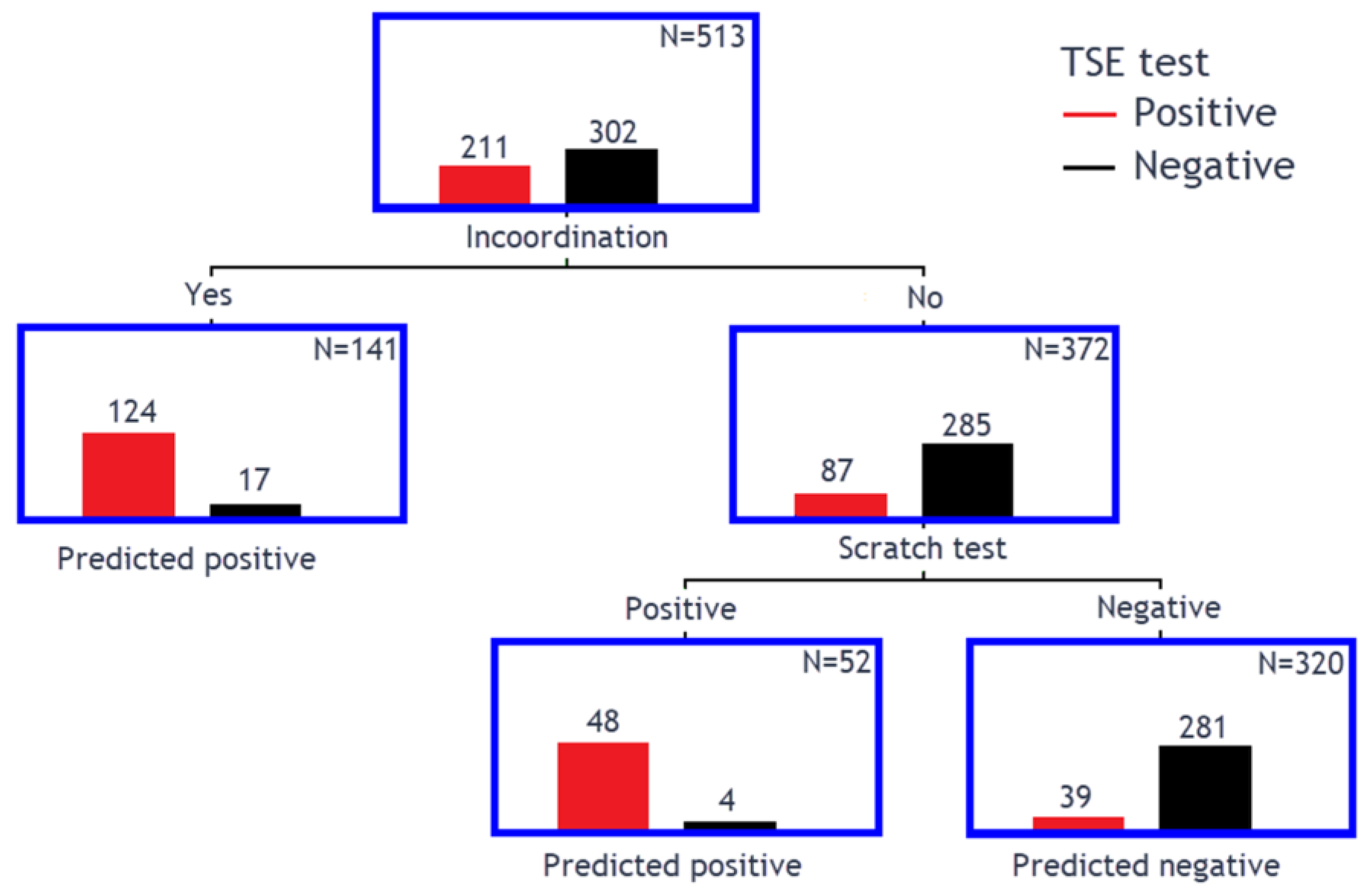
3.3. Classification Tree Model to Determine Clinical Markers for TSEs

This model utilised those examinations that had all clinical signs assessed, which were available for 513 sheep. Table 4 summarises the importance of each sign to generate the decision tree calculated by the software.
The resulting decision tree is shown in Figure 1, correctly predicting a TSE in 172 of 211 TSE-positive sheep (81.5% sensitivity) and TSE-freedom in 281 of 320 TSE-negative sheep (93.0% specificity). PPV and NPV were 89.1% and 87.8%, respectively.
Four TSE-positive and one TSE-negative sheep did not have incoordination or the scratch test assessed, which left a total of 997 sheep where the decision tree model could be applied: a TSE was correctly predicted in 242 of 308 TSE-positive sheep (78.6% sensitivity) and TSE-freedom in 652 of 689 TSE-negative sheep (94.6% specificity). PPV and NPV were 86.7% and 90.8%, respectively. Supplementary File Video S1 shows an experimentally infected atypical scrapie case (case 0005/22) with ataxia, and Supplementary File Video S2 shows a naturally infected classical scrapie case (case 0004/22) with a positive scratch test in the absence of ataxia.

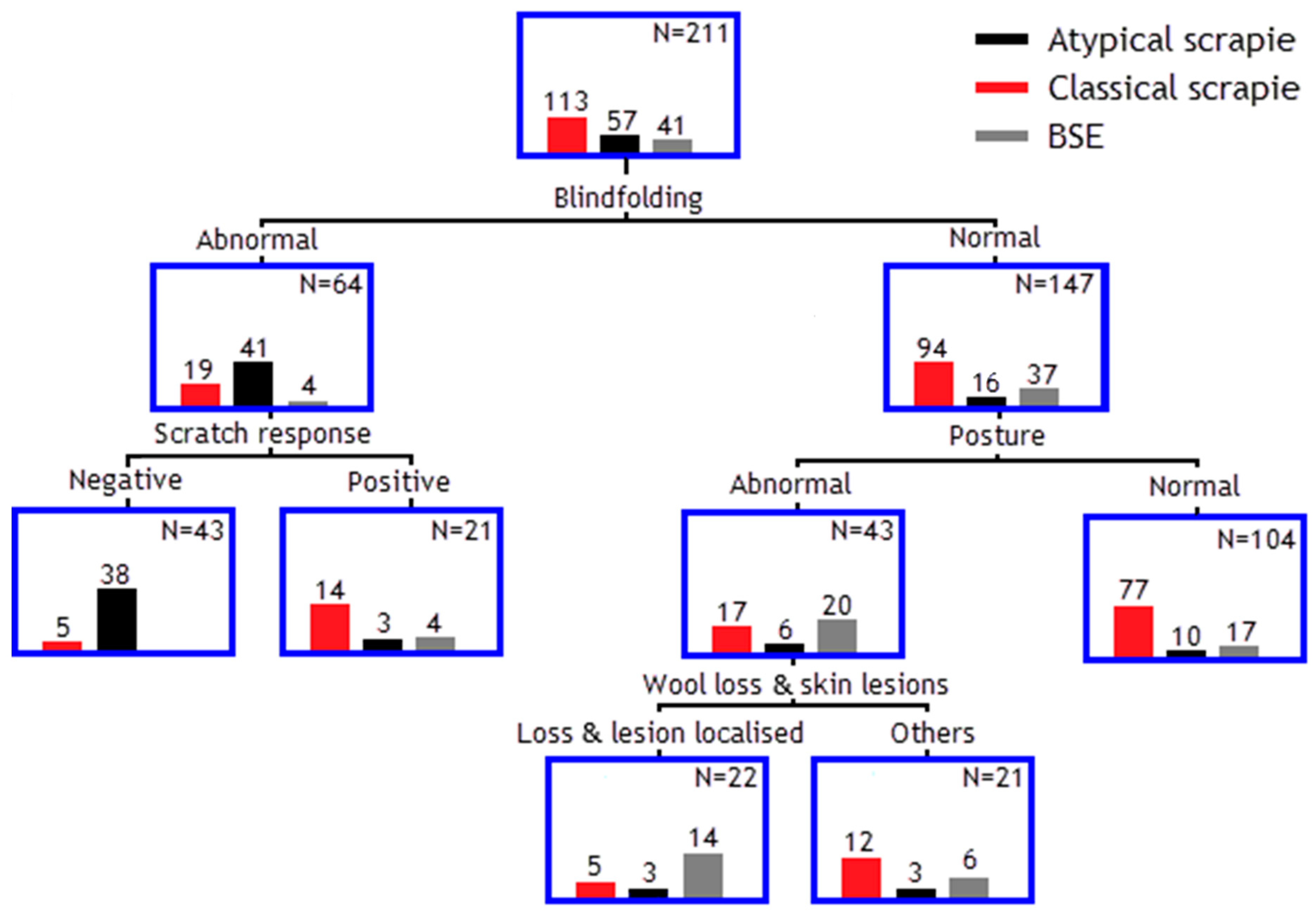
3.4. Classification Tree Model to Separate Different TSE Strains

This model was applied to 211 sheep that were diagnosed with a TSE (classical, atypical scrapie or BSE) and had all clinical signs assessed, with the outcome displayed in Figure 2. An abnormal blindfolding response was seen in the majority (71.9%) of atypical scrapie cases, and 92.7% of these did not have a scratch response (left branch of the decision tree). BSE and classical scrapie cases were more likely to be clinically unremarkable when blindfolded (90.2% of BSE cases and 83.1% of classical scrapie cases) and of these, postural deficits seemed to be more frequent in BSE cases (54.0%) than in classical scrapie cases (18.1%); see right branch of the decision tree.

4. Discussion

Compared to diagnostic laboratory tests to confirm TSEs, for example, scrapie with a test sensitivity of at least 94% [28], clinical test sensitivity is considerably lower. For example, clinical test sensitivity to diagnose BSE in cattle was only 50% on a largely BSE-negative cattle population, although specificity was 99.5% [29]. In a population with more BSE-affected cattle, various combinations of main signs had sensitivities that ranged from 85.7 to 94.0%, whilst specificities were low (36.4 to 63.6%) [30]. The sensitivities in the current study are comparable to that of clinical tests for BSE in cattle but with higher specificity. With a PPV and NPV over 87%, the criteria chosen for disease suspicion were better than those published for BSE in cattle when the combination of changes in behaviour, sensitivity and locomotion were taken into consideration (PPV: 84.0%, NPV: 66.7%) [30].
Currently validated ante-mortem tests, such as the examination of rectal biopsies for disease-associated prion protein (PrPSc) by screening tests or immunohistochemistry [31,32], are only useful in animals with peripheral PrPSc distribution that can be detected by these tests, which is absent in atypical scrapie and sheep with more resistant genotypes.
It is unlikely that a screening test will be available for sheep that is reliable, cheap and quick to carry out. Ultrasensitive prion detection tests, such as Real-Time Quaking Induced Conversion, are promising in diagnosing CJD in humans, although they are generally applied to cerebrospinal fluid samples, and false negative results can occur [33]. PrPSc has been successfully detected in highly susceptible (VRQ/VRQ) sheep using protein misfolding cyclic amplification. However, only a small number of sheep were tested, all with the same prion protein genotype and only exposed to classical scrapie. This does not allow a conclusion on its suitability to detect classical scrapie in more resistant genotypes and in animals infected with strains like atypical scrapie where peripheral distribution via blood may be below the detection limit if it occurs at all. As such, diagnosis in live animals is currently only possible by clinical examination and will continue to be important in countries without facilities and skills to carry out these laboratory tests.
Clinical diagnosis of scrapie requires the examiner to be familiar with its clinical presentation, which can be challenging if the disease prevalence is very low, and scrapie may not be included in the differential diagnosis of sheep with ill-thrift or neurological signs. Breeding for resistance has contributed to the reduction of cases world-wide. However, atypical scrapie cases continue to occur sporadically, even in countries with no evidence of classical scrapie, but are usually detected by active surveillance of healthy slaughtered sheep or fallen stock and not by reporting of clinical suspects (passive surveillance) [34], which is the only type of surveillance in many countries.
The short examination protocol tested here is easy to implement and very quick to perform, although it concentrates on clinical signs associated with a TSE. Contrary to a neurological examination, it does not test the function of specific neural pathways to determine possible lesion in the nervous system and reach a diagnosis, which requires more expertise in conducting the examination and interpreting its findings. When interpreting the results from the short clinical examination protocol to aid in the clinical diagnosis of scrapie in sheep, it was suggested that the presence of individual signs, such as a repeatable response to scratching, tremor, abnormal behaviour, circling, collapsing episodes, ataxia, or an absent menace response, may have various causes, but the combined occurrence of two or more of these signs is highly suggestive of scrapie in sheep or goats [25]. Test sensitivity was only 71%, meaning a considerable number of TSE-affected sheep were missed, but the specificity was high at 96%, so not many sheep would have been wrongly classified as TSE-positive. The classification and decision tree model demonstrated that actually only two signs appeared to be relevant: incoordination, which has been reported for BSE in sheep [18,19], classical [21,35] and atypical scrapie [4,15], and a positive scratch test, usually associated with pruritus, with is a feature of BSE in sheep [19] and classical scrapie, albeit genotype-dependent [21,22], but not atypical scrapie [4,15]. This is similar to TSEs in goats where the predominant clinical signs at clinical end-stage were either a positive scratch test or ataxia without signs of pruritus [36]. Considering only these two signs, scratch test and incoordination, increased sensitivity to 89.1% or 78.6% if the decision tree model was applied to all sheep that had both signs assessed but considerably reduced specificity (93.0% or 94.6%).
A repeatable response to scratching (positive scratch response, often also referred to as “nibble reflex”) may not always present in pruritic sheep with scrapie [22], and additional assessment of pruritus would require either longer passive observation to observe pruritic behaviour or observation of alopecia with or without skin lesions, which can occur in pruritic sheep with scrapie. In the current study, we only included generalized wool loss with or without skin lesions as an additional indicator of pruritus (more than two localised areas or at least one large region affected, see Figure 3) because of the more frequent observation of localised (288 sheep; 168 were TSE-negative) compared to generalised changes (32 sheep; three were TSE-negative). It was not considered relevant enough for the clinical decision tree.
Weight loss and skin lesions were not considered characteristic signs in scrapie in Italy [23], which is in agreement with our findings.
Few existing studies describe clinical examination protocols in sheep [23,24] to aid in the clinical diagnosis of scrapie, and all are based on classical scrapie. The number of sheep in these and other comparative studies [18,21,37] displaying specific clinical signs usually varies, which is not surprising since it depends on prion protein genotype [21,22], strain [18] and clinical duration [21,24], which may be shorter in experimental disease depending on route of inoculation and age at exposure. The sheep population in the reported study was deliberately heterogenous to include both naturally and experimentally infected animals using different routes of inoculation (oral and intracerebral), and different TSE isolates (BSE, classical and atypical scrapie) at different stages of incubation (clinical end-stage, early clinical or pre-clinical stage as evidenced by some sheep with detectable PrPSc only in lymphoid tissue). Whilst this protocol aimed at the clinical diagnosis in naturally occurring TSEs, there is currently no evidence that the clinical signs are different between experimental and natural disease using the same isolate, which is why experimentally inoculated animals were included. Furthermore, atypical scrapie is very rarely found in clinical suspects [5], and knowledge about the clinical presentation relies heavily on experimental disease [15,17]. Thus, this protocol is also suitable to be applied to experimental TSE studies in sheep to determine clinical progression and end-point, even in sheep exposed to isolates not naturally occurring in sheep [38].
Contrary to the decision support tool used for Belgian BSE cases [39], the data used to generate the classification tree model deliberately did not include age because survival time depends on strain and prion protein genotype. It also did not include prion protein genotype because it is not usually known in advance, and susceptibility varies between strains, and possibly also breed, because little is known about breed susceptibility due to the confounding factor of genotype (certain breeds have certain genotypes).
A classification tree model used for the diagnosis of sporadic CJD in Belgium showed that it had poor predictive power, but laboratory test results, in this case 14-3-3 protein in cerebrospinal fluid, were the main predictor of disease [40], which is unrealistic as a diagnostic test for sheep. A study more reminiscent of our first approach, where clinical sign combinations were used (e.g., progressive dementia in combination with myoclonus or at least two of the clinical features of CJD together with signs of lower motor neuron involvement), had a relative high sensitivity of 95.9% in diagnosing probable CJD, but specificity was poor (17.5%); PPV and NPV for the clinical diagnosis of possible and probable CJD were 84.3% and 57.1%, respectively [41]. The authors concluded that different diagnostic criteria used in different European countries had a major impact on the classification of patients with clinical suspicion of CJD. In this aspect, our results are not very different: the criteria proposed by Italian researchers for scrapie (combined positive scratch test and proprioceptive deficits) had a sensitivity of only 49.8%, whilst the specificity was very high at 99.7%, and PPV and NPV were above 92% [23]. Although the Italian study did not include atypical scrapie, the importance of scratch test and proprioceptive deficits that are most likely present in ataxic sheep seems to suggest that the clinical markers used in the Italian and our study are fairly similar in reaching a suspect diagnosis of scrapie.
Sensitivity and specificity are usually negatively correlated: the higher the sensitivity, the lower the specificity and vice-versa. Despite being short, the clinical protocol is unlikely to be applicable to scanning a larger population of sheep, e.g., prior to slaughter where veterinary inspection may be limited to visual inspection, but ataxia may be detectable if one of the two signs of the decision tree are considered. With a specificity of over 90%, the number of sheep that are erroneously declared clinical suspects is small, which is encouraging for a preliminary clinical diagnostic test.
In terms of predicting which strain may be present in sheep with TSEs, the decision tree analysis was less informative. It confirmed that an abnormal blindfolding response (e.g., swaying or circling) and a negative scratch test were the main features to distinguish atypical scrapie from other TSEs, which is not surprising because these were the clinical signs in studies reported by us [15], and no other studies have been published where blindfolding was tested in TSE cases. Pruritus is not a common feature of atypical scrapie [5], so a negative scratch test was expected. Differentiation between classical scrapie and BSE appeared to be more difficult, with postural abnormalities more frequent in BSE cases and localised wool and skin lesions providing further separation, but this is unlikely to be useful. A behavioural study in sheep that also utilised classification and regression tree analysis failed to produce clinical signs that would distinguish BSE from classical scrapie [42]. Differentiation of different TSE strains by the clinical presentation is, in general, difficult in sheep: whilst BSE and atypical scrapie are linked to single strains, classical scrapie is known to be caused by different strains [43] that are likely to cause different presentations, like the ataxic and pruritic form described in Japanese scrapie cases [44]. In addition, the case material was not uniform: natural cases may be more advanced because they are reported by the farmer, whereas experimental cases are monitored more frequently to determine clinical onset and end-point in order to prevent suffering as a condition in the ethical approval of these studies.

5. Conclusions

A short clinical examination to assess clinical signs associated with scrapie can be useful to aid in the clinical diagnosis of TSEs in sheep. The presence of two or more clinical signs associated with scrapie or—if only two signs are assessed—incoordination and the presence of a positive scratch test are highly suspicious of a TSE with adequate sensitivity and specificity for a clinical test.

Supplementary Materials

The following supporting information can be downloaded at: https://www.mdpi.com/article/10.3390/ani15091310/s1, Table S1: Sheep data; Video S1: Case 0005-22 Atypical scrapie, Video S2: Case 0004-22 Classical scrapie.

Author Contributions

Conceptualization, methodology, investigation, T.K.; data curation, L.J.P.; formal analysis, T.K. and L.J.P., writing—original draft preparation, review and editing, T.K. and L.J.P. All authors have read and agreed to the published version of the manuscript.

Funding

Data analysis and writing of this manuscript were funded by the Department for Environment, Food and Rural Affairs (grant number SE1963). The studies providing the animals were also funded by the Department for Environment, Food and Rural Affairs (grant numbers SE0213, SE0230, SE1847, SE1855, SE1865, SE1919, SE1931, SE1938, SE1945, SE1946, SE1953, SE1961, SE1962, TS5300, TS5700) and the European Union when APHA functioned as European Union reference laboratory for TSEs.

Institutional Review Board Statement

The animal studies where this protocol was used were approved by the Animal Welfare and Ethical Review Body of the Animal and Plant Health Agency Weybridge. This is a prerequisite for ethical approval and licensing by the British Home Office.

Informed Consent Statement

Not applicable.

Data Availability Statement

Data are provided as Supplementary File.

Acknowledgments

We acknowledge past and present staff of APHA who contributed to animal transport, husbandry, necropsy, genotyping and postmortem diagnosis. This study would not have been possible without the project leaders who provided the studies where the animals could be used.

Conflicts of Interest

The authors declare no conflicts of interest. The funders had no role in the design of the study; in the collection, analyses, or interpretation of data; in the writing of the manuscript; or in the decision to publish the results.

Abbreviations

The following abbreviations are used in this manuscript:
APHAAnimal and Plant Health Agency
BSEBovine spongiform encephalopathy
CJDCreutzfeldt–Jakob disease
NPVNegative predictive value
PrPScDisease-associated prion protein
PPVPositive predictive value
TSETransmissible spongiform encephalopathy
WOAHWorld Organisation for Animal Health

References

  1. Foster, J.D.; Hope, J.; Fraser, H. Transmission of bovine spongiform encephalopathy to sheep and goats. Vet. Rec. 1993, 133, 339–341. [Google Scholar] [CrossRef] [PubMed]
  2. Del Rio Vilas, V.J.; Böhning, D.; Kuhnert, R. A comparison of the active surveillance of scrapie in the European Union. Vet. Res. 2008, 39, 37. [Google Scholar] [CrossRef] [PubMed]
  3. Eloit, M.; Adjou, K.; Coulpier, M.; Fontaine, J.J.; Hamel, R.; Lilin, T.; Messiaen, S.; Andréoletti, O.; Baron, T.; Bencsik, A.; et al. BSE agent signatures in a goat. Vet. Rec. 2005, 156, 523–524. [Google Scholar] [CrossRef]
  4. Benestad, S.L.; Sarradin, P.; Thu, B.; Schönheit, J.; Tranulis, M.A.; Bratberg, B. Cases of scrapie with unusual features in Norway and designation of a new type, Nor98. Vet. Rec. 2003, 153, 202–208. [Google Scholar] [CrossRef] [PubMed]
  5. Benestad, S.L.; Arsac, J.N.; Goldmann, W.; Nöremark, M. Atypical/Nor98 scrapie: Properties of the agent, genetics, and epidemiology. Vet. Res. 2008, 39, 19. [Google Scholar] [CrossRef]
  6. Corona, C.; Vallino Costassa, E.; Iulini, B.; Caramelli, M.; Bozzetta, E.; Mazza, M.; Desiato, R.; Ru, G.; Casalone, C. Phenotypical variability in bovine spongiform encephalopathy: Epidemiology, pathogenesis, and diagnosis of classical and atypical forms. Prog. Mol. Biol. Transl. Sci. 2017, 150, 241–265. [Google Scholar]
  7. Simmons, M.M.; Moore, S.J.; Lockey, R.; Chaplin, M.J.; Konold, T.; Vickery, C.; Spiropoulos, J. Phenotype shift from atypical scrapie to CH1641 following experimental transmission in sheep. PLoS ONE 2015, 10, e0117063. [Google Scholar] [CrossRef]
  8. Betancor, M.; Marín, B.; Otero, A.; Hedman, C.; Romero, A.; Barrio, T.; Sevilla, E.; Douet, J.Y.; Huor, A.; Badiola, J.J.; et al. Detection of classical BSE prions in asymptomatic cows after inoculation with atypical/Nor98 scrapie. Vet. Res. 2023, 54, 89. [Google Scholar] [CrossRef]
  9. Konold, T.; Spiropoulos, J.; Hills, J.; Abdul, H.; Cawthraw, S.; Phelan, L.; McKenna, A.; Read, L.; Canoyra, S.; Marín-Moreno, A.; et al. Experimental transmission of ovine atypical scrapie to cattle. Vet. Res. 2023, 54, 98. [Google Scholar] [CrossRef]
  10. Kittelberger, R.; Chaplin, M.J.; Simmons, M.M.; Ramirez-Villaescusa, A.; McIntyre, L.; MacDiarmid, S.C.; Hannah, M.J.; Jenner, J.; Bueno, R.; Bayliss, D.; et al. Atypical scrapie/Nor98 in a sheep from New Zealand. J. Vet. Diagn. Investig. 2010, 22, 863–875. [Google Scholar] [CrossRef]
  11. Cook, R.; Bingham, J.; Besier, A.; Bayley, C.; Hawes, M.; Shearer, P.; Yamada, M.; Bergfeld, J.; Williams, D.; Middleton, D. Atypical scrapie in Australia. Aust. Vet. J. 2016, 94, 452–455. [Google Scholar] [CrossRef] [PubMed]
  12. Ulvund, M.J. Clinical findings in scrapie. In Prions in Humans and Animals; Hörnlimann, B., Riesner, D., Kretzschmar, H., Eds.; de Gruyter: Berlin, Germany, 2006; pp. 398–407. [Google Scholar]
  13. Onodera, T.; Hayashi, T. Diversity of clinical signs in natural scrapie cases occurring in Japan. Jpn. Agr. Res. Q. 1994, 28, 59–61. [Google Scholar]
  14. Konold, T.; Davis, A.; Bone, G.; Bracegirdle, J.; Everitt, S.; Chaplin, M.; Saunders, G.C.; Cawthraw, S.; Simmons, M.M. Clinical findings in two cases of atypical scrapie in sheep: A case report. BMC Vet. Res. 2007, 3, 2. [Google Scholar] [CrossRef] [PubMed]
  15. Simmons, M.M.; Konold, T.; Thurston, L.; Bellworthy, S.J.; Chaplin, M.J.; Moore, S.J. The natural atypical scrapie phenotype is preserved on experimental transmission and sub-passage in PRNP homologous sheep. BMC Vet. Res. 2010, 6, 14. [Google Scholar] [CrossRef] [PubMed]
  16. Simmons, H.A.; Simmons, M.M.; Spencer, Y.I.; Chaplin, M.J.; Povey, G.; Davis, A.; Ortiz-Pelaez, A.; Hunter, N.; Matthews, D.; Wrathall, A.E. Atypical scrapie in sheep from a UK research flock which is free from classical scrapie. BMC Vet. Res. 2009, 5, 8. [Google Scholar] [CrossRef]
  17. Cassmann, E.D.; Mammadova, N.; Moore, S.J.; Benestad, S.; Greenlee, J.J. Transmission of the atypical/Nor98 scrapie agent to Suffolk sheep with VRQ/ARQ, ARQ/ARQ, and ARQ/ARR genotypes. PLoS ONE 2021, 16, e0246503. [Google Scholar] [CrossRef]
  18. Houston, E.F.; Gravenor, M.B. Clinical signs in sheep experimentally infected with scrapie and BSE. Vet. Rec. 2003, 152, 333–334. [Google Scholar] [CrossRef]
  19. Konold, T.; Bone, G.; Vidal-Diez, A.; Tortosa, R.; Davis, A.; Dexter, G.; Hill, P.; Jeffrey, M.; Simmons, M.M.; Chaplin, M.J.; et al. Pruritus is a common feature in sheep infected with the BSE agent. BMC Vet. Res. 2008, 4, 16. [Google Scholar] [CrossRef]
  20. Baron, T.G.M.; Madec, J.Y.; Calavas, D.; Richard, Y.; Barillet, F. Comparison of French natural scrapie isolates with bovine spongiform encephalopathy and experimental scrapie infected sheep. Neurosci. Lett. 2000, 284, 175–178. [Google Scholar] [CrossRef]
  21. Healy, A.M.; Weavers, E.; McElroy, M.; Gomez-Parada, M.; Collins, J.D.; O’Doherty, E.; Sweeney, T.; Doherty, M.L. The clinical neurology of scrapie in Irish sheep. J. Vet. Intern. Med. 2003, 17, 908–916. [Google Scholar] [CrossRef]
  22. Konold, T.; Bone, G.; Ortiz-Pelaez, A.; Tortosa, R.; Clifford, D.; Dexter, G.; Simmons, M.M.; Spiropoulos, J.; Berthelin-Baker, C.F. Associations of clinical signs and prion protein genotypes in British sheep with scrapie. Dtsch. Tierarztl. Wochenschr. 2009, 116, 380–388. [Google Scholar]
  23. D’Angelo, A.; Maurella, C.; Bona, C.; Borrelli, A.; Caramelli, M.; Elena, C.M.; Jaggy, A.; Ru, G. Assessment of clinical criteria to diagnose scrapie in Italy. Vet. J. 2007, 174, 106–112. [Google Scholar] [CrossRef]
  24. Vargas, F.; Lujan, L.; Bolea, R.; Monleon, E.; Martin-Burriel, I.; Fernandez, A.; De Blas, I.; Badiola, J.J. Detection and clinical evolution of scrapie in sheep by 3rd eyelid biopsy. J. Vet. Intern. Med. 2006, 20, 187–193. [Google Scholar] [CrossRef] [PubMed]
  25. Konold, T.; Phelan, L. Clinical examination protocol to detect atypical and classical scrapie in sheep. J. Vis. Exp. 2014, e51101. [Google Scholar] [CrossRef]
  26. Ortiz-Pelaez, A.; Thompson, C.E.; Dawson, M. The impact of the National Scrapie Plan on the PRNP genotype distribution of the British national flock, 2002–2012. Vet. Rec. 2014, 174, 530. [Google Scholar] [CrossRef] [PubMed]
  27. Wells, G.A.H.; Hawkins, S.A.C. Animal models of transmissible spongiform encephalopathies: Experimental infection, observation and tissue collection. In Techniques in Prion Research, 1st ed.; Lehmann, S., Grassi, J., Eds.; Birkhäuser Verlag: Basel, Switzerland, 2004; pp. 37–71. [Google Scholar]
  28. Bozzetta, E.; Nappi, R.; Crudeli, S.; Meloni, D.; Varello, K.; Loprevite, D.; Melis, P.G.; Mazza, M.; Colussi, S.; Ingravalle, F.; et al. Comparative performance of three TSE rapid tests for surveillance in healthy sheep affected by scrapie. J. Virol. Methods 2011, 173, 161–168. [Google Scholar] [CrossRef]
  29. Braun, U.; Amrein, E.; Estermann, U.; Pusterla, N.; Schonmann, M.; Schweizer, T.; Ehrensperger, F.; Vandevelde, M.; Kihm, U. Reliability of a diagnosis of BSE made on the basis of clinical signs. Vet. Rec. 1999, 145, 198–200. [Google Scholar] [CrossRef]
  30. Braun, U.; Schicker, E.; Hörnlimann, B. Diagnostic reliability of clinical signs in cows with suspected bovine spongiform encephalopathy. Vet. Rec. 1998, 143, 101–105. [Google Scholar] [CrossRef]
  31. González, L.; Horton, R.; Ramsay, D.; Toomik, R.; Leathers, V.; Tonelli, Q.; Dagleish, M.P.; Jeffrey, M.; Terry, L. Adaptation and evaluation of a rapid test for the diagnosis of sheep scrapie in samples of rectal mucosa. J. Vet. Diagn. Investig. 2008, 20, 203–208. [Google Scholar] [CrossRef]
  32. González, L.; Dagleish, M.P.; Bellworthy, S.J.; Sisó, S.; Stack, M.J.; Chaplin, M.J.; Davis, L.A.; Hawkins, S.A.; Hughes, J.; Jeffrey, M. Postmortem diagnosis of preclinical and clinical scrapie in sheep by the detection of disease-associated PrP in their rectal mucosa. Vet. Rec. 2006, 158, 325–331. [Google Scholar] [CrossRef]
  33. Huang, B.; Shafiian, N.; Masi, P.J.; Gordon, M.L.; Franceschi, A.M.; Giliberto, L. Creutzfeldt-Jakob disease presenting as psychiatric disorder: Case presentation and systematic review. Front. Neurol. 2024, 15, 1428021. [Google Scholar] [CrossRef] [PubMed]
  34. European Food Safety Authority. The European Union summary report on surveillance for the presence of transmissible spongiform encephalopathies (TSE) in 2023. EFSA J. 2024, 22, e9097. [Google Scholar] [CrossRef]
  35. Vargas, F.; Bolea, R.; Monleon, E.; Acin, C.; Vargas, A.; De Blas, I.; Lujan, L.; Badiola, J.J. Clinical characterisation of natural scrapie in a native Spanish breed of sheep. Vet. Rec. 2005, 156, 318–320. [Google Scholar] [CrossRef]
  36. Konold, T.; Bone, G.E.; Phelan, L.J.; Simmons, M.M.; González, L.; Sisó, S.; Goldmann, W.; Cawthraw, S.; Hawkins, S.A.C. Monitoring of clinical signs in goats with transmissible spongiform encephalopathies. BMC Vet. Res. 2010, 6, 13. [Google Scholar] [CrossRef]
  37. Sharpe, A.; McElroy, M.; Bassett, H.; Sweeney, T. Clinical and pathological features of experimental scrapie in Irish Blackface Mountain sheep. Res. Vet. Sci. 2006, 80, 71–78. [Google Scholar] [CrossRef]
  38. Simmons, M.M.; Chaplin, M.J.; Konold, T.; Casalone, C.; Beck, K.E.; Thorne, L.; Everitt, S.; Floyd, T.; Clifford, D.; Spiropoulos, J. L-BSE experimentally transmitted to sheep presents as a unique disease phenotype. Vet. Res. 2016, 47, 112. [Google Scholar] [CrossRef] [PubMed]
  39. Saegerman, C.; Speybroeck, N.; Roels, S.; Vanopdenbosch, E.; Thiry, E.; Berkvens, D. Decision support tools for clinical diagnosis of disease in cows with suspected bovine spongiform encephalopathy. J. Clin. Microbiol. 2004, 42, 172–178. [Google Scholar] [CrossRef] [PubMed]
  40. Abrahantes, J.C.; Aerts, M.; van Everbroeck, B.; Saegerman, C.; Berkvens, D.; Geys, H.; Mintiens, K.; Roels, S.; Cras, P. Classification of sporadic Creutzfeldt-Jakob disease based on clinical and neuropathological characteristics. Eur. J. Epidemiol. 2007, 22, 457–465. [Google Scholar] [CrossRef]
  41. Brandel, J.P.; Delasnerie-Lauprêtre, N.; Laplanche, J.L.; Hauw, J.J.; Alpérovitch, A. Diagnosis of Creutzfeldt-Jakob disease: Effect of clinical criteria on incidence estimates. Neurology 2000, 54, 1095–1099. [Google Scholar] [CrossRef]
  42. Calatayud, F.; Maublanc, M.L.; Col, E.; Lantier, F.; Bernadet, P.; Berthon, P.; Lantier, I.; Barc, C.; Sarradin, P.; Andréoletti, O.; et al. Behavioural study of sheep infected by bovine spongiform encephalopathy. Rev. Med. Vet. 2010, 161, 363–371. [Google Scholar]
  43. Bruce, M.E. TSE strain variation. Br. Med. Bull. 2003, 66, 99–108. [Google Scholar] [CrossRef] [PubMed]
  44. Onodera, T.; Saeki, K. Japanese scrapie cases. Jpn. J. Infect. Dis. 2000, 53, 56–61. [Google Scholar] [CrossRef] [PubMed]
Figure 1. Decision tree based on 513 animals that had all signs assessed.
Animals 15 01310 g001
Figure 2. Decision tree based on 211 sheep to distinguish different TSE strains.
Animals 15 01310 g002
Figure 3. Clinical suspect (BSE-positive, sheep 0929/10) with wool loss affecting more than two areas. Areas of wool loss are visible behind the right shoulder, on the back, and left and right of the tail.
Animals 15 01310 g003
Table 1. TSE status grouped by infection or exposure.
TSE ResultClassical ScrapieAtypical ScrapieBSENo Exposure or InfectionTotal
Negative273 (39.6%)21 (3.0%)336 (48.7%)60 (8.7%)690
Positive202 (64.7%)59 (18.9%)51 (16.3%)0312
Total475 (47.4%)80 (8.0%)387 (38.6%)60 (6.0%)1002
Table 2. Clinical sign categories and examples.
Sign CategoryExamples
PostureWide-based hind limbs or crossed legs, crouching, low head carriage
BehaviourCollapsing when handled, teeth grinding, confusion, separation from others, spontaneous circling
Menace responseUnilaterally or bilaterally absent response
Scratch responseOnly a repeatable head movement or nibble response counted as positive
Wool and skin changesWool loss or lesion at poll, back, rump, side of chest or abdomen, localised or generalised (>2 areas or wider area, e.g., both flanks)
Body condition scoreScored from 0.5 to 5, score of <2.5 was considered poor
Blindfolding responseCircling, swaying head or body, loss of balance
IncoordinationWide-based movements of the hind limbs, hopping with both hind limbs, excessive swaying in the hind quarters, high stepping gait (hypermetria), drifting to one side
TremorTremor when restrained or undisturbed, either localized (head or ear) or general (whole body)
Table 3. Frequency of clinical signs in TSE-positive and -negative sheep.
Clinical SignTse PositiveTse NegativeTotal Assessed
Positive scratch test158 (51.0%)18 (2.6%)999
Incoordination156 (50.3%)20 (2.9%)1000
Abnormal behaviour155 (49.8%)83 (12.0%)1001
Tremor121 (38.9%)27 (3.9%)1001
Abnormal blindfolding response65 (30.7%)17 (5.6%)515
Poor body condition89 (28.7%)28 (4.1%)999
Abnormal posture88 (28.2%)21 (3.0%)1001
Wool loss & skin lesion78 (25.0%)27 (3.9%)1002
Absent menace response59 (19.0%)27 (3.9%)1000
Table 4. Importance of clinical signs to predict TSE status.
Clinical SignsImportance
Scratch test100%
Behaviour57%
Incoordination53%
Tremor37%
BCS34%
Wool loss and skin lesions26%
Posture26%
Abnormal blindfolding response21%
Menace response7%
Disclaimer/Publisher’s Note: The statements, opinions and data contained in all publications are solely those of the individual author(s) and contributor(s) and not of MDPI and/or the editor(s). MDPI and/or the editor(s) disclaim responsibility for any injury to people or property resulting from any ideas, methods, instructions or products referred to in the content.

