
Successful Management of Recurrent Pyothorax in a Cat: Clinical Findings with Medical and Surgical Approaches


Simple Summary
Abstract
1. Introduction
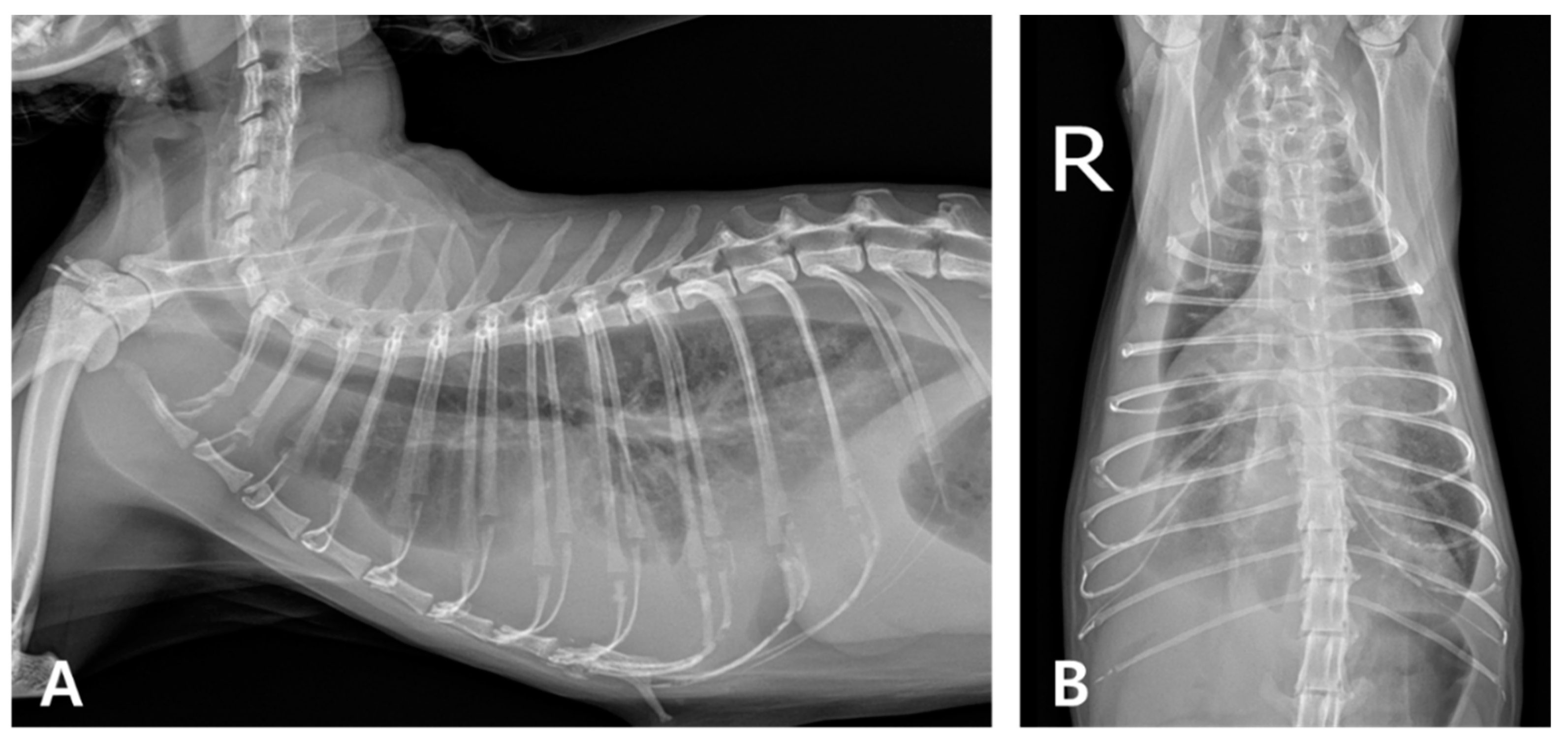
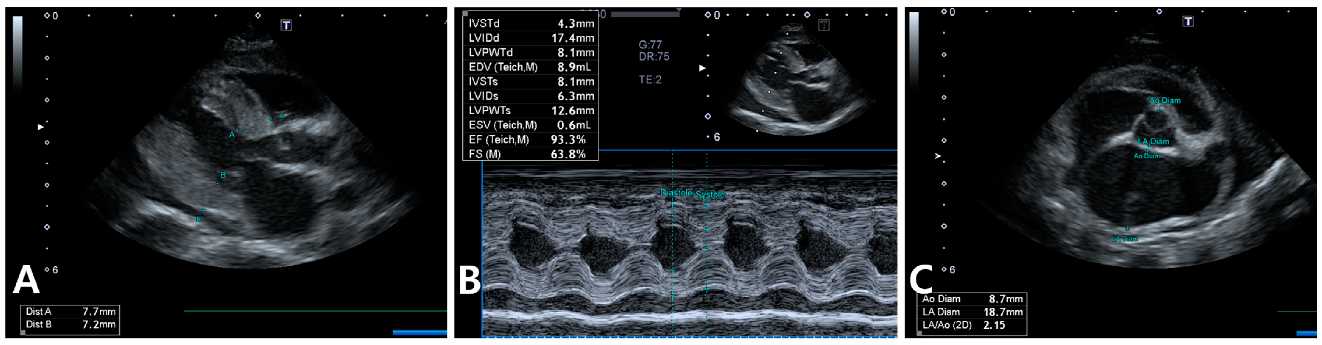
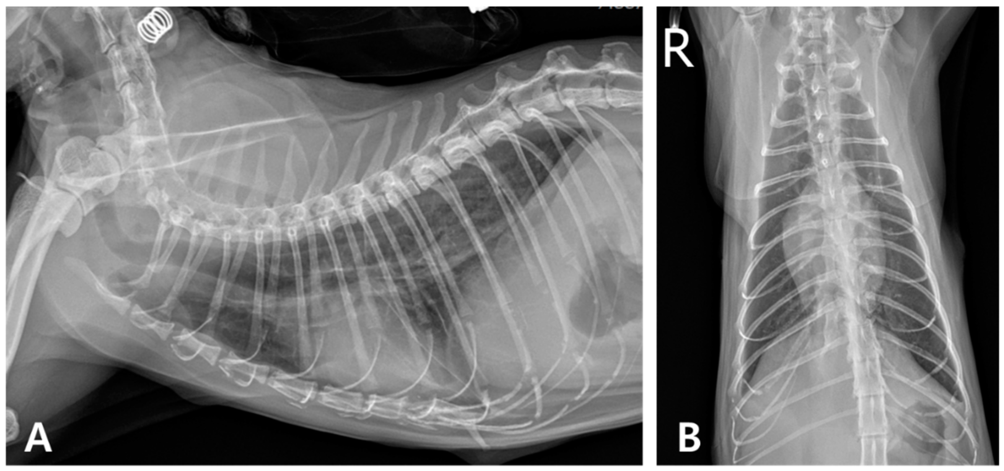
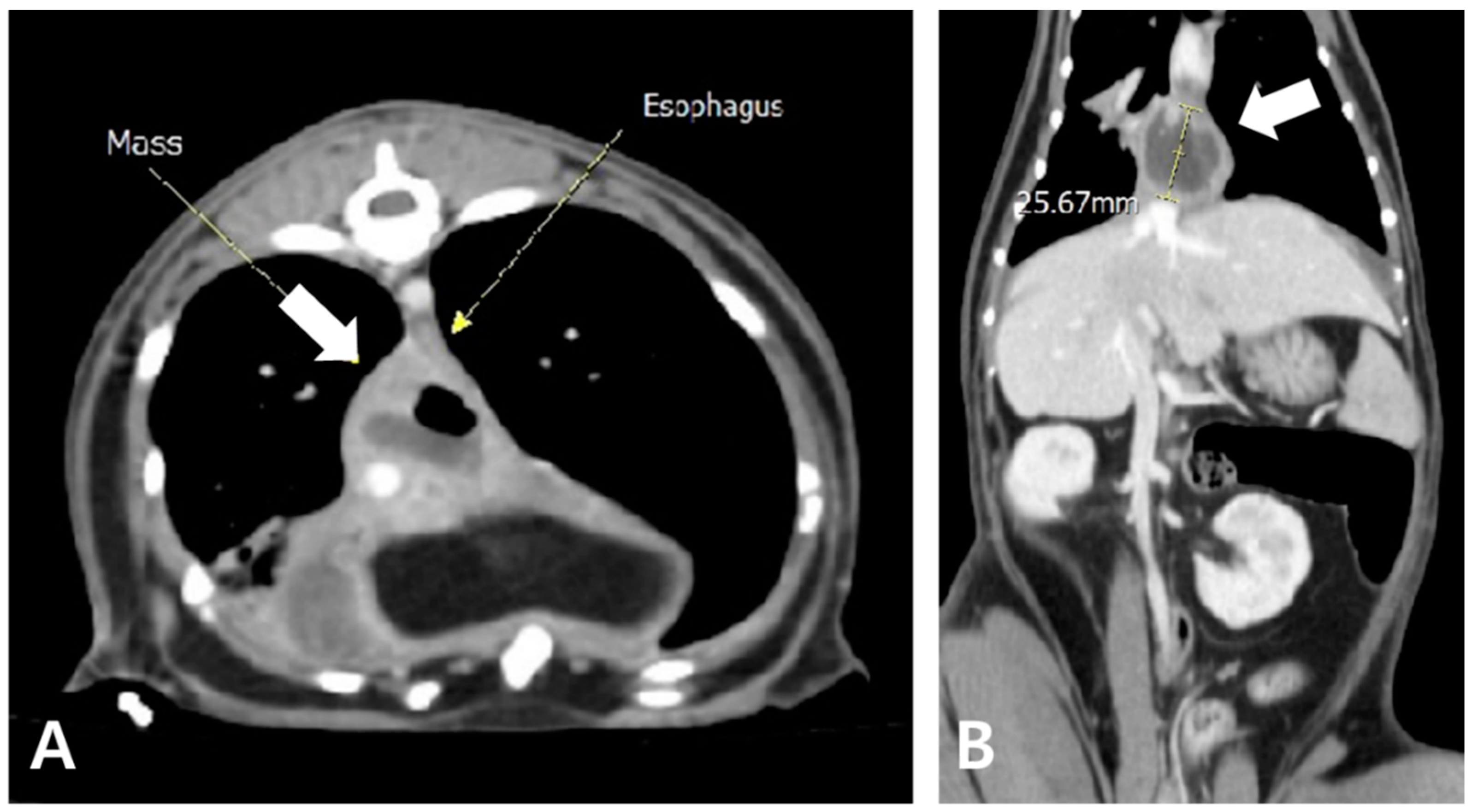
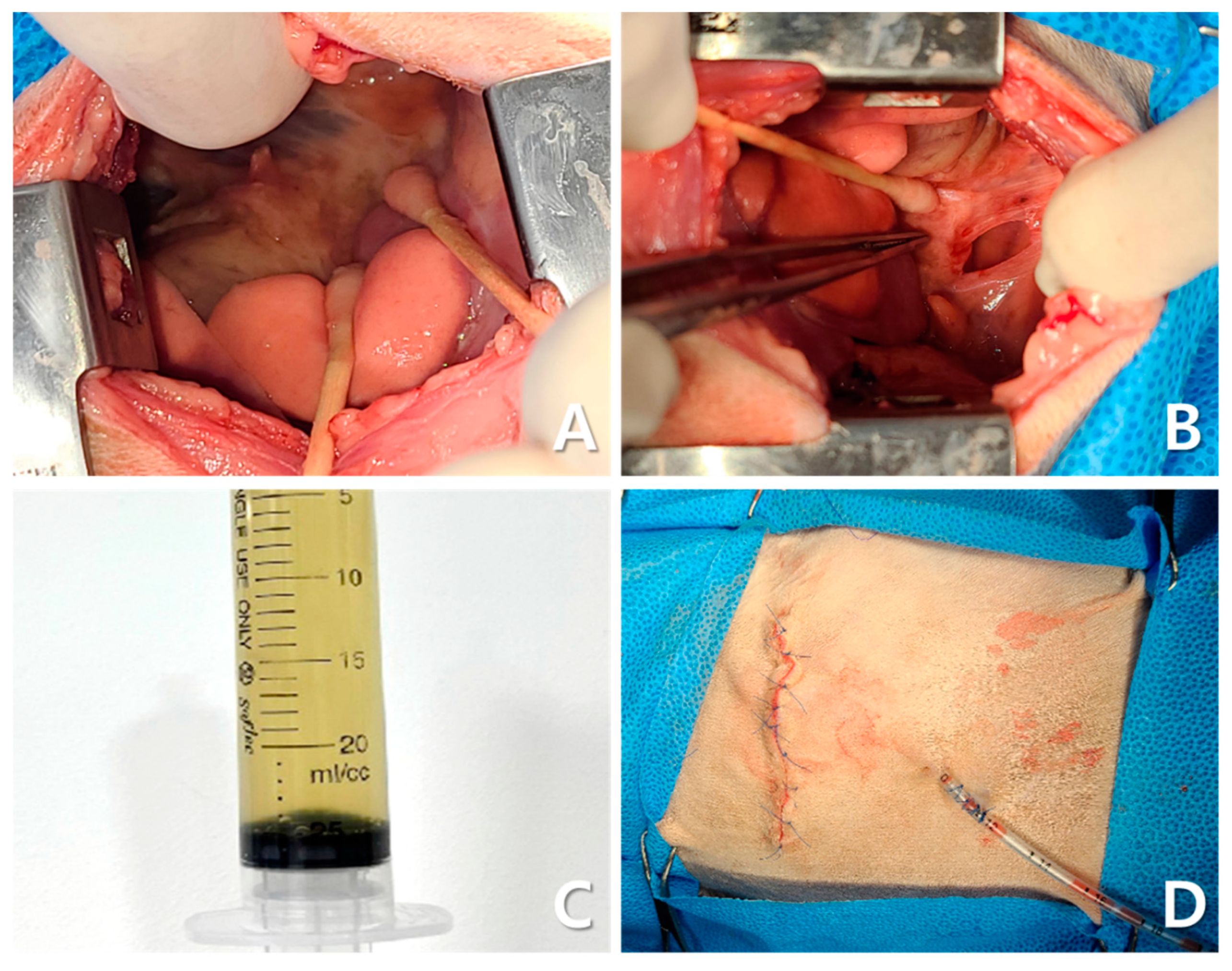
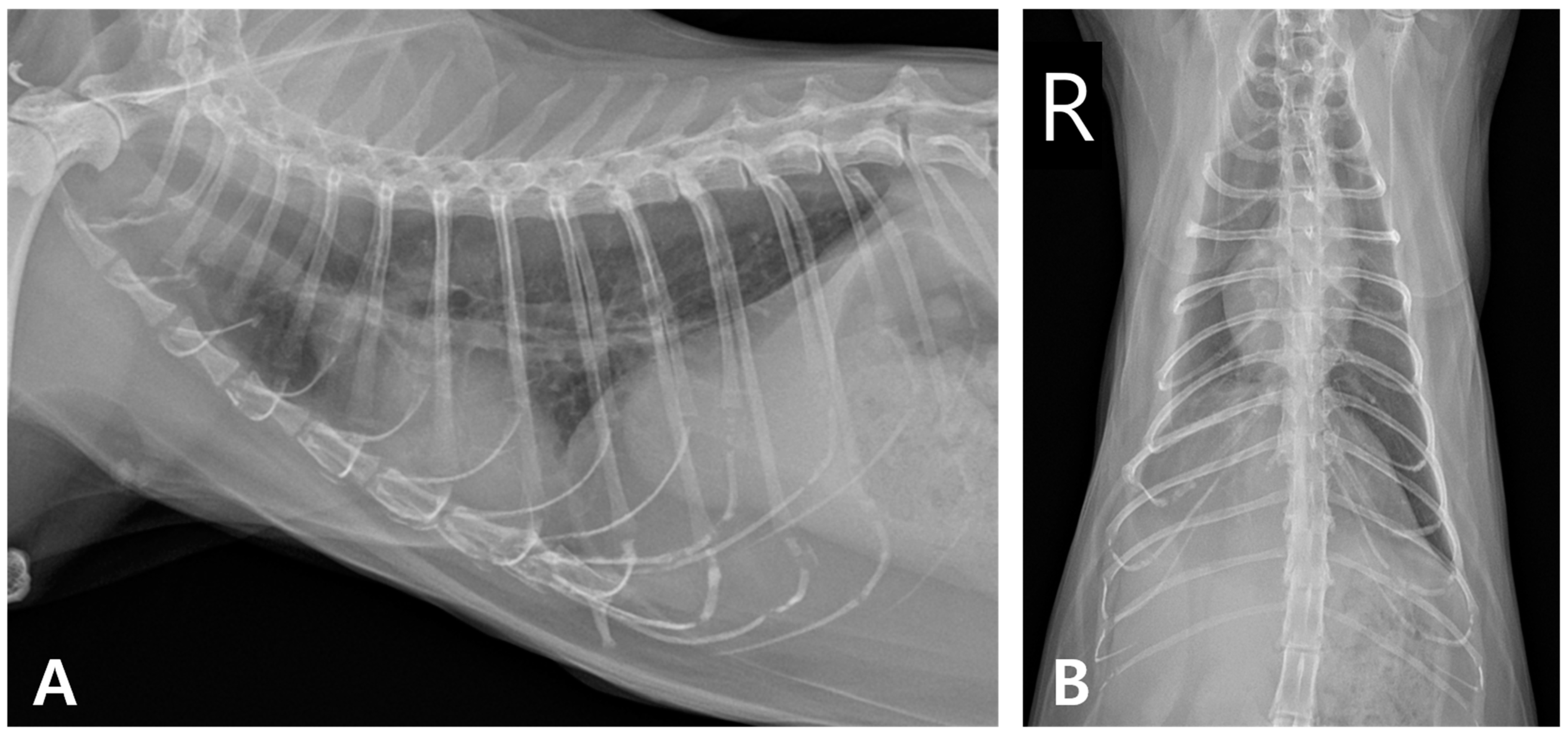
2. Case Description
3. Discussion
4. Conclusions
Author Contributions
Funding
Institutional Review Board Statement
Informed Consent Statement
Data Availability Statement
Acknowledgments
Conflicts of Interest
References
- Waddell, L.S.; Brady, C.A.; Drobatz, K.J. Risk factors, prognostic indicators, and outcome of pyothorax in cats: 80 cases (1986–1999). J. Am. Vet. Med. Assoc. 2002, 221, 819–824. [Google Scholar] [CrossRef] [PubMed]
- Barrs, V.R.; Allan, G.S.; Martin, P.; Beatty, J.A.; Malik, R. Feline pyothorax: A retrospective study of 27 cases in Australia. J. Feline Med. Surg. 2005, 7, 211–222. [Google Scholar] [CrossRef]
- Krämer, F.; Rainer, J.; Bali, M.S. Short- and long-term outcome in cats diagnosed with pyothorax: 47 cases (2009–2018). J. Small Anim. Pract. 2021, 62, 669–676. [Google Scholar] [CrossRef] [PubMed]
- Stillion, J.R.; Letendre, J.A. A clinical review of the pathophysiology, diagnosis, and treatment of pyothorax in dogs and cats. J. Vet. Emerg. Crit. Care 2015, 25, 113–129. [Google Scholar] [CrossRef]
- Nelson, R.W.; Couto, C.G. Small Animal Internal Medicine-E-Book; Elsevier Health Sciences: St. Louis, MO, USA, 2019. [Google Scholar]
- Little, S.E. The Cat-E-Book; Elsevier Health Sciences: St. Louis, MO, USA, 2024. [Google Scholar]
- Anastasio, J.; Sharp, C.; Needle, D. Histopathology of lung lobes in cats with pyothorax: 17 cases (1987–2010). In Proceedings of the 18th International Veterinary Emergency & Critical Care Symposium (IVECCS), San Antonio, TX, USA, 8–12 September 2012. [Google Scholar]
- Davies, C.; Forrester, S.D. Pleural effusion in cats: 82 cases (1987 to 1995). J. Small Anim. Pract. 1996, 37, 217–224. [Google Scholar] [CrossRef] [PubMed]
- Boothe, H.W.; Howe, L.M.; Boothe, D.M.; Reynolds, L.A.; Carpenter, M. Evaluation of outcomes in dogs treated for pyothorax: 46 cases (1983–2001). J. Am. Vet. Med. Assoc. 2010, 236, 657–663. [Google Scholar] [CrossRef]
- Fujii, Y.; Suwa, A.; Tsuyuki, Y.; Koyama, K.; Nio-Kobayashi, J.; Yoshii, K. The First Case of a Cat Infected with Burkholderia pseudomultivorans, a Member of the Burkholderia cepacia Complex. Vet. Sci. 2024, 11, 559. [Google Scholar] [CrossRef]
- Gruget, E.; Cabon, Q.; Gory, G. Description of the CT evolution of pyothorax and a caudal mediastinal paraesophageal empyema in a cat. Vet. Rec. Case Rep. 2022, 10, e221. [Google Scholar] [CrossRef]
- Jung, J.; Choi, M. Nonsurgical resolution of caudal mediastinal paraesophageal abscess in a cat. J. Vet. Med. Sci. 2015, 77, 499–502. [Google Scholar] [CrossRef]
- Sivacolundhu, R.K.; O’Hara, A.J.; Read, R.A. Thoracic actinomycosis (arcanobacteriosis) or nocardiosis causing thoracic pyogranuloma formation in three dogs. Aust. Vet. J. 2001, 79, 398–402. [Google Scholar] [CrossRef]
- Gendron, K.; McDonough, S.P.; Flanders, J.A.; Tse, M.; Scrivani, P.V. The pathogenesis of paraesophageal empyema in dogs and constancy of radiographic and computed tomography signs are linked to involvement of the mediastinal serous cavity. Vet. Radiol. Ultrasound 2018, 59, 169–179. [Google Scholar] [CrossRef]
- Brissot, H.N.; Burton, C.A.; Doyle, R.S.; Bray, J.P. Caudal mediastinal paraesophageal abscesses in 7 dogs. Vet. Surg. 2012, 41, 286–291. [Google Scholar] [CrossRef] [PubMed]
- MacPhail, C.M. Medical and surgical management of pyothorax. Vet. Clin. N. Am. Small Anim. Pract. 2007, 37, 975–988. [Google Scholar] [CrossRef]
- Tillett, W.S.; Sherry, S.; Read, C.T. The use of streptokinase-streptodornase in the treatment of chronic empyema; with an interpretive discussion of enzymatic actions in the field of intrathoracic diseases. J. Thorac. Surg. 1951, 21, 325–341. [Google Scholar] [CrossRef] [PubMed]
- Bouros, D.; Schiza, S.; Patsourakis, G.; Chalkiadakis, G.; Panagou, P.; Siafakas, N.M. Intrapleural streptokinase versus urokinase in the treatment of complicated parapneumonic effusions: A prospective, double-blind study. Am. J. Respir. Crit. Care Med. 1997, 155, 291–295. [Google Scholar] [CrossRef]
- Skeete, D.A.; Rutherford, E.J.; Schlidt, S.A.; Abrams, J.E.; Parker, L.A.; Rich, P.B. Intrapleural tissue plasminogen activator for complicated pleural effusions. J. Trauma 2004, 57, 1178–1183. [Google Scholar] [CrossRef]
- Maskell, N.A.; Davies, C.W.H.; Nunn, A.J.; Hedley, E.L.; Gleeson, F.V.; Miller, R.; Gabe, R.; Rees, G.L.; Peto, T.E.A.; Woodhead, M.A.; et al. U.K. Controlled trial of intrapleural streptokinase for pleural infection. N. Engl. J. Med. 2005, 352, 865–874. [Google Scholar] [CrossRef] [PubMed]
- Rahman, N.M.; Chapman, S.J.; Davies, R.J.O. The approach to the patient with a parapneumonic effusion. Clin. Chest Med. 2006, 27, 253–266. [Google Scholar] [CrossRef]
- Novo Matos, J.; Pereira, N.; Glaus, T.; Wilkie, L.; Borgeat, K.; Loureiro, J.; Silva, J.; Law, V.; Kranjc, A.; Connolly, D.J.; et al. Transient myocardial thickening in cats associated with heart failure. J. Vet. Intern. Med. 2018, 32, 48–56. [Google Scholar] [CrossRef]
- Hiramitsu, S.; Morimoto, S.; Kato, S.; Uemura, A.; Kubo, N.; Kimura, K.; Sugiura, A.; Itoh, T.; Hishida, H. Transient ventricular wall thickening in acute myocarditis: A serial echocardiographic and histopathologic study. Jpn. Circ. J. 2001, 65, 863–866. [Google Scholar] [CrossRef]
- Tsiamis, E.; Panagopoulou, V.; Aggeli, C.; Toutouzas, K.; Oikonomou, E.; Stefanadis, C.; Tousoulis, D. Segmental myocardial hypokinesis and hypertrophy as initial echocardiographic presentation of myocarditis. Int. J. Cardiol. 2014, 176, 1460–1461. [Google Scholar] [CrossRef] [PubMed]
- Zagrosek, A.; Wassmuth, R.; Abdel-Aty, H.; Rudolph, A.; Dietz, R.; Schulz-Menger, J. Relation between myocardial edema and myocardial mass during the acute and convalescent phase of myocarditis—A CMR study. J. Cardiovasc. Magn. Reson. 2008, 10, 19. [Google Scholar] [CrossRef] [PubMed]
- Ernandes, M.A.; Cantoni, A.M.; Armando, F.; Corradi, A.; Ressel, L.; Tamborini, A. Feline coronavirus-associated myocarditis in a domestic longhair cat. J. Feline Med. Surg. Open Rep. 2019, 5, 2055116919879256. [Google Scholar] [CrossRef]
- Rolim, V.M.; Casagrande, R.A.; Wouters, A.T.B.; Driemeier, D.; Pavarini, S.P. Myocarditis caused by feline immunodeficiency virus in Five Cats with hypertrophic cardiomyopathy. J. Comp. Pathol. 2016, 154, 3–8. [Google Scholar] [CrossRef]
- Joseph, J.L.; Oxford, E.M.; Santilli, R.A. Transient myocardial thickening in a Bartonella henselae-positive cat. J. Vet. Cardiol. 2018, 20, 198–203. [Google Scholar] [CrossRef] [PubMed]
- Romito, G.; Fracassi, F.; Cipone, M. Transient myocardial thickening associated with acute myocardial injury and congestive heart failure in two Toxoplasma gondii-positive cats. J. Feline Med. Surg. Open Rep. 2022, 8, 20551169221131266. [Google Scholar] [CrossRef]
- Demetriou, J.L.; Foale, R.D.; Ladlow, J.; McGrotty, Y.; Faulkner, J.; Kirby, B.M. Canine and feline pyothorax: A retrospective study of 50 cases in the UK and Ireland. J. Small Anim. Pract. 2002, 43, 388–394. [Google Scholar] [CrossRef]
- Sim, J.J.; Lau, S.F.; Omar, S.; Watanabe, M.; Aslam, M.W. A retrospective study on bacteriology, clinicopathologic and radiographic features in 28 cats diagnosed with pyothorax. Animals 2021, 11, 2286. [Google Scholar] [CrossRef]
- Jang, S.S.; Hirsh, D.C. Characterization, distribution, and microbiological associations of Fusobacterium spp. in clinical specimens of animal origin. J. Clin. Microbiol. 1994, 32, 384–387. [Google Scholar] [CrossRef]
- Talan, D.A.; Citron, D.M.; Abrahamian, F.M.; Moran, G.J.; Goldstein, E.J. Bacteriologic analysis of infected dog and cat bites. Emergency Medicine Animal Bite Infection Study Group. N. Engl. J. Med. 1999, 340, 85–92. [Google Scholar] [CrossRef]
- Citron, D.M. Update on the taxonomy and clinical aspects of the genus fusobacterium. Clin. Infect. Dis. 2002, 35 (Suppl. S1), S22–S27. [Google Scholar] [CrossRef] [PubMed]
- Camacho Zamora, P.; Román Cabello, M.; Álvarez García, P.; Vázquez Canal, E. Polymicrobial osteomyelitis on the limb of the hand after a cat bite. Rev. Esp. Quimioter. 2022, 35, 498–499. [Google Scholar] [CrossRef] [PubMed]
- Goldstein, E.J.C.; Citron, D.M.; Tyrrell, K.L.; Leoncio, E.S. In vitro activity of pexiganan and 10 comparator antimicrobials against 234 isolates, including 93 Pasteurella species and 50 anaerobic bacterial isolates recovered from animal bite wounds. Antimicrob. Agents Chemother. 2017, 61, e00246-17. [Google Scholar] [CrossRef]
- Elsheikha, H.M.; Kennedy, F.A.; Murphy, A.J.; Soliman, M.; Mansfield, L.S. Sarcocystosis of Sarcocystis felis in cats. J. Egypt. Soc. Parasitol. 2006, 36, 1071–1085. [Google Scholar] [PubMed]
- Matsuu, A.; Kanda, T.; Sugiyama, A.; Murase, T.; Hikasa, Y. Mitral stenosis with bacterial myocarditis in a cat. J. Vet. Med. Sci. 2007, 69, 1171–1174. [Google Scholar] [CrossRef]
- Simpson, K.E.; Devine, B.C.; Gunn-Moore, D. Suspected toxoplasma-associated myocarditis in a cat. J. Feline Med. Surg. 2005, 7, 203–208. [Google Scholar] [CrossRef]
- Kegler, K.; Nufer, U.; Alic, A.; Posthaus, H.; Olias, P.; Basso, W. Fatal infection with emerging apicomplexan parasite Hepatozoon silvestris in a domestic cat. Parasit. Vectors 2018, 11, 428. [Google Scholar] [CrossRef]
- Sharpe, A.N.; Gunther-Harrington, C.T.; Epstein, S.E.; Li, R.H.L.; Stern, J.A. Cats with thermal burn injuries from California wildfires show echocardiographic evidence of myocardial thickening and intracardiac thrombi. Sci. Rep. 2020, 10, 2648. [Google Scholar] [CrossRef]
- LeBlanc, N.; Scollan, K.F. Bacterial pericarditis in a cat. J. Feline Med. Surg. Open Rep. 2015, 1, 2055116915603077. [Google Scholar] [CrossRef]


| Initial Onset | Recurrent Onset | Tube Removal | 2 Weeks After Cessation of Lavage | 2 Months Post-Medication Withdrawal | 1 Year Post-Treatment | Reference Range | |
|---|---|---|---|---|---|---|---|
| Hematocrit (%) | 22 | 26.8 | 32.7 | 33.4 | 36.7 | 48.4 | 30.3–52.3 |
| White blood cells (k/μL) | 51.98 | 21.92 | 6.98 | 6.91 | 6.44 | 6.34 | 2.87–17.02 |
| Neutrophils (k/μL) | 0.45 | 12.3 | 2.82 | 4.41 | 2.38 | 2.43 | 2.30–10.29 |
| Lymphocytes (k/μL) | 37.43 | 8.98 | 2.67 | 1.71 | 1.56 | 2.01 | 0.92–6.88 |
| Monocytes (k/μL) | 13.84 | 0.44 | 0.58 | 0.43 | 0.18 | 0.16 | 0.05–0.67 |
| Eosinophils (k/μL) | 0.07 | 0.06 | 0.87 | 0.29 | 1.97 | 1.48 | 0.17–1.57 |
| Platelet (k/μL) | 386 | 239 | 456 | 371 | 270 | 282 | 151–600 |
| Total protein (g/dL) | 7.7 | 7.0 | - | - | 7.1 | 7.0 | 5.7–8.9 |
| Albumin (g/dL) | 2.4 | 2.6 | - | - | 3.1 | 2.8 | 2.2–4.0 |
| Globulin (g/dL) | 5.3 | 4.4 | - | - | 4.0 | 4.2 | 2.8–5.1 |
| A:G ratio | 0.4 | 0.6 | - | - | 0.8 | 0.6 | |
| BUN (mg/dL) | 43 | 26 | - | 49 | 37 | 36 | 9–29 |
| Creatinine (mg/dL) | 0.8 | 1.2 | - | 1.0 | 1.9 | 1.9 | 0.8–2.4 |
| SDMA (μg/dL) | 19 | 20 | 4 | 10 | - | 16 | 0–14 |
| fSAA (mg/mL) | 24.9 | 17.3 | <5 | <5 | <5 | <5 | <5 |
| NT-proBNP (μg/dL) | 1014.8 | 122 | - | - | <50 | 53.7 | <100 |
| Initial Onset | Recurrent Onset | |
|---|---|---|
| Total nucleated cell count (K/μL) | 239.93 | 21.25 |
| Total protein (g/dL) | 4.6 | 4.3 |
| Triglyceride (mg/dL) | <10 | <10 |
| Total cholesterol (mg/dL) | 74 | 106 |
Disclaimer/Publisher’s Note: The statements, opinions and data contained in all publications are solely those of the individual author(s) and contributor(s) and not of MDPI and/or the editor(s). MDPI and/or the editor(s) disclaim responsibility for any injury to people or property resulting from any ideas, methods, instructions or products referred to in the content. |
© 2025 by the authors. Licensee MDPI, Basel, Switzerland. This article is an open access article distributed under the terms and conditions of the Creative Commons Attribution (CC BY) license (https://creativecommons.org/licenses/by/4.0/).
Share and Cite
Jang, H.; Kim, S.; Lee, Y.; Park, J.; Kwon, H.; Kim, S.; Sohn, J.; Kim, J.-i.; Jung, D.-I. Successful Management of Recurrent Pyothorax in a Cat: Clinical Findings with Medical and Surgical Approaches. Animals 2025, 15, 1253. https://doi.org/10.3390/ani15091253
Jang H, Kim S, Lee Y, Park J, Kwon H, Kim S, Sohn J, Kim J-i, Jung D-I. Successful Management of Recurrent Pyothorax in a Cat: Clinical Findings with Medical and Surgical Approaches. Animals. 2025; 15(9):1253. https://doi.org/10.3390/ani15091253
Chicago/Turabian StyleJang, Hyomi, Seoyeon Kim, Yebeen Lee, Jongwon Park, Hyojun Kwon, Sunyoung Kim, Jiheui Sohn, Jong-in Kim, and Dong-In Jung. 2025. "Successful Management of Recurrent Pyothorax in a Cat: Clinical Findings with Medical and Surgical Approaches" Animals 15, no. 9: 1253. https://doi.org/10.3390/ani15091253
APA StyleJang, H., Kim, S., Lee, Y., Park, J., Kwon, H., Kim, S., Sohn, J., Kim, J.-i., & Jung, D.-I. (2025). Successful Management of Recurrent Pyothorax in a Cat: Clinical Findings with Medical and Surgical Approaches. Animals, 15(9), 1253. https://doi.org/10.3390/ani15091253