Article

A Quadruplex RT-qPCR for the Detection of Porcine Sapelovirus, Porcine Kobuvirus, Porcine Teschovirus, and Porcine Enterovirus G

1
Guangxi Key Laboratory of Animal Breeding, Disease Control and Prevention, College of Animal Science and Technology, Guangxi University, Nanning 530005, China
2
Guangxi Center for Animal Disease Control and Prevention, Nanning 530001, China
*
Authors to whom correspondence should be addressed.
These authors contributed equally to this work.
Animals 2025, 15(7), 1008; https://doi.org/10.3390/ani15071008
Submission received: 29 January 2025 / Revised: 27 March 2025 / Accepted: 28 March 2025 / Published: 31 March 2025

Simple Summary

Porcine sapelovirus (PSV), porcine kobuvirus (PKV), porcine teschovirus (PTV), and porcine enterovirus G (EV-G) are important viruses in the pig industry. These viruses play important roles in the establishment of similar clinical signs of diarrhea, encephalitis, and reproductive and respiratory disorders in pig herds. The differential detection of these viruses is crucial for the accurate diagnosis of these diseases. In this study, a quadruplex real-time quantitative RT-PCR (RT-qPCR) for the simultaneous detection of PSV, PKV, PTV, and EV-G was developed. The assay showed high sensitivity, strong specificity, and excellent repeatability. The assay was further assessed for its applicability through testing 1823 fecal samples collected from different pig farms. The positivity rates of PSV, PKV, PTV, and EV-G were 15.25% (278/1823), 21.72% (396/1823), 18.82% (343/1823), and 27.10% (494/1823), respectively, with coincidence rates higher than 99.01% for the reference RT-qPCR/RT-PCR. The results indicated that the developed assay provides a rapid, sensitive, and accurate method for the simultaneous detection of PSV, PKV, PTV, and EV-G.

Abstract

Porcine sapelovirus (PSV), porcine kobuvirus (PKV), porcine teschovirus (PTV), and porcine enterovirus G (EV-G) are all important viruses in the swine industry. These viruses play important roles in the establishment of similar clinical signs of diseases in pigs, including diarrhea, encephalitis, and reproductive and respiratory disorders. The early accurate detection of these viruses is crucial for dealing with these diseases. In order for the differential detection of these four viruses, specific primers and TaqMan probes were designed for the conserved regions in the 5′ untranslated region (UTR) of these four viruses, and one-step quadruplex reverse-transcription real-time quantitative PCR (RT-qPCR) for the detection of PSV, PKV, PTV, and EV-G was developed. The results showed that this assay had the advantages of high sensitivity, strong specificity, excellent repeatability, and simple operation. Probit regression analysis showed that the assay obtained low limits of detection (LODs) for PSV, PKV, PTV, and EV-G, with 146.02, 143.83, 141.92, and 139.79 copies/reaction, respectively. The assay showed a strong specificity of detecting only PSV, PKV, PTV, and EV-G, and had no cross-reactivity with other control viruses. The assay exhibited excellent repeatability of the intra-assay coefficient of variation (CV) of 0.28–1.58% and the inter-assay CV of 0.20–1.40%. Finally, the developed quadruplex RT-qPCR was used to detect 1823 fecal samples collected in Guangxi Province, China between January 2024 and December 2024. The results indicated that the positivity rates of PSV, PKV, PTV, and EV-G were 15.25% (278/1823), 21.72% (396/1823), 18.82% (343/1823), and 27.10% (494/1823), respectively, and there existed phenomena of mixed infections. Compared with the reference RT-qPCR/RT-PCR established for these four viruses, the coincidence rates for the detection results of PSV, PKV, PTV, and EV-G reached 99.51%, 99.40%, 99.51%, and 99.01%, respectively. In conclusions, the developed quadruplex RT-qPCR could simultaneously detect PSV, PKV, PTV, and EV-G, and provided an efficient and convenient detection method to monitor the epidemic status and variation of these viruses.

1. Introduction

Porcine sapelovirus (PSV) is classified in the Sapelovirus genus within the Picornaviridae family [1]. It is a positive-stranded RNA virus with a genome of 7.5 kb [2]. PSV can be isolated from the feces of healthy and diarrheic pigs [3]. The virus can cause severe diarrhea, respiratory distress, neurological symptoms, and reproductive failure in pigs [4]. The pathological changes such as atrophy of the villi are mainly found in the small intestine. Although, the virus is also detected in other organs, no significant pathological changes are found in these organs [5]. A recent study showed that PSV could infect human 293T cells, suggesting that PSV may have a potential risk of infection in humans [6]. PSV has been reported worldwide [2,3,4,5,7], including China [5,6,8,9].
Porcine kobuvirus (PKV) belongs to the Kobuvirus genus of the Picornaviridae family, and it is a single-stranded positive-sense RNA virus with a 8.1 kb genome [1,10]. Kobuvirus has been found in different animal species and humans around the world, and can cause diarrhea in both humans and animals, indicating the zoonotic potential of this pathogen [11,12]. Although PKV is an enterovirus, it can be present in the intestinal tissues with diarrhea or diarrheic subclinical pigs, and it can also induce other clinical signs such as respiratory distress [13]. A study found that the PKV-positive pigs were clinically healthy, while many pigs infected with PKV and porcine epidemic diarrhea virus (PEDV) showed clinical illness of diarrhea [13]. It has been shown that PKV can synergize with PEDV, and can exacerbate clinical signs and pathological damage to the infected pigs [14].
Porcine teschovirus (PTV) is a member of the Teschovirus genus within the Picornaviridae family. The genome of the virus is a single-stranded positive-sense RNA, 7.1 kb in size [1]. PTV-infected pigs usually show diarrhea or subclinical diarrhea, and other signs such as polioencephalomyelitis, reproductive disorders, and severe-to-moderate neurological symptoms [15,16,17]. PTV consists of 14 genotypes and may cause different damage to pigs depending on the viral genotypes. For example, PTV-1 is usually considered a genotype of porcine diarrhea and neurotropic signs [16,17], PTV-2 induces severe encephalomyelitis [18], and PTV-13 causes neurological symptoms in piglets [19]. In addition, some other genotypes of PTV have also been detected [20,21], but their pathogenic roles in pig herds require further investigation and evaluation.
Porcine enterovirus G (EV-G) belongs to the Enterovirus genus within the Picornaviridae family, and is a single-stranded positive-sense RNA virus with a 7.4 kb genome size [1]. Twenty genotypes of EV-G have been identified [22]. The first isolated genotype 2 EV-G, which can cause mild diarrhea, wasting, and other clinical signs in newborn piglets, was reported in Sichuan Province of China in 2023 [23]. In addition, EV-G can also induce neurological symptoms, and growth retardation [24]. It has been reported that there is extensive recombination between different genotypes of EV-G [25,26], and recombinant EV-G strains can persist in pig farms [27].
All four viruses, PSV, PKV, PTV, and EV-G, play important roles in the establishment of diarrhea in pigs, and PSV and PKV have the potential risk of infecting humans. The pigs infected with these four viruses may be subclinical or have similar signs of diarrhea and neurological symptoms. Co-infections of these viruses exacerbate the clinical signs and pathological changes of the diseases [6,13,14,28], which seriously jeopardize the health of pigs. It is difficult to distinguish these diseases relying on only the clinical signs and pathological changes, especially the existence of co-infections with these viruses. To date, there is no commercial kit to detect these four viruses in China. A multiplex RT-PCR assay was developed for the detection of PTV, PSV, and EV-G [29], but RT-PCR has shortcomings such as cumbersome operation and non-specificity of amplification. Therefore, multiplex real-time fluorescence quantitative RT-PCR (RT-qPCR) has gradually replaced multiplex RT-PCR, and become the major technique for the simultaneous detection of several viruses in clinical samples. Multiplex RT-qPCR can simultaneously detect multiple pathogens in the same sample in a short time, with the advantages of easy operation, strong specificity, and high sensitivity [30,31]. To date, no detection method for the simultaneous detection of PSV, PKV, PTV, and EV-G has been reported. In this study, a quadruplex RT-qPCR assay for the detection of PSV, PKV, PTV, and EV-G was developed, which could be used for the clinical detection and epidemiological investigation of these pathogens.

2. Materials and Methods

2.1. Reference Strains

The following vaccine strains were purchased from Keqian Biological Co., Ltd. (Wuhan, China): transmissible gastroenteritis virus (TGEV, H strain), porcine epidemic diarrhea virus (PEDV, CV777 strain), porcine circovirus type 2 (PCV2, SX07 strain), porcine reproductive and respiratory syndrome virus (PRRSV, TJM-F92 strain), foot-and-mouth disease virus (FMDV, O/Mya98/XJ/2010 strain), classical swine fever virus (CSFV, C strain), pseudorabies virus (PRV, Bartha-K61 strain), swine influenza virus (SIV, TJ strain), and porcine rotavirus (PoRV, NX strain).
The following positive samples were provided by our laboratory: PSV, PKV, PTV, EV-G, African swine fever virus (ASFV), and porcine deltacoronavirus (PDCoV).

2.2. Design of Primers and Probes

The genome sequences of PSV, PKV, PTV, and EV-G representative strains from different countries around the world were downloaded from GenBank in NCBI (https://www.ncbi.nlm.nih.gov/nucleotide/, accessed on 15 August 2023). The multiple sequence alignments were performed, and the conserved regions of the 5′ untranslated region (UTR) were selected for designing the specific primers and probes (Table 1), which are suitable for the detection of different strains of PSV, PKV, PTV, and EV-G from different countries. The genome accession number for the reference strains and the amplified gene fragments in the 5′ UTR of PSV, PKV, PTV, and EV-G are shown in Supplementary Figure S1.

2.3. Clinical Samples

Between January 2024 and December 2024, a total of 1823 fecal samples from diarrheic piglets were collected in Guangxi Province in China. These samples came from diarrheal piglets less than 3 months old in different intensive pig farms in 14 cities of Guangxi Province during different seasons in 2024. Three-to-five grams of feces was collected from each diarrheal piglet using an anal swab. All samples were sent to the laboratory at ≤4 °C conditions within six hours post collection.

2.4. Extraction of Nucleic Acid

All the fecal samples were placed in 1.5 mL centrifuge tubes, and 1.0 mL of phosphate-buffered saline (PBS, pH7.2) (1:4, w/v) was added to each tube, vortexed (2 min), and centrifuged at 4 °C (12,000 rpm, 5 min). The nucleic acids of the clinical samples or vaccine solutions were extracted according to the MiniBEST Viral RNA/DNA Extraction Kit Ver.5.0 (TaKaRa, Dalian, China; Cat No. 9766) using 200 µ L of supernatants or vaccine solutions. A 30 μL elution buffer was used to dissolve the extracted nucleic acids, which were used to generate standard plasmid constructs, or detect the target gene fragments.

2.5. Construction of Standard Plasmids

The synthesized viral RNAs of PSV, PKV, PTV, and EV-G corresponding to the target gene fragments for amplification were provided by Dalian TaKaRa Co., Ltd. (TaKaRa, Dalian, China). The RNAs were used for evaluating the sensitivity of the developed quadruplex RT-qPCR assay.
The standard plasmid constructs were generated according to the reported procedures [32] with minor modification. The extracted nucleic acids from PSV, PKV, PTV, and EV-G positive samples were reverse transcribed to cDNA using PrimeScript II 1st Strand cDNA Synthesis Kit (TaKaRa, Dalian, China; Cat No. 6210A). The corresponding target gene fragments were amplified from the cDNA using the specific primers in Table 1. The PCR products were purified using MiniBEST DNA Fragment Purification Kit Ver.4.0 (TaKaRa, Dalian, China; Cat No. 9762), ligated into pMD18-T vector (TaKaRa, Dalian, China; Cat No. 6011), and transformed into DH5α competent cells (TaKaRa, Dalian, China; Cat No. 9057). The positive clones were selected and incubated at 37 °C for 20–24 h, and then the plasmids were extracted using MiniBEST Plasmid Extraction Kit Ver.5.0 (TaKaRa, Dalian, China; Cat No. 9760). Finally, the recombinant standard plasmid constructs were named p-PSV, p-PKV, p-PTV, and p-EV-G. The supercoiled plasmids were treated with EcoRI endonuclease (TaKaRa, Dalian, China; Cat No. 1040A) to obtain linearized plasmids. Their concentrations were determined through a spectrophotometer at OD260/OD280 nm using the following equation:
concentration   ( copies / μ L )   =   6.02 × 10 23 × ( X   n g / µ L   × 10 9 ) p l a s m i d   l e n g t h   ( b p ) × 660 .

2.6. Optimization of Reaction System and Reaction Procedure

The reaction system for the quadruplex RT-qPCR assay was optimized using the ABI Q5 Real-Time System (Thermo Fisher scientific, Carlsbad, CA, USA). The primers and probes were diluted to 20 μM, and the total volume of the reaction system was 20 µ L : 10 μL 2× One Step RT-PCR Buffer III (TaKaRa, Dalian, China; Cat No. RR064A), 0.4   µ L Ex Taq HS (TaKaRa, Dalian, China; Cat No. RR064A), 0.4 µ L PrimeScript RT Enzyme Mix II (TaKaRa, Dalian, China; Cat No. RR064A), 2.0 µL of a mixture of the four standard plasmid constructs (108 copies/ µ L , and the final reaction concentration was 107 copies/ µ L in the reaction system), forward/reverse primers and probes for four viruses 0.2–0.5 µL, and distilled water to a final volume of 20 µ L . Fluorescence signals were collected at the end of each reaction cycle. The variables in this experiment were the concentrations of forward/reverse primers and probes, the temperatures of the reaction program, and reaction cycles to determine the optimal reaction system and reaction program. Their optimal conditions were based on the minimum cycling threshold (Ct) and the maximum ∆Rn at the end of the reaction.

2.7. Generation of Standard Curves

The plasmid constructs p-PSV, p-PKV, p-PTV, and p-EV-G were mixed at a ratio of 1:1:1:1, then 10-fold serially diluted. The mixtures with concentrations of 1.0 × 10⁸ to 1.0 × 10² copies/ µ L   (the final reaction concentration: 1.0 × 10⁷ to 1.0 × 101 copies/ µ L ) were used as templates to generate the standard curves of the developed quadruplex RT-qPCR assay.

2.8. Analytical Specificity Analysis

The total nucleic acids of PSV, PKV, PTV, EV-G, TGEV, PCV2, PRRSV, FMDV, CSFV, PRV, SIV, PoRV, ASFV, PEDV, and PDCoV were used as templates for specificity analysis of the developed quadruplex RT-qPCR assay. The plasmid constructs p-PSV, p-PKV, p-PTV, p-EV-G, the positive clinical samples, and nuclease-free distilled water were used as controls.

2.9. Analytical Sensitivity Analysis

The synthesized viral RNAs of PSV, PKV, PTV, and EV-G were mixed at a ratio of 1:1:1:1, and 10-fold serially diluted. The concentrations of the mixtures with 1.0 × 10⁸ to 1.0 × 101 copies/ µ L   (the final reaction concentration: 1.0 × 10⁷ to 1.0 × 100 copies/ µ L ) were used as templates for sensitivity analysis of the developed quadruplex RT-qPCR assay. The limits of detection (LODs) were determined.
In addition, the mixtures of synthesized viral RNAs of PSV, PKV, PTV, and EV-G with 500, 250, 125, and 62.5 copies/reaction were also used as templates for the sensitivity analysis of the developed assay using probit regression analysis.

2.10. Repeatability Analysis

The plasmid constructs p-PSV, p-PKV, p-PTV, and p-EV-G were mixed together at a ratio of 1:1:1:1, and 10-fold serially diluted. The plasmid construct concentrations of 1.0 × 10⁷, 1.0 × 10⁴, and 1.0 × 10² copies/ µ L (the final concentrations: 1.0 × 10⁶, 1.0 × 10³, and 1.0 × 101 copies/ µ L ) were used as templates for the determination of the coefficient of variation (CV) of the developed quadruplex RT-qPCR assay. The intra-assay tests were performed in triplicates, and the inter-assay tests were performed on three different days.

2.11. Assessment of the Developed Assay Using Clinical Samples

The 1823 fecal samples collected from diarrheic pigs in Guangxi Province were tested using the established quadruplex RT-qPCR for evaluating its applicability. In addition, these samples were also tested using the RT-qPCR assays previously reported for the detection of PSV [33], PKV [34], and PTV [35], and using the RT-PCR assay previously reported for the detection of EV-G [29]. The diagnostic sensitivity and specificity of the developed quadruplex RT-qPCR assay were assessed. The detection results of the developed assay and the reference assays were compared, and the coincidence rates were calculated.

3. Results

3.1. Preparation of the Standard Plasmids

To construct the standard plasmids, total nucleic acids were extracted from the positive clinical samples of PSV, PKV, PTV, and EV-G, then reverse transcribed into cDNA. The amplified target fragments of the 5′ UTR of PSV, PKV, PTV, and EV-G using the primers in Table 1 were purified and used for the construction of recombinant standard plasmid constructs. The obtained plasmid constructs were named p-PSV, p-PKV, p-PTV, and p-EV-G, and their concentrations were measured by a spectrophotometer. The initial concentrations of p-PSV, p-PKV, p-PTV, and p-EV-G were calculated as 4.83 × 10¹⁰, 5.23 × 10¹⁰, 5.45 × 10¹⁰, and 5.17 × 10¹⁰ copies/ µ L , respectively. Finally, they were diluted to 1.0 × 10¹⁰ copies/ µ L , and stored at −80 °C until use.

3.2. Attainment of the Optimal Reaction Parameters

To develope the quadruplex RT-qPCR, the p-PSV, p-PKV, p-PTV, and p-EV-G plasmid constructs were used as templates, and the One-Step PrimeScript RT-PCR Kit (TaKaRa, Dalian, China; Cat No. RR064A) was used as basic components. The reaction conditions of the quadruplex RT-qPCR assay were optimized, including primer and probe concentrations, annealing temperatures, and reaction cycles. After optimization, the reaction system and amplification procedures for the quadruplex RT-qPCR were obtained. The ingredients of the 20 µ L reaction system is shown in Table 2. The optimized reaction protocol was as follows: 42 °C for 5 min, 95 °C for 10 s; followed by 40 cycles of 95 °C for 5 s and 56 °C for 30 s. The fluorescence signals were collected after each cycle, and the samples with Ct values ≤36 were judged as positive samples.

3.3. Generation of the Standard Curves

The four plasmid constructs were mixed in equal proportions and serially diluted. The mixtures with concentrations of 1.0 × 10⁸–1.0 × 10² copies/ µ L (the final reaction concentration: 1.0 × 10⁷–1.0 × 101 copies/ µ L ) were used to generate the standard curves of the developed assay (Figure 1). The results indicated that the amplification efficiencies (Eff%) of the standard curves for PSV, PKV, PTV, and EV-G were 105.45%, 102.871%, 101.102%, and 98.776%, respectively; their correlation coefficients (R2) were 1, 0.999, 1, and 0.999, respectively, indicating excellent amplification efficiency and correlation coefficient (R2 ≥ 0.998) of the quadruplex RT-qPCR.

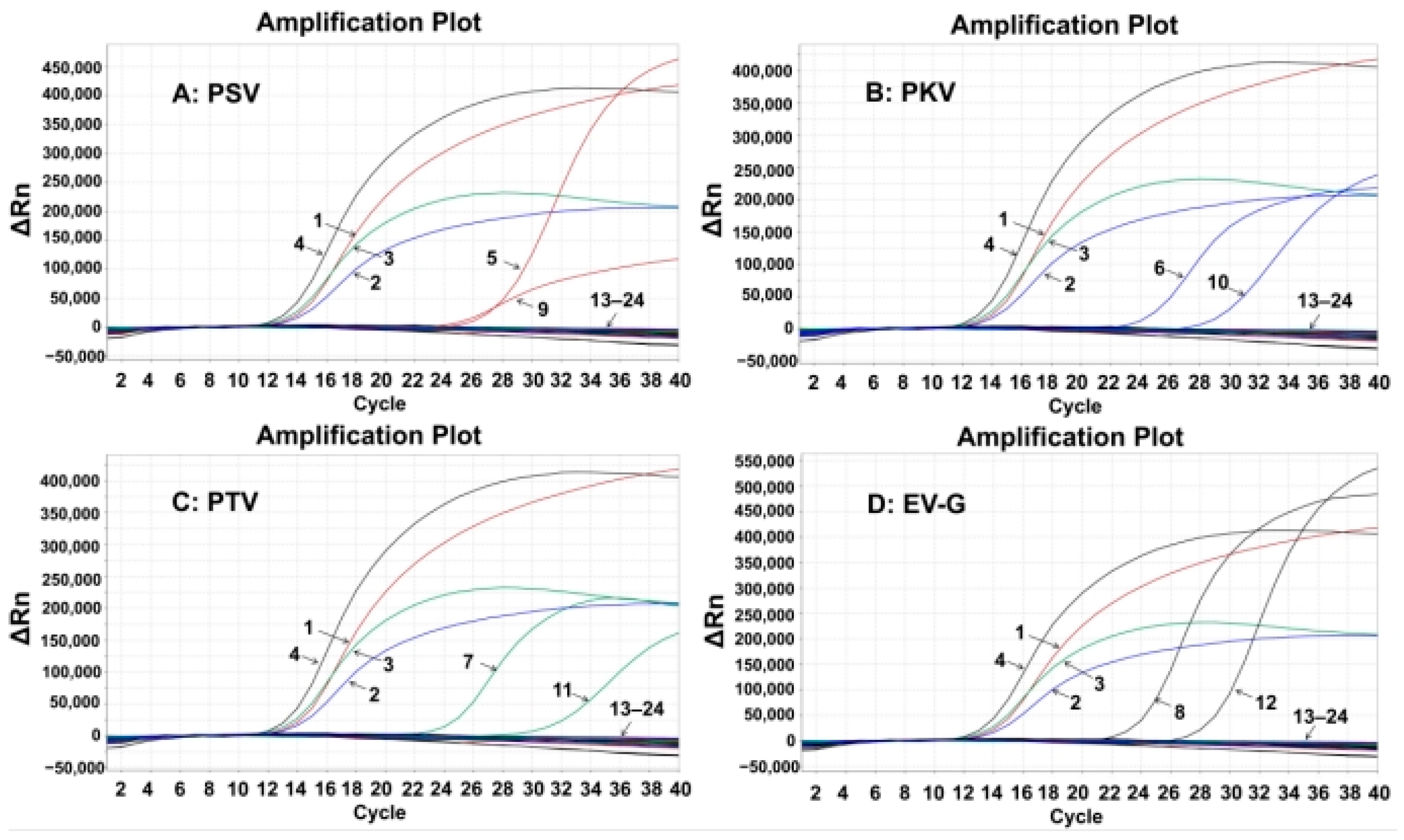
3.4. Specificity of the Quadruplex RT-qPCR

The specificity of the established assay was assessed using the total nucleic acids of PSV, PKV, PTV, EV-G, TGEV, PCV2, PRRSV, FMDV, CSFV, PRV, SIV, PoRV, ASFV, PEDV, and PDCoV. The results showed that the quadruplex RT-qPCR produced specific amplification curves only for PSV, PKV, PTV, and EV-G, but not for the other control viruses (Figure 2), indicating the strong specificity of the quadruplex RT-qPCR.

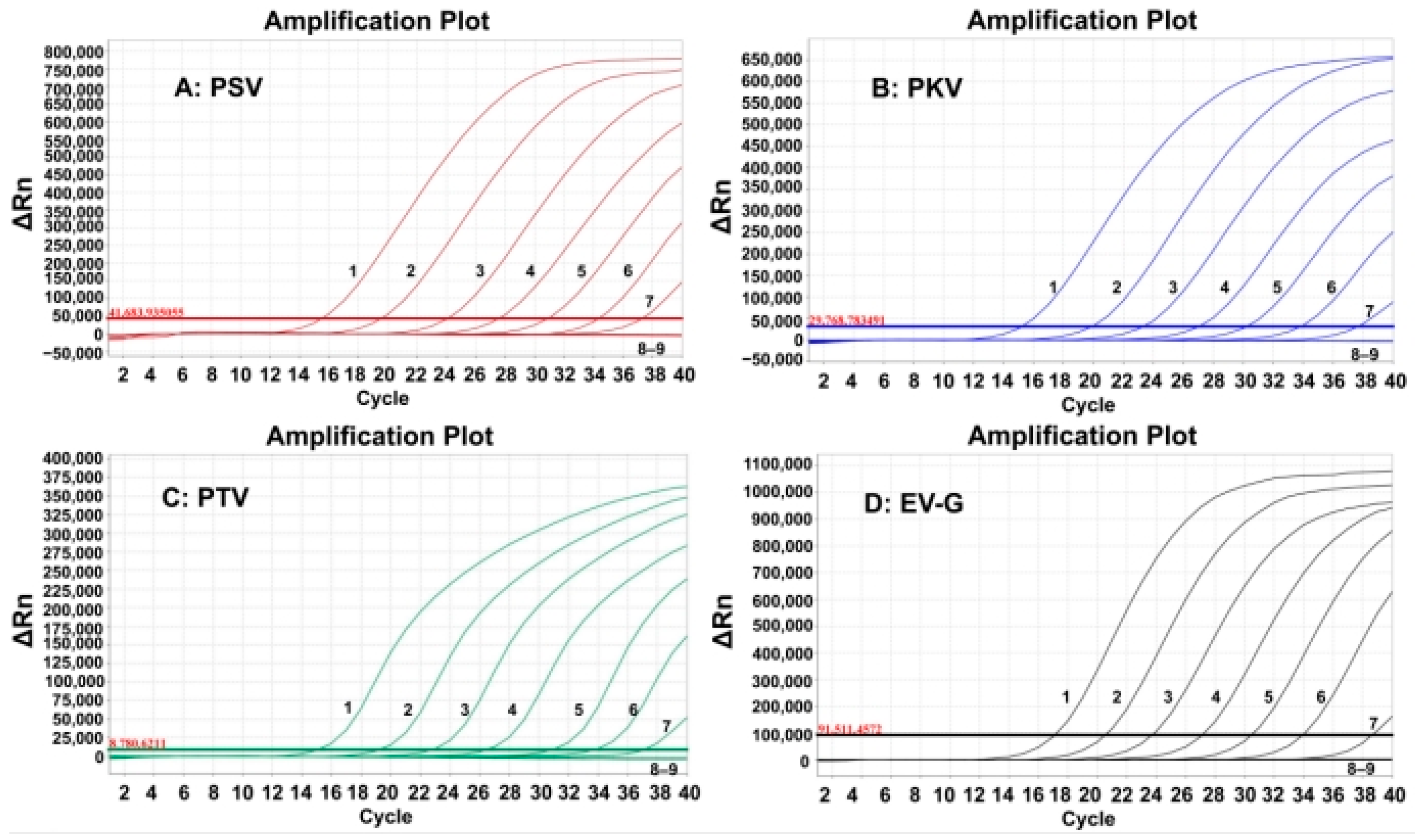
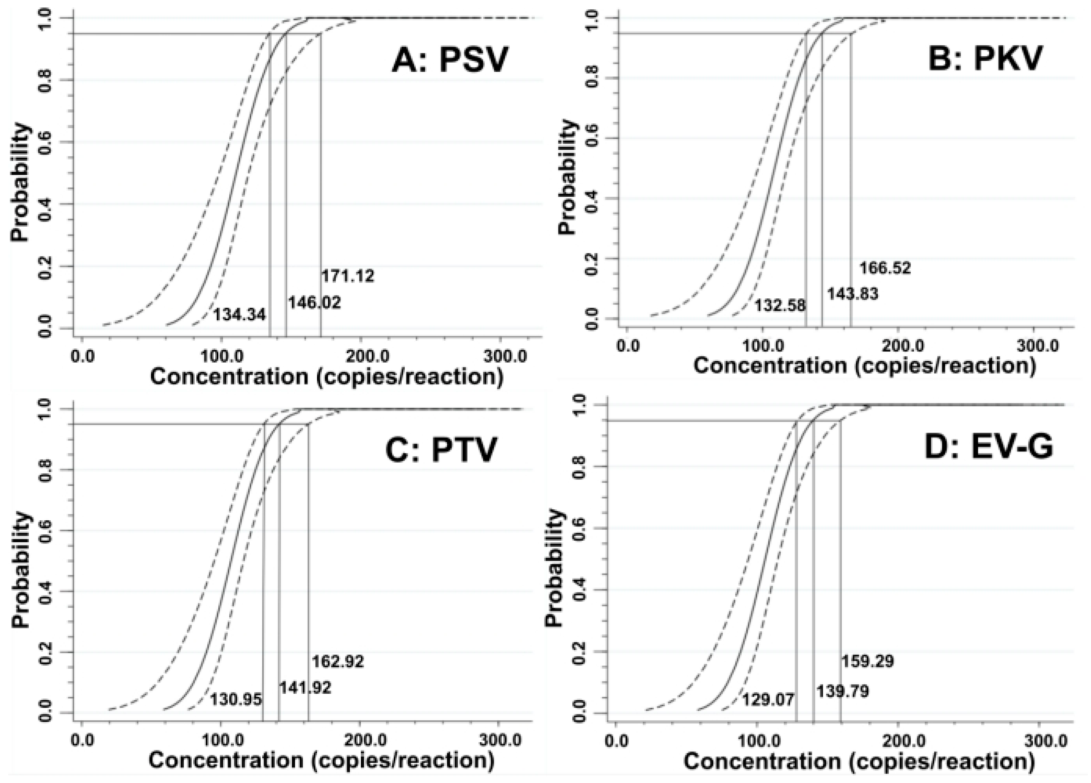
3.5. Sensitivity of the Quadruplex RT-qPCR

The sensitivity of the developed quadruplex RT-qPCR was evaluated using the synthesized viral RNAs. The mixtures of synthesized viral RNAs of PSV, PKV, PTV, and EV-G with concentrations from 1.0 × 10⁸ to 1.0 × 101 copies/ µ L (the final concentration: 1.0 × 10⁷ to 1.0 × 100 copies/ µ L ). The results indicated that the LODs were 1.0 × 101 copies/ µ L for all four synthesized viral RNAs (Figure 3).
The LODs were also assessed using probit regression analysis. The concentrations of 500, 250, 125, and 62.5 copies/reaction for the synthesized viral RNAs of PSV, PKV, PTV, and EV-G were selected as templates (Table 3). The results showed that PSV, PKV, PTV, and EV-G had LODs of 146.02 (95% confidence interval (CI) of 134.34–171.12), 143.83 (95% CI of 132.58–166.52), 141.92 (95% CI of 130.95–162.92), and 139.79 (95% CI of 129.07–159.29), respectively (Figure 4), indicating high sensitivity of the quadruplex RT-qPCR.

3.6. Repeatability of the Quadruplex RT-qPCR

The repeatability of the developed quadruplex RT-qPCR was analyzed using the mixtures of plasmid constructs p-PSV, p-PKV, p-PTV, and p-EV-G, with concentrations of 1.0 × 10⁷, 1.0 × 10⁴, and 1.0 × 10² copies/ µ L (the final concentrations: 1.0 × 10⁶, 1.0 × 10³, and 1.0 × 101 copies/ µ L ), respectively. The results indicated that the intra-assay CVs were 0.27–1.57%, and the inter-assay CVs were 0.21–1.41% (Table 4), indicating the excellent reproducibility of the developed assay.

3.7. Assessment Results of the Clinical Samples

The 1823 fecal samples collected from diarrheic piglets in Guangxi Province between January 2024 and December 2024 were tested using the developed quadruplex RT-qPCR assay. The results indicated that the positivity rates of PSV, PKV, PTV, and EV-G were 15.25% (278/1823), 21.72% (396/1823), 18.82% (343/1823), and 27.10% (494/1823), respectively. Furthermore, mixed infections of these viruses were also found in the clinical samples (Table 5). PTV + EV-G co-infections showed the highest positivity rate of dual infection at 13.76% (251/1823), and quadruple co-infections were detected in 1.91% (35/1823) of these fecal samples.
In addition, the reference RT-qPCR for the detection of PSV [33], PKV [34], and PTV [35], and the reference RT-PCR for the detection of EV-G [29] were used to test these 1823 fecal samples. The results showed that the positivity rates of PSV, PKV, PTV, and EV-G were 15.03% (274/1823), 21.56% (393/1823), 18.65% (340/1823), and 26.82% (488/1823), respectively. Compared with the reported reference methods, the developed quadruplex RT-qPCR assay showed diagnostic sensitivity and specificity for PSV, PKV, PTV, and EV-G at 99.27% and 99.61%, 98.98% and 99.51%, 99.12% and 99.60%, and 98.77% and 99.10%, respectively (Table 6). The two methods showed coincidence rates of 99.56%, 99.40%, 99.51%, and 99.01% for PSV, PKV, PTV, and EV-G, respectively (Supplementary Table S1).

4. Discussion

At present, many viruses can cause diarrhea in pig herds [36,37,38]; of which, PSV, PKV, PTV, and EV-G are common viruses that play important roles in the establishment of diarrhea in animals. PSV was initially discovered in the United Kingdom in 1958, and the first Chinese PSV strain was isolated in Shanghai Municipality in 2009 [39]. There is a widespread prevalence of PSV in porcine populations, and even a positivity rate as high as 79.3% in some areas [5,9,40]. PKV has been reported globally since its discovery in 2007 [41,42]. Even worse, the recombinant strains of PKV were found in the circulating strains [43,44]. The first outbreak of PTV occurred in Czechoslovakia in 1929, and different genotypes have been found. PTV1 has a high prevalence in wild and domestic pigs in some European countries [45]. In China, PTV2 infection was first identified in Heilongjiang Province in 2009 [46], and has also been found in Guangxi Province [47]. Recently, six strains of known genotypes, and four undefined genotype strains were obtained in Jiangxi Province, indicating there were more than two genotypes of PTV2 in China [48]. This indicated that PTV had genetic diversity in China. EV-G was first isolated in 1973. Diarrheic pigs infected with EV-G have been reported in many countries [28,49,50]. EV-G was also widespread in Guangxi Province, and had a high positivity rate in pig farms, even reaching a 100% positivity rate [51]. The prevalence of EV-G (24.37%, 97/398) was higher than those of PSV (5.77%, 23/398) and PTV (12.81%, 51/398), and there also existed co-infections of these three viruses in India [52]. These situations make it necessary and urgent to establish accurate detection methods, and perform investigation and epidemiology studies on these pathogens.
PSV, PKV, PTV, and EV-G play important roles in the establishment of diarrhea in pig herds, and they show similar clinical signs and pathological changes, which are hard to differentiate in clinical cases. Even worse, co-infections with these viruses are usually found [52,53], and these exacerbate the diarrhea and other clinical signs [14,28,52,54]. Current techniques used for the detection and identification of viruses usually include electron microscopy, virus isolation, and serological tests. However, they show the shortcomings of cumbersomeness, relatively low sensitivity, and susceptibility to environmental contamination. On the contrary, qPCR can detect the target nucleic acids based on Ct values. It has the advantages of easy operation, strong specificity, and high sensitivity, and has been widely used in different biological laboratories [30,31]. To date, there exist single RT-qPCR assays for the detection of PSV, PKV, and PTV [33,34,35], and a multiplex RT-PCR assay for the detection of PSV, PTV, and EV-G [29]. However, no multiplex RT-qPCR assay has been reported for the simultaneous detection of these four viruses until now. In this study, the standard plasmid constructs were prepared, the reaction conditions were optimized, the standard curves were generated, and the specificity, sensitivity, and reproducibility were evaluated. The results indicated that the developed quadruplex RT-qPCR assay specifically detected PSV, PKV, PTV, and EV-G; it showed low LODs of 146.02, 143.83, 141.92, and 139.79 copies/reaction for PSV, PKV, PTV, and EV-G, respectively, which had higher sensitivity than the reference RT-qPCR for PSV (934 copies/reaction), PKV (750 copies/reaction), PTV (200 copies/reaction) [33,34,35], and the non-quantitative analysis of EV-G due to RT-PCR [29]. The assay indicated intra-assay CVs of 0.27–1.57% and inter-assay CVs of 0.21–1.41%. Furthermore, 1823 fecal samples collected in Guangxi Province were validated using the established quadruplex RT-qPCR and the previously reported reference RT-qPCR/RT-PCR [29,33,34,35]. The detection results showed that their coincidence rates were higher than 99.01%. These results indicated that the quadruplex RT-qPCR assay had the advantages of easy operation, high specificity, excellent sensitivity, and good reproducibility, and was suitable for application to detect clinical samples. It is noteworthy that there exists genetic diversity in the prevalent strains of PSV, PKV, PTV, and EV-G, so the design of the specific primers and probes is very important for the development of a quadruplex RT-qPCR. In this study, the multiple sequence alignments of viral strains from different countries were performed. Then, the conserved regions in 5′ UTR of PSV, PKV, PTV, and EV-G were selected as the target fragments to design the specific primers and probes, which ensured the detection of different viral strains prevalent in various countries around the world. Of course, due to the continuous variation of the epidemic strains, it is necessary to continuously perform molecular epidemiological research. This ensures the grasping of the viral genetic variation and the adjustment of primers and probe sequences on time, and ensures the accurate detection of the epidemic strains.
In this study, 1823 fecal samples collected from different pig farms in Guangxi Province had positivity rates of 15.03%, 21.56%, 18.65%, and 26.82% for PSV, PKV, PTV, and EV-G, respectively. This suggested that these viruses have high positivity rates in Guangxi Province. These viruses have also been reported in many provinces of China [5,6,8,9,13,14,16,17,18,20,23,24,25], and many countries around the world [2,3,4,7,18,21,22,27,36,44,50,55,56]. In China, it has been found that PKV is usually co-infected with PEDV, and PKV may enhance the virulence of PEDV, resulting in more serious damage to the hosts [13,14]. In our previous study, the positivity rate of PEDV during 2020–2024 in Guangxi Province was 11.90% (397/5859) [57]. In this study, the 1823 fecal samples were also tested for PEDV using the RT-qPCR assay previously established in our laboratory [37]. The results showed that PEDV had a positivity rate of 5.38% (98/1823); PKV and PEDV had a co-infection rate of 3.46% (63/1823), while PKV (+)/PEDV (−) was 18.10% (330/1823), and PEDV (+)/PKV (−) was 1.92% (35/1823). The results showed that PKV and PEDV co-infection existed in Guangxi Province, and further research is needed to evaluate the synergism of PKV and PEDV. In this study, triple and quadruple co-infections of PSV, PKV, PTV, and EV-G were also detected in fecal samples, with the highest positivity rate of 13.76% (251/1823) for co-infections of PTV and EV-G. In the Czech Republic, co-infections of PTV and EV-G were also as high as 30.4% (17/56) in wild boars and 12.9% (16/124) in domestic pigs [28]. PSV, PTV, and EV-G co-infections have also been reported in India and Brazil [52,53]. At present, all four viruses are widely spread in different countries around the world, and co-infections of pigs with Picornaviridae viruses are common, and the harms of these viruses cannot be ignored. It is urgent to carry out appropriate prevention and control measures to curb the spread of these viruses and decrease the economic losses of these diseases.
Especially, PSV [6,7] and PKV [11,12,58,59] have the zoonotic potentials to infect humans. The widespread occurrence of PSV and PKV creates a risk of cross-species transmission, and induces huge harms to the pig industry and human health. This further highlights the urgency and importance of developing the quadruplex RT-qPCR for the rapid, accurate, and simultaneous detection of PSV, PKV, PTV, and EV-G. In addition, due to the zoonotic potentials of PSV and PKV, there is indeed a risk of these viruses being transmitted from pigs across species to humans. Therefore, it is extremely important to take strong prevention and control measures to prevent these viruses from being transmitted from pigs to humans. The measures include but are not limited to the following main measures: establish and implement strict biosecurity measures in pig farms; regularly clean and disinfect the external environment thoroughly; use personal protective measures for workers and veterinarians who have close contact with pigs; clean and disinfect vehicles and items before entering the farms; timely dispose of sick pigs to reduce their chances of contact with humans.

5. Conclusions

The developed quadruplex RT-qPCR assay for the detection of PSV, PKV, PTV, and EV-G is characterized by easy operation, high sensitivity, and strong specificity, and can be used for the accurate evaluation of PSV, PKV, PTV, and EV-G in clinical specimens. The detection results of clinical samples indicated that these viruses are widespread in Guangxi Province, southern China, and further experimental studies demonstrating their roles in the establishment of diarrhea in pigs would be interesting.

Supplementary Materials

The following supporting information can be downloaded at: https://www.mdpi.com/article/10.3390/ani15071008/s1, Figure S1: The multiple sequence alignments of PSV (A), PKV (B), PTV (C), and EV-G (D); Table S1: Agreements of the test results via different assays.

Author Contributions

B.L. contributed to experiment design, data analysis, and manuscript drafting. K.S. contributed to funding, experiment design, laboratory supervision, manuscript drafting, and manuscript editing. Y.S. and S.F. contributed to sample collection, methods, and manuscript drafting. Y.Y., W.L., and F.L. contributed to software and manuscript drafting. Z.W. and Y.W. contributed to laboratory supervision, manuscript drafting, and manuscript editing. All authors have read and agreed to the published version of the manuscript.

Funding

This study was supported by the Guangxi Key Laboratory of Animal Breeding, Disease Control and Prevention (Grant No. ABDC-b202304), Guangxi Science and Technology Bureau (Grant No. AB21238003), and Bama county program for talents in science and technology (Grant No. 20220020), Guangxi, China.

Institutional Review Board Statement

This study was approved by the Guangxi Center for Animal Disease Control and Prevention (CADC) (No. 2020-A-01) on 5 November 2020.

Informed Consent Statement

Written informed consent was obtained from the owners of the animals.

Data Availability Statement

The original contributions presented in this study are included in this article/Supplementary Materials; further inquiries can be directed to the corresponding authors.

Conflicts of Interest

The authors declare no conflicts of interest.

References

  1. Zell, R.; Delwart, E.; Gorbalenya, A.E.; Hovi, T.; King, A.M.Q.; Knowles, N.J.; Lindberg, A.M.; Pallansch, M.A.; Palmenberg, A.C.; Reuter, G.; et al. ICTV Virus Taxonomy Profile: Picornaviridae. J. Gen. Virol. 2017, 98, 2421–2422. [Google Scholar] [CrossRef] [PubMed]
  2. Son, K.Y.; Kim, D.S.; Kwon, J.; Choi, J.S.; Kang, M.I.; Belsham, G.J.; Cho, K.O. Full-Length Genomic Analysis of Korean Porcine Sapelovirus Strains. PLoS ONE 2014, 9, e107860. [Google Scholar] [CrossRef]
  3. Boros, Á.; László, Z.; Pankovics, P.; Marosi, A.; Albert, M.; Cságola, A.; Bíró, H.; Fahsbender, E.; Delwart, E.; Reuter, G. High Prevalence, Genetic Diversity and a Potentially Novel Genotype of Sapelovirus A (Picornaviridae) in Enteric and Respiratory Samples in Hungarian Swine Farms. J. Gen. Virol. 2020, 101, 609–621. [Google Scholar] [CrossRef]
  4. Kumari, S.; Ray, P.K.; Singh, R.; Desingu, P.A.; Varshney, R.; Saikumar, G. Pathological and Molecular Investigation of Porcine Sapelovirus Infection in Naturally Affected Indian Pigs. Microb. Pathog. 2019, 127, 320–325. [Google Scholar] [CrossRef]
  5. Chen, J.; Suo, X.; Cao, L.; Yuan, C.; Shi, L.; Duan, Y.; Zheng, H.; Wang, Q. Virome Analysis for Identification of a Novel Porcine Sapelovirus Isolated in Western China. Microbiol. Spectr. 2022, 10, e0180122. [Google Scholar] [CrossRef] [PubMed]
  6. Li, Y.; Du, L.; Jin, T.; Cheng, Y.; Zhang, X.; Jiao, S.; Huang, T.; Zhang, Y.; Yan, Y.; Gu, J.; et al. Characterization and Epidemiological Survey of Porcine Sapelovirus in China. Vet. Microbiol. 2019, 232, 13–21. [Google Scholar] [CrossRef]
  7. Arruda, P.H.; Arruda, B.L.; Schwartz, K.J.; Vannucci, F.; Resende, T.; Rovira, A.; Sundberg, P.; Nietfeld, J.; Hause, B.M. Detection of a Novel Sapelovirus in Central Nervous Tissue of Pigs with Polioencephalomyelitis in the USA. Transbound. Emerg. Dis. 2017, 64, 311–315. [Google Scholar] [CrossRef]
  8. Lu, S.J.; Ma, M.Y.; Yan, X.G.; Zhao, F.J.; Hu, W.Y.; Ding, Q.W.; Ren, H.J.; Xiang, Y.Q.; Zheng, L.L. Development and Application of a Low-Priced Duplex Quantitative PCR Assay Based on SYBR Green I for the Simultaneous Detection of Porcine Deltacoronavirus and Porcine Sapelovirus. Vet. Med. 2023, 68, 106–115. [Google Scholar] [CrossRef] [PubMed]
  9. Ibrahim, Y.M.; Zhang, W.; Werid, G.M.; Zhang, H.; Feng, Y.; Pan, Y.; Zhang, L.; Li, C.; Lin, H.; Chen, H.; et al. Isolation, Characterization, and Molecular Detection of Porcine Sapelovirus. Viruses 2022, 14, 349. [Google Scholar] [CrossRef]
  10. Liu, X.; Oka, T.; Wang, Q. Genomic Characterization of a US Porcine Kobuvirus Strain. Arch. Microbiol. 2015, 197, 1033–1040. [Google Scholar] [CrossRef]
  11. Khamrin, P.; Maneekarn, N.; Okitsu, S.; Ushijima, H. Epidemiology of Human and Animal Kobuviruses. Virusdisease 2014, 25, 195–200. [Google Scholar]
  12. Werid, G.M.; Ibrahim, Y.M.; Chen, H.; Fu, L.; Wang, Y. Molecular Detection and Genetic Characterization of Potential Zoonotic Swine Enteric Viruses in Northern China. Pathogens 2022, 11, 417. [Google Scholar] [CrossRef]
  13. Zang, Y.; Feng, B.; Huang, Z.; Zhao, D.; Qi, W.; Qiu, Y.; Qiu, M.; Li, C.; Lin, H.; Zheng, W.; et al. Epidemiologic and Genomic Characterizations of Porcine Kobuviruses in Diarrheic and Healthy Pigs. Animals 2023, 13, 3129. [Google Scholar] [CrossRef]
  14. Wu, S.; Gou, F.; Meng, J.; Jin, X.; Liu, W.; Ding, W.; Xu, W.; Gu, C.; Hu, X.; Cheng, G.; et al. Porcine Kobuvirus Enhances Porcine Epidemic Diarrhea Virus Pathogenicity and Alters the Number of Intestinal Lymphocytes in Piglets. Vet. Microbiol. 2024, 293, 110100. [Google Scholar]
  15. Oba, M.; Naoi, Y.; Ito, M.; Masuda, T.; Katayama, Y.; Sakaguchi, S.; Omatsu, T.; Furuya, T.; Yamasato, H.; Sunaga, F.; et al. Metagenomic Identification and Sequence Analysis of a Teschovirus A-Related Virus in Porcine Feces in Japan, 2014-2016. Infect. Genet. Evol. 2018, 66, 210–216. [Google Scholar] [CrossRef]
  16. Ma, H.; Zhang, M.; Wu, M.; Ghonaim, A.H.; Fan, S.; He, Q. Isolation and Genetic Characteristics of a Neurotropic Teschovirus Variant Belonging to Genotype 1 in Northeast China. Arch. Virol. 2021, 166, 1355–1370. [Google Scholar]
  17. Zhang, B.; Guo, R.; Xiao, L.; Zhong, C.; Yuan, X.; Huang, J.; Zhu, X.; Zhou, J.; Fan, B.; Xue, T.; et al. Analysis on the Genome of a Teschovirus Type 1 Isolates with Swine Diarrhea. Heliyon 2023, 9, e14710. [Google Scholar]
  18. Liang, W.; Wu, X.; Ding, Z.; Zhong, S.; Qian, X.; Ye, P.; Liu, H.; Chen, Z.; Zhang, J.; Cao, H.; et al. Identification of a Novel Porcine Teschovirus 2 Strain as Causative Agent of Encephalomyelitis in Suckling Piglets with High Mortality in China. BMC Vet. Res. 2023, 19, 2. [Google Scholar]
  19. Carnero, J.; Prieto, C.; Polledo, L.; Martínez-Lobo, F.J. Detection of Teschovirus Type 13 from Two Swine Herds Exhibiting Nervous Clinical Signs in Growing Pigs. Transbound. Emerg. Dis. 2018, 65, e489–e493. [Google Scholar] [PubMed]
  20. Yang, T.; Li, R.; Yao, Q.; Zhou, X.; Liao, H.; Ge, M.; Yu, X. Prevalence of Porcine Teschovirus Genotypes in Hunan, China: Identification of Novel Viral Species and Genotypes. J. Gen. Virol. 2018, 99, 1261–1267. [Google Scholar] [CrossRef] [PubMed]
  21. Bhat, S.; Kattoor, J.J.; Sircar, S.; VinodhKumar, O.R.; Thomas, P.; Ghosh, S.; Malik, Y.S. Detection and Molecular Characterization of Porcine Teschoviruses in India: Identification of New Genotypes. Indian J. Microbiol. 2024, 64, 963–972. [Google Scholar] [PubMed]
  22. Bhat, S.; Ansari, M.I.; Kattoor, J.J.; Sircar, S.; Dar, P.S.; Deol, P.; Vinodh Kumar, O.R.; Thomas, P.; Ghosh, S.; El Zowalaty, M.E.; et al. Emerging Porcine Enterovirus G Infections, Epidemiological, Complete Genome Sequencing, Evolutionary and Risk Factor Analysis in India. Virology 2024, 590, 109906. [Google Scholar]
  23. Xiao, D.; Zhang, L.; Li, S.; Liang, Y.; Wu, R.; Wen, Y.; Yan, Q.; Du, S.; Zhao, Q.; Han, X.; et al. Characterization, Phylogenetic Analysis, and Pathogenicity of a Novel Genotype 2 Porcine Enterovirus G. Virus Res. 2023, 335, 199185. [Google Scholar]
  24. Mi, X.; Yang, C.; Lu, Y.; Wang, H.; Qin, Q.; Chen, R.; Chen, Z.; Luo, Y.; Chen, Y.; Wei, Z.; et al. Isolation, Identification, and Evaluation of the Pathogenicity of a Porcine Enterovirus G Isolated from China. Front. Vet. Sci. 2021, 8, 712679. [Google Scholar]
  25. Van Dung, N.; Anh, P.H.; Van Cuong, N.; Hoa, N.T.; Carrique-Mas, J.; Hien, V.B.; Sharp, C.; Rabaa, M.; Berto, A.; Campbell, J.; et al. Large-Scale Screening and Characterization of Enteroviruses and Kobuviruses Infecting Pigs in Vietnam. J. Gen. Virol. 2016, 97, 378–388. [Google Scholar] [PubMed]
  26. Ibrahim, Y.M.; Zhang, W.; Wang, X.; Werid, G.M.; Fu, L.; Yu, H.; Wang, Y. Molecular Characterization and Pathogenicity Evaluation of Enterovirus G Isolated from Diarrheic Piglets. Microbiol. Spectr. 2023, 11, e0264323. [Google Scholar]
  27. Imai, R.; Rongduo, W.; Kaixin, L.; Borjigin, S.; Matsumura, H.; Masuda, T.; Ozawa, T.; Oba, M.; Makino, S.; Nagai, M.; et al. Novel Recombinant Porcine Enterovirus G Viruses Lacking Structural Proteins Are Maintained in Pig Farms in Japan. J. Vet. Med. Sci. 2023, 85, 252–265. [Google Scholar]
  28. Prodělalová, J. The Survey of Porcine Teschoviruses, Sapeloviruses and Enteroviruses B Infecting Domestic Pigs and Wild Boars in the Czech Republic between 2005 and 2011. Infect. Genet. Evol. 2012, 12, 1447–1451. [Google Scholar]
  29. Stäubli, T.; Rickli, C.I.; Torgerson, P.R.; Fraefel, C.; Lechmann, J. Porcine Teschovirus, Sapelovirus, and Enterovirus in Swiss Pigs: Multiplex RT-PCR Investigation of Viral Frequencies and Disease Association. J. Vet. Diagn. Investig. 2021, 33, 864–874. [Google Scholar]
  30. Hawkins, S.F.C.; Guest, P.C. Multiplex Analyses Using Real-Time Quantitative PCR. Methods Mol. Biol. 2017, 1546, 125–133. [Google Scholar]
  31. Kralik, P.; Ricchi, M. A Basic Guide to Real Time PCR in Microbial Diagnostics: Definitions, Parameters, and Everything. Front. Microbiol. 2017, 8, 108. [Google Scholar]
  32. Ma, Y.; Shi, K.; Chen, Z.; Shi, Y.; Zhou, Q.; Mo, S.; Wei, H.; Hu, L.; Mo, M. Simultaneous Detection of Porcine Respiratory Coronavirus, Porcine Reproductive and Respiratory Syndrome Virus, Swine Influenza Virus, and Pseudorabies Virus via Quadruplex One-Step RT-qPCR. Pathogens 2024, 13, 341. [Google Scholar] [CrossRef]
  33. Chen, Q.Y.; Sun, Z.H.; Che, Y.L.; Chen, R.J.; Wu, X.M.; Wu, R.J.; Wang, L.B.; Zhou, L.J. High Prevalence, Genetic Diversity, and Recombination of Porcine Sapelovirus in Pig Farms in Fujian, Southern China. Viruses 2023, 15, 1751. [Google Scholar] [CrossRef]
  34. Zhu, X.; Wang, Y.; Chen, J.; Zhang, X.; Shi, H.; Shi, D.; Gao, J.; Feng, L. Development of TaqMan Real-Time Reverse Transcription-Polymerase Chain Reaction for the Detection and Quantitation of Porcine Kobuvirus. J. Virol. Methods 2016, 234, 132–136. [Google Scholar]
  35. Zhang, C.; Wang, Z.; Hu, F.; Liu, Y.; Qiu, Z.; Zhou, S.; Cui, S.; Wang, M. The Survey of Porcine Teschoviruses in Field Samples in China with a Universal Rapid Probe Real-Time RT-PCR Assay. Trop. Anim. Health Prod. 2013, 45, 1057–1061. [Google Scholar]
  36. Smoľak, D.; Šalamúnová, S.; Jacková, A.; Haršányová, M.; Budiš, J.; Szemes, T.; Vilček, Š. Analysis of RNA Virome in Rectal Swabs of Healthy and Diarrheic Pigs of Different Age. Comp. Immunol. Microbiol. Infect. Dis. 2022, 90–91, 101892. [Google Scholar]
  37. Zhou, H.; Shi, K.; Long, F.; Zhao, K.; Feng, S.; Yin, Y.; Xiong, C.; Qu, S.; Lu, W.; Li, Z. A Quadruplex qRT-PCR for Differential Detection of Four Porcine Enteric Coronaviruses. Vet. Sci. 2022, 9, 634. [Google Scholar] [CrossRef]
  38. He, J.; Shi, K.; Shi, Y.; Yin, Y.; Feng, S.; Long, F.; Qu, S.; Song, X. Development of a Quadruplex RT-qPCR for the Detection of Porcine Astrovirus, Porcine Sapovirus, Porcine Norovirus, and Porcine Rotavirus A. Pathogens 2024, 13, 1052. [Google Scholar] [CrossRef]
  39. Lan, D.; Ji, W.; Yang, S.; Cui, L.; Yang, Z.; Yuan, C.; Hua, X. Isolation and Characterization of the First Chinese Porcine Sapelovirus Strain. Arch. Virol. 2011, 156, 1567–1574. [Google Scholar]
  40. Liu, J.; Li, B.; Tao, J.; Cheng, J.; Shi, Y.; Qiao, C.; Shen, X.; Liu, H. Development of an Indirect ELISA Method Based on the VP1 Protein for Detection of IgG Antibodies against Porcine Sapelovirus. Can. J. Vet. Res. 2023, 87, 176–183. [Google Scholar]
  41. Reuter, G.; Boldizsár, A.; Kiss, I.; Pankovics, P. Candidate New Species of Kobuvirus in Porcine Hosts. Emerg. Infect. Dis. 2008, 14, 1968–1970. [Google Scholar] [PubMed]
  42. Yu, J.; Jin, M.; Zhang, Q.; Li, H.; Li, D.; Xu, Z.; Li, J.; Cui, S.; Yang, S.; Liu, N.; et al. Candidate Porcine Kobuvirus, China. Emerg. Infect. Dis. 2009, 15, 823–825. [Google Scholar]
  43. Wei, R.; Shang, R.; Cheng, K.; Wang, S.; Yuan, X.; Wu, J.; Yu, Z. Phylogenetic Analysis and Molecular Characterization of the Co-Infection of the New Variant of the Porcine Epidemic Diarrhea Virus and the Novel Porcine Kobuvirus Isolated from Piglets with Diarrhea. Braz. J. Microbiol. 2023, 54, 2527–2534. [Google Scholar]
  44. Cui, Y.; Li, J.; Guo, J.; Pan, Y.; Tong, X.; Liu, C.; Wang, D.; Xu, W.; Shi, Y.; Ji, Y.; et al. Evolutionary Origin, Genetic Recombination, and Phylogeography of Porcine Kobuvirus. Viruses 2023, 15, 240. [Google Scholar] [CrossRef]
  45. Sytiuk, M.P.; Bezymennyy, M.V.; Halka, I.V.; Uhovskyy, V.V.; Muzykina, L.M.; Lavalley, M.; Nychyk, S.A.; Nedosekov, V.V.; Howard, M.W.; Bortz, E. Seroprevalence of Enzootic Teschen Disease in the Wild Boar Population in Ukraine. Vector Borne Zoonotic Dis. 2022, 22, 138–147. [Google Scholar]
  46. Wang, B.; Tian, Z.J.; Gong, D.Q.; Li, D.Y.; Wang, Y.; Chen, J.Z.; An, T.Q.; Peng, J.M.; Tong, G.Z. Isolation of Serotype 2 Porcine Teschovirus in China: Evidence of Natural Recombination. Vet. Microbiol. 2010, 146, 138–143. [Google Scholar] [PubMed]
  47. Li, Y.; Chen, S.; Shi, Y.; Huang, H.; Wang, W.; Zheng, M.; Zhao, C.; Zhang, X.; Lei, X.; Sun, W.; et al. Construction and Characterization of the Full-Length cDNA of an Infectious Clone of Emerging Porcine Teschovirus-2. Pathog. Dis. 2022, 80, ftac033. [Google Scholar]
  48. Yang, T.; Lu, Y.; Zhang, L.; Li, X. Identification of Novel Genotypes Belonging to the Species Teschovirus A from Indigenous Pigs in Western Jiangxi, China. Arch. Virol. 2020, 165, 993–1001. [Google Scholar]
  49. Vilar, M.J.; Peralta, B.; García-Bocanegra, I.; Simon-Grifé, M.; Bensaid, A.; Casal, J.; Segalés, J.; Pina-Pedrero, S. Distribution and Genetic Characterization of Enterovirus G and Sapelovirus A in Six Spanish Swine Herds. Virus Res. 2016, 215, 42–49. [Google Scholar]
  50. Janetanakit, T.; Chaiyawong, S.; Charoenkul, K.; Tangwangvivat, R.; Chamsai, E.; Udom, K.; Jairak, W.; Amonsin, A. Distribution and Genetic Diversity of Enterovirus G (EV-G) on Pig Farms in Thailand. BMC Vet. Res. 2021, 17, 277. [Google Scholar]
  51. Hong, D.; Bian, J.; Zeng, L.; Huang, S.; Qin, Y.; Chen, Y.; Wei, Z.; Huang, W.; Ouyang, K. A Novel VP1-Based Enzyme-Linked Immunosorbent Assay Revealed Widespread Enterovirus G Infections in Guangxi, China. J. Virol. Methods 2024, 325, 114873. [Google Scholar]
  52. Patel, S.K.; Agrawal, A.; Pathak, M.; Singh, A.; Varshney, R.; Rana, J.; Saikumar, G. Detection of Porcine Enteric Picornaviruses from Faecal Samples of Indian Pigs. Virusdisease 2022, 33, 102–107. [Google Scholar]
  53. Donin, D.G.; de Arruda Leme, R.; Alfieri, A.F.; Alberton, G.C.; Alfieri, A.A. First Report of Porcine Teschovirus (PTV), Porcine Sapelovirus (PSV) and Enterovirus G (EV-G) in Pig Herds of Brazil. Trop. Anim. Health Prod. 2014, 46, 523–528. [Google Scholar]
  54. Sawant, P.M.; Atre, N.; Kulkarni, A.; Gopalkrishna, V. Detection and Molecular Characterization of Porcine Enterovirus G15 and Teschovirus from India. Pathog. Dis. 2020, 78, ftaa039. [Google Scholar]
  55. Puente, H.; Arguello, H.; Cortey, M.; Gómez-García, M.; Mencía-Ares, O.; Pérez-Perez, L.; Díaz, I.; Carvajal, A. Detection and Genetic Characterization of Enteric Viruses in Diarrhoea Outbreaks from Swine Farms in Spain. Porc. Health Manag. 2023, 9, 29. [Google Scholar]
  56. Capai, L.; Piorkowski, G.; Maestrini, O.; Casabianca, F.; Masse, S.; de Lamballerie, X.; Charrel, R.N.; Falchi, A. Detection of Porcine Enteric Viruses (Kobuvirus, Mamastrovirus and Sapelovirus) in Domestic Pigs in Corsica, France. PLoS ONE 2022, 17, e0260161. [Google Scholar] [CrossRef]
  57. Shi, K.; Li, B.; Shi, Y.; Feng, S.; Yin, Y.; Long, F.; Pan, Y.; Wei, Y. Phylogenetic and Evolutionary Analysis of Porcine Epidemic Diarrhea Virus in Guangxi Province, China, during 2020 and 2024. Viruses 2024, 16, 1126. [Google Scholar] [CrossRef]
  58. Khamrin, P.; Maneekarn, N.; Hidaka, S.; Kishikawa, S.; Ushijima, K.; Okitsu, S.; Ushijima, H. Molecular Detection of Kobuvirus Sequences in Stool Samples Collected from Healthy Pigs in Japan. Infect. Genet. Evol. 2010, 10, 950–954. [Google Scholar] [CrossRef] [PubMed]
  59. Okitsu, S.; Khamrin, P.; Thongprachum, A.; Hidaka, S.; Kongkaew, S.; Kongkaew, A.; Maneekarn, N.; Mizuguchi, M.; Hayakawa, S.; Ushijima, H. Sequence Analysis of Porcine Kobuvirus VP1 Region Detected in Pigs in Japan and Thailand. Virus Genes 2012, 44, 253–257. [Google Scholar] [CrossRef] [PubMed]
Figure 1. The amplification curves of p-PSV (A), p-PKV (B), p-PTV (C), and p-EV-G (D), and the standard curves (E) of the developed quadruplex RT-qPCR. In subfigure (AD) 1–7, the final concentration of the plasmid constructs ranged from 107 to 101 copies/ µ L ; 8: distilled water.
Animals 15 01008 g001
Figure 2. Specificity evaluation of the developed quadruplex RT-qPCR for PSV (A), PKV (B), PTV (C), and EV-G (D). 1: p-PSV; 2: p-PKV; 3: p-PTV; 4: p-EV-G; 5–8: positive clinical samples of PSV, PKV, PTV, and EV-G; 9: PSV; 10: PKV; 11: PTV; 12: EV-G; 13–22: TGEV, PCV2, PRRSV, FMDV, CSFV, PRV, SIV, PoRV, ASFV, PEDV, and PDCoV; 23: negative fecal sample; 24: distilled water.
Animals 15 01008 g002
Figure 3. Sensitivity evaluation of the developed quadruplex RT-qPCR. In subfigures (AD) 1–8: the final reaction concentrations of the synthesized viral RNAs ranged from 107 to 100 copies/ µ L ; 9: negative control.
Animals 15 01008 g003
Figure 4. Sensitivity evaluation of the developed quadruplex RT-qPCR using probit regression analysis. The PSV (A), PKV (B), PTV (C), and EV-G (D) had LODs of 146.02, 143.83, 141.92, and 139.79 copies/reaction, respectively.
Animals 15 01008 g004
Table 1. The designed specific primers and probes.
Primer/ProbeSequence (5′ → 3′)GeneTm/°CProduct (bp)
PSV-FGACTGGGCCTATACTACCTGATA5′ UTR57.2140
PSV-RAGGTACACACGGGCTCTCTG60.6
PSV-PROX-TGGCCGCCTGTAACTAGTATAGTCAGT-BHQ263.6
PKV-FCGTGCTGAGTAATGGGATAGG5′ UTR57.0149
PKV-RTGCACTTCAGAGGTCAGAGAA57.9
PKV-PVIC-ATGAGTAGAGCATGGACTGCGGTG-BHQ163.4
PTV-FGGACTGCRTTGCATATCCCTA5′ UTR58.3137
PTV-RGACTATACAAAGTACAGACGGCCA58.9
PTV-PCY5-CTGTATGGGAATGCAGGACTGG-BHQ359.9
EV-G-FGGACCTAGTAGTGATAGGCTGTA5′ UTR57.1113
EV-G-RCTGAGCGAAACGCCAAGA57.2
EV-G-PFAM-GCCGAAGATGAACCCGTCCGTTAT-BHQ163.8
Note: In degenerate primers, R represents A or G. F—forward; R—reverse; P—probe. The same as follows.
Table 2. The components and their optimal parameters.
IngredientVolume (µL)Final Concentration (nM)
2× One-Step RT-PCR Buffer III10.0/
Ex Taq HS (5 U/µL)0.4/
PrimeScript RT Enzyme Mix II0.4/
PSV-F0.3300
PSV-R0.3200
PSV-P0.2200
PKV-F0.2200
PKV-R0.2200
PKV-P0.2200
PTV-F0.2200
PTV-R0.2200
PTV-P0.2200
EV-G-F0.2200
EV-G-R0.2200
EV-G- P0.2200
Total Nucleic Acid2.0/
Nuclease-Free Distilled WaterUp to 20.0/
Table 3. The Ct values and hit rates of the serially diluted synthesized viral RNAs.
RNACopies/ReactionNumber of SamplesQuadruplex RT-qPCR
Ct Value ( X ¯ ± SD)Hit Rate (%)
PSV5003634.93 ± 0.13100
2503635.49 ± 0.12100
1253635.89 ± 0.1375.0
62.536ND0
PKV5003634.86 ± 0.11100
2503635.41 ± 0.13100
1253635.83 ± 0.1577.8
62.536ND0
PTV5003634.73 ± 0.10100
2503635.37 ± 0.13100
1253635.76 ± 0.1580.6
62.536ND0
EV-G5003634.67 ± 0.14100
2503635.33 ± 0.10100
1253634.73 ± 0.1383.3
62.536ND0
Note: ND—not detected.
Table 4. Repeatability of the developed quadruplex RT-qPCR.
PlasmidConcentration (Copies/μL)Ct Values of Intra-AssayCt Values of Inter-Assay
X ¯ SDCV (%) X ¯ SDCV (%)
p-PSV1.0 × 10615.920.251.57%15.450.120.78%
1.0 × 10325.890.080.31%25.880.100.39%
1.0 × 10133.940.150.44%33.560.110.33%
p-PKV1.0 × 10615.850.191.20%15.260.171.11%
1.0 × 10326.170.130.50%26.040.080.31%
1.0 × 10133.650.250.74%33.830.070.21%
p-PTV1.0 × 10615.430.150.97%15.580.070.45%
1.0 × 10326.590.090.34%26.520.090.34%
1.0 × 10133.110.090.27%33.020.110.33%
p-EV-G1.0 × 10615.450.231.49%14.930.211.41%
1.0 × 10325.570.080.31%25.360.130.51%
1.0 × 10132.390.120.37%32.370.300.93%
Table 5. Assessment results of the clinical specimens from different cities in Guangxi Province.
RegionNumberPositive Sample
PSVPKVPTVEV-GS + KS + TS + EK + TK + ET + ES + K + TS + T + ES + K + EK + T + ES + K + T + E
Nanning197776152214312181012191
Liuzhou2409233314644910643353
Guilin23011100000000000
Wuzhou54000000000000000
Beihai20000000000000000
Fangchenggang8862482201106214111
Qinzhou50000000000000000
Guigang22168125641363140663088581140312711
Yulin12410272118422310600230
Baise48213777158202196410173681341961196819
Hezhou9338122957120351627020010
Hechi39205201100201000
Laibin100000000000000000
Chongzuo92131920000920600060
Total1823278 (15.25%)396 (21.72%)343 (18.82%)494 (27.10%)62 (3.40%)136 (7.46%)213 (11.68%)137 (7.51%)226 (12.39%)251 (13.76%)36 (1.97%)131 (7.18%)57 (3.12%)120 (6.58%)35 (1.91%)
Note: S + K denotes co-infection of PSV and PKV; S + T denotes co-infection of PSV and PTV; S + E denotes co-infection of PSV and EV-G; K + T denotes co-infection of PKV and PTV; K + E denotes co-infection of PKV and EV-G; T + E denotes co-infection of PTV and EV-G; S + K + T denotes co-infection of PSV, PKV, and PTV; S + T + E denotes co-infection of PSV, PTV, and EV-G; S + K + E denotes co-infection of PSV, PKV, and EV-G; K + T + E denotes co-infection of PKV, PTV, and EV-G; S + K + T + E denotes co-infection of PSV, PKV, PTV, and EV-G. In this study, the Ct values of the positive samples were as follows (Ct ± SD): PSV: 24.54 ± 4.91; PKV: 28.31 ± 3.57; PTV: 28.16 ± 4.05; EV-G: 26.10 ± 5.11.
Table 6. The diagnostic sensitivity and specificity of the developed assay.
The Developed AssayThe Reference AssayTotalDiagnostic Sensitivity
(95% CI)
Diagnostic Specificity
(95% CI)
PositiveNegative
PSVPositive272627899.27%
(97.38–99.80%)
99.61%
(99.16–99.82%)
Negative215431545
Total27415491823
PKVPositive389739698.98%
(97.41–99.60%)
99.51%
(98.99–99.76%)
Negative414231427
Total39314301823
PTVPositive337634399.12%
(97.44–99.70%)
99.60%
(99.12–99.81%)
Negative314771480
Total34014831823
EV-GPositive4821249498.77%
(97.34–99.44%)
99.10%
(98.44–99.49%)
Negative613231329
Total48813351823
Disclaimer/Publisher’s Note: The statements, opinions and data contained in all publications are solely those of the individual author(s) and contributor(s) and not of MDPI and/or the editor(s). MDPI and/or the editor(s) disclaim responsibility for any injury to people or property resulting from any ideas, methods, instructions or products referred to in the content.

