Communication

Genomic Mosaicism in Fowl Adenovirus 3 Strains

1
Ceva-Phylaxia Ltd., Szállás u. 5, H-1107 Budapest, Hungary
2
HUN-REN Veterinary Medical Research Institute, Hungária krt. 21, H-1143 Budapest, Hungary
3
National Laboratory for Infectious Animal Diseases, Antimicrobial Resistance, Veterinary Public Health and Food Chain Safety, Hungária krt. 21, H-1143 Budapest, Hungary
4
Department of Microbiology and Infectious Diseases, University of Veterinary Medicine, Hungária krt. 23–25, H-1143 Budapest, Hungary
5
One Health Institute, Faculty of Health Sciences, University of Debrecen, Nagyerdei krt. 98, H-4032 Debrecen, Hungary
6
National Laboratory of Virology, Szentágothai Research Centre, University of Pécs, H-7624 Pécs, Hungary
7
Institute of Biology, Faculty of Sciences, University of Pécs, H-7624 Pécs, Hungary
8
Department of Pharmacology and Toxicology, University of Veterinary Medicine, István Utca 2, H-1078 Budapest, Hungary
9
Department of Laboratory Medicine, Medical School, University of Pécs, H-7624 Pécs, Hungary
10
Molecular Medicine Research Group, Szentágothai Research Centre, University of Pécs, H-7624 Pécs, Hungary
*
Author to whom correspondence should be addressed.
These authors contributed equally to this work.
Animals 2025, 15(4), 508; https://doi.org/10.3390/ani15040508
Submission received: 22 December 2024 / Revised: 22 January 2025 / Accepted: 31 January 2025 / Published: 11 February 2025
(This article belongs to the Section Poultry)

Simple Summary

Adenoviral inclusion body hepatitis in chicken has been, in part, associated with serotypes of fowl adenovirus D. We determined the genome of 44 FAdV-D isolates collected from parts of the world, including 43 FAdV-2/-11 and a rare FAdV-3. This study focused on the sole FAdV-3 isolate, showing evidence of a mosaic structure of the viral genome. These findings highlight the need for full-genome analysis of rarely isolated virus variants to better reconstruct their evolution and epidemiology.

Abstract

Fowl adenovirus D is the main cause of inclusion body hepatitis in chickens. Whole genome sequencing was carried out to enrich the genomic sequence database using field isolates of FAdV-D. Out of 44 newly determined genomes, 43 were classified into FAdV-2/-11 and 2 into FAdV-3; no FAdV-9 was identified. Whole-genome based phylogeny showed that FAdV-3 was more distantly related to FAdV 9 and FAdV-2/-11 than FAdV-9 and FAdV-2/-11 to each other. Whole-genome sequence homology analysis revealed that the full-length FAdV-3 genome harbored a ~12 kbp fragment of the genome that shared moderate sequence homology with representative strains of other FAdV-D serotypes but high relatedness with only the FAdV-3 strain whose full-genome is available in GenBank. A closer look onto the fiber and the penton genes of our FAdV-3 isolate identified putative recombination events; both the fiber and the penton coding genes shared fragments originating from FAdV-9. Of interest, ORF19 displayed a close relationship with the homologous genomic region of some FAdV-E strains (amino acid sequence homology, up to 82%). Thus, although FAdV-3 is classified into FAdV-D, the genomic structure of FAdV-3 appears to result from multiple heterotypic and heterologous recombination events. This study highlights the unique origin of FAdV-3.

1. Introduction

All the evidence suggests that birds are the sole hosts of the genus Aviadenovirus [1]. The aviadenovirus particle is similar in shape and size to other adenoviruses, although some members of the genus carry two fibers at each apex of the icosahedron. The aviadenovirus genome is among the largest within the Adenoviridae, measuring approximately 38.6 to 45.8 kbp. The genomic organization of aviadenoviruses differs from that of other adenoviruses. The genus Aviadenovirus contains at least 21 species [1]. Aviadenoviruses of chicken, fowl adenoviruses (FAdVs), are classified into five species: Aviadenovirus ventriculi (fowl adenovirus A; FAdV-A), Aviadenovirus quintum (FAdV-B), Aviadenovirus hydropericardii (FAdV-C), Aviadenovirus gallinae (FAdV-D), and Aviadenovirus hepatitidis (FAdV-E). This classification is based primarily on the genomic organization and the phylogenetic relationship among strains [1]. FAdVs are globally prevalent, and commercial flocks of chickens can be heavily affected by FAdV infections. In chickens, the main FAdV-associated pathologies are gizzard erosions (GEs), hepatitis-hydropericardium syndrome (HHS) and inclusion body hepatitis (IBH) [2]. Field reports on the etiologic link have shown that members of FAdV-C and FAdV-A are mainly isolated from HHS and GE outbreaks, respectively, whereas FAdV-D and FAdV-E are most often isolated from IBH cases. However, the clinical spectrum is more complex, as many FAdV infections are subclinical, while others show non-specific manifestations. In the latter cases, FAdVs are usually identified as secondary pathogens whose contribution to the observed morbidities and mortalities is not fully understood [3].
FAdV-D comprises four serotypes, FAdV-2, -3, -9 and -11. Early reports have distinguished between the FAdV-2 and FAdV-11 serotypes based on serological data; however, more recent studies have been unable to determine the genetic basis for this separation using sequence data. FAdV-D infections have been reported from numerous countries worldwide, mainly from IBH outbreaks. For example, FAdV-2/-11 and FAdV-3 have been reported from Hungary, FAdV-2 from Japan, FAdV-3 and FAdV-11 from Korea, FAdV-11 from Australia, FAdV-2 from South Africa, FAdV-2 and 3 from Italy, FAdV-11 from Brazil and FAdV-2/-11 from China. Overall, FAdV-D serotypes 2 and 11 represent emerging strains in parts of the world, while serotypes 3 and 9 appear to be less prevalent. Furthermore, FAdV -2/-11 and -3 have been detected in wild birds, suggesting that avian species that belong to Anseriformes, Strigiformes, and Columbiformes may serve as reservoirs of these aviadenovirus serotypes or their very close relatives [4,5,6,7,8,9,10,11,12,13].
In a recent paper, we reported the wide geographic distribution of pathogenic FAdVs based on the results of a multi-year diagnostic study [3]. In that study, we used complex laboratory testing that included the sequencing and phylogenetic analysis of the viral DNA polymerase. This preliminary genetic characterization was suitable for the identification of FAdV species but was less informative from an epidemiologic and evolutionary viewpoint [3]. In the present study, we extended the number of full genomes of FAdV-D isolates and analyzed a rare variant, FAdV-3, in more detail. Additionally, the data generated will be useful to better understand the clinical role of serotypes and individual genome variants circulating in the field, and the data can be used in descriptive epidemiologic surveillance as well as in phylodynamic and phylogeographic analyses.

2. Materials and Methods

2.1. Virus Strains

FAdV-D isolates in this study represented a large strain collection comprising 125 partially characterized strains [3]. The primary identification of these 125 FAdV-D strains was based on a combination of laboratory techniques, such as virus isolation in embryonated eggs and cell cultures, and molecular detection techniques, such as conventional and real-time PCR, restriction fragment length polymorphism and/or the nucleotide sequencing of the amplified gene. All diagnostic procedures were carried out at the Scientific Support and Investigation Unit, Ceva-Phylaxia Co. Ltd., Budapest, Hungary, over the period spanning 2008 and 2019. Further details are shared in the recent publication by Kiss and coworkers [3].

2.2. Genome Sequencing

All FAdV-D isolates were kept at −70 °C until DNA extraction and genome sequencing. In brief, the consensus genome sequences were obtained by using next-generation sequencing. The adenoviral DNA was extracted from cell culture supernatants using the ZiXpress-32 Viral Nucleic Acid Extraction Kit and ZiXpress-32 Automated Nucleic Acid Purification Instrument (Zinexts Life Science Corp., New Taipei City, Taiwan). Libraries were prepared using the Illumina Nextera XT DNA Library Preparation Kit (Illumina, San Diego, CA, USA) and published protocols [14,15]. Sequencing was carried out on Illumina 500 equipment. Single-end reads of 150 nucleotides were generated.

2.3. Sequence Analysis

Genome assemblies were carried out by using the Geneious Prime® 2022.2.2 software (Biomatters Ltd., Auckland, New Zealand). The coding potential was predicted based on reference genomes with the same software package. Sequence similarities were compared to GenBank records by Basic Local Alignment Search Tool [16]. Multiple alignments of the individual genes as well as the complete genomes were prepared in the MAFFT aligner [17]. Gene-wise and whole genome-based phylogenies were performed with the maximum-likelihood method and optimized substitution models (whole genome, TPM2 + F + R8; DNA polymerase, HKY + F + R2; penton, HKY + F + I; fiber, TPM2 + F + R3; hexon, TN93 + R4 model) in MEGA 11 (version 11.0.13.) and IQ-TREE [18,19]. Bootstrap support values for the maximum likelihood trees were calculated using 1000 iterations. Genome-wide homology was assessed by sliding window analysis (window size, 200 bp; step, 20 bp; model, Jukes-Cantor) with the SimPlot++ tool [20].

2.4. Data Reposition

The assembled FAdV-D genome sequences were deposited in GenBank under the accession numbers PP471920 to PP471963. Additional information on FAdV-D isolates can be found in Table 1 and the Supplementary Materials.

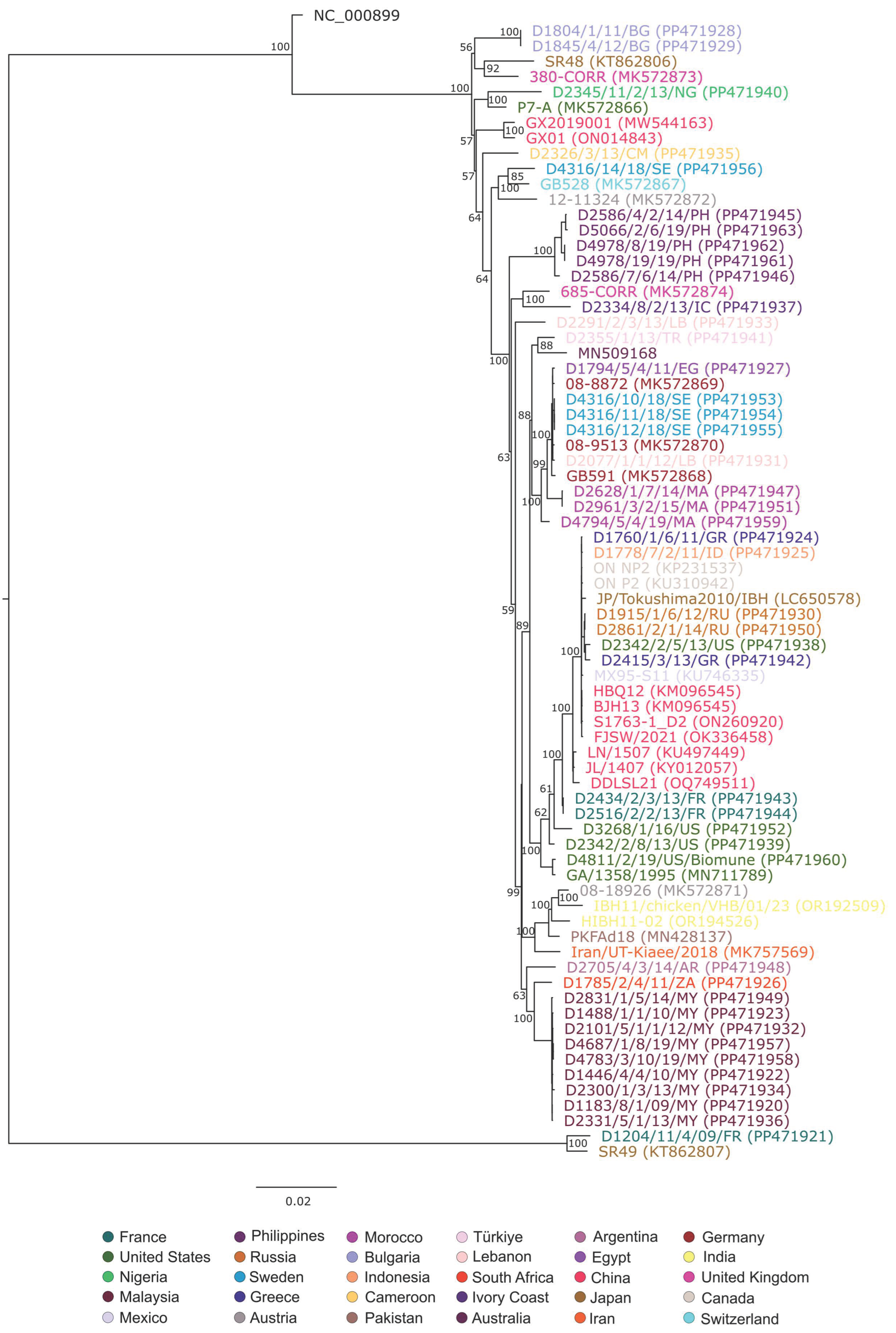
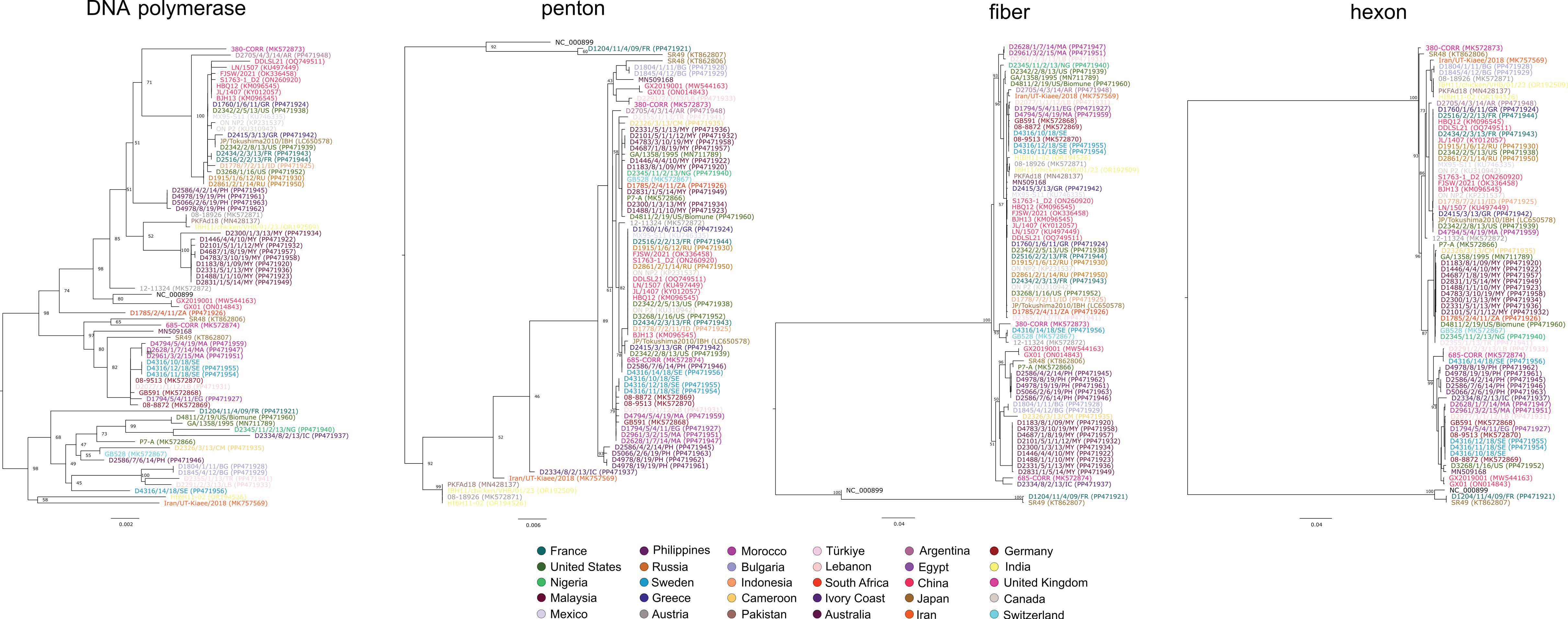
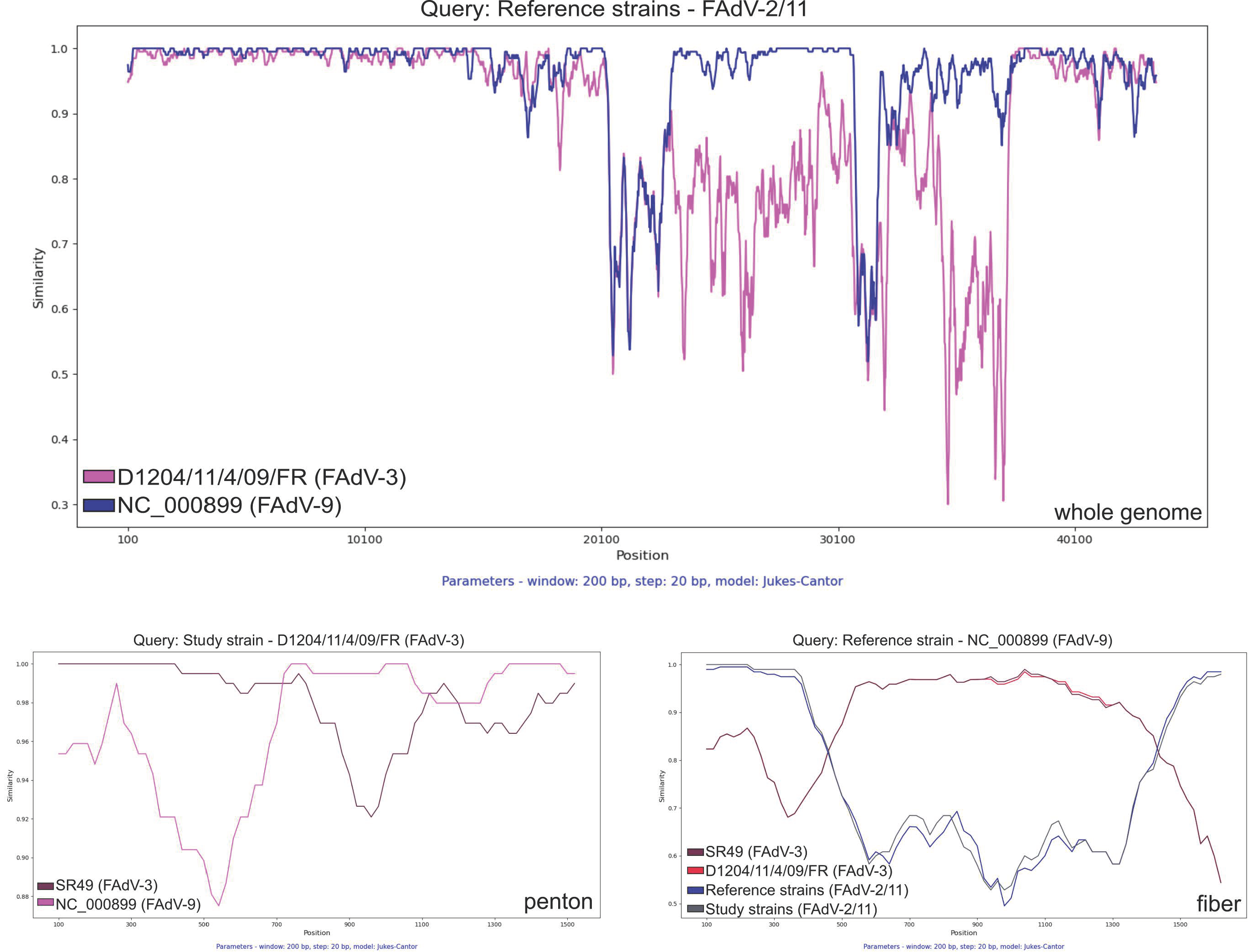
3. Results and Discussion

The FAdV-D isolates available for this study were collected from 18 countries and four continents (Table 1, Supplementary Materials). The 45 isolates initially selected represented 36% of all FAdV-D strains isolated from clinical and pathological samples between 2008 and 2019. The viral genome was successfully assembled for 44 out of 45 FAdV-D isolates; the genome of 1 isolate could not be fully determined due to the low number of specific sequence reads. Genome sequences were assembled with a sequence depth ranging from 13X to 3873X (median, 1344X). The length of consensus genomes varied between 43,219 bp and 45,138 bp (Supplementary Materials). Each viral genome was predicted to encode 36 ORFs. Nucleotide sequences were used as queries to determine the type specificity. As a result of a genome similarity search with the Blast engine and based on preliminary phylogenetic analyses, the study strains were grouped into two clusters. The larger branch contained 43 strains most closely related to the combined FAdV-2/-11 cluster; the detection of these strains spanned the entire collection period and the entire study area. A single isolate was typed as FAdV-3; this strain was detected in France in 2009. The fourth serotype of FAdV-D, FAdV-9, was not identified among the selected isolates. To gain insight into the relationship among globally circulating FAdV-D strains, the study strains (n = 44) were analyzed with several reference strains (n = 31), whose full genome was deposited in GenBank. The genome sequences and selected genes were analyzed phylogenetically; moreover, a gene- and genome-wide nucleotide sequence similarity search was performed by using sliding window analysis.
Phylogenetic analysis using whole-genome sequences was performed after the deletion of large unequally aligned genomic regions (spanning the region from nt 37,829 to 39,278 in strain SR48 [KT862806]), which resulted in significant sequence length differences among the genomes. Whole-genome based phylogeny (Figure 1) showed that all FAdV-2/-11 strains formed a large cluster containing numerous strains (sequence identity, ≥97.4%) and FAdV-9 clustered with this large branch (sequence identity range, 95.5% to 96.6%). A small cluster contained the two known FAdV-3 strains (SR49 and D1204/11/4/09/FR) with available full-genome sequences, and this cluster was more distantly related to all other FAdV-D genomes (sequence identity ranges, with the FAdV-2/-11 cluster, 89.5% to 90.5%; with FAdV-9, 91.9%).
Additional phylogenetic analyses were carried out using the nucleotide alignments of the DNA polymerase, the hexon, the penton and the fiber coding genes (Figure 2). These analyses revealed that DNA polymerase forms two statistically significant clusters; of interest, both branches contained a large number of FAdV-2/-11 strains. The sole FAdV-9 strain clustered with the larger group containing somewhat more taxa, whereas the two FAdV-3 strains showed a different branching pattern. In particular, the GenBank reference FAdV-3 strain, SR49, was genetically more closely related to the FAdV-9 strain than the other FAdV-3 strain sequenced in this study. The three main virion components expressed on the surface (i.e., the hexon, the fiber and the penton) showed some differences in the tree topologies. In particular, the FAdV-2/-11 cluster formed a common branch in the hexon and the fiber gene phylogenies and also formed a statistically supported branch in the penton tree. The position of FAdV-3 and FAdV-9 strains was slightly different. In the hexon tree, FAdV-3 and FAdV-9 formed a common cluster and the genetic distance between FAdV-3 and FAdV-9 was similar to that observed within the FAdV-2/-11 cluster (97.6–100% vs. 96.8–100%). In the fiber gene tree, although forming a common cluster, the sole FAdV-9 strain appeared to be more closely related to strains in the FAdV-2/-11 cluster than the FAdV-3. A similar pattern was seen in the penton tree; in addition, the FAdV-3 strain from France appeared to be in an intermingled position between the FAdV-9 and the reference FAdV-3 strain.
This peculiar pattern of gene-wise phylogenies prompted us to further investigate the inter-serotype sequence relationships, and we performed gene- and genome-wide sequence similarity analysis that permitted the identification of conserved and diversified genomic regions (Figure 3). Consistent with the report by Schachner and co-workers [21], a ~12 kbp segment spanning the hexon coding region to the end of the fiber coding region was uniquely diversified in the FAdV-3 strains, although as delineated in the phylogenetic analysis section, both the hexon- and the fiber coding regions in this diversified segment of FAdV-3 shared greater similarity with FAdV-9. However, when analyzing the fiber coding gene by SimPlot, the FAdV-9 strain shared greater similarity to both historical and more recent FAdV-2/-11 strains at the 5’ and 3’ ends and greater similarity to FAdV-3 strains in the central region of the gene, which is consistent with past recombination events in the fiber coding gene. Of interest, approximately half of the penton coding gene of the FAdV-3 strain from France shared similarity with the reference FAdV-3 strain, while the other half of the gene was more similar to the reference FAdV-9 strain, a finding that suggests an inter-serotype recombination event independent from the event observed in the fiber coding gene (Figure 1). Another genomic region, ORF19, coding for a lipase-like protein is very divergent and is often the subject of genetic analyses; these analyses have shown that recombination may occur in this region even between distantly related adenoviruses [11,22,23,24]. When analyzing the 723 amino acid long-predicted protein of ORF19, both FAdV-3 strains shared moderate genetic relatedness with FAdV-9 and FAdV-2/-11 strains (up to 65% identity) and greater sequence relatedness (up to 82% identity) with selected FAdV-E strains (e.g., 13-19395, 09-8330).
These data together suggest that the genome of FAdV-D serotypes, including the sparsely sequenced FAdV-3, evolved through multiple, consecutive recombination events involving both heterotypic FAdV-D serotypes and aviadenoviruses with either known serotypes (within FAdV-E) or unknown serotypes.

4. Conclusions

Our analyses confirmed the notion from epidemiologic surveillance conducted in parts of the world that the FAdV-2/-11 cluster represents a globally prevalent FAdV-D serotype; the vast majority of FAdV-D isolates in our strain collection (~98%) carried this type specificity. In addition, we confirmed the conserved nature of genomic organization within serotypes of FAdV-D. In general, the genome of FAdV-2/-11 strains was more similar to FAdV-9, while FAdV-3 had a long stretch in the central region of the genome that may have evolved from an ancient recombination event. Ancestral recombination events within and between homologous FAdV-D strains that involved genome fragments of various lengths may have been followed by more recent inter-serotype recombination events that further shaped the viral genome structure. The putative FAdV-3/FAdV-9 inter-serotype recombination event detected in the gene coding for the penton antigen appears to represent a more recent evolutionary leap. The question of whether similar major evolutionary leaps could lead to changes in the antigenic features of circulating FAdV-D strains could be the subject of future studies. The genome sequence data reported here may contribute to a better understanding of the evolutionary mechanisms, epidemiological dynamics and phylogeography of emerging FAdV-D strains and may help devise future control and prevention measures against FAdV-D-associated IBH outbreaks.

Supplementary Materials

The following supporting information can be downloaded at: https://www.mdpi.com/article/10.3390/ani15040508/s1, Table S1: Background information on FAdV-D strains sequenced in this study.

Author Contributions

Conceptualization, I.K., V.P. and K.B. (Krisztián Bányai); methodology, Z.H., S.J., K.B. (Krisztina Bali), M.D. and S.M.; software, Z.H., S.J., E.K. and K.B. (Krisztián Bányai); validation, T.M., I.K. and G.K.; data curation, Z.H., S.J., T.M., E.K. and K.B. (Krisztián Bányai); writing—original draft preparation, Z.H., S.J. and K.B. (Krisztián Bányai); writing—review and editing, T.M., S.M., G.K., I.K. and V.P.; supervision, V.P., G.K., T.M., S.M. and K.B. (Krisztián Bányai); project administration, Z.H., S.J., T.M., I.K. and K.B. (Krisztián Bányai); funding acquisition, S.J., G.K. and K.B. (Krisztián Bányai). All authors have read and agreed to the published version of the manuscript.

Funding

This work was supported by the National Scientific Research Fund of Hungary (OTKA, K120201). Additional support was provided by the National Research, Development and Innovation Office, Hungary, under grant RRF-2.3.1-21-2022-00001 and RRF-2.3.1-21-2022-00010. Szilvia Jakab was supported by the ÚNKP-23-4 New National Excellence Program of the Ministry for Culture and Innovation from the source of the National Research, Development and Innovation Fund.

Institutional Review Board Statement

Not applicable.

Informed Consent Statement

Not applicable.

Data Availability Statement

The dataset generated for this study can be found in GenBank (see Section 2.4 and Supplementary Materials).

Conflicts of Interest

Z.H., T.M., I.K. and V.P. are employees of Ceva-Phylaxia Ltd.; however, the employer had no role in the design of the study or in the collection, analyses, or interpretation of data or in the writing and publishing of the manuscript. The remaining authors declare that the research was conducted in the absence of any commercial or financial relationships that could be construed as potential conflicts of interest.

References

  1. Benkő, M.; Aoki, K.; Arnberg, N.; Davison, A.J.; Echavarría, M.; Hess, M.; Jones, M.S.; Kaján, G.L.; Kajon, A.E.; Mittal, S.K.; et al. ICTV virus taxonomy profile: Adenoviridae 2022. J. Gen. Virol. 2022, 103, 001721. [Google Scholar] [CrossRef] [PubMed]
  2. El-Shall, N.A.; El-Hamid, H.S.A.; Elkady, M.F.; Ellakany, H.F.; Elbestawy, A.R.; Gado, A.R.; Geneedy, A.M.; Hasan, M.E.; Jaremko, M.; Selim, S.; et al. Epidemiology, pathology, prevention, and control strategies of inclusion body hepatitis and hepatitis-hydropericardium syndrome in poultry: A comprehensive review. Front. Vet. Sci. 2022, 9, 963199. [Google Scholar] [CrossRef]
  3. Kiss, I.; Homonnay, Z.G.; Mató, T.; Bányai, K.; Palya, V. Research note: An overview on distribution of fowl adenoviruses. Poult. Sci. 2021, 100, 101052. [Google Scholar] [CrossRef]
  4. Kaján, G.L.; Kecskeméti, S.; Harrach, B.; Benkő, M. Molecular typing of fowl adenoviruses, isolated in Hungary recently, reveals high diversity. Vet. Microbiol. 2013, 167, 357–363. [Google Scholar] [CrossRef]
  5. Nakamura, K.; Mase, M.; Yamamoto, Y.; Takizawa, K.; Kabeya, M.; Wakuda, T.; Matsuda, M.; Chikuba, T.; Yamamoto, Y.; Ohyama, T.; et al. Inclusion body hepatitis caused by fowl adenovirus in broiler chickens in Japan, 2009–2010. Avian Dis. 2011, 55, 719–723. [Google Scholar] [CrossRef] [PubMed]
  6. Choi, K.S.; Kye, S.J.; Kim, J.Y.; Jeon, W.J.; Lee, E.K.; Park, K.Y.; Sung, H.W. Epidemiological investigation of outbreaks of fowl adenovirus infection in commercial chickens in Korea. Poult. Sci. 2012, 91, 2502–2506. [Google Scholar] [CrossRef]
  7. Steer, P.A.; Kirkpatrick, N.C.; O’Rourke, D.; Noormohammadi, A.H. Classification of fowl adenovirus serotypes by use of high-resolution melting-curve analysis of the hexon gene region. J. Clin. Microbiol. 2009, 47, 311–321. [Google Scholar] [CrossRef] [PubMed]
  8. Maartens, L.H.; Joubert, H.W.; Aitchison, H.; Venter, E.H. Inclusion body hepatitis associated with an outbreak of fowl adenovirus type 2 and type 8b in broiler flocks in South Africa. J. S. Afr. Vet. Assoc. 2014, 85, e1–e5. [Google Scholar] [CrossRef] [PubMed]
  9. Pizzuto, M.S.; De Battisti, C.; Marciano, S.; Capua, I.; Cattoli, G. Pyrosequencing analysis for a rapid classification of fowl adenovirus species. Avian Pathol. 2010, 39, 391–398. [Google Scholar] [CrossRef] [PubMed]
  10. Batista, E.B.; Kunert Filho, H.C.; Withoeft, J.A.; de Oliveira Cunha, A.L.; Fonseca, A.; Casagrande, R.A. Fowl aviadenovirus (FAdV-11) as the causative agent of a vertical outbreak of inclusion body hepatitis in commercial broiler breeders in Brazil. Microbe 2024, 3, 100102. [Google Scholar] [CrossRef]
  11. Li, S.; Zhao, R.; Yang, Q.; Wu, M.; Ma, J.; Wei, Y.; Pang, Z.; Wu, C.; Liu, Y.; Gu, Y.; et al. Phylogenetic and pathogenic characterization of current fowl adenoviruses in China. Infect. Genet. Evol. 2022, 105, 105366. [Google Scholar] [CrossRef]
  12. Niu, D.; Feng, J.; Duan, B.; Shi, Q.; Li, Y.; Chen, Z.; Ma, L.; Liu, H.; Wang, Y. Epidemiological survey of avian adenovirus in China from 2015 to 2021 and the genetic variability of highly pathogenic Fadv-4 isolates. Infect. Genet. Evol. 2022, 101, 105277. [Google Scholar] [CrossRef] [PubMed]
  13. Niczyporuk, J.S.; Kozdruń, W.; Czekaj, H.; Styś-Fijoł, N.; Piekarska, K. Detection of fowl adenovirus D strains in wild birds in Poland by Loop-Mediated Isothermal Amplification (LAMP). BMC Vet. Res. 2020, 16, 58. [Google Scholar] [CrossRef] [PubMed]
  14. Olasz, F.; Mészáros, I.; Marton, S.; Kaján, G.L.; Tamás, V.; Locsmándi, G.; Magyar, T.; Bálint, Á.; Bányai, K.; Zádori, Z. A simple method for sample preparation to facilitate efficient whole-genome sequencing of African swine fever virus. Viruses 2019, 11, 1129. [Google Scholar] [CrossRef] [PubMed]
  15. Bali, K.; Bálint, Á.; Farsang, A.; Marton, S.; Nagy, B.; Kaszab, E.; Belák, S.; Palya, V.; Bányai, K. Recombination events shape the genomic evolution of infectious bronchitis virus in Europe. Viruses 2021, 13, 535. [Google Scholar] [CrossRef] [PubMed]
  16. Altschul, S.F.; Gish, W.; Miller, W.; Myers, E.W.; Lipman, D.J. Basic local alignment search tool. J. Mol. Biol. 1990, 215, 403–410. [Google Scholar] [CrossRef] [PubMed]
  17. Katoh, K.; Misawa, K.; Kuma, K.; Miyata, T. MAFFT: A novel method for rapid multiple sequence alignment based on fast Fourier transform. Nucleic Acids Res. 2002, 30, 3059–3066. [Google Scholar] [CrossRef]
  18. Tamura, K.; Stecher, G.; Kumar, S. MEGA11: Molecular evolutionary genetics analysis version 11. Mol. Biol. Evol. 2021, 38, 3022–3027. [Google Scholar] [CrossRef]
  19. Nguyen, L.T.; Schmidt, H.A.; von Haeseler, A.; Minh, B.Q. IQ-TREE: A fast and effective stochastic algorithm for estimating maximum-likelihood phylogenies. Mol. Biol. Evol. 2015, 32, 268–274. [Google Scholar] [CrossRef]
  20. Samson, S.; Lord, É.; Makarenkov, V. SimPlot++: A Python application for representing sequence similarity and detecting recombination. Bioinformatics 2022, 38, 3118–3120. [Google Scholar] [CrossRef] [PubMed]
  21. Schachner, A.; Gonzalez, G.; Endler, L.; Ito, K.; Hess, M. Fowl adenovirus (FAdV) recombination with intertypic crossovers in genomes of FAdV-D and FAdV-E, displaying hybrid serological phenotypes. Viruses 2019, 11, 1094. [Google Scholar] [CrossRef] [PubMed]
  22. Zheng, W.; Teng, X.; Jiang, T.; Tang, W.; Jiang, L.; Zhu, H.; Yu, X.; Chen, G.; Wang, J.; Zhang, J.; et al. Genome analysis of a novel avian atadenovirus reveals a possible horizontal gene transfer. Virology 2024, 593, 109999. [Google Scholar] [CrossRef] [PubMed]
  23. Homonnay, Z.; Jakab, S.; Bali, K.; Kaszab, E.; Mató, T.; Kiss, I.; Palya, V.; Bányai, K. Genome sequencing of a novel variant of fowl adenovirus B reveals mosaicism in the pattern of homologous recombination events. Arch. Virol. 2021, 166, 1477–1480. [Google Scholar] [CrossRef] [PubMed]
  24. Marek, A.; Kaján, G.L.; Kosiol, C.; Benkő, M.; Schachner, A.; Hess, M. Genetic diversity of species Fowl aviadenovirus D and Fowl aviadenovirus E. J. Gen. Virol. 2016, 97, 2323–2332. [Google Scholar] [CrossRef] [PubMed]
Figure 1. Nucleotide sequence-based phylogenetic analysis of FAdV-D strains using the whole genome sequence (color codes of taxa; identical colors indicate shared geographical origin).
Animals 15 00508 g001
Figure 2. Nucleotide sequence based phylogenetic analysis of FAdV-D strains using the DNA polymerase, the penton, the fiber and the hexon genes (color codes of taxa; identical colors indicate shared geographical origin).
Animals 15 00508 g002
Figure 3. Simplot analyses show the sequence similarities along the whole genome (upper panel) and some genes (i.e., penton, fiber; lower panels) among the selected strains.
Animals 15 00508 g003
Table 1. Information on FAdV-D strains sequenced in this study.
Strain IDYear of IsolationCountry of OriginIsolated from SampleProduction Type/Age Sero-/Genotype
D1183/8/1/09/MY2009Malaysiabursabroiler/D45FAdV-2/FAdV-11
D1204/11/4/09/FR2009Franceliverslow-grown broiler/D56FAdV-3
D1446/4/4/10/MY2010Malaysiacecal tonsilbroiler/D34FAdV-2/FAdV-11
D1488/1/1/10/MY2010Malaysialiverbroiler/D20FAdV-2/FAdV-11
D1760/1/6/11/GR2011GreeceliverbroilerFAdV-2/FAdV-11
D1778/7/2/11/ID2011Indonesiaproventriculusbroiler/D20FAdV-2/FAdV-11
D1785/2/4/11/ZA2011South Africaliverbroiler embryoFAdV-2/FAdV-11
D1794/5/4/11/EG2011Egyptcecal tonsilbroiler/D39FAdV-2/FAdV-11
D1804/1/11/BG2011Bulgarialiverbroiler/D10FAdV-2/FAdV-11
D1845/4/12/BG2012Bulgarialiverbroiler/D10FAdV-2/FAdV-11
D1915/1/6/12/RU2012Russialiverbroiler/D19FAdV-2/FAdV-11
D2101/5/1/1/12/MY2012Malaysiacecal tonsilbroiler/D42FAdV-2/FAdV-11
D2291/2/3/13/LB2013Lebanoncecal tonsilbroiler/D42FAdV-2/FAdV-11
D2300/1/3/13/MY2013Malaysiacecal tonsilbroiler/D36FAdV-2/FAdV-11
D2326/3/13/CM2013Cameroonintestinebroiler/D42FAdV-2/FAdV-11
D2331/5/1/13/MY2013Malaysialiverbroiler/D35FAdV-2/FAdV-11
D2334/8/2/13/IC2013Ivory Coastcecal tonsilbroiler/D42FAdV-2/FAdV-11
D2342/2/5/13/US2013United StatesliverbroilerFAdV-2/FAdV-11
D2342/2/8/13/US2013United StatesliverbroilerFAdV-2/FAdV-11
D2345/11/2/13/NG2013Nigeriacecal tonsilbroiler/D32FAdV-2/FAdV-11
D2355/1/13/TR2013Türkiyececal tonsilbroiler/D40FAdV-2/FAdV-11
D2434/2/3/13/FR2013Francececal tonsilbroiler/D29FAdV-2/FAdV-11
D2516/2/2/13/FR2013Franceliverbroiler/D26FAdV-2/FAdV-11
D2586/4/2/14/PH2014Philippinescecal tonsilbroiler/D35FAdV-2/FAdV-11
D2586/7/6/14/PH2014Philippinescecal tonsilbroiler/D29FAdV-2/FAdV-11
D2628/1/7/14/MA2014Moroccocecal tonsilbroiler/D36FAdV-2/FAdV-11
D2705/4/3/14/AR2014Argentinaproventriculusbroiler/D38FAdV-2/FAdV-11
D2831/1/5/14/MY2014Malaysiacecal tonsilbroiler/D38FAdV-2/FAdV-11
D2861/2/1/14/RU2014Russiagizzardbroiler/D20FAdV-2/FAdV-11
D2961/3/2/15/MA2015Moroccokidneybroiler/D36FAdV-2/FAdV-11
D3268/1/16/US2016United Statesliverbroiler/D7FAdV-2/FAdV-11
D4316/10/18/SE2018Swedencecal tonsiln. a.FAdV-2/FAdV-11
D4316/11/18/SE2018Swedenlivern. a.FAdV-2/FAdV-11
D4316/12/18/SE2018Swedenlivern. a.FAdV-2/FAdV-11
D4316/14/18/SE2018Swedenlivern. a.FAdV-2/FAdV-11
D4687/1/8/19/MY2019Malaysiacecal tonsilbroiler/D38FAdV-2/FAdV-11
D4783/3/10/19/MY2019Malaysiacecal tonsilbroiler/D37FAdV-2/FAdV-11
D4794/5/4/19/MA2019Moroccoliverbroiler/D40FAdV-2/FAdV-11
D2077/1/1/12/LB2012Lebanoncecal tonsilbroiler/D39FAdV-2/FAdV-11
D2415/3/13/GR2013Greecececal tonsilbroiler/D45FAdV-2/FAdV-11
D4811/2/19/US2019United Stateslivern. a.FAdV-2/FAdV-11
D4978/19/19/PH2019Philippinesliverbroiler/D32FAdV-2/FAdV-11
D4978/8/19/PH2019Philippinesliverbroiler/D32FAdV-2/FAdV-11
D5066/2/6/19/PH2019Philippinesbursabroiler/D21FAdV-2/FAdV-11
n. a.: not available.
Disclaimer/Publisher’s Note: The statements, opinions and data contained in all publications are solely those of the individual author(s) and contributor(s) and not of MDPI and/or the editor(s). MDPI and/or the editor(s) disclaim responsibility for any injury to people or property resulting from any ideas, methods, instructions or products referred to in the content.

