Article

Lactic Acid Bacteria Metabolites Modulate Immune Response Against Staphylococcus haemolyticus-Infected RAW264.7 Murine Macrophage: A Novel Approach for Bovine Mastitis

by
Nitsanat Cheepchirasuk
1,
Sureeporn Suriyaprom
2,
Thida Kaewkod
1,
Varachaya Intachaisri
2,
Thararat Chitov
1,
Aussara Panya
1,
Witaya Suriyasathaporn
3,4 and
Yingmanee Tragoolpua
1,*
1
Department of Biology, Faculty of Science, Chiang Mai University, Chiang Mai 50200, Thailand
2
Office of Research Administration, Chiang Mai University, Chiang Mai 50200, Thailand
3
Faculty of Veterinary Medicine, Chiang Mai University, Chiang Mai 50100, Thailand
4
Overseas Division Cambodia Office, Asian Satellite Campus Institute, Nagoya University, Nagoya 464-0813, Japan
*
Author to whom correspondence should be addressed.
Animals 2025, 15(22), 3338; https://doi.org/10.3390/ani15223338
Submission received: 21 September 2025 / Revised: 13 November 2025 / Accepted: 14 November 2025 / Published: 19 November 2025

Simple Summary

Bovine mastitis, a painful udder infection in dairy cows, is a major economic burden for farmers. Although antibiotics are commonly used, their overuse of the antibiotics leads to resistance and milk safety concerns. Natural compounds from beneficial lactic acid bacteria that might help cows fight Staphylococcus haemolyticus, a common mastitis pathogen, were investigated in this study. In lab tests using mouse immune cells, these compounds boosted defenses by increasing nitric oxide (a natural antibacterial agent) and activating inflammation-related immune signals. Metabolites from Enterococcus faecalis were especially effective. These results suggested that such natural compounds could become antibiotic-free treatments for mastitis. This approach would benefit dairy farmers by lowering costs, improvement of cow welfare, and providing consumers with safer milk by reducing antibiotic use. Therefore, the compounds from the beneficial bacteria have potential as a sustainable solution for mastitis control.

Abstract

Bovine mastitis, an inflammation of the mammary gland, is recognized as the most prevalent disease in dairy cattle, leading to significant economic losses due to the reduction in quality and yield of milk. While antibiotic treatment remains to be the primary control method but their use of antibiotics contributes to the emergence of antibiotic-resistant bacteria and possesses potential health risks to consumers. Enhancement of host immune responses represents a promising alternative strategy for combating pathogenic bacteria. This study aimed to evaluate the immunomodulatory potential of metabolites derived from lactic acid bacteria (LAB) in murine macrophage RAW264.7 cells infected with Staphylococcus haemolyticus, a causative agent of bovine mastitis. Notably, LAB-derived metabolites demonstrated significant, strain-specific immunostimulatory activity. A comparative metabolomic analysis confirmed that each strain possessed a unique metabolic profile, providing a chemical basis for these various responses. The most pronounced effects were observed with metabolites from the isolated strain Enterococcus faecalis, which markedly increased NO production. Furthermore, these metabolites upregulated the expression of key inflammatory genes, e.g., iNOS, COX-2, TNF-α, and IL-6 and enhanced the protein levels of iNOS and COX-2. These findings suggest that LAB metabolites, particularly those from E. faecalis, may offer a novel therapeutic approach for enhancing immune defenses against mastitis-causing pathogens.

1. Introduction

Bovine mastitis is a prevalent and economically significant disease in dairy cattle, characterized by inflammation of the mammary gland that is primarily caused by bacterial infections [1,2]. Among the various pathogens associated with mastitis, Staphylococcus haemolyticus and other coagulase-negative staphylococci (CNS) have been increasingly implicated in both subclinical and clinical forms [3,4]. Notably, S. haemolyticus is recognized as an emerging opportunistic pathogen with strong biofilm-forming ability and multidrug resistance, which enables it to persist within the mammary gland and evade host immune clearance. Infected cows often exhibit chronic or recurrent mastitis, leading to reduced milk yield, altered milk composition, and significant economic losses in dairy production. Moreover, the bacteria contain transferable resistance genes, posing a potential threat to animal health and raising public health concerns due to the possible transmission of antimicrobial resistance between animals and humans [5,6]. Gram-positive bacterial infections often trigger a delayed or insufficient host immune response, which can exacerbate disease severity [7]. Conventional treatment of bovine mastitis mostly relies on antibiotics to eliminate the causative bacteria and mitigate mammary gland inflammation. While data from the National Institute for Research in Dairying (NIRD) indicate that antibiotics effectively reduce bacterial load and clinical symptoms, this approach has critical limitations, including the emergence of antibiotic resistance, milk residues, and high recurrence rates [8]. Moreover, the overuse of antibiotics in dairy farming raises environmental and public health concerns [4]. Given these challenges, strengthening the host immune response to enable rapid and effective pathogen clearance is essential for disease control. Immunomodulatory strategies might present a viable alternative, offering potential improvements in mastitis management while reducing dependence on antimicrobial therapy.
Probiotics, particularly lactic acid bacteria (LAB), have garnered considerable attention for their immunomodulatory potential due to their ability to modulate host immune responses [9,10]. LAB are commonly found in fermented foods and the gut microbiota. They are recognized for their ability to enhance both innate and adaptive immunity [11,12]. These bacteria stimulate the production of key cytokines, enhance the activity of macrophages and dendritic cells, and promote immune homeostasis. Importantly, LAB reduce inflammation by regulating the overproduction of pro-inflammatory cytokines while upregulating regulatory cytokines like IL-10 [13], thereby mitigating excessive immune activation [14]. This immunoregulatory capacity makes LAB as promising candidates for supporting immune function, particularly in the context of infections and inflammatory conditions. Given these properties, probiotic-based interventions, especially those involving LAB, are being explored for both prophylactic and therapeutic applications. Their potential of the metabolite to complement or reduce reliance on conventional antibiotic treatments highlights their growing importance in contemporary medical and nutritional approaches.
Despite the well-documented immunomodulatory effects of lactic acid bacteria (LAB), the specific interaction between LAB-derived metabolites and Staphylococcus haemolyticus in the context of immune regulation has not been systematically investigated and the significant knowledge remains unknown. To address this, the present study aims to evaluate the potential of LAB-derived metabolites to modulate immune responses in RAW264.7 macrophage cells following S. haemolyticus infection.

2. Materials and Methods

2.1. Reagents and Chemicals

De Man, Rogosa, and Sharpe (MRS) medium and brain heart infusion medium were purchased from HiMedia (Dindori, Nashik, India). Dulbecco’s Modified Eagle’s Medium (DMEM), fetal bovine serum, and penicillin-streptomycin were obtained from Gibco (Grand Island, NY, USA). 3-(4,5-Dimethylthiazol-2-yl)-2,5-diphenyltetrazolium bromide (MTT) was purchased from Bio Basic (Toronto, ON, Canada). Additionally, 2,2-diphenyl-1-picrylhydrazyl (DPPH), 2,2′-azino-bis (3-ethylbenzothiazoline-6-sulfonic acid) (ABTS), 6-hydroxy-2,5,7,8-tetramethylchroman-2-carboxylic acid (Trolox), and gallic acid monohydrate were acquired from Sigma-Aldrich (St. Louis, MO, USA). Anti-iNOS/NOS II polyclonal, anti-COX-2 polyclonal, and anti-β-actin monoclonal antibodies, as well as peroxidase-conjugated secondary antibody, were purchased from EMD Millipore Corporation (Temecula, CA, USA).

2.2. Bacterial Strain

This study utilized selected lactic acid bacteria (LAB) strains isolated from raw milk, including Enterococcus faecalis TCAN02 (GenBank accession: OM992233.1) and Lactobacillus plantarum AD73 (GenBank accession: OM807268.1). All isolates were precisely identified through 16S rRNA gene sequencing, with their sequences deposited in the National Center for Biotechnology Information (NCBI) database. Additionally, reference strains L. plantarum TISTR 2070 and L. casei TISTR 1340 were included in the study. The Staphylococcus haemolyticus strain, isolated from bovine mastitis cases, was generously provided by Prof. Dr. Witaya Suriyasathaporn from Faculty of Veterinary Medicine, Chiang Mai University, Thailand.

2.3. Preparation of LAB-Derived Metabolites

LAB-derived metabolites were isolated from culture supernatants of both isolated and reference LAB strains [15]. The bacterial cultures were grown in De Man, Rogosa, and Sharpe (MRS) broth at a concentration of 1.0 × 10⁸ CFU/mL (100 mL total volume) and incubated for 48 h at 37 °C under static conditions. Following incubation, the culture supernatant was separated by centrifugation at 3000× g for 10 min at 4 °C. The cell-free supernatant was then sterilized by passage through a 0.22 µm pore-size membrane filter. The filtered supernatant was concentrated by lyophilization at −50 °C under reduced pressure until complete dehydration was achieved. The metabolite powder was obtained and reconstituted in sterile ultrapure water to a final concentration of 200 mg/mL and stored at −80 °C until use.

2.4. Preparation of Inactivated S. haemolyticus Bacterial Cells

The inactivation of S. haemolyticus bacterial cells was performed according to the method described by Dehlink et al. (2007) [16] with modifications. Briefly, S. haemolyticus was cultured in 100 mL of brain heart infusion (BHI) broth for 24 h at 37 °C. Bacterial cells were harvested by centrifugation at 3000× g for 15 min at 4 °C and washed twice with phosphate-buffered saline (PBS). The cell pellet was then inactivated by incubation with 4% paraformaldehyde (PFA) for 2 h on ice. Complete inactivation was verified by plating the treated cells on BHI agar plates and incubating at 37 °C for 48 h, with observation of non-bacterial growth confirming successful inactivation.

2.5. Cell Culture

Murine macrophage RAW 264.7 cells (ATCC TIB-71) were maintained in Dulbecco’s Modified Eagle Medium (DMEM) supplemented with 10% (v/v) heat-inactivated fetal bovine serum (FBS) and 1% (v/v) penicillin-streptomycin antibiotics. The cells were cultured under standard conditions in a humidified incubator with 5% CO2 at 37 °C, with media changes performed every 2–3 days. Cells were routinely subcultured when 80–90% cell confluence was observed using cell scrapers for the further experiment.

2.6. Cell Viability

Cell viability was assessed by measuring mitochondrial dehydrogenase activity using the MTT (3-(4,5-dimethylthiazol-2-yl)-2,5-diphenyltetrazolium bromide) assay [17]. RAW 264.7 macrophages were seeded in 96-well plates at a density of 1.0 × 10⁵ cells/well and allowed to adhere for 24 h prior to treatment. Cells were then exposed to LAB-derived metabolite solutions (100 µL/well) at concentrations ranging from 11 to 20 mg/mL. Following 24 h of incubation, the culture medium was carefully aspirated and replaced with MTT solution. After 2–4 h of additional incubation at 37 °C, the formazan crystals were solubilized with 200 µL of dimethyl sulfoxide (DMSO) per well. Absorbance was measured using a microplate reader (DYNEX Technologies, Chantilly, VA, USA) at 540 nm with a reference wavelength of 630 nm. Cell viability was expressed as a percentage relative to untreated control cells (set as 100% viability).

2.7. Infection Studies with Inactivated S. haemolyticus

For bacterial challenge experiments, RAW 264.7 cells were co-cultured with S. haemolyticus (I/A) at multiplicity of infection (MOIs) ratios ranging from 1.56 to 50 for 24 h. Following infection, cells were gently washed twice with 100 µL phosphate-buffered saline (PBS) before assessing cell viability using the MTT assay as described.

2.8. Measurement of Nitric Oxide Production

Nitric oxide (NO) production was measured in RAW 264.7 murine macrophages using the Griess assay [18] to evaluate the effects of LAB-derived metabolites under both basal and S. haemolyticus (I/A)-stimulated conditions. Cells were seeded in 96-well plates at a density of 1.0 × 10⁵ cells/well and allowed to adhere for 24 h. The culture medium was then replaced with non-toxic concentrations of LAB-derived metabolites and incubated for an additional 24 h. Following incubation, 100 µL of culture supernatant was collected and mixed with an equal volume of Griess reagent (1% sulfanilamide in 5% phosphoric acid containing 0.1% N-(1-naphthyl) ethylenediamine dihydrochloride). After 15 min of incubation at room temperature in the dark, absorbance was measured at 550 nm using a microplate reader. Appropriate controls were included: LAB-derived metabolite solutions alone served as reference blanks to account for potential interference, while cells treated with 5 ng/mL lipopolysaccharide (LPS) provided a positive control (set as 100% NO production).

2.9. Morphological Analysis of Macrophage Activation

Morphological changes in RAW264.7 macrophages were assessed by Giemsa staining [19]. Cells were seeded in 24-well plates at a density of 5.0 × 10⁵ cells/well in complete DMEM (10% FBS, 1% penicillin/streptomycin) and allowed to adhere for 24 h at 37 °C in a 5% CO2 humidified incubator. Then, cells were treated with subtoxic concentrations of LAB-derived metabolites in combination with subtoxic doses of S. haemolyticus. Following 24 h of incubation under standard culture conditions, cells were washed twice with PBS, fixed with 4% paraformaldehyde, and stained with Giemsa solution. Stained cells were visualized using an inverted light microscope

2.10. Analysis of Inflammatory Gene Expression by Quantitative Real-Time PCR

Gene expression levels of iNOS, COX-2, IL-6, and TNF-α were analyzed using quantitative reverse transcription polymerase chain reaction (qRT-PCR). RAW 264.7 murine macrophages were seeded in 24-well plates at a density of 5.0 × 10⁵ cells/well and allowed to adhere for 24 h prior to treatment. Cells were then stimulated with LAB-derived metabolites (4, 8, and 16 mg/mL) in the presence of inactivated S. haemolyticus (I/A) at a subtoxic doses with multiplicity of infection (MOI) of 25, followed by incubation for 24 h under standard culture conditions (37 °C, 5% CO2, humidified atmosphere). Total RNA was isolated using TRIzol® reagent (Invitrogen, Carlsbad, CA, USA) according to the manufacturer’s protocol. cDNA synthesis was performed using the ReverTra Ace® qPCR RT Master Mix (TOYOBO, Osaka, Japan). Quantitative PCR amplification was carried out using the SensiFAST™ SYBR® No-ROX Kit (BIOLINE, London, UK) on a real-time PCR system. The β-actin gene served as the endogenous reference control for normalization, with primer sequences provided in Table 1. Relative gene expression was calculated using the 2(−ΔΔCt) method.

2.11. Immunoblotting Analysis of Protein Expression

Protein expression levels of iNOS and COX-2 were evaluated in RAW 264.7 murine macrophages by Western blot analysis. Cells were seeded in 12-well plates at a density of 1.0 × 10⁶ cells/well and allowed to adhere for 24 h prior to treatment with LAB-derived metabolites. Following 24 h of incubation under standard culture conditions (37 °C, 5% CO2), cells were lysed and total protein was extracted. Equal amounts of protein were separated by SDS-PAGE and transferred to PVDF membranes. Membranes were blocked with 5% non-fat dry milk in TBST (Tris-buffered saline with 0.1% Tween-20) for 1 h at room temperature, followed by overnight incubation at 4 °C with primary antibodies against iNOS (polyclonal, 1:1000), COX-2 (polyclonal, 1:1000), and β-actin (monoclonal, 1:1000). After washing, membranes were incubated for 1 h at room temperature with species-appropriate horseradish peroxidase (HRP)-conjugated secondary antibodies (goat anti-mouse or anti-rabbit IgG, 1:1000). Protein bands were visualized using Ultrascene Pico Chemiluminescent Substrate (Bio-helix, New Taipei City, Taiwan) and detected using the ImageQuant LAS 500 Chemiluminescent Imaging System (GE Healthcare, Boston, MA, USA). Band intensities were quantified using ImageJ software version 1.54p (NIH, Bethesda, MD, USA) and normalized to β-actin expression levels, with results expressed as fold-changes relative to untreated controls (set as 1.0).

2.12. DPPH Radical Scavenging Assay

The antioxidant capacity of LAB-derived metabolites was evaluated using the 2,2-diphenyl-1-picrylhydrazyl (DPPH) radical scavenging assay [24]. Briefly, serial dilutions of the metabolite samples were prepared and mixed with 0.1 mM DPPH solution. The reaction mixture was mixed and incubated in the dark at room temperature for 20 min to allow complete reaction. The absorbance was then measured at 517 nm using a microplate reader, with methanol serving as the blank control. The antioxidant capacity was quantified against a gallic acid standard curve and expressed as milligrams of gallic acid equivalents per gram of LAB-derived metabolites (mg GAE/g metabolites). All measurements were performed in triplicate.

2.13. ABTS Radical Scavenging Assay

The antioxidant capacity of LAB-derived metabolites was further evaluated using the 2,2′-azino-bis (3-ethylbenzothiazoline-6-sulfonic acid) (ABTS) radical cation decolorization assay [25]. The ABTS radical cation (ABTS•+) was generated by reacting 7 mM ABTS stock solution with 2.45 mM potassium persulfate (final concentration) in distilled water, followed by 12–16 h of incubation in the dark at room temperature. Prior to analysis, the ABTS•+ solution was diluted with phosphate buffer (pH 7.4) to an absorbance of 0.70 ± 0.02 at 734 nm. For the assay, LAB-derived metabolite samples (5 μL) were mixed with 195 μL of diluted ABTS•+ solution in a 96-well microplate and incubated for exactly 10 min at room temperature protected from light. Absorbance was then measured at 734 nm using a microplate reader. Trolox (6-hydroxy-2,5,7,8-tetramethylchroman-2-carboxylic acid) was used as an antioxidant standard, and results were expressed as mg of Trolox equivalents (TE) per gram of LAB-derived metabolites (mg TE/g metabolites).

2.14. Metabolomic Analysis Using LC-MS/MS

Metabolomic analysis was performed using an Agilent 6545XT AdvanceBio QTOF LC-MS system (Agilent Technologies, Santa Clara, California, USA). Chromatographic separation was achieved on a Poroshell 120 EC-C18 column (2.1 × 100 mm; 2.7 µm) maintained at 50 °C. The mobile phase consisted of 0.1% formic acid in water (A) and 0.1% formic acid in acetonitrile (B). A gradient elution was employed at a flow rate of 0.4 mL/min as follows: 0% B for 0.5 min, increased to 55% B at 10.5 min, increased to 75% B at 12.5 min, and further increased to 100% B at 14.0 min. This was held at 100% B for 3 min before re-equilibrating to 0% B at 17.5 min for a total run time of 20 min. The injection volume was 10 µL.
Mass spectrometry was performed in both positive and negative high-resolution modes. The ion source parameters were set as follows: drying gas temperature at 325 °C with a flow rate of 13 L/h, sheath gas temperature at 275 °C with a flow rate of 12 L/h, and nebulizer pressure at 45 psi. The capillary voltage was set to 4000 V in positive mode and 3000 V in negative mode. Data were acquired in MS1 mode over a mass range of 40–1700 m/z and in MS2 mode over a range of 25–1000 m/z, at an acquisition rate of 3.35 spectra/s. The collision energy was set to 20 eV for positive mode and 10 eV for negative mode. Up to 10 precursors per cycle were selected for fragmentation with a precursor threshold of 5000 counts. Continuous internal calibration was performed using reference masses at m/z 121.0509 and 922.0098 in positive mode, and m/z 112.9856 and 1033.9881 in negative mode.

2.15. Statistical Analysis

All experimental data were analyzed using GraphPad Prism version 10 (GraphPad Software, Inc., La Jolla, CA, USA). Results are presented as mean ± standard deviation (SD) of at least three independent experiments, each performed in triplicate. For comparisons between two groups, statistical significance was determined using unpaired two-tailed t-test. Multiple group comparisons were analyzed by one-way analysis of variance (ANOVA) followed by Tukey’s post hoc test. In all analyses, a p-value < 0.05 was considered statistically significant.

3. Results

3.1. Cytotoxicity Evaluation of LAB-Derived Metabolites on RAW264.7 Murine Macrophages

The cytotoxicity of LAB-derived metabolites from four bacterial strains were systematically evaluated in RAW264.7 murine macrophages using the MTT assay (Figure 1). Following 24 h exposure, all treatment demonstrated dose-dependent effects on cell viability, though with distinct strain-specific patterns. L. casei TISTR1340 metabolites exhibited cytotoxicity, with a CC80 of 16.98 ± 0.145 mg/mL, suggesting relatively low toxicity that might be high biocompatibility within the tested concentration range. E. faecalis TCAN02 metabolites induced the cytotoxic response at a CC80 of 16.70 ± 0.291 mg/mL. Similarly, L. plantarum TISTR2070 showed comparable effects at CC80 of 16.08 ± 0.11 mg/mL. Notably, L. plantarum AD73 displayed significantly higher cytotoxic potential, as evidenced by its lower CC80 of 15.58 ± 0.311 mg/mL. These findings demonstrated that while all tested metabolites affect macrophage viability in a concentration-dependent manner, the magnitude of cytotoxicity varies substantially between strains, with L. plantarum AD73 emerging as the most cytotoxic and L. casei TISTR1340 as the lowest toxicity.

3.2. Cytotoxicity Evaluation of Inactivated S. haemolyticus (I/A) on RAW264.7 Murine Macrophages

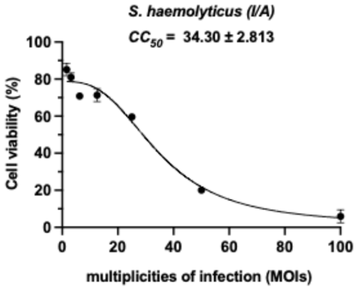
The cytotoxic effects of inactivated S. haemolyticus (I/A) on RAW264.7 murine macrophages were evaluated across a range of multiplicities of infection (MOI). As shown in Figure 2, macrophage viability was maintained at ≥80% at MOIs up to 12.5. Notably, cell viability remained above 50% at an MOI of 25, demonstrating the dose-dependent nature of the bacterial challenge, indicating a relatively safe concentration range for subsequent experiments.

3.3. Nitric Oxide (NO) Production on RAW264.7 Murine Macrophages

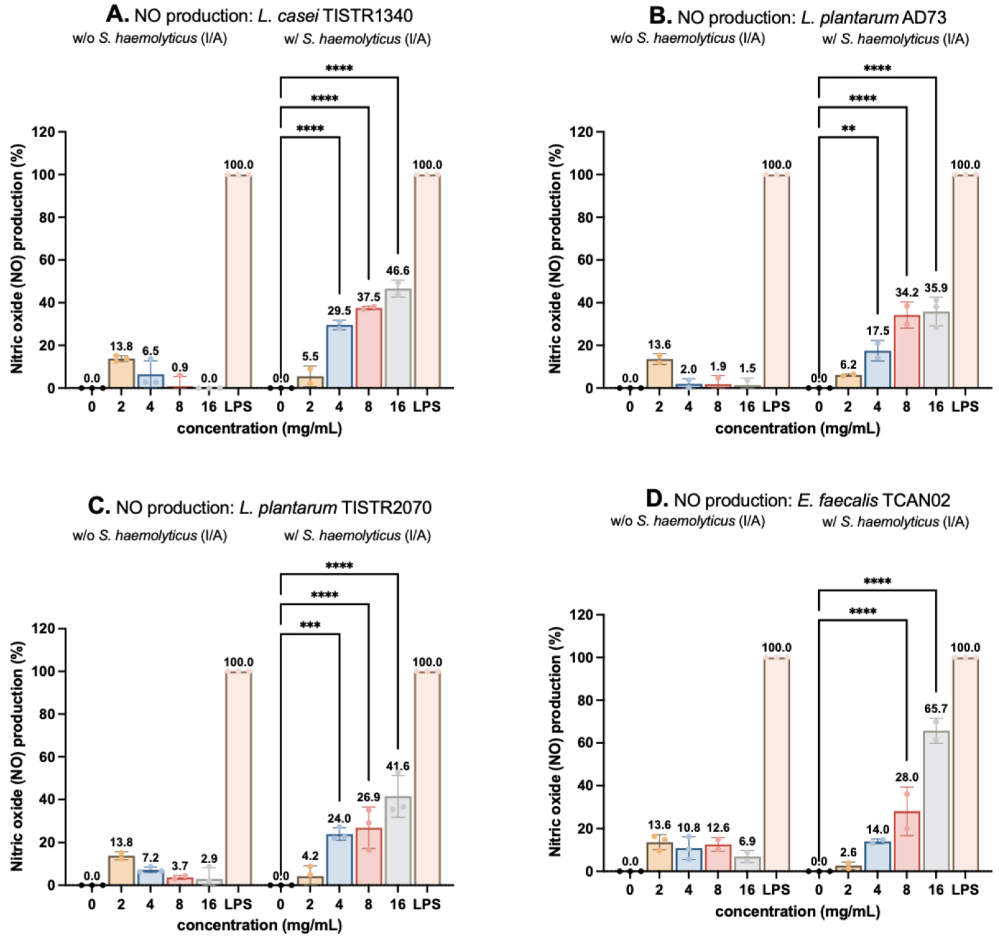
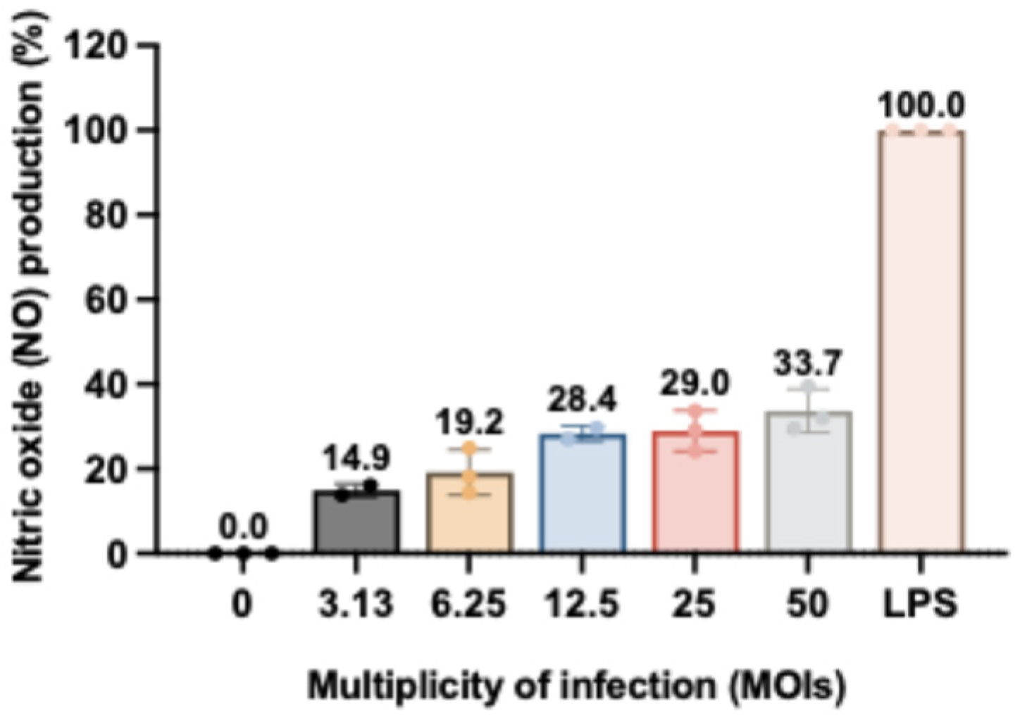
The immunomodulatory effects of LAB-derived metabolites were assessed by measuring NO production in RAW264.7 macrophages using the Griess assay. Cells were treated with subtoxic concentrations of metabolites (2–16 mg/mL) in the presence or absence of S. haemolyticus (I/A), with LPS (5 ng/mL) serving as a positive control (100% NO production). While treatment alone induced minimal NO release, co-stimulation with S. haemolyticus (I/A) triggered dose-dependent increases in NO production (Figure 3D). Enterococcus faecalis TCAN02 metabolites elicited the strongest response (65.68% of LPS-induced NO), followed by Lactobacillus casei TISTR1340, L. plantarum TISTR2070, and L. plantarum AD73 metabolites. Notably, S. haemolyticus alone at MOI 50 induced moderate NO production (33.68%), confirming its immunostimulatory potential (Figure 4). These results demonstrated that LAB metabolites synergized with bacterial challenge to enhance macrophage activation, with strain-specific efficacy.

3.4. LAB-Derived Metabolites Induce Distinct Morphological Activation in S. haemolyticus (I/A)-Challenged RAW264.7 Murine Macrophages

Morphological changes in RAW264.7 macrophages under various co-stimulation conditions were assessed by Giemsa staining, as depicted in Figure 5. The results revealed that LAB-derived metabolites induced characteristic activation-associated morphological changes in S. haemolyticus-challenged RAW264.7 macrophages. Untreated control cells maintained a flattened, adherent morphology with extensive cytoplasmic projections. However, co-treatment of macrophages with LAB metabolites and S. haemolyticus (I/A) exhibited dose-dependent alterations, including cellular rounding, membrane ruffling, and reduced substrate adhesion, which is hallmarks of macrophage activation. Notably, L. plantarum AD73 and E. faecalis TCAN02 metabolites at 8 mg/mL elicited morphological changes comparable to LPS-stimulated positive controls, suggesting immunostimulatory capacity.
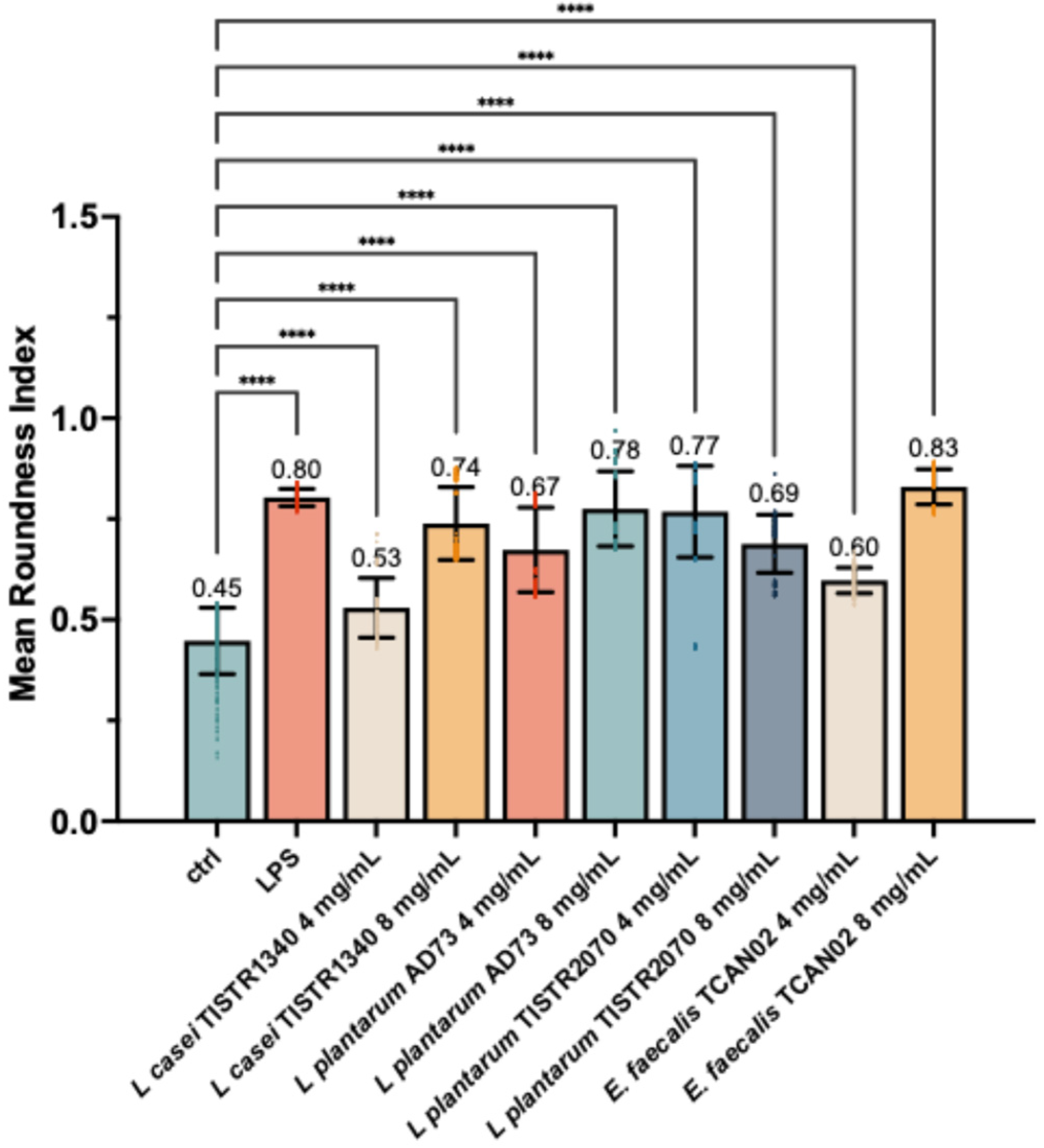
For quantitative validation for these morphological observations, cells were measured the cell circularity metric using ImageJ software (Figure 6). The analysis confirmed that unstimulated cells maintained a low circularity score, consistent with their flattened, adherent morphology.
In contrast, the LPS-stimulated positive control showed a significant increase in cell circularity, indicative of cellular rounding. Similarly, all LAB-derived metabolites induced a statistically significant, dose-dependent increase in cell circularity. Notably, treatment with 8 mg/mL of metabolites from L. plantarum AD73 and E. faecalis TCAN02 resulted in the most pronounced cellular rounding, achieving activation levels comparable to the LPS control. These quantitative data statistically confirm that LAB-derived metabolites promote significant morphological activation in S. haemolyticus-challenged macrophages.

3.5. Modulation of Pro-Inflammatory Cytokine Gene Expression on RAW264.7 Murine Macrophages

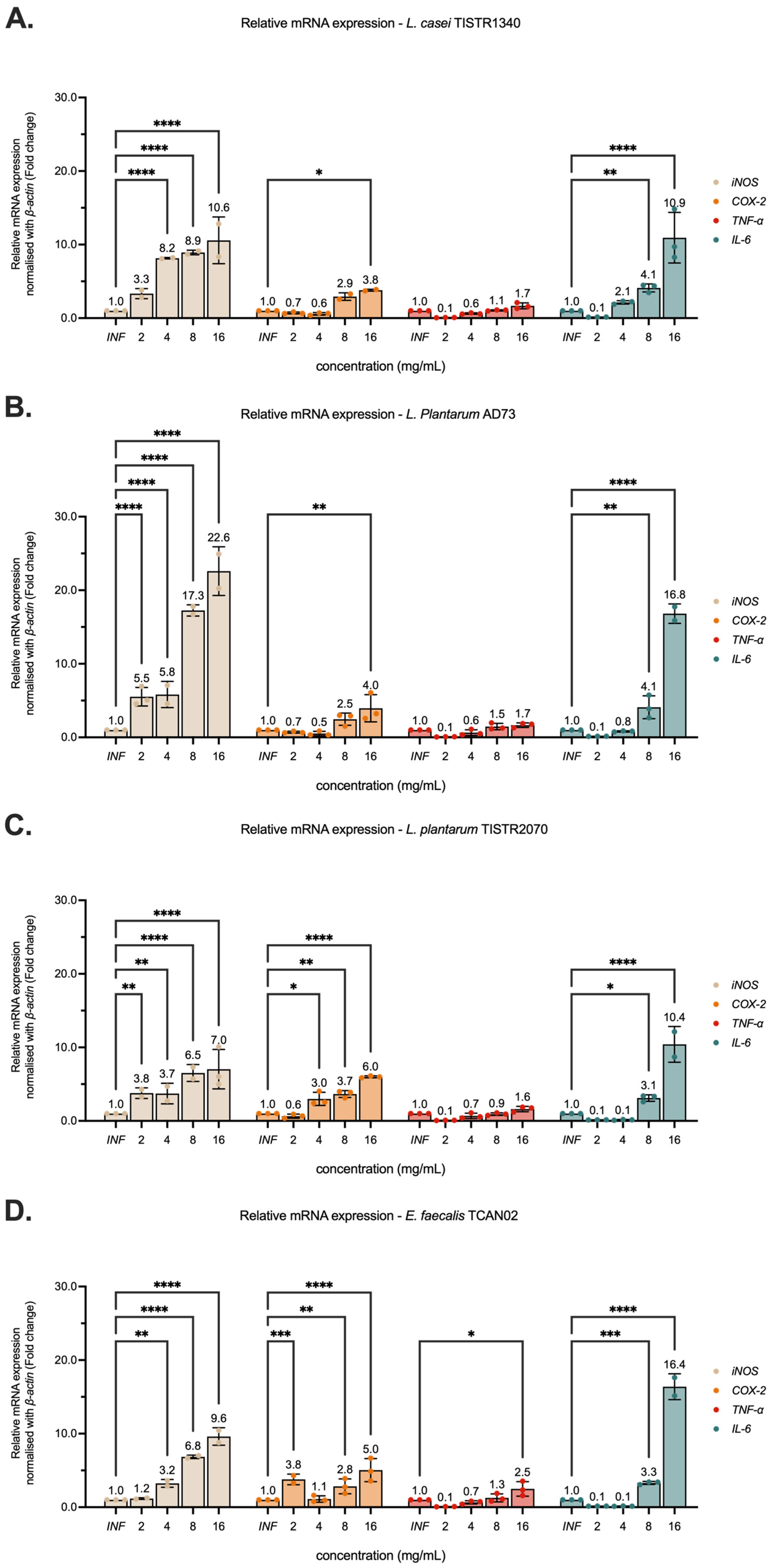
qRT-PCR analysis demonstrated that LAB-derived metabolites (2–16 mg/mL) significantly enhanced the mRNA expression of pro-inflammatory mediators (iNOS, COX-2, TNF-α, and IL-6) in RAW 264.7 macrophages infected with S. haemolyticus (I/A) (Figure 7). Notably, L. plantarum AD73 metabolites exhibited the strongest immunostimulatory effects, inducing iNOS and IL-6 expression by 22.59-fold and 16.82-fold, respectively (p < 0.01 versus untreated controls). In contrast, L. plantarum TISTR2070 metabolites showed preferential upregulation of COX-2 at a value of 18.43 ± 1.1-fold, p < 0.05. While all tested LAB strains (including L. casei TISTR1340 and E. faecalis TCAN02) increased cytokine expression in a dose-dependent manner. The observation of strain-specific patterns of immunostimulatory effects suggests distinct immunomodulatory mechanisms. These findings highlight the potential of LAB-derived metabolites to enhance macrophage inflammatory responses during bacterial challenge. The metabolites from L. plantarum AD73 demonstrated particularly potent activity.

3.6. LAB-Derived Metabolites Potently Upregulate Inflammatory Mediators on RAW264.7 Murine Macrophages Exposed to S. haemolyticus

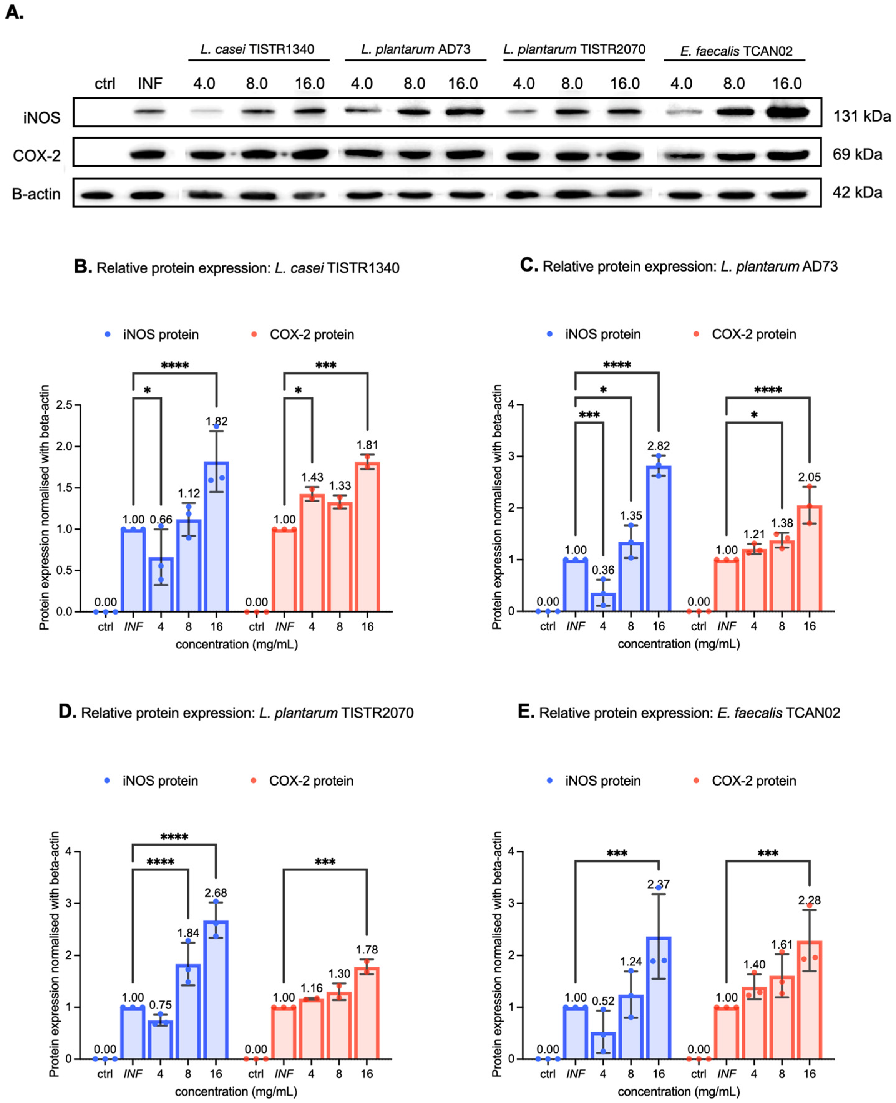
Immunoblotting analysis demonstrated that LAB-derived metabolites significantly enhanced the expression of inflammatory proteins iNOS and COX-2 in RAW264.7 macrophages challenged with S. haemolyticus (I/A). All treatments exhibited dose-dependent effects (4–16 mg/mL), with particularly responses observed at the highest concentration (16 mg/mL) (Figure 8 and Figures S1–S3). Notably, metabolites from L. plantarum TISTR2070 increased iNOS expression by 50% at 8 mg/mL, while both L. plantarum TISTR2070 and L. casei TISTR1340 metabolites enhanced iNOS levels more than twice (>100% increase) and elevated COX-2 by more than 50% at 16 mg/mL. Among isolated strains, L. plantarum AD73 metabolites (16 mg/mL) upregulated iNOS (>50%) and COX-2 (100%) expression, while E. faecalis TCAN02 metabolites showed the most potent immunomodulatory effects, increasing both inflammatory markers by over 100%. These findings demonstrate that LAB metabolites can substantially amplify macrophage inflammatory responses to bacterial challenge, with the magnitude of effect varying significantly between different bacterial strains. The strong upregulation of iNOS and COX-2 suggests LAB metabolites may enhance antimicrobial defenses through potent activation of inflammatory pathways.

3.7. Antioxidant Activity of LAB-Derived Metabolites

The antioxidant potential of LAB-derived metabolites was evaluated using DPPH and ABTS radical scavenging assays, revealing distinct strain-specific activities (Table 2). DPPH assay results demonstrated moderate antioxidant capacity across all strains ranging of 1.25–1.84 mg GAE/g sample. LAB-derived metabolites from L. casei TISTR1340 showed the highest capacity at a value of 1.84 ± 0.21 mg GAE/g sample. In contrast, ABTS assay revealed higher antioxidant capacity ranging from 7.44–14.97 mg TE/g sample. LAB-derived metabolites from L. plantarum AD73 exhibited the most potent effects at a value of 14.97 ± 2.52 mg TE/g sample.

3.8. Multivariate Analysis Reveals Distinct Metabolic Profiles Between Bacterial Species

Untargeted LC-MS metabolomics analysis revealed that the four bacterial species induced distinct and highly significant metabolic shifts in the culture medium, as evidenced by multivariate statistical analysis of both positive and negative ionization mode. The results from the supernatants of E. faecalis TCAN02 (Group a), L. casei TISTR1340 (Group b), L. plantarum AD73 (Group c), and L. plantarum TISTR2070 (Group d) were compared against each other.
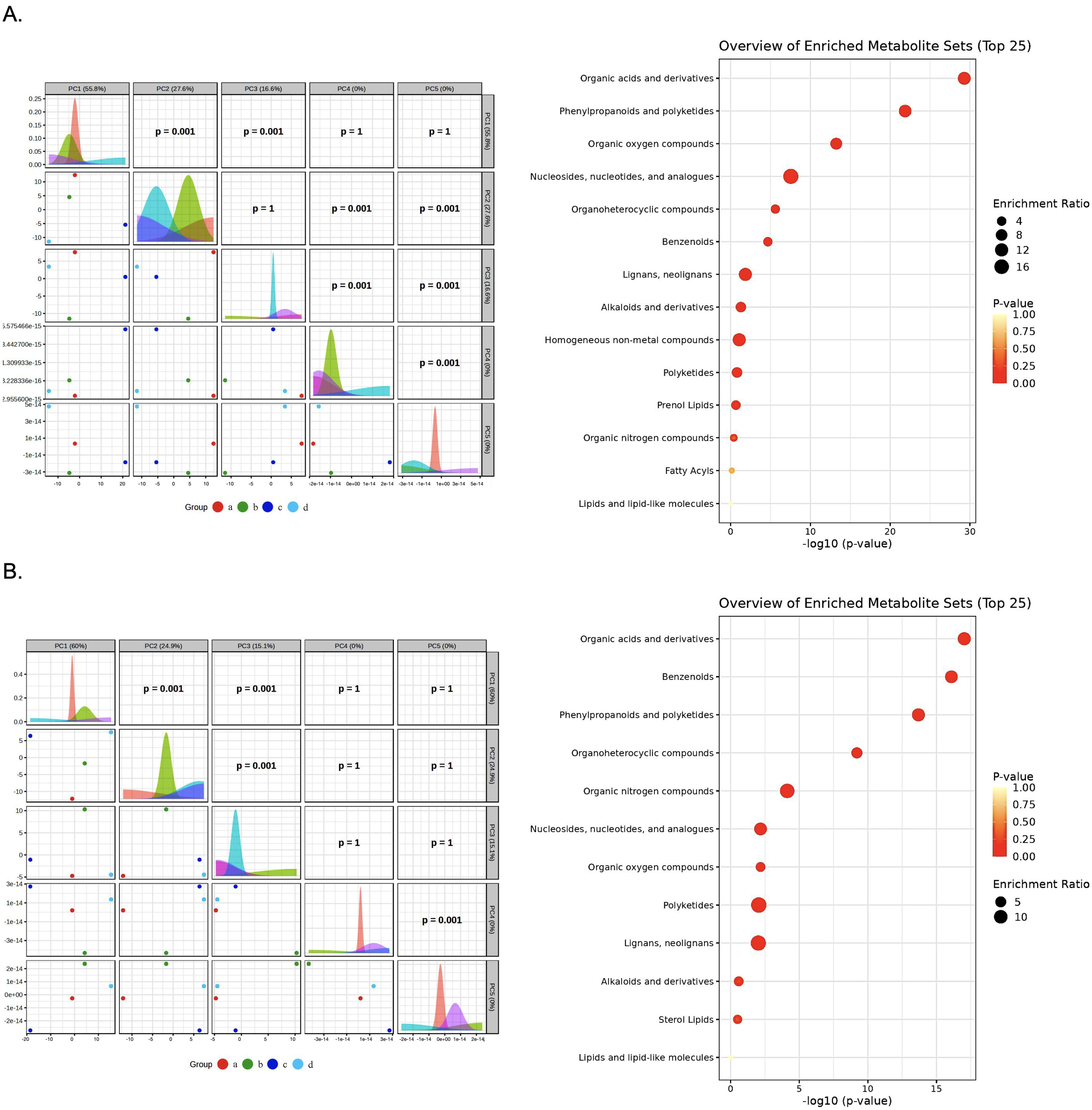
Principal Component Analysis (PCA) demonstrated a clear and consistent segregation of the samples into two primary clusters (Figure 9). This separation was remarkably robust, appearing in both negative and positive ionization modes. The first cluster was composed exclusively of E. faecalis TCAN02 (a) and L. casei TISTR1340 (b), indicating a high degree of metabolic similarity between them. The second, more dispersed cluster, contained L. plantarum AD73 (c) and L. plantarum TISTR2070 (d). This primary separation was driven by the first principal component (PC1) in both modes, which accounted for over 50% of the total variance, highlighting a major metabolic divergence between the (a, b) and (c, d) groups. The primary axis of variation, PC1, which accounted for a majority of the total variance (58.8% in negative and 60.0% in positive mode). The second principal component (PC2) further resolved the subtle differences between Groups a, b, and c, which formed a tighter cluster, indicating a higher degree of metabolic similarity among them. The statistical significance of these groupings was confirmed with p-values of 0.001 for the key component comparison.

3.9. Hierarchical Clustering and Identification of Metabolites

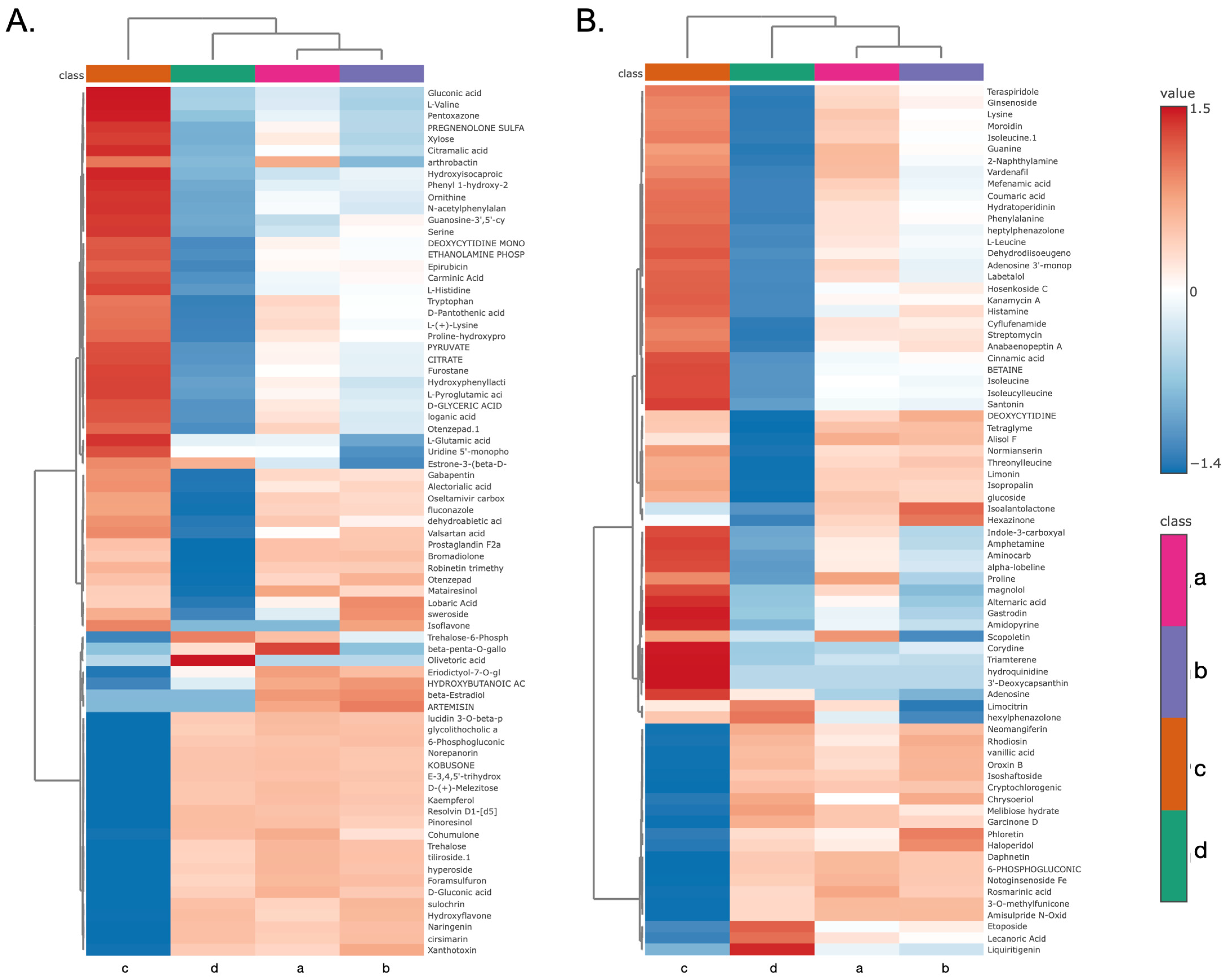
To investigate the specific molecular features driving these separations, hierarchical clustering analysis was performed on the top 75 differential metabolites. The resulting heatmaps and sample dendrograms explicitly corroborated the PCA findings, with the primary branch of the dendrogram separating Group C from a larger cluster containing Groups A, B, and D. Additionally, there is very similarity between Group A and B (Figure 10 and Table 3).
The metabolomic profiling conducted under both negative (Figure 10A) and positive (Figure 10B) ionization modes revealed distinct metabolic signatures among the four sample groups. Group D was characterized by a pronounced upregulation of metabolites within the upper cluster in both modes—for instance, olivetoric acid in the negative mode, and limocitrin and ginsenoside in the positive mode. In contrast, Group A exhibited a higher relative abundance of specific metabolites, including matairesinol in the negative mode, and rosmarinic acid and scopoletin in the positive mode. Group C presented a distinct metabolic profile, marked by elevated levels of glutamic acid and gluconic acid in the negative mode, and gastrodin and cinnamic acid in the positive mode. Notably, Group B displayed a unique metabolic phenotype, characterized by the presence of lobaric acid and sweroside in the negative mode, and phloretin in the positive mode.

3.10. Enrichment Analysis Define

To interpret these molecular changes into a broader biochemical context, metabolite set enrichment analysis (MSEA) was conducted on the significantly altered features. The analysis of both negative (Figure 9A) and positive (Figure 9B) mode data consistently identified organic acids and derivatives as the most significantly enriched metabolite set. Furthermore, significant enrichment was also observed for phenylpropanoids and polyketides and benzenoids, which aligned with the observed differential abundance of aromatic amino acids. This consistent enrichment across both analytical modes indicates that the metabolic differences between the experimental groups are fundamentally rooted in the differential regulation of central carbon and secondary metabolic pathways involving these key chemical classes.

4. Discussion

Bovine mastitis represents a globally prevalent disease with significant economic consequences for dairy production [26]. This condition, primarily caused by bacterial infections of the udder, substantially impacts milk production, increases veterinary costs, and raises concerns about antibiotic residue contamination in milk [27]. Severe cases may result in mortality of affected animals [28]. The causative pathogens are categorized as either contagious or environmental agent. Notable contagious pathogens include Staphylococcus aureus, Streptococcus agalactiae, and S. haemolyticus, which spread during lactation and exacerbate disease transmission [29]. While antibiotic therapy remains the primary treatment approach, its use presents limitations including the development of antibiotic-resistant bacteria and potential host toxicity [30]. Although antibacterial activity of probiotic metabolites has been reported [31], as well as their influence on immune responses in colorectal adenocarcinoma [32]. There are no reports description their immunomodulatory effects on macrophage cells, particularly in the context of pathogenic bacteria-induced bovine mastitis infection. These challenges highlight the importance of enhancing host immune responses as an alternative strategy for mastitis control.
The immune system is an integrated defense network comprising specialized organs, cells, and molecules that safeguard the host while maintaining homeostasis [31]. Macrophages are pivotal effectors of this system, functioning as sentinels in innate immunity, mediators of antigen presentation, and regulators of cytokine-driven responses. By coordinating pathogen clearance and immune modulation, they play a critical role in both host defense and immune balance [32]. In recent years, probiotics have gained considerable attention as potential immunomodulatory agents, and their mechanisms of action have been increasingly explored. One of their most recognized functions is the regulation of host immune responses [33]. Moreover, accumulating evidence indicates that certain probiotic strains exert strain-specific immunomodulatory effects on host immunity and immune cell function, partly through interactions with Toll-like receptors (TLRs) that activate interferon (IFN) signaling pathways [34].
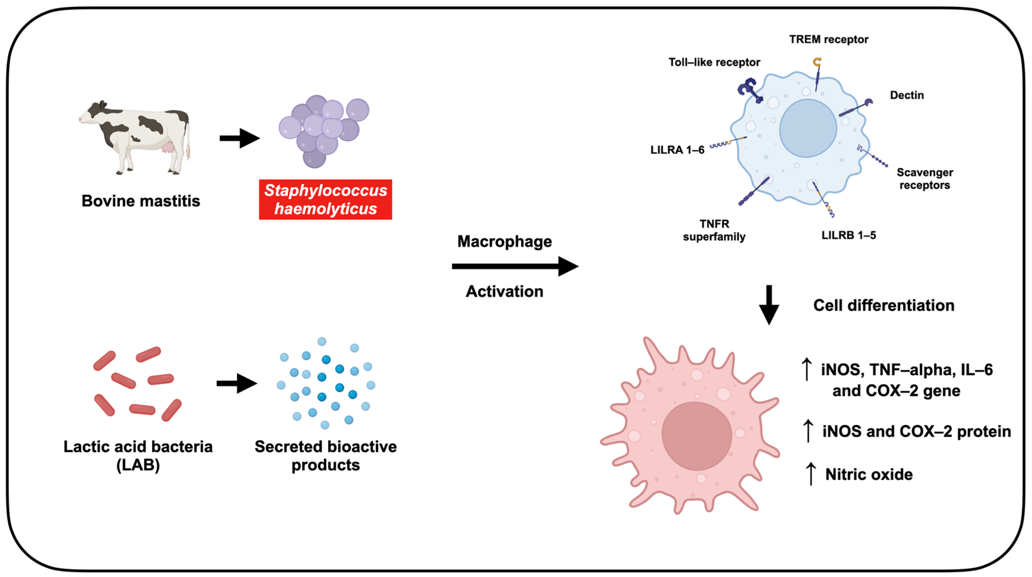
This study investigated the immune-stimulating potential of LAB-derived metabolites in S. haemolyticus-exposed RAW264.7 murine macrophages (Figure 11). RAW264.7 macrophage cell line was used as a standard in vitro model in many studies. The key pathways for investigation including nitric oxide (NO) production via iNOS and the expression of pro-inflammatory mediators such as COX-2, TNF-α, and IL-6 were highly conserved across mammalian species, including mice and cattle [35]. It is widely accepted and well characterized for study of fundamental macrophage immunology [36]. Moreover, the use of macrophage cell line ensures high reproducibility and consistency for the initial screening study. However, bovine macrophage should be used for further studied in the future.
The effect of LAB-derived metabolite on the murine macrophage RAW264.7 cell was determined through mitochondrial activity by MTT assay. The assay quantified mitochondrial activity through conversion of MTT [3-(4,5-dimethylthiazol-2-yl)-2,5-diphenyltetrazolium bromide] to insoluble formazan by mitochondrial reductase [37]. Results demonstrated strain-dependent effects on macrophage viability, with L. plantarum TISTR 2070 and AD73 metabolites exhibiting greater potency with higher CC80 compared to L. casei TISTR 1340 and E. faecalis TCAN02 metabolites. Co-incubation studies with S. haemolyticus (I/A) revealed maintained macrophage viability (>50%) at MOI 25, indicating low responses under bacterial challenge conditions. The immunomodulatory effects of LAB-derived metabolites were further evaluated through nitric oxide (NO) production in RAW264.7 macrophages. NO functions as a key signaling molecule involved in diverse cellular processes, including inflammatory responses and immune regulation [34]. Previous research has established its role in macrophage immune function, antiviral activity, and anti-cancer effects [37,38]. These properties make NO production a relevant biomarker for assessing immune modulation by LAB-derived metabolites.
NO production in RAW264.7 macrophages was quantified using the Griess assay, where nitrite reacts with sulphanilamide under acidic conditions to form a diazonium salt that couples with N-(1-naphthyl)ethylenediamine (NED), producing a colored azo dye [39]. At MOI 25, S. haemolyticus (I/A) alone induced 28.99% NO production relative to LPS controls that induced 100% NO production. However, co-treatment of S. haemolyticus (I/A) with LAB-derived metabolites, particularly from E. faecalis TCAN02, significantly enhanced NO production to 65.68%. Notably, LAB metabolites alone did not stimulate NO production in unchallenged macrophages, indicating their immune-activating effects specifically require bacterial co-stimulation. These findings demonstrate that LAB-derived metabolites potentiate macrophage immune responses primarily during pathogenic bacterial challenge rather than a non-specific stimulation.
Additionally, the expression levels of iNOS, COX-2, TNF-α, and IL-6 genes were evaluated by qRT-PCR using β-actin as a reference gene. LAB-derived metabolites upregulated the expression of all examined genes, with particularly pronounced effects on iNOS and IL-6. TNF-α promotes inflammatory cell recruitment through regulation of leukocyte and vascular adhesion molecule expression [40], while IL-6 has been shown to mitigate tissue inflammation in hypersensitivity pneumonitis and reduce lung injury from oxygen toxicity and endotoxin exposure [41,42]. These observations are consistent with the potential role of LAB-derived metabolites in enhancing immune responses against bacterial pathogens. Immunoblotting analysis was performed to confirm the immunostimulatory activity of LAB-derived metabolites through evaluation of iNOS and COX-2 protein expression. The results demonstrated dose-dependent upregulation of both iNOS and COX-2 proteins following treatment with LAB-derived metabolites. In contrast, macrophages exposed to S. haemolyticus (I/A) showed lower expression levels of these proteins, indicating a diminished immune response in the absence of LAB metabolites. Interestingly, treatment with 2.0 mg/mL of LAB-derived metabolites from E. faecalis TCAN02 resulted in a higher COX-2 mRNA expression level compared with 4.0 and 8.0 mg/mL of LAB-derived metabolites. However, the corresponding protein expression exhibited a clear dose-dependent increase. This apparent discrepancy between mRNA and protein levels may be attributed to several biological factors. Both mRNA and protein were measured at the 24 h time point in order to determine the levels of COX-2 transcription and translation. Thus, the elevated COX-2 mRNA expression observed when treatment with 2.0 mg/mL of LAB-derived metabolites may represent a transient transcription occurring around 24 h. However, when treatment at the higher concentrations of LAB-derived metabolites such as 8.0 mg/mL, the COX-2 mRNA level may occur earlier and decline by the time of measurement. Moreover, COX-2 protein accumulation is influenced by both synthesis and degradation processes. The dose-dependent increase in COX-2 protein detected by Western blot suggests that higher concentrations of E. faecalis-derived metabolites may enhance translational efficiency or stabilize the COX-2 protein, thereby reducing its degradation [41,42].
This study of LAB-derived metabolites revealed distinct profiles among different bacterial strains, leading to different immunomodulatory capacity. For more understanding the chemical profile of this variation, the comparative metabolic analysis was performed using LC-MS/MS. The results clearly demonstrated that the four LAB strains possessed unique and distinct metabolic profiles, which were directly responsible for their differential biological activities. The Principal Component Analysis (PCA) confirmed that the metabolic fingerprints of the four strains were statistically different. The clear separation of the groups in both positive and negative ion modes indicated that each strain produced a unique combination of metabolites. This fundamental of chemical dissimilarity explained why they elicited variation in responses in the macrophage. The metabolite set enrichment analysis (MSEA) further identified the major classes of compounds that defined these profiles. Across all strains, organic acids and derivatives, benzenoids, phenylpropanoids and polyketides were among the most significantly enriched classes. This is a critical finding, as these categories contain a vast number of known bioactive molecules, including phenolic acids and flavonoids, which are well-documented for their immunomodulatory effects.
Notably, several key compounds were identified in the LAB-derived metabolite profiles, including matairesinol from E. faecalis and cinnamic acid from L. plantarum. While the present study evaluated the effects of the whole metabolite mixtures, it is possible that these individual components are the primary contributors to the observed immunomodulatory responses [43,44]. Therefore, these compounds should be isolated and analyzed in order to determine whether individual compound can stimulate the immune response or synergistic interaction among multiple metabolites are required. The future investigations would provide more precise understanding of the molecular mechanisms underlying LAB-mediated immune modulation.
The potent ability of E. faecalis TCAN02 to induce nitric oxide (NO) production is likely driven by its unique metabolic signature. Matairesinol, identified in this strain, has been reported to promote M1 macrophage polarization in THP-1 cells [45]. Additionally, previous study showed effect of rosmarinic acid to enhance immune responses in ovalbumin-induced intestinal allergy mouse models [46], that might further contribute to this effect. Similarly, the strong upregulation of iNOS and IL-6 gene expression observed in response to L. plantarum AD73 metabolites can be attributed to its distinct chemical profile, which exhibits comparatively high levels of glutamic acid, gluconic acid, gastrodin, and cinnamic acid relative to the other strains. With respect to antioxidant activity, the metabolic profiles also help to explain the divergent results observed in the antioxidant assays. The superior ABTS radical scavenging activity of L. plantarum AD73 correlates with the high abundance of specific phenolic compounds. In contrast, the enhanced DPPH radical scavenging capacity of L. casei TISTR1340 metabolites may be associated with its production of distinct organic acids or other specialized small molecular metabolites.
These findings demonstrate that LAB-derived metabolites induce immune responses in murine macrophages. When macrophages infected with S. haemolyticus (I/A) were treated with LAB-derived metabolites, NO production increased significantly. The expression levels of inflammatory mediator genes including inducible nitric oxide synthase (iNOS), cyclooxygenase-2 (COX-2), tumor necrosis factor-alpha (TNF-α), and interleukin-6 (IL-6)—were elevated, along with iNOS and COX-2 protein levels. LAB-derived metabolites activated the host immune response specifically in the presence of pathogenic bacteria, as no induction of NO production or upregulation of inflammatory genes/proteins occurred in uninfected macrophages. This pathogen-dependent activation highlights the therapeutic potential of LAB-derived metabolites for bacterial infections.
On the other hand, it is important to acknowledge certain limitations of the present study. A primary methodological limitation is the number of replicates used for our in vitro assays. The experiments were consistently performed with three independent replicates (n = 3). While this is a common practice for initial screening studies and our data yielded statistically significant trends, this relatively small sample size may impact the overall statistical power and broad reproducibility of the results. Therefore, the future studies should validate these promising findings using a larger number of data and confirm the reliability of these metabolites before proceeding to primary cell or in vivo models. Moreover, antioxidant evaluation was performed as in vitro chemical assays (DPPH and ABTS) to confirm the radical scavenging potential of the metabolites. However, the intracellular antioxidant capacity within the macrophages was not assessed in this study. For further study, a cell-based assay using a probe such as DCFH-DA would be a critical next step to confirm if this chemical activity translates to a biologically relevant reduction in reactive oxygen species (ROS) within the target cells.

5. Conclusions

This study reveals that LAB-derived metabolites can selectively enhance immune responses in S. haemolyticus-infected macrophages, without activating uninfected cells. The metabolites significantly boosted nitric oxide (NO) production and upregulated key inflammatory mediators (iNOS, COX-2, TNF-α, and IL-6) at both gene and protein levels. Crucially, LAB-derived metabolites revealed different profile between different bacterial strain led to different immunomodulatory capacity. These findings demonstrate that LAB metabolites can precisely stimulate immune defenses against bacterial infection. Importantly, this targeted immunomodulation suggests potential for developing LAB-based therapies as alternatives to conventional antibiotics in bovine mastitis treatment, potentially overcoming issues of antibiotic resistance and toxicity. Further research should explore their efficacy in whole-animal models and dairy cattle.

Supplementary Materials

The following supporting information can be downloaded at: https://www.mdpi.com/article/10.3390/ani15223338/s1, Figure S1: Western blot membrane iNOS protein expression, Figure S2: Western blot membrane COX-2 protein expression, Figure S3: Western blot membrane Beta-actin protein expression.

Author Contributions

Conceptualization, N.C., T.K. and Y.T.; methodology, N.C., S.S., T.K. and V.I.; formal analysis, N.C., S.S., V.I. and Y.T.; investigation, N.C., S.S., T.C., A.P., W.S. and Y.T.; writing—original draft preparation, N.C. writing—review and editing, N.C., S.S., A.P., T.C. and Y.T.; supervision, S.S. and Y.T.; funding acquisition, W.S. and Y.T. All authors have read and agreed to the published version of the manuscript.

Funding

This research was funded by Fundamental Fund 2024, Chiang Mai University [Grant No. FF066/2567].

Institutional Review Board Statement

Not applicable.

Informed Consent Statement

Not applicable.

Data Availability Statement

The original contributions presented in this study are included in the article/Supplementary Material. Further inquiries can be directed to the corresponding author.

Acknowledgments

The authors would like to thank the Department of Biology, Faculty of Science, and Fundamental Fund 2024, Chiang Mai University.

Conflicts of Interest

The authors declare no conflicts of interest.

Abbreviations

The following abbreviations are used in this manuscript:
ABTS2, 2-azinobis (3-ethylbenzothiazoline-6 -sulfonic acid)
COX-2Cyclooxygenase-2
DMEMDulbecco’s modified eagle’s medium
DPPH2, 2-diphenyl-1-picrylhydrazyl
IL-6Interleukin-6
iNOSInducible nitric oxide synthase
MTT3-(4,5-dimethylthizaol-2-yl)-2,5-diphenyl tetrazolium bromide
RT-qPCRQuantitative reverse transcription polymerase chain reaction
TNF- αTumor necrosis factor-alpha

References

  1. Sinha, M.K.; Thombare, N.; Mondal, B. Subclinical mastitis in dairy animals: Incidence, economics, and predisposing factors. Sci. World J. 2014, 2014, 523984. [Google Scholar] [CrossRef]
  2. Sathiyabarathi, M.; Jeyakumar, S.; Manimaran, A.; Jayaprakash, G.; Pushpadass, H.A.; Sivaram, M.; Ramesha, K.; Das, D.; Kataktalware, M.A.; Prakash, M.A. Infrared thermography: A potential noninvasive tool to monitor udder health status in dairy cows. Vet. World 2016, 9, 1075. [Google Scholar] [CrossRef]
  3. Pyörälä, S.; Taponen, S. Coagulase-negative staphylococci—Emerging mastitis pathogens. Vet. Microbiol. 2009, 134, 3–8. [Google Scholar] [CrossRef]
  4. Persson Waller, K.; Aspán, A.; Nyman, A.; Persson, Y.; Grönlund Andersson, U. CNS species and antimicrobial resistance in clinical and subclinical bovine mastitis. Vet. Microbiol. 2011, 152, 112–116. [Google Scholar] [CrossRef]
  5. Shoaib, M.; Xu, J.; Meng, X.; Wu, Z.; Hou, X.; He, Z.; Shang, R.; Zhang, H.; Pu, W. Molecular epidemiology and characterization of antimicrobial-resistant Staphylococcus haemolyticus strains isolated from dairy cattle milk in Northwest, China. Front. Cell. Infect. Microbiol. 2023, 13, 1183390. [Google Scholar] [CrossRef] [PubMed]
  6. Ajose, D.J.; Abolarinwa, T.O.; Oluwarinde, B.O.; Montso, P.K.; Fayemi, O.E.; Aremu, A.O.; Ateba, C.N. Whole genome sequence analysis of multi-drug resistant and biofilm-forming Staphylococcus haemolyticus isolated from bovine milk. BMC Microbiol. 2024, 24, 426. [Google Scholar] [CrossRef] [PubMed]
  7. Tietze, K.; Dalpke, A.; Morath, S.; Mutters, R.; Heeg, K.; Nonnenmacher, C. Differences in innate immune responses upon stimulation with gram-positive and gram-negative bacteria. J. Periodontal Res. 2006, 41, 447–454. [Google Scholar] [CrossRef] [PubMed]
  8. Health, National Immunisation Office (NIO). National Database of Antibiotic Resistant Organisms (NDARO). 2022. Available online: https://www.ncbi.nlm.nih.gov/pathogens/antimicrobial-resistance/ (accessed on 17 July 2025).
  9. Galdeano, C.M.; Perdigón, G. Role of viability of probiotic strains in their persistence in the gut and in mucosal immune stimulation. J. Appl. Microbiol. 2004, 97, 673–681. [Google Scholar] [CrossRef]
  10. Perdigón, G.; Fuller, R.; Raya, R. Lactic acid bacteria and their effect on the immune system. Curr. Issues Intest. Microbiol. 2001, 2, 27–42. [Google Scholar]
  11. Cristofori, F.; Dargenio, V.N.; Dargenio, C.; Miniello, V.L.; Barone, M.; Francavilla, R. Anti-inflammatory and immunomodulatory effects of probiotics in gut inflammation: A door to the body. Front. Immunol. 2021, 12, 578386. [Google Scholar] [CrossRef]
  12. Azad, M.A.K.; Sarker, M.; Wan, D. Immunomodulatory effects of probiotics on cytokine profiles. BioMed Res. Int. 2018, 2018, 8063647. [Google Scholar] [CrossRef]
  13. Wallace, T.D.; Bradley, S.; Buckley, N.D.; Green-Johnson, J.M. Interactions of Lactic Acid Bacteria with Human Intestinal Epithelial Cells: Effects on Cytokine Production. J. Food Prot. 2003, 66, 466–472. [Google Scholar] [CrossRef]
  14. Weid, T.v.d.; Bulliard, C.; Schiffrin, E.J. Induction by a Lactic Acid Bacterium of a Population of CD4+ T Cells with Low Proliferative Capacity That Produce Transforming Growth Factor β and Interleukin-10. Clin. Diagn. Lab. Immunol. 2001, 8, 695–701. [Google Scholar] [CrossRef]
  15. Arrioja-Bretón, D.; Mani-López, E.; Palou, E.; López-Malo, A. Antimicrobial activity and storage stability of cell-free supernatants from lactic acid bacteria and their applications with fresh beef. Food Control 2020, 115, 107286. [Google Scholar] [CrossRef]
  16. Dehlink, E.; Domig, K.J.; Loibichler, C.; Kampl, E.; Eiwegger, T.; Georgopoulos, A.; Kneifel, W.; Urbanek, R.; Szepfalusi, Z. Heat-and Formalin-Inactivated Probiotic Bacteria Induce Comparable Cytokine Patterns in Intestinal Epithelial Cell–Leucocyte Cocultures. J. Food Prot. 2007, 70, 2417–2421. [Google Scholar] [CrossRef]
  17. Zhao, F.; Wang, L.; Liu, K. In vitro anti-inflammatory effects of arctigenin, a lignan from Arctium lappa L., through inhibition on iNOS pathway. J. Ethnopharmacol. 2009, 122, 457–462. [Google Scholar] [CrossRef]
  18. Suriyaprom, S.; Srisai, P.; Intachaisri, V.; Kaewkod, T.; Pekkoh, J.; Desvaux, M.; Tragoolpua, Y. Antioxidant and Anti-Inflammatory Activity on LPS-Stimulated RAW 264.7 Macrophage Cells of White Mulberry (Morus alba L.) Leaf Extracts. Molecules 2023, 28, 4395. [Google Scholar] [CrossRef] [PubMed]
  19. Ponnusamy, D.; Clinkenbeard, K.D. Yersinia pestis Intracellular Parasitism of Macrophages from Hosts Exhibiting High and Low Severity of Plague. PLoS ONE 2012, 7, e42211. [Google Scholar] [CrossRef] [PubMed]
  20. Cho, Y.-S.; Lee, S.-H.; Kim, S.-K.; Ahn, C.-B.; Je, J.-Y. Aminoethyl-chitosan inhibits LPS-induced inflammatory mediators, iNOS and COX-2 expression in RAW264. 7 mouse macrophages. Process Biochem. 2011, 46, 465–470. [Google Scholar] [CrossRef]
  21. Deng, Y.; Li, L.; Zhu, J.H.; Li, P.P.; Deng, Y.X.; Luo, H.H.; Yang, Y.Y.; He, B.C.; Su, Y. COX-2 promotes the osteogenic potential of BMP9 through TGF-β1/p38 signaling in mesenchymal stem cells. Aging 2021, 13, 11336–11351. [Google Scholar] [CrossRef]
  22. Taddesse, Y.; Im, E.J.; Kwak, D.; Lee, Y.C.; Hyun, E.; Hong, M.; Jiao, P.; Jia, Q.; Goo, Y.-K.; Hong, S.-B. Stellera chamaejasme methanolic extract attenuates nitric oxide production and enhance heme oxygenase 1 expression in murine macrophages. Chiang Mai J. Sci. 2017, 44, 858–868. [Google Scholar]
  23. He, J.; Li, J.; Liu, H.; Yang, Z.; Zhou, F.; Wei, T.; Dong, Y.; Xue, H.; Tang, L.; Liu, M. Scandoside exerts anti-inflammatory effect via suppressing NF-κB and MAPK signaling pathways in LPS-induced RAW 264.7 macrophages. Int. J. Mol. Sci. 2018, 19, 457. [Google Scholar] [CrossRef] [PubMed]
  24. Sittisart, P.; Chitsomboon, B. Intracellular ROS Scavenging Activity and Downregulation of Inflammatory Mediators in RAW264.7 Macrophage by Fresh Leaf Extracts of Pseuderanthemum palatiferum. J. Evid. Based Complement. Altern. Med. 2014, 2014, 309095. [Google Scholar] [CrossRef]
  25. Yao, F.; Xue, Q.; Li, K.; Cao, X.; Sun, L.; Liu, Y. Phenolic Compounds and Ginsenosides in Ginseng Shoots and Their Antioxidant and Anti-Inflammatory Capacities in LPS-Induced RAW264.7 Mouse Macrophages. Int. J. Mol. Sci. 2019, 20, 2951. [Google Scholar] [CrossRef]
  26. Reshi, A.A.; Husain, I.; Bhat, S.; Rehman, M.U.; Razak, R.; Bilal, S.; Mir, M.R. Bovine mastitis as an evolving disease and its impact on the dairy industry. Int. J. Curr. Res. Rev. 2015, 7, 48. [Google Scholar]
  27. Gomes, F.; Henriques, M. Control of bovine mastitis: Old and recent therapeutic approaches. Curr. Microbiol. 2016, 72, 377–382. [Google Scholar] [CrossRef]
  28. Menzies, F.D.; Gordon, A.W.; McBride, S.H. An epidemiological study of bovine toxic mastitis. In Proceedings of the British Mastitis Conference 2003, Lancashire, UK, 15–17 September 2003; pp. 1–13. [Google Scholar]
  29. Leroy, F.; Van Coillie, E.; Braem, G.; Piessens, V.; Verbist, B.; De Vuyst, L.; De Vliegher, S. Subtyping of Staphylococcus haemolyticus isolates from milk and corresponding teat apices to verify the potential teat-skin origin of intramammary infections in dairy cows. J. Dairy Sci. 2015, 98, 7893–7898. [Google Scholar] [CrossRef]
  30. Awandkar, S.; Bhikane, A.; Kulkarni, M. Antibiotic resistance trends in clinical bovine mastitis. Biolife 2013, 1, 139–143. [Google Scholar]
  31. Kim, D.-M.; Kim, Y.; Seo, J.-W.; Lee, J.; Park, U.; Ha, N.-Y.; Koh, J.; Park, H.; Lee, J.-W.; Ro, H.-J. Enhanced eosinophil-mediated inflammation associated with antibody and complement-dependent pneumonic insults in critical COVID-19. Cell Rep. 2021, 37, 109798. [Google Scholar] [CrossRef]
  32. Geng, L.; Hu, W.; Liu, Y.; Wang, J.; Zhang, Q. A heteropolysaccharide from Saccharina japonica with immunomodulatory effect on RAW 264.7 cells. Carbohydr. Polym. 2018, 201, 557–565. [Google Scholar] [CrossRef]
  33. Liu, W.-B.; Lin, Z.-W.; Zhou, Y.; Ye, B.-C. Overexpression of capsular polysaccharide biosynthesis protein in Lactobacillus plantarum P1 to enhance capsular polysaccharide production for Di-n-butyl phthalate adsorption. J. Microbiol. Biotechnol. 2021, 31, 1545. [Google Scholar] [CrossRef]
  34. Cizeikiene, D.; Jagelaviciute, J. Investigation of antibacterial activity and probiotic properties of strains belonging to Lactobacillus and Bifidobacterium genera for their potential application in functional food and feed products. Probiotics Antimicro. Prot. 2021, 13, 1387–1403. [Google Scholar] [CrossRef] [PubMed]
  35. Kumar, P.; Nagarajan, A.; Uchil, P.D. Analysis of cell viability by the MTT assay. Cold Spring Harb. Protoc. 2018, 2018, pdb-prot095505. [Google Scholar] [CrossRef]
  36. Mayer, B.; Hemmens, B. Biosynthesis and action of nitric oxide in mammalian cells. Trends Biochem. Sci. 1997, 22, 477–481. [Google Scholar] [CrossRef]
  37. Granger, D.L.; Hibbs, J.; Perfect, J.R.; Durack, D.T. Specific amino acid (L-arginine) requirement for the microbiostatic activity of murine macrophages. J. Clin. Investig. 1988, 81, 1129–1136. [Google Scholar] [CrossRef]
  38. Nathan, C.; Shiloh, M.U. Reactive oxygen and nitrogen intermediates in the relationship between mammalian hosts and microbial pathogens. Proc. Natl. Acad. Sci. USA 2000, 97, 8841–8848. [Google Scholar] [CrossRef]
  39. Sun, J.; Zhang, X.; Broderick, M.; Fein, H. Measurement of nitric oxide production in biological systems by using Griess reaction assay. Sensors 2003, 3, 276–284. [Google Scholar] [CrossRef]
  40. Denis, M. Interleukin-6 in mouse hypersensitivity pneumonitis: Changes in lung free cells following depletion of endogenous IL-6 or direct administration of IL-6. J. Leukoc. Biol. 1992, 52, 197–201. [Google Scholar] [CrossRef] [PubMed]
  41. Ulich, T.R.; Yin, S.; Guo, K.; Yi, E.S.; Remick, D.; del Castillo, J. Intratracheal injection of endotoxin and cytokines. II. Interleukin-6 and transforming growth factor beta inhibit acute inflammation. Am. J. Pathol. 1991, 138, 1097–1101. [Google Scholar] [PubMed]
  42. Tsan, M.F.; White, J.E.; Del Vecchio, P.J.; Shaffer, J.B. IL-6 enhances TNF-alpha- and IL-1-induced increase of Mn superoxide dismutase mRNA and O2 tolerance. Am. J. Physiol. 1992, 263, L22–L26. [Google Scholar] [CrossRef]
  43. Lee, A.G.; Kang, S.; Im, S.; Pak, Y.K. Cinnamic acid attenuates peripheral and hypothalamic inflammation in high-fat diet-induced obese mice. Pharmaceutics 2022, 14, 1675. [Google Scholar] [CrossRef]
  44. Ryyti, R.; Hämäläinen, M.; Leppänen, T.; Peltola, R.; Moilanen, E. Phenolic compounds known to be present in lingonberry (Vaccinium vitis-idaea L.) enhance macrophage polarization towards the anti-inflammatory M2 phenotype. Biomedicines 2022, 10, 3045. [Google Scholar] [CrossRef] [PubMed]
  45. Chaudhary, A.; Patil, P.; Raina, P.; Kaul-Ghanekar, R. Matairesinol repolarizes M2 macrophages to M1 phenotype to induce apoptosis in triple-negative breast cancer cells. Immunopharm. Immunotoxicol. 2025, 47, 8–22. [Google Scholar] [CrossRef] [PubMed]
  46. Yang, Q.; Jia, B.; Shang, J.; Wang, X.; Xu, L.; Liu, X.; Fang, M.; Zeng, F.; Zeng, H.-L.; Gong, Z. Effects of rosmarinic acid on immune response and intestinal microbiota in ovalbumin-induced intestinal allergy mice. J. Sci. Food Agric. 2024, 104, 3002–3012. [Google Scholar] [CrossRef] [PubMed]
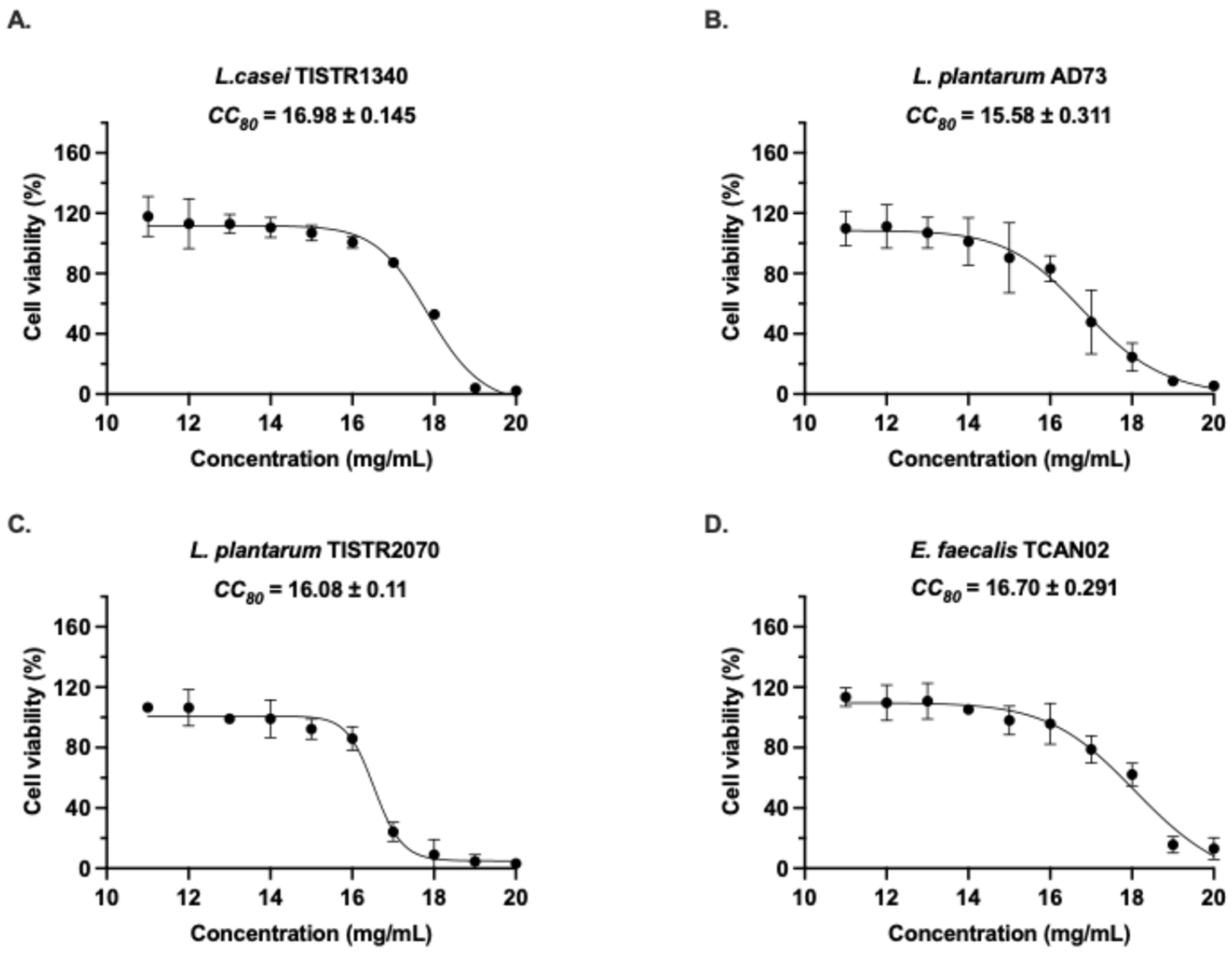
Figure 1. Cytotoxic effects of LAB-derived metabolites on RAW264.7 macrophage viability. Cells were treated for 24 h with increasing concentrations (11–20 mg/mL) of metabolites from (A) L. casei TISTR1340, (B) L. plantarum AD73, (C) L. plantarum TISTR2070, and (D) Enterococcus faecalis TCAN02. Cell viability was assessed by MTT assay and expressed as percentage relative to untreated controls (dotted line at 100%). Data represent mean ± SD of three independent experiments (n = 3), each performed in triplicate.
Animals 15 03338 g001
Figure 2. Cell viability of RAW264.7 macrophages after 24 h exposure to inactivated S. haemolyticus at varying MOIs. Dose-dependent cytotoxicity of inactivated S. haemolyticus in RAW264.7 macrophages. Cells were exposed to increasing multiplicities of infection (MOI: 1.56–100) of inactivated S. haemolyticus for 24 h, with viability assessed by MTT assay. Data represent mean ± SD of three independent experiments (n = 3), each performed in triplicate. Dotted line indicates cell viability threshold.
Animals 15 03338 g002
Figure 3. Modulation of nitric oxide (NO) production in RAW264.7 macrophages by LAB-derived metabolites. Cells were treated for 24 h with metabolites (2–16 mg/mL) from (A) Lactobacillus casei TISTR1340, (B) L. plantarum AD73, (C) L. plantarum TISTR2070, or (D) Enterococcus faecalis TCAN02, either alone or in combination with inactivated S. haemolyticus. LPS (5 ng/mL) served as a positive control (100% response). Data represent mean ± SD of triplicate experiments. ** indicates p < 0.005; *** indicates p < 0.0005; and **** indicates p < 0.0001.
Animals 15 03338 g003
Figure 4. Immunostimulatory effect of inactivated S. haemolyticus on nitric oxide (NO) production in RAW264.7 murine macrophages. Cells were treated with inactivated S. haemolyticus (I/A) for 24 h, and NO concentration in the culture supernatant was measured. LPS (5 ng/mL) served as a positive control (100% response). Data represent mean ± SD of triplicate experiments.
Animals 15 03338 g004
Figure 5. Morphological activation of RAW264.7 macrophages induced by LAB-derived metabolites during S. haemolyticus (I/A) challenge. Giemsa-stained cells showing: (A) Resting morphology in unstimulated controls, (B) Classical activation with LPS (positive control), and (CJ) Dose-dependent morphological changes (4–8 mg/mL) with L. casei TISTR1340 (C,D), L. plantarum AD73 (E,F), L. plantarum TISTR2070 (G,H), and E. faecalis TCAN02 (I,J) metabolites during bacterial co-stimulation.
Animals 15 03338 g005
Figure 6. Quantitative analysis of macrophage activation by cell circularity. The circularity of cells from representative images was quantified using ImageJ software. Data are presented as mean ± SD (n = 3 independent experiments, with >100 cells analyzed per group per experiment). Statistical significance relative to the vehicle control group was determined by One-way ANOVA followed by Tukey’s post hoc test. **** indicates p < 0.0001.
Animals 15 03338 g006
Figure 7. Modulation of pro-inflammatory cytokine gene expression by LAB-derived metabolites in RAW264.7 macrophages. Cells were treated for 24 h with metabolites (2–16 mg/mL) from (A) Lactobacillus casei TISTR1340, (B) L. plantarum AD73, (C) L. plantarum TISTR2070, or (D) Enterococcus faecalis TCAN02, with S. haemolyticus (I/A). Gene expression levels of TNF-α, IL-6, iNOS, and COX-2 were quantified by qRT-PCR and normalized to β-actin. Data represent mean ± SD of triplicate experiments. * indicates p < 0.05; ** indicates p < 0.005; *** indicates p < 0.0005; and **** indicates p < 0.0001.
Animals 15 03338 g007
Figure 8. Modulation of iNOS and COX-2 protein expression by LAB-derived metabolites in S. haemolyticus-challenged macrophages. (A) Representative Western blots show dose-dependent up-regulation of iNOS and COX-2 protein levels after 24 h co-treatment with LAB metabolites. Relative protein expressions of iNOS and COX-2 were demonstrated after co-treatment with (B) Lactobacillus casei TISTR1340, (C) L. plantarum AD73, (D) L. plantarum TISTR2070, or (E) Entero-coccus faecalis TCAN02 metabolites (4.0–16.0 mg/mL). Protein levels were analyzed by immunoblotting with β-actin as loading control. Densitometric quantification represents mean ± SD of three independent experiments. * indicates p < 0.05; *** indicates p < 0.0005; and **** indicates p < 0.0001.
Animals 15 03338 g008
Figure 9. Comparative metabolomic profiling of Lactic Acid Bacteria (LAB)-derived metabolites. Analysis was performed by LC-MS in (A) negative ion mode and (B) positive ion mode. Principal Component Analysis (PCA) correlation matrix showing the metabolic separation between the four LAB strains including E. faecalis (a), L. casei (b), L. paracasei (c), and L. plantarum (d). Metabolite Set Enrichment Analysis (MSEA) dot plot of the top 25 enriched metabolite sets, ranked by statistical significance (−log10 (p-value)). The dot size represents the enrichment ratio, and the color indicates the p-value.
Animals 15 03338 g009
Figure 10. Heatmap and hierarchical clustering of significant metabolites identified in (A) negative ion mode and (B) positive ion mode. Columns correspond to the four LAB strains including E. faecalis (a), L. casei (b), L. paracasei (c), and L. plantarum (d), and rows correspond to individual metabolites. The relative abundance of metabolites is normalized (z-score) and visualized with a color scale where red indicates high abundance and blue indicates low abundance. The top dendrogram illustrates the similarity between the metabolic profiles of the strains, while the side dendrogram showed groups metabolites with similar expression patterns.
Animals 15 03338 g010
Figure 11. LAB-derived metabolites activate immune responses in RAW264.7 macrophages by promoting cell differentiation and stimulating nitric oxide production—a key host defense mechanism against Staphylococcus haemolyticus infection in bovine mastitis.
Animals 15 03338 g011
Table 1. Primer for qRT-PCR analysis.
GenesSense Primer Sequence 5′-3′Antisense Primer Sequence 5′-3′References
iNOSTTCCAGAATCCCTGGACAAGCTGGTCAAACTCTTGGGGTTCG[20]
COX-2AGAAGGAAATGGCTGCAGAAGCTCGGCTTCCAGTATTGAG[21]
TNF-αAGCCCCCAGTCTGTATCCTTCCATTCGAGGCTCCAGTGAATTCG[20]
IL-6GCTGGAGTCACAGAAGGAGTGGCATAACGCACTAGGTTTGCC[22]
β-actinTGCTGTCCCTGTATGCCTCTGGCTGTAGCCACGCTCGGTCA[23]
Table 2. Antioxidant activity of LAB-derived metabolites assessed by DPPH and ABTS radical scavenging assays.
Bacterial StrainAntioxidant Activity
DPPH Assay (mg GAE/g Sample)ABTS Assay (mg TE/g Sample)
L. casei TISTR 13401.84 ± 0.21 *12.11 ± 0.99
L. plantarum TISTR 20701.65 ± 0.159.64 ± 0.13
L. plantarum AD731.59 ± 0.0114.97 ± 2.52 **
E. faecalis TCAN021.25 ± 0.367.44 ± 0.10
Data presented as mean ± SD of three independent experiments (n = 3). GAE = gallic acid equivalents; TE = Trolox equivalents. * Significantly higher than other strains in each assay (p < 0.05) ** Significantly higher than other strains in each assay (p < 0.01).
Table 3. Key candidate metabolites identified in LAB supernatants and their predominant strain association.
Mode of DetectionMetabolitesOntologyPredominant Strain(s)
Negative modegluconic acidcarbohydrate-derived acidsL. plantarum AD73
glutamic acidAlpha amino acids and derivativesL. plantarum AD73
KaempferolFlavonoid-O-glycosidesE. faecalis TCAN02
lobaric acidDepsides and depsidonesL. casei TISTR1340
matairesinolPhenylpropanoidE. faecalis TCAN02
Olivetoric acidPhenolic AcidL. plantarum TISTR2070
swerosidesecoiridoid glycosidesL. casei TISTR1340
Positive
mode
AdenosinePurine nucleosidesL. plantarum AD73
Chrysoeriol3′-O-methylated flavonoidsE. faecalis TCAN02
Cinnamic acidHydroxycinnamic acidsL. plantarum AD73
gastrodinPhenolic glucosideL. plantarum AD73
GenisteinIsoflavonesE. faecalis TCAN02
ginsenosideTriterpenoidsL. plantarum TISTR2070
Isoscopoletin8-prenylated flavonesL. plantarum AD73
limocitrinFlavonolsL. plantarum TISTR2070
LuteolinFlavonoid 8-C-glycosidesE. faecalis TCAN02
phloretin2′-Hydroxy-dihydrochalconesL. casei TISTR1340
Rosmarinic acidCoumaric acids and derivativesE. faecalis TCAN02
scopoletin7-hydroxycoumarinsE. faecalis TCAN02
vanillic acidM-methoxybenzoic acids and derivativesL. plantarum AD73
Disclaimer/Publisher’s Note: The statements, opinions and data contained in all publications are solely those of the individual author(s) and contributor(s) and not of MDPI and/or the editor(s). MDPI and/or the editor(s) disclaim responsibility for any injury to people or property resulting from any ideas, methods, instructions or products referred to in the content.