Article

Comparative Chromosome Painting Clarifies the Intraspecific Chromosomal Variation in Two Ctenomys Species (Rodentia: Ctenomyidae)

by
Thays Duarte de Oliveira
1,†,
Natasha Ávila Bertocchi
2,†,
Luciano Cesar Pozzobon
2,
Ivanete de Oliveira Furo
3,
Edivaldo Herculano Corrêa de Oliveira
4,5,
Jorge C. Pereira
6,7,
Malcolm A. Ferguson-Smith
8,
Rafael Kretschmer
2 and
Thales R. O. de Freitas
1,2,*
1
Departamento de Zoologia, Instituto de Biociências, Universidade Federal do Rio Grande do Sul, Porto Alegre 91501-970, Rio Grande do Sul, Brazil
2
Departamento de Genética, Instituto de Biociências, Universidade Federal do Rio Grande do Sul, Porto Alegre 91501-970, Rio Grande do Sul, Brazil
3
Coordenação de Biodiversidade (COBIO), Instituto Nacional de Pesquisas da Amazônia, Manaus 69060-001, Amazonas, Brazil
4
Instituto de Ciências Exatas e Naturais, Universidade Federal do Pará, Belém 66075-110, Pará, Brazil
5
Laboratório de Cultura de Tecidos e Citogenética, SEAMB, Instituto Evandro Chagas, Ananindeua 67030-000, Pará, Brazil
6
Animal and Veterinary Research Centre (CECAV), University of Trás-os-Montes and Alto Douro (UTAD), 5000-801 Vila Real, Portugal
7
Genetics4U, University of Trás-os-Montes and Alto Douro, 5000-801 Vila Real, Portugal
8
Cambridge Resource Centre for Comparative Genomics, Department of Veterinary Medicine, University of Cambridge, Cambridge CB3 0ES, UK
*
Author to whom correspondence should be addressed.
These authors contributed equally to this work.
Animals 2025, 15(21), 3091; https://doi.org/10.3390/ani15213091 (registering DOI)
Submission received: 1 October 2025 / Revised: 22 October 2025 / Accepted: 22 October 2025 / Published: 24 October 2025

Simple Summary

Ctenomys minutus and Ctenomys lami are rodents known for their karyotypic diversity. The C. minutus diploid number ranges from 2n = 42–50, divided into seven parental cytotypes. While C. lami diploid number ranges from 2n = 50–58, it is divided into four parental cytotypes. To understand the chromosomal evolution of these species, we used whole-chromosome painting with chromosomic probes from Ctenomys flamarioni. We identified chromosomal rearrangements shared among C. minutus and C. lami, and within different cytotypes from within each species. Moreover, several conserved chromosomes across all three Ctenomys species were observed. These rearrangements shed light on the chromosomal evolution of the genus Ctenomys, particularly within the torquatus group, suggesting that these rearrangements have a role on diversification.

Abstract

Background: Ctenomys is a subterranean rodent genus known for exhibiting the highest levels of chromosome variation, both among species (2n = 10 to 70) and within species. Ctenomys minutus is particularly notable for its extensive chromosomal diversity, comprising the greatest number of described cytotypes within this genus. In contrast, Ctenomys lami presents the highest degree of karyotypic variation within a comparatively restricted geographic range. Both species inhabit the coastal plain of southern Brazil: C. minutus occurs in dunes and sandy fields extending from Laguna (Santa Catarina State) to São José do Norte (Rio Grande do Sul State), whereas C. lami is restricted to the “Coxilha das Lombas” region, which lies parallel to the distribution of C. minutus in Rio Grande do Sul State. Despite their close evolutionary relationship and the absence of external morphological differences, the mechanism underlying their karyotypic divergence remains poorly understood. Methods: In this study, we applied whole-chromosome painting using probes from Ctenomys flamarioni to investigate chromosomal evolution in C. minutus and C. lami. Results: The resulting homology maps revealed a variety of chromosomal rearrangements that differentiate cytotypes both within and between these species. Comparative analyses demonstrated substantial karyotypic divergence from C. flamarioni, although some entire chromosomes and large chromosomal segments remained conserved between C. minutus and C. lami. Our findings underscore the critical role of chromosomal rearrangements in shaping the diversification of Ctenomys. Additionally, we identified shared chromosomal rearrangements in C. minutus and C. lami, which are likely restricted to the torquatus group. Conclusions: These rearrangements provide new insights into the processes driving chromosomal evolution in genus Ctenomys.

1. Introduction

Chromosomes are fundamental to the structural organization of genomes, making chromosomal studies crucial for deciphering evolutionary trajectories and understanding karyotype differentiation [1,2,3,4,5,6]. Numerous speciation models suggest chromosomal rearrangements can act as barriers to gene flow, reducing genetic exchange between populations (review in [7]). Additionally, these rearrangements may contribute to the preservation of co-adapted gene complexes by inhibiting recombination within the affected chromosomal regions [7,8,9].
Most taxa undergo minimal karyotype changes during speciation, and their karyotypes typically remain conserved at the genus or family level, as seen in Felidae species [10]. However, some groups exhibit considerable interspecific karyotypic variation. Ctenomys is particularly notable among mammals for its exceptional chromosomal diversity, with diploid numbers (2n) ranging from 2n = 10 to 70 [11]. Furthermore, species within this genus, such as Ctenomys pearsoni, Ctenomys minutus, and Ctenomys lami, also show significant interspecific variation [12,13,14].
Ctenomys is the most diverse genus of subterranean rodents, comprising at least 65 species [15]. Phylogenetically, it is divided into eight monophyletic groups: boliviensis, frater, mendocinus, opimus, magellanicus, talarum, torquatus, and tucumanus [16]. Among these, the torquatus group, which includes eight species, exhibits the second-highest level of karyotypic variation within the genus. Diploid numbers ranging from 2n = 42 to 70 with fundamental numbers (FN) varying from 68 to 84 [17]. Many species in this group are karyotypically polymorphic, displaying multiple cytotypes, primarily due to Robertsonian (Rb) translocations. Notable examples include Ctenomys torquatus (2n = 40 to 46, FN = 72), C. pearsoni (2n = 56–70, FN = 76 to 84), C. lami (2n = 54 to 58), and C. minutus (2n = 42 to 50) [17]. C. minutus and C. lami are sister species within the torquatus group and are endemic to the coastal plain of southern Brazil [16,18,19].
Despite their chromosomal differences, C. minutus and C. lami are externally indistinguishable. C. lami was recognized as a distinct species from C. minutus based on variations in geographic distribution, karyotype organization, and skull morphology [18]. The karyotypic divergence between these species extends beyond the difference in diploid numbers, encompassing chromosomal rearrangements contributing to intraspecific polymorphism. These rearrangements also lead to distinct chromosomal morphologies, which may be unique for each species [18].
Ctenomys minutus is a species endemic to southern Brazil, found explicitly in the dunes and sandy fields along the coastal plains of the Santa Catarina (Laguna) and Rio Grande do Sul States (São José do Norte) [20]. This species is notable for its exceptional karyotypic diversity, with 45 distinct karyotypes described, more than any other species in the genus. These karyotypes result from varying combinations of 2n ranging from 42 to 50 and FN from 68 to 80 [14,18,19]. Seven primary cytotypes parapatrically distributed across the region from north to south are identified: 2n = 50a, 48a, 46a, 42, 46b, 48b, and 50b—(designated here as I to VII) [14,21]. The presence of four intraspecific hybrid zones has resulted in the formation of intermediate karyotypes, which exhibit combinations of these parental diploid numbers [14,20,21,22,23,24].
Each cytotype of C. minutus is linked to a specific geographic area along the coastal plain of southern Brazil, exhibiting either a contiguous distribution or being separated by geographic barriers or paleochannels [25]. The 2n = 50 cytotype characterizes the distribution extremes in the North and South. The standard karyotype is the northernmost cytotype (cytotype I-2n = 50a), from which diploid numbers gradually decrease to 2n = 42 (cytotype IV) due to Robertsonian rearrangements. Conversely, toward the southernmost distribution, the diploid number increases to 2n = 50b (cytotype VII). Cytotypes with the same diploid number are differentiated as “a” or “b” based on structural rearrangements in distinct chromosomes, including tandem fusions/fissions, as well as paracentric or pericentric inversions [20,21,22,23,26].
Ctenomys lami has a highly restricted geographic range, occupying an area of approximately 78 km × 12 km of sandy fields in the “Coxilha das Lombas” in Rio Grande do Sul State, Brazil [13,18,27]. Despite this limited distribution, it exhibits one of the highest karyotypic variability known within the genus Ctenomys, with five distinct diploid numbers (2n = 54, 55, 56, 57, and 58) and ten fundamental numbers (FN = 74 to 82 and 84), resulting in a total of 26 different karyotypes within just 936 km2 [13,18]. These variations in diploid numbers are primarily due to Robertsonian rearrangements, centric fusions/fissions, and pericentric inversions [13]. As seen in C. minutus, cytotypes designated as “a” or “b” differ due to rearrangements that affect different chromosomes [13].
Chromosome painting, using whole-chromosome probes, has revolutionized comparative cytogenetics, especially in rodents, by illuminating regions of homology between species [28]. For example, this technique has elucidated the evolutionary processes in the genus Ellobius, which showcases remarkable karyotypic diversity. It has helped to reveal the driving forces behind diversification and speciation in these species [26]. Yet, comparative cytogenetic studies using chromosome painting remain scarce when it comes to Ctenomys, a genus with equally intriguing chromosomal variation. While some species have been studied with banding techniques (see [29]), only one investigation applied chromosome painting, focusing on C. minutus and C. flamarioni [30]. In this study, Kubiak et al. [30] performed chromosome painting on a single cytotype of C. minutus (2n = 46a—cytotype II) to compare parental karyotypes (C. flamarioni and C. minutus), as well as their hybrids, shedding light on the origins of these hybrid forms.
Our study builds on this work, offering a deep look into chromosomal evolution within Ctenomys by focusing on two closely related species, C. minutus and C. lami. These species’ striking karyotypic diversity, indistinguishable external appearances, and limited geographic ranges present a fascinating opportunity to explore how chromosomal rearrangements may drive speciation. What is even more exciting is the unique chromosomal traits and intraspecific variability they exhibit. By employing the set of C. flamarioni chromosome probes [30], we dive into the chromosomal evolution of both C. minutus and C. lami.

2. Materials and Methods

2.1. Sample Collection

Thirteen C. minutus and eight C. lami individuals were collected in Rio Grande do Sul and Santa Catarina States, Brazil (Table 1; Figure 1). Animals were captured using Oneida Victor No.0 plot traps and euthanized in accordance with the guidelines of the American Society of Mammalogists’ Animal Care and Use Committee [31]. All procedures were approved by the Ethics Committee for the Use of Animals (CEUA-UFRGS, protocol No. 35828) and authorized by the Brazilian Environmental Agency (IBAMA, approval No. 14690-1).

2.2. Chromosomal Preparations and Karyotype

Chromosome preparations were obtained from fibroblast cultures following the method of Verma and Babu [32], with modifications. Briefly, kidney and/or lung biopsies were enzymatically digested with collagenase and cultured at 37 °C in Dulbecco’s Modified Eagle Medium–high glucose (DMEM; GIBCO) supplemented with 20% fetal bovine serum (GIBCO), penicillin (100 U/mL), and streptomycin (100 µg/mL). After a 1h incubation with colchicine, cells were exposed to a hypotonic solution (0.075 M KCl) for 8 min and subsequently fixed in methanol/acetic acid (3:1). For each specimen, ~30 metaphases were examined to determine diploid number and chromosome morphology.

2.3. Fluorescence In Situ Hybridization (FISH)

Fluorescence in situ hybridization experiments were carried out on all specimens and cytotypes of C. minutus and C. lami using whole-chromosome probes from C. flamarioni (CFL 1–23 and X), obtained by flow sorting at the Cambridge Resource Centre for Comparative Cytogenetics (Cambridge, UK) [30]. Probes were amplified and labeled with biotin or digoxigenin using DOP-PCR. Hybridization procedures followed Kubiak et al. [30]. Biotin-labeled probes were detected with Cy3-avidin, and digoxigenin-labeled probes with anti-rabbit FITC. Slides were counterstained with Fluoroshield™ containing DAPI (Sigma-Aldrich, St. Louis, MO, USA). Images were captured using a ZEISS Axiophot epifluorescence microscope (Carl Zeiss, Jena, Germany) with ZEN BLUE (Carl Zeiss, Jena, Germany) software (https://www.zeiss.com/microscopy/en/products/software/zeiss-zen.html (accessed on 21 October 2025)) and processed with Adobe Photoshop CS6.

3. Results

3.1. Karyotypes

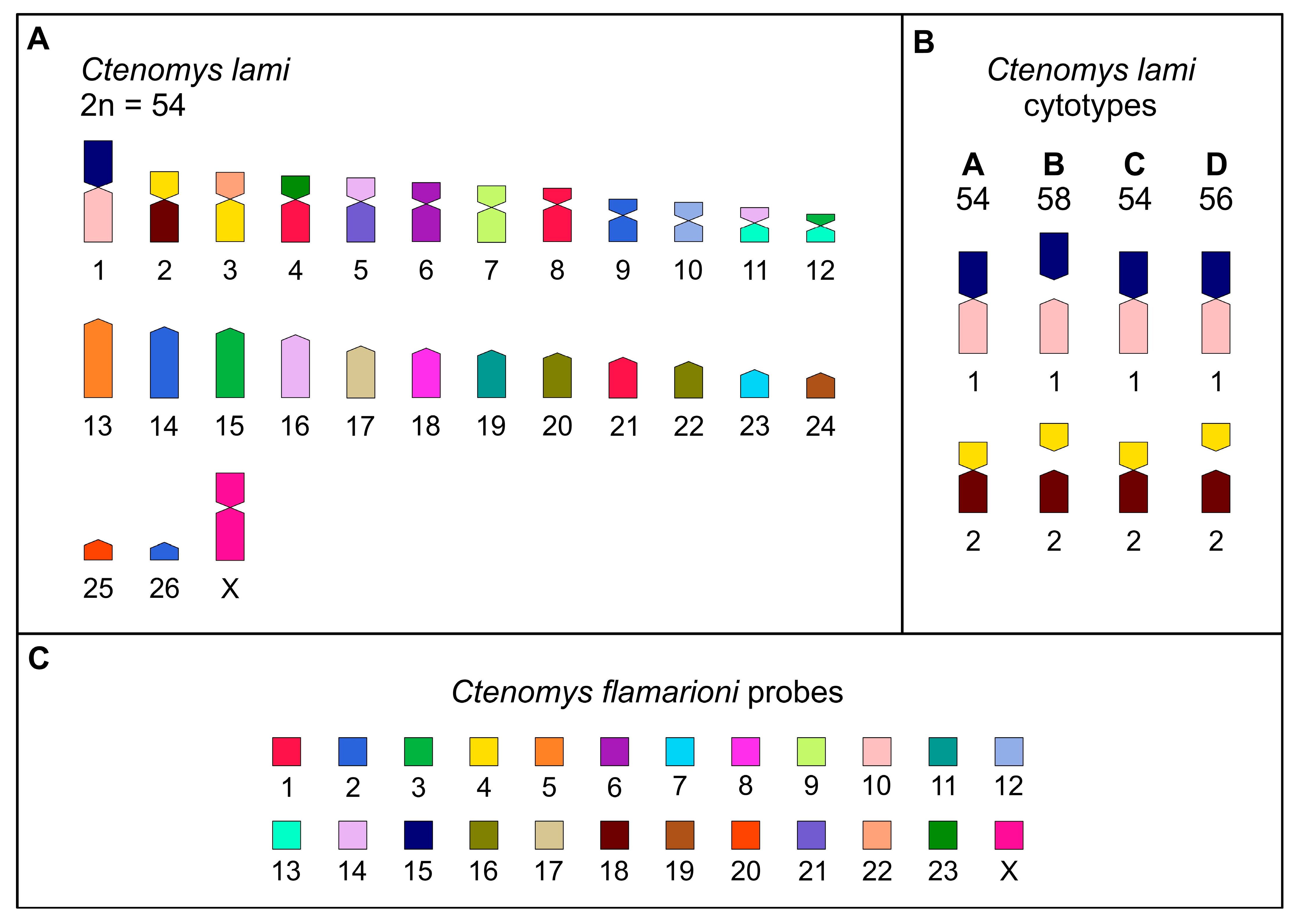
The karyotypes observed in our sample of C. minutus corresponded to cytotypes 50a, 48a, 46a, 42, 46b, 48b, and 50b, as previously described by Lopes et al. [14], Freygang et al. [20] and Freitas [22]. For C. lami individuals, the karyotypes align with those previously described by Freitas [13,18], with individuals exhibiting the cytotypes 2n = 54, 2n = 56, and 2n = 58, captured in blocks A and D, block C, and block B, respectively.

3.2. Comparative Chromosome Painting

Chromosome-specific paints from C. flamarioni (CFL), corresponding to all autosomes and the X chromosome, were successfully hybridized to the metaphases of C. minutus and C. lami (Figure 2). The results allowed for the construction of comparative chromosome maps for all cytotypes of both species, C. minutus (CMI) and C. lami (CLA) (Table 2; Figure 3 and Figure 4).
Karyotype 50a (cytotype I) is regarded as the standard karyotype for C. minutus [22]. Using this reference, we established a comparative homology map between C. flamarioni and C. minutus (cytotype I; Figure 3 and Figure 4A). In cytotype I, seven autosomes exhibited homology to only one C. flamarioni probe: CFL6 with CMI7; CFL9 with CMI9; CFL10 with CMI17; CFL12 with CMI12; CFL15 with CMI20; CFL17 with CMI21; and CFL22 with CMI23. Notably, the homology of CFL10 and CFL15 is exclusive to cytotype I (50a), as in other cytotypes these chromosomes are involved in fusion events. Additionally, CFL19 corresponds to the short arm of chromosome 2 (CMI2p). In cytotypes III, V, VI, and VII, chromosome 2 undergoes fission, resulting in 2p as an independent chromosome, thereby preserving complete homology with CFL19. Apart from these homology regions, the remaining C. minutus chromosomes show seven fissions (CFL3 with CMI14p and CMI18; CFL4 with CMI5p and CMI19; CFL13 with CMI13q and CMI14q; CFL14 with CMI2q, CMI8p and CMI13p; and CFL16 with CMI4p and CMI16) and nine fusions (CFL7/5, CFL19/14, CFL 20/8, CFL16/11, CFL4/18, CFL 23/1, CFL14/21, CFL14/13, CFL3/14) (Table 2; Figure 3 and Figure 4).
For C. lami, cytotype 54 (Block A) is considered the standard karyotype for the species. Based on this reference, we constructed a homology map between C. flamarioni and C. lami (cytotype Block A; Figure 3 and Figure 5A). Ten C. lami autosomes exhibited homology to C. flamarioni chromosomes, as follows: CFL5 with CLA13; CFL6 with CLA6; CFL7 with CLA23; CFL8 with CLA18; CFL9 with CLA7; CFL11 with CLA19; CFL12 with CLA10; CFL17 with CLA17; CFL19 with CLA24; and CFL20 with CLA25 (Table 2; Figure 3). In cytotype 56b (Block D) and cytotype 58 (Block B), in addition to these chromosomes, homology was also detected for CFL10 and CFL15, due to the fission of chromosome 1, which gives rise to two distinct chromosomes in these cytotypes. Furthermore, in cytotype 58 (Block B), beyond the homologies identified in cytotype 56b, an additional homology was observed involving CFL18, as a consequence of chromosome 2 fission, which also generates two chromosomes in this block. The remaining C. lami chromosomes present seven fissions (CFL3 with CLA12p and CLA15, CFL4 with CLA12p and CLA15, CFL13 with CLA11q and CLA12q, CFL14 with CLA5p, CLA11p and CLA16, and CFL16 with CLA20 and CLA22) and seven fusions: CFL15/10, CLF4/18, CLF22/4, CLF23/1, CLF15/21, CLF14/13, CLF3/13 (Table 2; Figure 3 and Figure 5).

4. Discussion

4.1. Comparative Chromosome Painting

Cross-species chromosome painting has been applied to the study of karyotypes in more than 100 rodent species (reviewed in [28]). Nevertheless, Zoo-FISH data for the genus Ctenomys remain scarce. To date, chromosome-specific probes from C. flamarioni have been hybridized exclusively to metaphases of C. minutus (cytotype II, 2n = 46a), revealing extensive chromosomal rearrangements between C. flamarioni and C. minutus cytotype II [30]. Our Zoo-FISH experiments provide the first comprehensive comparison of the genome of C. flamarioni with the parental cytotypes of C. minutus and C. lami.
We identified four chromosomes (CFL6, CFL9, CFL12, and CFL17) that are conserved across C. flamarioni, all cytotypes of C. minutus, and C. lami (Figure 3, Figure 4 and Figure 5). In contrast, seven large C. flamarioni chromosomes (CFL1, CFL2, CFL3, CFL4, CFL13, CFL14, and CFL16) are fragmented into two or three distinct chromosome pairs in C. minutus and C. lami, while remaining conserved between these two species (Figure 3). The absence of complete 1:1 synteny supports the hypothesis that karyotypes in Ctenomys are highly rearranged [11,33,34,35,36,37], in stark contrast with other rodent genera characterized by highly conserved karyotypes, such as Peromyscus (all species with 2n = 48) and Oxymycterus (all species with 2n = 54 and FN = 62–64) [38,39,40].
Multiple chromosomal rearrangements, including fissions and inversions, were detected. Among them were specific rearrangements within C. minutus cytotypes, such as the fission and inversion of CMI2p in system “b”, which enabled the reconstruction of the differentiation of cytotypes sharing the same 2n (Figure 3 and Figure 4). Several fusion events were also observed, including CMI17/20 (CFL10 and CFL15), CMI19/23 (CFL4 and CFL22), and CMI22/24/16 (CFL1, CFL2, and CFL16) (Figure 3 and Figure 5), consistent with the results of Kubiak et al. [30]. In C. lami, rearrangements of chromosomes 1 and 2 were evident: CFL10 corresponds to CLA1q, CFL15 to CLA1p, part of CFL4 to CLA2p, and CFL18 to CLA2q (Figure 3). These findings are corroborated by G-banding analyses reported by Freyganag et al. [20] and Freitas [13].
Taken together with previous comparative chromosome painting studies, our results demonstrate that Zoo-FISH provides a resolution of rearrangement patterns comparable to G-banding in C. minutus and C. lami. Nonetheless, while probes derived from C. flamarioni are highly effective for identifying interchromosomal rearrangements and assessing chromosomal variability, they are less suitable for detecting intrachromosomal changes. For this purpose, higher-resolution approaches, such as bacterial artificial chromosome (BAC) probes or other region-specific markers, are more appropriate [41,42,43].

4.2. Comparative C. minutus x C. lami

Morphological, molecular, and cytogenetic studies have substantially advanced our understanding of taxonomy, phylogenetic relationships, and karyotypic patterns within the genus Ctenomys. Lopes et al. [14] has emphasized the intrinsic association of geographic barriers in the fixation of chromosomal rearrangements in this group, as they are fixated by genetic drift and after the removal of the geographic barrier causes reduced gene flow between different cytotypes. Using mitochondrial DNA from representatives across thirty distinct localities, their study revealed that individuals with a 2n = 50a karyotype occupy a basal position, whereas those with 2n = 42 represent a more derived condition within the population.
This striking diversity is closely tied to the complex geography of the region, characterized by a mosaic of lakes, lagoons, rivers, swamps, sandy fields, and dunes that act as natural barriers promoting speciation. However, in certain hybrid zones, contact between recently diverged species enables gene flow among populations, which in turn contributes to the emergence of distinct cytotypes [14,44].
In the present study, by comparing the rearrangements identified in C. minutus and C. lami, we provide insights into the chromosomal changes involved in their karyotypic differentiation. We demonstrate that shared rearrangements between these two species may be associated with a recent speciation process in which chromosomal changes play a pivotal role, as previously hypothesized by [21]. This view is further supported by the ecological context of these species: the presence of strong geographic barriers is consistent with an allopatric model of speciation, followed by the accumulation of diverse chromosomal rearrangements [13,21].
Our data support the hypothesis that C. lami (2n = 58) possesses a karyotype more similar to the putative common ancestor of the two species. From an evolutionary perspective, given that fusions are generally more frequent than fissions, karyotypes with higher diploid numbers are typically considered closer to the ancestral state [45]. Accordingly, C. minutus (2n = 42) would represent a more derived condition, characterized by a reduced diploid number and a higher number of chromosomal rearrangements relative to C. lami [13,18]. Nevertheless, establishing which karyotype is ancestral and which is derived remains a challenge due to the rapid and recent divergence observed within Ctenomys [16].
Overall, our findings provide a framework for investigating chromosomal evolution in Ctenomys and can be extended to other species within the genus. Such approaches may contribute to species delimitation, particularly in groups from central-western Brazil. Future studies should combine comparative chromosome painting with whole-genome sequencing to achieve higher-resolution analyses, as has been successfully demonstrated in other taxa [29,46,47].

5. Conclusions

In this study, we provide a comparative chromosomal painting analysis of two endemic tuco-tuco species from southern Brazil, C. minutus and C. lami, which display some of the greatest karyotypic diversity described within the genus. Our results reinforce the hypothesis that chromosomal rearrangements have played a role in the diversification and speciation of Ctenomys. We demonstrate that C. minutus and C. lami share specific chromosomal rearrangements.
Beyond the interspecific comparison, our data contribute to reconstructing the evolutionary dynamics of Ctenomys karyotypes, highlighting the importance of fusions and fissions in shaping their remarkable chromosomal variation. Expanding comparative cytogenetic analyses to additional species will be essential for elucidating the broader evolutionary history of the genus.
Finally, integrating classical and molecular cytogenetic techniques with whole-genome sequencing and assembly will provide the resolution needed to detect intrachromosomal rearrangements, uncover the mechanisms driving chromosomal evolution, and clarify the role of these processes in the speciation of Ctenomys.

Author Contributions

Conceptualization, T.D.d.O., R.K. and T.R.O.d.F.; methodology, T.D.d.O., R.K., L.C.P., N.Á.B., I.d.O.F., J.C.P. and M.A.F.-S.; software, T.D.d.O. and L.C.P.; validation, T.D.d.O. and L.C.P.; formal analysis, T.D.d.O., R.K., N.Á.B. and I.d.O.F.; investigation, T.D.d.O. and R.K.; resources, R.K., M.A.F.-S. and T.D.d.O.; data curation, T.D.d.O.; writing—original draft preparation, T.D.d.O., R.K., L.C.P., N.Á.B., I.d.O.F., J.C.P., E.H.C.d.O., M.A.F.-S. and T.R.O.d.F.; writing—review and editing, T.D.d.O., R.K., L.C.P., N.Á.B., I.d.O.F., J.C.P., E.H.C.d.O., M.A.F.-S. and T.R.O.d.F.; visualization, T.D.d.O., R.K., N.Á.B., L.C.P. and I.d.O.F.; supervision, T.R.O.d.F.; project administration, T.R.O.d.F.; funding acquisition, R.K. and T.R.O.d.F. All authors have read and agreed to the published version of the manuscript.

Funding

This research was funded by Coordenação de Aperfeiçoamento de Pessoal de Nível Superior (CAPES), grant number 88882.439377/2019-01 (T.D.d.O.), and Conselho Nacional de Desenvolvimento Científico e Tecnológico (CNPq), grant number 166809/2017-9 (N.Á.B.), 153212/2018-7 (R.K.), 304068/2025-0 (R.K), Proc. 171459/2023-7 (L.C.P.), and Proc. 406747/2023-7 (T.R.O.d.F.).

Institutional Review Board Statement

This study was conducted in accordance with the guidelines of the American Society of Mammalogists’ Animal Care and Use Committee (Sikes and the Animal Care and Use Committee of the American Society of Mammalogists 2016). All procedures were approved by the Ethics Committee for the Use of Animals (CEUA-UFRGS, protocol No. 35828, approved on 2020) and authorized by the Brazilian Environmental Agency (IBAMA, approval No. 14690-1, approved on 2017).

Informed Consent Statement

Not applicable.

Data Availability Statement

All original contributions from this study are included in this article. Additional information is available from the corresponding author upon request.

Acknowledgments

We would like to thank Cristina Araujo Matzenbacher for the technical support.

Conflicts of Interest

The authors declare no conflicts of interest.

References

  1. Bakloushinskaya, I.; Lyapunova, E.A.; Saidov, A.S.; Romanenko, S.A.; O’Brien, P.C.M.; Serdyukova, N.A.; Ferguson-Smith, M.A.; Matveevsky, S.N.; Bogdanov, A.S. Rapid Chromosomal Evolution in Enigmatic Mammal with XX in Both Sexes, the Alay Mole Vole Ellobius alaicus Vorontsov et al., 1969 (Mammalia, Rodentia). Comp. Cytogenet. 2019, 13, 147–177. [Google Scholar] [CrossRef] [PubMed]
  2. Beklemisheva, V.R.; Romanenko, S.A.; Biltueva, L.S.; Trifonov, V.A.; Vorobieva, N.V.; Serdukova, N.A.; Rubtsova, N.V.; Brandler, O.V.; O’Brien, P.C.M.; Yang, F.; et al. Reconstruction of Karyotype Evolution in Core Glires. I. The Genome Homology Revealed by Comparative Chromosome Painting. Chromosome Res. 2011, 19, 549–565. [Google Scholar] [CrossRef]
  3. Graphodatsky, A.S.; Yang, F.; Dobigny, G.; Romanenko, S.A.; Biltueva, L.S.; Perelman, P.L.; Beklemisheva, V.R.; Alkalaeva, E.Z.; Serdukova, N.A.; Ferguson-Smith, M.A.; et al. Tracking Genome Organization in Rodents by Zoo-FISH. Chromosome Res. 2008, 16, 261–274. [Google Scholar] [CrossRef]
  4. Sitnikova, N.A.; Romanenko, S.A.; O’Brien, P.C.M.; Perelman, P.L.; Fu, B.; Rubtsova, N.V.; Serdukova, N.A.; Golenishchev, F.N.; Trifonov, V.A.; Ferguson-Smith, M.A.; et al. Chromosomal Evolution of Arvicolinae (Cricetidae, Rodentia). I. The Genome Homology of Tundra Vole, Field Vole, Mouse and Golden Hamster Revealed by Comparative Chromosome Painting. Chromosome Res. 2007, 15, 447–456. [Google Scholar] [CrossRef]
  5. Lemskaya, N.A.; Romanenko, S.A.; Golenishchev, F.N.; Rubtsova, N.V.; Sablina, O.V.; Serdukova, N.A.; O’Brien, P.C.M.; Fu, B.; Yiğit, N.; Ferguson-Smith, M.A.; et al. Chromosomal Evolution of Arvicolinae (Cricetidae, Rodentia). III. Karyotype Relationships of Ten Microtus Species. Chromosome Res. 2010, 18, 459–471. [Google Scholar] [CrossRef]
  6. Pavlova, S.V.; Lebedev, V.S.; Yakushov, V.D.; Zhu, Y.; Fang, Y.; Sun, Y.-H.; Sheftel, B.I. High Diversity of Small Insectivorous Mammals on Qinghai–Tibet Plateau and First Description of Karyotype for Four Endemics of China. Sci. Rep. 2021, 11, 24496. [Google Scholar] [CrossRef]
  7. Rieseberg, L.H. Chromosomal Rearrangements and Speciation. Trends Ecol. Evol. 2001, 16, 351–358. [Google Scholar] [CrossRef]
  8. Noor, M.A.F.; Grams, K.L.; Bertucci, L.A.; Reiland, J. Chromosomal Inversions and the Reproductive Isolation of Species. Proc. Natl. Acad. Sci. USA 2001, 98, 12084–12088. [Google Scholar] [CrossRef] [PubMed]
  9. Navarro, A.; Barton, N.H. Chromosomal Speciation and Molecular Divergence--Accelerated Evolution in Rearranged Chromosomes. Science 2003, 300, 321–324. [Google Scholar] [CrossRef]
  10. Eroğlu, H.E. The Comparison of the Felidae Species with Karyotype Symmetry/Asymmetry Index (S/AI). Punjab Univ. J. Zool. 2017, 32, 229–235. [Google Scholar]
  11. Reig, O.A.; Bush, C.; Ortells, M.O.; Contreras, J.R. An Overview of Evolution, Systematic, Population Biology, Cytogenetics, Molecular Biology and Speciation in Ctenomys. Prog. Clin. Biol. Res. 1990, 335, 71–96. [Google Scholar]
  12. Tomasco, I.H.; Lessa, E.P. Phylogeography of the Tuco-Tuco Ctenomys pearsoni: mtDNa Variation and Its Implication for Chromosomal Differentiation. In The Quintessential Naturalist: Honoring the Life and Legacy of Oliver P. Pearson; Kelt, D., Kaspin, D., Eds.; California Scholarship Online: Oakland, CA, USA, 2007. [Google Scholar]
  13. De Freitas, T.R.O. Ctenomys lami: The Highest Chromosome Variability in Ctenomys (Rodentia, Ctenomyidae) Due to a Centric Fusion/Fission and Pericentric Inversion System. Acta Theriol. 2007, 52, 171–180. [Google Scholar] [CrossRef]
  14. Lopes, C.M.; Ximenes, S.S.F.; Gava, A.; De Freitas, T.R.O. The Role of Chromosomal Rearrangements and Geographical Barriers in the Divergence of Lineages in a South American Subterranean Rodent (Rodentia: Ctenomyidae: Ctenomys minutus). Heredity 2013, 111, 293–305. [Google Scholar] [CrossRef]
  15. Teta, P.; D’Elía, G. Uncovering the Species Diversity of Subterranean Rodents at the End of the World: Three New Species of Patagonian Tuco-Tucos (Rodentia, Hystricomorpha, Ctenomys). PeerJ 2020, 8, e9259. [Google Scholar] [CrossRef] [PubMed]
  16. Parada, A.; D’Elía, G.; Bidau, C.J.; Lessa, E.P. Species Groups and the Evolutionary Diversification of Tuco-Tucos, Genus Ctenomys (Rodentia: Ctenomyidae). J. Mammal. 2011, 92, 671–682. [Google Scholar] [CrossRef]
  17. Buschiazzo, L.M.; Caraballo, D.A.; Labaroni, C.A.; Teta, P.; Rossi, M.S.; Bidau, C.J.; Lanzone, C. Comprehensive Cytogenetic Analysis of the Most Chromosomally Variable Mammalian Genus from South America: Ctenomys (Rodentia: Caviomorpha: Ctenomyidae). Mamm. Biol. 2022, 102, 1963–1979. [Google Scholar] [CrossRef]
  18. Freitas, T.R.O. Tuco-Tucos (Rodentia, Octodontidae) in Southern Brazil: Ctenomys lami Spec. Nov. Separated from C. minutus Nehring 1887. Stud. Neotrop. Fauna Environ. 2001, 36, 1–8. [Google Scholar] [CrossRef]
  19. Buschiazzo, L.M.; Caraballo, D.A.; Cálcena, E.; Longarzo, M.L.; Labaroni, C.A.; Ferro, J.M.; Rossi, M.S.; Bolzán, A.D.; Lanzone, C. Integrative Analysis of Chromosome Banding, Telomere Localization and Molecular Genetics in the Highly Variable Ctenomys of the Corrientes Group (Rodentia; Ctenomyidae). Genetica 2018, 146, 403–414. [Google Scholar] [CrossRef]
  20. Freygang, C.C.; Marinho, J.R.; De Freitas, T.R.O. New Karyotypes and Some Considerations about the Chromosomal Diversification of Ctenomys minutus (Rodentia: Ctenomyidae) on the Coastal Plain of the Brazilian State of Rio Grande Do Sul. Genetica 2004, 121, 125–132. [Google Scholar] [CrossRef] [PubMed]
  21. De Freitas, T.R.O. Cytogenetics Status of Four Ctenomys Species in the South of Brazil. Genetica 2006, 126, 227–235. [Google Scholar] [CrossRef]
  22. Freitas, T.R.O.D. Chromosome Polymorphism in Ctenomys minutus (Rodentia-Octodontidae). Braz. J. Genet. 1997, 20, 1–7. [Google Scholar] [CrossRef]
  23. Gava, A.; Freitas, T.R.O. Inter and Intra-Specific Hybridization in Tuco-Tucos (Ctenomys) from Brazilian Coastal Plains (Rodentia: Ctenomyidae). Genetica 2003, 119, 11–17. [Google Scholar] [CrossRef]
  24. Fornel, R.; Cordeiro-Estrela, P.; De Freitas, T.R.O. Skull Shape and Size Variation in Ctenomys minutus (Rodentia: Ctenomyidae) in Geographical, Chromosomal Polymorphism, and Environmental Contexts. Biol. J. Linn. Soc. 2010, 101, 705–720. [Google Scholar] [CrossRef]
  25. Weschenfelder, J.; Baitelli, R.; Corrêa, I.C.S.; Bortolin, E.C.; Dos Santos, C.B. Quaternary Incised Valleys in Southern Brazil Coastal Zone. J. S. Am. Earth Sci. 2014, 55, 83–93. [Google Scholar] [CrossRef]
  26. De Freitas, T.R.O. Speciation Within the Genus Ctenomys: An Attempt to Find Models. In Tuco-Tucos; Freitas, T.R.O.D., Gonçalves, G.L., Maestri, R., Eds.; Springer International Publishing: Cham, Switzerland, 2021; pp. 43–66. ISBN 978-3-030-61678-6. [Google Scholar]
  27. Lopes, C.M.; De Freitas, T.R.O. Human Impact in Naturally Patched Small Populations: Genetic Structure and Conservation of the Burrowing Rodent, Tuco-Tuco (Ctenomys lami). J. Hered. 2012, 103, 672–681. [Google Scholar] [CrossRef]
  28. Romanenko, S.A.; Perelman, P.L.; Trifonov, V.A.; Graphodatsky, A.S. Chromosomal Evolution in Rodentia. Heredity 2012, 108, 4–16. [Google Scholar] [CrossRef]
  29. Graphodatsky, A.S.; Perelman, P.L.; O’Brien, S.J. (Eds.) Atlas of Mammalian Chromosomes, 1st ed.; Wiley: Hoboken, NJ, USA, 2020; ISBN 978-1-119-41803-0. [Google Scholar]
  30. Kubiak, B.B.; Kretschmer, R.; Leipnitz, L.T.; Maestri, R.; De Almeida, T.S.; Borges, L.R.; Galiano, D.; Pereira, J.C.; De Oliveira, E.H.C.; Ferguson-Smith, M.A.; et al. Hybridization between Subterranean Tuco-Tucos (Rodentia, Ctenomyidae) with Contrasting Phylogenetic Positions. Sci. Rep. 2020, 10, 1502. [Google Scholar] [CrossRef]
  31. Sikes, R.S.; The Animal Care and Use Committee of the American Society of Mammalogists. Guidelines of the American Society of Mammalogists for the Use of Wild Mammals in Research and Education. J. Mammal. 2016, 97, 663–688. [Google Scholar] [CrossRef]
  32. Verma, R.; Babu, A. Human Chromosomes: Principles & Techniques; McGraw-Hill, Inc.: New York, NY, USA, 1996; Volume 43, p. 134. [Google Scholar]
  33. Reig, O.A.; Kiblisky, P. Chromosome Multiformity in the Genus Ctenomys (Rodentia, Octodontidae): A Progress Report. Chromosoma 1969, 28, 211–244. [Google Scholar] [CrossRef] [PubMed]
  34. Reig, O.A. Karyotypic Repatterning as One Triggering Factor in Cases of Explosive Speciation. In Evolutionary Biology of Transient Unstable Populations; Fontdevila, A., Ed.; Springer: Berlin/Heidelberg, Germany, 1989; pp. 246–289. ISBN 978-3-642-74527-0. [Google Scholar]
  35. Ortells, M. Phylogenetic Analysis of G-Banded Karyotypes among the South American Subterranean Rodents of the Genus Ctenomys (Caviomorpha: Octodontidae), with Special Reference to Chromosomal Evolution and Speciation. Biol. J. Linn. Soc. 1995, 54, 43–70. [Google Scholar] [CrossRef]
  36. Slamovits, C.H.; Cook, J.A.; Lessa, E.P.; Susana Rossi, M. Recurrent Amplifications and Deletions of Satellite DNA Accompanied Chromosomal Diversification in South American Tuco-Tucos (Genus Ctenomys, Rodentia: Octodontidae): A Phylogenetic Approach. Mol. Biol. Evol. 2001, 18, 1708–1719. [Google Scholar] [CrossRef] [PubMed]
  37. Bidau, C.J.; Martí, D.A.; Giménez, M.D. Two Exceptional South American Models for the Study of Chromosomal Evolution: The Tucura Dichroplus Pratensis and the Tucotucos of the Genus Ctenomys. Hist. Nat. 2003, II, 53–72. [Google Scholar]
  38. Romanenko, S.A.; Sitnikova, N.A.; Serdukova, N.A.; Perelman, P.L.; Rubtsova, N.V.; Bakloushinskaya, I.Y.; Lyapunova, E.A.; Just, W.; Ferguson-Smith, M.A.; Yang, F.; et al. Chromosomal Evolution of Arvicolinae (Cricetidae, Rodentia). II. The Genome Homology of Two Mole Voles (Genus ellobius), the Field Vole and Golden Hamster Revealed by Comparative Chromosome Painting. Chromosome Res. 2007, 15, 891–897. [Google Scholar] [CrossRef]
  39. Romanenko, S.A.; Lyapunova, E.A.; Saidov, A.S.; O’Brien, P.C.M.; Serdyukova, N.A.; Ferguson-Smith, M.A.; Graphodatsky, A.S.; Bakloushinskaya, I. Chromosome Translocations as a Driver of Diversification in Mole Voles Ellobius (Rodentia, Mammalia). Int. J. Mol. Sci. 2019, 20, 4466. [Google Scholar] [CrossRef]
  40. Oliveira Da Silva, W.; Malcher, S.M.; Pereira, A.L.; Pieczarka, J.C.; Ferguson-Smith, M.A.; O’Brien, P.C.M.; Mendes-Oliveira, A.C.; Geise, L.; Nagamachi, C.Y. Chromosomal Signatures Corroborate the Phylogenetic Relationships within Akodontini (Rodentia, Sigmodontinae). Int. J. Mol. Sci. 2020, 21, 2415. [Google Scholar] [CrossRef]
  41. Trifonov, V.A.; Kosyakova, N.; Romanenko, S.A.; Stanyon, R.; Graphodatsky, A.S.; Liehr, T. New Insights into the Karyotypic Evolution in Muroid Rodents Revealed by Multicolor Banding Applying Murine Probes. Chromosome Res. 2010, 18, 265–275. [Google Scholar] [CrossRef]
  42. Rocchi, M.; Archidiacono, N.; Schempp, W.; Capozzi, O.; Stanyon, R. Centromere Repositioning in Mammals. Heredity 2012, 108, 59–67. [Google Scholar] [CrossRef]
  43. Romanenko, S.; Serdyukova, N.; Perelman, P.; Pavlova, S.; Bulatova, N.; Golenishchev, F.; Stanyon, R.; Graphodatsky, A. Intrachromosomal Rearrangements in Rodents from the Perspective of Comparative Region-Specific Painting. Genes 2017, 8, 215. [Google Scholar] [CrossRef]
  44. Torgasheva, A.A.; Basheva, E.A.; Gomez Fernandez, M.J.; Mirol, P.; Borodin, P.M. Chromosomes and Speciation in Tuco-Tuco (Ctenomys, Hystricognathi, Rodentia). Vestn. VOGiS 2016, 20, 408–415. [Google Scholar] [CrossRef]
  45. Baker, R.J.; Bickham, J.W. Karyotypic Evolution in Bats: Evidence of Extensive and Conservative Chromosomal Evolution in Closely Related Taxa. Syst. Biol. 1980, 29, 239–253. [Google Scholar] [CrossRef]
  46. Le Scouarnec, S.; Gribble, S.M. Characterising Chromosome Rearrangements: Recent Technical Advances in Molecular Cytogenetics. Heredity 2012, 108, 75–85. [Google Scholar] [CrossRef] [PubMed]
  47. Warren, W.C.; Jasinska, A.J.; García-Pérez, R.; Svardal, H.; Tomlinson, C.; Rocchi, M.; Archidiacono, N.; Capozzi, O.; Minx, P.; Montague, M.J.; et al. The Genome of the Vervet (Chlorocebus aethiops sabaeus). Genome Res. 2015, 25, 1921–1933. [Google Scholar] [CrossRef] [PubMed]
Figure 1. Geographic origin of the Ctenomys minutus and C. lami specimens analyzed in this study. Map based on the image of the coastal plain of southern Brazil. The circles and the squares indicate the approximate locations of the C. minutus and C. lami samples, respectively, as listed in Table 1, indicating the Blocks/Cytotypes.
Animals 15 03091 g001
Figure 2. Representative FISH experiments with C. flamarioni (CFL) probes (AF) in different cytotypes of C. minutus an (GI) C. lami. C. minutus: (A) showing fusion of CFL19/14 and fission of CFL14 on cytotype I (2n = 50a), (B,C) showing fission of CFL14 on cytotype VI (2n = 48b) and V (2n = 46b), (D) showing fusion of CFL14/19 on cytotype IV (2n = 42) (E,F) showing fission of CFL2 on cytotype V (2n = 46b) and IV (2n = 42). C. lami: (GI) Only homologies are shown. Cytotypes used are indicated in the lower-left corner of the images. The probes used are indicated in the lower-right corner of the images. Chromosomes were counterstained with DAPI (blue), and the probes were labelled with Biotin-CY3 (red) and digoxigenin-FITC (green).
Animals 15 03091 g002
Figure 3. Homology map of standard cytotype of C. minutus (Block I—50a; orange) and C. lami (Block A—54; purple) with C. flamarioni. The number on the right side of each chromosome is the homology with the probes of C. flamarioni.
Animals 15 03091 g003
Figure 4. Homology map of C. minutus with C. flamarioni. (A) Homology map of standard cytotype of C. minutus (cytotype I—50a) with C. flamarioni. (B) Representation of the chromosomal variation from cytotypes I to VII of C. minutus, the cytotypes are arranged according to their geographic distribution as shown in Figure 1. (C) Each colour represents the homologous chromosome of C. flamarioni.
Animals 15 03091 g004
Figure 5. Homology map of C. lami with C. flamarioni. (A) Homology map of standard cytotype of C. lami (block A 54) with C. flamarioni. (B) Representation of the chromosomal variation from cytotypes A to D of C. lami, the cytotypes are arranged according to their geographic distribution as shown in Figure 1. (C) Each colour represents the homologous chromosome of C. flamarioni.
Animals 15 03091 g005
Table 1. Number of specimens, sex, and location of each species collected.
SpeciesBlocks/CytotypesIndividuals/SexLocalityGeographic
Coordinate
C. lamiA (54)2 ♀Parque do Itapuã (RS)30°20′47.4″ S 51°01′35.6″ W
B (58)2 ♀Passo do Vigário (RS)30°13′13.1″ S 50°59′43.6″ W
C (54)2 ♀Lombas (RS)30°01′32.4″ S 50°39′16.6″ W
D (56b)2 ♀Chico Lomã (RS)29°56′49.8″ S 50°35′48.7″ W
C. minutusI (50a)1 ♀Jaguaruna (SC)28°41′53.02″ S 49°01′33.86″ W
II (48a)2 ♀Praia do barro (RS)29°42′14.86″ S 49°58′51.86″ W
III (46a)2 ♂♀Bacupari (RS)30°28′41.01″ S 50°27′13.92″ W
IV (42)2 ♀Mostardas (RS)31°06′ 17″ S 50°55′20″ W
V (46b)2 ♂♀Tavares (RS)31°17′ 58,9″ S 51°05′47,6″ W
VI (48b)2 ♀Bojuru (RS)31°39′10.7″ S 51°26′14.8″ W
VII (50b)2 ♀São José do Norte (RS)32°04′34.47″ S 52°02′31.47″ W
Table 2. C. flamarioni probes and its homologous regions in the standard karyotypes of C. minutus (2n = 50a) and C. lami (2n = 54).
C. flamarioni (2n = 48)C. minutus I (2n = 50a)C. lami A (2n = 54)
16q, 10, 224q, 8, 21
211, 15, 249, 14, 26
314p, 1812p, 15
45p, 192p, 3q
51q13
676
71p23
83q18
997
10171q
114q19
121210
1313q, 14q11q, 12q
142q, 8p, 13p5p, 11p, 16
15201p
164p, 1620, 22
172117
185q2q
192p24
203p25
218q5q
22233p
236p4p
XXX
Disclaimer/Publisher’s Note: The statements, opinions and data contained in all publications are solely those of the individual author(s) and contributor(s) and not of MDPI and/or the editor(s). MDPI and/or the editor(s) disclaim responsibility for any injury to people or property resulting from any ideas, methods, instructions or products referred to in the content.