Article

Nanozinc Ecotoxicity in the Freshwater Invasive Bivalve Limnoperna fortunei Under a Climate Change Scenario

by
Analía Ale
1,2,*,
Victoria S. Andrade
3,
Florencia M. Rojas Molina
3,4,
Luciana Montalto
3,4,
Lucía M. Odetti
1,4,
Pablo E. Antezana
5,
Martín F. Desimone
6 and
María Fernanda Simoniello
1
1
Cátedra de Toxicología, Farmacología y Bioquímica Legal, Facultad de Bioquímica y Ciencias Biológicas, Universidad Nacional del Litoral (FBCB-UNL), Santa Fe S3000, Argentina
2
Consejo Nacional de Investigaciones Científicas y Técnicas (CONICET), Buenos Aires C142F, Argentina
3
Instituto Nacional de Limnología, Consejo Nacional de Investigaciones Científicas y Técnicas (CONICET), Universidad Nacional del Litoral (INALI-CONICET-UNL), Santa Fe S3000, Argentina
4
Facultad de Humanidades y Ciencias, Universidad Nacional del Litoral (FHUC-UNL), Santa Fe S3000, Argentina
5
Instituto de Bioquímica y Medicina Molecular, Facultad de Farmacia y Bioquímica, Universidad de Buenos Aires (FFyB-UBA), Consejo Nacional de Investigaciones Científicas y Técnicas (CONICET), Buenos Aires C1113, Argentina
6
Instituto de Química y Metabolismo del Fármaco, Facultad de Farmacia y Bioquímica, Universidad de Buenos Aires (IQUIMEFA-FFyB-UBA), Consejo Nacional de Investigaciones Científicas y Técnicas (CONICET), Buenos Aires C1113, Argentina
*
Author to whom correspondence should be addressed.
Animals 2025, 15(18), 2734; https://doi.org/10.3390/ani15182734
Submission received: 27 August 2025 / Revised: 13 September 2025 / Accepted: 15 September 2025 / Published: 19 September 2025

Simple Summary

Zinc oxide nanoparticles (ZnONP) are among most applied nanomaterials worldwide. When released into aquatic environments, they exert ecotoxicological effects on associated biota. Climate change affects every corner of the world, with temperatures expected to rise by up to 4 °C by the end of this century. Therefore, we aimed to evaluate a battery of biomarkers in soft tissue (enzyme tissue damage and oxidative stress) and filtration rate in the freshwater invasive bivalve Limnoperna fortunei after exposure to 0 (control), 25, and 250 µg ZnONP/L at 27 and 31 °C. Inhibition of tissue-damage-related enzyme activities was evidenced at both temperatures. Significant changes were observed in oxidative-stress-related enzyme activities, which depended on the temperature evaluated, while no lipid peroxidation was evidenced. Filtration rate showed differences between control groups at both temperatures, and a significant decrease was observed at 31 °C after exposure to both ZnONP concentrations.

Abstract

In a changing world where temperature is expected to increase, emerging nanopollutants could affect the biota in complex ways. With zinc oxide nanoparticles (ZnONP) being one of the most applied nanomaterials, we exposed the freshwater invasive bivalve Limnoperna fortunei to 0 (control), 25, and 250 µg/L of ZnONP at 27 or 31 °C for 96 h. In parallel, a 24 h bioassay was performed to calculate filtration rate. After 96 h, in soft tissue of the bivalves, tissue-damage-related enzyme activities (aspartate aminotransferase and alkaline phosphatase) were inhibited at both concentrations and temperatures. Oxidative stress was observed through increased superoxide dismutase activity after both ZnONP concentrations at 27 °C and decreased catalase activity after 250 µg/L at 31 °C, while glutathione-S-transferase activity showed opposing significant tendencies depending on temperature. After 6 h, the filtration rate differed significantly between control groups, as it was higher at 31 °C. However, in case of 31 °C, bivalves exposed to ZnONP drastically decreased their filtration rate compared to control. Our study highlights nanotoxicological implications of ZnONP; as even at environmentally relevant concentrations (such as the lowest applied in this study), they exert deleterious effects on freshwater organisms, which could be worsened in a climate-change scenario.

1. Introduction

Metal-based nanomaterials (NM), including nanoparticles (NP), have been extensively studied and applied in various sectors due to their unique properties. In particular, zinc oxide nanoparticles (ZnONP), or nanozinc, are third most produced NM after silica and Ti-based ones, with a global production of up to 36,000 tons per year [1]. Nanozinc has diverse applications and is employed, for example, in solar cells, optoelectronic devices, biomedicine, antibacterial materials, and personal care products [2]. In parallel with their growing production and applications, it has been estimated that the most important ZnONP flow (95%) goes to wastewater (mainly through prevalent use in cosmetics) and ultimately reaches aquatic environments [3,4]. However, predicted environmental concentrations for surface water are in the low range of ≤0.17 µg/L for European rivers [5], or 0.5 µg/L for the European Union according to the NanoFATE project, while in municipal wastewaters these values could reach 10 µg/L [6].
As with many of the NM studied in nanotoxicity research, when ZnONP become available to non-target biota they may exert deleterious effects and alter their natural homeostasis [4,7,8]. In this sense, benthic invertebrates, especially filter and suspension feeders, have gained importance in nanotoxicology because when they reach the natural aquatic systems, NM are likely to form agglomerates which become deposited and accumulated in sediment, increasing the biological risk for the associated organisms [4,9]. Nevertheless, nanotoxicity in benthic organisms has been poorly explored, with emphasis on freshwater systems [4,10]. Moreover, in comparison with other NP, nanozinc has been insufficiently addressed as reviewed by Cazenave et al. [11] and Gutierrez et al. [9], who explained that only 11% and 12% (approximately) of studies conducted on fish and invertebrates, respectively, evaluated ZnONP toxicity among other metal-based NP (with Ag- and TiO2NP being the most evaluated ones). Few studies on freshwater mollusks have revealed deleterious effects after nanozinc exposure. It has been reported that different ZnONP concentrations generated morphological changes, bioaccumulation, inflammation, genotoxicity, and neurotoxicity in Dreissena bugensis and Limnoperna fortunei [12,13]. Filtration rate under exposure to NP has been poorly addressed, despite the fact that it constitutes a key tool for evaluating toxicity. In this sense, a decrease in such behavior was evidenced in the case of high concentrations of ZnONP (2.5–25 mg/L) in the marine mussel Mytilaster lineatus and the estuarine one Dreissena polymorpha [14].
In the context of increasing emerging nanopollutants, climate change reaches every corner of the Earth with its imminent threats to the natural systems, particularly the freshwater ones [15,16,17]. The Intergovernmental Panel on Climate Change (IPCC) [18] reported an increase in the mean atmospheric temperature up to 4 °C by the year 2100. In this sense, a recent review carried out by Gutierrez et al. [19] explains that temperature increase within the framework of climate change is remarkably associated with synergistic toxic interactions with other pollutants and stressors, and emphasizes the need for intensifying research or emergent contaminants in a changing world.
Limnoperna fortunei, commonly named the “golden mussel”, is a freshwater invasive bivalve species established and naturalized in the aquatic ecosystems worldwide [20]. Therefore, due to its widespread distribution, high availability, size, and sedentary lifestyle, this species has been used as test organism for laboratory studies to evaluate the toxicity of many kinds of pollutants, including NP [13,21,22,23]. The aim of this study was to assess the ecotoxicity of the emerging nanopollutant ZnONP (0, 25, and 250 µg/L) in the golden mussel (L. fortunei) under different temperatures in a climate change context (27 and 31 °C). For this purpose, a battery of biomarkers was evaluated, which consisted in biochemical parameters (enzyme tissue damage and oxidative stress) and behavioral responses (filtration rate). The novelty of this work lies in addressing the lack of ecotoxicological assessments on freshwater mollusks, despite their known sensitivity to nanopollutants, under a scenario representing a warming world.

2. Materials and Methods

2.1. Nanozinc Characterization

The nanoparticles were purchased from Sigma-Aldrich® (Steinheim, Germany) (Product number 721077), guaranteeing their purity and stability. According to the Certificate of Analysis, the particles were correctly dispersed in the medium (H2O), with a pH 8.9, and were 40 nm-sized. The stock concentration was reported to be 19% wt.
ZnONP were characterized by transmission electron microscopy (TEM) using a Zeiss EM109T (Oberkochen, Germany) electron microscope. Briefly, a drop of the sample was placed on carbon-coated copper grids and allowed to dry for a few minutes. The hydrodynamic diameter and zeta potential of the NP were also determined using a Brookhaven Instruments dynamic light scattering (DLS) instrument (Nashua, NH, USA). For statistical accuracy, all measurements were performed in triplicate.

2.2. Bivalves and Exposure Conditions

During the summer season (March 2024), L. fortunei adults (mean length: 26.59 ± 0.41 mm; weight: 1.40 ± 0.04 g; N = 100) were manually collected from a dock located in the Santa Fe River (a secondary channel of the Middle Paraná River, 31°38′34.90″ S; 60°41′6.22″ W) (Figure S1). The acclimation procedure followed that described by Cazenave [22]. The organisms were transferred to the laboratory in plastic containers filled with natural river water, separated, and brushed to remove the biofilm. The animals were randomly separated into two groups, and for the first 24 h, they were kept in a medium composed of half natural river water and half with dechlorinated tap water, with continuous oxygenation at 27 or 31 °C (in incubators). Then, the bivalves were kept in dechlorinated tap water for 48 h, under a 16/8 h photoperiod (light/darkness) and with algae-based food (Tetradesmus obliquus algae culture) ad libitum until 24 h before starting the final experiments. When an individual was attached and showed signs of valve activity in response to physical stimuli, it was considered healthy. The temperatures were chosen according to the mean temperature in their habitat during the summer season, the optimal temperature reported for the species, and the tolerance limit for survival [24,25,26], plus 4 °C according to the IPCC predictions [18]: 27 and 31 °C. The tap water conditions were monitored during the entire acclimation period and experiments, which remained consistent (pH 7.1 ± 0.03, conductivity 261.25 ± 16.02 µS/cm, constant aeration), by employing a portable multiparameter device (Hach® HQ4300) (Loveland, CO, USA).
The ZnONP concentrations were selected according to the LC50-96 h value estimated as 11.31 mg/L (confidence interval: 5.39–17.23) (Figure S2) [27], representing ~2% and ~0.2%. For this purpose, four nanozinc concentrations were selected (plus a control group) with a dilution factor of 10: 0.025, 0.250, 2.50, and 25 mg ZnONP/L. Furthermore, the lowest concentration was considered to be environmentally relevant as the predicted Zn concentration for urban surface waters is on the order of 10–50 µg/L [28,29].

2.3. Experimental Designs

One battery of bioassays consisted of 96 h exposures to 0 (control), 25, and 250 µg ZnONP/L at 27 or 31 °C. Each experimental unit was composed of 5 individuals (in 250 mL aquaria with dechlorinated tap water and constant aeration) and was replicated five times. Renewals of all treatments were made every 24 h. The exposure was conducted by directly adding ZnONP into the aquaria after preparing the corresponding dissolutions (in ultrapure water), which were prepared daily prior to their application to prevent any particle transformation and/or agglomeration. The animals were not fed during the experiment, and the photoperiod was consistent with the acclimation process (16/8 h light/darkness). At the end, the soft tissue of each animal was removed and stored at −80 °C until further analysis.
Separately, a second battery of bioassays aimed to calculate the filtration rate of L. fortunei. It consisted of 24 h exposure to the aforementioned concentrations and temperature conditions in the absence of light (to prevent algal growth). Each experimental unit consisted of a 250 mL aquarium with a predetermined algae concentration (T. obliquus) and two individuals, which was replicated five times for each ZnONP concentration and temperature. Phytoplankton samples were taken from each aquarium at 0 (at the beginning before the bivalves’ addition), 3, 6, 12, and 24 h (end of experiment) and fixed with a 1% acidified Lugol’s solution. The number of algae was counted in a Neubauer chamber under a compound microscope. Furthermore, experimental units containing only algae (without bivalves) were included as controls to assess algal growth in the aquaria, since their data were used to correct initial algal concentrations in the filtration algorithm. Before starting this experiment, a flocculation test (with algae and ZnONP) was conducted to verify that the ZnONP did not alter the algae dispersion or behavior in the medium [30].

2.4. Tissue Damage and Oxidative Stress-Related Biomarkers

For the 96 h exposure, the soft tissue of each bivalve was homogenized to determine enzyme tissue damage and oxidative stress markers (n = 5 per biomarker type) [31,32]. Enzyme activities of alanine aminotransferase (ALT), aspartate aminotransferase (AST), and alkaline phosphatase (ALP) were measured using the methodology proposed by Reitman & Frankel [33] with commercial kits (Wiener Lab®) (Rosario, Argentina). Oxidative stress was assessed by determining the enzyme activities of superoxide dismutase (SOD) [34], catalase (CAT) [35,36], and glutathione S-transferase (GST) [37]. Lipid peroxidation levels (LPO) were determined using the thiobarbituric acid reactive substances (TBARS) assay according to Yagi [38]. Each sample was measured in triplicate, and all results were expressed relative to protein content determined with a commercial kit (Wiener Lab®).

2.5. Filtration Rate

The filtration rate of L. fortunei was estimated based on the predetermined algae concentration within the treatments at the following time points: 3, 6, 12, and 24 h for each temperature (27 or 31 °C), following the equation proposed by Jørgensen [39]:
F = V ·     [ ln C i C f   l n ( C i C f ) N   ·     T ]
where F is the filtration rate (mL/individual/h), V is the volume of water in the experiment (mL), N is the number of organisms per aquarium, T is the total filtration time (h), Ci and Cf are the algae concentrations with bivalves at the beginning and end of each time period, and C′i and C′f are the algae concentrations in the control aquarium (without bivalves) for the beginning and end of each time period.

2.6. Statistical Analysis

The lethal concentration 50 (LC50) was estimated based on the lethality data recorded after 96 h of ZnONP exposure through Probit analyses performed with the “drc” R studio package (version 2025.05.0+496) [40].
Data are reported as mean ± standard error. Shapiro-Wilks and Levene’s tests were applied to evaluate normality and homogeneity of variance, respectively. Variables without normal distribution were transformed using log10 and tested again, prior to parametric analysis. For statistical comparisons among the treatments at each temperature, 1-way ANOVA was performed, considering a significance of p < 0.05. Then, Tukey post-test was used for normally distributed data, and the Kruskal–Wallis test for non-normally distributed data. To evaluate interactions between treatments and temperature, a 2-way ANOVA (p < 0.05 significance) was employed. In these cases, statistical analysis was performed using the InfoStat software (version 2008) (Universidad Nacional de Córdoba, Argentina).
The filtration rate data were analyzed by applying a generalized linear mixed model (GLM) with repeated measures, lognormal distribution, and Tukey post-test with glmmTMB package in R Studio software (version 2025.05.0+496).

3. Results

3.1. Nanozinc Characterization

Transmission electron microscopy (TEM) revealed that the ZnONP had an average primary diameter of approximately 27 nm. Despite this small size, a noticeable degree of agglomeration was present in the images (Figure 1a). Further analysis by dynamic light scattering (DLS) confirmed the extensive aggregation of the nanoparticles in aqueous suspension. The measured hydrodynamic diameter was significantly larger, at 630 ± 207 nm, highlighting the formation of aggregates in the colloidal system (Figure 1b). To assess the stability of these aggregates, we measured the zeta potential, which was found to be −26.09 ± 0.79 mV. This value falls within the range of moderate colloidal stability, meaning the electrostatic repulsion is insufficient to prevent aggregation over time, particularly with changes in pH or ionic strength [41,42]. The combined DLS and zeta potential data therefore indicate that the nanoparticles are prone to dynamic aggregation in solution, which can significantly influence their bioavailability and ultimate toxicity.

3.2. Tissue Damage and Oxidative Stress-Related Biomarkers

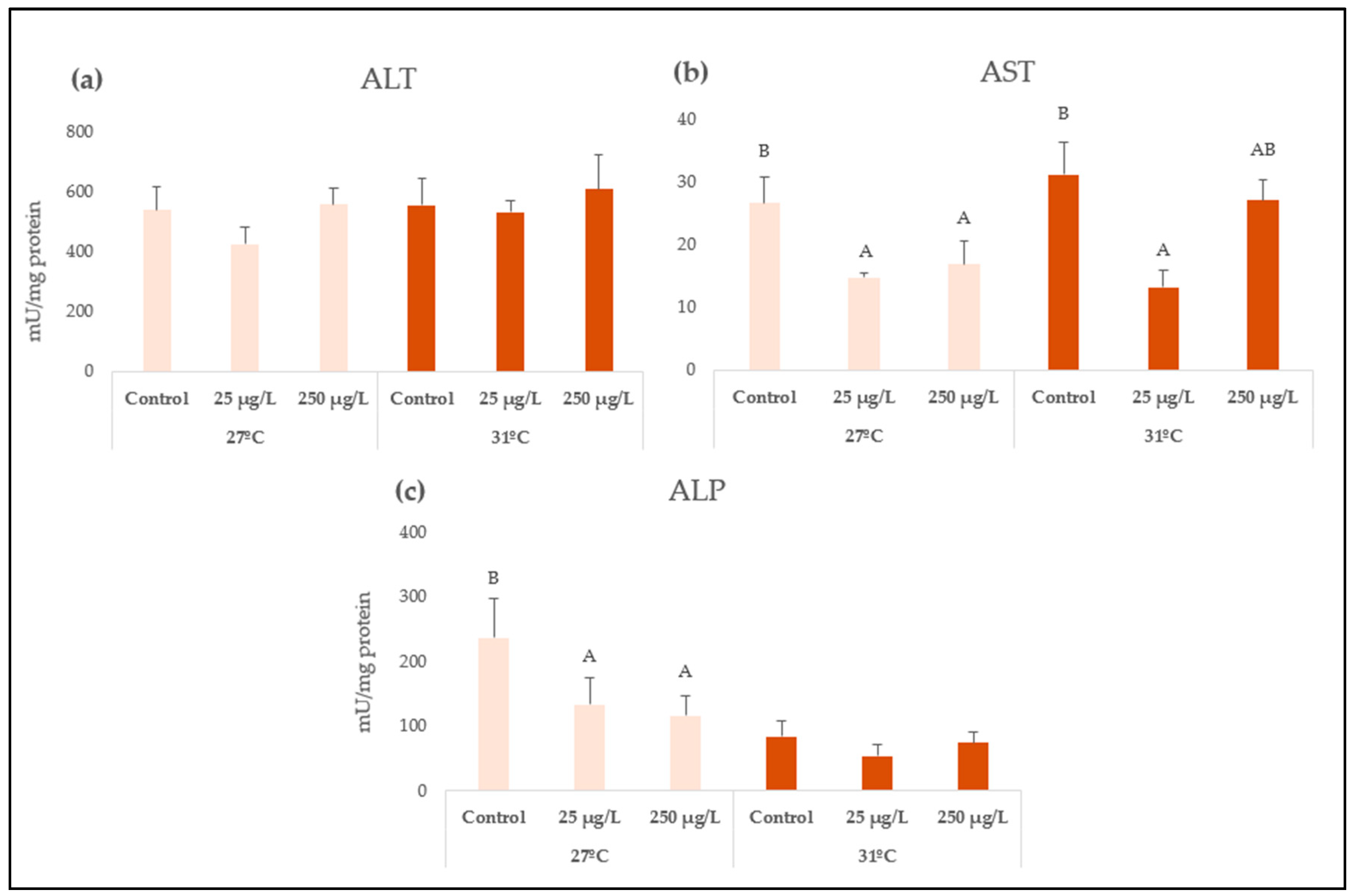
After 96 h of exposure to ZnONP at 27 °C, decreased enzyme activities of AST and ALP were observed after both concentrations (p = 0.0484 and p = 0.0467, respectively), while no changes were observed for ALT. At 31 °C, AST enzyme activity also significantly decreased only after the lowest nanozinc concentration (25 µg/L) (p = 0.0125), while no changes were observed for either ALT or ALP enzyme activities (Figure 2).
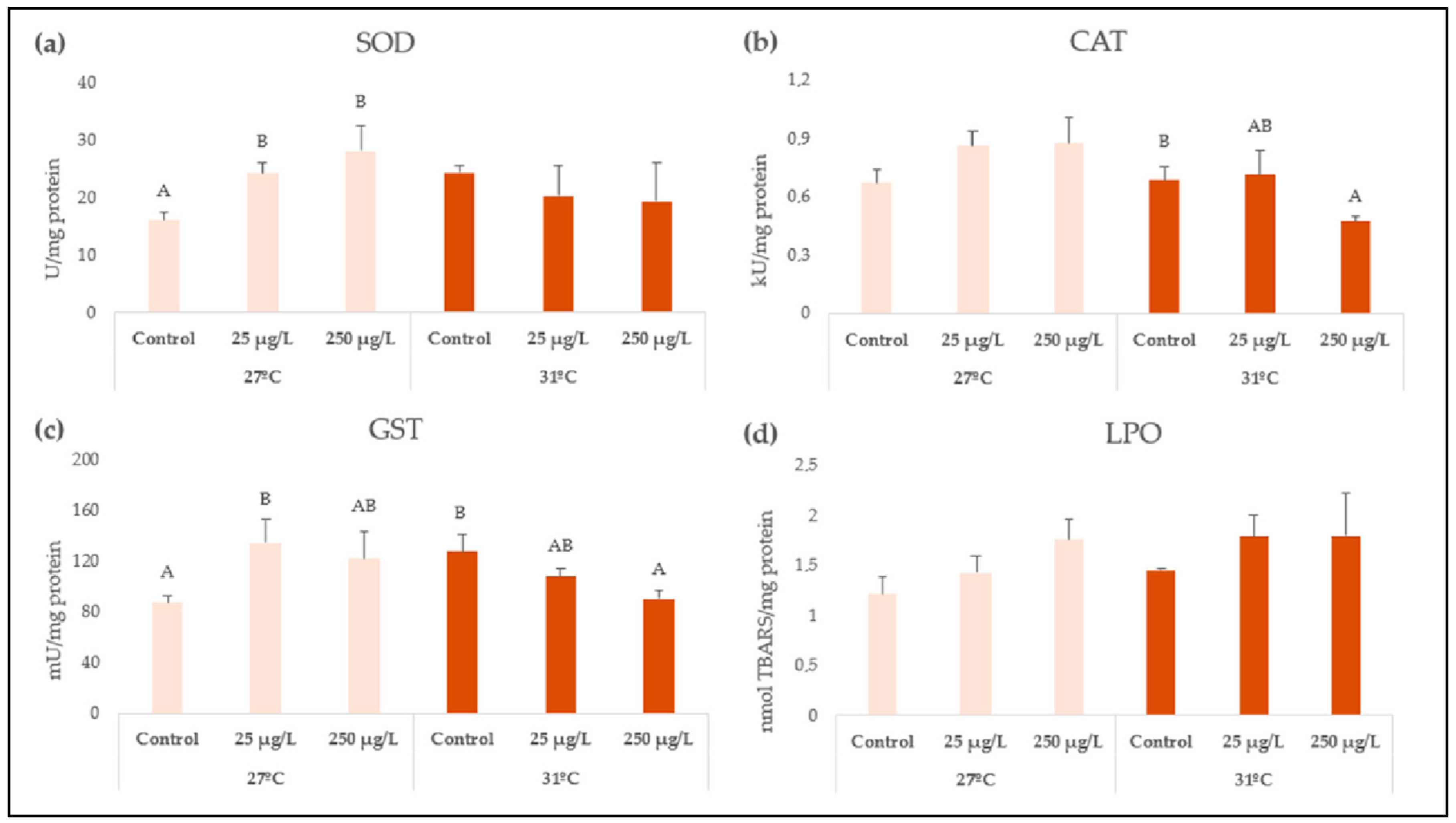
Oxidative stress-related biomarkers are shown in Figure 3. At the lowest temperature, while no changes were observed for CAT, SOD enzyme activity was increased after exposures to both nanozinc concentrations (p = 0.0083), and GST also showed increased activity, which was significant in the case of 25 µg/L. Conversely, at the highest temperature, CAT and GST enzyme activities decreased at the highest concentration (250 µg/L) (p = 0.0305, p = 0.0487, respectively); however, SOD showed no difference among the treatments. Interactions between the temperature and nanozinc concentrations were significant for SOD (p = 0.0026), CAT (p = 0.0373), and GST (p = 0.0015) enzyme activities (Table 1). Finally, LPO levels showed no differences in comparison with their respective control groups.

3.3. Filtration Rate

The filtration rate mean values are shown in Figure 4. Data at 24 h were excluded from the analysis as no algae were observed at this time. A significant interaction between temperature and time was observed (h) (p = 0.0086). After 6 h, the control filtration rate was significantly higher at 31 °C compared to 27 °C (p = 0.0196). Furthermore, after 6 h at the highest temperature, the filtration rates of L. fortunei were drastically decreased following exposure to both ZnONP concentrations (25 and 250 µg/L) in comparison to the control group (p = 0.0215 and p = 0.0198, respectively). A summary of the GLM modeling results is shown in Table 2.

4. Discussion

Zinc oxide nanoparticles are among the most produced NM worldwide, despite the fact that their ecotoxicology has been poorly addressed within the available literature [1,9,11]. The high volumes of release into aquatic environments have posed them as a threat to both humans and ecosystems [43]. In particular, we emphasize the need for assessing nanozinc toxicity under freshwater conditions and in benthic organisms like mussels, as they were stated to be particularly sensitive to nanopollutants given their filter-feeding habits [10].
The significant difference between the primary particle size (27 nm) and the hydrodynamic diameter (630 ± 207 nm) is a critical finding that has direct implications for the observed nanotoxicity. The DLS and zeta potential data confirm that ZnONP are not present as individual, discrete particles in the test medium but rather as larger aggregates. This aggregation is a key factor in their environmental fate and bioavailability. The −26.09 mV zeta potential, indicating moderate colloidal stability, suggests that these aggregates are dynamic and can undergo further changes, potentially leading to sedimentation. The ecotoxicological responses observed in L. fortunei (such as altered enzyme activities and filtration rate) are therefore likely a response to exposure to these larger agglomerates rather than to the primary individual NP. These findings emphasize the importance of characterizing NP under relevant exposure conditions to accurately interpret their toxic effects and bridge the gap between laboratory results and real-world environmental risks.
Nanozinc has unique properties, such as high reactivity and bioavailability, that bring a complex cascade of effects that become more challenging to elucidate under changing scenarios [44]. In this context, the ZnONP toxicity has been underestimated, as it was previously suggested that non-toxic concentrations of Limnoperna fortunei were in the range of 10–50 mg/L [13]; however, according to our results, the LC50-96 h for this species is much lower (11.31 mg/L), and exposure to 25 mg/L caused the mortality of the 100% of the tested organisms (Figure S2). These remarkable differences between results may lie in the intrinsic characteristics of the applied NP. The particles used in this study were dispersed in H2O and generated agglomerations when dispersed in the medium. According to Khan et al. [43], ZnONP particles are prone to agglomerate and sediment in heterogeneous matrices, which depend on the temperature (among other variables such as the presence of organic matter). Furthermore, it has been proved that these particles suffer from both increased agglomeration and sedimentation at higher temperatures, thus, affecting the bioavailability for the associated aquatic biota as they become less mobile and easily settle down [4]. Therefore, agglomeration processes could be a key phenomenon that needs further research, especially in a climate-change context of rising temperatures. While studies on another metal-based NP (AgNP) showed that agglomeration in the presence of organic matter and algae may mitigate the toxic effects in freshwater microcrustacean (inhabiting the water column) in terms of life-history traits [30,45], the toxicity mechanisms may be different for benthic and sedentary organisms like the bivalves because it may increase the bioavailability. Another key aspect in terms of exposure conditions consists of the sonication of the particles. In the other study, the ZnONP were sonicated prior to exposure in order to enhance their dispersion in the medium [13], while in this study we did not because: it has been proved that such a procedure has altered (even increased) the toxicity of other metallic NP [46,47], it does not represent the NM reaching the aquatic media, and media with low ionic strength (as freshwater media) guarantee a suitable dispersion.
Tissue damage was generated in the soft tissue of L. fortunei by inhibition of enzyme-related activities, which were more evident at 27 °C. To the best of our knowledge, no report is available assessing enzyme tissue damage in mussels exposed to ZnONP, despite the fact that the biomarkers were described to be particularly sensitive in terms of nanopollutants and other stressors (e.g., metals, crude oil, anaerobiosis) [48,49,50,51,52]. Few studies have assessed these biomarkers in freshwater organisms, which make it difficult to discuss these results. Although few studies have been conducted on nanozinc toxicity, Wu and Sokolova [8] exposed Mytilus edulis marine mussels to ZnONP and increased temperature, and evidenced an overall blunting of cell responses. Therefore, it could be hypothesized that the temperature may blunt the mechanisms against tissue damage in L. fortunei, and thus the intrinsic effects of the nanopollutant may be masked by the increased temperature alone. In the snail Biomphalaria alexandrina exposed to high ZnONP concentrations (7, 35 mg/L), the transaminases (ALT, AST) and ALP were found to be drastically increased in hemolymph and soft tissue, and they were related to muscle damage, intestinal and hepatopancreatic injuries, and toxic hepatitis [53]. In the serum of Cyprinus carpio fish exposed to nanozinc via intraperitoneal injection (10–20 µg/g), increased activity of the transaminases and ALP were found, and it was related to kidney-related function alterations [54]. Overall, we highlight the much lower concentrations employed in the present study, which may generate differential responses in the organisms for these biomarkers. Furthermore, we consider that waterborne exposure is more environmentally relevant for assessing ecotoxicity than via injection; therefore, this makes it difficult to discuss the obtained findings given the poorly available literature. Lastly, we highlight that a significant decrease in AST in the soft tissue of L. fortunei was found at both temperatures analyzed, even at the lowest concentration assayed, which is environmentally relevant (25 µg/L).
Oxidative stress has been widely assessed in terms of nanotoxicity since it is considered one of the main toxicity mechanisms, as ZnONP cellular metabolism generates reactive oxygen species (ROS) and the associated oxidative damage [55]. It must be pointed out that antioxidant-related enzyme activities significantly interacted with the nanozinc concentration and the temperature tested, suggesting that climate change threatens the aquatic ecosystem in complex ways. It has been demonstrated that NP-induced toxicity is related to activation or inhibition of antioxidant enzymes in the case of cytotoxicity triggered by ROS. In this regard, SOD enzyme is the first one to deal with oxyradicals and is responsible for catalyzing the dismutation of the superoxide radical O2− to H2O2 [55]. Our results showed an increase in enzyme activity after both nanozinc concentrations at 27 °C; however, at 31 °C, its activity remained similar to the control values. The lack of differences in the enzyme activity at the highest temperature could also be explained by a masked effect caused by the high temperature alone toward the nanopollutant, which generated an overall blunt in cell responses [8]. Accordingly, increased SOD activity was observed in the digestive gland of the freshwater mussels Unio tumidus exposed for 14 days to ZnONP (concentration expressed as 3.1 µM), which was explained by an enzyme activation due to ROS overproduction [56]. Another study evaluating environmentally relevant concentrations of ZnONP (1 and 10 µg/L) in the marine clam Ruditapes philippinarum also reported increased SOD activity in gills and digestive gland [57]. An interesting report carried out by Lai et al. [7] evaluated the interactive effects of ZnONP (concentrations expressed as 0.01 and 3 mg Zn/L) with different ranges of temperature and salinity in the marine mussel Xenostrobus secures. The authors found decreased activity of SOD and interaction only with salinity; however, another study that evaluated the ecotoxicity of CeO2NP in the marine bivalve Mytilus galloprovincialis explained that temperature altered the organisms’ biochemical functions [58]. Key differences in terms of toxicity must be contemplated regarding media with low or high ionic strength (freshwater vs. marine media), making it difficult to assess our findings given the lack of studies on freshwater mussels exposed to nanozinc.
CAT is another important antioxidant enzyme, widely reported, that protects the cell from ROS damage by reducing H2O2 into water and oxygen; thus, it is crucial to maintain cell homeostasis [59]. Our results showed decreased activity in the soft tissue of L. fortunei exposed to the highest ZnONP concentration (250 µg/L) at 31 °C (while no changes were observed at the optimal temperature). As this biomarker showed a significant interaction between the nanozinc exposure, and also for temperature alone, the high temperature could have exacerbated the nanotoxic effects of the particles, as suggested by Morossetti et al. [58] for CeO2NP. Accordingly, it was already mentioned that ZnONP could suffer from both agglomeration and sedimentation at higher temperatures [4]. In our study, the TEM analysis showed agglomeration of the ZnONP; thus, a higher temperature could have exacerbated it, leading to higher sedimentation and augmented bioavailability for L. fortunei at 31 °C (in comparison with 27 °C). CAT activity inhibition following exposure to nanozinc has been explained by enhanced oxygen free-radical production, and this result is consistent with that reported by Fahmy [53] in hemolymph and soft tissue of the freshwater snail B. alexandrina exposed to higher concentrations (7 and 35 mg ZnONP/L). Our results are also consistent with the findings by other authors in muscle of freshwater fish species exposed to nanozinc [60,61].
Remarkably, opposite tendencies were evidenced for GST activity, as it increased at 27 °C but decreased at 31 °C, indicating a clear effect determined by the temperature. Conversely, in the marine mussel X. securis exposed to 3 mg ZnONP/L, the opposite behavior was observed (GST activity increased under higher temperatures) [7]; once again, we highlight the difference in terms of the ionic strength of the medium. This enzyme has a key role in the detoxification process, which conjugates reduced glutathione (GSH) with xenobiotics. Therefore, its inhibition (observed at the higher temperature) was explained by a direct action of the metal on the enzyme or indirectly via the ROS production which causes depletion of GSH [57]. The overactivity of GST at the optimal temperature for L. fortunei (27 °C) could be due to the need for detoxifying the effects provoked by the metal (Zn ions released by the particles) or the ROS overproduction; however, synergistic effects could have occurred at the highest temperature, bringing additional stress which the enzyme was unable to cope with; therefore, its inhibition happened instead. No effects in terms of lipid peroxidation were observed after exposure to nanozinc at either 27 °C or 31 °C. In other studies conducted on freshwater species exposed to higher ZnONP concentrations, increased LPO levels were observed [53,62]; however, the high tolerance of L. fortunei, the exposure time, and the low concentrations selected in this study were not sufficient to cause lipid oxidative damage in the soft tissue of the organisms.
Filtration rate is a crucial characteristic for bivalves that engage in filter feeding and is subject to regulation in response to various environmental factors. In this sense, it has been studied that, despite the high tolerance of L. fortunei, temperature is a key factor modifying this behavior [63]. A study conducted on two marine/estuarine mussels exposed to high ZnONP concentrations (2.5–50 mg/L) concluded that changes in the filtration rate are an appropriate indicator for the measurement of nanopollutants [14]. Accordingly, we observed that the control groups for each temperature differed in their filtration rate (it was higher at 31 °C than at 27 °C) as a result of increased physiological processes and metabolism at higher temperature [64]. However, while no differences among the treatments were found at the optimal temperature, a drastic decrease in the filtration rate was observed at 31 °C for both nanozinc concentrations tested. According to Quevedo et al. [65], macroinvertebrate assemblages are sensitive to thermal changes in a context of climate change (even when temperature does not exceed 3 °C). This could be a crucial threat for filter-feeding species with ultimate implications for their body storage, as has been proven for L. fortunei exposed to Cu and a wide range of temperatures [22]. The low filtration rate could be explained by a synergistic effect provoked by the nanozinc exposure and the higher temperature (not evidenced at 27 °C). In the case of a harmful environment, the bivalves are capable of minimizing their water contact by reducing the valve opening period and narrowing valve gap amplitude, resulting in a reduced filtration rate [7]. Studies on mollusks exposed to other metal-based NP (TiO2NP, CuONP) also reported a decreased filtration rate, which was related to a reduction in energy consumption [66].
It has also been documented that L. fortunei is able to selectively ingest and retain particles spanning a broad size range (approximately 1–1000 µm), while smaller particles (≤1 µm) are typically aggregated and expelled as pseudofeces [67,68]. In this sense, exposure to TiO2NP has been reported to stimulate the secretion of mucus filaments in L. fortunei and M. edulis, suggesting an adaptive mechanism to entrap the particles, thus limiting their assimilation and mitigating the toxic effects [69,70]. Furthermore, another behavioral mechanism described in bivalves to reduce the pollutant uptake and maintain the homeostasis under stress conditions is the valve closure [67]. Therefore, these defense mechanisms could have prevented the filtration rate alteration in L. fortunei under the optimal temperature; however, they were not sufficient to prevent deleterious effects at 31 °C, as the joint effect with the nanopollutant could have exceeded the bivalves’ tolerance to stress. Lastly, it has been explained that particle accumulation may have an effect on ciliary beating and muscular changes in gills, which are physiologically controlled by the nervous system [71]; in this regard, further studies are needed on ZnONP and their neurological effects on mollusks.

5. Conclusions

Overall, we highlight the ecotoxicological implications of nanozinc in a climate-change context, even at environmentally relevant concentrations (such as the lowest one tested in this study). Nanozinc was proven to cause tissue damage, disrupt the redox homeostasis, and alter the filter-feeding habits in the golden mussel L. fortunei; these effects may become more complex in a changing world of rising temperatures. We consider that bivalves are key organisms for evaluating exposure to nanopollutants, as their sensitivity and filter-feeding habits are valuable for obtaining early responses. With the freshwater habitats being poorly represented among the available literature, complex mechanisms for emerging contaminants like the NM are needed to be elucidated in the near future.

Supplementary Materials

The following supporting information can be downloaded at: https://www.mdpi.com/article/10.3390/ani15182734/s1, Figure S1: Map showing the collection area of L. fortunei in Santa Fe River, Argentina (31°38′34.90″ S; 60°41′6.22″ W), Figure S2: Lethal concentration 50 (LC50, mg ZONP/L), 95% confidence intervals, and dose–response curve in L. fortunei.

Author Contributions

Conceptualization, A.A., V.S.A., L.M.O. and F.M.R.M.; methodology, A.A., V.S.A., L.M.O., M.F.S., P.E.A., M.F.D., L.M.O. and F.M.R.M.; software, A.A. and V.S.A.; validation, A.A. and V.S.A.; investigation, A.A., V.S.A., L.M. and F.M.R.M.; resources, A.A., L.M., F.M.R.M. and M.F.D.; data curation, A.A. and V.S.A.; writing—original draft preparation, A.A.; writing—review and editing A.A., V.S.A., L.M.O., M.F.S., P.E.A., M.F.D., L.M.O. and F.M.R.M.; visualization, A.A. and V.S.A.; supervision, A.A.; project administration, A.A. and F.M.R.M.; funding acquisition, A.A. and F.M.R.M. All authors have read and agreed to the published version of the manuscript.

Funding

This research was funded by Agencia Nacional de Promoción Científica y Tecnológica (PICT 2020-01206, PI: A.A.), Universidad Nacional del Litoral (CAI+D 50620190100047LI, PI: F.R.M.), Universidad de Buenos Aires (UBACYT 20020150100056BA, PI: M.F.D), and CONICET (PIP 11220200102598CO, PI: M.F.D).

Institutional Review Board Statement

The animal study protocol was approved by the Institutional Ethics Committee of CONICET (resolution 1047, 1 July 2005).

Informed Consent Statement

Not applicable.

Data Availability Statement

The raw data supporting the conclusions of this article will be made available by the authors upon request.

Conflicts of Interest

The authors declare no conflicts of interest.

References

  1. Ale, A.; Andrade, V.S.; Gutierrez, M.F.; Ayech, A.; Monserrat, J.M.; Desimone, M.F.; Cazenave, J. Metal-based nanomaterials in aquatic environments: What do we know so far about their ecotoxicity? Aquat. Toxicol. 2024, 275, 107069. [Google Scholar] [CrossRef]
  2. Wu, F.; Deng, Y.; Sokolov, E.P.; Falfushynska, H.; Glänzer, A.; Xie, L.; Sokolova, I.M. Nanopollutants (nZnO) amplify hypoxia-induced cellular stress in a keystone marine bivalve, Mytilus edulis. Environ. Res. 2025, 274, 121346. [Google Scholar] [CrossRef]
  3. Gottschalk, F.; Lassen, C.; Kjoelholt, J.; Christensen, F.; Nowack, B. Modeling flows and concentrations of nine engineered nanomaterials in the Danish environment. Int. J. Environ. Res. Public Health 2015, 12, 5581–5602. [Google Scholar] [CrossRef] [PubMed]
  4. Yung, M.M.N.; Mouneyrac, C.; Leung, K.M.Y. Ecotoxicity of Zinc Oxide Nanoparticles in the Marine Environment. In Encyclopedia of Nanotechnology; Bhushan, B., Ed.; Springer: Dordrecht, The Netherlands, 2014. [Google Scholar]
  5. Hong, H.; Adam, V.; Nowack, B. Form-Specific and Probabilistic Environmental Risk Assessment of 3 Engineered Nanomaterials (Nano-Ag, Nano-TiO2, and Nano-ZnO) in European Freshwaters. Environ. Toxicol. Chem. 2021, 40, 2629–2639. [Google Scholar] [CrossRef] [PubMed]
  6. Sun, T.Y.; Gottschalk, F.; Hungerbühler, K.; Nowack, B. Comprehensive probabilistic modelling of environmental emissions of engineered nanomaterials. Environ. Pollut. 2014, 185, 69–76. [Google Scholar] [CrossRef] [PubMed]
  7. Lai, R.W.S.; Zhou, G.J.; Yung, M.M.N.; Djurišić, A.B.; Leung, K.M.Y. Interactive effects of temperature and salinity on toxicity of zinc oxide nanoparticles towards the marine mussel Xenostrobus securis. Sci. Total Environ. 2023, 889, 164254. [Google Scholar] [CrossRef]
  8. Wu, F.; Sokolova, I.M. Immune responses to ZnO nanoparticles are modulated by season and environmental temperature in the blue mussels Mytilus edulis. Sci. Total Environ. 2021, 801, 149786. [Google Scholar] [CrossRef]
  9. Gutierrez, M.F.; Ale, A.; Andrade, V.; Bacchetta, C.; Rossi, A.; Cazenave, J. Metallic, metal oxide, and metalloid nanoparticles toxic effects on freshwater microcrustaceans: An update and basis for the use of new test species. Water Environ. Res. 2021, 93, 2505–2526. [Google Scholar] [CrossRef]
  10. Skawina, A.; Agnieszka, D.; Bonk, A.; Paterczyk, B.; Nowakowska, J. Tracking the micro- and nanoplastics in the terrestrial-freshwater food webs. Bivalves as sentinel species. Sci. Total Environ. 2024, 917, 170468. [Google Scholar] [CrossRef]
  11. Cazenave, J.; Ale, A.; Bacchetta, C.; Rossi, A.S. Nanoparticles Toxicity in Fish Models. Curr. Pharm. Des. 2019, 25, 3927–3942. [Google Scholar] [CrossRef]
  12. Auclair, J.; Turcotte, P.; Gagnon, C.; Gagné, F. The influence of surface waters on the bioavailability and toxicity of copper oxide nanoparticles to freshwater mussels. Invertebr. Surviv. J. 2021, 18, 33–45. [Google Scholar] [CrossRef]
  13. Girardello, F.; Leite, C.C.; Touguinha, L.B.; Roesch-Ely, M.; da Silva, C.K.H.; de Oliveira, R.M.; Borges, D.L.G.; Villela, I.V.; Fernandes, A.N.; Salvador, M.; et al. ZnO nanoparticles alter redox metabolism of Limnoperna fortunei. Environ. Sci. Pollut. Res. 2021, 28, 69416–69425. [Google Scholar] [CrossRef]
  14. Qadermarzi, A.; Pouladi, M.; Rezamand, A.; Hoseinifar, S.H.; Hedayati, A.A. Investigation of sub-acute levels of zinc oxide nanoparticles on the filtration rate of Mytilaster lineatus and Dressina polymorpha in the short term. Nusant. Biosci. 2018, 10, 53–57. [Google Scholar] [CrossRef]
  15. Johnson, M.F.; Albertson, L.K.; Algar, A.C.; Dugdale, S.J.; Edwards, P.; England, J.; Gibbins, C.; Kazama, S.; Komori, D.; MacColl, A.D.C.; et al. Rising water temperature in rivers: Ecological impacts and future resilience. Wiley Interdiscip. Rev. Water 2024, 11, e1724. [Google Scholar] [CrossRef]
  16. Van Vliet, M.T.; Thorslund, J.; Strokal, M.; Hofstra, N.; Flörke, M.; Ehalt Macedo, H.; Nkwasa, A.; Tang, T.; Kaushal, S.S.; Kumar, R.; et al. Global river water quality under climate change and hydroclimatic extremes. Nat. Rev. Earth Environ. 2023, 4, 687–702. [Google Scholar] [CrossRef]
  17. Gebrewahid, Y.; Eyasu, G.; Meresa, E.; Abrehe, S.; Darcha, G.; Manaye, A.; Tesfamariam, B. Modeling the ecological niche of rhamnus prinoides L. under climate change scenarios: A MaxEnt modeling approach. Geol. Ecol. Landsc. 2024, 1–17. [Google Scholar] [CrossRef]
  18. IPCC. Summary for Policymarkers: Synthesis Andreport. In Climate Change 2023: Synthesis Report; Contribution of Working Groups I, II and III to the Sixth Assessment Report of the Intergovernmental Panel on Climate Change; Core Writing Team, Lee, H., Romero, J., Eds.; IPCC: Geneva, Switzerland, 2023; pp. 1–34. [Google Scholar] [CrossRef]
  19. Gutierrez, M.F.; Andrade, V.S.; Ale, A.; Monserrat, J.M.; Roa-Fuentes, C.A.; Herrera-Martínez, Y.; Bacchetta, C.; Cazenave, J.; Rossi, A.S.; Nandini, S.; et al. Responses of freshwater organisms to multiple stressors in a climate change scenario: A review on small-scale experiments. Environ. Sci. Pollut. Res. 2025, 32, 4431–4444. [Google Scholar] [CrossRef]
  20. Barbosa, F.G. The scientific literature on Limnoperna fortune (Dunker 1857) from 1982 to 2012. An. Acad. Bras. Cienc. 2014, 86, 1373–1383. [Google Scholar] [CrossRef]
  21. Castro, M.; Garreta, C.; Arocena, R. Urban effluents affect the invasive bivalve Limnoperna fortunei (Dunker 1857) fitness in a large Pampasic river (Río Negro, Uruguay). Environ. Monit. Assess. 2024, 196, 48. [Google Scholar] [CrossRef]
  22. Cazenave, J.; Rossi, A.S.; Ale, A.; Montalto, L.; Gutierrez, M.F.; Rojas Molina, F. Does temperature influence on biomarker responses to copper exposure? The invasive bivalve Limnoperna fortunei (Dunker 1857) as a model. Comp. Biochem. Physiol. Part-C Toxicol. Pharmacol. 2025, 287, 110059. [Google Scholar] [CrossRef] [PubMed]
  23. Girardello, F.; Leite, C.C.; Branco, C.S.; Roesch-Ely, M.; Fernandes, A.N.; Salvador, M.; Henriques, J.A.P. Antioxidant defences and haemocyte internalization in Limnoperna fortunei exposed to TiO2 nanoparticles. Aquat. Toxicol. 2016, 176, 190–196. [Google Scholar] [CrossRef] [PubMed]
  24. Perepelizin, P.V.; Boltovskoy, D. Thermal tolerance of Limnoperna fortunei to gradual temperature increase and its applications for biofouling control in industrial and power plants. Biofouling 2011, 27, 667–674. [Google Scholar] [CrossRef]
  25. Iriondo, M.H.; Paira, A.R. Physical Geography of the Basin. In The Middle Paraná River; Iriondo, M.H., Paggi, J.C., Parma, M.J., Eds.; Springer: Berlin/Heidelberg, Germany, 2007. [Google Scholar]
  26. Boltovskoy, D.; Paolucci, E.; MacIsaac, H.J.; Zhan, A.; Xia, Z.; Correa, N. What We Know and Don’t Know About the Invasive Golden Mussel Limnoperna Fortunei; Springer International Publishing: Cham, Switzerland, 2025; Volume 852, ISBN 0123456789. [Google Scholar]
  27. Ritz, C.; Baty, F.; Streibig, J.C.; Gerhard, D. Dose-response analysis using R. PLoS ONE 2015, 10, e0146021. [Google Scholar] [CrossRef]
  28. Gagnon, C.; Turcotte, P.; Trépanier, S.; Gagné, F.; Cejka, P.J. Impacts of municipal wastewater oxidative treatments: Changes in metal physical speciation and bioavailability. Chemosphere 2014, 97, 86–91. [Google Scholar] [CrossRef]
  29. Clara, M.; Windhofer, G.; Weilgony, P.; Gans, O.; Denner, M.; Chovanec, A.; Zessner, M. Identification of relevant micropollutants in Austrian municipal wastewater and their behaviour during wastewater treatment. Chemosphere 2012, 87, 1265–1272. [Google Scholar] [CrossRef]
  30. Andrade, V.S.; Ale, A.; Antezana, P.E.; Desimone, M.F.; Cazenave, J.; Gutierrez, M.F. Ecotoxicity of nanosilver on cladocerans and the role of algae provision. Environ. Sci. Pollut. Res. 2023, 30, 27137–27149. [Google Scholar] [CrossRef]
  31. Bacchetta, C.; Cazenave, J.; Parma, M.J.; Biancucci, G.F. Biochemical stress responses in tissues of the cichlid fish Cichlasoma dimerus exposed to a commercial formulation of endosulfan. Arch. Environ. Contam. Toxicol. 2011, 61, 453–460. [Google Scholar] [CrossRef]
  32. Reglero, M.M.; Taggart, M.A.; Monsalve-González, L.; Mateo, R. Heavy metal exposure in large game from a lead mining area: Effects on oxidative stress and fatty acid composition in liver. Environ. Pollut. 2009, 157, 1388–1395. [Google Scholar] [CrossRef] [PubMed]
  33. Reitman, S.; Frankel, S. A Colorimetric Method for the Determination of Serum Glutamic Oxalacetic and Glutamic Pyruvic Transaminases. Am. J. Clin. Pathol. 1957, 28, 56–63. [Google Scholar] [CrossRef]
  34. Misra, H.P.; Fridovich, I. The role of superoxide anion in the autoxidation of epinephrine and a simple assay for superoxide dismutase. J. Biol. Chem. 1972, 247, 3170–3175. [Google Scholar] [CrossRef] [PubMed]
  35. Aebi, H. Catalase in vitro. Method. Enzymol. 1984, 105, 121–126. [Google Scholar]
  36. Poletta, G.L.; Simoniello, M.F.; Mudry, M.D. Biomarkers of oxidative damage and antioxidant defense capacity in Caiman latirostris blood. Comp. Biochem. Physiol. Part-C Toxicol. Pharmacol. 2016, 179, 29–36. [Google Scholar] [CrossRef]
  37. Habig, W.H.; Pabst, M.J.; Jakoby, W.B. Glutathione S-Transferases. J. Biol. Chem. 1974, 249, 7130–7139. [Google Scholar] [CrossRef]
  38. Yagi, K. A simple fluorometric assay for lipoperoxide in blood plasma. Biochem. Med. 1976, 212–216. [Google Scholar] [CrossRef]
  39. Barker Jørgensen, C. Bivalve filter feeding revisited. Mar. Ecol. Prog. Ser. 1996, 142, 287–302. [Google Scholar] [CrossRef]
  40. Finney, D. Probit Analysis; Cambridge University Press: London, UK, 1971. [Google Scholar]
  41. Clogston, J.D.; Patri, A.K. Zeta Potential Measurement. Methods Mol. Biol. 2011, 697, 63–70. [Google Scholar] [CrossRef]
  42. Khani, O.; Mohammadi, M.; Khaz’ali, A.R.; Aghdam, M.A. Effect of pH value and zeta potential on the stability of CO2 foam stabilized by SDS surfactant and SiO2, ZnO and Fe2O3 nanoparticles. Sci. Rep. 2025, 15, 10302. [Google Scholar] [CrossRef]
  43. Khan, R.; Inam, M.A.; Zam, S.Z.; Park, D.R.; Yeom, I.T. Assessment of key environmental factors influencing the sedimentation and aggregation behavior of zinc oxide nanoparticles in aquatic environment. Water 2018, 10, 660. [Google Scholar] [CrossRef]
  44. Wu, F.; Sokolov, E.P.; Timm, S.; Sokolova, I.M. Synergistic impacts of nanopollutants (nZnO) and hypoxia on bioenergetics and metabolic homeostasis in a marine bivalve Mytilus edulis. Environ. Sci. Nano 2024, 12, 576–596. [Google Scholar] [CrossRef]
  45. Andrade, V.S.; Ale, A.; Antezana, P.E.; Desimone, M.F.; Cazenave, J.; Gutierrez, M.F. Environmental factors modify silver nanoparticles ecotoxicity in Chydorus eurynotus (Cladocera). Ecotoxicology 2024, 33, 683–696. [Google Scholar] [CrossRef] [PubMed]
  46. Laban, G.; Nies, L.F.; Turco, R.F.; Bickham, J.W.; Sepúlveda, M.S. The effects of silver nanoparticles on fathead minnow (Pimephales promelas) embryos. Ecotoxicology 2010, 19, 185–195. [Google Scholar] [CrossRef]
  47. Lee, W.M.; Ha, S.W.; Yang, C.Y.; Lee, J.K.; An, Y.J. Effect of fluorescent silica nanoparticles in embryo and larva of Oryzias latipes: Sonic effect in nanoparticle dispersion. Chemosphere 2011, 82, 451–459. [Google Scholar] [CrossRef]
  48. Kokhan, A.S.; Soldatov, A.A.; Golovina, I.V.; Bogdanovich, V.Y.; Shalagina, N.E.; Rychkova, V.N. Parameters of Energy Metabolism and Adenylate System in Mytilus galloprovincialis Tissues under Moderate Hypoxia. J. Evol. Biochem. Phys. 2023, 59, 1986–1994. [Google Scholar] [CrossRef]
  49. Li, Y.; Gu, Z.; Liu, H.; Shen, H.; Yang, J. Biochemical response of the mussel Mytilus coruscus (Mytiloida: Mytilidae) exposed to in vivo sub-lethal copper concentrations. Chin. J. Oceanol. Limnol. 2012, 30, 738–745. [Google Scholar] [CrossRef]
  50. Blasco, J.; Puppo, J. Effect of heavy metals (Cu, Cd and Pb) on aspartate and alanine aminotransferase in Ruditapes philippinarum (Mollusca: Bivalvia). Comp. Biochem. Physiol.-C Pharmacol. Toxicol. Endocrinol. 1999, 122, 253–263. [Google Scholar] [CrossRef]
  51. Narvia, M.; Rantamäki, P. Aminotransferases in the bivalve mollusc Mytilus edulis L. and short term effects of crude oil in brackish water. Biomarkers 1997, 2, 253–258. [Google Scholar] [CrossRef] [PubMed]
  52. Brandts, I.; Teles, M.; Gonçalves, A.P.; Barreto, A.; Franco-Martinez, L.; Tvarijonaviciute, A.; Martins, M.A.; Soares, A.M.V.M.; Tort, L.; Oliveira, M. Effects of nanoplastics on Mytilus galloprovincialis after individual and combined exposure with carbamazepine. Sci. Total Environ. 2018, 643, 775–784. [Google Scholar] [CrossRef] [PubMed]
  53. Fahmy, S.R.; Abdel-Ghaffar, F.; Bakry, F.A.; Sayed, D.A. Ecotoxicological effect of sublethal exposure to zinc oxide nanoparticles on freshwater snail Biomphalaria alexandrina. Arch. Environ. Contam. Toxicol. 2014, 67, 192–202. [Google Scholar] [CrossRef]
  54. Rasheed, A.; Javed Iqbal, K.; Safdar, A.; Nasir, A.; Jabeen, R.; sami, A.; Tara, N.; Ali, S.; Zeeshan, M.; Abbas, S.; et al. Toxicological effects of zinc oxide nanoparticles on hemato-biochemical profile of common carp (Cyprinus carpio). J. King Saud Univ.-Sci. 2023, 35, 102835. [Google Scholar] [CrossRef]
  55. Zhao, X.; Ren, X.; Zhu, R.; Luo, Z.; Ren, B. Zinc oxide nanoparticles induce oxidative DNA damage and ROS-triggered mitochondria-mediated apoptosis in zebrafish embryos. Aquat. Toxicol. 2016, 180, 56–70. [Google Scholar] [CrossRef]
  56. Falfushynska, H.; Gnatyshyna, L.; Yurchak, I.; Sokolova, I.; Stoliar, O. The effects of zinc nanooxide on cellular stress responses of the freshwater mussels Unio tumidus are modulated by elevated temperature and organic pollutants. Aquat. Toxicol. 2015, 162, 82–93. [Google Scholar] [CrossRef]
  57. Marisa, I.; Matozzo, V.; Munari, M.; Binelli, A.; Parolini, M.; Martucci, A.; Franceschinis, E.; Brianese, N.; Marin, M.G. In vivo exposure of the marine clam Ruditapes philippinarum to zinc oxide nanoparticles: Responses in gills, digestive gland and haemolymph. Environ. Sci. Pollut. Res. 2016, 23, 15275–15293. [Google Scholar] [CrossRef]
  58. Morosetti, B.; Freitas, R.; Pereira, E.; Hamza, H.; Andrade, M.; Coppola, F.; Maggioni, D.; Della Torre, C. Will temperature rise change the biochemical alterations induced in Mytilus galloprovincialis by cerium oxide nanoparticles and mercury? Environ. Res. 2020, 188, 109778. [Google Scholar] [CrossRef] [PubMed]
  59. Regoli, F.; Hummel, H.; Amiard-Triquet, C.; Larroux, C.; Sukhotin, A. Trace metals and variations of antioxidant enzymes in arctic bivalve populations. Arch. Environ. Contam. Toxicol. 1998, 35, 594–601. [Google Scholar] [CrossRef] [PubMed]
  60. Abdelazim, A.M.; Saadeldin, I.M.; Swelum, A.A.A.; Afifi, M.M.; Alkaladi, A. Oxidative stress in the muscles of the fish Nile tilapia caused by zinc oxide nanoparticles and its modulation by vitamins C and E. Oxid. Med. Cell. Longev. 2018, 2018, 6926712. [Google Scholar] [CrossRef]
  61. Su, L.; Li, H.; Wang, J.; Wu, J.; Wan, J.; He, Y.; Liu, J. Zinc oxide nanoparticles cause hepatotoxicity in rare minnow (Gobiocypris rarus) via ROS-mediated oxidative stress and apoptosis activation and inhibition of lipid peroxidation. Aquac. Rep. 2024, 38, 102317. [Google Scholar] [CrossRef]
  62. Borase, H.P.; Patil, S.V.; Singhal, R.S. Moina macrocopa as a non-target aquatic organism for assessment of ecotoxicity of silver nanoparticles: Effect of size. Chemosphere 2019, 219, 713–723. [Google Scholar] [CrossRef]
  63. Zhang, J.; Xu, M.; Yang, Y. Assessing the effects of environmental factors on filtration rate of golden mussel (Limnoperna fortunei). Ecol. Indic. 2024, 158, 111544. [Google Scholar] [CrossRef]
  64. Casas, S.M.; Filgueira, R.; Lavaud, R.; Comeau, L.A.; La Peyre, M.K.; La Peyre, J.F. Combined effects of temperature and salinity on the physiology of two geographically-distant eastern oyster populations. J. Exp. Mar. Bio. Ecol. 2018, 506, 82–90. [Google Scholar] [CrossRef]
  65. Quevedo, L.; Ibáñez, C.; Caiola, N.; Mateu, D. Effects of thermal pollution on benthic macroinvertebrate communities of a large Mediterranean River. J. Entomol. Zool. Stud. 2018, 6, 500–507. [Google Scholar]
  66. Basti, L.; Nagai, S.; Watanabe, S.; Oda, T.; Tanaka, Y. Neuroenzymatic activity and physiological energetics in Manila clam, Ruditapes philippinarum, during short-term sublethal exposure to harmful alga, Heterocapsa circularisquama. Aquat. Toxicol. 2016, 176, 76–87. [Google Scholar] [CrossRef]
  67. Rojas Molina, F.; Paggi, J.C.; Devercelli, M. Zooplanktophagy in the natural diet and selectivity of the invasive mollusk Limnoperna fortunei. Biol. Invasions 2010, 12, 1647–1659. [Google Scholar] [CrossRef]
  68. Frau, D.; Molina, F.R.; Mayora, G. Feeding selectivity of the invasive mussel Limnoperna fortunei (Dunker, 1857) on a natural phytoplankton assemblage: What really matters? Limnology 2016, 17, 47–57. [Google Scholar] [CrossRef]
  69. Bourgeault, A.; Legros, V.; Gonnet, F.; Daniel, R.; Paquirissamy, A.; Bénatar, C.; Spalla, O.; Chanéac, C.; Renault, J.P.; Pin, S. Interaction of TiO2 nanoparticles with proteins from aquatic organisms: The case of gill mucus from blue mussel. Environ. Sci. Pollut. Res. 2017, 24, 13474–13483. [Google Scholar] [CrossRef] [PubMed]
  70. Manske Nunes, S.; Josende, M.E.; González-Durruthy, M.; Pires Ruas, C.; Gelesky, M.A.; Romano, L.A.; Fattorini, D.; Regoli, F.; Monserrat, J.M.; Ventura-Lima, J. Different crystalline forms of titanium dioxide nanomaterial (rutile and anatase) can influence the toxicity of copper in golden mussel Limnoperna fortunei? Aquat. Toxicol. 2018, 205, 182–192. [Google Scholar] [CrossRef] [PubMed]
  71. Saidani, W.; Sellami, B.; Khazri, A.; Mezni, A.; Dellali, M.; Joubert, O.; Sheehan, D.; Beyrem, H. Metal accumulation, biochemical and behavioral responses on the Mediterranean clams Ruditapes decussatus exposed to two photocatalyst nanocomposites (TiO2 NPs and AuTiO2NPs). Aquat. Toxicol. 2019, 208, 71–79. [Google Scholar] [CrossRef]
Figure 1. (a) TEM and (b) DLS analyses of the ZnONP.
Animals 15 02734 g001
Figure 2. Tissue-damage-related enzyme activities: (a) alanine aminotransferase (ALT), (b) aspartate aminotransferase (AST), and (c) alkaline phosphatase (ALP) in soft tissue of L. fortunei exposed to 0, 25 and 250 µg ZnONP/L at 27 or 31 °C for 96 h. The values are expressed as means ± SE. Means not sharing the same capital letter (A or B) are significantly different at p < 0.05.
Animals 15 02734 g002
Figure 3. Oxidative stress-related enzyme activities: (a) superoxide dismutase (SOD), (b) catalase (CAT), and (c) glutathione S-transferase (GST), and (d) lipid peroxidation levels (LPO) in soft tissue of L. fortunei exposed to 0, 25 and 250 µg ZnONP/L at 27 or 31 °C for 96 h. The values are expressed as means ± SE. Means not sharing the same capital letter (A or B) are significantly different at p < 0.05.
Animals 15 02734 g003
Figure 4. Filtration rate of L. fortunei exposed to 0, 25 and 250 µg ZnONP/L at (a) 27 or (b) 31 °C for 24 h. The values are expressed as means ± SE. Means not sharing the same capital letter (A or B) are significantly different at p < 0.05. The hash sign (#) means statistical difference between filtrations rates in the control groups at 27 or 31 °C at p < 0.05.
Animals 15 02734 g004
Table 1. Summary of the 2-way ANOVA applied to evaluate the effects of nanozinc concentration, temperature, and their interaction (Conc × Temp) on biomarker responses of L. fortunei. Significant effects are in bold at p < 0.05.
BiomarkerFactordfF Valuep Value
ALTConcentration20.890.4246
Temperature10.810.3786
Conc × Temp20.160.8555
ASTConcentration28.50.0018
Temperature12.270.1462
Conc × Temp21.410.2655
ALPConcentration22.280.1242
Temperature19.950.0043
Conc × Temp21.270.3001
SODConcentration21.410.2663
Temperature10.640.4325
Conc × Temp28.110.0026
CATConcentration21.690.2091
Temperature112.410.002
Conc × Temp23.860.0373
GSTConcentration21.790.1917
Temperature12.210.152
Conc × Temp28.960.0015
LPOConcentration21.90.1713
Temperature11.260.2727
Conc × Temp20.260.7705
Table 2. Summary of the GLM applied to evaluate the effects of nanozinc concentrations and temperature after 3, 6 and 12 h on filtration rate of L. fortunei. Significant effects are in bold at p < 0.05.
Contrast RatioSEt Ratiop Value
3 h
Control27 °C/0.025 µg/L27 °C1.1770.4450.4310.9979
Control27 °C/0.250 µg/L27 °C1.5110.6650.9380.9337
Control27 °C/control31 °C0.7660.242−0.8430.9570
Control27 °C/0.025 µg/L31 °C1.0740.3880.1971.0000
Control27 °C/0.250 µg/L31 °C0.9060.304−0.2950.9997
0.025 µg/L27 °C/0.250 µg/L27 °C1.2840.5950.5390.9941
0.025 µg/L31 °C/control31 °C0.6500.226−1.2360.8160
0.025 µg/L31 °C/0.025 µg/L31 °C0.9120.355−0.2360.9999
0.025 µg/L31 °C/0.250 µg/L31 °C0.7690.281−0.7170.9785
0.250 µg/L31 °C/control31 °C0.5070.211−1.6310.5845
0.250 µg/L31 °C/0.025 µg/L31 °C0.7110.321−0.7560.9729
0.250 µg/L31 °C/0.250 µg/L31 °C0.5990.258−1.1880.8395
Control31 °C/0.025 µg/L31 °C1.4020.4591.0330.9035
Control31 °C/0.250 µg/L31 °C1.1830.3530.5620.0028
0.025 µg/L31 °C/0.250 µg/L31 °C0.8440.292−0.4910.9962
6 h
Control27 °C/0.025 µg/L27 °C1.1700.5630.3270.9995
Control27 °C/0.250 µg/L27 °C1.1230.5320.2450.9999
Control27 °C/control31 °C0.3170.107−3.4040.0196
Control27 °C/0.025 µg/L31 °C1.2790.6460.4880.9963
Control27 °C/0.250 µg/L31 °C1.1770.5670.3390.9994
0.025 µg/L27 °C/0.250 µg/L27 °C0.9590.488−0.0811.0000
0.025 µg/L31 °C/control31 °C0.2710.1044−3.3920.0202
0.025 µg/L31 °C/0.025 µg/L31 °C1.0930.5880.1651.0000
0.025 µg/L31 °C/0.250 µg/L31 °C1.0060.5190.0111.0000
0.250 µg/L31 °C/control31 °C0.2820.107−3.3520.0223
0.250 µg/L31 °C/0.025 µg/L31 °C1.1390.6060.2450.9999
0.250 µg/L31 °C/0.250 µg/L31 °C1.0480.5350.0931.0000
Control31 °C/0.025 µg/L31 °C4.0381.6703.3660.0215
Control31 °C/0.250 µg/L31 °C3.7121.4303.4000.0198
0.025 µg/L31 °C/0.250 µg/L31 °C0.9200.496−0.1541.0000
12 h
Control27 °C/0.025 µg/L27 °C0.9980.239−0.0071.0000
Control27 °C/0.250 µg/L27 °C2.0420.7361.9810.3736
Control27 °C/control31 °C1.0350.2510.1431.0000
Control27 °C/0.025 µg/L31 °C1.4070.3971.2110.8284
Control27 °C/0.250 µg/L31 °C1.3530.3731.0950.8798
0.025 µg/L27 °C/0.250 µg/L27 °C2.0450.7361.9870.3704
0.025 µg/L31 °C/control31 °C1.0370.2511.1501.0000
0.025 µg/L31 °C/0.025 µg/L31 °C1.4090.3971.2180.8252
0.025 µg/L31 °C/0.250 µg/L31 °C1.3550.3741.1020.8771
0.250 µg/L31 °C/control31 °C0.5070.184−1.8670.4386
0.250 µg/L31 °C/0.025 µg/L31 °C0.6890.268−0.9590.9277
0.250 µg/L31 °C/0.250 µg/L31 °C0.6630.255−1.0710.8893
Control31 °C/0.025 µg/L31 °C1.3590.3881.0750.8879
Control31 °C/0.250 µg/L31 °C1.3070.3650.9580.9280
0.025 µg/L31 °C/0.250 µg/L31 °C0.9620.301−0.1251.0000
Disclaimer/Publisher’s Note: The statements, opinions and data contained in all publications are solely those of the individual author(s) and contributor(s) and not of MDPI and/or the editor(s). MDPI and/or the editor(s) disclaim responsibility for any injury to people or property resulting from any ideas, methods, instructions or products referred to in the content.