Article

Clinical and Molecular Characterization of Feline Sporotrichosis in the Brazilian Amazon: PCR-Based Identification of Sporothrix brasiliensis

by
Nayara Fátima Lazameth-Diniz
1,
Danielle Barreto de Almeida
1,
Flávia da Silva Fernandes
1,
Adriana Oliveira da Silva Queiroz
1,
Érica Simplicio de Souza
2,
Kátia Santana Cruz
3,
Ani Beatriz Jackisch Matsuura
4,
Hagen Frickmann
5,6,* and
João Vicente Braga de Souza
1,*
1
Laboratory of Mycology, Coordination of Society, Environment and Health, National Institute of Amazonian Research (INPA), Manaus 69067-375, AM, Brazil
2
School of Technology, Amazonas State University (UEA), Manaus 69065-001, AM, Brazil
3
Fundação de Medicina Tropical Dr. Heitor Vieira Dourado (FMT-HVD), Manaus 69040-000, AM, Brazil
4
Leônidas and Maria Deane Institute (ILMD), Oswaldo Cruz Foundation (Fiocruz Amazônia), Manaus 69057-070, AM, Brazil
5
Department of Medical Microbiology, Virology and Hygiene, University Medicine Rostock, 18057 Rostock, Germany
6
Department of Microbiology and Hospital Hygiene, Bundeswehr Hospital Hamburg, 22049 Hamburg, Germany
*
Authors to whom correspondence should be addressed.
Animals 2025, 15(15), 2318; https://doi.org/10.3390/ani15152318
Submission received: 8 July 2025 / Revised: 30 July 2025 / Accepted: 5 August 2025 / Published: 7 August 2025
(This article belongs to the Section Veterinary Clinical Studies)

Simple Summary

Sporotrichosis is a fungal disease that affects both animals and humans. In recent years, cases in domestic cats have increased in the Brazilian Amazon, raising public health concerns. This study analyzed cats diagnosed with sporotrichosis in Manaus, focusing on their clinical signs and behavioral features. In the laboratory, we tested different methods for extracting DNA from the fungus and assessed the efficiency of molecular techniques to identify the fungal species involved. Additionally, we used in silico tools and genetic analysis to confirm the identity of the fungus. Our results showed that most infected cats were young, male, and free-roaming, and had skin lesions. The laboratory tests revealed phenol–chloroform DNA extraction methods and its genetic markers were effective for fungal identification. Molecular analysis demonstrated that the predominant species in the region is Sporothrix brasiliensis, a fungus known for its zoonotic potential. These findings can help to improve diagnostic tools and to provide surveillance information essential for controlling the spread of the disease in the Amazon region.

Abstract

Sporotrichosis is a zoonotic fungal infection with increasing incidence in the Brazilian Amazon, primarily affecting domestic cats and posing risks to human health. This study characterized the clinical and epidemiological profiles of 29 feline sporotrichosis cases in Manaus and optimized molecular diagnostic methods for Sporothrix species identification. Most affected cats were young (86.2% aged 1–3 years), male (82.7%), and free-roaming or semi-indoor (44.8% each), frequently presenting cutaneous lesions localized at the nasal planum (23.3%), ears (7%), eyes (2.3%) and other facial areas (18.6%). Three DNA extraction methods were compared; the phenol–chloroform protocol yielded the highest DNA concentration and purity, and ITS1ITS4 primers showed an adequate sensibility for PCR detection. In silico RFLP profiles using common restriction enzymes showed limited discriminatory power among Sporothrix species. ITS sequencing of four high-quality amplicons confirmed all isolates as Sporothrix brasiliensis. Antifungal susceptibility testing of all isolates revealed geometric mean MICs of 0.25 µg/mL for ketoconazole, 0.57 µg/mL for itraconazole, 7.27 µg/mL for amphotericin B, and 64 µg/mL for fluconazole, respectively. These findings provide clinical, molecular, and therapeutic information supporting the diagnosis and surveillance of feline sporotrichosis in the Amazon, reinforcing the need for ongoing veterinary and public health monitoring.

1. Introduction

The genus Sporothrix includes several thermally dimorphic species with diverse ecological roles, ranging from saprophytic forms in soil and plant debris to human and animal pathogens like S. schenckii and S. brasiliensis [1,2]. Notably, S. brasiliensis has emerged as a hyperendemic zoonotic pathogen in Brazil and neighboring countries, exhibiting high virulence in feline and human hosts. It is now well recognized that cats play a central role in transmission, often contaminating domestic and environmental areas through scratches, bites, or lesions, thereby sustaining epidemic cycles in zoonotic hotspots [1,2]. Recently, the Brazilian Amazon has experienced an alarming increase in feline sporotrichosis cases, raising significant public health concerns due to potential zoonotic transmission [2,3,4,5]. The clinical presentations in cats often include multiple cutaneous and subcutaneous lesions, frequently progressing to severe disseminated forms, complicating clinical management and containment efforts. As domestic cats play a crucial epidemiological role in facilitating fungal transmission to humans due to direct contact, accurate diagnosis and effective monitoring strategies are desirable to support disease control [5,6,7]. However, traditional diagnostic methods like fungal culture require considerable time and resources, underscoring the need for more time- and cost-efficient molecular diagnostic approaches [6].
Previous studies have assessed molecular methodologies in order to improve sporotrichosis diagnostics. Historic investigations highlighted the utility of ribosomal DNA regions, particularly of the internal transcribed spacer (ITS) region, as reliable genetic markers for fungal identification [8,9]. The ITS region provides sufficient variability to differentiate between many closely related species, which is critical given the genetic similarities within the Sporothrix genus [9,10]. Additionally, polymerase chain reaction (PCR) techniques, coupled with restriction fragment length polymorphism (RFLP) analyses, have been suggested as promising tools for rapid and accurate fungal species differentiation [9].
Despite advancements in molecular diagnostics [11,12,13], critical gaps remain, particularly in applying these techniques to clinical isolates from diverse geographic regions like the Amazon. Many molecular diagnostic protocols were developed and validated in regions with varying epidemiological contexts, limiting their direct applicability to Amazonian isolates. Furthermore, there is a lack of comprehensive comparative analyses evaluating different DNA extraction methods and sensitivities of primers apparently specific to S. brasiliensis isolates from the Amazon. Another notable gap is the limited evaluation of PCR-RFLP’s discriminatory power in differentiating closely related Sporothrix species prevalent in endemic regions, calling for additional studies to investigate diagnostic specificity and practicality.
This study aimed at addressing these gaps by evaluating clinical and epidemiological characteristics of feline sporotrichosis cases in Manaus, Amazon region, and optimizing molecular diagnostic methodologies. Specifically, the objectives were as follows: (1) to characterize cats epidemiologically and clinically affected by sporotrichosis; (2) to compare the efficiency and sensitivity of different DNA extraction methods as well as the diagnostic accuracy of PCR primers targeting the ITS region; (3) to in silico assess the discriminatory power of PCR-RFLP for Sporothrix species differentiation; (4) to confirm the identity of regional isolates on a molecular level by applying phylogenetic analysis; and (5) to determine antifungal susceptibility profiles of regional isolates. This study hypothesized that feline sporotrichosis cases in Manaus are predominantly caused by Sporothrix brasiliensis and that optimized molecular methods (including DNA extraction protocols, ITS-PCR, and phylogenetic confirmation) can improve diagnostic accuracy, supporting epidemiological surveillance and antifungal therapeutic management in the Amazon region.

2. Materials and Methods

2.1. Cats Included in the Study

The study included 29 domestic cats (Felis catus) clinically diagnosed with sporotrichosis, presenting cutaneous lesions, and being assessed at the private veterinary clinic ‘Gatum’ located in Manaus, Amazonas, Brazil (03°06′07″ S, 60°01′30″ W) during July and August 2022. Typical lesions were ulcerative and nodular, often exudative, and primarily located on the face, limbs, and tail. Animals previously treated with antifungal agents or corticosteroids were excluded. Ethical approval was obtained from the Ethics Committee on Animal Use (CEUA) at the Instituto Nacional de Pesquisas da Amazônia (INPA) under protocol number CEUA/INPA 017/2023. Written informed consent was obtained from each cat owner prior to sample collection.

2.2. Reference Strains of the Sporothrix Genus and Others

Reference strains used in this study included Sporothrix schenckii ATCC CFP-00746, Sporothrix brasiliensis ATCC MYA-4606, Candida albicans ATCC 10231, and Aspergillus niger ATCC 16404, obtained from the American Type Culture Collection (ATCC, Manassas, VA, USA).

2.3. Procedures

2.3.1. Clinical and Epidemiological Characterization

Clinical samples were obtained applying sterile swabs (Cral®, Brazil, Cotia, SP, Brazil) on cutaneous lesions. The swabs were transferred immediately to sterile saline solution 0.9% w/v (Isofarma®, Eusébio, CE, Brazil), and the samples were processed within two hours after collection. Lesions were documented, and epidemiological data were collected, including age, sex, behavioral features (indoor, semi-indoor, or free-roaming), and clinical manifestations. Sabouraud dextrose agar (SDA, Difco, Detroit, MI, USA) supplemented with chloramphenicol (0.5 g/L) was used for fungal isolation. Agar plates were incubated at 25 °C for 7–14 days, and the choice of fungal colonies for further assessments was initially guided by colony morphological analysis conducted by experienced investigators [14,15].

2.3.2. DNA Extraction and PCR Amplification

To evaluate the efficiency of different DNA extraction protocols applied with Sporothrix spp., 200 mg volumes of fungal biomass (cultivated on Sabouraud dextrose agar) were subjected to three distinct extraction methods:
(a)
Silica Column-Based Extraction: DNA was extracted using the DNeasy Blood & Tissue Kit (Qiagen, Hilden, Germany) following the manufacturer’s protocol. Briefly, fungal material was incubated with 20 µL of proteinase K and 180 µL of buffer AL at 56 °C for 30 min. After the addition of 200 µL of absolute ethanol, the lysate was transferred to spin columns containing silica membranes. Wash steps were performed with 500 µL each of buffers AW1 and AW2. Genomic DNA was eluted with 100 µL of elution buffer ATE (Tris-EDTA, pH 9.0).
(b)
Salt Precipitation-Based Extraction: The protocol employed the MasterPure DNA Purification Kit for Blood (Epicentre, Madison, WI, USA). Fungal cells were lysed with sodium dodecyl sulfate (SDS) and proteinase K, followed by protein precipitation using 3 M sodium acetate. After centrifugation, the DNA-containing supernatant was precipitated with absolute ethanol and washed twice with 70% ethanol. The DNA pellet was air-dried and resuspended in 100 µL of TE buffer (Tris-EDTA).
(c)
Phenol–Chloroform Extraction with Mechanical Disruption: This protocol [16] was adapted for Sporothrix spp. to improve cell wall lysis efficiency. Fungal biomass (~200 mg) was transferred to microtubes containing 0.45 mm glass beads and 500 µL of lysis buffer. Samples were subjected to thermal shock (85 °C for 15 min), frozen at −80 °C for 30 min, and then incubated in a boiling water bath at 100 °C for 60 min. Afterwards, the biomass was mechanically disrupted using sterile cotton-free swabs. An additional 500 µL of lysis buffer was added (total volume: 1 mL), and the mixture was vortexed for 30 s. After adding 20 µL of proteinase K, samples were incubated at 56 °C for 1 h. Next, 500 µL of phenol–chloroform–isoamyl alcohol (25:24:1) was added, and the mixture was gently inverted 100 times to homogenize it. Vortexing was avoided to prevent DNA shearing. After centrifugation (13,500 rounds per min (rpm), 15 min), the aqueous phase was transferred to new tubes containing 500 µL of isopropanol and mixed by inversion (50 times). DNA was precipitated by centrifugation (13,500 rpm, 15 min), followed by two washing steps with 500 µL of 70% ethanol. The pellet was air-dried at 65 °C (10 min) and resuspended in 100 µL of TE buffer.
DNA Quantification and Quality Assessment: DNA concentration and purity were evaluated using a Gene Quant pro RNA/DNA Calculator device (GE Healthcare, Piscataway, NJ, USA) at 260 nm. A 1:50 dilution was prepared by mixing 4 µL of crude DNA with 196 µL of Milli-Q water from Milli-Q® water purification system (Merck Millipore, Burlington, MA, USA). Absorbance ratios A260/280 and A260/230 were recorded. DNA samples were then diluted to the desired concentrations for PCR using the equation C1 × V1 = C2 × V2, and stored in 500 µL microtubes with TE buffer at 4 °C until use.
PCR Amplification: Amplification of fungal DNA was performed using three primer pairs: ITS1ITS4, NL1NL4, and ITS5NL4 [10]. PCR was conducted in a Mastercycler Nexus Gradient thermal cycler (Eppendorf, Hamburg, Germany). Each 50 µL reaction contained the following: 25 µL of PCR Master Mix (Promega, Madison, WI, USA), 1 µL of each primer (10 µM), 2 µL of genomic DNA (20 ng to 0.0002 ng), and nuclease-free water. The cycling conditions were as follows: initial denaturation at 95 °C for 3 min; 35 cycles of denaturation at 95 °C for 30 s, annealing at 56 °C for 30 s, extension at 72 °C for 1 min; and a final extension at 72 °C for 10 min. PCR products were visualized on 1.5% agarose gels stained with GelRed (Biotium, Fremont, CA, USA).

2.3.3. In Silico RFLP Analysis

In silico restriction analysis was performed using the NEBcutter V3.0 online tool (New England BioLabs, Ipswich, MA, USA). ITS sequences from Sporothrix brasiliensis CBS 120339, Sporothrix schenckii CBS 359.36, Sporothrix globosa CBS 120340, Aspergillus niger ATCC 16888, and Candida albicans CBS 562 were retrieved from the GenBank database. Analyses were performed using complete ITS sequences of each species. The default NEBcutter parameters for double-stranded DNA and recognition sites were applied, with fragment sizes calculated in base pairs (bp). The restriction enzymes evaluated included HaeIII, MspI, HinfI, DdeI, and BsaI. Predicted fragment sizes were documented for comparisons of discriminatory potential [17].

2.3.4. Phylogenetic Analysis

Only four isolates yielded high-quality ITS amplicons suitable for sequencing, primarily due to low amplification success (~70% of samples) and suboptimal DNA quality in others. Amplicons were purified using 20% polyethylene glycol (PEG8000) and subjected to bidirectional Sanger sequencing applying the BigDye Terminator v3.1 Cycle Sequencing Kit (Thermo Fisher Scientific, Waltham, MA, USA), following the manufacturer’s instructions. Sequencing reactions were precipitated using ethanol/EDTA and analyzed on an ABI PRISM 3130xL Genetic Analyzer (Thermo Fisher Scientific). The internal transcribed spacer (ITS) sequences obtained from the isolates were checked, compiled, and edited using the BioEdit software v7.0.9.0 (Ibis Biosciences, Carlsbad, CA, USA). The curated sequences were deposited in GenBank (https://www.ncbi.nlm.nih.gov/genbank/, accessed on 6 July 2025). To identify the species, the obtained ITS sequences were compared with reference sequences available in the NCBI database using the BLASTN algorithm. Multiple sequence alignment was conducted using the MUSCLE algorithm implemented in the MEGA X software package (Molecular Evolutionary Genetics Analysis, version 10, State College, PA, USA). Phylogenetic relationships were inferred by the neighbor-joining (NJ) method, with bootstrap support calculated from 1000 replicates. Aspergillus niger and Candida albicans were included as outgroup taxa to root the tree.

2.3.5. Antifungal Susceptibility Testing

Antifungal susceptibility testing was performed on 29 S. brasiliensis isolates according to the Clinical and Laboratory Standards Institute (CLSI) guidelines M27-Ed4 and M38-Ed3 [18,19], with minor adaptations. The antifungal agents tested included amphotericin B (AMB), itraconazole (ITR), ketoconazole (CTZ), and fluconazole (FLZ), selected due to their clinical relevance for treatment of feline sporotrichosis in Brazil.
Stock solutions of AMB, ITR, and CTZ were prepared in 100% dimethyl sulfoxide (DMSO), while FLZ was directly solubilized in sterile Roswell Park Memorial Institute 1640 medium (RPMI 1640) buffered with 0.165 M 3-(N-morpholino)propanesulfonic acid (MOPS) (pH 7.0). All stock solutions were sterilized by filtration through 0.22 µm membranes, aliquoted, and stored at −20 °C for up to four weeks. Serial two-fold dilutions were prepared in RPMI 1640 medium in 96-well flat-bottom microplates to achieve final concentration ranges of 0.03–16 µg/mL for AMB and ITR, 0.03–8 µg/mL for CTZ, and 0.125–64 µg/mL for FLZ.
Fungal suspensions were prepared from 7-day-old cultures grown on Sabouraud dextrose agar at 25 °C. Conidia were harvested in sterile saline, filtered through sterile gauze to remove hyphal fragments, and adjusted to a final concentration of 1–5 × 10⁴ CFU/mL using a hemocytometer and confirmed by viable count assessment. Each well was inoculated with 100 µL of the standardized suspension, and plates were incubated at 35 °C for 48–72 h.
The minimum inhibitory concentration (MIC) was defined as the lowest drug concentration that resulted in complete inhibition (100%) of visible fungal growth for amphotericin B and a significant reduction (≥50%) in azoles, according to CLSI criteria. Quality control was performed in parallel using Candida parapsilosis ATCC 22019 and Candida krusei ATCC 6258 to ensure accuracy and reproducibility of the assay.
MIC values were determined visually, and results were expressed as ranges and geometric means (µg/mL) for each antifungal agent. All tests were performed in triplicate.

2.4. Statistical Analysis

Statistical analysis was performed using GraphPad Prism v.9.0 (GraphPad Software Inc., San Diego, CA, USA). Data distribution was assessed using the Shapiro–Wilk normality test. DNA concentration and purity data were analyzed using ANOVA followed by Tukey’s multiple comparisons test. The significance level was set at p < 0.05.

3. Results

3.1. Clinical and Epidemiological Characteristics

The first objective of this study was to evaluate the clinical and epidemiological characteristics of domestic cats diagnosed with sporotrichosis in Manaus, Brazilian Amazon. The main findings are summarized in Table 1 and illustrated in Figure 1.
The majority of affected cats were young, with 25 animals (86.2%) aged between one and three years. Male cats predominated, representing 82.7% (24/29) of cases. In terms of behavioral classification, 44.8% (13/29) of the cats were semi-indoor and another 44.8% were free-roaming. Regarding lesion distribution, 55.2% (16/29) of the cats presented localized lesions restricted to a single anatomical site, such as the nasal planum, face, limbs, or tail. The remaining 44.8% (13/29) exhibited disseminated lesions affecting multiple anatomical regions, including combinations of the head, limbs, trunk, ears, and paw pads. The nasal planum (23.3%, 10/43), face (18.6%, 8/43), and paws (16.3%, 7/43) were the most commonly affected sites, as shown in Table 1. In summary, most affected cats were young males with free-roaming behavior, and lesions were predominantly localized at the nasal planum, face, and limbs.
Ulcerative and nodular lesions with crusts and secretions ranging from serous to bloody were characteristic. An example of a typical clinical lesion, along with the corresponding fungal culture and microscopic morphology of S. brasiliensis isolated from a feline case, is presented in Figure 1.

3.2. DNA Extraction Testing and PCR Amplification Efficiency

The second objective of this study was to evaluate the efficiency of three DNA extraction methods applied to fungal isolates of medical importance. For this assay, we selected one regional isolate (S02) along with four well-characterized ATCC reference strains. The main results are presented in Figure 2.
Regarding DNA concentration (Figure 2A), the phenol–chloroform method yielded the highest mean DNA concentrations across all isolates, with values ranging from 30.5 ± 2.1 ng/µL for Sporothrix schenckii ATCC CFP-00746 to 47.8 ± 1.7 ng/µL for Aspergillus niger ATCC 16404. The Silica Column method produced intermediate DNA concentrations, with values between 12.4 ± 1.3 ng/µL and 28.9 ± 1.6 ng/µL. The Acetate Precipitation method showed the lowest DNA yields, with concentrations ranging from 4.2 ± 0.8 ng/µL to 9.5 ± 1.2 ng/µL. In terms of DNA purity (Figure 2B), the phenol–chloroform and Silica Column methods provided the highest 260/280 absorbance ratios, ranging from 1.6 ± 0.1 to 1.9 ± 0.1 across the different isolates, while the Acetate Precipitation method showed lower purity values, ranging between 1.3 ± 0.1 and 1.5 ± 0.1.
To complement the DNA extraction analysis, the amplification efficiency of three primer pairs targeting different regions of the ribosomal DNA of S. brasiliensis S02 was evaluated. The ITS1ITS4 primer pair demonstrated a successful amplification of the target region at all tested DNA concentrations, ranging from 20 ng to 0.0002 ng. The NL1NL4 primers also showed good amplification efficiency, with visible and well-defined bands down to 0.002 ng. The ITS5NL4 primers produced amplification products with satisfactory band intensity at DNA concentrations as low as 0.02 ng, indicating lower sensitivity compared to the other primer sets.
Overall, the phenol–chloroform method produced significantly higher DNA concentrations and purity compared to other methods (p < 0.05), and ITS1ITS4 and NL1NL4 primers demonstrated the best sensitivity for PCR detection within the conducted comparison.

3.3. In Silico RFLP Analysis

To evaluate the discriminatory potential of different restriction enzymes applied to the ITS region of fungal species, an in silico analysis was performed using the NEBcutter V3.0 software with TYPE strain sequences deposited in GenBank. The predicted fragment sizes generated by the enzymes HaeIII, MspI, HinfI, DdeI, and RsaI for Sporothrix brasiliensis CBS 120339, Sporothrix schenckii CBS 359.36, Sporothrix globosa CBS 120340, Aspergillus niger ATCC 16888, and Candida albicans CBS 562 are presented in Table 2.
Among the in silico-assessed Sporothrix species, the digestion profiles obtained with each enzyme had highly similar bp. For example, digestion with HaeIII produced five main fragments for S. brasiliensis (82, 33, 276, 21, and 116 bp), S. schenckii (81, 33, 276, 21, and 114 bp), and S. globosa (97, 33, 277, 21, and 113 bp). MspI generated fragment pairs of 463–65 bp, 462–63 bp, and 479–62 bp for these same species, respectively. Similarly, HinfI produced profiles consisting of five fragments, with the largest fragments ranging between 253 and 258 bp. Although these differences were detected in silico, most fragment sizes were very close, with variations under 20 bp, which may limit the practical differentiation in electrophoresis of these species applying PCR-RFLP. In contrast, A. niger and C. albicans exhibited distinct fragmentation patterns for all tested enzymes, as observed with HaeIII (six fragments for A. niger, two fragments of 69 and 426 bp for C. albicans) and DdeI (187–389 bp for A. niger and 397–98 bp for C. albicans), allowing their clear differentiation from the Sporothrix spp. group.
In summary, in silico RFLP profiles showed minimal differences among Sporothrix species, demonstrating the limited discriminatory power of this method under practical conditions.

3.4. Phylogenetic Analysis

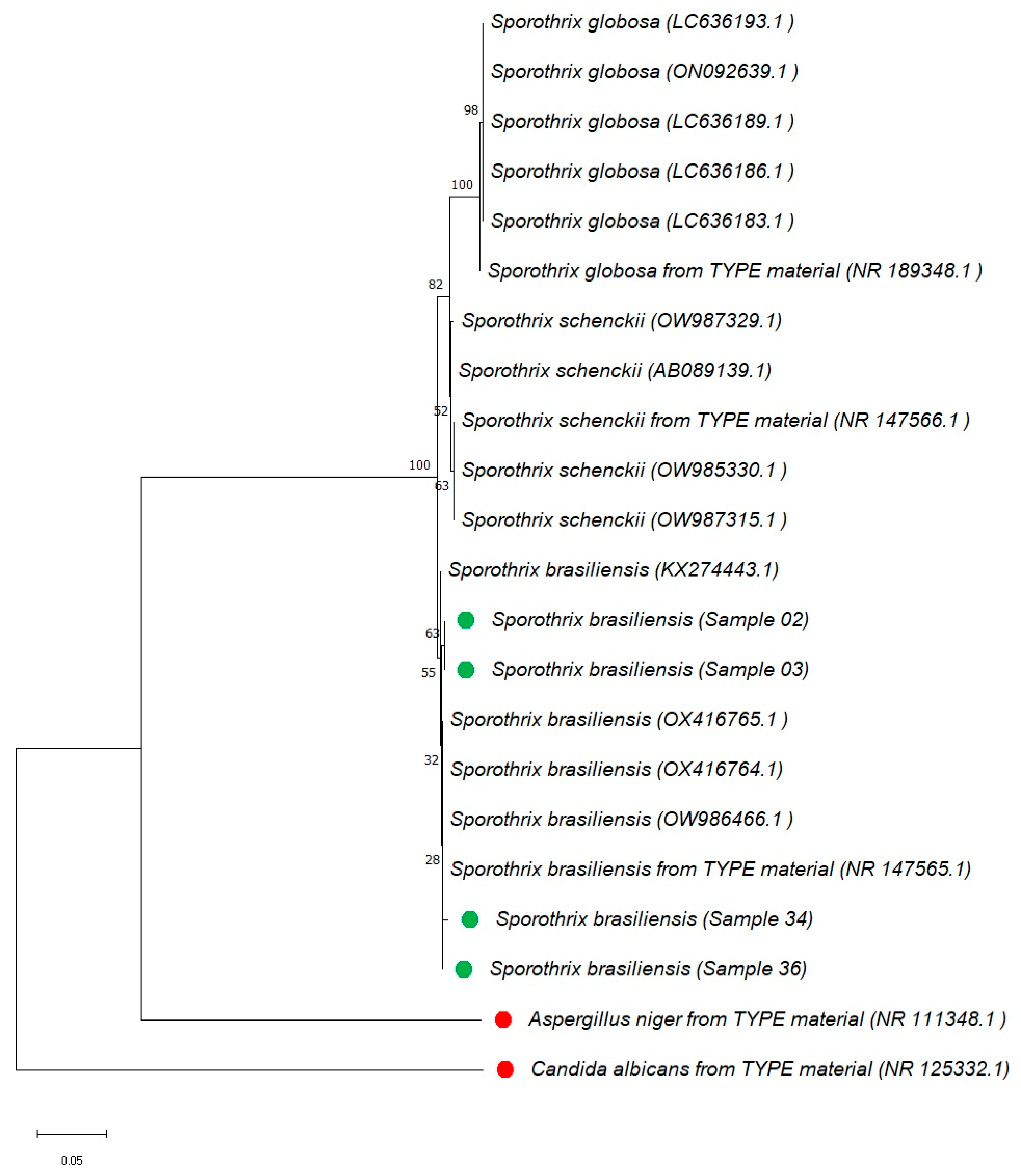
The fourth objective of this study was to confirm the phylogenetic identity of four feline Sporothrix isolates from Manaus using internal transcribed spacer (ITS)-based analysis. Although more isolates were initially processed (n = 29), in our described experimental conditions, only four produced ITS amplicons of sufficient quality for sequencing. The ITS region was selected based on the broad acceptance of ITS for species-level identification of Sporothrix spp. [8]. The neighbor-joining tree was constructed in MEGA X (version 10) with 1000 bootstrap replicates and is shown in Figure 3. All four isolates are grouped within the S. brasiliensis isolates.
The analysis included ITS sequences of reference strains of S. brasiliensis, S. schenckii, and S. globosa obtained from the NCBI database. All four regional isolates from the Amazon are clustered within the S. brasiliensis cluster, along with the reference strains. The isolates used as outgroups, Aspergillus niger ATCC 16888 and Candida albicans CBS 562, are grouped separately and are highlighted in red in Figure 3. The regional isolates analyzed in this study are shown in green. All sequences used in this analysis are publicly available in the NCBI database (https://www.ncbi.nlm.nih.gov/genbank/): Sample 36-PV991410; Sample 03-PV991411; Sample 34-PV991412; Sample 02-PV991379; accessed on 6 July 2025.

3.5. Antifungal Susceptibility Profile (MIC Determination)

To evaluate in vitro drug susceptibility, minimum inhibitory concentrations (MICs) were determined for 29 S. brasiliensis isolates using the CLSI broth microdilution method. The assays included amphotericin B, itraconazole, ketoconazole, and fluconazole. Results are summarized in Table 3. In short, antifungal susceptibility testing indicated low MIC values for itraconazole and ketoconazole, and consistently high MICs for fluconazole. All isolates exhibited MIC values of 64 µg/mL for fluconazole, without variation observed among tested samples.

4. Discussion

This study presents clinical, epidemiological, and molecular data on feline sporotrichosis cases in the Amazon region, contributing to diagnostic standardization. Clinically, most affected cats were young males exhibiting ulcerative and nodular lesions, frequently on the nasal planum, face, and limbs, with either localized or disseminated distribution. The phenol–chloroform extraction method yielded the highest DNA quality and concentration across isolates. Minimum DNA concentrations for successful amplification were defined using pan-fungal primer pairs ITS1ITS4, NL1NL4, and ITS5NL4. Although ITS-based PCR-RFLP showed limitations in species discrimination, ITS sequencing enabled the identification of Sporothrix brasiliensis in feline isolates. This represents one of the first regional applications of ITS sequencing in the Amazon. Preliminary antifungal susceptibility data indicated low MIC values for itraconazole.
The predominance of young (1–3 years; 86.2%) male (82.7%) cats exhibiting territorial or free-roaming behavior observed in this study aligns with the epidemiological profile described in other endemic regions. Gremião et al. [5], studying feline sporotrichosis in Rio de Janeiro, reported that most affected animals were young, outdoor-dwelling, and aggressive males, frequently engaged in fighting, which facilitates transmission through scratches and bites. Similarly, Etchecopaz et al. [20], analyzing emerging cases in Argentina, highlighted the role of male territorial behavior in the dissemination of S. brasiliensis. Rodrigues et al. [2] further emphasized that lesions commonly affect the head, face, limbs, and tail—areas associated with combat wounds—supporting the behavioral hypothesis of transmission. In our cohort, the frequent involvement of the nasal planum, forelimbs, and ears reinforces this pattern, suggesting that behavioral traits and anatomical exposure contribute significantly to disease progression and lesion distribution.
Regarding DNA extraction, our findings demonstrated superior efficiency of the phenol–chloroform method, yielding significantly higher DNA concentrations and purity compared to Silica Column and Acetate Precipitation methods. These results are corroborated by previous studies [16,21], which also highlighted the effectiveness of phenol–chloroform in removing proteins and contaminants. The consistent purity values (260/280 ratio between 1.6 and 2.0) obtained confirmed the method’s suitability for nucleic acid amplification-based assays. Nonetheless, the toxic nature of phenol–chloroform necessitates careful handling, suggesting safer alternatives, such as silica columns. Despite their slightly lower DNA yield, such columns could be more practical in clinical diagnostic routine.
In silico PCR-RFLP analyses showed that commonly used restriction enzymes like HaeIII, MspI, HinfI, and DdeI produced highly similar fragment patterns among Sporothrix species, with limited fragment size differences (less than 20 bp). This finding aligns with earlier studies [4,22,23], emphasizing PCR-RFLP’s limitations due to minor fragment size variations, making this approach insufficient for reliable species differentiation in practical scenarios. Genes such as β-tubulin and calmodulin have demonstrated greater accuracy in differentiating Sporothrix species [4,22,23] and could be integrated in research workflows.
Phylogenetic analysis based on the internal transcribed spacer (ITS) region was used to confirm the molecular identity of Sporothrix isolates recovered from feline cases in the Amazon region. Although multi-locus sequencing schemes have been proposed to improve resolution within the Sporothrix complex, several studies continue to support the discriminatory potential of the ITS region. Rodrigues et al. [24] and Zhou et al. [8] highlighted its ability to separate the main clinically relevant species, such as S. brasiliensis, S. schenckii, and S. globosa, using ITS-based phylogenies. More recently, Carvalho et al. [25] reinforced its practical application by demonstrating ITS clustering consistency with species assignment. In this study, all four feline isolates analyzed showed high-quality ITS amplification and sequencing, clustering consistently within the S. brasiliensis clade. The limited number of sequences obtained (4/29 isolates) is primarily attributable to difficulties in ITS amplification in approximately 70% of the samples and to the suboptimal quality of extracted DNA in others. These findings are in line with previous reports of the predominance of S. brasiliensis as the primary etiological agent in feline sporotrichosis across Brazil [4,25,26,27]. The widespread use of ITS in diagnostic laboratories, coupled with ITS representation in public databases, supports the continued relevance of ITS as a phylogenetic marker for initial species delimitation in Sporothrix infections.
Antifungal susceptibility testing of S. brasiliensis isolates showed MIC values consistent with previous studies. The geometric mean MIC for itraconazole (0.57 µg/mL) and ketoconazole (0.25 µg/mL) were comparable to those previously reported [6,28]. Amphotericin B assessment even showed higher MICs (7.27 µg/mL) compared to the results published by Etchecopaz et al. [20]. Fluconazole consistently exhibited limited activity (64 µg/mL), aligning with previously documented resistance profiles [2,6]. These findings highlight regional consistency of low itraconazole MIC values.
Sporotrichosis is an emerging fungal infection which has been a cause of growing concern in Brazil since the late 1990s. Now, it represents the most prevalent and most widely distributed implantation mycosis globally. In Manaus, Amazonas State, surveillance data recorded 4301 confirmed cases between August 2020 and December 2023, including 3403 animal cases (99.6% in cats) and 898 human cases, with cases reported across all districts of the city. During this period, human cases doubled and animal cases quadrupled, with a clear spatial association between animal and human cases [29]. These findings highlight an urgent need for coordinated veterinary and municipal monitoring programs to enable early detection, improve epidemiological mapping, and support targeted interventions to reduce transmission to humans. Although transmission of S. brasiliensis in Manaus is primarily linked to direct contact with infected cats, the possibility of indirect or nosocomial transmission should also be further investigated. Recent reports highlight environmental contamination in veterinary clinics and fomites as potential sources of infection, suggesting a need for strict biosafety measures [6].
This study presents several limitations that should be acknowledged. First, the relatively small number of analyzed isolates restricts the generalizability of the findings and may not fully represent the genetic variability of Sporothrix spp. in the Amazon region. Second, although the phenol–chloroform method proved effective for DNA extraction, its known toxicity limits its applicability in routine clinical settings, highlighting a need for safer alternatives. Third, DNA quality assessment was based solely on yield and purity. Further analyses focusing on DNA integrity and amplification efficiency would strengthen the conclusions. Additionally, only the ITS region was used for molecular identification, which, despite its broad use and database representation, has demonstrated limitations in discriminating closely related species within the Sporothrix complex. The absence of multi-locus approaches using markers such as β-tubulin or calmodulin may have constrained the phylogenetic resolution.

5. Conclusions

This study contributed to the assessment of the ongoing feline sporotrichosis outbreak in the Brazilian Amazon and confirmed S. brasiliensis as the predominant species involved. In particular, it provided details on clinical and epidemiological features of the affected cats, supporting available case definitions. The study’s contributions to the standardization of molecular diagnostic approaches may support regional species identification. In detail, the phenol–chloroform method demonstrated the best DNA extraction efficiency in the conducted comparison, and the ITS1ITS4 primers were adequate for PCR detection. Although PCR-RFLP showed limited discriminatory power within the Sporothrix genus, phylogenetic analysis based on ITS sequences allowed species identification by clustering within the S. brasiliensis clade. These findings may contribute to the standardization of the molecular diagnosis of S. brasiliensis as well as to the epidemiological surveillance of sporotrichosis in endemic regions, supporting optimized disease prevention and control strategies.

Author Contributions

Data curation, N.F.L.-D., D.B.d.A., F.d.S.F. and A.O.d.S.Q.; formal analysis, N.F.L.-D., D.B.d.A., F.d.S.F., A.O.d.S.Q. and J.V.B.d.S.; investigation, N.F.L.-D., D.B.d.A., F.d.S.F. and J.V.B.d.S.; resources, A.O.d.S.Q.; methodology, H.F. and J.V.B.d.S.; supervision, H.F. and J.V.B.d.S.; visualization, H.F. and J.V.B.d.S.; writing—original draft preparation, H.F. and J.V.B.d.S.; writing—review and editing, É.S.d.S., K.S.C., A.B.J.M. and H.F.; project administration, É.S.d.S., K.S.C., A.B.J.M. and J.V.B.d.S.; funding acquisition, É.S.d.S., K.S.C., A.B.J.M. and J.V.B.d.S. All authors have read and agreed to the published version of the manuscript.

Funding

This research was funded by the Fundação de Amparo à Pesquisa do Estado do Amazonas (FAPEAM) through the following grants: (1) POSGRAD/UEA 2021—Support for Naira Sulany Oliveira de Sousa; (2) PPSUS—Research Program for the Unified Health System (Notice No. 001/2017); (3) CT&I Productivity—Call for Research Productivity in Science, Technology, and Innovation (Notice No. 013/2022); (4) UNIVERSAL AMAZONAS—Celebrating 20 years of FAPEAM (Notice No. 001/2023); and (5) 003/2022-JOVENS DOUTORES.

Institutional Review Board Statement

All procedures involving animals were conducted following ethical standards and were approved by the Ethics Committee on Animal Use of the Instituto Nacional de Pesquisas da Amazônia (INPA) under protocol number CEUA/INPA 017/2023 on 10 April 2023. All clinical procedures, including lesion sampling, were performed under veterinary supervision, following local animal welfare regulations.

Informed Consent Statement

Written informed consent was obtained from the owners of all animals included in the study.

Data Availability Statement

The data presented in this study are available on request from the corresponding author. The ITS sequences of S. brasiliensis obtained from feline clinical isolates have been deposited in the NCBI GenBank database (https://www.ncbi.nlm.nih.gov/genbank/) under the following accession numbers: Sample 36-PV991410; Sample 03-PV991411; Sample 34-PV991412; Sample 02-PV991379, accessed on 6 July 2025.

Acknowledgments

The authors would like to thank the Fundação de Amparo à Pesquisa do Estado do Amazonas (FAPEAM), the Coordenação de Aperfeiçoamento de Pessoal de Nível Superior (CAPES), and the Conselho Nacional de Desenvolvimento Científico e Tecnológico (CNPq) for their support. Special thanks go to Ana Cláudia Cortez (INPA), Evelyn Cesar Campelo (FVS-AM), Carla Silvana da Silva Santos (FMT-HVD), Alex Panizza Jalkh (FMT-HVD), Maria Eduarda Grisolia (ILMD Fiocruz Amazônia), and Marla Jalene Alves (ILMD Fiocruz Amazônia) for their valuable contributions. We are also grateful to all cat owners who consented to participate in this study.

Conflicts of Interest

The authors declare no conflicts of interest.

Abbreviations

The following abbreviations are used in this manuscript:
ITSInternal Transcribed Spacer
SDSSodium Dodecyl Sulfate
rpmRevolutions Per Minute
AMBAmphotericin B
ITRItraconazole
CTZClotrimazole
RPMIRoswell Park Memorial Institute (medium)
MOPS3-(N-morpholino)propanesulfonic acid
bpBase Pairs

References

  1. Gremião, I.D.F.; Marques, M.; Oliveira, E.; de Miranda, L.H.M.; Freitas, D.F.S.; Pereira, S.A. Geographic Expansion of Sporotrichosis, Brazil. Emerg. Infect. Dis. 2020, 26, 621. [Google Scholar] [CrossRef]
  2. Rodrigues, A.M.; Hagen, F.; de Camargo, Z.P. A Spotlight on Sporothrix and Sporotrichosis. Mycopathologia 2022, 187, 407–411. [Google Scholar] [CrossRef]
  3. Mesquita, V.A.; Talhari, S.; Leturiondo, A.L.; de Souza, G.C.; de Brito, E.M.; de Andrade, S.L.; de Fernandes, D.C.L.; Frota, M.Z.M.; Cruz, R.C.d.S.; Guimarães, J.D.A.R.; et al. Zoonotic Sporotrichosis Outbreak: Emerging Public Health Threat in the Amazon State, Brazil. PLoS Negl. Trop. Dis. 2024, 18, e0012328. [Google Scholar] [CrossRef]
  4. Rodrigues, A.M.; Della Terra, P.P.; Gremião, I.D.; Pereira, S.A.; Orofino-Costa, R.; de Camargo, Z.P. The Threat of Emerging and Re-Emerging Pathogenic Sporothrix Species. Mycopathologia 2020, 185, 813–842. [Google Scholar] [CrossRef]
  5. Boechat, J.S.; Oliveira, M.M.E.; Gremião, I.D.F.; Almeida-Paes, R.; de Machado, A.C.S.; Zancopé-Oliveira, R.M.; de Oliveira, R.V.C.; Morgado, D.S.; Corrêa, M.L.; Figueiredo, A.B.F.; et al. Sporothrix brasiliensis and Feline Sporotrichosis in the Metropolitan Region of Rio de Janeiro, Brazil (1998–2018). J. Fungi 2022, 8, 749. [Google Scholar] [CrossRef] [PubMed]
  6. Dib, I.; Gremião, F.; Martins Da Silva Da Rocha, E.; Montenegro, H.; Aroldo, C.; Borges Carneiro, J.; Xavier, M.O.; Rodrigues De Farias, M.; Monti, F.; Mansho, W.; et al. Guideline for the Management of Feline Sporotrichosis Caused by Sporothrix brasiliensis and Literature Revision. Braz. J. Microbiol. 2021, 52, 107–124. [Google Scholar] [CrossRef]
  7. Maschio-Lima, T.; Domiciano, M.; Marques, R.; Thiago, H.L.; Lemes, H.; Seron Brizzotti-Mazuchi, N.; Maicon, H.C.; Caetano, H.; Gottardo De Almeida, B.; Letícia, M.B.; et al. Clinical and Epidemiological Aspects of Feline Sporotrichosis Caused by Sporothrix brasiliensis and in Vitro Antifungal Susceptibility. Vet. Res. Commun. 2021, 45, 171–179. [Google Scholar] [CrossRef] [PubMed]
  8. Zhou, X.; Rodrigues, A.M.; Feng, P.; De Hoog, G.S. Global ITS Diversity in the Sporothrix Schenckii Complex. Fungal Divers. 2014, 66, 153–165. [Google Scholar] [CrossRef]
  9. Rodrigues, A.M.; De Hoog, G.; Zhang, Y.; De Camargo, Z.P. Emerging Sporotrichosis Is Driven by Clonal and Recombinant Sporothrix Species. Emerg. Microbes Infect. 2014, 3, e32. [Google Scholar] [CrossRef]
  10. White, T.J.; Bruns, T.; Lee, S.; Taylor, J.W. Amplification and Direct Sequencing of Fungal Ribosomal RNA Genes for Phylogenetics. In PCR Protocols: A guide to Methods and Applications; Innis, M.A., Gelfand, D.H., Sninsky, J.J.W.T.J., Eds.; Academic in Press: New York, NY, USA, 1990; pp. 315–322. [Google Scholar]
  11. do Prado, C.M.; Razzolini, E.; Santacruz, G.; Ojeda, L.; Geraldo, M.R.; Segovia, N.; Pereira Brunelli, J.; Vicente, V.A.; Svoboda, W.K.; Queiroz-Telles, F. First Cases of Feline Sporotrichosis Caused by Sporothrix brasiliensis in Paraguay. J. Fungi 2023, 9, 972. [Google Scholar] [CrossRef]
  12. Zhang, M.; Li, F.; Li, R.; Gong, J.; Zhao, F. Fast Diagnosis of Sporotrichosis Caused by Sporothrix Globosa, Sporothrix Schenckii, and Sporothrix brasiliensis Based on Multiplex Real-Time PCR. PLoS Negl. Trop. Dis. 2019, 13, e0007219. [Google Scholar] [CrossRef]
  13. Amadei, S.; Campos, J.; Bertão-Santos, A.; Frentzel, A.; Ávila, H.; Monti, F.S.; Farias, M.R. A Novel Approach for Feline Sporotrichosis Pathogen Detection Based on Loop-Mediated Isothermal Amplification. Vet. Dermatol. 2025, 36, 474–484. [Google Scholar] [CrossRef]
  14. Lacaz, C.; Porto, E.; Costa, J.; Heins-Vaccari, E.; Melo, N. Tratado de Micologia Médica; Sarvier: São Paulo, Brazil, 2002. [Google Scholar]
  15. Sidrim, J.; Rocha, M. Micologia Médica à Luz de Autores Conteporâneos; Guanaba Koogan: Rio de Janeiro, Brazil, 2004. [Google Scholar]
  16. Sambrook, J.; Russell, D.W. Purification of Nucleic Acids by Extraction with Phenol: Chloroform. Cold Spring Harb. Protoc. 2006, 2006, pdb-prot4455. [Google Scholar] [CrossRef]
  17. Vincze, T.; Posfai, J.; Roberts, R.J. NEBcutter: A Program to Cleave DNA with Restriction Enzymes. Nucleic Acids Res. 2003, 31, 3688. [Google Scholar] [CrossRef]
  18. Espinel-Ingroff, A.; Chowdhary, A.; Gonzalez, G.M.; Guinea, J.; Hagen, F.; Meis, J.F.; Iii, G.R.T.; Turnidgeh, J. Multicenter Study of Isavuconazole Mic Distributions and Epidemiological Cutoff Values for the Cryptococcus Neoformans-Cryptococcus Gattii Species Complex Using the Clsi M27-A3 Broth Microdilution Method. Antimicrob. Agents Chemother. 2015, 59, 666–668. [Google Scholar] [CrossRef]
  19. CLSI M27M44S; Performance Standards for Antifungal Susceptibility Testing of Yeasts. Clinical and Laboratory Standards Institute (CLSI): Wayne, PA, USA, 2022.
  20. Etchecopaz, A.; Toscanini, M.A.; Gisbert, A.; Mas, J.; Scarpa, M.; Iovannitti, C.A.; Bendezú, K.; Nusblat, A.D.; Iachini, R.; Cuestas, M.L. Sporothrix brasiliensis: A Review of an Emerging South American Fungal Pathogen, Its Related Disease, Presentation and Spread in Argentina. J. Fungi 2021, 7, 170. [Google Scholar] [CrossRef]
  21. Sampaio, I.D.L.; Freire, A.K.L.; Ogusko, M.M.; Salem, J.I.; De Souza, J.V.B. Selection and Optimization of PCR-Based Methods for the Detection of Histoplasma Capsulatum Var. Capsulatum. Rev. Iberoam. Micol. 2012, 29, 34–39. [Google Scholar] [CrossRef]
  22. Rodrigues, A.M.; de Hoog, G.S.; de Camargo, Z.P. Sporothrix Species Causing Outbreaks in Animals and Humans Driven by Animal–Animal Transmission. PLoS Pathog. 2016, 12, e1005638. [Google Scholar] [CrossRef] [PubMed]
  23. de Sousa, D.R.T.; da Silva Santos, C.S.; Wanke, B.; da Silva, R.M.; dos Santos, M.C.; Cruz, K.S.; Monte, R.L.; Nocker, A.; de Souza, J.V.B. PCR-RFLP as a Useful Tool for Diagnosis of Invasivemycoses in a Healthcare Facility in the North of Brazil. Electron. J. Biotechnol. 2015, 18, 231–235. [Google Scholar] [CrossRef]
  24. Rodrigues, A.M.; de Hoog, G.S.; de Camargo, Z.P. Molecular Diagnosis of Pathogenic Sporothrix Species. PLoS Negl. Trop. Dis. 2015, 9, e0004190. [Google Scholar] [CrossRef] [PubMed]
  25. de Carvalho, J.A.; Monteiro, R.C.; Hagen, F.; de Camargo, Z.P.; Rodrigues, A.M. Trends in Molecular Diagnostics and Genotyping Tools Applied for Emerging Sporothrix Species. J. Fungi 2022, 8, 809. [Google Scholar] [CrossRef] [PubMed]
  26. Gremião, I.D.F.; Miranda, L.H.M.; Reis, E.G.; Rodrigues, A.M.; Pereira, S.A. Zoonotic Epidemic of Sporotrichosis: Cat to Human Transmission. PLoS Pathog. 2017, 13, e1006077. [Google Scholar] [CrossRef] [PubMed]
  27. Naiff, R.D.; Barrett, T.V.; Naiff, M.D.F.; Ferreira, L.C.D.L.; Arias, J.R. New Records of Histoplasma Capsulatum from Wild Animals in the Brazilian Amazon. Rev. Inst. Med. Trop. São Paulo 1996, 38, 273–277. [Google Scholar] [CrossRef] [PubMed]
  28. Waller, S.B.; Dalla Lana, D.F.; Quatrin, P.M.; Ferreira, M.R.A.; Fuentefria, A.M.; Mezzari, A. Antifungal Resistance on Sporothrix Species: An Overview. Clin. Microbiol. Rev. 2021, 52, 73–80. [Google Scholar] [CrossRef]
  29. Valente, R.M.; de Verçosa, J.V.M.; de Souza, E.R.; Gordiano, N.P.; Barroso, L.d.C.; Carvalho, S.M.d.S.; Alves, M.J.; Fonseca, F.R.; Grisolia, M.E.; de Almeida, M.E.M.; et al. Expansion of Human and Animal Sporotrichosis in Manaus, Amazonas State, Brazil. Cad. Saude Publica 2025, 41, e00180024. [Google Scholar] [CrossRef]
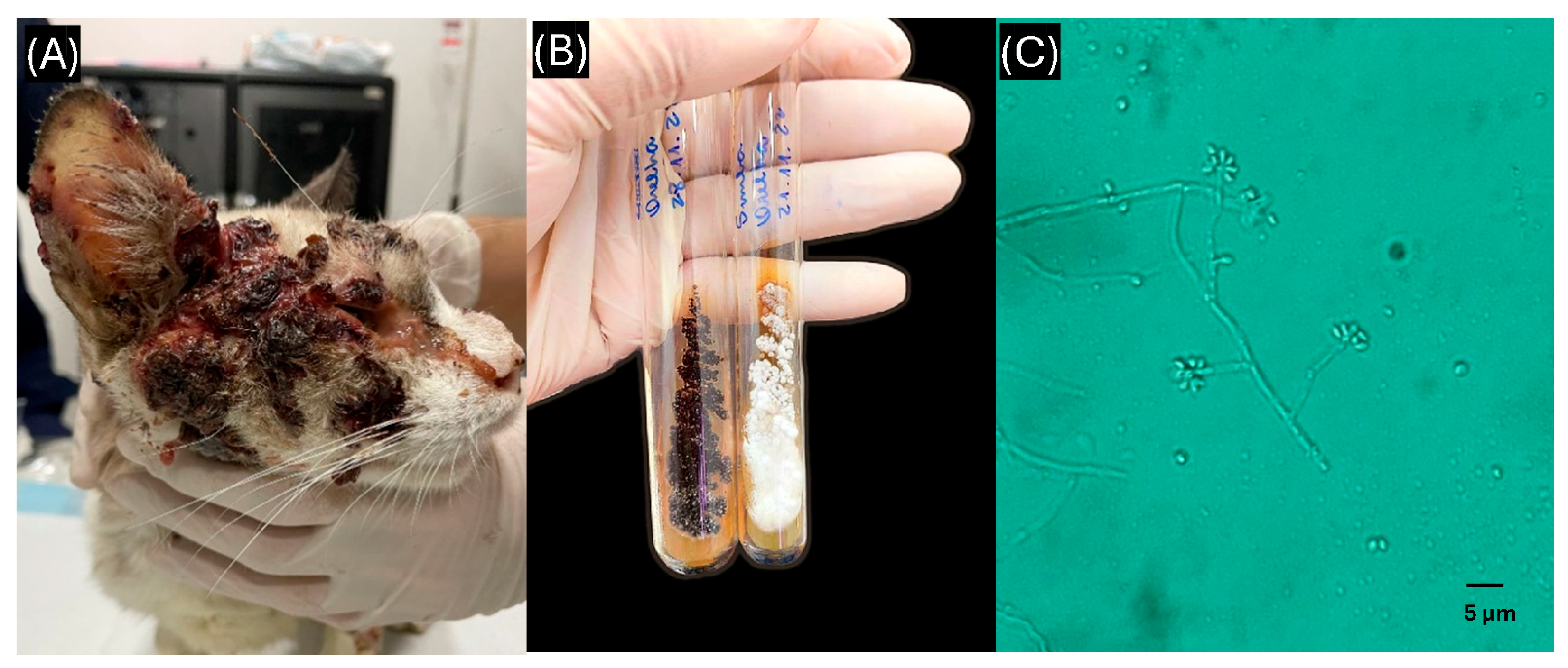
Figure 1. Clinical lesion, fungal culture, and microscopic morphology of Sporothrix brasiliensis isolated from a feline case. (A) Feline with a lesion on the lateral facial region showing cutaneous involvement with dissemination to the ocular area, ear, and nasal planum swelling; (B) colony of S. brasiliensis grown on Sabouraud agar after incubation, obtained from a sample collected from the ocular and ear regions; (C) microscopic morphology of S. brasiliensis displaying the characteristic “daisy-like” arrangement, observed at 400× magnification.
Animals 15 02318 g001
Figure 2. Comparative evaluation of DNA extraction methods applied to fungal isolates of Sporothrix schenckii ATCC CFP-00746, Sporothrix brasiliensis ATCC MYA-4606, a regional Sporothrix isolate (Sample 2), Candida albicans ATCC 10231, and Aspergillus niger ATCC 16404. (A) Mean DNA concentration (ng/µL) obtained using the phenol–chloroform, Silica Column, and Acetate Precipitation methods. (B) Mean DNA purity, expressed as the 260/280 nm absorbance ratio. Error bars represent the standard deviation from measurements performed in triplicate.
Animals 15 02318 g002
Figure 3. Phylogenetic analysis based on the ITS region using the neighbor-joining method with bootstrap support, performed applying the MEGA (Molecular Evolutionary Genetics Analysis) X software package. The tree was constructed using the internal transcribed spacer (ITS) molecular marker, based on the alignment of Sporothrix species sequences available in the NCBI database. Regional isolates from the Amazon region included in this study are shown in green. Aspergillus niger and Candida albicans were used as outgroup controls and are shown in red. All sequence data are available in the NCBI database.
Animals 15 02318 g003
Table 1. Epidemiological and clinical characteristics of domestic cats diagnosed with sporotrichosis in a veterinary clinic of spontaneous demand, Manaus, Brazilian Amazon.
Epidemiological and Clinical CharacteristicsSporotrichosis Positive Isolates (%)
Age
6 months1/29 (3.4%)
8 months1/29 (3.4%)
1 year6/29 (20.7%)
2 years14/29 (48.3%)
3 years5/29 (17.2%)
4 years1/29 (3.4%)
7 years1/29 (3.4%)
Sex
Male24/29 (82.7%)
Female5/29 (17.2%)
Behavioral features
Indoor3/29 (10.3%)
Semi-indoor13/29 (44.8%)
Free-roaming13/29 (44.8%)
Clinical Manifestation
Localized lesions (lesions confined to a single anatomical region)16/29 (55.2%)
Disseminated lesions13/29 (44.8%)
Anatomical distribution of all reported lesions 1
Nasal planum10/43 (23.3%)
Face (excluding nasal planum, eyes and ears)8/43 (18.6%)
Paws (including forelimbs and hindlimbs)7/43 (16.3%)
Ribs and thoracic flank4/43 (9.3%)
Ears (auricle)3/43 (7.0%)
Tail3/43 (7.0%)
Eye and periocular region1/43 (2.3%)
Penile region1/43 (2.3%)
Post-surgical abdominal site1/43 (2.3%)
Dorsum (thoracolumbar)1/43 (2.3%)
Paw pads2/43 (4.7%)
Total number of lesion sites identified43/29 cats 2
1 Some cats presented lesions in more than one anatomical region. 2 The total number of lesion sites exceeds the number of animals due to multifocal skin involvement in 13/29 cats.
Table 2. In silico predicted fragment sizes (bp) from ITS region digestion with commercial restriction enzymes (HaeIII, MspI, HinfI, and DdeI) for five medically important fungal species: Sporothrix brasiliensis CBS 120339, Sporothrix schenckii CBS 359.36, Sporothrix globosa CBS 120340, Aspergillus niger ATCC 16888, and Candida albicans CBS 562. Analyses were performed using the NEBcutter V3.0 tool based on amplified ITS sequences, with fragment sizes expressed in base pairs (bp), aiming at species differentiation by PCR-RFLP.
EnzymeS. brasiliensis CBS 120339S. schenckii CBS 359.36S. globosa CBS 120340A. niger ATCC 16888C. albicans CBS 562
HaeIII (GG/CC)82, 33, 276, 21, 11681, 33, 276, 21, 11497, 33, 277, 21, 11347, 45, 15, 29, 291, 73, 7669, 426
MspI (C/CGG)463, 65462, 63479, 62111, 21, 15, 93, 101, 42, 32, 86, 75276, 219
HinfI (G/ANTC)130, 60, 16, 64, 258129, 61, 16, 64, 255145, 63, 16, 64, 25319, 208, 16, 56, 248, 29162, 16, 56, 8, 137, 116
DdeI (C/TNAG)154, 374153, 372169, 372187, 389397, 98
BsaI (G/TAC)22, 415, 9122, 414, 8941, 412, 8873, 503495
Table 3. Minimum inhibitory concentrations (MICs) of antifungal agents against Sporothrix brasiliensis isolates obtained from feline clinical cases in veterinary clinics in Manaus (Brazilian Amazon).
Antifungal AgentMIC Range (µg/mL)Geometric Mean (µg/mL)
Ketoconazole0.125–0.50.25
Itraconazole0.25–10.57
Amphotericin B4–87.27
Fluconazole6464
Disclaimer/Publisher’s Note: The statements, opinions and data contained in all publications are solely those of the individual author(s) and contributor(s) and not of MDPI and/or the editor(s). MDPI and/or the editor(s) disclaim responsibility for any injury to people or property resulting from any ideas, methods, instructions or products referred to in the content.