Article

The Role of Gonadotropins and Growth Factor in Regulating Ras During Maturation in Cumulus–Oocyte Complexes of Pigs

1
College of Animal Life Sciences, Kangwon National University, Chuncheon 24341, Republic of Korea
2
College of Veterinary Sciences, Kangwon National University, Chuncheon 24341, Republic of Korea
3
School of ICT, University of Tasmania, Hobart, TAS 7005, Australia
*
Author to whom correspondence should be addressed.
Animals 2025, 15(14), 2100; https://doi.org/10.3390/ani15142100
Submission received: 11 June 2025 / Revised: 6 July 2025 / Accepted: 14 July 2025 / Published: 16 July 2025
(This article belongs to the Special Issue Health of the Ovaries, Uterus, and Mammary Glands in Animals)

Simple Summary

Some studies have indicated that Ras affects follicular differentiation into the corpus luteum post-ovulation and early embryonic development, but studies on Ras in porcine immature oocytes are limited. In this study, we determined the changes in Ras and its GTPases during maturation of porcine cumulus–oocyte complexes (COCs) following treatment with follicle-stimulating hormone (FSH), luteinizing hormone (LH), and epidermal growth factor (EGF). We demonstrated that gonadotropins and growth factors that induce COC maturation promote cumulus cell proliferation and meiotic resumption in oocytes via Ras-mediated signaling pathways.

Abstract

Oocytes and cumulus cells undergo meiotic resumption and proliferation via gonadotropins and growth factors during maturation, and various small G proteins are activated when COCs undergo physiological changes. This study investigated the influence of gonadotropins and growth factors on Ras and its GTPases during porcine COC maturation. Unmatured COCs were treated with FSH, LH, or EGF for 44 h. The mRNA expression levels of the Ras subfamily (H-Ras, K-Ras, N-Ras, and R-Ras), its GTPases (RASA1 and SOS1), and proliferation factors (ERK, CCNB1, and Cdc2) were analyzed using RT-PCR. In contrast to other Ras subfamilies, R-Ras expression is upregulated during COC maturation. We evaluated the effects of FSH, LH, and EGF at various concentrations that most effectively regulated the expression of R-Ras and GTPases. The results demonstrated that 0.5 µg/mL FSH, 10 IU/mL human chorionic gonadotropin (hCG), and 10 ng/mL EGF effectively enhanced R-Ras expression and cell proliferation. FSH supplementation during porcine COC maturation significantly upregulated R-Ras and ERK expression, independent of LH and EGF, and downregulated Cdc2 expression. These results indicated that FSH regulates R-Ras expression, thereby promoting cell proliferation during COC maturation. These results provide fundamental knowledge for understanding the role of Ras and its family members in the development of follicular environments in pigs.

1. Introduction

Oocytes within the follicles exist in an immature state and are stimulated to mature by various gonadotropins and growth factors, ultimately leading to ovulation [1,2]. At this stage, the oocyte does not exist independently but as a cumulus–oocyte complex (COC) [3,4]. This complex facilitates communication between the oocyte and cumulus cells (CCs) by directly affecting gene expression and protein synthesis, leading to the differentiation and expansion of CCs as well as oocyte maturation [5]. Follicle-stimulating hormone (FSH) promotes the development of immature follicles, while luteinizing hormone (LH) resumes the arrested meiosis of oocytes, leading to their maturation into mature oocytes [6,7]. Furthermore, gonadotropins induce meiosis in arrested oocytes as well as stimulate ovarian cells such as the theca, granulosa, and cumulus cells to produce epidermal growth factor (EGF) [8,9]. The complex of EGF and the EGF receptor (EGFR) induces the proliferation of cumulus cells through the activation of the MAPK/ERK signaling pathway [10,11]. This promotes the rapid proliferation of immature COCs during maturation, along with notable morphological changes, as well as indirectly supporting cumulus cell expansion and nuclear maturation of oocytes [12,13]. Generally, the cell doubling time is 18 h in cumulus cells, and rapid mitosis is maintained for 36–40 h before ovulation in pigs [14]. This specific physiological phenomenon occurs only in reproductive somatic cells with gonadotropin receptors, and their cell signaling differs from that of other somatic cells.
Ras (rat sarcoma virus) is a guanosine triphosphate (GTP)-binding protein involved in various cellular signaling pathways such as cell proliferation, survival, molecular transport, and differentiation [15,16]. The small family of Ras proteins includes H-Ras, K-Ras, N-Ras, and R-Ras, all of which are important in intracellular signaling, trafficking, proliferation, and survival [17,18]. Improper expression of these proteins can lead to cellular pathological conditions. In practice, approximately 27% of all cancers are attributed to the inappropriate expression of Ras proteins [19,20]. Ras functions as a cellular molecular switch, transitioning between its active form, Ras-GTP, and its inactive form, Ras-guanosine diphosphate (GDP), and regulates various cellular processes [17,21]. When the inactive form, Ras-GDP, transitions to the active form, Ras-GTP, Ras is facilitated by the exchange of GDP for GTP by a protein called the guanine nucleotide exchange factor (GEF). Conversely, Ras-GTP is hydrolyzed to Ras-GDP with the assistance of Ras-GTPase-activating protein (GAP) [17,22]. Therefore, disruption in the regulation of Ras activity/inactivity by GEF and GAP can result in negative implications for cellular pathology. For example, if Ras remains persistently activated owing to the overexpression of Ras GEF or the loss of function of Ras GAP, cellular carcinogenesis can be induced [23]. Ras is a well-known oncogene, and several studies have focused on tumorigenesis caused by Ras mutations [24,25]. However, studies on Ras expression in oocytes and ovaries are limited. Although some studies have indicated that Ras affects follicular differentiation into corpus luteum post-ovulation [26] and early embryonic development [27], studies on Ras in porcine immature oocytes are limited.
We hypothesized that Ras and its regulators (GEFs and GAPs) would be involved in rapid proliferation during the maturation period of porcine COCs and that the interaction between G-protein coupled receptors and gonadotropins or growth factors involved in COC maturation could potentially activate Ras and Ras GTPase. Therefore, we determined the changes in Ras and its GTPases during porcine COC maturation and investigated the influence of FSH, LH, and EGF on Ras signaling and proliferation-related factors during porcine COC maturation.

2. Materials and Methods

2.1. Collection of Porcine COCs

The porcine ovarian samples used in the experiment were collected from immature gilt in the slaughterhouse (Pochen farm, Pocheon, Republic of Korea) and transported to the laboratory in 0.9% saline solution at 38.5 °C within 2 h. Follicular fluid measuring 3–6 mm was aspirated using an 18-gauge needle after three washes with 0.85% saline solution. The COCs were washed three times with phosphate-buffered saline with polyvinyl alcohol (PBS-PVA), collected in the follicular fluid under a stereo microscope (SZX16, Olympus Tokyo, Japan), and COCs (50–60 per sample and a total of six replication) were cultured in 650 µL tissue culture medium 199 (TCM-199; Invitrogen, Carlsbad, CA, USA) with 0.1% PVA (Sigma, St. Louis, MO, USA), 0.57 mM cysteine (Sigma), 10 ng/mL EGF (Sigma), 10% porcine follicular fluid (pFF), 0.5 µg/mL FSH (Sigma), and 10 IU/mL human chorionic gonadotropin (hCG; Intervet, The Netherlands) for 22 h at 38.5 °C in 5.0% CO2. The collected COCs exhibiting a homogenous ooplasm and 3 to 4 compact layers of cumulus cells were selected for IVM [28]. The culture medium was replaced with a hormone-free TCM-199 culture medium, and further incubation was conducted for an additional 22 h at 38.5 °C in a 5.0% CO2 environment [29]. The degree of cumulus expansion in COCs was assessed based on their morphological characteristics at 0, 22, and 44 h after the initiation of in vitro maturation (IVM), and the area of COCs was quantitatively measured using ImageJ 1.54p software (NCBI, Bethesda, MD, USA). The relative mRNA expression levels were normalized in the 0 h treatment group.

2.2. Treatment with FSH, LH, and EGF

The COCs were collected and washed three times with PBS-PVA before being transferred to a fresh culture medium consisting of TCM-199 supplemented with 1.0% pFF. The COCs that exhibited a homogenous ooplasm and 3 to 4 compact layers of cumulus cells were selected for IVM [28]. Each experiment was performed in four replicates. To assess the independent roles of FSH, LH, and EGF in COC (30–40 per sample and a total of four replications) maturation, the cultures were treated with varying concentrations of FSH (0.0, 0.1, 0.5, and 1.0 µg/mL), hCG (0.0, 1.0, 10, and 20 IU/mL), and EGF (0.0, 1.0, 10, and 20 ng/mL) for a total incubation period of 44 h. The minimum and maximum effective concentrations of these hormones and growth factors for porcine COC maturation were determined using previously established methods [30,31]. Subsequently, additional COCs were cultured in 500 µL of TCM-199 medium with FSH, hCG, and EGF at 38.5 °C in a 5.0% CO2 environment for 44 h.

2.3. Quantitative Reverse Transcription PCR

Total RNA was extracted from the COCs using an easy-spin Total RNA Extraction kit (iNtRON Biotechnology, Inc., Seongnam-si, Republic of Korea). The RNA concentration was measured using an EzDrop 1000C spectrophotometer (Blue-Ray Biotech, New Taipei City, Taiwan). Based on the measured RNA concentration, cDNA was synthesized using the PrimeScript™ 1st strand cDNA Synthesis Kit (Takara, Tokyo, Japan). PCR was performed using Veriti 96-well Thermal Cycler (Applied Biosystems, Waltham, MA, USA) with AccuPower® PCR PreMix & Master Mix (Bioneer, Seoul, Republic of Korea), following the manufacturer’s protocol. Gene expressions of gonadotropin and growth factor receptors (FSHR, LHR, and EGFR), Ras subfamily (H-Ras, K-Ras, N-Ras, and R-Ras), Ras GEFs (RasGEF-1B and SOS1), Ras GAPs (RasGAP and RASA1), and proliferation factors (ERK, CCNB1, and Cdc2) were analyzed according to the PCR conditions (Table 1). The amplified products were separated by electrophoresis on a 1.5% agarose gel containing RedSafe Nucleic Acid Staining Solution (iNtRON) at 100 V for 20 min and photographed under UV illumination. Relative mRNA expression levels were normalized to β-actin, and ImageJ software (NCBI) was used for image analysis. The primers are listed in Table 1.

2.4. Statistical Analysis

Each experiment was repeated at least thrice. Data are presented as mean ± standard error. Data were analyzed using t-test or one-way analysis of variance (ANOVA) followed by Tukey’s multiple comparison test, using SAS ver. 9.4 software. Differences were considered statistically significant at p < 0.05.

3. Results

3.1. Changes in Ras and Its Regulators During Maturation in Porcine COCs

At 0 h of maturation, cumulus cells formed a compact structure around the oocyte (Figure 1A). Subsequently, at 22 h of maturation, a notable increase in cumulus cell proliferation was observed (Figure 1B). Cumulus cells exhibited significant proliferation and expansion (Figure 1C) at 44 h of maturation. The expansion of cumulus cells significantly (p < 0.05) increased during the maturation of porcine COCs (Figure 1D). FSHR mRNA expression was significantly (p < 0.05) higher at 44 h than at 0 h (Figure 1E) of maturation in the COCs. The expression of LHR mRNA significantly (p < 0.05) increased at 22 and 44 h compared to that at 0 h of maturation; however, no significant difference was observed between 22 and 44 h (Figure 1E). Conversely, EGFR mRNA expression levels did not differ significantly throughout COC maturation (Figure 1E). Changes in the expression levels of the Ras family genes were measured according to the maturation time of COCs, as shown in Figure 1F,H. For R-Ras mRNA, a significant (p < 0.05) increase was observed in its expression as the maturation time (22 and 44 h) of porcine COCs progressed (Figure 1F). Conversely, no significant differences were observed in expression levels according to the maturation time of porcine COCs for H-Ras, K-Ras, and N-Ras mRNAs (Figure 1F). Additionally, the mRNA expression levels of RasGEF-1B and SOS1 (Ras GEF), RasGAP and RASA1 (Ras GAP), and ERK were measured during the maturation of porcine COCs (Figure 1G). During the maturation of porcine COCs, the mRNA expression of SOS1 remained unchanged, whereas that of RASA1 significantly decreased (p < 0.05) as maturation advanced (Figure 1G). ERK expression of ERK significantly increased (p < 0.05) as COC maturation progressed (Figure 1G).

3.2. Influences of FSH on the Expression of Ras and Its Regulator Genes in Porcine COCs

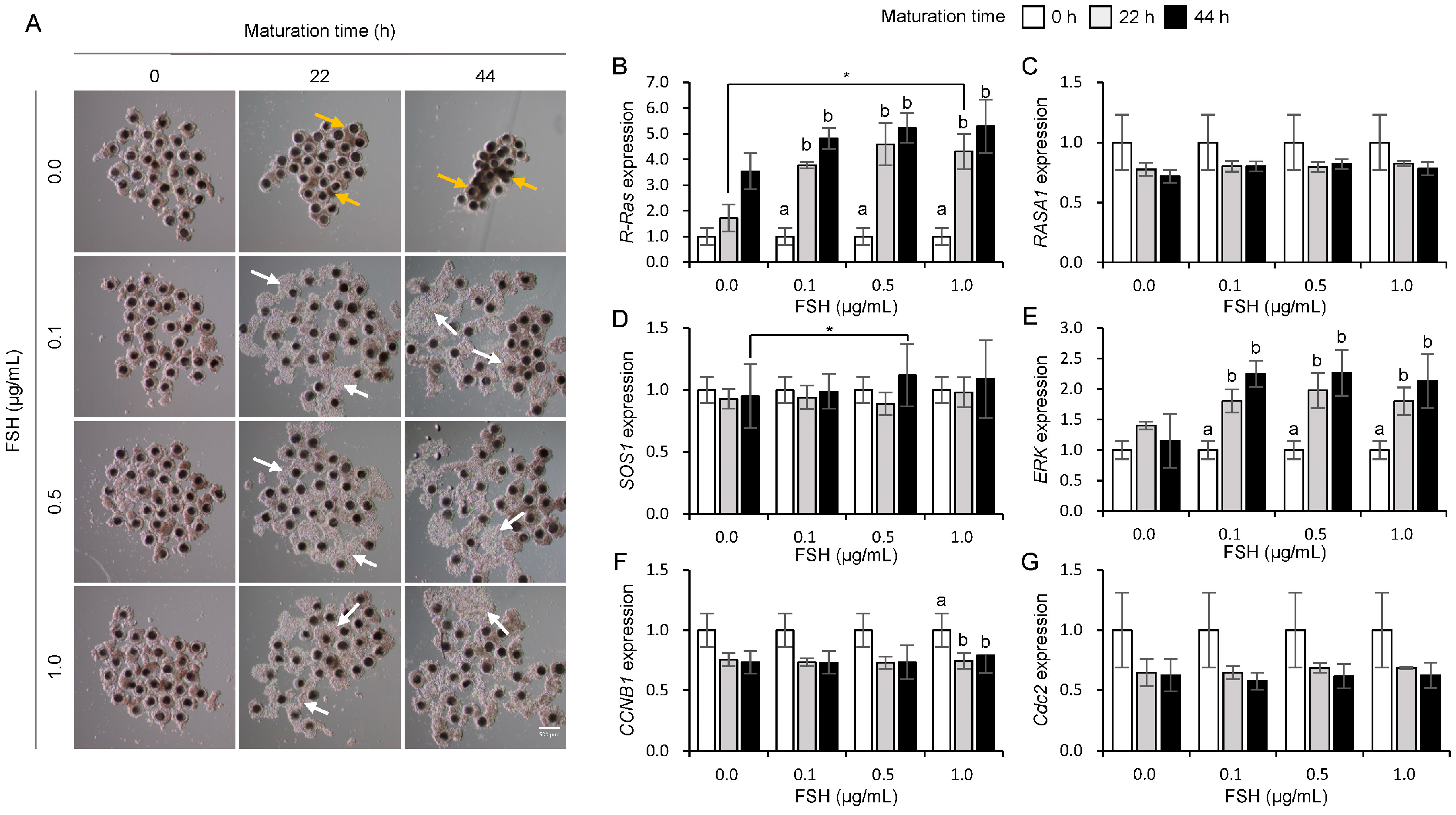
Figure 2A shows the morphological changes in COCs during maturation in response to FSH treatment. The size of cumulus cells increased in the 0.1, 0.5, and 1.0 µg/mL FSH treatment groups (Figure 2A, white arrows); however, the control group (0.0 µg/mL FSH, Figure 2A, yellow arrows) did not increase at 22 and 44 h of maturation in porcine COCs. At both 22 h and 44 h, R-Ras mRNA expression showed a concentration-dependent increase, with the 1.0 µg/mL FSH treatment resulting in a significant increase (p < 0.05) in R-Ras mRNA expression at 22 h of maturation in COCs (Figure 2B). However, RASA1 mRNA expression levels did not differ with the FSH concentration (Figure 2C). The SOS1 expression at 44 h of COC maturation significantly increased (p < 0.05) in the 0.5 µg/mL FSH treatment group compared to that in the 0.0 µg/mL FSH treatment group (Figure 2D). The ERK mRNA expression significantly increased (p < 0.05) at 22 and 44 h compared to that at 0 h when COCs were treated with 0.1, 0.5, and 1.0 µg/mL FSH (Figure 2E). The expression of CCNB1 mRNA significantly decreased (p < 0.05) at 22 and 44 h compared to that at 0 h in the 1.0 µg/mL FSH treatment group (Figure 2F). In contrast, no significant differences was observed in Cdc2 expression across the FSH treatment group (Figure 2G).

3.3. Influences of hCG on the Expression of Ras and Its Regulator Genes in Porcine COCs

Figure 3A shows the morphological changes observed in the COCs during maturation in response to hCG treatment. After 22 h of hCG treatment, most COCs in the 0 and 0.1 IU/mL groups exhibited a shrunken morphology (Figure 3A, yellow arrows). In contrast, the 10 and 20 IU/mL hCG-treated groups showed shrunken (Figure 3A, yellow arrows) and expanded (Figure 3A, white arrows) COCs. At 44 h of maturation, most COCs in the 0 IU/mL hCG group exhibited shrunken morphology (Figure 3A, yellow arrows). However, the 0.1, 10, and 20 IU/mL hCG groups showed both shrunken (Figure 3A, yellow arrows) and expanded (Figure 3A, white arrows) forms, with a greater proportion of expansion observed particularly in the 10 and 20 IU/mL groups. As shown in Figure 3B, no significant changes in R-Ras expression were observed across different hCG concentrations during COC maturation. However, RASA1 (Figure 3C) and SOS1 (Figure 3D) expression significantly (p < 0.05) decreased at 44 h of oocyte maturation in the 10 IU/mL hCG-treated group. Notably, at 44 h of maturation, RASA1 expression was significantly reduced in the 10 and 20 IU/mL hCG treatment groups compared to the 0.0 IU/mL control group (p < 0.05). A clear concentration-dependent increase in ERK mRNA expression was observed (Figure 3E), with the 10 and 20 IU/mL hCG groups showing significantly higher levels at 44 h than the 0.0 IU/mL group (p < 0.05). In contrast, CCNB1 (Figure 3F) and Cdc2 (Figure 3G) did not show significant changes in expression at different maturation stages after treatment with hCG alone.

3.4. Influences of EGF on the Expression of Ras and Its Regulator Genes in Porcine COCs

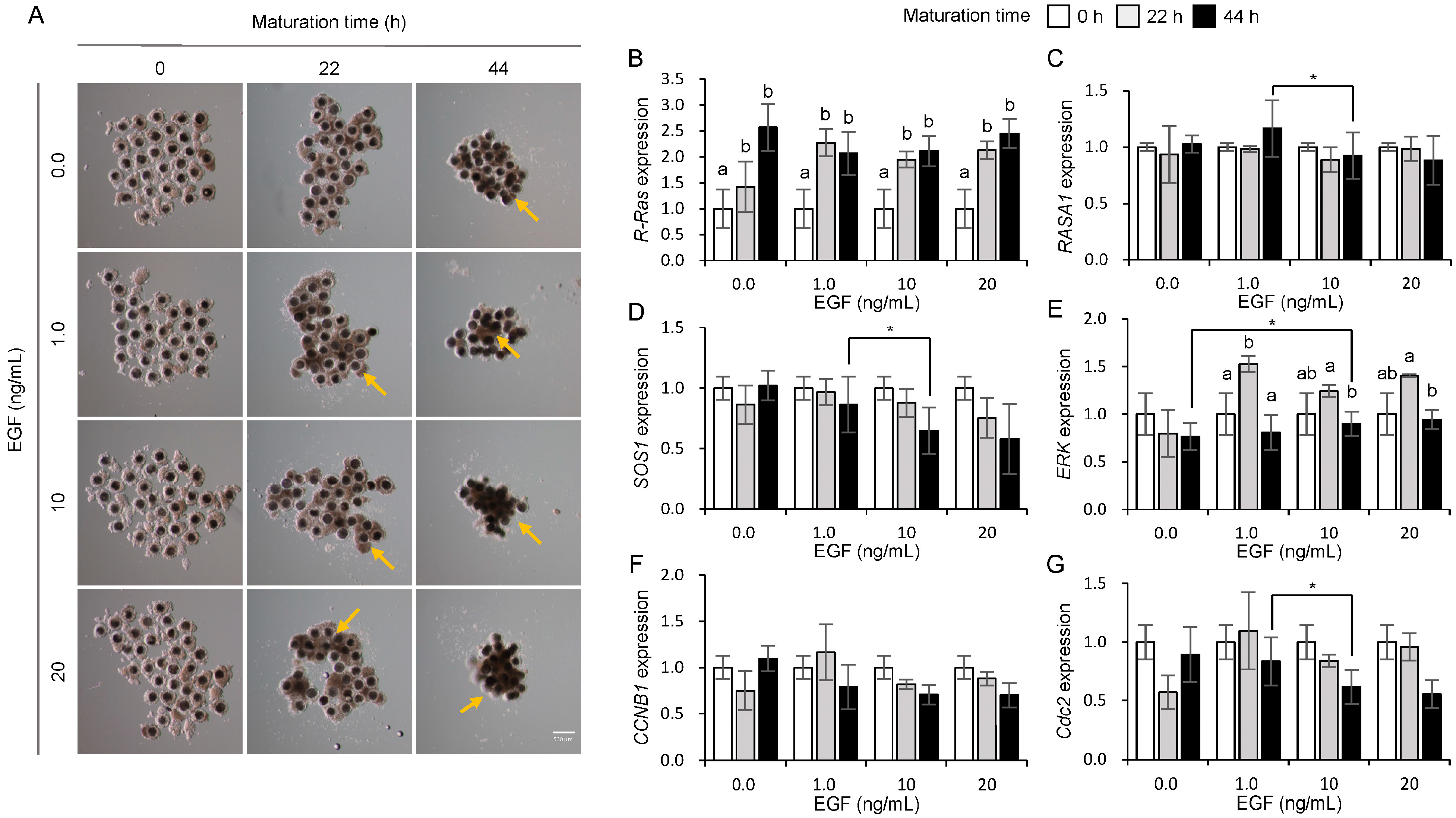
Figure 4A shows the morphological changes observed in the COCs during maturation in response to EGF treatment. A more compact and aggregated morphology (Figure 4A, yellow arrows) of COCs was observed in the 1.0, 10, and 20 ng/mL EGF treatment groups, whereas such morphological changes were not apparent in the control group (0.0 ng/mL EGF, Figure 4A) at 22 and 44 h of maturation in porcine COCs. As shown in Figure 4B, R-Ras expression significantly increased (p < 0.05) in the 1.0, 10, and 20 ng/mL EGF treatment groups at 22 and 44 h compared with that at 0 h (Figure 4B). However, Figure 4C,D indicate that the expression of RASA1 and SOS1 at 44 h significantly decreased (p < 0.05) in the group treated with 10 ng/mL EGF compared to the other groups. ERK (Figure 4E) expression significantly increased (p < 0.05) during the early phase (0–22 h) but decreased during the late phase (22–44 h) of oocyte maturation in the 1.0, 10, and 20 ng/mL EGF treatment groups, possibly as a regulatory mechanism to control excessive cell proliferation induced by R-Ras upregulation. In particular, a significant increase in ERK expression was observed at 44 h in the 10 ng/mL EGF-treated group compared to the 0.0 ng/mL group (p < 0.05). The expression of CCNB1 mRNA showed no significant difference (Figure 4F), whereas Cdc2 expression significantly decreased at 44 h in the 10 ng/mL EGF treatment group (Figure 4G).

3.5. Mechanism of Ras Regulation by Gonadotropins and EGF

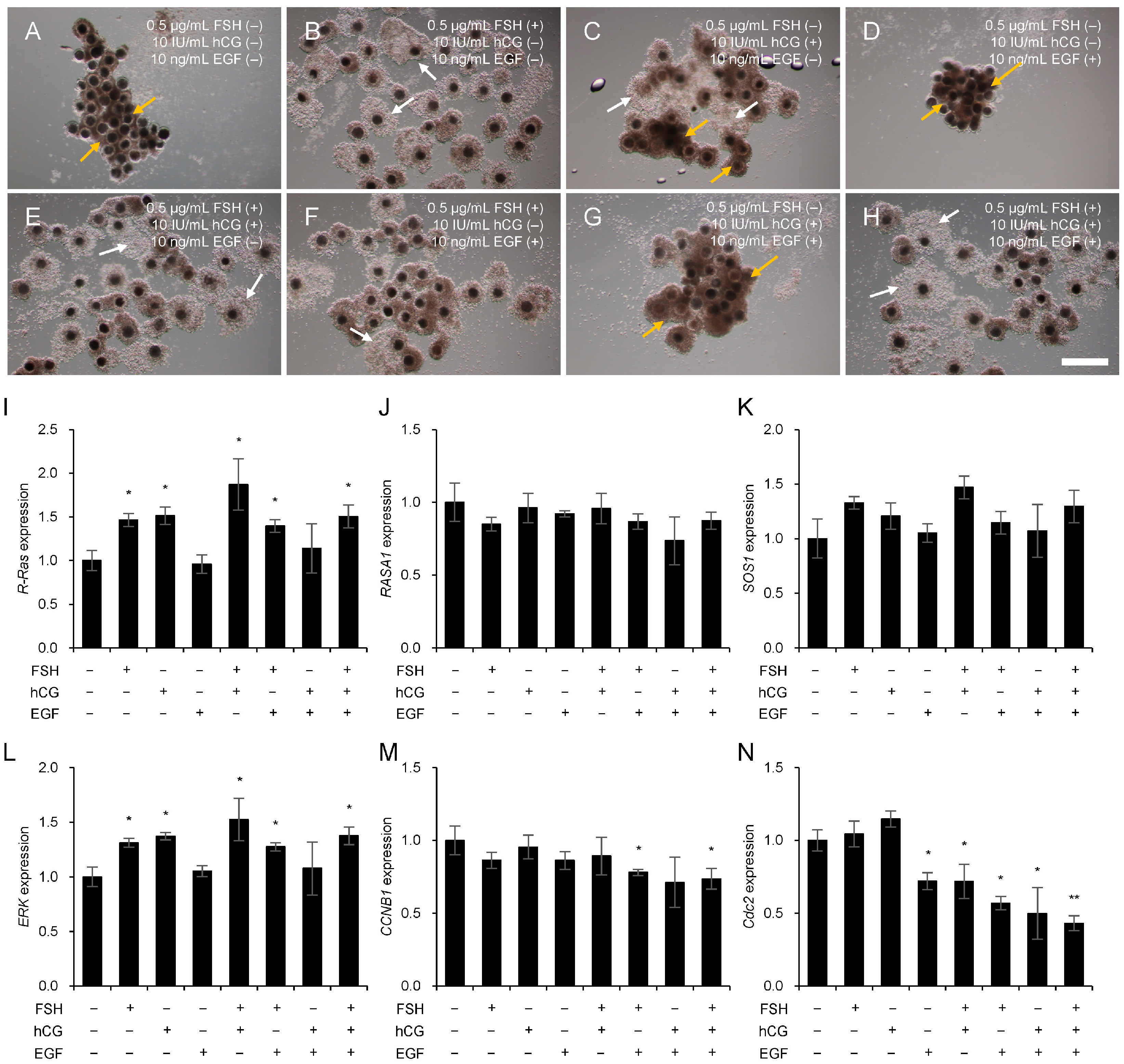
Based on the previous results, 0.5 µg/mL FSH, 10 IU/mL hCG, and 10 ng/mL EGF were used to determine changes in Ras and its regulator genes in porcine COCs. Figure 5 shows the expression changes in Ras, Ras GTPase regulators, and cell proliferation factors during porcine COC maturation, depending on the presence or absence of FSH, hCG, and EGF. Figure 5A shows the morphological changes in porcine COCs during maturation with or without gonadotropin and EGF treatment. Cumulus cell expansion was observed in treatment groups supplemented with FSH and hCG (Figure 5A, white arrows). In contrast, shrinkage of cumulus cells was observed in the EGF treatment group without FSH supplementation (Figure 5A, yellow arrows). R-Ras mRNA expression was significantly increased (p < 0.05) in the gonadotropin-treated groups, particularly in the presence of FSH (Figure 5B). In contrast, no significant changes were observed in RASA1 (Figure 5C) or SOS1 (Figure 5D) levels, regardless of gonadotropin or growth factor treatment. Similarly to R-Ras, ERK mRNA expression was significantly upregulated (p < 0.05) in the gonadotropin-treated groups, particularly in the presence of FSH (Figure 5E). The mRNA expression levels of CCNB1 (Figure 5F) and Cdc2 (Figure 5G) significantly decreased (p < 0.05) after the combined treatment with gonadotropins and growth factors.

4. Discussion

The Ras subfamily is the founding member of a large superfamily of small monomeric GTPases that regulate diverse cellular processes [32]. The transition between the active GTP-bound and inactive GDP-bound states of small GTPases is controlled by GEFs, which stimulate the exchange of GDP with GTP, and by GAPs, which terminate the active state by stimulating GTP hydrolysis [23,33]. The Ras/MAPK/ERK signaling pathway is crucial in cell proliferation, differentiation, and oocyte maturation, mainly through the activation of receptor tyrosine kinases (RTKs), such as EGFR [34,35]. In the ovarian follicles, FSH and LH are essential for the proliferation, differentiation, and maturation of COCs [36,37,38]. FSH is particularly critical for stimulating the expression of EGF-like factors, which then activate ERK1/2 signaling in cumulus cells, leading to cumulus expansion and oocyte meiotic resumption [39,40]. During ovulation, LH binds to its receptor (LHR) and induces the release of EGFR ligands, further activating the ERK/MAPK pathway and promoting the resumption of meiosis in oocytes [5,41]. The binding of EGF to EGFR and RTK subsequently initiates multiple downstream signaling pathways, including MAPK and PI3K, which mediate cumulus cell expansion and meiotic progression, as well as follicular development and ovulation [12,42,43,44]. Although these pathways have been studied extensively, the role of Ras in porcine COC maturation remains unclear.
In this study, we investigated changes in the expression of Ras and Ras GTPases during porcine COC maturation. Our analysis of H-Ras, K-Ras, N-Ras, and R-Ras expression levels revealed that only R-Ras exhibited significant changes during maturation. Additionally, ERK expression was upregulated in response to Ras activation, whereas RasGAP expression progressively decreased as the COCs matured. These findings indicate that R-Ras functions as a specific regulatory factor during porcine COC maturation. Although K-Ras expression has been previously identified in mouse ovaries [45], no studies have investigated the changes in R-Ras expression during porcine COC maturation.
To further investigate the role of R-Ras in COC maturation, we examined the interaction between Ras signaling and gonadotropins or growth factors within the follicular environment. To identify the optimal concentrations for the effective modulation of Ras and Ras GTPase expression, COCs were cultured with various concentrations of FSH. The results demonstrated that R-Ras and ERK expression significantly increased in all FSH-treated groups. These results support previous findings that FSHR inhibition reduces Ras production in sheep COCs [46]. Although SOS1 expression did not exhibit notable changes across FSH-treated groups, a significant upregulation was observed in the group treat with 0.5 µg/mL FSH compared to the untreated control. These results indicate that the FSH-induced Ras upregulation likely contributed to increased ERK expression, but this alone may not be sufficient for complete oocyte maturation and development. Subsequently, COCs were treated with a range of hCG concentrations. Although R-Ras expression remained unchanged across different hCG concentrations, the group treated with 10 IU/mL hCG exhibited significant alterations in SOS1 and RASA1 expression. Notably, despite the absence of changes in R-Ras expression, hCG treatment resulted in increased ERK expression, which we hypothesized was mediated by observed variations in RASA1 and SOS1 expression. This finding is consistent with previous studies that reported that the absence of FSHR in immature granulosa cells reduces LHR mRNA transcription levels, thereby impairing follicular maturation, and that the LH surge can transiently activate the Ras-ERK signaling pathway [47]. Based on these results, 10 IU/mL of hCG was the most effective concentration for modifying Ras GTPase expression. Additionally, treatment with EGF at various concentrations revealed that the most pronounced differences in R-Ras and ERK expression were observed in the 10 ng/mL EGF-treated group. This result is consistent with the established role of EGF as a general regulator of the Ras/ERK signaling pathway [48,49,50]. However, the expressions of RASA1 and SOS1 decreased in the 10 ng/mL EGF-treated group. This result indicates that the decrease in GEF expression is due to the negative feedback induced by the upregulation of R-Ras, and the reduction in GAP is likely attributable to signaling deficiencies caused by excessive R-Ras expression.
During porcine COC maturation, R-Ras gene expression was modulated in the presence or absence of gonadotropin. Although we did not confirm differences in Ras and Ras GTPase expression at the protein level, this study suggests that Ras signaling may be important in mediating gonadotropin- and growth factor-induced oocyte maturation and cumulus cell expansion. Considering that Ras activity is primarily regulated at the posttranslational level, further validation through protein-level analyses is essential. In future studies, we plan to establish oocyte models with regulated Ras signaling and subsequently investigate the effects of hormone treatments to further elucidate the signal transduction mechanisms involved in oocyte development, as well as to clarify the role of R-Ras in porcine follicular development.
Based on these findings, we analyzed the expression changes of Ras and Ras GTPases during porcine COC maturation following individual or combined treatments with FSH (0.5 µg/mL), hCG (10 IU/mL), and EGF (10 ng/mL). Notably, regardless of the addition of hCG or EGF, all FSH-treated groups exhibited increased R-Ras expression and significant upregulation of ERK expression. These findings strongly indicate that gonadotropin-mediated Ras signaling is intricately involved in follicular maturation. The expression levels of SOS1 and RASA1 were not significantly altered, possibly because these factors regulate Ras activity through protein–protein interactions [51] rather than de novo synthesis or degradation, making precise gene-level analysis challenging. Additionally, the cell cycle regulators CCNB1 and Cdc2 exhibited changes in expression in response to FSH, hCG, and EGF treatments, suggesting that Ras signaling during COC maturation may influence cumulus cell proliferation and the resumption of meiosis.
This study effectively captured the specific expression dynamics of R-Ras during porcine COC maturation and elucidated a molecular regulatory mechanism by which R-Ras is modulated by gonadotropins and growth factors within the follicular environment. Although these results highlight the potential role of R-Ras, further investigation is necessary to evaluate its functional activity at the protein level, considering that Ras and Ras GTPases regulate their activity through conformational changes between GTP- and GDP-bound states [52]. Despite these limitations, our study provides compelling genetic evidence that FSH directly affects Ras expression. The findings of this study contribute to reproductive physiology and medicine by elucidating the regulatory pathway for Ras signaling. This study advances our understanding of hormonal regulation in the reproductive system and contributes to the development of therapeutic approaches for infertility and ovarian dysfunction.

5. Conclusions

In conclusion, this study effectively captured R-Ras-specific signaling events during porcine COC maturation. We demonstrated that gonadotropins and growth factors that induce COC maturation promote cumulus cell proliferation and meiotic resumption in oocytes via Ras-mediated signaling pathways. These findings provide novel insights into the molecular control of follicular maturation and indicate that R-Ras is a key regulator.

Author Contributions

Conceptualization, S.L. and S.-H.L.; methodology, E.S. and M.S.; validation, E.S. and M.S.; writing—original draft preparation, E.S. and M.S.; writing—review and editing, E.S. and S.-H.L.; visualization, E.S. and M.S.; supervision, S.-H.L. and H.-T.C.; project administration, S.-H.L. and H.-T.C. All authors have read and agreed to the published version of the manuscript.

Funding

This work was supported by the Innovative Human Resource Development for Local Intellectualization program through the Institute of Information and Communications Technology Planning and Evaluation (IITP) grant funded by the Korean government (MSIT) (IIPT-2025-RS-2023-00260267).

Institutional Review Board Statement

Ethical review and approval were waived for this study because the ovaries used were obtained post-mortem from pigs that had been slaughtered at a certified commercial abattoir for food production purposes, in compliance with national animal welfare regulations. Therefore, the use of animal tissues in this study does not fall under the scope of the Laboratory Animal Act, and IACUC approval was not required.

Informed Consent Statement

Not applicable.

Data Availability Statement

All data generated or analyzed during this study are included in this published article.

Conflicts of Interest

The authors declare no conflicts of interest.

References

  1. Lonergan, P.; Fair, T. Maturation of oocytes in vitro. Annu. Rev. Anim. Biosci. 2016, 4, 255–268. [Google Scholar] [CrossRef]
  2. Robker, R.L.; Hennebold, J.D.; Russell, D.L. Coordination of ovulation and oocyte maturation: A good egg at the right time. Endocrinology 2018, 159, 3209–3218. [Google Scholar] [CrossRef]
  3. Richani, D.; Dunning, K.R.; Thompson, J.G.; Gilchrist, R.B. Metabolic co-dependence of the oocyte and cumulus cells: Essential role in determining oocyte developmental competence. Hum. Reprod. Update 2021, 27, 27–47. [Google Scholar] [CrossRef]
  4. Macaulay, A.D.; Gilbert, I.; Scantland, S.; Fournier, E.; Ashkar, F.; Bastien, A.; Saadi, H.A.S.; Gagné, D.; Sirard, M.-A.; Khandjian, É.W.; et al. Cumulus cell transcripts transit to the bovine oocyte in preparation for maturation. Biol. Reprod. 2016, 94, 16. [Google Scholar] [CrossRef] [PubMed]
  5. Turathum, B.; Gao, E.-M.; Chian, R.-C. The function of cumulus cells in oocyte growth and maturation and in subsequent ovulation and fertilization. Cells 2021, 10, 2292. [Google Scholar] [CrossRef] [PubMed]
  6. Arroyo, A.; Kim, B.; Yeh, J. Luteinizing hormone action in human oocyte maturation and quality: Signaling pathways, regulation, and clinical impact. Reprod. Sci. 2020, 27, 1223–1252. [Google Scholar] [CrossRef] [PubMed]
  7. Field, S.L.; Dasgupta, T.; Cummings, M.; Orsi, N.M. Cytokines in ovarian folliculogenesis, oocyte maturation and luteinisation. Mol. Reprod. Dev. 2014, 81, 284–314. [Google Scholar] [CrossRef]
  8. Shimada, M.; Hernandez-Gonzalez, I.; Gonzalez-Robayna, I.; Richards, J.S. Paracrine and autocrine regulation of epidermal growth factor-like factors in cumulus oocyte complexes and granulosa cells: Key roles for prostaglandin synthase 2 and progesterone receptor. Mol. Endocrinol. 2006, 20, 1352–1365. [Google Scholar] [CrossRef]
  9. Su, Y.-Q.; Sugiura, K.; Li, Q.; Wigglesworth, K.; Matzuk, M.M.; Eppig, J.J. Mouse oocytes enable LH-induced maturation of the cumulus-oocyte complex via promoting EGF receptor-dependent signaling. Mol. Endocrinol. 2010, 24, 1230–1239. [Google Scholar] [CrossRef]
  10. Yong, H.; Oh, H.-I.; Lee, S.-H.; Cheong, H.-T.; Yalng, B.-K.; Park, C.-K. Treatment of epidermal growth factor (EGF) enhances nuclear maturation of porcine oocytes and stimulates expression of ER/Golgi transport proteins. Dev. Reprod. 2017, 21, 131–138. [Google Scholar] [CrossRef]
  11. Uhm, S.; Gupta, M.; Yang, J.; Chung, H.-J.; Min, T.; Lee, H. Epidermal growth factor can be used in lieu of follicle-stimulating hormone for nuclear maturation of porcine oocytes in vitro. Theriogenology 2010, 73, 1024–1036. [Google Scholar] [CrossRef]
  12. Richani, D.; Gilchrist, R.B. The epidermal growth factor network: Role in oocyte growth, maturation and developmental competence. Hum. Reprod. Update 2018, 24, 1–14. [Google Scholar] [CrossRef]
  13. Baszary, C.D.; Moniharapon, M. Epidermal Growth Factor as Trigger Mitotic Cleavage in Goat Cumulus Cell. J. Phys. Conf. Ser. 2020, 1463, 012019. [Google Scholar] [CrossRef]
  14. Hunter, M. Oocyte maturation and ovum quality in pigs. Rev. Reprod. 2000, 5, 122–130. [Google Scholar] [CrossRef] [PubMed]
  15. Rebollo, A.; Martı, C. Ras proteins: Recent advances and new functions. Blood. J. Am. Soc. Hematol. 1999, 94, 2971–2980. [Google Scholar] [CrossRef]
  16. Simanshu, D.K.; Nissley, D.V.; McCormick, F. RAS proteins and their regulators in human disease. Cell 2017, 170, 17–33. [Google Scholar] [CrossRef] [PubMed]
  17. Muñoz-Maldonado, C.; Zimmer, Y.; Medová, M. A comparative analysis of individual RAS mutations in cancer biology. Front. Oncol. 2019, 9, 1088. [Google Scholar] [CrossRef]
  18. Prior, I.A.; Hood, F.E.; Hartley, J.L. The frequency of Ras mutations in cancer. Cancer Res. 2020, 80, 2969–2974. [Google Scholar] [CrossRef]
  19. Pylayeva-Gupta, Y.; Grabocka, E.; Bar-Sagi, D. RAS oncogenes: Weaving a tumorigenic web. Nat. Rev. Cancer 2011, 11, 761–774. [Google Scholar] [CrossRef]
  20. Hobbs, G.A.; Der, C.J.; Rossman, K.L. RAS isoforms and mutations in cancer at a glance. J. Cell Sci. 2016, 129, 1287–1292. [Google Scholar] [CrossRef]
  21. McCormick, F. K-Ras protein as a drug target. J. Mol. Med. 2016, 94, 253–258. [Google Scholar] [CrossRef]
  22. Papke, B.; Der, C.J. Drugging RAS: Know the enemy. Science 2017, 355, 1158–1163. [Google Scholar] [CrossRef] [PubMed]
  23. Vigil, D.; Cherfils, J.; Rossman, K.L.; Der, C.J. Ras superfamily GEFs and GAPs: Validated and tractable targets for cancer therapy? Nat. Rev. Cancer 2010, 10, 842–857. [Google Scholar] [CrossRef] [PubMed]
  24. Takashima, A.; Faller, D.V. Targeting the RAS oncogene. Expert Opin. Ther. Targets 2013, 17, 507–531. [Google Scholar] [CrossRef] [PubMed]
  25. Samatar, A.A.; Poulikakos, P.I. Targeting RAS–ERK signalling in cancer: Promises and challenges. Nat. Rev. Drug Discov. 2014, 13, 928–942. [Google Scholar] [CrossRef]
  26. Fan, H.-Y.; Liu, Z.; Mullany, L.K.; Richards, J.S. Consequences of RAS and MAPK activation in the ovary: The good, the bad and the ugly. Mol. Cell. Endocrinol. 2012, 356, 74–79. [Google Scholar] [CrossRef]
  27. Zhang, Y.; Duan, X.; Cao, R.; Liu, H.-L.; Cui, X.-S.; Kim, N.-H.; Rui, R.; Sun, S.-C. Small GTPase RhoA regulates cytoskeleton dynamics during porcine oocyte maturation and early embryo development. Cell Cycle 2014, 13, 3390–3403. [Google Scholar] [CrossRef]
  28. Gorczyca, G.; Wartalski, K.; Romek, M.; Samiec, M.; Duda, M. The Molecular Quality and Mitochondrial Activity of Porcine Cumulus–Oocyte Complexes Are Affected by Their Exposure to Three Endocrine-Active Compounds under 3D In Vitro Maturation Conditions. Int. J. Mol. Sci. 2022, 23, 4572. [Google Scholar] [CrossRef]
  29. Lee, S.-H.; Park, C.-K. Effect of magnetized extender on sperm membrane integrity and development of oocytes in vitro fertilized with liquid storage boar semen. Anim. Reprod. Sci. 2015, 154, 86–94. [Google Scholar] [CrossRef]
  30. Shimada, M.; Nishibori, M.; Isobe, N.; Kawano, N.; Terada, T. Luteinizing hormone receptor formation in cumulus cells surrounding porcine oocytes and its role during meiotic maturation of porcine oocytes. Biol. Reprod. 2003, 68, 1142–1149. [Google Scholar] [CrossRef]
  31. Abeydeera, L.R.; Wang, W.-H.; Cantley, T.C.; Rieke, A.; Prather, R.S.; Prather, R.S. Presence of epidermal growth factor during in vitro maturation of pig oocytes and embryo culture can modulate blastocyst development after in vitro fertilization. Mol. Reprod. Dev. Inc. Gamete Res. 1998, 51, 395–401. [Google Scholar] [CrossRef]
  32. Goitre, L.; Trapani, E.; Trabalzini, L.; Retta, S.F. The Ras superfamily of small GTPases: The unlocked secrets. Ras Signal. Methods Protoc. 2014, 1120, 1–18. [Google Scholar] [CrossRef]
  33. Toma-Fukai, S.; Shimizu, T. Structural insights into the regulation mechanism of small GTPases by GEFs. Molecules 2019, 24, 3308. [Google Scholar] [CrossRef]
  34. Li, M.; Liang, C.-G.; Xiong, B.; Xu, B.-Z.; Lin, S.-L.; Hou, Y.; Chen, D.-Y.; Schatten, H.; Sun, Q.-Y. PI3-kinase and mitogen-activated protein kinase in cumulus cells mediate EGF-induced meiotic resumption of porcine oocyte. Domest. Anim. Endocrinol. 2008, 34, 360–371. [Google Scholar] [CrossRef] [PubMed]
  35. Yamashita, Y.; Okamoto, M.; Ikeda, M.; Okamoto, A.; Salkai, M.; Gunji, Y.; Nishimura, R.; Hishinuma, M.; Shimada, M. Protein kinase C (PKC) increases TACE/ADAM17 enzyme activity in porcine ovarian somatic cells, which is essential for granulosa cell luteinization and oocyte maturation. Endocrinology 2014, 155, 1080–1090. [Google Scholar] [CrossRef] [PubMed]
  36. Dekel, N.; Lawrence, T.S.; Gilula, N.B.; Beers, W.H. Modulation of cell-to-cell communication in the cumulus-oocyte complex and the regulation of oocyte maturation by LH. Dev. Biol. 1981, 86, 356–362. [Google Scholar] [CrossRef] [PubMed]
  37. Byskov, A.G.; Andersen, C.Y.; Hossaini, A.; Guoliang, X. Cumulus cells of oocyte-cumulus complexes secrete a meiosis-activating substance when stimulated with FSH. Mol. Reprod. Dev. Inc. Gamete Res. 1997, 46, 296–305. [Google Scholar] [CrossRef]
  38. Yokoo, M.; Sato, E. Cumulus-oocyte complex interactions during oocyte maturation. Int. Rev. Cytol. 2004, 235, 251–282. [Google Scholar] [CrossRef]
  39. Shimada, M.; Terada, T. FSH and LH induce progesterone production and progesterone receptor synthesis in cumulus cells: A requirement for meiotic resumption in porcine oocytes. Mol. Hum. Reprod. 2002, 8, 612–618. [Google Scholar] [CrossRef]
  40. Blaha, M.; Nemcova, L.; Kepkova, K.V.; Vodicka, P.; Prochazka, R. Gene expression analysis of pig cumulus-oocyte complexes stimulated in vitro with follicle stimulating hormone or epidermal growth factor-like peptides. Reprod. Biol. Endocrinol. 2015, 13, 113. [Google Scholar] [CrossRef]
  41. Kalous, J.; Tetkova, A.; Kubelka, M.; Susor, A. Importance of ERK1/2 in regulation of protein translation during oocyte meiosis. Int. J. Mol. Sci. 2018, 19, 698. [Google Scholar] [CrossRef]
  42. Sugimura, S.; Ritter, L.J.; Rose, R.D.; Thompson, J.G.; Smitz, J.; Mottershead, D.G.; Gilchrist, R.B. Promotion of EGF receptor signaling improves the quality of low developmental competence oocytes. Dev. Biol. 2015, 403, 139–149. [Google Scholar] [CrossRef]
  43. Kim, J.-W.; Park, H.-J.; Jung, J.-M.; Yang, S.-G.; Kim, M.-J.; Kim, I.-S.; Jega, H.-G.; Koo, D.-B. Reversible effects of exogenous GM3 on meiotic maturation and cumulus cells expansion of porcine cumulus-oocyte complexes. J. Embryo Transf. 2018, 33, 287–296. [Google Scholar] [CrossRef]
  44. Reizel, Y.; Elbaz, J.; Dekel, N. Sustained activity of the EGF receptor is an absolute requisite for LH-induced oocyte maturation and cumulus expansion. Mol. Endocrinol. 2010, 24, 402–411. [Google Scholar] [CrossRef]
  45. Fan, H.-Y.; Richards, J.S. Minireview: Physiological and pathological actions of RAS in the ovary. Mol. Endocrinol. 2010, 24, 286–298. [Google Scholar] [CrossRef]
  46. Wei, S.; Shen, X.; Lai, L.; Liang, H.; Deng, Y.; Gong, Z.; Che, T. FSH receptor binding inhibitor impacts K-Ras and c-Myc of ovarian cancer and signal pathway. Oncotarget 2018, 9, 22498. [Google Scholar] [CrossRef] [PubMed]
  47. Fan, H.-Y.; Shimada, M.; Liu, Z.; Cahill, N.; Noma, N.; Wu, Y.; Gossen, J.; Richards, J.S. Selective expression of KrasG12D in granulosa cells of the mouse ovary causes defects in follicle development and ovulation. Development 2008, 135, 2127–2137. [Google Scholar] [CrossRef] [PubMed]
  48. Martinelli, E.; Morgillo, F.; Troiani, T.; Ciardiello, F. Cancer resistance to therapies against the EGFR-RAS-RAF pathway: The role of MEK. Cancer Treat. Rev. 2017, 53, 61–69. [Google Scholar] [CrossRef] [PubMed]
  49. Garrington, T.P.; Johnson, G.L. Organization and regulation of mitogen-activated protein kinase signaling pathways. Curr. Opin. Cell Biol. 1999, 11, 211–218. [Google Scholar] [CrossRef]
  50. Cook, S.J.; McCormick, F. Inhibition by cAMP of Ras-dependent activation of Raf. Science 1993, 262, 1069–1072. [Google Scholar] [CrossRef]
  51. Hennig, A.; Markwart, R.; Esparza-Franco, M.A.; Ladds, G.; Rubio, I. Ras activation revisited: Role of GEF and GAP systems. Biol. Chem. 2015, 396, 831–848. [Google Scholar] [CrossRef]
  52. Hall, B.E.; Bar-Sagi, D.; Nassar, N. The structural basis for the transition from Ras-GTP to Ras-GDP. Proc. Natl. Acad. Sci. USA 2002, 99, 12138–12142. [Google Scholar] [CrossRef]
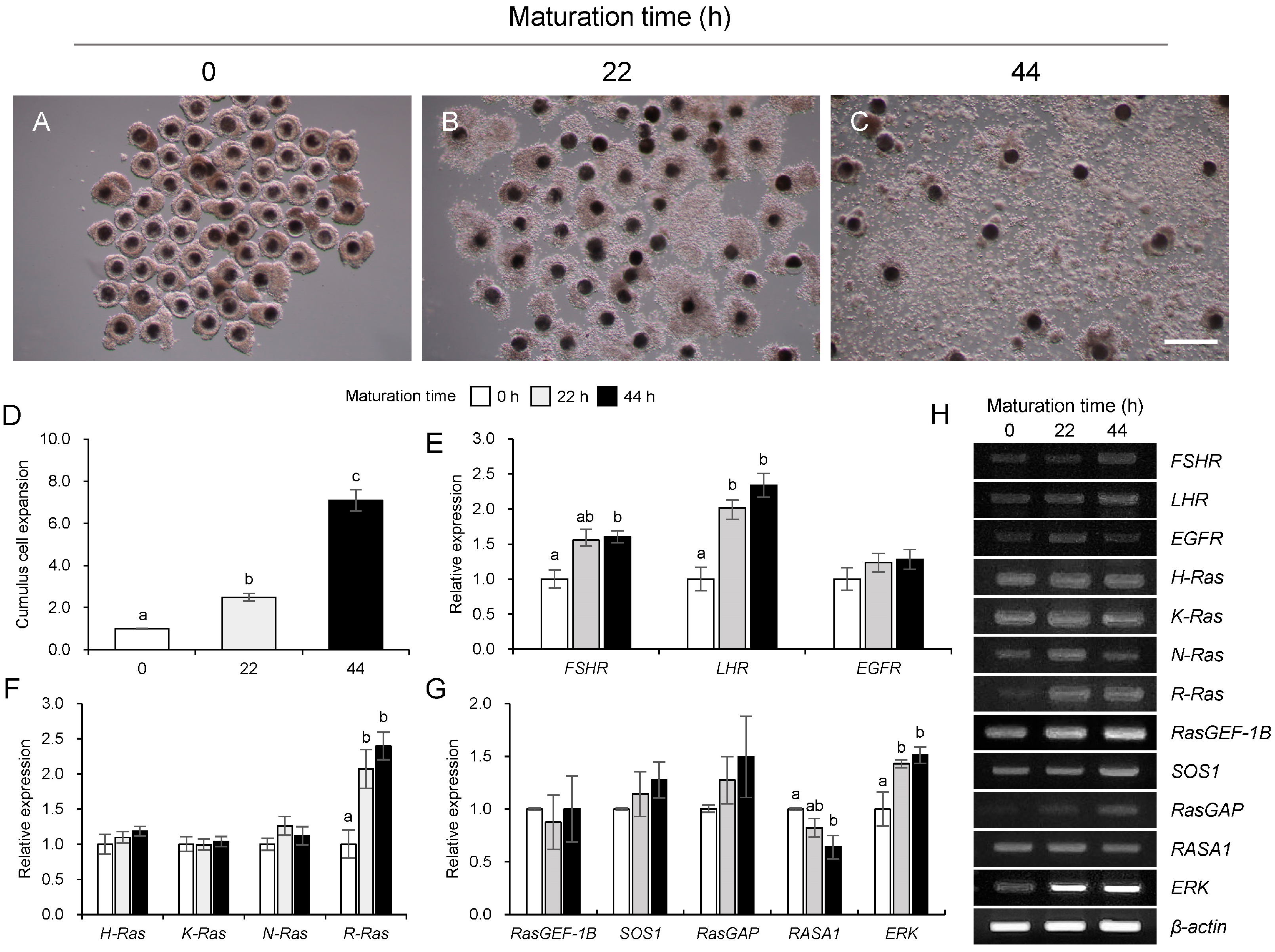
Figure 1. Changes in Ras subfamily and it GTPase genes during maturation in porcine cumulus–oocyte complexes (COCs). Morphology of COCs at 0 h (A), 22 h (B), and 44 h (C) of maturation. The relative expansion of cumulus cell size during the maturation in porcine COCs (D). Expression of gonadotropin receptors (FSHR and LHR) and epidermal growth factor (EGFR) genes (E), Ras subfamily (H-Ras, K-Ras, N-Ras, and R-Ras) genes (F), Ras GEF (RasGEF-1B and SOS1), Ras GAP (RasGAP and RASA1), and ERK genes (G) during maturation. Representative RT-PCR gel image showing the mRNA expression of all analyzed genes (H). a–c Bars with different letters are significantly different (p < 0.05, n = 6). Scale bar = 500 μm.
Animals 15 02100 g001
Figure 2. Influence of follicle stimulating hormone (FSH) on morphological changes (A) and mRNA expression of R-Ras (B), RASA1 (C), SOS1 (D), ERK (E), CCNB1 (F), and Cdc2 (G) during maturation in porcine cumulus–oocyte complexes (COCs). White arrows indicate expanded cumulus cells, and yellow arrows indicate shrunken cumulus cells. a, b Bars with different letters indicate significant differences in gene expression levels within the same concentration. An asterisk (*) indicates significant differences in gene expression levels between different concentration treatment groups (p < 0.05, n = 4). Scale bar = 500 μm.
Animals 15 02100 g002
Figure 3. Influence of human chorionic gonadotropin (hCG) on morphological changes (A) and mRNA expression of R-Ras (B), RASA1 (C), SOS1 (D), ERK (E), CCNB1 (F), and Cdc2 (G) during maturation in porcine cumulus–oocyte complexes (COCs). White arrows indicate expanded cumulus cells, and yellow arrows indicate shrunken cumulus cells. a, b Bars with different letters indicate significant differences in gene expression levels within the same concentration. An asterisk (*) indicates significant differences in gene expression levels between different concentration treatment groups (p < 0.05, n = 4). Scale bar = 500 μm.
Animals 15 02100 g003
Figure 4. Influence of epidermal growth factor (EGF) on morphological changes (A) and mRNA expression of R-Ras (B), RASA1 (C), SOS1 (D), ERK (E), CCNB1 (F), and Cdc2 (G) during maturation in porcine cumulus–oocyte complexes (COCs). Yellow arrows indicate shrunken cumulus cells. a, b Bars with different letters indicate significant differences in gene expression levels within the same concentration. An asterisk (*) indicates significant differences in gene expression levels between different concentration treatment groups (p < 0.05, n = 4). Scale bar = 500 μm.
Animals 15 02100 g004
Figure 5. Effects of follicle stimulating hormone (FSH), human chorionic gonadotropin (hCG), and epidermal growth factor (EGF) on morphological changes (AH) and mRNA expression of R-Ras (I), RASA1 (J), SOS1 (K), ERK (L), CCNB1 (M), and Cdc2 (N) during maturation in porcine cumulus–oocyte complexes (COCs). The treatment concentrations of FSH, hCG, and EGF were determined based on the results of previous experiments, and the COCs were treated with a combination of 0.5 μg/mL FSH, 10 IU/mL hCG, and 10 ng/mL EGF. White arrows indicate expanded cumulus cells, and yellow arrows indicate shrunken cumulus cells. An asterisk (*) indicates significant differences in gene expression levels between different concentration treatment groups (p < 0.05, n = 4). Scale bar = 500 μm.
Animals 15 02100 g005
Table 1. Information on primer condition.
GenesSequence (5′–3′)Annealing Temp. (°C)CycleProduct Size (bp)
β-actinF: GGACTTCGAGCAGGAGATGG6030233
R: GCACCGTGTTGGCGTAGAGG
FSHRF: GATCCTGATCACCAGCCAATAC6032217
R: GACTGAGAGCTCACTAGCAAAG
LHRF: CTGTCCTCTTTGTTCTCCTGAC6032191
R: AGCAACACTACACCCATTCC
EGFRF: CCTTGGGAACTTGGAGATCACCTAC6032344
R: TGTTGCTTAGAAAGTCGCTGTTGAC
H-RasF: CGCCATCAACAACACCAAATC6032194
R: TGGCTGAGGTCTCGATGTAA
K-RasF: CAATGAGGGACCAGTACATGAG6032205
R: GCTAAGTCCTGAGCCTGTTT
N-RasF: TACAAACTGGTGGTGGTTGG6032219
R: GCTAAGTCCTGAGCCTGTTT
R-RasF: CCCACTATTGAAGACTCCTACAC6032205
R: GAAGTCATCTCGGTCCTTGAC
RasGEF-1BF: GGCAGCACAAAGGTCTTTAAC6035201
R: GGACACTCCACTTGTTTCCA
SOS1F: CTGCTCACCTTACACCCAATAG6035231
R: CACCACAGCTACCCTTTCTT
RasGAPF: CAAACGCACGAAGTCACAAC6035203
R: CTGGCTTGATGATGGAGTCTT
RASA1F: TACTTCCACCGACACTGAGATA6035221
R: CTGCACAGACTTAGCCACTAAT
ERKF: GACCAGCTCAACCACATTCT6032213
R: CCGACAGAAGCCAAGATAACA
CCNB1F: GTGTCAGGCTTTCTCTGATGT6032199
R: CCAGTCAATTAGGATGGCTCTC
Cdc2F: GGTGTTCCTAGTACTGCCATTC6032179
R: GAATCCATGAACTGACCAGGAG
Disclaimer/Publisher’s Note: The statements, opinions and data contained in all publications are solely those of the individual author(s) and contributor(s) and not of MDPI and/or the editor(s). MDPI and/or the editor(s) disclaim responsibility for any injury to people or property resulting from any ideas, methods, instructions or products referred to in the content.