Article

Bacillus spp. Potentiate the Virulence and Intracellular Invasion of A. paragallinarum in Chickens

1
Institute of Animal Husbandry and Veterinary Medicine, Hubei Academy of Agricultural Sciences, Wuhan 430064, China
2
Institute of Animal Husbandry and Veterinary Medicine, Beijing Academy of Agriculture and Forestry Sciences, Beijing 100097, China
3
College of Veterinary Medicine, China Agricultural University, Beijing 100193, China
*
Authors to whom correspondence should be addressed.
These authors contributed equally to this work.
Animals 2025, 15(14), 2076; https://doi.org/10.3390/ani15142076 (registering DOI)
Submission received: 4 June 2025 / Revised: 9 July 2025 / Accepted: 11 July 2025 / Published: 14 July 2025

Simple Summary

Bacterial coinfection poses severe threats to poultry health. One common bacterium, Avibacterium paragallinarum, relies on extracellular growth factors acquired from other organisms or its surrounding environment and is more susceptible to coinfection. In this study, Bacillus promotes the growth and infection of Avibacterium paragallinarum in vitro and in vivo by metabolites, especially some Bacillus strains isolated from probiotics. This study highlights the necessity for enhanced safety assessments of probiotic Bacillus strains to evaluate their potential role in facilitating coinfections.

Abstract

Coinfection poses severe threats to poultry health, particularly due to the complexity and resilience of multispecies interactions, increasing the difficulty of treatment. Haemophilus spp., a heterotrophic bacterium, heavily relies on extracellular growth factors acquired from other organisms or its surrounding environment. Although coinfections by Avibacterium paragallinarum and Bacillus have been reported, the underlying mechanism of the cooperative interaction remains poorly understood. In this study, we characterized the growth-promoting properties and nicotinamide adenine dinucleotide production of some Bacillus species, including probiotic Bacillus, to evaluate the feasibility of A. paragallinarum coinfection in vitro. Meanwhile, we determined the minimum inhibitory concentration (MIC) and population dynamics of cocultured Bacillus and A. paragallinarum to assess the effect of bacterial interactions on antibiotic efficacy. Additionally, we demonstrated that B. cereus aggravates rhinitis symptoms in chickens infected with A. paragallinarum. Our findings reveal that Bacillus spp.-derived metabolites sustain A. paragallinarum growth and enhance its survival, thereby highlighting the infection risks associated with Bacillus colonization in the respiratory tract.

1. Introduction

Coinfections frequently involve synergistic interactions among pathogens, commensals, or opportunistic bacteria through cross-feeding and cross-protection mechanisms [1]. In multispecies communities, complex interactions facilitate metabolite sharing, which diminishes antibiotic efficacy against co-inhabiting microbes [2,3,4]. Metabolic exchanges shape the structure and function of microbial communities, playing a crucial role in ecological and environmental interactions [5]. For instance, opportunistic Klebsiella pneumoniae cross-feeds Acinetobacter baumannii with sugar fermentation by-products, enhancing the production of dual-species biofilms that confer resistance to antibiotic treatments [6,7]. Such metabolite exchange among community strains can enhance bacterial survival or virulence in resource-limited environments [8,9,10]. These cooperative behaviors among bacteria exacerbate the progression of bacterial infections, leading to more severe diseases and increasing the challenges of antibiotic treatment.
The respiratory tract harbors trillions of bacteria, serving as the second-largest microbial reservoir in the host and acting as gatekeepers of respiratory health [11]. Infectious coryza (IC), an acute respiratory infection in poultry, is caused by Avibacterium paragallinarum—a nutrient-deficient pathogen reliant on environmental nicotinamide adenine dinucleotide (NAD+) for growth [12]. While vaccination remains the most effective control strategy, antibiotic treatment shows limited efficacy. Previous studies demonstrate that the resident bacteria Staphylococcus chromogenes provides the NAD+ and releases the NAD+ from host cells to promote the survival and growth of A. paragallinarum [13]. Furthermore, a positive correlation between Bacillus spp. isolates and A. paragallinarum has been observed [14], suggesting Bacillus spp. may facilitate A. paragallinarum infection in the chicken respiratory tract. This potential role is particularly relevant given that certain Bacillus species are promising antibiotic alternatives in the feed industry due to their ability to modulate microbial communities [15]. However, some Bacillus strains isolated from probiotic products have been associated with sepsis, intestinal inflammation, and liver damage in various mouse models [16]. Given the limited research on Bacillus in the respiratory tract, it remains unknown whether and how Bacillus spp. assist the survival and infection of A. paragallinarum in vivo.
In this study, we investigated the in vitro growth-promoting abilities of Bacillus spp.—including probiotic Bacillus—on A. paragallinarum and elucidated the underlying mechanisms. We observed a significant increase in bacterial numbers when cocultured Bacillus spp. and A. paragallinarum, this function was attributed to Bacillus-derived NAD+ production. Furthermore, B. cereus CAU492 and B. licheniformis YC3-2 provided antibiotic protection against A. paragallinarum, as evidenced by an increased minimum inhibitory concentration in the coculture. Additionally, the presence of Bacillus cereus CAU492 significantly aggravated clinical symptoms and increased the burden of A. paragallinarum in chickens. Our findings indicate that B. cereus and B. licheniformis present in the respiratory tract promote the replication and infection of A. paragallinarum through NAD+-feeding and antibiotic protection mechanisms.

2. Materials and Methods

2.1. Coculture of Various Bacillus Strains and A. paragallinarum

Bacillus [16] and A. paragallinarum [13] strains were used in this study, previously isolated from various sources, including IC chickens, pig farms, meat products, and probiotic products (Table S1). Overnight cultures of Bacillus and A. paragallinarum were adjusted to a McFarland turbidity (McF) of 0.5. The bacteria density of A. paragallinarum was then diluted to 106 CFU/mL. Next, 100 µL of the bacterial suspensions were spread onto Tryptic Soy Agar (TSA, Land Bridge Technology, Beijing, China) plates supplemented with 5% sheep blood. After air-drying, 5 µL aliquots of Bacillus cultures were spotted onto the center of each TSA agar plate. The plates were incubated at 37 °C under 5% CO2 for 24 h. After incubation, the growth radii of both the central Bacillus colonies and the surrounding A. paragallinarum were measured accurately.

2.2. Hemolytic Activity Assessment of Bacillus Strains

The hemolytic activity assessment method referenced established protocols [17]. Briefly, Bacillus strains were grown overnight in Tryptic Soy Broth (TSB, Land Bridge Technology, Beijing, China) at 37 °C with shaking at 200 rpm and adjusted to 0.5 McF. Then, 5 µL of each Bacillus strain was carefully spotted onto the center of TSA agar plates supplemented with 5% sheep blood. The plates were incubated at 37 °C for 24 h, after which the radius of the hemolytic zone surrounding each Bacillus colony was measured at the specified time points.

2.3. Coculture of Bacillus Strains and A. paragallinarum In Vitro

The murine alveolar macrophage cell line MH-S (ATCC CRL-2019) was used to assess the cellular entry capacity of A. paragallinarum. Cells were seeded in 24-well plates at 0.5 × 10⁶ cells per well and cultured overnight at 37 °C under 5% CO2. Overnight cultures of the A. paragallinarum strains X1-1S-1, B. cereus CAU 492, and B. licheniformis YC 3-2 were washed once with sterile phosphate-buffered saline (PBS) and resuspended in basal cell culture medium to a final concentration of 2 × 10⁶ CFU/mL. Prior to infection, the MH-S cells were washed with PBS twice. For single infections (MOI = 1), the wells received 250 μL of A. paragallinarum suspension plus 250 μL culture medium to achieve a multiplicity of infection. In the coinfection group, 250 μL of each bacterial suspension was added to achieve an MOI of 1 for both strains. The cells were incubated for 6 h at 37 °C in a CO2 incubator. After incubation, the culture medium was collected for serial dilution plating. The cells were washed twice with PBS, lysed with 0.1% Triton X-100 (Beyotime Biotechnology Co., Shanghai, China), and the cell lysate was plated on TSA-Van plus agar plates to enumerate the A. paragallinarum and TSA agar plates for Bacillus quantification.

2.4. NAD Measurement

Overnight cultures of Bacillus strains were adjusted at 0.5 McF followed by a 1:100 dilution in fresh TSB for 8 h shaking at 37 °C and 200 rpm. After incubation, the fermented supernatants of these Bacillus cultures were obtained by centrifugation at 8000× g for 5 min at 4 °C. The concentrations of extracellular total NAD (both NAD+ and NADH) levels in the Bacillus supernatants were measured using an NAD+/NADH Assay Kit with WST-8 (Beyotime Biotechnology Co., Shanghai, China) according to the manufacturer’s instructions.

2.5. Antibiotic Susceptibility Testing

The minimum inhibitory concentration (MIC) of antibiotics against A. paragallinarum and Bacillus strains was determined using the micro-broth method as described by the Clinical and Laboratory Standards Institute (CLSI) 2023 guidelines [18]. Bacillus and A. paragallinarum strains were cultured overnight at 37 °C with shaking at 200 rpm and adjusted to 0.5 McF in cation-adjusted Mueller–Hinton broth (CMHB, GuanDao Biotech Co., Shanghai, China) containing 5% serum and 20 µg/mL of NAD+. Cefotamine, Ofloxacin, Doxycycline, Gentamicin, and Ampicillin were diluted twofold in CAMHB containing 20 µg/mL NAD+ and 1% (v/v) sterile-filtered heat-inactivated chicken serum (Solarbio Life Science Co., Beijing, China). Next, 100 µL of bacterial suspension (5 × 10⁵ CFU/mL in CAMHB plus broth) was mixed with an equal volume of the diluted antibiotic and incubated for 16–18 h at 37 °C in a 5% CO2 atmosphere. Escherichia coli American Type Culture Collection (ATCC) 25922 and Staphylococcus aureus ATCC 29213 served as the quality control strains. Following incubation, the bacterial number of A. paragallinarum was counted on TSA plates containing 10 µg/mL of vancomycin, 5% fetal bovine serum (FBS), and 20 µg/mL NAD+. The population of Bacillus was measured using TSA plates.

2.6. DNA Sequencing and Genome Analysis

The genomic DNA of A. paragallinarum was extracted using the TIANamp Bacterial DNA Kit (Tiangen Biotech, Beijing, China). The DNA was then fragmented to prepare the library and subjected to sequencing using Illumina NovaSeq 6000 (Illumina, San Diego, CA, USA) in a paired-end model. The virulence factors and antimicrobial resistance genes of A. paragallinarum were identified based on VFDB, ResFinder, and the Comprehensive Antibiotic Resistance (CARD) database using BLASTn -2.10.0+ with a cut-off of 60% of coverage and 80% of identity. The whole-genome sequences of B. cereus CAU492 and B. licheniformis YC3-2 were deposited at GenBank under accession numbers PRJNA1134002 and PRJNA1133239. The A. paragallinarum used in this study had been submitted before under accession number PRJNA648655.

2.7. Infection Challenge Experiments in Birds

For the animal experiments, six-week-old specific pathogen-free (SPF) chickens were purchased from Boehringer Ingelheim Vital Biotechnology Co., Ltd. (Beijing, China) The project strictly adhered to ethical guidelines for laboratory animals and implemented the principles of the 3Rs (Replacement, Reduction, and Refinement) to ensure the appropriate handling of the animals and the collection of animal tissue samples for the construction of a chicken infectious rhinitis animal infection model.
The bird infection test followed the procedures in a previous study [13]. The chickens were randomly assigned to six groups, control, B. cereus CAU492 only, B. licheniformis YC3-2 only, A. paragallinarum only, B. cereus CAU492 coinfected with A. paragallinarum, and B. licheniformis YC3-2 coinfected with A. paragallinarum, with five chickens in each group. The chickens were allowed to acclimate to the environment for one week prior to infection, following standardized feeding protocols. Two strains of Bacillus spp. (B. cereus CAU492 or B. licheniformis YC3-2) and A. paragallinarum 1X-1S-1 were all cultured overnight. After incubation, bacterial density was determined; the bacteria were centrifuged and resuspended using sterile phosphate-buffered saline (PBS) to achieve a final concentration of 5 × 10⁸ CFU/mL. The chickens were pretreated with a bacteria-free solution containing 32 µg/mL of vancomycin by injection in the infraorbital sinuses before infection. Subsequently, the chickens were injected with Bacillus spp., followed by infection with A. paragallinarum the next day. On the final day of the experiment, the clinical symptoms of chicken sinusitis were assessed and scored [19] based on the following scale: 0 for no clinical signs; 1 for mild signs (slight facial swelling); 2 for moderate signs (facial swelling and nasal discharge); 3 for severe signs (facial swelling, nasal discharge, ocular discharge, half-closed eyes). All the chickens were humanely euthanized via the inhalation of carbon dioxide gas, and nasal and sinus tissues were collected for bacterial counting. Some tissues were fixed with 4% paraformaldehyde for hematoxylin and eosin (H&E) staining. These experiments were performed as three independent experiments at different intervals.

2.8. Statistical Analysis

Statistical analysis of the data was performed using GraphPad Prism 9 software. p-values were calculated using an unpaired t-test with Bonferroni correction between two groups or a nonparametric one-way ANOVA among multiple groups. Linear modeling (LM) was performed in the R package (version 4.1.3).

3. Results

3.1. Bacillus Facilitates the Growth of A. paragallinarum Under NAD-Deficiency Conditions

NAD is an essential factor for the growth and survival of A. paragallinarum, which is a pathogen known for growth-factor deficiency. Previous studies have confirmed that certain Bacillus species can enhance the survival of A. paragallinarum in vitro [14]. However, the prevalence of this trait across Bacillus species and the comparative efficacy of different strains remain unclear. To address this gap, we conducted in vitro assays to assess whether distinct Bacillus strains potentiate A. paragallinarum growth and to elucidate their mechanistic role in infection pathogenesis. A total of 51 Bacillus strains (representing 17 species) were included in the study. Growth promotion was quantified via the radius of A. paragallinarum growth, which ranged from 0.3 to 1.0 cm (Figure S1a). Comparative analysis revealed significant inter-strain variability in growth-promoting activity, with 24 Bacillus strains promoting growth radii > 0.7 cm (Figure 1). Additionally, we also measured the microcolony radii of Bacillus strains, revealing that species in the B. cereus group formed relatively larger bacterial colonies than other Bacillus groups (Figure S1b). This prompted investigation of a potential correlation between the Bacillus colony size and growth-promoting capacity. Linear regression analysis demonstrated a significant negative correlation (Pearson’s r = −0.51, p < 0.05; Figure S1c), indicating an inverse relationship between the Bacillus colony dimensions and A. paragallinarum growth enhancement.
Given that host cells serve as a substantial nutrient pool for pathogen infection, we further evaluated the hemolysis activity of the Bacillus species. All the species in the B. cereus group exhibited hemolysis zones, indicating robust hemolytic capacity. In contrast, some species in the B. subtilis group (B. amyloliquefaciens and B. velezensis) displayed only minimal hemolytic activity. Some strains exhibited comparable high levels of hemolysis with B. cereus (Figure S1d), like Bacillus sp. 29HD 1S-1. Combined growth-promoting and hemolytic abilities, we screened seven strains of Bacillus (B. cereus 2-1, B. cereus 10-3, B. cereus CAU492, B. licheniformis YC3-2, Bacillus sp. 29HD 1S-1, Bacillus sp. 31HD 1S-6, B. pumilus CAU497, B. oceanisediuminis 22-7) for the next experiments. Considering the growth-promoting ability of Bacillus strains and their hemolytic activity, we supposed that B. cereus is more likely to promote the invasion of A. paragallinarum compared to other Bacillus species.

3.2. NAD Derived from Bacillus Promotes the Growth of A. paragallinarum

Given that Bacillus spp. promotes the growth of A. paragallinarum, we speculated that Bacillus produces some kinds of diffusible metabolic cofactors required by A. paragallinarum. To investigate this mechanism, we further measured the NAD content in the culture supernatants, as it has been previously reported that various Bacillus species can cause the leakage of NAD. Our findings revealed that the supernatants of the different Bacillus species exhibited varying concentrations of total NAD (both NAD+ and NADH) (Figure 2a). Notably, B. licheniformis YC3-2, B. cereus CAU492 and B. oceanisediuminis 22-7 exhibited significantly higher total NAD levels than other strains. These results confirm that Bacillus species can release NAD extracellularly. Interestingly, these strains also exhibited elevated NAD+ production (Figure 2b), with B. licheniformis YC3-2 displaying particularly highest levels. The NAD production profile aligned with different growth-promotion ability of Bacillus strains, suggesting that Bacillus-derived NAD likely sustains the survival of A. paragallinarum in chickens with infectious coryza.

3.3. Bacillus Enhances the Invasion of A. paragallinarum In Vitro

To investigate whether Bacillus promotes the invasion of A. paragallinarum in cells, we conducted coculture assays with B. cereus CAU492, B. licheniformis YC3-2 and A. paragallinarum on MH-S cells. B. cereus CAU492 was found to increase both extracellular and intracellular bacterial numbers of A. paragallinarum (Figure 3a). On the other hand, B. licheniformis YC3-2 increased the load of A. paragallinarum more significantly in extracellular than intracellular (Figure 3b). Compared to B. cereus CAU492, B. licheniformis YC3-2 exhibited a significantly greater enhancement in the growth of A. paragallinarum. These findings demonstrate that Bacillus promotes the invasion and proliferation of A. paragallinarum in cells.

3.4. Bacillus spp. Confer Protection on A. paragallinarum Against Antibiotics

Based on the threat of antimicrobial resistance posed by antibiotics in farms [20], we assessed the growth fitness and antibiotic tolerance of A. paragallinarum when cocultured with Bacillus in the presence of seven kinds of antibiotics. First, we measured the minimum inhibitory concentration (MIC) of A. paragallinarum and Bacillus against these antibiotics. Our results showed that A. paragallinarum was highly sensitive to Cefotaxime, Ofloxacin, Gentamicin, and Ampicillin. The MIC values of B. cereus CAU492 against Ofloxacin and Gentamicin were below 0.25 and 0.5 µg/mL, respectively, while the MICs for the remaining antibiotics were at least 64 µg/mL. B. licheniformis YC3-2 displayed resistance to Cefotaxime but increased sensitivity to Ofloxacin, Gentamicin, and Ampicillin (MIC values below 0.125 and 0.25 µg/mL, respectively; Table S2). According to the diagrams, we recorded the MICs of mono-culture and coculture wells and selected wells for bacteria counting (Figure 4a). In the presence of Cefotaxime and Ampicillin, the MIC values of the cocultures were either consistent with or higher than those of Bacillus alone (Figure 4b). Further, we counted the bacterial number of A. paragallinarum in these groups of wells and found a significant increase-from undetectable (0 CFU) to 1250 and 2250 CFU-following Cefotaxime treatment (Table S3). This represented an increase of more than 1000 times compared to the cocultured group alone (Figure 4c). Similarly, the number of A. paragallinarum increased from 6 to more than 106 CFUs in the coculture after the Ampicillin treatment. Interestingly, we simultaneously observed that A. paragallinarum increased the MIC value of B. cereus CAU492 against Doxycycline (Figure 4b), indicating bidirectional antibiotic tolerance modulation. These results indicate that Bacillus significantly enhances A. paragallinarum survival under antibiotic pressure. Collectively, our findings suggest that Bacillus plays an assistant role in the respiratory tract colonization of A. paragallinarum by protecting the pathogens from antibiotic-induced eradication.
To further investigate the underlying mechanism of Bacillus in enhancing the antibiotic resistance and infection of A. paragallinarum, we analyzed the resistance genes and virulence genes carried by A. paragallinarum and Bacillus via genomic analysis. The results revealed that A. paragallinarum carried the tetracycline resistance gene tet(B) and the virulence genes (lpxC, manB, yhxB, gmhA, and lpcA). B. cereus CAU492 harbors the resistance genes bcII, bcI, fosB, and vanZF and the virulence genes cytK, bas3190, nheC, nheA, and inhA. Among them, B. licheniformis YC 3-2 carried only the resistance genes ermD, bcrA, bcrB, bcrC (Table S4). Notably, genomic analysis suggested the potential involvement of β-lactamase genes (bcII and bcI) in the ability of B. cereus CAU492 to hydrolyze cephalosporin antibiotics, contributing to the survival of A. paragallinarum in the presence of Cefotaxime and Ampicillin by reducing antibiotic concentration in the environment. Additionally, B. cereus CAU492 carries the biosynthesis gene of the hemolytic enterotoxin (nhe) that could exacerbate A. paragallinarum infections by producing enterotoxin. However, B. licheniformis YC 3-2 lacks the β-lactamase gene and still enhances the survival of A. paragallinarum in cefotaxime treatment, possibly mediated by other mutualistic mechanisms.

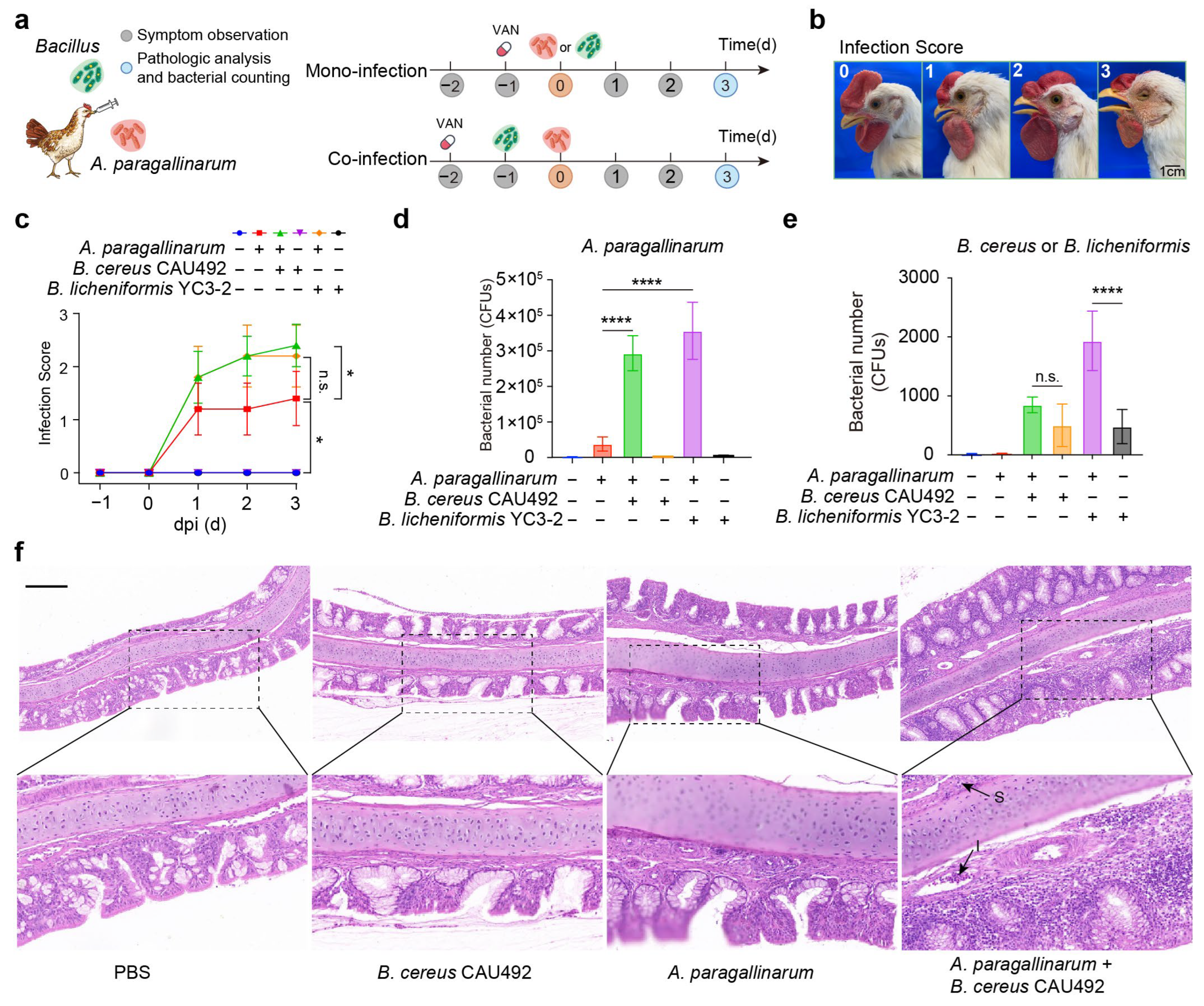
3.5. Bacillus Aggravates the Disease Severity in A. paragallinarum-Infected Chickens

Given that Bacillus supported the growth and invasion of A. paragallinarum in vitro, we subsequently conducted in vivo experiment to investigate whether their positive interactions enhance infection severity. According to the schematic diagram (Figure 5a), six-week-old SPF chickens were randomly divided into six groups (n = 5 per group) and infected with A. paragallinarum in the presence or absence of Bacillus. Clinical symptoms were carefully evaluated using a 0–3 severity (Figure 5b), reflecting the intensity of nasal inflammation. Our results revealed that A. paragallinarum coinfected with Bacillus caused more severe clinical symptoms of nasal inflammation in chickens than the mono-infection (Figure 5c). Meanwhile, B. licheniformis YC3-2 or B. cereus CAU492 caused no visible symptoms of nasal inflammation when infected alone. This finding suggests that Bacillus exacerbates A. paragallinarum-induced infection in chickens.
To evaluate the cross-feeding dynamics between Bacillus and A. paragallinarum, we quantified the bacterial load in the nasal cavity and infraorbital sinuses. Our findings illustrated that both B. licheniformis YC3-2 and B. cereus CAU492 significantly increased A. paragallinarum populations (Figure 5d). Similarly, the presence of A. paragallinarum increased the population of B. cereus CAU492, though the difference was not significant, B. licheniformis YC3-2 exhibited a more pronounced increase when coinfected with A. paragallinarum (Figure 5e). Notably, histopathological analysis revealed that coinfections induced submucosa thickening, extensive inflammatory cells infiltration, and a disappeared villus structure of epithelial cells (Figure 5f). These results confirm that the introduction of Bacillus spp. promote A. paragallinarum infection through a combination of metabolic support and protective mechanisms.

4. Discussion

The prevailing occurrence of coinfections generally stems from bacteria-positive interactions, including symbiosis and collaboration [21], which are often mediated by various bacterial metabolites produced by each member of the community. These behaviors related to signaling communication among bacteria are ubiquitous and important to ecosystem stability such as exchanging metabolic products and sharing genes [22,23], especially when engaged in auxotrophic and prototrophic bacteria [24]. Such symbiotic interactions may be prevalent in humans and animals, resulting in the exacerbation of coinfections. Some Bacillus species are reported to serve as probiotic microbiota engaged in colonization resistance and the antagonism of pathogens by various metabolites [25,26]. While toxin- and mobile antimicrobial-resistant genes also have been found in Bacillus probiotics, that pose a risk in application [16]. This study found a coinfection between Bacillus and A. paragallinarum that revealed a new risk of Bacillus. Although probiotic Bacillus strains have exhibited in vitro growth-promoting effects on A. paragallinarum, their oral administration typically restricts interaction with respiratory pathogens. This limitation may diminish their potential to exacerbate infections relative to Bacillus strains from other sources. Nevertheless, the potential risk of Bacillus-mediated enhancement of A. paragallinarum infections warrants continued attention.
Respiratory infections stand as a prevalent contributor to heightened mortality rates in poultry across the globe [27]. Whereas the bulk of coinfection research has concentrated on viral coinfections [28] and viral–bacterial coinfections [29], investigations into respiratory bacterial coinfections have been comparatively limited. Bacterial cooperation and collaboration often emerge as seemingly “costless” behaviors among bacteria within a community [22]. A. paragallinarum, an opportunistic pathogen auxotrophic for NAD, relies on the support of host cells or bacteria in the surrounding environment for this essential cofactor during survival and infection [30]. Based on its specificity, research has identified instances of coinfection between A. paragallinarum and other bacteria, such as A. paragallinarum and Gallibacterium anatis being coinfected in chickens, causing the aggravation of infectious coryza [31]. These coinfection cases reveal mutualistic behavior by metabolite sharing that is vital for the survival of pathogen persistence. Our findings demonstrate that various Bacillus species exhibit distinct growth-promoting behaviors on A. paragallinarum through abundant NAD+ synthesized by Bacillus species, as bacterial NAD+ serves as an essential redox factor in bacterial energy generation and the synthesis of metabolic products [32,33]. Extracellular NAD+ was detected in the Bacillus cultures, indicating that Bacillus can synthesize its own energy and excrete excess NAD+ into the environment. Based on the importance of NAD+ in regulating cellular metabolism and proliferation [34,35], the behavior of the NAD release process may be involved in specific metabolic pathways in Bacillus, such as its biofilm formation [36]. However, whether the secretion behavior is active or passive and the underlying regulatory mechanism remain to be further investigated. Furthermore, the hemolytic properties of Bacillus represent virulence factors that damage host cells and may contribute to the exacerbation of A. paragallinarum infections.
Compared to B. licheniformis YC3-2 in our work, B. cereus CAU492 carries multiple enterotoxin genes, including nheA and nheC, potentially leading to more serious infections in cellular models. Research has demonstrated that some B. cereus strains are capable of producing a heat-stable emetic toxin called cereulide and carry mobile antimicrobial resistance genes, posing a significant risk to both humans and animals [16,37]. Therefore, the development of Bacillus cereus as a probiotic requires rigorous safety evaluation for its potential to cause infection and coinfection. Crucially, the growth-promoting behaviors exhibited by Bacillus species imply that synergistic Bacillus colonizing the respiratory tract may serve an assisting role in the colonization and infection processes of A. paragallinarum. Interestingly, alongside the increased bacterial population of A. paragallinarum while in the coculture, there is also a notable augmentation in the population of B. licheniformis YC3-2 (Figure 5e), indicating that the presence of A. paragallinarum may facilitate the reproduction of Bacillus species in vivo.
Antibiotic resistance is frequently mediated by bacterial metabolites through siderophore production, antibiotic hydrolysis, and biofilm formation [38,39]. For instance, Pseudomonas aeruginosa produces pyoverdine and enterobacteria siderophore enterobactin to displace the iron hijack transporters of antibiotic cefiderocol for the cross-protection of susceptible P. aeruginosa [40]. Similarly, Enterococcus and Pseudomonas augment bacterial resistance to antibiotics through biofilm protection [41]. Our study predicted β-lactamase in B. cereus CAU492, an enzyme capable of hydrolyzing β-lactam antibiotics [42], may contribute to enhancing A. paragallinarum survival during cephalosporin or ampicillin exposure. This phenomenon is akin to certain strains of E. coli producing β-lactamase, which enables Salmonella Typhimurium to evade antibiotic eradication [43]. The antibiotic protection of B. licheniformis YC3-2 may contribute by biofilm formation [44] or other resistant genes, such as the bcrA, bcrB, or bcrC genes, which mediated Bacitractin resistance [45]. Remarkably, the cocultured bacteria displayed heightened resistance to antibiotic doxycycline, likely due to the presence of the tetracycline resistance gene tet(B) in A. paragallinarum, which confers tetracycline antibiotic resistance [46,47]. Collectively, these mechanisms demonstrate how bacterial metabolite exchange provides cross-protection to members engaged in antibiotic battles within niches.

5. Conclusions

To our knowledge, this study represents a novel investigation of the impact of Bacillus species on respiratory bacterial infections. Through experimental observations, we have revealed a syntrophic mechanism in which B. cereus and B. licheniformis promote the virulence of A. paragallinarum by supplying essential nutrients (NAD+) and antibiotic-resistant substances. These discoveries not only expand our understanding of bacterial coinfection mechanisms but also provide critical guidance for the rational development and clinical application of Bacillus-based probiotics.

Supplementary Materials

The following supporting information can be downloaded at: https://www.mdpi.com/article/10.3390/ani15142076/s1, Figure S1: Bacillus promotes A. paragallinarum growth; Table S1: Sources of bacteria used in this study; Table S2: Minimum inhibitory concentration for mono-cultures and cocultures (1:1) of Bacillus and A. paragallinarum; Table S3: Bacterial number of A. paragallinarum in mono-culture and coculture; Table S4: The presence of resistance genes and toxin genes in A. paragallinarum and Bacillus spp.

Author Contributions

Eight authors participated in this study. Conceptualization, J.Z. and Y.L.; methodology, T.G.; software, Y.C.; validation, W.L.; formal analysis, W.L.; resources, K.Y.; investigation, T.G.; data curation, J.Z. and Y.L.; writing—original draft preparation, J.Z. and Y.L.; writing—review and editing, J.Z., Y.C. and K.Z.; visualization, K.Z. and D.Z.; supervision, K.Y.; project administration, K.Z. and D.Z.; funding acquisition, J.Z. and D.Z. All authors have read and agreed to the published version of the manuscript.

Funding

This research was funded by the National Natural Science Foundation of China (No. 32402901), China Postdoctoral Science Foundation (2023M741106), the Important Science and Technology Project of Hubei Province (2024BBA004), and Hubei Agricultural Science and Technology Innovation Center (2024-620-000-001-013).

Institutional Review Board Statement

The animal study protocol was approved by the State Council of the People’s Republic of China on 14 November 1988. The study was also conducted in accordance with the guidelines of the Animal Welfare Committee of Beijing Academy of Agriculture and Forestry Sciences (BAAFS), under the approved Animal Care Protocol (ID: IHVM11-2308-46).

Informed Consent Statement

Not applicable.

Data Availability Statement

The original contributions presented in this study are included in the article/Supplementary Material. Further inquiries can be directed to the corresponding authors.

Acknowledgments

We gratefully acknowledge the Beijing Academy of Agriculture and Forestry Sciences, Institute of Animal Husbandry and Veterinary Medicine, for its support in facilitating the animal experimentation.

Conflicts of Interest

The authors declare no conflict of interest.

Abbreviations

The following abbreviations are used in this manuscript:
CLSIClinical and Laboratory Standards Institute
NAD+Nicotinamide adenine dinucleotide
MICMinimum inhibitory concentration
McFMcFarland turbidity
MOIMultiplicity of infection

References

  1. Tay, W.H.; Chong, K.K.; Kline, K.A. Polymicrobial-Host Interactions during Infection. J. Mol. Biol. 2016, 428, 3355–3371. [Google Scholar] [CrossRef]
  2. Pathak, A.; Angst, D.C.; Leon-Sampedro, R.; Hall, A.R. Antibiotic-degrading resistance changes bacterial community structure via species-specific responses. ISME J. 2023, 17, 1495–1503. [Google Scholar] [CrossRef] [PubMed]
  3. Pearl Mizrahi, S.; Goyal, A.; Gore, J. Community interactions drive the evolution of antibiotic tolerance in bacteria. Proc. Natl. Acad. Sci. USA 2023, 120, e2209043119. [Google Scholar] [CrossRef] [PubMed]
  4. Abushaheen, M.A.; Muzaheed; Fatani, A.J.; Alosaimi, M.; Mansy, W.; George, M.; Acharya, S.; Rathod, S.; Divakar, D.D.; Jhugroo, C.; et al. Antimicrobial resistance, mechanisms and its clinical significance. Dis. Mon. 2020, 66, 100971. [Google Scholar] [CrossRef] [PubMed]
  5. Kost, C.; Patil, K.R.; Friedman, J.; Garcia, S.L.; Ralser, M. Metabolic exchanges are ubiquitous in natural microbial communities. Nat. Microbiol. 2023, 8, 2244–2252. [Google Scholar] [CrossRef]
  6. Semenec, L.; Cain, A.K.; Dawson, C.J.; Liu, Q.; Dinh, H.; Lott, H.; Penesyan, A.; Maharjan, R.; Short, F.L.; Hassan, K.A.; et al. Cross-protection and cross-feeding between Klebsiella pneumoniae and Acinetobacter baumannii promotes their co-existence. Nat. Commun. 2023, 14, 702. [Google Scholar] [CrossRef]
  7. Oliveira, M.; Cunha, E.; Tavares, L.; Serrano, I.P. aeruginosa interactions with other microbes in biofilms during co-infection. AIMS Microbiol. 2023, 9, 612–646. [Google Scholar] [CrossRef]
  8. Bottery, M.J.; Pitchford, J.W.; Friman, V.P. Ecology and evolution of antimicrobial resistance in bacterial communities. ISME J. 2021, 15, 939–948. [Google Scholar] [CrossRef]
  9. Kim, H.-J.; Na, S.W.; Alodaini, H.A.; Al-Dosary, M.A.; Nandhakumari, P.; Dyona, L. Prevalence of multidrug-resistant bacteria associated with polymicrobial infections. J. Infect. Public. Health 2021, 14, 1864–1869. [Google Scholar] [CrossRef]
  10. Cordero, O.; Wildschutte, H.; Kirkup, B.; Proehl, S.; Ngo, L.; Hussain, F.; Le Roux, F.; Mincer, T.; Polz, M. Ecological populations of bacteria act as socially cohesive units of antibiotic production and resistance. Science 2012, 337, 1228–1231. [Google Scholar] [CrossRef]
  11. Man, W.H.; de Steenhuijsen Piters, W.A.; Bogaert, D. The microbiota of the respiratory tract: Gatekeeper to respiratory health. Nat. Rev. Microbiol. 2017, 15, 259–270. [Google Scholar] [CrossRef]
  12. Jeong, O.M.; Kang, M.S.; Blackall, P.J.; Jeon, B.W.; Kim, J.H.; Jeong, J.; Lee, H.J.; Kim, D.W.; Kwon, Y.K.; Kim, J.H. Genotypic divergence of Avibacterium paragallinarum isolates with different growth requirements for nicotinamide adenine dinucleotide. Avian Pathol. 2020, 49, 153–160. [Google Scholar] [CrossRef]
  13. Wu, Y.; Wang, Y.; Yang, H.; Li, Q.; Gong, X.; Zhang, G.; Zhu, K. Resident bacteria contribute to opportunistic infections of the respiratory tract. PLoS Pathog. 2021, 17, e1009436. [Google Scholar] [CrossRef] [PubMed]
  14. Zhu, J.; Chen, Y.; Wu, Y.; Wang, Y.; Zhu, K. Commensal bacteria contribute to the growth of multidrug-resistant Avibacterium paragallinarum in chickens. Front. Microbiol. 2022, 13, 1010584. [Google Scholar] [CrossRef] [PubMed]
  15. Arreguin-Nava, M.A.; Graham, B.D.; Adhikari, B.; Agnello, M.; Selby, C.M.; Hernandez-Velasco, X.; Vuong, C.N.; Solis-Cruz, B.; Hernandez-Patlan, D.; Latorre, J.D.; et al. Evaluation of in ovo Bacillus spp. based probiotic administration on horizontal transmission of virulent Escherichia coli in neonatal broiler chickens. Poult. Sci. 2019, 98, 6483–6491. [Google Scholar] [CrossRef]
  16. Cui, Y.; Wang, S.; Ding, S.; Shen, J.; Zhu, K. Toxins and mobile antimicrobial resistance genes in Bacillus probiotics constitute a potential risk for One Health. J. Hazard. Mater. 2020, 382, 121266. [Google Scholar] [CrossRef] [PubMed]
  17. Deng, F.; Chen, Y.; Sun, T.; Wu, Y.; Su, Y.; Liu, C.; Zhou, J.; Deng, Y.; Wen, J. Antimicrobial resistance, virulence characteristics and genotypes of Bacillus spp. from probiotic products of diverse origins. Food Res. Int. 2021, 139, 109949. [Google Scholar] [CrossRef]
  18. A, A.J.; Suresh, A. Oral microbial shift induced by probiotic Bacillus coagualans along with its clinical perspectives. J. Oral. Biol. Craniofacial Res. 2023, 13, 398–402. [Google Scholar] [CrossRef]
  19. Morales-Erasto, V.; Falconi-Agapito, F.; Luna-Galaz, G.A.; Saravia, L.E.; Montalvan-Avalos, A.; Soriano-Vargas, E.E.; Fernandez-Diaz, M. Coinfection of Avibacterium paragallinarum and Ornithobacterium rhinotracheale in Chickens from Peru. Avian Dis. 2016, 60, 75–78. [Google Scholar] [CrossRef]
  20. O’Meara, S. Antimicrobial resistance. Nature 2020, 586, S49. [Google Scholar] [CrossRef]
  21. Mestre, A.; Poulin, R.; Hortal, J. A niche perspective on the range expansion of symbionts. Biol. Rev. Camb. Philos. Soc. 2020, 95, 491–516. [Google Scholar] [CrossRef] [PubMed]
  22. Ona, L.; Giri, S.; Avermann, N.; Kreienbaum, M.; Thormann, K.M.; Kost, C. Obligate cross-feeding expands the metabolic niche of bacteria. Nat. Ecol. Evol. 2021, 5, 1224–1232. [Google Scholar] [CrossRef] [PubMed]
  23. Ponomarova, O.; Patil, K.R. Metabolic interactions in microbial communities: Untangling the Gordian knot. Curr. Opin. Microbiol. 2015, 27, 37–44. [Google Scholar] [CrossRef]
  24. Giri, S.; Ona, L.; Waschina, S.; Shitut, S.; Yousif, G.; Kaleta, C.; Kost, C. Metabolic dissimilarity determines the establishment of cross-feeding interactions in bacteria. Curr. Biol. 2021, 31, 5547–5557.E6. [Google Scholar] [CrossRef]
  25. Zhu, J.; Chen, Y.; Imre, K.; Arslan-Acaroz, D.; Istanbullugil, F.R.; Fang, Y.; Ros, G.; Zhu, K.; Acaroz, U. Mechanisms of probiotic Bacillus against enteric bacterial infections. One Health Adv. 2023, 1, 21. [Google Scholar] [CrossRef]
  26. Cui, Y.; Zhu, J.; Li, P.; Guo, F.; Yang, B.; Su, X.; Zhou, H.; Zhu, K.; Xu, F. Assessment of probiotic Bacillus velezensis supplementation to reduce Campylobacter jejuni colonization in chickens. Poult. Sci. 2024, 103, 103897. [Google Scholar] [CrossRef]
  27. Sid, H.; Benachour, K.; Rautenschlein, S. Co-infection with Multiple Respiratory Pathogens Contributes to Increased Mortality Rates in Algerian Poultry Flocks. Avian Dis. 2015, 59, 440–446. [Google Scholar] [CrossRef]
  28. Samy, A.; Naguib, M.M. Avian Respiratory Coinfection and Impact on Avian Influenza Pathogenicity in Domestic Poultry: Field and Experimental Findings. Vet. Sci. 2018, 5, 23. [Google Scholar] [CrossRef]
  29. Wu, Z.; Chen, C.; Zhang, Q.; Bao, J.; Fan, Q.; Li, R.; Ishfaq, M.; Li, J. Arachidonic acid metabolism is elevated in Mycoplasma gallisepticum and Escherichia coli co-infection and induces LTC4 in serum as the biomarker for detecting poultry respiratory disease. Virulence 2020, 11, 730–738. [Google Scholar] [CrossRef]
  30. Mouahid, M.; Bisgaard, M.; Morley, A.J.; Mutters, R.; Mannheim, W. Occurrence of V-factor (NAD) independent strains of Haemophilus paragallinarum. Vet. Microbiol. 1992, 31, 363–368. [Google Scholar] [CrossRef]
  31. Paudel, S.; Hess, M.; Hess, C. Coinfection of Avibacterium paragallinarum and Gallibacterium anatis in Specific-Pathogen-Free Chickens Complicates Clinical Signs of Infectious Coryza, Which Can Be Prevented by Vaccination. Avian Dis. 2017, 61, 55–63. [Google Scholar] [CrossRef]
  32. Shimizu, M. NAD(+)/NADH homeostasis affects metabolic adaptation to hypoxia and secondary metabolite production in filamentous fungi. Biosci. Biotechnol. Biochem. 2018, 82, 216–224. [Google Scholar] [CrossRef]
  33. Arjes, H.A.; Vo, L.; Dunn, C.M.; Willis, L.; DeRosa, C.A.; Fraser, C.L.; Kearns, D.B.; Huang, K.C. Biosurfactant-mediated membrane depolarization maintains viability during oxygen depletion in Bacillus subtilis. Curr. Biol. 2020, 30, 1011–1022.e6. [Google Scholar] [CrossRef] [PubMed]
  34. Choi, D. NADH-Reductive Stress in Cancer Cell Proliferation. Mol. Cells 2023, 46, 473–475. [Google Scholar] [CrossRef] [PubMed]
  35. Lee, H.; Yoon, H. Mitochondrial sirtuins: Energy dynamics and cancer metabolism. Mol. Cells 2024, 47, 100029. [Google Scholar] [CrossRef] [PubMed]
  36. Qin, Y.; He, Y.; She, Q.; Larese-Casanova, P.; Li, P.; Chai, Y. Heterogeneity in respiratory electron transfer and adaptive iron utilization in a bacterial biofilm. Nat. Commun. 2019, 10, 3702. [Google Scholar] [CrossRef]
  37. Cui, Y.; Liu, Y.; Liu, X.; Xia, X.; Ding, S.; Zhu, K. Evaluation of the Toxicity and Toxicokinetics of Cereulide from an Emetic Bacillus cereus Strain of Milk Origin. Toxins 2016, 8, 156. [Google Scholar] [CrossRef]
  38. Galdino, A.C.M.; Vaillancourt, M.; Celedonio, D.; Huse, K.; Doi, Y.; Lee, J.S.; Jorth, P. Evolved bacterial siderophore-mediated antibiotic cross-protection. Res. Sq. 2023. [Google Scholar] [CrossRef]
  39. Xu, H.; Tan, C.; Li, C.; Li, J.; Han, Y.; Tang, Y.; Lei, C.; Wang, H. ESBL- Escherichia coli extracellular vesicles mediate bacterial resistance to beta-lactam and mediate horizontal transfer of bla(CTX-M-55). Int. J. Antimicrob. Agents 2024, 63, 107145. [Google Scholar] [CrossRef]
  40. Galdino, A.C.M.; Vaillancourt, M.; Celedonio, D.; Huse, K.; Doi, Y.; Lee, J.S.; Jorth, P. Siderophores promote cooperative interspecies and intraspecies cross-protection against antibiotics in vitro. Nat. Microbiol. 2024, 9, 631–646. [Google Scholar] [CrossRef]
  41. Lee, K.; Lee, K.M.; Kim, D.; Yoon, S.S. Molecular Determinants of the Thickened Matrix in a Dual-Species Pseudomonas aeruginosa and Enterococcus faecalis Biofilm. Appl. Environ. Microbiol. 2017, 83, e01182-17. [Google Scholar] [CrossRef] [PubMed]
  42. Zhao, X.; Ruelens, P.; Farr, A.D.; de Visser, J.; Baraban, L. Population dynamics of cross-protection against beta-lactam antibiotics in droplet microreactors. Front. Microbiol. 2023, 14, 1294790. [Google Scholar] [CrossRef] [PubMed]
  43. Perlin, M.H.; Clark, D.R.; McKenzie, C.; Patel, H.; Jackson, N.; Kormanik, C.; Powell, C.; Bajorek, A.; Myers, D.A.; Dugatkin, L.A.; et al. Protection of Salmonella by ampicillin-resistant Escherichia coli in the presence of otherwise lethal drug concentrations. Proc. Biol. Sci. 2009, 276, 3759–3768. [Google Scholar] [CrossRef] [PubMed]
  44. Falcon Garcia, C.; Kretschmer, M.; Lozano-Andrade, C.N.; Schonleitner, M.; Dragos, A.; Kovacs, A.T.; Lieleg, O. Metal ions weaken the hydrophobicity and antibiotic resistance of Bacillus subtilis NCIB 3610 biofilms. NPJ Biofilms Microbiomes 2020, 6, 1. [Google Scholar] [CrossRef]
  45. Cao, M.; Helmann, J.D. Regulation of the Bacillus subtilis bcrC bacitracin resistance gene by two extracytoplasmic function sigma factors. J. Bacteriol. 2002, 184, 6123–6129. [Google Scholar] [CrossRef]
  46. Yang, S.; Fan, K.; Lin, W.; Wang, J.; Lin, M.; Yang, S.; Jiang, Y.; Huang, X.; Chen, W.; Huang, C. Identification of a multiple drug-resistance gene island in the Haemophilus parasuis chromosome. J. Glob. Antimicrob. Resist. 2020, 22, 422–425. [Google Scholar] [CrossRef]
  47. Lancashire, J.F.; Terry, T.D.; Blackall, P.J.; Jennings, M.P. Plasmid-encoded Tet B tetracycline resistance in Haemophilus parasuis. Antimicrob. Agents Chemother. 2005, 49, 1927–1931. [Google Scholar] [CrossRef]
Figure 1. Commensal Bacillus promoting A. paragallinarum growth. The differences in positive interaction between various genera of Bacillus spp. and A. paragallinarum. Bacillus was selected by the hemolysis and growth-promotion ability. The radii of hemolysis, growth-promotion, and plaque were measured when mono- or cocultured with A. paragallinarum on sheep blood plate. Hemolytic activity was color-coded in blue, growth promotion in red, and Bacillus plaque formation in green.
Animals 15 02076 g001
Figure 2. Bacillus species release NAD into extracellular. Extracellular concentrations of total NAD (including NAD+ and NADH) (a) and NAD+ (b) in Bacillus supernatant after 8 h of culturing. The mean of three biological replicates is shown, and error bars represent the standard deviation (SD) (n = 3).
Animals 15 02076 g002
Figure 3. Bacillus enhances the cellular invasion capacity of A. paragallinarum. The extracellular (a) and intracellular (b) counts of A. paragallinarum were quantified during mono-infection or coinfection with Bacillus. Boxplots represent the distribution of A. paragallinarum (CFUs, n ≥ 3), * p < 0.05, ** p < 0.01, *** p < 0.001, n.s. means no significance.
Animals 15 02076 g003
Figure 4. Bacillus reduces the antibiotic efficacy against A. paragallinarum. (a) Diagrams of experimental records. (b) The MIC values of A. paragallinarum 1X-1S-1, B. cereus CAU492, and B. licheniformis YC3-2. (c) Bacterial numbers of A. paragallinarum in the presence of either cefotaxime or ampicillin. **** p < 0.0001).
Animals 15 02076 g004
Figure 5. Bacillus aggravates A. paragallinarum-induced infection in chickens. (a) Schematic illustration of the experimental workflow. (b) An infection score was used to determine the severity of symptoms, including the swelling of the infraorbital sinus, decreased food intake, and loss of body weight. (c) Bacillus aggravates the symptoms induced by A. paragallinarum. (d,e) Bacterial burdens of A. paragallinarum (d), B. cereus CAU492 or B. licheniformis YC3-2 (e) in the nasal cavity and infraorbital sinuses of poultry. (f) Tissue damage in the nasal cavity of chicken coinfected with B. cereus CAU492 and A. paragallinarum. Arrows indicated thickening of the submucosa (S) and infiltration of inflammatory cells (I). Scale bar = 200 µm. p values were calculated using an unpaired t-test, * p < 0.05, **** p < 0.0001), n.s. means no significance. The mean of three biological replicates is shown, and error bars represent the standard deviation (SD) (n = 5).
Animals 15 02076 g005
Disclaimer/Publisher’s Note: The statements, opinions and data contained in all publications are solely those of the individual author(s) and contributor(s) and not of MDPI and/or the editor(s). MDPI and/or the editor(s) disclaim responsibility for any injury to people or property resulting from any ideas, methods, instructions or products referred to in the content.

