Article

Integrated Analysis of Differential Expression Profiles of miRNA and mRNA in Gonads of Scatophagus argus Provides New Insights into Sexually Biased Gene Expression

by
Yaling Lei
1,2,3,
Kaizhi Jiao
1,2,3,
Yuanqing Huang
1,2,3,
Yuwei Wu
1,2,3,
Gang Shi
1,2,3,
Hongjuan Shi
1,2,3,
Huapu Chen
1,2,3,
Siping Deng
1,2,3,
Guangli Li
1,2,3,
Wenjing Tao
4,5,* and
Dongneng Jiang
1,2,3,*
1
Guangdong Research Center on Reproductive Control and Breeding Technology of Indigenous Valuable Fish Species, Fisheries College, Guangdong Ocean University, Zhanjiang 524088, China
2
Key Laboratory of Marine Ecology and Aquaculture Environment of Zhanjiang, Fisheries College, Guangdong Ocean University, Zhanjiang 524088, China
3
Guangdong Provincial Key Laboratory of Aquatic Animal Disease Control and Healthy Culture, Fisheries College, Guangdong Ocean University, Zhanjiang 524088, China
4
Key Laboratory of Freshwater Fish Reproduction and Development (Ministry of Education), Southwest University, Chongqing 400715, China
5
Key Laboratory of Aquatic Science of Chongqing of Life Sciences, Southwest University, Chongqing 400715, China
*
Authors to whom correspondence should be addressed.
Animals 2025, 15(11), 1564; https://doi.org/10.3390/ani15111564
Submission received: 24 March 2025 / Revised: 22 May 2025 / Accepted: 24 May 2025 / Published: 27 May 2025
(This article belongs to the Section Animal Reproduction)

Simple Summary

Scatophagus argus (S. argus) is a key aquaculture species in southern China, with females growing faster than males. Limited knowledge of its sex determination and differentiation hinders sex-controlled breeding. MicroRNAs (miRNAs) regulate these processes in vertebrates, but no research exists on their role in S. argus. This study analyzed miRNA/mRNA expression in S. argus gonads, identifying 2210 miRNAs (482 sex-differentially expressed) targeting 3340 genes to form 13,773 regulatory pairs. Key sex-related genes (Foxl2, Gdf9, Gsdf, Sox3) showed coordinated or inverse expression patterns with their regulatory miRNAs. The species-specific miRNA and gene regulatory network revealed non-conserved mechanisms across fish. The present study advances the understanding of sexual dimorphism and provides potential targets for sex-controlled breeding in this important marine aquaculture species.

Abstract

The Scatophagus argus (S. argus) is a valuable aquaculture species in southern China, with females exhibiting significantly faster growth rates than males. However, the limited understanding of its sex determination and differentiation mechanisms poses challenges for sex-controlled breeding. MicroRNAs (miRNAs), key post-transcriptional regulators, are known to modulate critical pathways governing sex determination and differentiation across several vertebrates. However, there is currently no research on miRNAs related to sex determination and differentiation in S. argus. In this study, we analyzed the expression profiles of miRNA and mRNA in the gonads of adult S. argus using high-throughput sequencing. Our analysis identified 2210 miRNAs, including 482 differentially expressed miRNAs (DEMs) between sexes. These DEMs targeted 3340 differentially expressed genes (DEGs), generating 13,773 regulatory interaction pairs. The expression of some DEGs related to sex determination and differentiation was found to be either positively or negatively correlated with expression of DEMs that might regulate them. The novel_miR_110/Foxl2, novel_miR_802/Gdf9, and novel_miR_1263/Gdf9 show opposing differential expression trends, whereas sar-miR-143-5p-4/Gsdf, sar-miR-143-5p-5/Gsdf, and novel_miR_379/Sox3 show consistent trends. The regulatory relationship between miRNA and gene in the gonads does not seem to be conserved among different fish species. This work advances our understanding of the molecular mechanisms underlying the sexual dimorphism of gonadal gene expression in S. argus. The identified miRNA–gene interactions may serve as potential targets for future sex-control strategies, contributing to advancements in aquaculture practices for this species.

1. Introduction

The mechanisms of sex determination (SD) in fish can be broadly categorized into three main types: genetic sex determination (GSD), environmental sex determination (ESD), and a mixed mode involving the synergistic regulation of both genetic and environmental factors [1,2]. In GSD systems, key sex-determining genes located on sex chromosomes (such as Amhy and DMY/Dmrt1bY) or polygenic factors on autosomes regulate gonadal development through hierarchical regulatory networks [3,4,5]. These master genes drive sexual differentiation by activating sex-specific signaling pathways (e.g., TGF-β promoting testis development and Wnt/β-catenin facilitating ovarian formation) while modulating steroid hormone biosynthesis [6,7]. Extensive research has revealed an intricate bidirectional regulatory network where sex-determining genes and differentiation genes maintain developmental homeostasis through mutual antagonism: male pathway genes (Dmrt1, Amh, etc.) suppress female pathway genes (Foxl2, Cyp19a1a, etc.), and vice versa [2,8]. A well-documented example is found in Oreochromis niloticus (O. niloticus), where the Amhy gene initiates male development by upregulating Dmrt1 while simultaneously repressing Foxl2 and Cyp19a1a expression, ultimately activating Gsdf to drive testicular differentiation [9,10,11]. While substantial progress has identified core regulatory components, our understanding remains incomplete regarding their precise regulatory networks. Particularly, the epigenetic control mechanisms governing these processes (encompassing dynamic DNA methylation, histone modifications, and non-coding RNA regulation) are still in the early stages of investigation, representing a critical frontier for future research in this field.
Non-coding RNAs (ncRNAs), an important epigenetic modification, have the ability to regulate post-transcriptional mechanisms that influence sex-related gene expression [12,13,14]. MicroRNAs (miRNAs) are endogenous ncRNAs with a length of approximately 22 nucleotides (nt). MiRNAs primarily regulate gene expression by binding to the 3′ untranslated region (UTR) of target mRNAs. In animals, this binding typically leads to translational repression and/or mRNA degradation (via deadenylation) [15,16,17]. These interactions generally establish a negative regulatory relationship between miRNAs and their target mRNA. MiRNAs exert post-transcriptional regulatory functions in both plant and animal species [18]. Deletion of miR-17~92 in mice causes sex reversal, highlighting the key role of miRNAs in sex determination [19]. In Bactrocera dorsalis (B. dorsalis), miR-1-3p is essential for male determination during early embryogenesis, serving as an intermediate factor in the male sex-determination pathway with Bdtra (the female sex-determining gene) as its target. When miR-1-3p is knocked out, the expression of Bdtra and the female-specific splice variant of doublesex (Bddsx) is upregulated, inducing sexual reversal in XY individuals to phenotypic females [20]. Emerging evidence highlights the critical regulatory role of miRNAs in fish sex determination and differentiation through targeted gene modulation. In Epinephelus coioides (E. coioides), miR-26a-5p directly targets and suppresses Cyp19a1a expression, establishing a regulatory axis where estrogen (E2) treatment downregulates miR-26a-5p to promote Cyp19a1a expression, thereby facilitating female sex reversal in males [21]. Similarly, in Monopterus albus (M. albus), miR-430 coordinately regulates multiple key genes (Cyp19a1b, Cyp17, and Foxl2) involved in steroidogenesis and sexual differentiation [22]. Another notable example in Cynoglossus semilaevis (C. semilaevis) demonstrates that miR-196a-4 inhibits Lgr8 expression, significantly impacting testicular development [23]. While these findings reveal species-specific miRNA-mediated regulatory mechanisms in sex development, the evolutionary conservation of these miRNA and gene interactions across fish species requires further investigation. Elucidating these miRNA regulatory networks will significantly advance our understanding of vertebrate sexual development mechanisms.
The rapid development of sequencing technology and bioinformatics has paved the way for a new approach to the joint analysis of multi-omics, providing a novel perspective on the regulation of gene expression in relation to other studies [24,25]. Recent studies highlight the crucial role of miRNA–mRNA regulatory networks in fish reproduction: In Salmo salar (S. salar), stage-specific miRNA–mRNA networks dynamically regulate spermatogenesis, revealing post-transcriptional control of sperm development [26]. In Gobiocypris rarus (G. rarus), 17α-methyltestosterone (17α-MT)-induced sex reversal involves negatively correlated miRNA–mRNA pairs that modulate steroid biosynthesis pathways [27]. Remarkably, Oryzias latipes (O. latipes) embryos exhibit adaptive miRNA-mRNA network restructuring in response to environmental changes, highlighting their regulatory plasticity [28]. These findings establish miRNA-mediated post-transcriptional regulation as a key mechanism in gonadal development, sex determination, and environmental adaptation, offering insights for aquaculture improvement.
Scatophagus argus (S. argus) is an economically important aquaculture and ornamental species in southern China due to its high nutritional value and colorful appearance [29,30]. S. argus has an XX–XY sex determination system. It exhibits sexual growth dimorphism, with females growing faster than males, so breeding all-female populations would be beneficial for economic purposes [31,32]. To establish an all-female breeding system in S. argus, the successful production of sex-reversed XX males for crossing with normal XX females is crucial for generating all-female progeny. However, conventional hormonal treatments using androgen 17α-MT and aromatase inhibitor letrozole (Le) fail to induce complete sex reversal in XX individuals [31]. This limitation may be attributed to the absence of functional Dmrt1 (whereas the Y chromosome encodes intact Dmrt1Y, the X chromosome carries a truncated Dmrt1ΔX variant) [32]. The inability to produce viable XX neo-males currently represents a major technical constraint in developing sex-control techniques for S. argus aquaculture. The technique of sex control in S. argus faces bottlenecks. Therefore, more research should be conducted in the field of sex determination and differentiation to address the challenges of sex-controlled breeding in the S. argus, and understanding the expression and regulation of miRNA on sex-related genes may help to solve this problem in S. argus. This study predicted the target regulatory relationship between gonadal differentially expressed miRNAs (DEMs) and differentially expressed genes (DEGs) in S. argus, and identified its miRNAs that might regulate key sex determination and differentiation genes. It deepens our understanding of the molecular regulatory mechanism of sex determination and differentiation in S. argus, and provides a theoretical basis for further studies on its sex-control breeding.

2. Materials and Methods

2.1. Experimental Animals

The experimental fish, healthy adult S. argus, were procured from Dongfeng Aquatic Market in Zhanjiang, Guangdong Province, China. A total of six specimens, comprising three females (designated as F1, F2, and F3) and three males (designated as M1, M2, and M3), were selected for this study. The fish were anesthetized with 100 mg/L 4-allyl-2-methoxyphenol (E809010, Macklin, Shanghai, China) prior to sampling. After completing body weight and length measurements (Supplementary Table S1), gonadal tissue samples were immediately collected through dissection. For RNA samples: in females, the gonadal capsule was removed and the central parenchymal tissue was collected (50–100 mg/tube); in males, the entire gonadal tissue including the capsule was collected. All RNA samples were flash-frozen in liquid nitrogen and stored at −80 °C. For histological samples: gonadal tissues with intact capsules were cut into standardized 5 × 5 × 3 mm cubic blocks, fixed in Bouin’s solution for 24 h, followed by paraffin embedding and sectioning. Sex and gonadal stage were determined by hematoxylin and eosin staining using established histological methods [33]. The samples used in this experiment are the same as those were used for the integrated analysis of gonadal methylome and transcriptome by Jiao et al. (2024) [34]. Both male and female gonads are in stage IV according to histological observation [34]. No endangered or protected species were used in this study. All animal experiments were conducted in accordance with the guidelines of the Animal Research and Ethics Committee of Guangdong Ocean University (201903004) (Supplementary Document S1).

2.2. Total RNA Extraction

Total RNA was extracted from gonadal tissues using TRIzol reagent (Invitrogen, Carlsbad, CA, USA) according to the following protocol: The tissues were homogenized in 1 mL of TRIzol, followed by the addition of 0.2 mL chloroform and centrifugation for phase separation. The aqueous phase containing RNA was precipitated with isopropanol, washed with 75% ethanol, and finally dissolved in RNase-free water. RNA quality was assessed by measuring concentration (Nanodrop 2000, Thermo Scientific, Wilmington, DE, USA), integrity (Agilent 2100 Bioanalyzer, Agilent Technologies, Santa Clara, CA, USA), and purity (LabChip GX, PerkinElmer, Waltham, MA, USA). Only high-quality RNA samples meeting the following criteria were used for subsequent Illumina sequencing and miRNA analysis: total quantity ≥ 1.5 μg (sufficient for two library constructions), concentration ≥ 200 ng/μL, volume ≥ 10 μL, with OD260/280 ratio of 1.7–2.5, OD260/230 ratio of 0.5–2.5, normal 260 nm absorption peak, RIN value ≥ 8, and 28S/18S ratio ≥ 1.0 (preferably ≥ 2.0) with flat or minimally elevated baseline.

2.3. Library Preparation and Small RNA (sRNA) Sequencing

Six small RNA libraries were constructed, comprising three biological replicates for each sex, using the VAHTSTM Small RNA Library Prep Kit (Illumina, San Diego, CA, USA).The procedure included: (1) Sequential ligation of 3′ and 5′ adapters (3′NEXTflex Adenylated Adapter and 5′SR Adaptor) at 28 °C for 1 h each; (2) Reverse transcription using M-MuLV Reverse Transcriptase (RNase H-) at 42 °C for 60 min; (3) PCR amplification with LongAmp Taq 2× Master Mix and Illumina-specific primers (15 cycles: 94 °C/15 s, 65 °C/15 s, 72 °C/15 s); (4) Size selection (138–150 bp) on 6% TBE polyacrylamide gels (Thermo Fisher Scientific, USA); (5) Purification using VAHTSTM DNA Clean Beads (Vazyme, Nanjing, China); (6) Quality control on Qsep-400 system (BiOptic Inc., Taipei, China; concentration > 1 ng/μL, no adapter dimers). The qualified libraries were sequenced on Illumina NovaSeq 6000 (PE150; Biomarker Technologies Co., Ltd., Beijing, China), with raw data deposited in NCBI SRA (PRJNA1149578).

2.4. miRNAs Identification

Raw sequencing data were preprocessed using fastp [35] to generate clean reads through the removal of adapter contamination, poly-N sequences (>10%), and low-quality bases (Phred score < 20). The resulting high-quality reads, ranging from 18–30 nt in length, were subsequently evaluated for standard quality parameters including Q20, Q30 scores, GC content, and sequence duplication levels. All subsequent analyses were based on the basis of high-quality clean reads. Clean reads were aligned against the Silva (http://www.arb-silva.de/, accessed on 4 March 2024), GtRNAdb (https://lowelab.ucsc.edu/GtRNAdb/, accessed on 4 March 2024), Rfam (http://rfam.xfam.org/, accessed on 4 March 2024), and Repbase (https://www.girinst.org/repbase/, accessed on 4 March 2024) databases using Bowtie (v1.0.0) [36] to filter out non-coding RNAs (ncRNAs) and repetitive sequences, including ribosomal RNAs (rRNAs), transfer RNAs (tRNAs), small nuclear RNAs (snRNAs), and small nuclear ribonucleoproteins (snoRNAs). Unannotated reads containing miRNA were obtained. The unannotated reads were aligned to the S. argus reference genome (accession number: GWHAOSK00000000.1; available at https://bigd.big.ac.cn/gwh/, accessed on 4 March 2024) using Bowtie (version 1.0.0), yielding mapped reads with genomic position information (miRNA initial position information). Subsequently, the successfully mapped reads were analyzed against the miRBase database (v22) (http://www.mirbase.org/, accessed on 25 March 2024) to identify conserved miRNAs through sequence homology comparison with known mature miRNA sequences. The identification of known miRNAs was conducted by permitting up to one mismatch in the upstream 2-nt and downstream 5-nt regions during sequence alignment Strict matching of seed regions ensures specificity of target identification; high tolerance for mismatches at both ends is to accommodate sequencing errors or inter-species microvariation. To identify novel miRNAs from remaining mapped reads that do not align to known miRNAs in miRBase database, we performed precursor-based prediction through the following workflow: First, we extracted genomic regions containing these mapped reads and extended them by 200 bp upstream and 50 bp downstream to capture potential precursor hairpin structures and Dicer cleavage sites. These extended sequences were then analyzed using miRDeep2 (v2.0.5) [37]: (1) The Mapper module aligned the remaining mapped reads to the extended sequences to determine their precise locations, abundance, and mismatch patterns (thereby defining pre-miRNA boundaries); (2) The Quantifier module quantified mapped reads on precursors and evaluated candidate reliability using randfold software (v2.0) scores. High-scoring precursors meeting miRNA biogenesis criteria were identified as novel miRNA precursors, with their reads classified as novel miRNAs.
All miRNAs identified from the gonads of S. argus, including both known and novel miRNAs, were comprehensively analyzed. The analysis encompassed length distribution, base deviation at the first nucleotide position, nucleotide bias at each position, and the identification of miRNAs undergoing base editing using isomiRID software (v1.0) [38]. Additionally, miRNA family analysis was conducted for both known and novel miRNAs using miRDeep2 (v2.0.5). This analysis required perfect matching of the seed sequence (2–8 nt) to classify miRNAs as belonging to the same family, leveraging sequence similarity for accurate annotation and classification.

2.5. MiRNA Expression and Differential Expression Analysis

The distribution and proportion of reads corresponding to identified miRNAs in the gonads were statistically analyzed following normalization of miRNA expression levels using the Trusted Platform Module (TPM) algorithm [39]. DEMs were identified using DESeq2 software (v1.6.3) [40], with miRNAs meeting the criteria of |log2(FC)| > 2 and a corrected p-value < 0.01 classified as DEMs.

2.6. Verification of miRNA Expression by Real-Time Quantitative PCR (RT-qPCR)

To validate the DEMs identified in this study, the expression levels of randomly selected miRNAs were quantified using RT-qPCR in both ovarian and testicular tissues. A total of eight DEMs were selected for verification, including four miRNAs that were significantly upregulated in the ovary and three miRNAs that exhibited significant upregulation in the testis. Quantification was performed using the poly(A)-tailing-based RT-qPCR method. This approach employed miRNA-specific forward primers designed against mature sequences and a universal reverse primer (R: GATCGCCCTTCTACGTCGTAT) complementary to the 3′-adapter sequence [41]. The forward primers were designed using the online primer design system from Sangon Biotech (Shanghai, China) (https://store.sangon.com/newPrimerDesign, accessed on 7 June 2024). For RT-qPCR analysis, cDNA was synthesized using the TransScript® miRNA First-Strand cDNA Synthesis SuperMix (TransGen Biotech, Beijing, China). Subsequently, the synthesized cDNA was utilized as a template for RT-qPCR amplification with the PerfectStart® Green qPCR SuperMix (TransGen Biotech, China). The RT-qPCR conditions were set as follows: initial denaturation at 95 °C for 3 min, followed by 40 cycles of denaturation at 95 °C for 30 s, annealing at 58 °C for 30 s, and extension at 72 °C for 15 s. The relative expression levels of miRNAs were quantified using the 2−ΔΔCt method, with U6 serving as the internal reference gene. Results are presented as the mean ± standard deviation of triplicate measurements. Statistical comparisons of miRNA expression levels between testis and ovaries were performed using an independent samples t-test, with a significance threshold of p < 0.05. All primers were synthesized by Sangon Biotech Co., Ltd. (Shanghai, China). Detailed sequence information is provided in Table 1.

2.7. Target Gene Prediction

To identify target DEGs regulated by DEMs, we performed differential expression analysis on transcriptome data from the same samples (PRJNA906196 and PRJNA1030442) [34], using the same reference genome as the miRNA analysis (GWHAOSK00000000.1; https://bigd.big.ac.cn/gwh/, accessed on 4 March 2021). Significant DEGs were identified with thresholds of |log2FC| > 2 and FDR < 0.01. Given that miRNA-mediated gene regulation primarily occurs through sequence complementarity with 3′UTRs, we extracted annotated 3′UTR sequences from the reference genome using TBtools-ll (v2.025). For genes lacking 3′UTR annotations, we developed a Python (v3.11.4) pipeline to obtain these regions, defined as sequences between stop codons and polyadenylation signals. This generated a comprehensive DEG-specific 3′UTR dataset for subsequent analysis. The binding sites of DEMs to the 3′UTRs of DEGs were predicted based on the complementarity between miRNAs and the 3′UTRs, as well as the free energy of RNA–RNA duplexes. Predictions were performed using miRanda (v3.3a) [42] and TargetScan (v5.2) [43] with specific parameters. For miRanda, the minimum free energy (S) threshold was set at ≥150 kcal/mol, and the Gibbs free energy change (ΔG) threshold was ≤−25 kcal/mol. For TargetScan, target prediction is primarily based on complementarity between the miRNA 5′ end seed region (2–8 nt) and the 3′UTR of target genes.

2.8. GO and KEGG Enrichment Analysis

After establishing the targeting relationships between DEMs and DEGs, the DEGs containing binding sites for DEMs were functionally analyzed using gene annotation data from the Gene Ontology (GO) and Kyoto Encyclopedia of Genes and Genomes (KEGG) databases. This analysis aimed to identify additional functional categories associated with the candidate target genes. Significantly enriched terms and pathways were defined as those with a corrected p < 0.05 in the GO and KEGG enrichment analyses.

3. Results

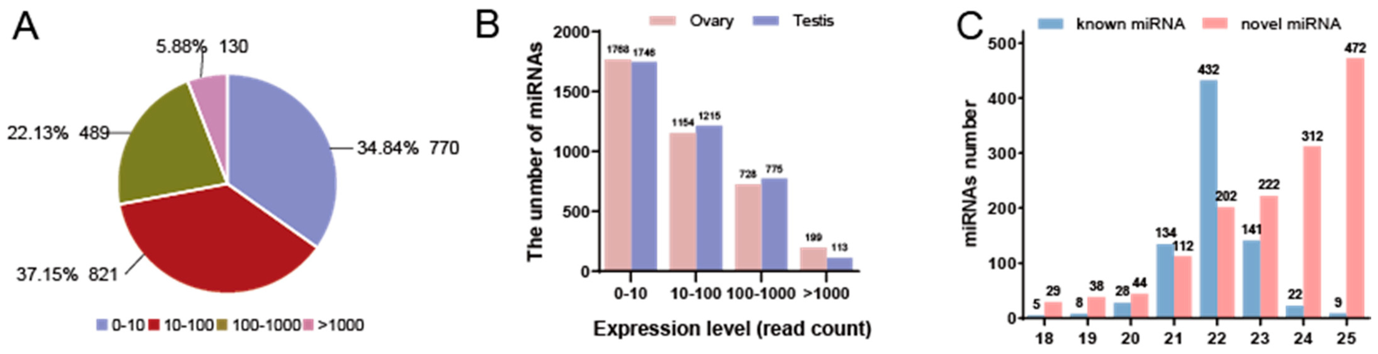
3.1. Construction of miRNA Library

Six small RNA libraries were constructed and sequenced. After sequencing, a total of 2.4 Gb of raw sequencing data was obtained from six small RNA libraries, with an average Q30 value exceeding 95.93% across all libraries. After the removal of contaminants and poor-quality sequences, a total of 76.60 Mb of clean reads was obtained, with more than 9.88 Mb of clean reads for each sample (Table 2).

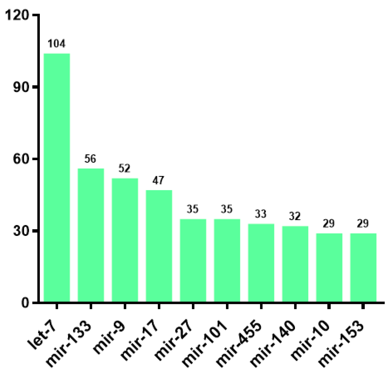
3.2. Identification and Classification of miRNAs

Through comparison with the miRBase database and analysis of miRNA biosignatures, a total of 2210 miRNAs were identified, comprising 779 known miRNAs and 1431 novel miRNA candidates (Supplementary Table S2). Among these miRNAs, 1161 miRNAs were annotated and classified into 218 families, and the ten most abundant miRNA families were let-7, mir-133, mir-9, mir-17, mir-27, mir-455, mir-140, mir-10, mir-30, and mir-153 (Figure 1).
TPM normalization analysis revealed that the majority of miRNAs (37.15%, n = 821) exhibited expression levels from 10–100, followed by those in the 0–10 range (34.84%, n = 770). miRNAs with expression >1000 accounted for the smallest proportion (5.88%, n = 130) (Figure 2A). In addition, the proportion of miRNAs with different ranges of expression levels was similar in the ovary and testis (Figure 2B). In detail, the majority of known miRNAs had length 22 nt, whereas newly predicted miRNAs showed a peak at 25 nt (Figure 2C). Nucleotide composition analysis revealed that a consistent uracil (U) bias in both known and novel miRNAs, accounting for 30.66% and 30.33%, respectively. While conserved miRNAs exhibited a nucleotide preference order of U > adenine (A) > guanine (G) > cytosine (C), novel miRNAs displayed a distinct pattern of U > G > C > A (Supplementary Figure S1).

3.3. Identification and Validation of Differentially Expressed miRNAs (DEMs)

Differential expression analysis identified 482 DEMs displaying sexually dimorphic expression patterns, comprising 179 testis-biased and 303 ovary-biased miRNAs (Figure 3). Among these, 13 DEMs exhibited exclusive ovarian expression while 17 were uniquely expressed in testicular tissue. To validate the expression profiles of these miRNAs, eight DEMs were randomly selected for RT-qPCR verification. The results showed that novel_miR_1049 (Ovarian-specific expression), novel_miR_1203, novel_miR_1006, and novel_miR_826 were significantly upregulated in the ovary (p < 0.05), whereas sar-miR-10d-1, novel_miR_579, novel_miR_110, and novel_miR_1112 were significantly upregulated in the testis (p < 0.05) (Figure 4) (Supplementary Figure S2). The expression patterns detected by RT-qPCR were consistent with the sequencing results, confirming the accuracy and reliability of the sequencing data.

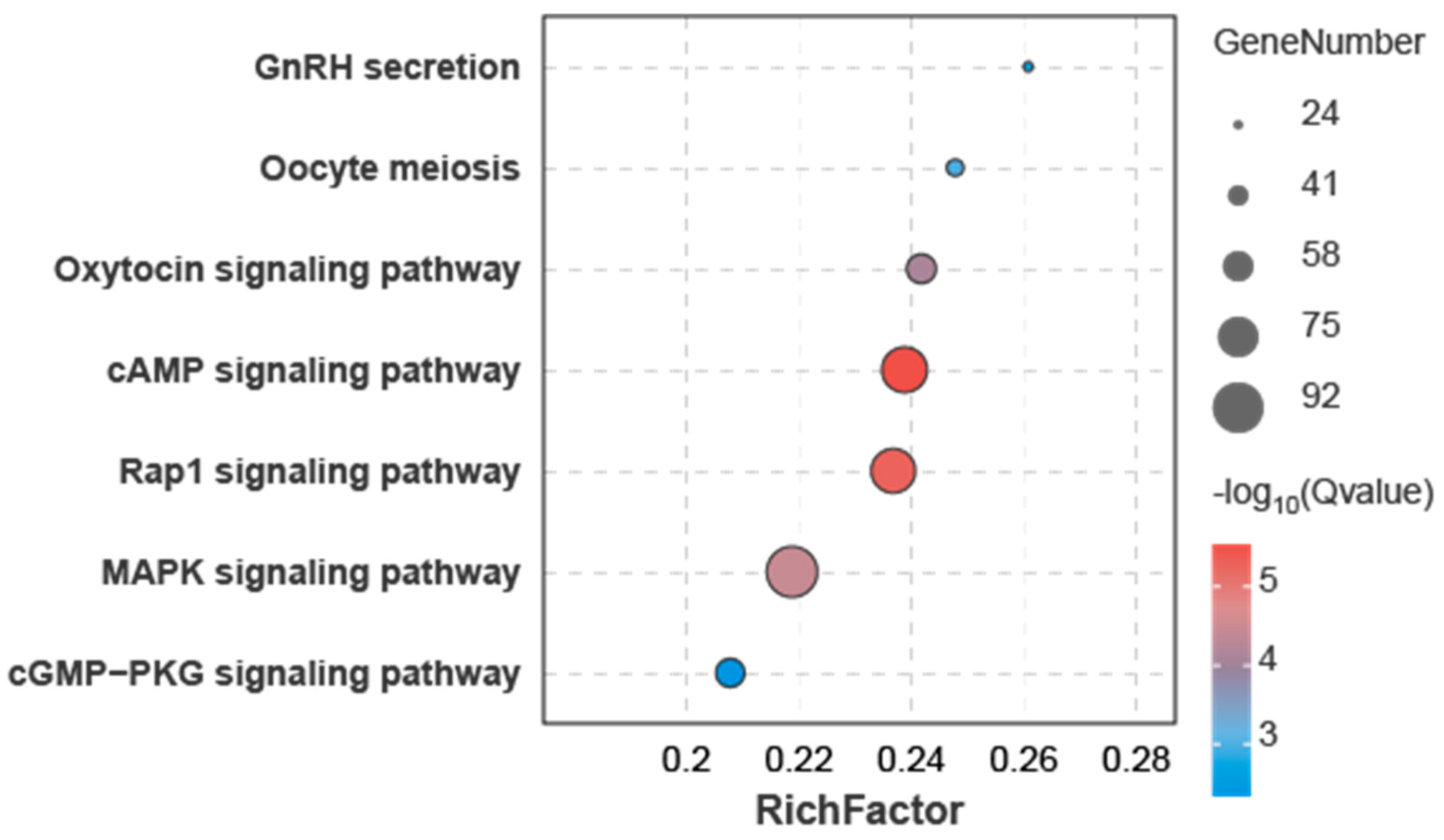
3.4. Joint miRNA and Transcriptome Analysis

Transcriptome profiling of the corresponding samples identified 9048 DEGs exhibiting sex-biased expression patterns in our previous studies [34]. Integrative analysis of DEMs and DEGs identified 462 DEMs potentially targeting 3340 DEGs through 3′UTR binding sites, forming 13,773 DEM–DEG regulatory pairs. Of these, 9403 pairs (68.27%) showed inverse differential expression trends, while 4370 pairs (31.73%) displayed consistent trends (Supplementary Table S3). Functional enrichment analysis of the 3340 DEGs identified significant associations with two GO terms and 18 KEGG pathways (Supplementary Table S4). These DEGs were particularly enriched in several pathways related to gonadal development and function, including the cAMP signaling pathway, Rap1 signaling pathway, MAPK signaling pathway, oxytocin signaling pathway, oocyte meiosis, GnRH secretion, and cGMP-PKG signaling pathway (Figure 5).

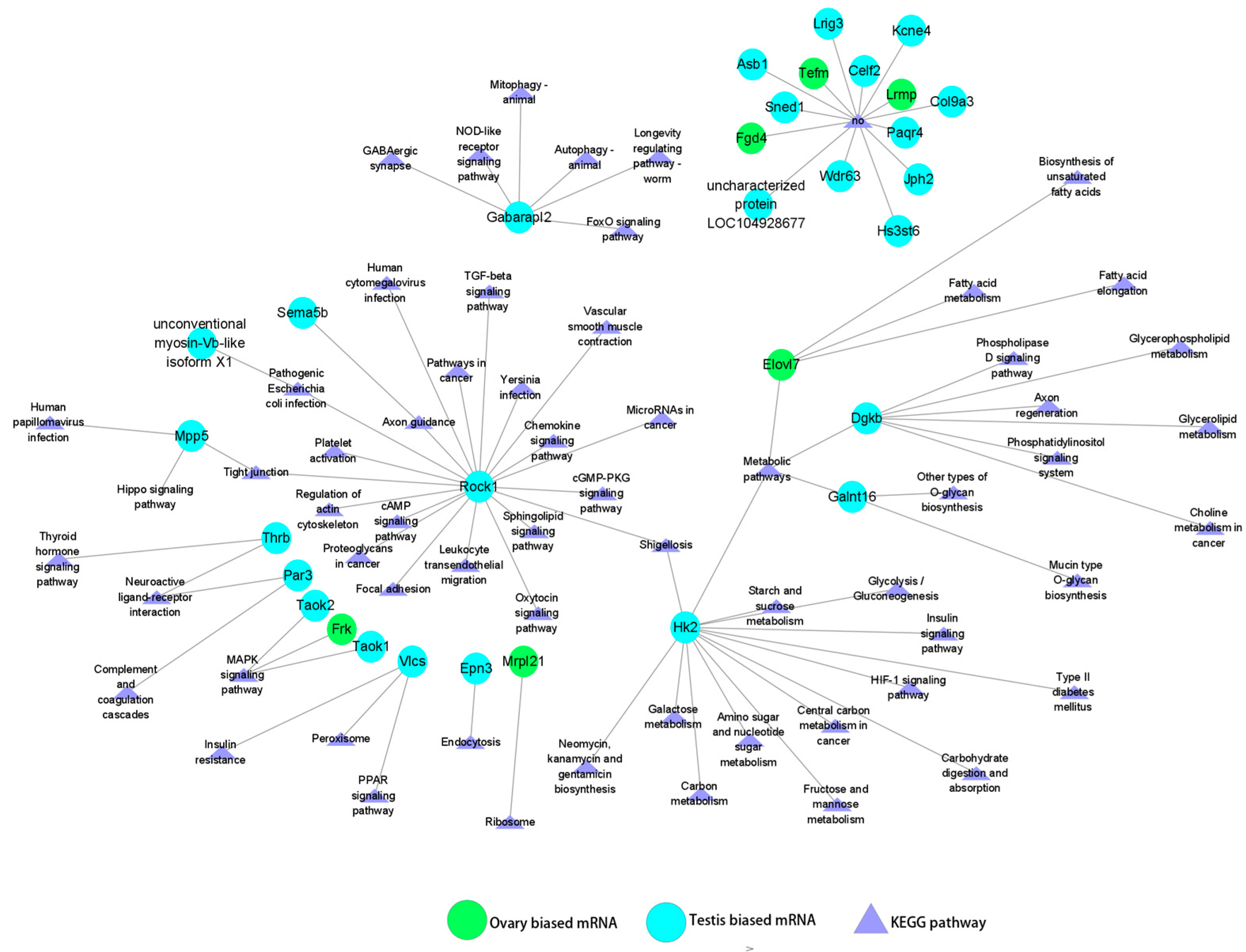
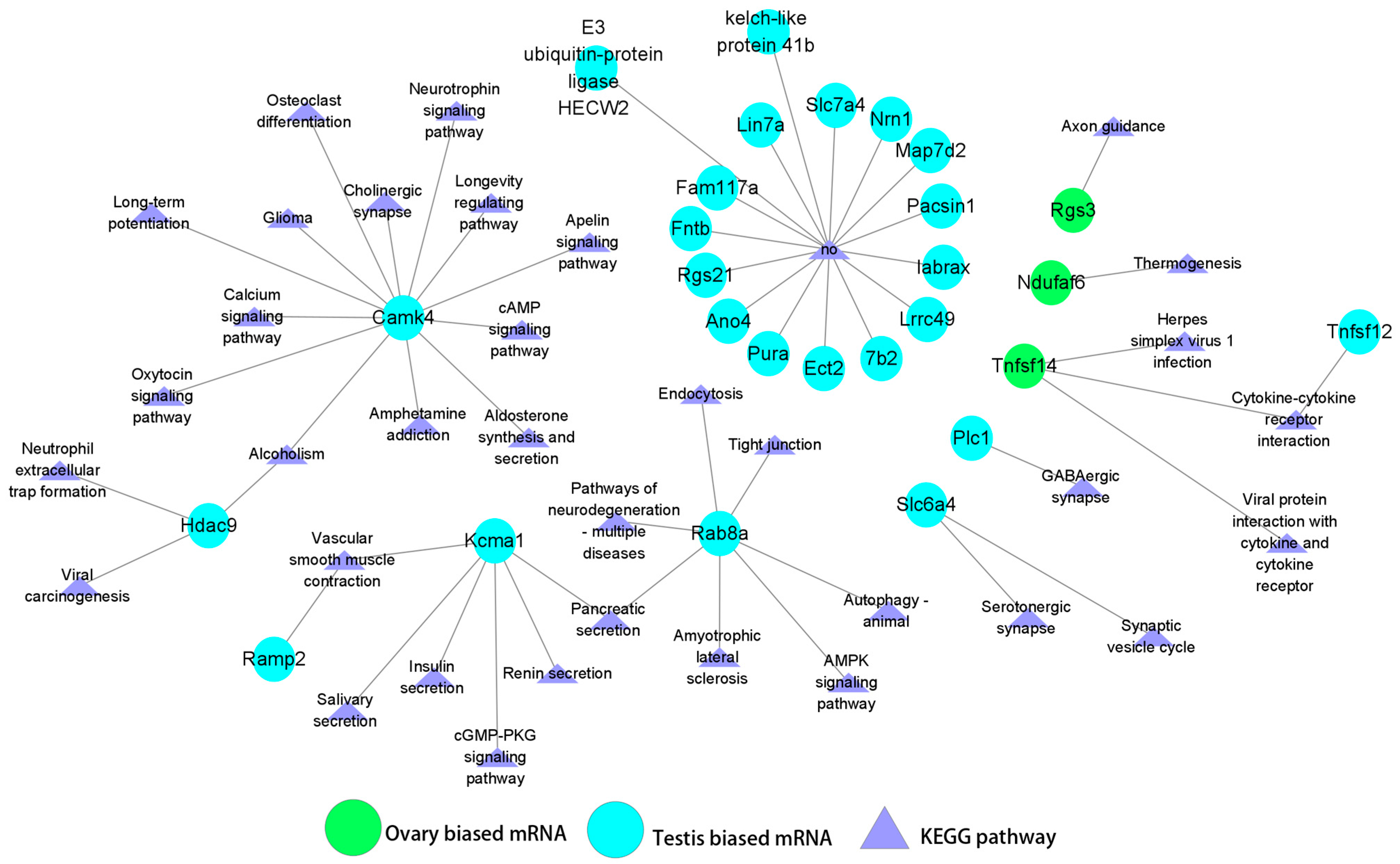
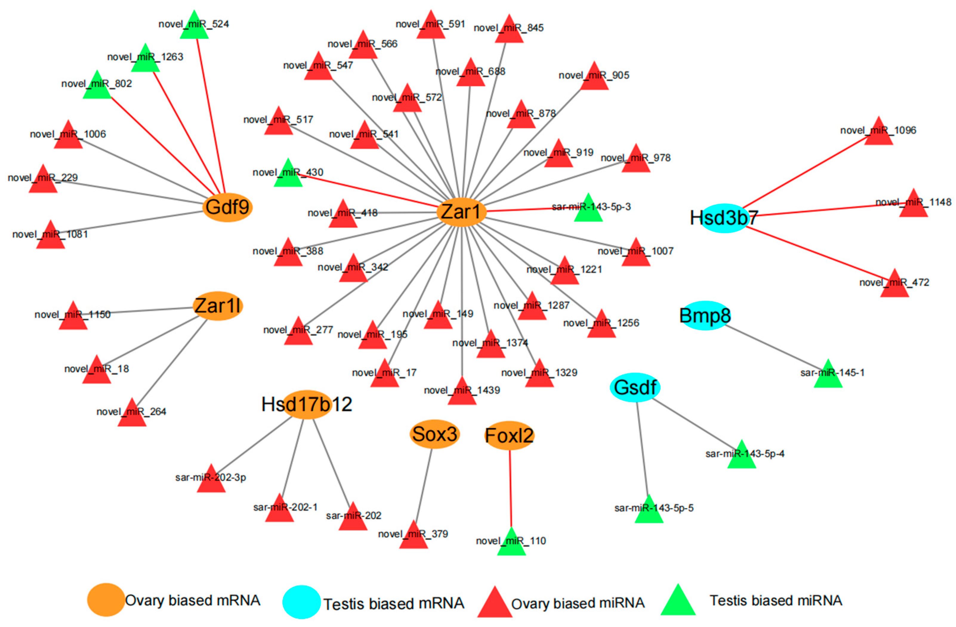
3.5. Regulatory Relationships Between miRNAs (DEMs) and Sex-Biased Genes (DEGs)

In the ovary, novel_miR_1351 exhibited the highest expression level among the DEMs, and was predicted to target 31 DEGs. Among these 31 DEGs, the majority (25 genes, 80.65%) displayed testis-biased expression, including Rock1, Hk2, Thrb, and Par3, while a smaller proportion (6 genes, 19.35%) showed ovarian-biased expression, such as Frk, Mrpl21, and Elovl7. Notably, certain target genes may play a role in the process of gonadal development. For instance, Rock1 is a component of the TGF-β, cAMP, cGMP-PKG, and oxytocin signaling pathways [6,44,45,46], which are closely associated with gonadal development and sex determination. Additionally, Taok1, Taok2, and Frk are members of the MAPK signaling pathway, which has also been shown to be involved in the regulation of gonadal development [47] (Figure 6). In contrast, sar-miR-143-3p exhibited a distinct expression pattern, showing the highest expression level in the testis. This miRNA was predicted to target 27 DEGs, of which the majority (24 genes, 88.89%) displayed testis-biased expression, including Camk4, Kcmal, Rab8a, and Slc6a4, while only 3 genes (Rgs3, Ndufaf6, and Tnfsf14) showed ovarian-biased expression (Figure 7).
Based on previous studies of S. argus, 21 key sex-related genes were screened for their potential regulatory relationships with miRNAs (Supplementary Table S5). The results showed that three male development-associated genes (Gsdf, Hsd3b7, and Bmp8) (DEGs) demonstrated binding sites with 6 DEMs, while six female development-related genes (Zar1, Zar1l, Gdf9, Hsd17b12, Sox3, and Foxl2) (DEGs) exhibited binding sites with 42 DEMs. Among these interactions, 9 DEM–DEG pairs showed inverse differential expression trends, whereas 39 DEM–DEG pairs displayed consistent trends (Figure 8). Among them, miRNAs showed an inverse expression trend with Foxl2/novel_miR_110, Gdf9/novel_miR_802 and Gdf9/novel_miR_1263, while miRNAs showed the same expression trend with Gsdf/sar-miR-143-5p-4, Gsdf/sar-miR-143-5p-5 and Sox3/novel_miR_379 showed the same expression trend.

4. Discussion

At present, high-throughput sRNA sequencing has emerged as a prevalent methodology for miRNA identification. This advanced approach has enhanced the discovery and characterization of a substantial number of miRNAs across diverse tissues in various fish species. In this study, we delineated the regulatory networks between miRNAs and their target genes in S. argus gonads through an integrated analysis of DEMs and DEGs utilizing next-generation sequencing technology. The findings yield novel insights into the potential regulatory mechanisms of miRNAs in regulating gene expression during gonadal development, including sex determination and differentiation in S. argus. Moreover, these data establish a molecular foundation for advancing sex-controlled breeding strategies in S. argus.
Our analysis identified a total of 2210 miRNAs in S. argus, of which 1431 were novel miRNAs, representing a substantial proportion (64.75%) of the total miRNAs. Comparative analysis revealed interspecies variation in miRNA profiles across different fish species. For example, Trachinotus ovatus (T. ovatus) exhibited 279 miRNAs (100 novel, 35.84%), Acipenser schrencki (A. schrenckii) harbored 730 miRNAs (51 novel, 6.99%), and Trachinotus blochii (T. blochii) contained 1453 miRNAs (69 novel, 4.81%), demonstrating divergence in miRNA composition [48,49,50]. Notably, S. argus demonstrated both the highest number of DEMs and the greatest proportion of novel miRNAs among the four analyzed species. It is particularly relevant to mention that the identification of DEMs in S. argus was conducted using more stringent screening criteria (Supplementary Table S6). However, comparisons of miRNA sequencing data across these four species may be influenced by differences in experimental conditions, sequencing depth, and annotation methods. Notably, despite these considerations, sar-miR-214-3p-2 emerged as a conserved miRNA across all species, albeit with distinct expression patterns: exhibiting testis-biased expression in S. argus (identified as a DEM in our study), ovary-biased expression in T. ovatus, and no significant sex-biased expression in A. schrenckii or T. blochii. In S. argus specifically, we identified 117 potential target genes of sar-miR-214-3p-2, including 6 genes associated with sex-related signaling pathways (Supplementary Table S7). These findings, while highlighting potential species-specific regulatory roles, underscore the need for standardized comparative approaches and functional validation to determine whether miR-214-3p-2′s regulatory function is evolutionarily conserved among teleosts.
Emerging evidence reveals that miRNA-mediated negative regulation plays a fundamental role in sexual development across diverse species. In the gonad of the S. argus, a high proportion of predicted DEMs demonstrate inverse expression patterns relative to their target DEGs. This observation is consistent with established DEM–DEG regulatory mechanisms reported in previous studies [51,52]. This inverse correlation suggests that highly expressed miRNAs may function through translational inhibition or mRNA degradation of their target mRNAs, thereby downregulating gene expression, whereas lowly expressed miRNAs exhibit weakened translational repression or degradative effects on their target genes. Typically, miRNAs exert their regulatory effects by binding to target mRNAs, establishing a negative regulatory relationship. For instance, in Paralichthys olivaceus (P. olivaceus), miR-202-5p exhibits marked testis-specific expression, showing significantly higher abundance in male gonads compared to ovarian tissues. Molecular characterization revealed that miR-202-5p directly targets Cbx2, a key transcriptional regulator of gonadal development. This miRNA–mRNA interaction establishes a clear negative regulatory relationship, wherein miR-202-5p-mediated suppression of Cbx2 appears to facilitate spermatogenic processes [53]. A conserved regulatory paradigm was identified in the B. dorsalis, where miR-1-3p demonstrates sexually dimorphic expression, being preferentially expressed in male embryos during the critical sex determination period. Comprehensive functional analyses, including target prediction and experimental validation, confirmed that miR-1-3p negatively regulates the master sex-determining gene Bdtra. Through a combination of embryonic microinjection and gene editing approaches, researchers conclusively demonstrated that miR-1-3p serves as a critical upstream modulator of male sex determination in this dipteran species [20].
In the ovaries of the S. argus, novel_miR_1351, a member of the miR-10 family, demonstrates the highest expression level among all differentially expressed miRNAs (DEMs). Furthermore, the genes predicted to be targeted by novel_miR_1351 are significantly enriched in pathways associated with gonad development (Figure 6), suggesting a potential regulatory role of this miRNA in ovarian function and development. This finding aligns with previous studies demonstrating that the miR-10 family plays critical regulatory roles in ovarian granulosa cells (GCs) across various species, including human, mouse, rat, and pig. miR-10a and miR-10b have been demonstrated to regulate the proliferation and apoptosis of GCs by targeting Bdnf [54]. Similarly, in pig ovarian GCs, miR-10a-5p has been shown to suppress steroid hormone synthesis through its targeting of Creb1 [55]. These findings highlight the conserved and multifaceted regulatory roles of the miR-10 family in ovarian function across species. In the present study, although Bdnf and Creb1 were not identified as DEGs, target prediction analysis using miRanda revealed that Creb1 is potentially regulated by 10 miRNAs. These include novel_miR_380 and novel_miR_1227 (belonging to the miR-263 family), as well as novel_miR_125, novel_miR_1342, novel_miR_1396, novel_miR_175, novel_miR_246, novel_miR_407, novel_miR_511, and novel_miR_619. Notably, among these, only novel_miR_246 was identified as a DEM, exhibiting higher expression in the testis compared to the ovary. This suggests a potential tissue-specific regulatory role of novel_miR_246 in modulating Creb1 expression. However, no miRNA–mRNA targeting relationships were predicted for Bdnf in the S. argus, suggesting that its regulation in this species may involve alternative mechanisms or remain unidentified under the current analysis framework. On the other hand, sar-miR-143-3p is the most highly expressed miRNA in the testis of the S. argus. Previous studies have identified Kras and Fshr as target genes of miR-143 in mouse and bovine GCs, respectively. Overexpression of miR-143 has been shown to downregulate the expression of P450scc, 3β-HSD, and StAR, leading to reduced levels of progesterone (P4) and estradiol (E2) in bovine GCs, whereas inhibition of miR-143 produces the opposite effect [56,57]. Notably, the sequence of sar-miR-143-3p is identical to that of mouse miR-143 (Mmu-miR-143-3p), while bovine miR-143 (bta-miR-143) differs by an additional G at the 3′ end. However, in this study, neither Kras nor Fshr were predicted as target genes of sar-miR-143-3p (Figure 7). Instead, Kras was predicted to be regulated by novel_miR_1430, novel_miR_288, novel_miR_292, and novel_miR_1215, while Fshr was predicted to be targeted by novel_miR_108, novel_miR_1216, novel_miR_238, novel_miR_482, novel_miR_58, novel_miR_635, and novel_miR_649. These findings suggest potential species-specific differences in miRNA-mediated regulatory networks.
The DEMs with target sites on critical genes involved in sex determination and differentiation were also predicted in the S. argus. As mentioned earlier, Foxl2 is a key gene in the female pathway. In mice, Foxl2 expression in ovarian granulosa cells is negatively regulated by miR-133b, influencing granulosa cell function and follicular development [58]. In O. niloticus gonads at 5 days after hatching (dah), a DEM (miR-7977) has been predicted to regulate Foxl2 [59]. In the S. argus, the predicted regulatory DEM for Foxl2 is novel_miR_110, which differs from the miRNAs identified in mice and O. niloticus. Another crucial gene in the female pathway is Cyp19a1/Cyp19a1a. In pigs, miR-10b has been shown to regulate Cyp19a1, inhibiting its expression and function in granulosa cells [60]. In O. niloticus gonads at 5 dah, a DEM, miR-30a, has been predicted to regulate Cyp19a1a [59]. Similarly, in the E. coioides, miR-26a-5p, which is highly expressed in the testis, can downregulate Cyp19a1a expression [21]. However, no regulatory miRNAs for Cyp19a1 were predicted in the S. argus. These observations indicate that the regulatory miRNAs for Cyp19a1 vary significantly among pigs, O. niloticus, and E. coioides. Collectively, these findings underscore the species-specific nature of miRNA regulatory mechanisms governing female pathway-related genes, highlighting the diversity and complexity of miRNA-mediated regulation across different species.
One key objective of this study was to identify miRNAs that might regulate Dmrt1Y, a sex-determining gene in S. argus [31,32]. While our bioinformatic analyses failed to predict any miRNAs targeting Dmrt1Y in this species, computational studies have identified potential Dmrt1-regulating miRNAs in several other fish species. These include miR-212 (a DEM) in O. niloticus and multiple DEMs (asc-miR-159a, 2779, 2779-1, and 203b-3p) in A. schrenckii [48,59]. Notably, in Takifugu rubripes (T. rubripes), experimental validation has confirmed that fru-miRNA-122 (a DEM) directly downregulates Dmrt1 expression [14]. Gsdf, a gene downstream of Dmrt1 in the S. argus [61], is predicted to be targeted by sar-miR-143-5p-4 and sar-miR-143-5p-5 in this species. Similarly, the T. rubripes suggest that Gsdf may be regulated by miR-730, fru-miR-216b, and several DEMs, including novel-m0524-5p, m0272-5p, m0408-5p, and m0087-3p [14]. Another gene involved in gonad development, Amh, exhibits male-biased sexual dimorphism in expression. In Cyprinus carpio (C. carpio); miR-153b-3p, which shows sexually dimorphic expression in the gonads; inhibits Amh expression, thereby modulating male germ cell proliferation and differentiation during spermatogenesis [13]. Bioinformatic predictions identified DEMs (miR-456 and miR-138) as potential regulators targeting Amh in 60 dah O. niloticus [62], while DEMs (miR-96 and miR-449) were predicted to be regulatory factors of Amh in 5 dah O. niloticus [59]. However, in the S. argus, no miRNAs were predicted to regulate Amh. These results highlight the lack of conservation in miRNA-mediated regulation of genes involved in sex determination and differentiation across fish species, underscoring the diversity and complexity of these regulatory networks.
The observed interspecies differences in miRNA regulation of the male sex-determination gene Dmrt1Y and other gonad development-related genes (Foxl2, Cyp19a1/Cyp19a1a, Gsdf, and Amh) in S. argus may arise from both technical and biological factors. Since the predicted regulatory relationships between these genes and DEMs have not been experimentally validated in either this study or some comparative studies, the observed variations could result from technical differences in miRNA and 3′UTR sequencing analyses (including sequencing platforms, library preparation methods, and bioinformatics pipelines), as well as discrepancies in target prediction algorithms and their parameter settings. When the predicted gene–DEM regulatory pairs exhibit minimal technical artifacts or have been experimentally validated across species, these observed differences may reflect species-specific characteristics in the miRNA regulatory mechanisms of gonad development-related genes from a biological perspective. In cases where highly conserved miRNAs are identified among the compared species, the differential gene–DEM regulatory relationships may result from species-specific variations in the 3′UTRs of conserved genes. Conversely, when miRNAs show low sequence similarity across species, the observed interspecies differences could originate from either the miRNAs themselves, the 3′UTRs of conserved genes, or both exhibiting species-specific features.

Supplementary Materials

The following supporting information can be downloaded at: https://www.mdpi.com/article/10.3390/ani15111564/s1. Table S1. Samples information; Table S2. All miRNA sequence information and sex−biased expression tables. Table S3. DEM-DEG predicted interaction pairs. Table S4. Predicted GO and KEGG regulatory networks of DEGs in DEM-DEG interaction pairs. Table S5. Expression profiles of 21 key sex-related genes in stage IV gonads of both sexes. Table S6. Criteria for screening DEMs in S. argus, T. ovatus, A. schrenckii, and T. blochii. Table S7. Six sex-related pathway genes targeted by sar-miR-214-3p-2 in S. argus. Figure S1. miRNA nucleotide bias analyses; Figure S2. Sexually dimorphic expression of DEMs validated by RT-qPCR. Supplementary Document S1. Approval of Animal Use Protocol, IACUC, GDOU.

Author Contributions

Conceptualization, Y.L., G.L., W.T. and D.J.; Methodology, Y.L., K.J., H.C. and W.T.; Validation, Y.L., Y.W. and S.D.; Formal analysis, G.S., H.S. and H.C.; Investigation, G.S., H.S. and S.D.; Resources, G.S., S.D. and G.L.; Data curation, Y.L., K.J. and Y.W.; Writing—original draft, Y.L. and Y.H.; Writing—review and editing, Y.H. and D.J.; Supervision, G.L., W.T. and D.J.; Project administration, D.J.; Funding acquisition, D.J. All authors have read and agreed to the published version of the manuscript.

Funding

This study was supported by grants from the National Natural Science Foundation of China [grant numbers: 32172971 and 32273131]; Guangdong Basic and Applied Basic Research Foundation [grant number: 2024A1515010328]; Guangdong Provincial Science and Technology Programme [grant number: 2023B0202010005]; Youth science and technology innovation talent of guangdong TeZhi plan talent [grant number: 2023TQ07A888].

Institutional Review Board Statement

All animal experiments were conducted in accordance with the guidelines of the Animal Research and Ethics Committee of Guangdong Ocean University (201903004, 4 March 2019).

Informed Consent Statement

Not applicable.

Data Availability Statement

The data that support the findings of this study are available upon reasonable request. The raw reads used in this article have been deposited into the Sequence Read Archive (SRA) of the NCBI database under BioProject accession number: PRJNA1149578.

Conflicts of Interest

The authors declare no conflicts of interest.

References

  1. Baroiller, J.F.; D’Cotta, H. The reversible sex of gonochoristic fish: Insights and consequences. Sex. Dev. 2016, 10, 242–266. [Google Scholar] [CrossRef] [PubMed]
  2. Nagahama, Y.; Chakraborty, T.; Paul-Prasanth, B.; Ohta, K.; Nakamura, M. Sex determination, gonadal sex differentiation, and plasticity in vertebrate species. Physiol. Rev. 2021, 101, 1237–1308. [Google Scholar] [CrossRef]
  3. Hattori, R.S.; Strüssmann, C.A.; Fernandino, J.I.; Somoza, G.M. Genotypic sex determination in teleosts: Insights from the testis-determining amhy gene. Gen. Comp. Endocrinol. 2013, 192, 55–59. [Google Scholar] [CrossRef]
  4. Matsuda, M.; Nagahama, Y.; Shinomiya, A.; Sato, T.; Matsuda, C.; Kobayashi, T.; Morrey, C.E.; Shibata, N.; Asakawa, S.; Shimizu, N.; et al. DMY is a Y-specific DM-domain gene required for male development in the medaka fish. Nature 2002, 417, 559–563. [Google Scholar] [CrossRef]
  5. Liew, W.C.; Orbán, L. Zebrafish sex: A complicated affair. Brief. Funct. Genom. 2014, 13, 172–187. [Google Scholar] [CrossRef] [PubMed]
  6. Yu, H.; Du, X.; Chen, X.; Liu, L.; Wang, X. Transforming growth factor-β (TGF-β): A master signal pathway in teleost sex determination. Gen. Comp. Endocrinol. 2024, 355, 114561. [Google Scholar] [CrossRef]
  7. Sreenivasan, R.; Jiang, J.; Wang, X.; Bártfai, R.; Kwan, H.Y.; Christoffels, A.; Orbán, L. Gonad differentiation in zebrafish is regulated by the canonical Wnt signaling pathway. Biol. Reprod. 2014, 90, 45. [Google Scholar] [CrossRef] [PubMed]
  8. Hattori, R.S.; Murai, Y.; Oura, M.; Masuda, S.; Majhi, S.K.; Sakamoto, T.; Fernandino, J.I.; Somoza, G.M.; Yokota, M.; Strüssmann, C.A. A Y-linked anti-Müllerian hormone duplication takes over a critical role in sex determination. Proc. Natl. Acad. Sci. USA 2012, 109, 2955–2959. [Google Scholar] [CrossRef]
  9. Jiang, D.N.; Yang, H.H.; Li, M.H.; Shi, H.J.; Zhang, X.B.; Wang, D.S. gsdf is a downstream gene of dmrt1 that functions in the male sex determination pathway of the Nile tilapia. Mol. Reprod. Dev. 2016, 83, 497–508. [Google Scholar] [CrossRef]
  10. Dai, S.; Qi, S.; Wei, X.; Liu, X.; Li, Y.; Zhou, X.; Xiao, H.; Lu, B.; Wang, D.; Li, M. Germline sexual fate is determined by the antagonistic action of dmrt1 and foxl3/foxl2 in tilapia. Development 2021, 148, dev199380. [Google Scholar] [CrossRef]
  11. Liu, X.; Dai, S.; Wu, J.; Wei, X.; Zhou, X.; Chen, M.; Tan, D.; Pu, D.; Li, M.; Wang, D. Roles of anti-Müllerian hormone and its duplicates in sex determination and germ cell proliferation of Nile tilapia. Genetics 2022, 220, iyab237. [Google Scholar] [CrossRef] [PubMed]
  12. Tachiwana, H.; Saitoh, N. Nuclear long non-coding RNAs as epigenetic regulators in cancer. Curr. Med. Chem. 2021, 28, 5098–5109. [Google Scholar] [CrossRef] [PubMed]
  13. Ma, W.; Chen, K.; Jiang, M.; Jia, S.; Chen, J.; Tao, B.; Song, Y.; Li, Y.; Wang, Y.; Xiao, W.; et al. MiR-153b-3p regulates the proliferation and differentiation of male germ cells by targeting amh in common carp (Cyprinus carpio). Aquaculture 2021, 535, 736420. [Google Scholar] [CrossRef]
  14. Shen, X.; Yan, H.; Hu, M.; Zhou, H.; Wang, J.; Gao, R.; Liu, Q.; Wang, X.; Liu, Y. The potential regulatory role of the non-coding RNAs in regulating the exogenous estrogen-induced feminization in Takifugu rubripes gonad. Aquat. Toxicol. 2024, 273, 107022. [Google Scholar] [CrossRef]
  15. Bartel, D.P. MicroRNAs: Genomics, biogenesis, mechanism, and function. Cell 2004, 116, 281–297. [Google Scholar] [CrossRef]
  16. Meijer, H.A.; Kong, Y.W.; Lu, W.T.; Wilczynska, A.; Spriggs, R.V.; Robinson, S.W.; Godfrey, J.D.; Willis, A.E.; Bushell, M. Translational repression and eIF4A2 activity are critical for microRNA-mediated gene regulation. Science 2013, 340, 82–85. [Google Scholar] [CrossRef]
  17. Gu, W.; Xu, Y.; Xie, X.; Wang, T.; Ko, J.H.; Zhou, T. The role of RNA structure at 5′ untranslated region in microRNA-mediated gene regulation. RNA 2014, 20, 1369–1375. [Google Scholar] [CrossRef]
  18. Winter, J.; Jung, S.; Keller, S.; Gregory, R.I.; Diederichs, S. Many roads to maturity: microRNA biogenesis pathways and their regulation. Nat. Cell Biol. 2009, 11, 228–234. [Google Scholar] [CrossRef]
  19. Hurtado, A.; Mota-Gómez, I.; Lao, M.; Real, F.M.; Jedamzick, J.; Burgos, M.; Lupiáñez, D.G.; Jiménez, R.; Barrionuevo, F.J. Complete male-to-female sex reversal in XY mice lacking the miR-17~92 cluster. Nat. Commun. 2024, 15, 3809. [Google Scholar] [CrossRef]
  20. Peng, W.; Yu, S.; Handler, A.M.; Tu, Z.; Saccone, G.; Xi, Z.; Zhang, H. miRNA-1-3p is an early embryonic male sex-determining factor in the Oriental fruit fly Bactrocera dorsalis. Nat. Commun. 2020, 11, 932. [Google Scholar] [CrossRef]
  21. Yu, Q.; Peng, C.; Ye, Z.; Tang, Z.; Li, S.; Xiao, L.; Liu, S.; Yang, Y.; Zhao, M.; Zhang, Y.; et al. An estradiol-17β/miRNA-26a/cyp19a1a regulatory feedback loop in the protogynous hermaphroditic fish, Epinephelus coioides. Mol. Cell. Endocrinol. 2020, 504, 110689. [Google Scholar] [CrossRef] [PubMed]
  22. Zhang, L.; Yang, Q.; Xu, W.; Wu, Z.; Li, D. Integrated analysis of miR-430 on steroidogenesis-related gene expression of larval rice field eel Monopterus albus. Int. J. Mol. Sci. 2021, 22, 6994. [Google Scholar] [CrossRef] [PubMed]
  23. Tang, L.; You, W.; Wang, Q.; Huang, F.; Shao, C. MicroRNA ssa-mir-196a-4 decreases lgr8 expression in testis development of Chinese tongue sole (Cynoglossus semilaevis). Comp. Biochem. Physiol. B Biochem. Mol. Biol. 2022, 258, 110695. [Google Scholar] [CrossRef]
  24. Zhao, L.; Dong, Q.; Luo, C.; Wu, Y.; Bu, D.; Qi, X.; Luo, Y.; Zhao, Y. DeepOmix: A scalable and interpretable multi-omics deep learning framework and application in cancer survival analysis. Comput. Struct. Biotechnol. J. 2021, 19, 2719–2725. [Google Scholar] [CrossRef]
  25. O’Connor, L.M.; O’Connor, B.A.; Lim, S.B.; Zeng, J.; Lo, C.H. Integrative multi-omics and systems bioinformatics in translational neuroscience: A data mining perspective. J. Pharm. Anal. 2023, 13, 836–850. [Google Scholar] [CrossRef] [PubMed]
  26. Skaftnesmo, K.O.; Edvardsen, R.B.; Furmanek, T.; Crespo, D.; Andersson, E.; Kleppe, L.; Taranger, G.L.; Bogerd, J.; Schulz, R.W.; Wargelius, A. Integrative testis transcriptome analysis reveals differentially expressed miRNAs and their mRNA targets during early puberty in Atlantic salmon. BMC Genom. 2017, 18, 801. [Google Scholar] [CrossRef]
  27. Liu, S.; Yang, Q.; Chen, Y.; Liu, Q.; Wang, W.; Song, J.; Zheng, Y.; Liu, W. Integrated analysis of mRNA- and miRNA-Seq in the ovary of rare minnow Gobiocypris rarus in response to 17α-methyltestosterone. Front. Genet. 2021, 12, 695699. [Google Scholar] [CrossRef]
  28. Lai, K.P.; Tam, N.Y.K.; Chen, Y.; Leung, C.T.; Lin, X.; Tsang, C.F.; Kwok, Y.C.; Tse, W.K.F.; Cheng, S.H.; Chan, T.F.; et al. miRNA-mRNA integrative analysis reveals the roles of miRNAs in hypoxia-altered embryonic development- and sex determination-related genes of medaka fish. Front. Mar. Sci. 2021, 8, 736362. [Google Scholar] [CrossRef]
  29. Assan, D.; Wang, Y.; Mustapha, U.F.; Ndandala, C.B.; Li, Z.; Li, G.L.; Chen, H. Neuropeptide Y in spotted scat (Scatophagus argus), characterization and functional analysis towards feed intake regulation. Fishes 2022, 7, 111. [Google Scholar] [CrossRef]
  30. Chen, H.; Jiang, D.; Li, Z.; Wang, Y.; Yang, X.; Li, S.; Li, S.; Yang, W.; Li, G. Comparative Physiological and Transcriptomic profiling offers insight into the sexual dimorphism of hepatic metabolism in size-dimorphic spotted Scat (Scatophagus argus). Life 2021, 11, 589. [Google Scholar] [CrossRef]
  31. Mustapha, U.F.; Huang, Y.; Huang, Y.Q.; Assan, D.; Shi, H.J.; Jiang, M.Y.; Deng, S.P.; Li, G.L.; Jiang, D.N. Gonadal development and molecular analysis revealed the critical window for sex differentiation, and E2 reversibility of XY-male spotted scat, Scatophagus argus. Aquaculture 2021, 544, 737147. [Google Scholar] [CrossRef]
  32. Huang, Y.-Q.; Zhang, X.-H.; Bian, C.; Jiao, K.-Z.; Zhang, L.; Huang, Y.; Yang, W.; Li, Y.; Shi, G.; Huang, Y.; et al. Allelic variation and duplication of the dmrt1 were associated with sex chromosome turnover in three representative Scatophagidae fish species. Commun. Biol. 2025, 8, 627. [Google Scholar] [CrossRef] [PubMed]
  33. Fischer, A.H.; Jacobson, K.A.; Rose, J.; Zeller, R. Hematoxylin and eosin staining of tissue and cell sections. CSH Protoc. 2008, 4, pdb-prot4986. [Google Scholar] [CrossRef]
  34. Jiao, K.; Li, Y.; Huang, Y.; Ndandala, C.B.; Shi, G.; Deng, S.; Shi, H.; Chen, H.; Li, G.; Jiang, D. Integrated analysis of the gonadal methylome and transcriptome provides new insights into the expression regulation of sex determination and differentiation genes in spotted scat (Scatophagus argus). Aquaculture 2024, 589, 740974. [Google Scholar] [CrossRef]
  35. Chen, S.; Zhou, Y.; Chen, Y.; Gu, J. fastp: An ultra-fast all-in-one FASTQ preprocessor. Bioinformatics 2018, 34, i884–i890. [Google Scholar] [CrossRef]
  36. Langmead, B.; Trapnell, C.; Pop, M.; Salzberg, S.L. Ultrafast and memory-efficient alignment of short DNA sequences to the human genome. Genome Biol. 2009, 10, R25. [Google Scholar] [CrossRef]
  37. Friedlander, M.R.; Mackowiak, S.D.; Li, N.; Chen, W.; Rajewsky, N. miRDeep2 accurately identifies known and hundreds of novel microRNA genes in seven animal clades. Nucleic Acids Res. 2012, 40, 37–52. [Google Scholar] [CrossRef]
  38. De Oliveira, L.F.; Christoff, A.P.; Margis, R. isomiRID: A framework to identify microRNA isoforms. Bioinformatics 2013, 29, 2521–2523. [Google Scholar] [CrossRef]
  39. Li, B.; Ruotti, V.; Stewart, R.M.; Thomson, J.A.; Dewey, C.N. RNA-Seq gene expression estimation with read mapping uncertainty. Bioinformatics 2009, 26, 493–500. [Google Scholar] [CrossRef]
  40. Love, M.I.; Huber, W.; Anders, S. Moderated estimation of fold change and dispersion for RNA-seq data with DESeq2. Genome Biol. 2014, 15, 550. [Google Scholar] [CrossRef]
  41. Shi, R.; Chiang, V.L. Facile means for quantifying microRNA expression by real-time PCR. Biotechniques 2005, 39, 519–525. [Google Scholar] [CrossRef] [PubMed]
  42. Betel, D.; Wilson, M.; Gabow, A.; Marks, D.S.; Sander, C. The microRNA.org resource: Targets and expression. Nucleic Acids Res. 2008, 36, D149–D153. [Google Scholar] [CrossRef]
  43. Lewis, B.P.; Shih, I.H.; Jones-Rhoades, M.W.; Bartel, D.P.; Burge, C.B. Prediction of mammalian microRNA targets. Cell 2003, 115, 787–798. [Google Scholar] [CrossRef]
  44. Takahashi, T.; Ogiwara, K. cAMP signaling in ovarian physiology in teleosts: A review. Cell Signal 2023, 101, 110499. [Google Scholar] [CrossRef]
  45. Li, J.; Wang, Y.; Zhou, W.; Li, X.; Chen, H. The role of PKG in oocyte maturation of zebrafish. Biochem. Biophys. Res. Commun. 2018, 505, 530–535. [Google Scholar] [CrossRef] [PubMed]
  46. Gao, J.; Qin, Y.; Schimenti, J.C. Gene regulation during meiosis. Trends Genet. 2024, 40, 326–336. [Google Scholar] [CrossRef]
  47. Xiao, S.; Cui, J.; Cao, Y.; Zhang, Y.; Yang, J.; Zheng, L.; Zhao, F.; Liu, X.; Zhou, Z.; Liu, D.; et al. Adolescent exposure to organophosphate insecticide malathion induces spermatogenesis dysfunction in mice by activating the HIF-1/MAPK/PI3K pathway. Environ. Pollut. 2024, 363, 125209. [Google Scholar] [CrossRef] [PubMed]
  48. Zhang, X.; Li, L.; Jiang, H.; Ma, J.E.; Li, J.; Chen, J. Identification and differential expression of microRNAs in testis and ovary of Amur sturgeon (Acipenser schrenckii). Gene 2018, 658, 36–46. [Google Scholar] [CrossRef]
  49. He, P.; Wei, P.; Chen, X.; Lin, Y.; Peng, J. Identification and characterization of microRNAs in the gonad of Trachinotus ovatus using Solexa sequencing. Comp. Biochem. Physiol. Part D Genom. Proteom. 2019, 30, 312–320. [Google Scholar] [CrossRef]
  50. Shi, L.; Song, F.; Zhang, K.; Gu, Y.; Hu, J.; Sun, J.; Wang, Z.; Zhou, L.; Luo, J. Identification and Characterization of Sex-Biased miRNAs in the Golden Pompano (Trachinotus blochii). Animals 2022, 12, 3342. [Google Scholar] [CrossRef]
  51. Guo, K.; Liang, Z.; Li, F.; Wang, H. Comparison of miRNA and gene expression profiles between metastatic and primary prostate cancer. Oncol. Lett. 2017, 14, 6085–6090. [Google Scholar] [CrossRef] [PubMed]
  52. Zhao, T.; Ding, S.; Hao, Z.; Wang, X.; Zhan, Y.; Chang, Y. Integrated miRNA-mRNA analysis provides potential biomarkers for selective breeding in bay scallop (Argopecten irradians). Genomics 2021, 113, 2744–2755. [Google Scholar] [CrossRef] [PubMed]
  53. Shen, F.F.; Chao, Q.H.; Huang, Q.Y.; Zhang, J.L. Expression of MIR-202-5P in gonads and verification of targeting relationship between MIR-202-5P and CBX 2 in Paralichthys olivaceus. Acta Hydrobiol. Sin. 2021, 45, 741–748. [Google Scholar] [CrossRef]
  54. Jiajie, T.; Yanzhou, Y.; Hoi-Hung, A.C.; Zi-Jiang, C.; Wai-Yee, C. Conserved miR-10 family represses proliferation and induces apoptosis in ovarian granulosa cells. Sci. Rep. 2017, 7, 41304. [Google Scholar] [CrossRef]
  55. Gao, L.; Zhang, L.; Zhang, Y.; Madaniyati, M.; Shi, S.; Huang, L.; Song, X.; Pang, W.; Chu, G.; Yang, G. miR-10a-5p inhibits steroid hormone synthesis in porcine granulosa cells by targeting CREB1 and inhibiting cholesterol metabolism. Theriogenology 2023, 212, 19–29. [Google Scholar] [CrossRef]
  56. Zhang, L.; Zhang, X.; Zhang, X.; Lu, Y.; Li, L.; Cui, S. MiRNA-143 mediates the proliferative signaling pathway of FSH and regulates estradiol production. J. Endocrinol. 2017, 234, 1–14. [Google Scholar] [CrossRef]
  57. Zhang, Z.; Chen, C.Z.; Xu, M.Q.; Zhang, L.Q.; Liu, J.B.; Gao, Y.; Jiang, H.; Yuan, B.; Zhang, J.B. MiR-31 and miR-143 affect steroid hormone synthesis and inhibit cell apoptosis in bovine granulosa cells through FSHR. Theriogenology 2019, 123, 45–53. [Google Scholar] [CrossRef]
  58. Dai, A.; Sun, H.; Fang, T.; Zhang, Q.; Wu, S.; Jiang, Y.; Ding, L.; Yan, G.; Hu, Y. MicroRNA-133b stimulates ovarian estradiol synthesis by targeting Foxl2. FEBS Lett. 2013, 587, 2474–2482. [Google Scholar] [CrossRef]
  59. Tao, W.; Sun, L.; Shi, H.; Cheng, Y.; Jiang, D.; Fu, B.; Conte, M.A.; Gammerdinger, W.J.; Kocher, T.D.; Wang, D. Integrated analysis of miRNA and mRNA expression profiles in tilapia gonads at an early stage of sex differentiation. BMC Genom. 2016, 17, 328. [Google Scholar] [CrossRef]
  60. Li, Q.; Du, X.; Pan, Z.; Zhang, L.; Li, Q. The transcription factor SMAD4 and miR-10b contribute to E2 release and cell apoptosis in ovarian granulosa cells by targeting CYP19A1. Mol. Cell. Endocrinol. 2018, 476, 84–95. [Google Scholar] [CrossRef]
  61. Jiang, D.N.; Mustapha, U.F.; Shi, H.J.; Huang, Y.Q.; Si-Tu, J.X.; Wang, M.; Deng, S.P.; Chen, H.P.; Tian, C.X.; Zhu, C.H.; et al. Expression and transcriptional regulation of gsdf in spotted scat (Scatophagus argus). Comp. Biochem. Physiol. B Biochem. Mol. Biol. 2019, 233, 35–45. [Google Scholar] [CrossRef] [PubMed]
  62. Wang, W.; Liu, W.; Liu, Q.; Li, B.; An, L.; Hao, R.; Zhao, J.; Liu, S.; Song, J. Coordinated microRNA and messenger RNA expression profiles for understanding sexual dimorphism of gonads and the potential roles of microRNA in the steroidogenesis pathway in Nile tilapia (Oreochromis niloticus). Theriogenology 2016, 85, 970–978. [Google Scholar] [CrossRef] [PubMed]
Figure 1. Statistics of the top 10 most abundant miRNA families.
Animals 15 01564 g001
Figure 2. Overview of miRNA expression characteristics in gonads of S. argus using high-throughput sequencing. (A) Proportion and number of miRNAs in different expression levels classified by read count. (B) Comparison of the number of miRNAs in different groups classified by read number in ovary and testis. (C) Length distribution of known miRNA and novel miRNA.
Animals 15 01564 g002
Figure 3. Heat map of DEMs between testis and ovaries of spotted scat. A total of 482 DEMs were identified with |log2(FC)| ≥ 2 and a corrected p < 0.01. Rows represent different miRNAs, while columns correspond to testis (M) and ovary (F). The expression data for each miRNA were derived from three biological replicates.
Animals 15 01564 g003
Figure 4. Comparison of expression levels of eight DEMs using miRNA-seq and qRT-PCR. To calculate the log2FC (log2 fold change) between male and female gonadal samples using the 2−ΔΔCt method from RT-qPCR data (n = 3).
Animals 15 01564 g004
Figure 5. Gonadal DEMs target KEGG pathways enriched by DEGs.
Animals 15 01564 g005
Figure 6. Target prediction of DEGs and KEGG analysis of novel_miR-1351, the most abundant DEM in ovary.
Animals 15 01564 g006
Figure 7. Target prediction DEGs and KEGG analysis of sar-miR-143-3p, the most abundant DEM in testis.
Animals 15 01564 g007
Figure 8. The interacted regulatory network between sex-related target genes and differentially expressed miRNAs. Black lines indicate that miRNAs and mRNAs show the same bias in differential expression in the gonad, while red lines indicate the opposite bias.
Animals 15 01564 g008
Table 1. Primer sequences of miRNAs for the qPCR analysis.
miRNAPrimers (5′–3′)
novel_miR_1049GCTGTTCGGCTGAGCCTC
novel_miR_1203AAAGAGCACAGTGGCCTCCATAATC
novel_miR_1006TCTCTGATGCGGGCCATGACCAGTC
novel_miR_826TGCTGTGGTAGCCATGCTGGTT
sar-miR-10d-1TACCCTGTAGAACCGAATGTGT
novel_miR_579TCGTGTCTTGTGTTGCAGCCAGT
novel_miR_110TCCTCATTGTGCATGCTGTGTG
novel_miR_1112TGTGATATTGTTTCATGTGATTCG
Universal primer RGATCGCCCTTCTACGTCGTAT
U6FGCCACTTCGGCAGCACATAC
U6RTTGCGTGTCATCCTTGC
Table 2. Classification of the obtained small RNAs.
IndexesF1(%)F2(%)F3(%)M1(%)M2(%)M3(%)
Raw reads18,797,84311,972,63711,847,428.59,939,29811,847,428.512,599,932
Clean reads18,690,270
(100%)
11,935,960
(100%)
11,774,117
(100%)
9,888,937
(100%)
11,795,830
(100%)
12,518,068
(100%)
rRNA1,261,728
(6.75%)
435,138
(3.65%)
395,408
(3.36%)
231,912
(2.35%)
155,958
(1.32%)
192,147
(1.53%)
snoRNA13,422
(0.07%)
8637
(0.07%)
1671
(0.01%)
1100
(0.01%)
1087
(0.01%)
1456
(0.01%)
tRNA103,138
(0.55%)
71,488
(0.60%)
38,397
(0.33%)
92,592
(0.94%)
179,553
(1.52%)
139,963
(1.12%)
Repbase248,340
(1.33%)
164,456
(1.38%)
123,269
(1.05%)
27,764
(0.28%)
30,414
(0.26%)
36,912
(0.29%)
Unannotated17,063,642
(91.30%)
11,256,241
(94.30%)
11,215,372
(95.25%)
9,535,569
(96.42%)
11,428,818
(96.89%)
12,147,590
(97.05%)
Mapped reads12,561,1628,134,8198,376,6436,582,4477,773,6228,573,809
Raw reads (Sequencing raw data), Clean reads (High-quality sequences), Ribosomal RNAs (rRNAs), Transporter RNAs (tRNAs), Small nucleolar RNAs (snoRNAs), Repbase, Unannotated reads (containing miRNAs), Mapped reads (Unannotated reads compared to the reference genome).
Disclaimer/Publisher’s Note: The statements, opinions and data contained in all publications are solely those of the individual author(s) and contributor(s) and not of MDPI and/or the editor(s). MDPI and/or the editor(s) disclaim responsibility for any injury to people or property resulting from any ideas, methods, instructions or products referred to in the content.

