Article

Environmental Risks and Toxicity of Fipronil and Imidacloprid Used in Pets Ectoparasiticides

1
Veterinary Teaching Hospital, Department of Veterinary Sciences, University of Pisa, 56124 Pisa, Italy
2
Consortium for the Interuniversity Center of Marine Biology and Applied Ecology “G. Bacci”, 57127 Livorno, Italy
3
Department of Veterinary Medicine and Animal Science, University of Milan, 26900 Lodi, Italy
4
Department of Environment and Health, Italian National Institute of Health, 00161 Roma, Italy
5
Department of Experimental and Clinical Biomedical Sciences, University of Florence, 50134 Florence, Italy
*
Authors to whom correspondence should be addressed.
Animals 2025, 15(11), 1533; https://doi.org/10.3390/ani15111533
Submission received: 2 April 2025 / Revised: 12 May 2025 / Accepted: 21 May 2025 / Published: 23 May 2025

Simple Summary

Fipronil (FIP) and imidacloprid (IMID) are common pet treatments for parasites, but their environmental impact is often overlooked. This study monitored their presence in wastewater from an animal shelter in Italy and assessed their effects on marine species and human cells. The findings revealed that FIP and IMID persist in water and can harm aquatic organisms, including copepods and mussels, while also causing toxic effects on human skin cells. These results suggest a need for stricter regulations and further research to understand the long-term environmental and health risks of these chemicals.

Abstract

Fipronil (FIP) and imidacloprid (IMID) are two of the most commonly used ectoparasiticides to control parasites in pets. Compared with those of farm animals, their environmental risks have generally been considered low because of their limited use; however, the growing pet population and evolving treatment practices make this assumption challenging. To assess these risks, water samples were collected at an animal shelter in Italy to monitor the abundance of ectoparasiticides in aquatic environments. Additionally, laboratory-based ecotoxicological assays were carried out on a range of marine non-target species across different trophic levels (algae, copepods, and mussels). In vitro toxicity tests on human epithelial cell cultures were also implemented to examine potential cytotoxic effects at the levels of human exposure detectable in a domestic setting after pet treatment. Wastewater samples from the shelter contained 0.18 µg L−1 of IMID, 0.50 µg L−1 of FIP, and 0.20 µg L−1 of FIP-sulfone, with these concentrations remaining stable for 60 days. Chronic exposure to FIP and IMID at 30.0 µg L−1 impaired the mobility of the copepods. The EC10 and EC20 values were determined to be 1.7 (0.06–6.59) µg L−1 and 2.8 (0.436–8.51) µg L−1 for FIP and 2.6 (0.80–6.33) µg L−1 and 7.6 (3.12–15.8) µg L−1 for IMID, respectively. FIP and IMID exposure led to lipid peroxidation in the digestive glands and gills of mussels, whereas only IMID exposure increased acetylcholinesterase activity in the digestive glands at concentrations between 0.5 and 5.0 µg L−1. Additionally, both fipronil and imidacloprid triggered the production of reactive oxygen species and lipid peroxidation and decreased the viability of human keratinocyte cells in a concentration-dependent manner. These findings highlight the persistence and potential risks of FIP and IMID, stressing the need for stricter regulations and further research on chronic environmental exposure to safeguard ecosystems and public health.

Graphical Abstract

1. Introduction

Ectoparasiticides are key to protecting the health of companion animals and their owners from ectoparasites and associated diseases [1]. Ectoparasiticides represent the second-largest segment of the global animal health market: parasiticides for companion animals have a market share comparable to that of livestock, and ectoparasiticides account for 49% of the market share [2]. Generally, they offer prolonged efficacy, enabling prophylactic use to prevent reinfestation [3].
Despite the availability of newer compounds such as isoxazolines [4], fipronil (FIP) and imidacloprid (IMID) are still used owing to their efficacy and affordability [5,6]. When topically applied as spot-on formulations, IMID and FIP spread rapidly over the skin via translocation. This process allows the active compounds to cover the animal’s body surface without entering systemic circulation. IMID and FIP become sequestered within hair follicles, sebaceous glands, and skin, where they are gradually released with sebum, thus maintaining prolonged efficacy against parasites.
FIP is a broad-spectrum phenylpyrazole pesticide that controls fleas and ticks in domestic animals through topical formulations, such as sprays and spot-ons [7]. FIP acts on insect gamma-aminobutyric acid (GABA) receptors, where it induces neurotoxicity by blocking GABA-regulated chloride channels, causing hyperexcitation in the insect nervous system. In the environment, FIP degrades through several pathways, such as reduction, oxidation, photolysis, and hydrolysis processes, each resulting in different by-products: FIP-sulfide, FIP-sulfone, FIP-desulfinyl, and FIP-amide, respectively [8,9]. Although FIP is highly selective for insect GABA receptors, its primary metabolite, FIP-sulfone, can still interact with mammalian GABA receptors. Moreover, FIP-sulfone can accumulate in tissues, potentially causing toxic effects in both target and non-target organisms [10].
IMID is a neonicotinoid insecticide that binds to nicotinic acetylcholine receptors (nAChRs). Although neonicotinoids are generally considered safe for mammals because of their greater affinity for insect nAChRs and lower affinity for mammalian nAChRs, IMID degrades into the environment primarily through photolysis, microbial degradation, and hydrolysis into more toxic forms, such as desnitroimidacloprid (DNI) [11]. DNI partially bypasses the insecticide’s intended selectivity, potentially heightening neurotoxic risks even for non-target species [12].
The environmental impact of veterinary medicinal products (VMPs) for companion animals is considered negligible according to regulations given that these animals are not intensively farmed, treatments are administered individually, and overall product use is lower compared to food animals. As a result, VMPs for pets do not require a Phase II Environmental Risk Assessment (ERA), which evaluates the potential long-term environmental impacts and the persistence of the product in various ecosystems under conditions of use [13,14].
However, the rapid growth in companion animal populations, combined with new treatments, has prompted re-evaluation of these assumptions. In 2023, the European Medicines Agency (EMA) released a Concept Paper [1], proposing potential updates to environmental risk assessments (ERAs) for parasiticides used on companion animals, including the IMID and FIP. The paper suggests that both substances may soon be added to a “watch list” owing to their potential toxicity to non-target species. This highlights the inadequacy of current ERA protocols in the changing environmental landscape. Recent findings also indicate that the growing number of treated animals and the increased frequency of product use may have intensified the environmental presence of these parasiticides beyond initial estimates, underscoring the need for a comprehensive reassessment of associated ecological risks [15,16,17].
In a recent paper, Perkins et al. [16] reported that pet ectoparasiticides can reach aquatic environments both via down-the-drain (DTD) pathways, through wastewater, and directly when dogs enter waterways. Studies from the U.S. and the EU have revealed the near-ubiquitous presence of FIP and IMID in wastewater effluents, implicating VMPs as important contributors.
The transfer of FIP to wastewater when bathing spot-on-treated dogs has been demonstrated qualitatively [18,19,20]. Budd et al. [20] provided the first direct evidence that topical parasiticides can be a significant source of wastewater pollution. They reported pesticide concentrations in the outflow from a pet-grooming facility, a laundromat, and a pest control operation in California. Their aim was to assess the relative contributions of these sites to a sewer catchment. Significantly high concentrations of FIP, its metabolites, and IMID were observed at the pet-grooming facility, with relatively minor concentrations at the other sites.
The situation is compounded by the fact that wastewater treatment plants (WWTPs) either do not exist, are outdated or are not designed to remove micropollutants such as VMPs. Conventional WWTPs target organic matter, nutrients, and pathogens but lack ozonation, activated carbon, or advanced oxidation processes needed to remove persistent substances such as FIP and IMID. In rural or economically disadvantaged areas, financial and technical limitations, along with insufficient regulation, often result in untreated or only partially treated wastewater being discharged into the environment [21]. Even advanced plants may not fully eliminate these compounds, thus leading to their widespread occurrence in aquatic ecosystems [22].
Pet ectoparasiticides can also contaminate the home environment, particularly carpets, sofas, and blankets, and residues may persist for days or even weeks, depending on the drug’s chemical composition. Airborne dispersion from sprays and powders can cause the medication to spread and settle on such textile surfaces, and medications absorbed into the pet’s fur can be deposited when the pet rubs itself against furniture or lies down on fabrics. Moreover, after topical medications (e.g., spot-ons, sprays, powders) are administered to pets, active ingredients can be transferred to household surfaces through direct contact.
This indirect exposure poses potential risks to humans, especially young children, individuals with allergies, and those who are immunocompromised [23]. The accumulation of these substances may cause skin irritation or allergic reactions from direct contact with contaminated fabrics. Inhalation of residual particles or accidental ingestion, particularly in young children who play on carpets or put contaminated objects in their mouths, also poses significant risks [17].
Therefore, it is crucial to study the risks of human exposure to ectoparasiticides in the home environment to ensure the safety of pet owners [24,25]. Furthermore, residues that enter domestic and shelter environments can eventually enter aquatic ecosystems, disrupting local biodiversity and harming nontarget species, potentially leading to ecological decline [9,26].
The aim of our study was thus to assess the environmental and ecological impact of FIP and IMID, which are used as ectoparasiticides in companion animals. The concentrations of these chemicals in wastewater from a shelter in Italy were determined following routine animal treatments. Sampling took place at the wastewater collection pit, thus enabling the analysis of water leaving the facility before entering the aquatic environment.
The environmental toxicity of these chemicals was assessed through laboratory-based ecotoxicological assays and biochemical analyses of marine non-target species across various trophic levels, including algae, copepods, and mussels. Marine model organisms were selected because of the limited data available for freshwater organisms and because the marine environment serves as the ultimate receiving water body. To investigate the potential toxicity of these chemicals to human skin, in vitro toxicity tests were also conducted on human epithelial cell cultures (HaCaT cells).

2. Materials and Methods

2.1. Field Activities

Water Sampling

Water sampling was conducted at a wastewater collection pit located at a shelter in Mulazzo (MS), Italy, following a treatment campaign with topical ectoparasiticides. A total of 35 dogs were treated with antiparasitic products during the campaign. Specifically, 15 dogs were treated with FRONTLINE COMBO® [FIP (9.8%) and (S)-methoprene (8.8%)—Boehringer Ingelheim Animal Health Italia S.p.A., Milan, Italy] at a dosage of one pipette (0.67 mL) per dog. The other 20 dogs received ADVANTIX® [IMID (10%) and Permethrin (50%)–Elanco Italia S.p.A., Florence, Italy] at a dosage of one pipette (0.4 mL) per dog.
Twenty-four hours after treatment and twice daily thereafter, the dogs’ boxes were showered and cleaned to maintain the hygiene standards necessary for animal care, following the standard procedures of the shelter. The wastewater generated from the washing process was collected from the wastewater collection pit. This pit functions as a containment system, receiving water from the grates located upstream, adjacent to the dog enclosures, for further treatment. In the pit, solid materials (such as feces and fur) are separated from the liquid phase, effectively removing debris.
Sampling was performed one day before treatment (T0) and subsequently at 7, 15, 30, and 60 days post-treatment (T7–T60). At each time point, one liter of water was collected directly into sterile 1 L plastic bottles, transported to the laboratory without any pre-processing, and immediately stored at −20 °C until further analysis.
The sampling process followed the shelter’s routine wastewater collection procedures without influencing the selection or timing of treatments during the study period.

2.2. Laboratory Activities

2.2.1. List of Reagents Used in the Laboratory Experiments

Fipronil (FIP) (batch number BCC47159), imidacloprid (IMID) (batch number BCCJ5226), fipronil (pyrazole-13C3, cyano-13C) and imidacloprid-d4, which were used as internal standards, were purchased as Supelco® reagents from Merck® (Milan, Italy). The reagents and materials used for in vitro tests were provided by Merck KGaA (Darmstadt, Germany) and included Dulbecco’s modified Eagle’s medium (DMEM) as the culture medium, antibiotics (penicillin and streptomycin), L-glutamine, fetal bovine serum (FBS), trypsin-EDTA solution, and phosphate-buffered saline (PBS). To assess cell viability, the MTT assay (1-(4,5-dimethylthiazol-2-yl)-3,5-diphenylformazan) was employed. For the quantification of the intracellular levels of reactive oxygen species (ROS), the fluorescent probe 2′,7′-dichlorofluorescin diacetate (DCFDA) was used. Finally, lipid peroxidation (LPO) levels were quantified using the fluorescent probe C11-BODIPY, which is specific for lipid peroxidation and was purchased from Thermo Fisher Scientific (Milan, Italy). All reagents were used following the manufacturers’ instructions to ensure the repeatability and reliability of the experiments. Sterile disposable plastic for cell culture was purchased from Sarstedt (Verona, Italy).

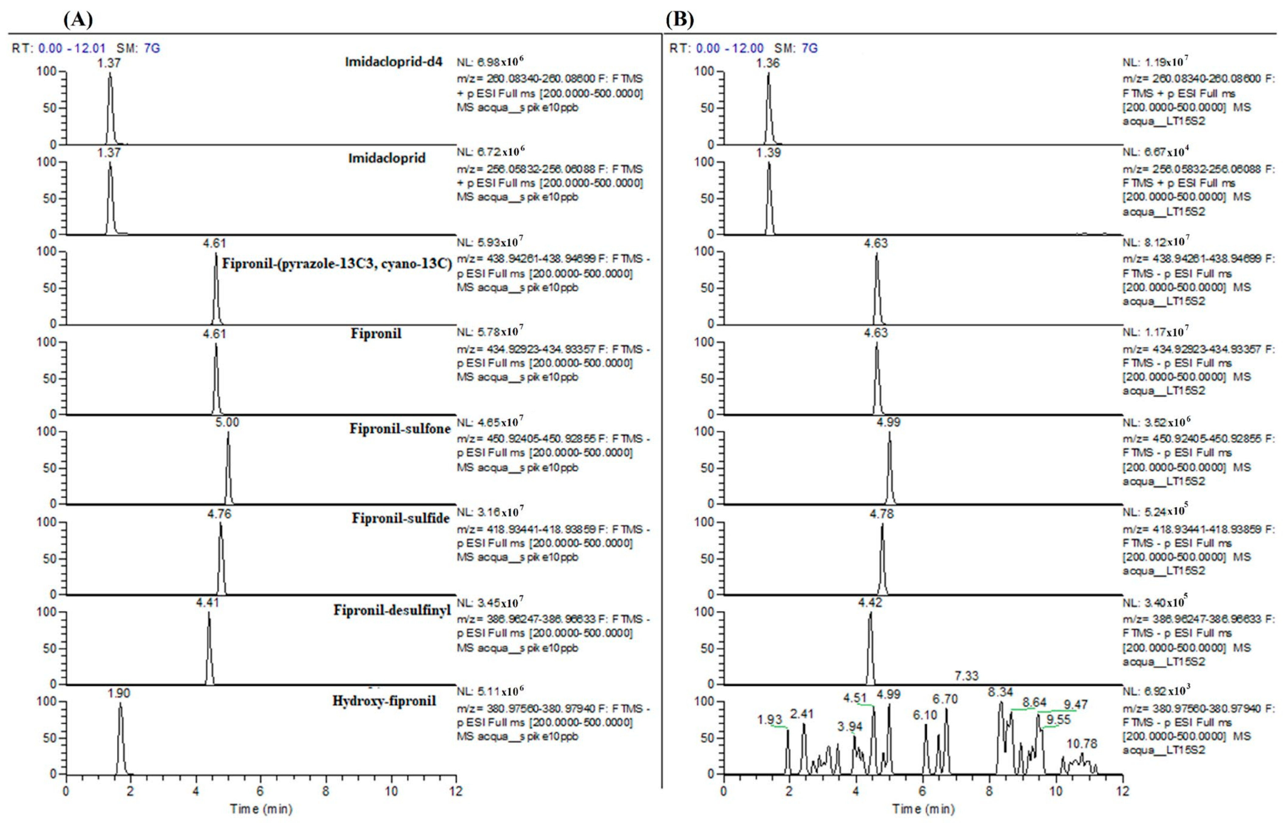
2.2.2. Chemical Quantification of Fipronil and Imidacloprid

Chemical Quantification in Water Samples
Stock solutions (1 mg mL−1) and daily working solutions (10 and 100 ng mL−1) were prepared in methanol and kept at −20 °C and 4 °C, respectively.
An aliquot of 5 mL wastewater samples was added to the labeled internal standards Fipronil-(pyrazole-13C3, cyano-13C) and imidacloprid-d4 at a concentration of 5 ng mL−1 in the matrix. The samples were purified by using Oasis HLB SPE cartridges under vacuum. The SPE cartridges were preconditioned with 3 mL of methanol or 3 mL of Milli-Q water, and then the samples were loaded. Finally, the samples were eluted with 5 mL of methanol and collected in a 15 mL polypropylene tube. The eluate was evaporated in a rotary vacuum evaporator at 40 °C. The dried extract was reconstituted in 200 μL of methanol/water with 0.1% formic acid (60:40 v/v) and transferred to a vial.
The quantification analysis was performed by a Vanquish liquid chromatograph (Thermo Fisher Scientific, Waltham, MA, USA) coupled to an Orbitrap Exploris 120 high-resolution mass spectrometer (Thermo Fisher Scientific) equipped with a heated electrospray ionization (HESI) source. Analytes were chromatographically separated on a Raptor ARC-18 5 μm, 120 × 2.1 mm column (Restek, Bellefonte, PA, USA). The mobile phase was composed of phases A (water with 0.1% formic acid) and B (MeOH). The gradient started with 60% B, which was maintained for one minute. After six minutes, it was increased to 95% and was maintained for two minutes. After 30 s, the initial conditions were reached and maintained until the 12th minute. The flow rate was 0.3 mL min−1. The injection volume was 10 μL.
With respect to the detector parameters, the ion transfer tube and vaporizer temperatures were set at 330 and 280 °C, respectively, the sheath and auxiliary gas temperatures were 35 and 15 arbitrary units, and the electrospray voltages were 3.5 kV for positive mode and 3.0 kV for negative mode. The full scan acquisition was combined with a product ion scan mode for the confirmatory response, which was based on an inclusion list.
The full scan worked with a resolution of 60,000 full width at half maximum (FWHM), a scan range of 200–500 m/z, a standard gain control (AGC), an RF lens at 70%, and auto maximum injection time. The confirmatory acquisition operated at 15,000 FWHM, with a standard AGC target, auto maximum injection time, and an isolation window of 1 m/z. Fragmentation of the precursors was optimized with a two-step normalized collision energy (20 and 45 eV). The parent ion exact masses and main product ions of the compounds investigated are listed in Table S1 of the Supplementary Materials.
Validation of the Analytical Method
Validation was performed according to the SANTE/11312/2021 [27] Guidelines. In brief, the method was validated through the selectivity, linearity, sensitivity, recovery, precision, and evaluation of the matrix effect.
The selectivity was investigated by injecting extracted blank samples, and the absence of interferences close to the expected retention times of analytes with an S/N < 3 was verified.
The linearity was evaluated both in the solvent and via matrix-matched calibration curves, which are the last curves used to quantify real samples. It was determined by spiking both solvent and blank samples with the standard working solution in duplicate at five calibration points (0.05, 1.0, 3.0, 5.0 and 10 µg L−1).
The limit of quantification (LOQ) was set at the lowest level, with a recovery within the range of 70–120% and a relative standard deviation (RSD) ≤ 20%, whereas the recovery was assessed by comparing the concentrations of analytes spiked before extraction in the matrix with those spiked at the end of the extraction protocol in six replicates at the LOQ (0.05 µg L−1) and at 1 µg L−1.
The intraday repeatability was assessed through six replicates at the LOQ and at 1 µg L−1 and expressed as a coefficient of variation (CV%). The interday precision was evaluated in six replicates on three different working days at the same concentrations.
The matrix effect, expressed as a percentage, was calculated by comparing the peak areas of analytes spiked in blank samples after the extraction protocol to the peak areas of standards in a neat solution.

2.2.3. Ecotoxicological Test Battery for Marine Species

The endpoints of the ecotoxicological assays included growth inhibition in the marine algae Phaeodactylum tricornutum, acute and chronic toxicity evaluations of Acartia tonsa, and in vitro biochemical responses of Mytilus galloprovincialis. These species play different ecological roles—primary producers, consumers, and filter feeders—spanning a wide range of trophic levels in aquatic ecosystems. By utilizing this combination of organisms in ecotoxicological testing, a more comprehensive understanding of the potential ecological risks associated with chemicals can be achieved, aiding in more accurate predictions of their impact on the environment and food webs. For both assays, the working concentration series was based on environmentally relevant concentrations (ERCs) of 0.03 µg L−1, as reported in the literature [1]. In the M. galloprovincialis biochemical response assay, the range of FIP and IMID concentrations was determined on the basis of the ECx values derived from the previous two tests.
Phaeodactylum tricornutum Growth Inhibition
A stock solution (500 mg L−1) was prepared using 0.1% DMSO for FIP or ultrapure water for IMID. The working concentration series were 3.0, 0.3, 0.03, 0.003, and 0.0003 µg L−1 for FIP and 3000, 300, 30, 3.0, 0.3, 0.03, 0.003, and 0.0003 µg L−1 for IMID.
The growth inhibition of the marine algae P. tricornutum was evaluated following ISO 10253:2016 [28]. P. tricornutum Bholin (CCAP 1052/1A) was the test strain used and was purchased from CCAP (Culture Collection of Algae and Protozoa—Scottish Association for Marine Science/SAMS Research Services Ltd., Oban, UK). P. tricornutum algae were cultured in ASTM-enriched saltwater medium (ASTM-ESM, ASTM E1218, 2012, [29]), and working batches were prepared by inoculating 2 mL of algal culture in 20 mL of fresh ASTM medium (20 ± 2° C, under continuous illumination (6000–8000 lx)). After 72 h, the algal batches were diluted to a concentration of 106 cells mL−1.
For the growth inhibition bioassay, three replicates of algae were exposed to FIP and IMID concentrations for a period of 72 h at 20 ± 2 °C under continuous illumination (6000–8000 lx). The absorbance at 670 nm was measured in each well with a spectrophotometer, making use of 1 cm optic-path plastic cuvettes. The algal concentration, expressed as cells mL−1, was calculated from the absorbance values using the equation originally defined by Rodrigues et al. [30] and later modified by the marine research center CIBM (Livorno, Italy) group:
C e l l s ×   m L 1 = A b s 670 10 7
Potassium dichromate was used as a reference toxicant for this species, with an EC50 of 27.29 mg/L [95% confidence interval (CI) = 25.52 − 29.27].
Acartia tonsa Toxicity Assays
The FIP and IMID solutions were prepared using the same methodology as that of the test conducted for P. tricornutum at the concentrations specified (FIP: 3.0, 0.3, 0.03, 0.003, and 0.0003 µg L−1; IMID: 3000, 300, 30, 3.0, 0.3, 0.03, 0.003, and 0.0003 µg L−1). The acute and chronic toxicity of FIP and IMID were evaluated in the copepod A. tonsa following two different protocols:
  • Acute toxicity assay: The acute toxicity of the FIP and IMID dilution series was tested in accordance with UNICHIM 2365:2012 [31]. Briefly, six replicates of 2 mL for each assessed concentration were placed in a 24-well plate. Five freshly collected fertilized A. tonsa eggs/nauplii were placed in each well and incubated at 20 ± 1 °C for 48 h in darkness, for a total of 30 eggs/nauplii per sample dilution. The number of immobilized individuals was counted at both 24 and 48 h. Ni2+ was used as the reference toxicant (EC50 0.243 mg Ni2+ L−1, 95% C.I. = 0.201–0.299 mg Ni2+ L−1).
  • Chronic toxicity assay: The chronic toxicity of the FIP and IMID concentration series was tested in accordance with UNICHIM 2366:2012 [32]. Briefly, three replicates of 30 mL for each assessed concentration were placed in a 50 mL glass beaker. A total of 10 freshly collected A. tonsa fertilized eggs/nauplii were placed in each well and incubated at 20 ± 1 °C for 7 days, with a photoperiod of 12:10 h (light:darkness). Each sample/dilution was renewed twice during the exposure (every two days). At the end of the exposure period, the number of immobilized individuals was counted. Ni2+ was used as a reference toxicant (EC50 of 0.037 mg Ni2+ L−1, 95% C.I. = 0.028–0.050 mg Ni2+ L−1).
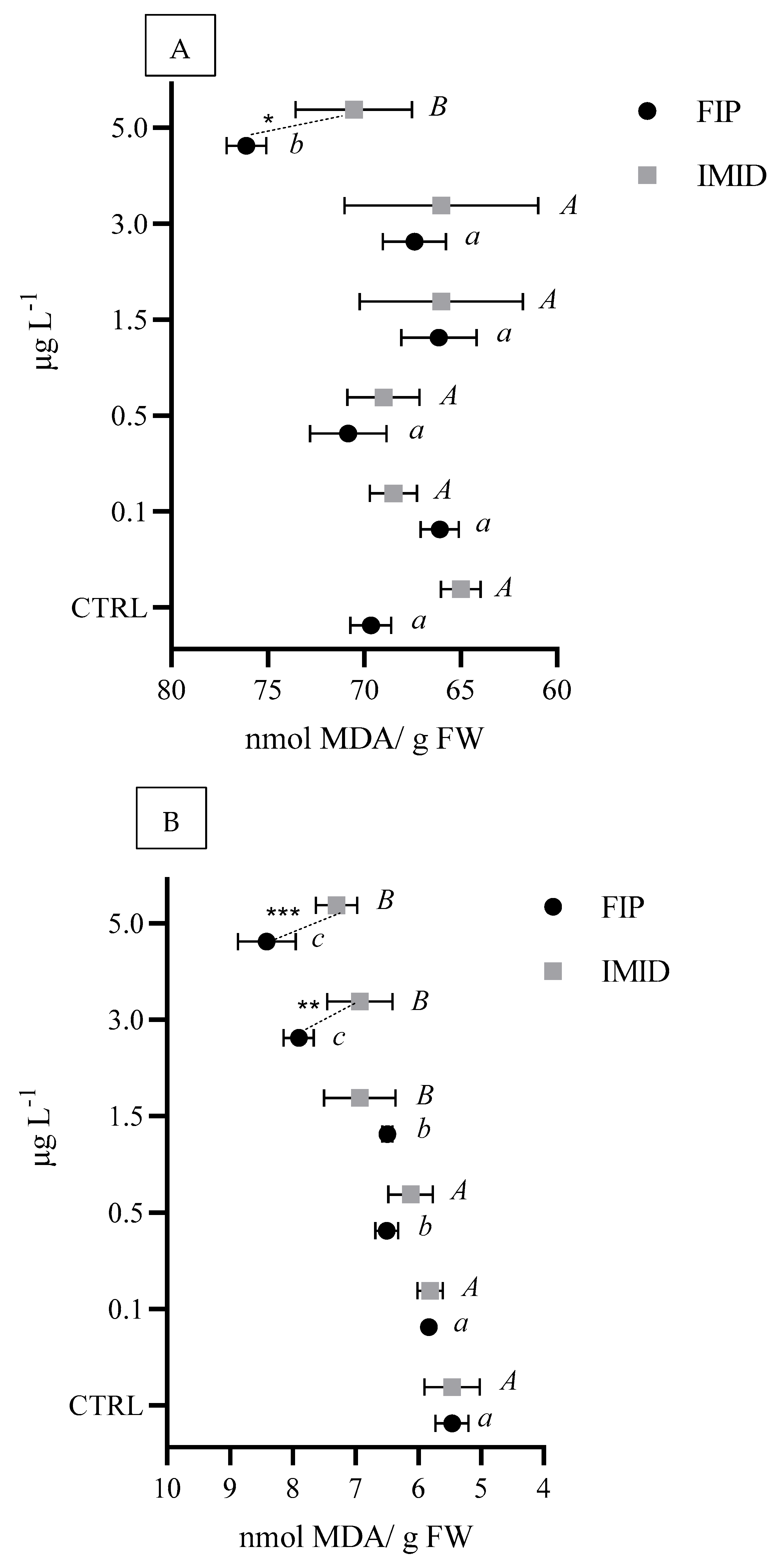
In Vitro Biochemical Responses of Mytilus galloprovincialis
For the M. galloprovincialis biochemical response assay, a range of FIP and IMID concentrations was selected based on the ECx values calculated from previous two tests. The selected test concentrations for both FIP and IMID were 0.1, 0.5, 1.5, 3.0, and 5.0 µg L−1. Adult individuals of M. galloprovincialis were purchased from an aquaculture farm located in the Gulf of La Spezia (NW Tyrrhenian coast, Italy) and transported to the laboratory under dry conditions. Mussels of similar size (shell lengths ranging from 60 to 70 mm) and the same sex (males) were selected.
The digestive gland and gill tissues were immediately collected separately from the mussels using sterile surgical instruments. Subsequently, subcellular S9 fractions were prepared by pooling the gills and digestive glands of 10 mussels per replicate. The excised tissues were weighed and placed into 50 mL sterile tubes. The fresh tissues were then homogenized with a phosphate buffer (50 mM potassium dihydrogen phosphate (K2HPO4) and 50 mM dipotassium phosphate (K3PO4), pH 8.00) at a ratio of 1:4 (v/v) using an Elvehjem potter. The homogenized tissues were centrifuged at 9000× g at 4 °C for 20 min to obtain the S9 fraction, which was then stored at −80 °C until use. S9 fractions from the gills and digestive gland were directly exposed to 0.1, 0.5, 1.5, 3.0, or 5.0 µg L−1 of FIP and IMID to assess the specific biochemical responses.
For each ectoparasiticide and concentration tested, six replicates, each containing 1 mg of protein, were incubated with the respective concentrations of FIP and IMID for 30 min at 25 °C. After incubation, the biochemical parameters were analyzed. Each sample was analyzed in triplicate using 96-well microplates, with a total reaction volume of 250 µL. The resulting absorbance was measured using a Synergy HT microplate reader (BioTek® Inc Winooski, VT, USA), with wavelength values determined by the type of biomarker.
The total protein content was measured using the Lowry method [33]. Lipid peroxidation (LPO) was assessed by quantifying thiobarbituric acid-reactive substances (TBARS) at a wavelength of 532 nm, according to Ohkawa et al. [34]. The enzymatic activity of acetylcholinesterase (AChE) was evaluated at a wavelength of 412 nm following Ellman et al. [35]. The detailed methodology for all biomarkers can be found in Giannessi et al. [36].

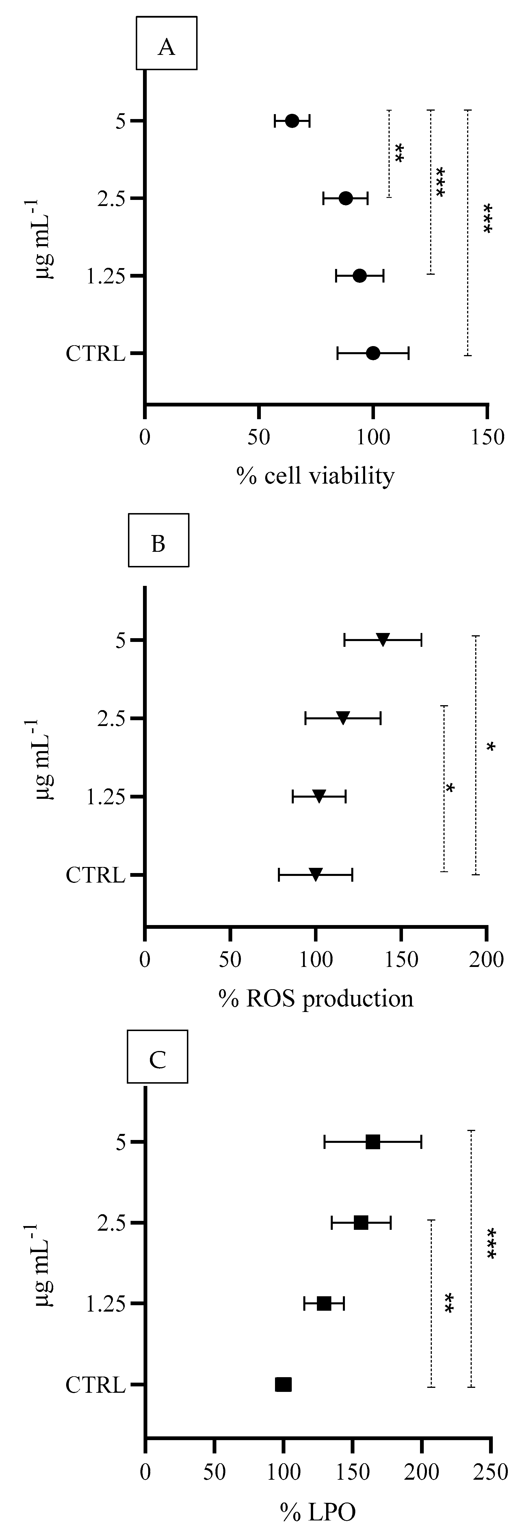
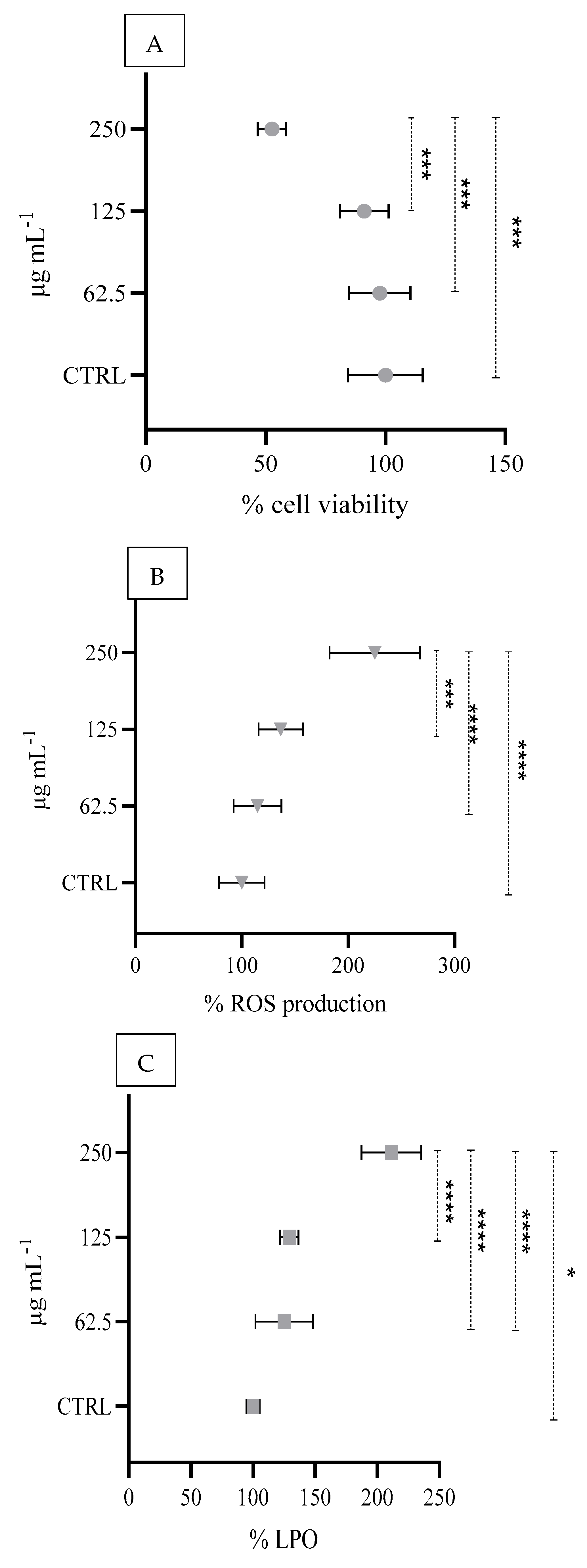
2.2.4. In Vitro Cell-Based Approaches

In vitro tests on cultured cells were conducted using IMID at concentrations of 62.5, 125, and 250 μg mL−1 and FIP at concentrations of 1.25, 2.5, and 5.0 μg mL−1 (with 0.1% DMSO) over a 24 h exposure period. The test solutions were prepared from a 500 mg L−1 stock solution in the respective medium and the potential topical effects resulting from skin exposure to both compounds were simulated.
Before each biomarker was measured, HaCaT cells [Cell Line Service (CLS, catalogue number 300493)] were cultured in a 96-well plate at a density of 1 × 10⁴ cells per well in complete medium and then incubated overnight at 37 °C and 5% CO2. Following incubation, the cells were exposed to various concentrations of FIP and IMID in serum-free DMEM for 24 h, with untreated cells serving as the control group (CTRL). The samples were then subjected to biomarker analyses.
Cell Viability Assay
At the end of the 24 h treatment, the culture medium was discarded, and 100 µL of MTT solution (0.5 mg mL−1 in PBS) was added to each well. The plates were subsequently incubated in the dark at 37 °C for 1 h, enabling metabolically active cells to convert MTT into formazan crystals. The formazan crystals were then dissolved via the addition of 100 μL of dimethyl sulfoxide (DMSO) to each well, with gentle shaking to ensure complete dissolution of the crystals. Optical density was then measured at a wavelength of 595 nm using a microplate reader to assess cell viability, and the resulting data are expressed as a percentage of the CTRL, as reported in the following formula:
C e l l   v i a b i l i t y % = 100 × O D s a m p l e O D c o n t r o l
Intracellular ROS Detection
Intracellular reactive oxygen species (ROS) levels were assessed via the use of the cell-permeable fluorescent probe 2,7-dichlorodihydrofluorescein diacetate (DCFDA). At the end of the 24 h treatment, a 10 μM DCFDA probe was added to each well in PBS, and the cells were incubated in the dark for 1 h at 37 °C to facilitate probe uptake and reaction with intracellular ROS. Fluorescence was subsequently measured at excitation and emission wavelengths of 485 nm and 538 nm, respectively, using a Biotek Synergy 1H plate reader (Agilent Technologies, Santa Clara, CA, USA). The ROS levels detected were normalized to cell viability, with the data expressed as a percentage with respect to the CTRL.
Detection of Lipid Peroxidation (LPO) Levels
The levels of lipid peroxidation (LPO) were investigated using the fluorescent probe C11-BODIPY following the manufacturer’s instructions. At the end of the 24 h treatment, a 1 μM C11-BODIPY solution in PBS was added to each well, and the cells were incubated in the dark for one hour at 37 °C. Fluorescence was subsequently measured using a Biotek Synergy 1H plate reader (Agilent Technologies, Santa Clara, CA, USA). The excitation and emission wavelengths used were 488 nm and 510 nm, respectively. The detected LPO levels were normalized to cell viability, and the untreated sample was used as a control. The data obtained are expressed as percentages with respect to the CTRL.

2.3. Statistical Analyses

The assumptions of normality and homogeneity of variance were tested via the Shapiro-Wilk test and Bartlett test, respectively. The results indicated that the data did not follow a normal distribution.
Statistical analyses of Phaeodactylum tricornutum growth inhibition and Acartia tonsa toxicity results were performed using the freeware tool MOSAIC (https://mosaic.univ-lyon1.fr/, accessed on 24 April 2025), developed by Université Claude Bernard Lyon 1 for ecotoxicological modeling.
EC10/20/50 values were calculated using the MOSAICbioacc module by fitting a dose-response model (3-parameter log-logistic model) through Bayesian inference. The model provided EC10/20/50 estimates along with 95% confidence intervals, and model fit was assessed with integrated diagnostic tools.
The effects of different concentrations of each ectoparasiticide were evaluated via one-way ANOVA, followed by Tukey’s post hoc test (p ≤ 0.05) for multiple comparisons. Graphs and statistical analyses were generated using GraphPad Prism 9.5.1® (www.graphpad.com, accessed on 20 April 2025).
To compare the effects of FIP and IMID at each concentration on the biomarkers of interest in each M. galloprovincialis tissue (LPO levels and AChE activity in the gills and digestive glands), two-way ANOVA was applied, followed by Tukey’s post hoc test (p ≤ 0.05) for multiple comparisons. Graphs and statistical analyses were generated using GraphPad Prism 9.5.1® (www.graphpad.com, accessed on 20 April 2025).
For HaCaT cultured cells, the effects of different concentrations of each ectoparasiticide on various biomarkers (MTT, ROS production, and LPO levels) were evaluated via one-way ANOVA, followed by Tukey’s post hoc test (p ≤ 0.05) for multiple comparisons. Graphs and statistical analyses were generated using GraphPad Prism 9.5.1® (www.graphpad.com, accessed on 20 April 2025).

3. Results

3.1. Fipronil and Imidacloprid Concentrations in Wastewater

The concentrations of IMID, FIP, and FIP-sulfone in the wastewater samples remained stable for at least 60 days after the animals were treated, with average values of 0.18, 0.50 and 0.20 µg L−1, respectively (Table 1). Other metabolites investigated, such as FIP-sulfide, FIP-desulfinil, and hydroxy-FIP, were found at concentrations < 0.07 µg L−1 or below the LOQ. In Figure 1, the extracted full scan chromatograms of the internal standards and all analytes at 10 µg L−1 in matrix (A) in comparison with a positive sample (B) are reported as an example.

3.2. Validation of the Analytical Method for Fipronil and Imidacloprid

The analytical method was validated in terms of selectivity, linearity, sensitivity, recovery, precision, and evaluation of the matrix effect. The validation data are reported in Table 2.

3.3. Ecotoxicological Test Battery Responses

3.3.1. Phaeodactylum tricornutum Growth Inhibition

No significant effects were detected in the growth inhibition assay for P. tricornutum at any of the tested FIP or IMID concentrations (Tables S1 and S2).

3.3.2. Acartia tonsa Toxicity Assays

No significant effects were detected in the A. tonsa acute toxicity assay compared with the control at both 24 and 48 h of FIP and IMID exposure (Table S3).
In contrast, a significant reduction in naupliar mobility (p < 0.001) was observed for both FIP and IMID after 7 days of exposure at concentrations of 0.003, 0.03, 0.3, and 3.0 µg L−1. This led to the calculation of EC10 and EC20 values of 1.7 (0.06–6.59) and 2.8 (0.436–8.51) µg L−1, respectively, for FIP and EC10, EC20 and EC50 values of 2.59 (0.80–6.33), 7.60 (3.12–15.8) and 47.50 (25.30–90.10) µg L−1, respectively, for IMID (Table 3).

3.3.3. Biochemical Markers of Mytilus galloprovincialis

A significant increase in LPO levels was observed in the digestive gland after exposure to 5 µg L−1 of both FIP and IMID, with significantly higher LPO levels with FIP than with IMID exposure (Figure 2A). The increase in LPO was more pronounced in the gills than in the digestive glands, with a significant increase in the levels starting at 0.5 µg L−1 for both FIP and IMID. Compared with IMID, FIP caused significantly greater damage at both 3.0 µg L−1 and 5.0 µg L−1 (Figure 2B).
A significant increase in AChE activity (p < 0.05) was observed only in the digestive gland exposed to IMID in the range of 0.5–5.0 µg L−1 (Figure 3A). No alterations in AChE activity were detected in the gills exposed to both ectoparasiticides (Figure 3B).

3.4. In Vitro Cell-Based Responses

A significant concentration-dependent reduction in cell viability (% MTT) of 94.2%, 88.0% and 64.6% was observed at FIP concentrations of 5.0, 2.5 and 1.25 μg mL−1, respectively (Figure 4A). Compared with those in the control, significant increases in ROS production and LPO levels were detected at FIP concentrations of 2.5 and 5 μg mL−1 (Figure 4B,C).
A significant reduction in cell viability of 97.7, 91.2 and 52.6% was observed at 250, 125 and 62.5 μg mL−1 concentrations of IMID, respectively (Figure 5A), along with an increase in ROS production (Figure 5B) and LPO levels (Figure 5C) compared with those of the control.
Figure S1 presents cell morphology images of the control alongside those exposed to the highest concentrations of IMID and FIP.

4. Discussion

4.1. Fipronil and Imidacloprid Concentrations in Wastewater

Although the water quantification in our study was limited to the wastewater outflow area of the shelter, where wastewater is specifically generated by routine pet and box care activities, our results provide a realistic scenario for assessing how these chemicals enter the environment through everyday pet care practices. The results can also be used for monitoring the stability of compound concentrations over time.
Compared with the literature, our data highlight that the concentrations of IMID, FIP and FIP metabolites were significantly higher—between 10 and 100 times greater—than those reported in European studies of freshwater surface waters. The average IMID concentrations in freshwater, reported by the ECHA, range from 0.01 to 0.03 µg L−1, which are notably lower than the concentrations observed in our study (0.18 µg L−1).
In contrast to freshwater environments, data on IMID concentrations in marine waters are less prevalent. However, Liu et al. [37] reported that IMID concentrations in marine waters ranged from undetectable to 0.636 µg L−1, with a median concentration of 0.00414 µg L−1.
Both FIP and IMID remained stable in water for at least 60 days, confirming their persistence in aqueous environments, as shown by Perkins et al. [16,17]. Data reported by the EMA [1] indicate that IMID, FIP and their metabolite concentrations typically remain below risk thresholds in environmental settings. However, the concentrations found in our study—FIP at 0.50 µg L−1 and FIP-sulfone at 0.20 µg L−1—were significantly higher than those typically reported in the EMA’s environmental data.

4.2. Ecotoxicological Assessment

In the present study, the endpoints for the marine ecotoxicological assays included growth inhibition of the marine algae P. tricornutum, acute and chronic toxicity assessments of the marine copepod A. tonsa, and in vitro biochemical responses of the marine mussel M. galloprovincialis. These species represent distinct ecological functions—primary producers, consumers, and filter feeders—covering a broad range of trophic levels in marine ecosystems. The use of this combination of organisms in ecotoxicological testing provides a more comprehensive understanding of the potential ecological risks posed by chemicals, facilitating better predictions of their impact on the environment and food webs.
Our decision to use non-target marine species as model organisms was a result of the scarcity of available data in the literature for marine environments in contrast to the more extensive studies conducted on freshwater species. Additionally, the marine environment represents the ultimate receiving body for many pollutants, making it crucial to assess the potential ecological risks in these ecosystems.
From an ecotoxicological perspective, FIP concentrations in European freshwaters are generally lower than 0.05 µg L−1, whereas ecotoxicological effects have been observed at 0.50 µg L−1 [38]. In our study, toxicological assessments using M. galloprovincialis demonstrated that FIP and IMID, at concentrations of 0.50 and 1.50 µg L−1, respectively, increased the levels of LPO in both the gills and digestive glands. Compared with the literature, the only study available on mussels reported that sublethal concentrations of FIP induced oxidative stress and histopathological damage in Unio delicatus [39]. In another model species, the fish Cyprinus carpio, FIP also led to increased LPO levels of 0.65 mg L−1 over exposure periods of 7, 30, and 90 days [40].
Similarly, IMID has been shown to increase LPO levels in the brain tissue of the neotropical fish Prochilodus lineatus [41] and in the gills of the freshwater mussel Unio mancus [42]. In our study, IMID increased ACh enzyme activity in the mussel digestive gland.
Recent studies have emphasized the growing concern regarding neonicotinoid contamination in aquatic environments and its potential effects on nontarget organisms, such as bivalves. Pagano et al. [43] reviewed the impact of neonicotinoids, including IMID, on aquatic invertebrates, with a specific focus on M. galloprovincialis. Pagano’s review highlights various biochemical effects of neonicotinoids, including alterations in enzyme activity and potential impacts on mussel health due to exposure to environmentally relevant concentrations. Similarly, Kuchovská et al. [44] examined the effects of IMID and other substances on the Pacific oyster (Magallana gigas), revealing how neonicotinoids influence bivalves and how their gene expression is associated with toxicity. Together, these findings, along with our findings, confirm the significant impact of FIP and IMID on non-target aquatic organisms at different trophic levels and highlight the need for further investigation into their ecological effects.
ECx and LCx values are essential for evaluating the toxicity of chemicals in aquatic ecosystems and organisms. In the literature, the LC50 values for FIP range from 2.18 to 19.12 μg L−1 when different model species are used [45,46]. Our findings are consistent with these values, with EC10 and EC20 values of 1.7 µg L−1 and 2.8 µg L−1, respectively, for A. tonsa exposed to FIP. When comparing our marine model organism to other freshwater crustacean species, A. tonsa displayed greater sensitivity to FIP than Ceriodaphnia dubia [47,48], Daphnia magna [49,50], and Simocephalus elizabethae [51], exhibiting LC50 values ranging from 0.98 to 19.12 μg L−1. These findings suggest that marine crustaceans may be more vulnerable to certain contaminants, such as FIP, potentially due to differences in environmental stressors, osmoregulatory processes, or adaptive mechanisms between marine and freshwater species.
Most studies have focused only on freshwater settings, creating a substantial gap in knowledge about the effects of FIP in saline or brackish environments. This lack of data is particularly relevant to our findings, as toxicity mechanisms and bioavailability may differ in marine environments, highlighting the need for further research to clarify FIP’s ecological impacts across various aquatic ecosystems.
Similarly, in invertebrates, IMID causes significant sublethal effects [52,53,54]. Our study revealed EC10 and EC20 values of 2.59 µg L−1 and 7.6 µg L−1, respectively, for A. tonsa following chronic exposure, which are consistent with the values reported in the literature. In a similar study, Picone et al. [55], for example, reported an EC50 of 8.84 μg L−1 for the same species. Together with the literature, our findings demonstrate that the calculated ECx values exceed the European Commission’s proposed standards for long-term exposure (8.3 ng L−1) [56]. These results suggest that current regulations may have underestimated the potential long-term effects of IMID on marine invertebrates.

4.3. In Vitro Cell-Based Toxicity

HaCaT cells, a human keratinocyte cell line derived from normal skin, are well suited for testing antiparasitic toxicity due to their similarity to native keratinocytes given that they preserve essential features such as proliferation and differentiation. These cells demonstrate functional competence in critical cellular processes, including cytotoxicity and inflammation, making them valuable for comprehensive drug evaluations. In our study, the potential cytotoxicity of FIP and IMID in HaCaT human keratinocytes was investigated simulating acute exposure. The analysis of cellular responses to FIP and IMID revealed that both chemicals triggered oxidative stress mechanisms, as evidenced by high levels of ROS and LPO, which ultimately led to reduced cell viability in exposed skin cells.
Regarding IMID exposure, our concentration range (62.5, 125 and 250 μg mL−1) was based on cytotoxicity data reported in the literature using the same cell line. For example, Leri et al. [57] and Silva et al. [58] reported oxidative stress responses in HaCaT cells exposed to IMID at concentrations of 200 μg mL−1. IMID-induced cytotoxicity has also been reported in other cell lines, including Caco-2, HepG2, A431, SK-MEL-5, and RAW 264.7, with IC50 values exceeding 200 μg mL−1, further supporting its potential cytotoxic effects at the cellular level.
Similarly, we selected FIP concentrations based on direct evidence indicating that FIP exposure can cause skin irritation in humans [59]. Although no studies have specifically reported oxidative stress responses in keratinocyte cultures, exposure to 40 μg mL−1 FIP in SH-SY5Y human neuronal cells induced cytotoxic effects. Furthermore, a concentration range of 20–80 μg mL−1 resulted in growth inhibition in human epithelial Caco-2 cells and reduced MTT activity [60]. Notably, the concentrations tested in the present study (0.25, 2.5, and 5.0 μg mL−1) were up to two times lower than those reported in previous studies that induced cytotoxicity in various cell models. These findings suggest that even at lower concentrations, FIP may exert toxic effects, potentially through oxidative stress mechanisms, thus reinforcing the need for further investigation into its impact on human skin cells. Overall, assessing their impact on keratinocyte function and skin barrier integrity could offer valuable insights into their potential dermatotoxicity. This knowledge would support the implementation of stricter preventive measures and strengthen the importance of adhering to usage guidelines to minimize health risks for humans.

5. Conclusions

Our analysis of wastewater samples from the Italian shelter provided valuable insights into the concentrations of IMID, FIP, and their metabolites in untreated water, establishing a baseline for effluent flowing directly from the facility. Our results indicated that these compounds remained stable for up to 60 days post-treatment, suggesting significant environmental persistence. Ecotoxicological tests on marine organisms revealed toxic effects of both FIP and IMID in copepods and bivalves, with EC10 and EC20 levels that could harm aquatic life, especially with prolonged exposure. Biochemical damage in mussel gills was observed at FIP concentrations as low as 0.5 µg L−1 and IMID at 1.5 µg L−1, emphasizing the vulnerability of marine species to these compounds. Furthermore, mussels emerged as a valuable model species for studying the impact of these contaminants, contributing crucial new insights to the literature.
However, no significant effects were observed in the growth inhibition assay for P. tricornutum at any of the concentrations tested for FIP or IMID. Previous studies have suggested that algae may possess inherent resistance or differences in metabolic pathways that reduce their sensitivity to these compounds [61,62].
Our findings are consistent with those outlined in EMA documents, underscoring the urgent need for effective monitoring and preventive strategies. This is especially important considering the growing population of companion animals and evolving management practices, which contribute to the increasing environmental burden through the widespread use and potential accumulation of ectoparasiticides in ecosystems.
Strengthening regulatory frameworks and promoting sustainable alternatives could mitigate the risks associated with the environmental persistence and toxicity of these substances. The in vitro cytotoxicity results for FIP and IMID further emphasize the need for strict adherence to usage instructions to minimize health risks to humans. Overall, these results highlight the interconnected impacts of ectoparasiticide treatments on environmental, animal, and human health, supporting the need for a One Health approach to assess, regulate, and mitigate the risks associated with their use.

Supplementary Materials

The following supporting information can be downloaded at: https://www.mdpi.com/article/10.3390/ani15111533/s1, Table S1: Algal concentration ± standard deviation; mean growth rate (µ) and percentage of growth rate inhibition with respect to control (I%) of P. tricornutum after 72 ± 2 h of fipronil exposure; Table S2: Algal concentration ± standard deviation; mean growth rate (µ) and percentage of growth rate inhibition with respect to control (I%) of P. tricornutum after 72 ± 2 h of imidacloprid exposure; Table S3: Percentage of immobilization (and relative standard deviation) of A. tonsa nauplii after 24 and 48 h of exposure to fipronil and imidacloprid. Figure S1: Phase-contrast images of HaCaT cultured cell morphology: (A) control (90% confluence); (B) exposed to 5 µg mL−1 FIP; (C) exposed to 250 µg mL−1 IMID, captured using a Leica Microsystems Model (Mateo TL, Wetzlar, Germany) microscope with a 4X objective.

Author Contributions

Conceptualization, L.M., C.P. and L.I.; Methodology, L.D.M., M.O., M.N., M.C., C.P. and M.V.; Validation, D.D. and L.I.; Formal analysis, L.D.M., M.O., M.N. and M.V.; Investigation, L.D.M., M.N., M.C., D.D., I.L., L.M., V.M. and C.P.; Data curation, L.D.M., M.O., M.N., M.C., D.D., I.L., L.M., V.M., M.V. and L.I.; Writing—original draft, L.D.M.; Writing—review & editing, L.D.M., M.O., M.N., M.C., L.M.C., D.D., I.L., L.M., V.M., C.P., M.V., R.E.V. and L.I.; Supervision, C.P. and L.I.; Project administration, L.D.M. and L.I.; Funding acquisition, L.M. and L.I. All authors have read and agreed to the published version of the manuscript.

Funding

This research received no external funding.

Institutional Review Board Statement

No ethical statement is required for this study. In Italy, the use of animals for scientific purposes is regulated by Legislative Decree No. 26 of 4 March 2014, which transposes Directive 2010/63/EU. This regulation mandates authorization from the Ministry of Health for research involving vertebrate animals and certain invertebrates, such as cephalopods. However, the legislation does not specifically mention other aquatic invertebrates, such as crustaceans or non-cephalopod mollusks. Therefore, for these organisms, no formal authorization or ethical statement is required for their use in laboratories.

Informed Consent Statement

Not applicable.

Data Availability Statement

The original contributions presented in this study are included in this article. Further inquiries can be directed to the corresponding author.

Acknowledgments

We sincerely thank the staff and collaborators of the Groppoli-Mulazzo (MS, Italy) Municipal Animal Shelter for their invaluable support, participation, and for granting us access to their facilities, which made the sampling activities possible. We also extend our gratitude to Marco Del Torto for his assistance in identifying animal shelters willing to participate in the project and for facilitating connections with the relevant authorities and veterinarians. Their contributions were essential to the successful completion of this study.

Conflicts of Interest

The authors declare no conflicts of interest.

References

  1. European Medicines Agency (EMA)/Committee for Medicinal Products for Veterinary Use (CVMP). Reflection Paper on the Environmental Risk Assessment of Ectoparasiticidal Veterinary Medicinal Products Used in Cats and Dogs. 2023. Available online: https://www.ema.europa.eu/en/documents/scientific-guideline/reflection-paper-environmental-risk-assessment-ectoparasiticidal-veterinary-medicinal-products-used-cats-and-dogs_en.pdf (accessed on 2 April 2025).
  2. Grand View Research. Animal Health Market Size, Share & Trends Analysis Report by Product (Biologics, Pharmaceuticals), by Animal Type (Companion, Production), by Distribution Channel, by End-Use, By Region, and Segment Forecasts, 2025–2030. 2023. Available online: https://www.grandviewresearch.com (accessed on 2 April 2025).
  3. European Scientific Counsel Companion Animal Parasites (ESCCAP). Control of Ectoparasites in Dogs and Cats, ESCCAP Guideline 03, 7th ed.; ESCCAP: Malvern, UK, 2022; pp. 1–33. Available online: https://www.esccap.org/link-document/63/ (accessed on 2 April 2025).
  4. Pfister, K.; Armstrong, R. Systemically and cutaneously distributed ectoparasiticides: A review of the efficacy against ticks and fleas on dogs. Parasites Vectors 2016, 9, 436. [Google Scholar] [CrossRef] [PubMed]
  5. Lavan, R.; Armstrong, R.; Normile, D.; Vaala, W. Adherence to veterinary recommendations for ectoparasiticides purchased by cat owners in the USA. Parasites Vectors 2020, 13, 541. [Google Scholar] [CrossRef] [PubMed]
  6. Yuskiv, I.D.; Tishyn, O.L.; Yuskiv, L.L. Evaluation of efficacy of a drug based on fipronil, ivermectin, and pyriproxyfen against ecto- and endoparasitic invasions of dogs and cats. Regul. Mech. Biosyst. 2024, 15, 113–118. [Google Scholar] [CrossRef]
  7. Ziliotto, L.; Luna, S.P.L.; Filho, D.A.A.; Resende, L.O.; Aun, A.G.; Braz, M.G. Genotoxicity assessment of fipronil (Frontline Plus®). Pesq. Vet. Bras. 2017, 37, 257–260. [Google Scholar] [CrossRef]
  8. Cravedi, J.P.; Delous, G.; Zalko, D.; Viguié, C.; Debrauwer, L. Chemosphere disposition of fipronil in rats. Chemosphere 2013, 93, 2276–2283. [Google Scholar] [CrossRef]
  9. Singh, N.S.; Sharma, R.; Singh, S.K.; Singh, D.K. A comprehensive review of environmental fate and degradation of fipronil and its toxic metabolites. Environ. Res. 2021, 199, 111316. [Google Scholar] [CrossRef]
  10. Zhao, X.; Yeh, J.Z.; Salgado, V.L.; Narahashi, T. Sulfone metabolite of fipronil blocks γ-aminobutyric acid- and glutamate-activated chloride channels in mammalian and insect neurons. J. Pharmacol. Exp. Ther. 2005, 314, 363–373. [Google Scholar] [CrossRef]
  11. Wan, Y.; Han, Q.; Wang, Y.; He, Z. Five degradates of imidacloprid in source water, treated water, and tap water in Wuhan, central China. Sci. Total Environ. 2020, 741, 140227. [Google Scholar] [CrossRef]
  12. Bass, C.; Denholm, I.; Williamson, M.S.; Nauen, R. The global status of insect resistance to neonicotinoid insecticides. Pestic. Biochem. Physiol. 2015, 121, 78–87. [Google Scholar] [CrossRef]
  13. European Medicines Agency (EMA). Environmental Impact Assessment (EIA) for Veterinary Medicinal Products (VMPs)—Phase I. 2000. Available online: https://www.ema.europa.eu/en/environmental-impact-assessment-veterinary-medicinal-products-support-vich-guidelines-gl6-gl38-scientific-guideline (accessed on 2 April 2025).
  14. Koslowski, S.; Latapy, C.; Auvray, P.; Blondel, M.; Meijer, L. Long-term fipronil treatment induces hyperactivity in female mice. Int. J. Environ. Res. Public Health 2020, 17, 1579. [Google Scholar] [CrossRef]
  15. Ferreira, T.P.; Bauerfeldt, G.F.; Castro, R.N.; Magalhães, V.S.; Alves, M.C.; Scott, F.B.; Cid, Y.P. Determination of fipronil and fipronil-sulfone in surface waters of the Guandu River Basin by high-performance liquid chromatography with mass spectrometry. Bull. Environ. Contam. Toxicol. 2022, 108, 225–233. [Google Scholar] [CrossRef] [PubMed]
  16. Perkins, R.; Barron, L.; Glauser, G.; Whitehead, M.; Woodward, G.; Goulson, D. Down-the-drain pathways for fipronil and imidacloprid applied as spot-on parasiticides to dogs: Estimating aquatic pollution. Sci. Total Environ. 2024, 917, 170175. [Google Scholar] [CrossRef] [PubMed]
  17. Perkins, R.; Whitehead, M.; Civil, W.; Goulson, D. Potential role of veterinary flea products in widespread pesticide contamination of English rivers. Sci. Total Environ. 2021, 755, 143560. [Google Scholar] [CrossRef] [PubMed]
  18. de la Casa-Resino, I.; Villegas Grondona, J.M.; Puig Infante, A.; Haro Castuera, A.; Bartolom’e Rodríguez, A.; Herrera Hern’andez, E.M.; Rubio, C.F.; Caixach, J.; Carapeto García, R. Residues of Imidacloprid in Surface Water Treatment Plants and Watercourses in Spain: Are Veterinary Medicinal Products (VMPs) a Significant Source of Emission? Spanish Agency of Medicines and Medical Devices (AEMPS). 2022. Available online: https://www.aemps.gob.es/publicaciones/publica/docs/Imidacloprid.pdf (accessed on 6 May 2025).
  19. Finckh, S.; Beckers, L.-M.; Busch, W.; Carmona, E.; Dulio, V.; Kramer, L.; Krauss, M.; Posthuma, L.; Schulze, T.; Slootweg, J.; et al. A risk based assessment approach for chemical mixtures from wastewater treatment plant effluents. Environ. Int. 2022, 164, 107234. [Google Scholar] [CrossRef]
  20. Budd, R.; Teerlink, J.; Alaimo, C.; Wong, L.; Young, T.M. Sub-sewershed monitoring to elucidate down-the-drain pesticide sources. Environ. Sci. Technol. 2023, 57, 5404–5413. [Google Scholar] [CrossRef]
  21. UNESCO. Wastewater: The Untapped Resource. The United Nations World Water Development Report 2017. United Nations Educational, Scientific and Cultural Organization (UNESCO); UNESCO: Paris, France, 2017. [Google Scholar]
  22. Burdon, F.J.; Munz, N.A.; Reyes, M.; Focks, A.; Joss, A.; Räsänen, K.; Altermatt, F. Agricultural and urban land uses drive in-stream concentrations and fluxes of pesticides at the catchment scale. Sci. Total Environ. 2019, 682, 737–746. [Google Scholar]
  23. Giannelli, A.; Schnyder, M.; Wright, I.; Charlier, J. Control of companion animal parasites and impact on One Health. One Health 2024, 18, 100679. [Google Scholar] [CrossRef]
  24. Testa, C.; Salis, S.; Rubattu, N.; Roncada, P.; Miniero, R.; Brambilla, G. Occurrence of fipronil in residential house dust in the presence and absence of pets: A hint for a comprehensive toxicological assessment. J. Environ. Sci. Health Part B 2019, 54, 441–448. [Google Scholar] [CrossRef]
  25. Dos Santos, G.C.M.; Scott, F.B.; Campos, D.R.; Magalhaes, V.D.S.; Borges, D.A.; Miranda, F.R.; Alves, M.C.C.; Pereira, G.A.; Moreira, L.O.; Lima, E.A.S.; et al. Oral pharmacokinetic profile of fipronil and efficacy against flea and tick in dogs. J. Vet. Pharmacol. Ther. 2022, 45, 23–33. [Google Scholar] [CrossRef]
  26. Anthe, M.; Valles-Ebeling, B.; Achtenhagen, J.; Arenz-Leufen, M.; Atkinson, J.; Starp, M.; Corsing, C. Development of an aquatic exposure assessment model for imidacloprid in sewage treatment plant discharges arising from use of veterinary medicinal products. Environ. Sci. Eur. 2020, 32, 147. [Google Scholar] [CrossRef]
  27. European Commission. European Commission SANTE/11312/2021: Analytical Quality Control and Method Validation Procedures for Pesticide Residues Analysis in Food and Feed. Available online: https://food.ec.europa.eu/system/files/2023-11/pesticides_mrl_guidelines_wrkdoc_2021-11312.pdf (accessed on 2 April 2025).
  28. ISO 10253:2016; Water Quality—Marine Algal Growth Inhibition Test with Skeletonema sp. and Phaeodactylum tricornutum. ISO: Geneva, Switzerland, 2016.
  29. ASTM E1218-04(2012); Standard Guide for Conducting Static Toxicity Tests with Microalgae. ASTM: West Conshohocken, PA, USA, 2012.
  30. Rodrigues, L.H.R.; Arenzon, A.; Raya-Rodriguez, M.T.; Fontoura, N.F. Algal density assessed by spectrophotometry: A calibration curve for the unicellular algae Pseudokirchneriella subcapitata. J. Environ. Chem. Ecotoxicol. 2011, 3, 225–228. [Google Scholar]
  31. UNICHIM M.U. 2365:2012; Qualità dell’acqua: Determinazione Dell’inibizione Della Mobilità di Naupli di Acartia tonsa Dana (Crustacea: Copepoda) Dopo 24 h e 48 h di Esposizione. Associazione per l‘Unificazione del Settore dell’Industria Chimica: Milan, Italy, 2012; p. 22.
  32. UNICHIM M.U. 2366:2012; Qualità Dell’acqua: Determinazione Dell’inibizione Della Mobilità di Naupli di Acartia tonsa Dana (Crustacea: Copepoda) Dopo 7 Giorni di Esposizione. Associazione per l‘Unificazione del Settore dell’Industria Chimica: Milan, Italy, 2012; p. 24.
  33. Lowry, O.H.; Rosebrough, N.J.; Farr, A.L.; Randall, R.J. Protein measurement with the Folin phenol reagent. J. Biol. Chem. 1951, 193, 265–275. [Google Scholar] [CrossRef] [PubMed]
  34. Ohkawa, H.; Ohishi, N.; Yagi, K. Assay for lipid peroxides in animal tissues by thiobarbituric acid reaction. Anal. Biochem. 1979, 95, 351–358. [Google Scholar] [CrossRef] [PubMed]
  35. Ellman, G.L.; Courtney, K.D.; Andres, V., Jr.; Featherstone, R.M. A new and rapid colorimetric determination of acetylcholinesterase activity. Biochem. Pharmacol. 1961, 7, 88–95. [Google Scholar] [CrossRef]
  36. Giannessi, J.; De Marchi, L.; Meucci, V.; Intorre, L.; Monni, G.; Baratti, M.; Pretti, C. Subcellular tissue-specific responses of Mytilus galloprovincialis to fluoroquinolone antibiotics. Environ. Toxicol. Pharmacol. 2023, 104, 104306. [Google Scholar] [CrossRef]
  37. Liu, S.; Wang, T.; Lu, J.; Li, Z. Seawater quality criteria derivation and ecological risk assessment for the neonicotinoid insecticide imidacloprid in China. Mar. Pollut. Bull. 2023, 190, 114871. [Google Scholar] [CrossRef]
  38. Dulio, V.; Alygizakis, N.; Ng, K.; Schymanski, E.L.; Andres, S.; Vorkamp, K.; Hollender, J.; Finckh, S.; Aalizadeh, R.; Ahrens, L.; et al. Beyond target chemicals: Updating the NORMAN prioritisation scheme to support the EU chemicals strategy with semi-quantitative suspect/non-target screening data. Environ. Sci. Eur. 2024, 36, 113. [Google Scholar] [CrossRef]
  39. Arslan, P.; Günal, A.Ç. Does fipronil affect on aquatic organisms? Physiological, biochemical, and histopathological alterations of non-target freshwater mussel species. Water 2023, 15, 334. [Google Scholar] [CrossRef]
  40. Clasen, V.L.; Loro, R.; Cattaneo, B.; Moraes, T.; Lópes, L.A.; de Avila, R.; Zanella, G.B.; Reimche, B.; Baldisserotto, B. Effects of the commercial formulation containing fipronil on the non-target organism Cyprinus carpio: Implications for rice-fish cultivation. Ecotoxicol. Environ. Saf. 2012, 77, 45–51. [Google Scholar] [CrossRef]
  41. Vieira, C.E.D.; Pérez, M.R.; Acayaba, R.D.A.; Raimundo, C.C.M.; dos Reis Martinez, C.B. DNA damage and oxidative stress induced by imidacloprid exposure in different tissues of the Neotropical fish Prochilodus lineatus. Chemosphere 2018, 195, 125–134. [Google Scholar] [CrossRef]
  42. Yoloğlu, E. Alterations in some biochemical responses of freshwater mussels in acute imidacloprid exposure. Adiyaman Univ. J. Sci. 2019, 9, 213–229. [Google Scholar] [CrossRef]
  43. Pagano, M.; Stara, A.; Aliko, V.; Faggio, C. Impact of neonicotinoids to aquatic invertebrates—In vitro studies on Mytilus galloprovincialis: A review. J. Mar. Sci. Eng. 2020, 8, 801. [Google Scholar] [CrossRef]
  44. Kuchovská, E.; Morin, B.; López-Cabeza, R.; Barré, M.; Gouffier, C.; Bláhová, L.; Cachot, J.; Bláha, L.; Gonzalez, P. Comparison of imidacloprid, propiconazole, and nanopropiconazole effects on the development, behavior, and gene expression biomarkers of the Pacific oyster (Magallana gigas). Sci. Total Environ. 2021, 764, 142921. [Google Scholar] [CrossRef] [PubMed]
  45. Stark, R.I.; Vargas, M. Toxicity and hazard assessment of fipronil to Daphnia pulex. Ecotoxicol. Environ. Saf. 2005, 62, 11–16. [Google Scholar] [CrossRef]
  46. Wu, C.H.; Lu, C.W.; Hsu, T.H.; Wu, W.J.; Wang, S.E. Neurotoxicity of fipronil affects sensory and motor systems in zebrafish. Pestic. Biochem. Physiol. 2021, 177, 104896. [Google Scholar] [CrossRef]
  47. Konwick, B.J.; Fisk, A.T.; Garrison, A.W.; Avants, J.K.; Black, M.C. Acute enantioselective toxicity of fipronil and its desulfinyl photoproduct to Ceriodaphnia dubia. Environ. Toxicol. Chem. 2005, 24, 2350–2355. [Google Scholar] [CrossRef]
  48. Qin, G.; Presley, S.M.; Anderson, T.A.; Gao, W.; Maul, J.D. Effects of predator cues on pesticide toxicity: Toward an understanding of the mechanism of the interaction. Environ. Toxicol. Chem. 2011, 30, 1926–1934. [Google Scholar] [CrossRef]
  49. Hayasaka, D.; Korenaga, T.; Suzuki, K.; Sánchez-Bayo, F.; Goka, K. Differences in susceptibility of five cladoceran species to two systemic insecticides, imidacloprid and fipronil. Ecotoxicology 2012, 21, 421–427. [Google Scholar] [CrossRef]
  50. U.S. EPA—United States Environmental Protection Agency. US EPA Database. 2018. Available online: https://www.epa.gov (accessed on 2 April 2025).
  51. Stevens, M.M.; Burdett, A.S.; Mudford, E.M.; Helliwell, S.; Doran, G. The acute toxicity of fipronil to two non-target invertebrates associated with mosquito breeding sites in Australia. Acta Trop. 2011, 117, 125–130. [Google Scholar] [CrossRef]
  52. European Food Safety Authority (EFSA). Conclusion on the peer review of the pesticide risk assessment for bees for the active substance imidacloprid considering all uses other than seed treatments and granules. EFSA J. 2015, 13, 4211. [Google Scholar]
  53. European Food Safety Authority (EFSA). Peer review of the pesticide risk assessment for the active substance imidacloprid in light of confirmatory data submitted. EFSA J. 2016, 14, e04607. [Google Scholar]
  54. Merga, L.B.; Van den Brink, P.J. Ecological effects of imidacloprid on a tropical freshwater ecosystem and subsequent recovery dynamics. Sci. Total Environ. 2021, 784, 147167. [Google Scholar] [CrossRef] [PubMed]
  55. Picone, M.; Distefano, G.G.; Marchetto, D.; Russo, M.; Baccichet, M.; Zangrando, R.; Gambaro, A.; Ghirardini, A.V. Inhibition of larval development of marine copepods Acartia tonsa by neonicotinoids. Toxics 2022, 10, 158. [Google Scholar] [CrossRef] [PubMed]
  56. Smit, C.E.; Posthuma-Doodeman, C.J.A.M.; van Vlaardingen, P.L.A.; de Jong, F.M.W. Ecotoxicity of imidacloprid to aquatic organisms: Derivation of water quality standards for peak and long-term exposure. Hum. Ecol. Risk Assess. 2015, 21, 1608–1630. [Google Scholar] [CrossRef]
  57. Leri, M.; Vasarri, M.; Barletta, E.; Schiavone, N.; Bergonzi, M.C.; Bucciantini, M.; Degl’Innocenti, D. The protective role of oleuropein aglycone against pesticide-induced toxicity in a human keratinocytes cell model. Int. J. Mol. Sci. 2023, 24, 14553. [Google Scholar] [CrossRef]
  58. Silva, A.M.; Martins-Gomes, C.; Silva, T.L.; Coutinho, T.E.; Souto, E.B.; Andreani, T. In vitro assessment of pesticides toxicity and data correlation with pesticides physicochemical properties for prediction of toxicity in gastrointestinal and skin contact exposure. Toxics 2022, 10, 378. [Google Scholar] [CrossRef]
  59. Raabe, H.A.; Costin, G.E.; Allen, D.G.; Lowit, A.; Corvaro, M.; O’Dell, L.; Breeden-Alemi, J.; Page, K.; Perron, M.; Silva, T.F.; et al. Human relevance of in vivo and in vitro skin irritation tests for hazard classification of pesticides. Cutan. Ocul. Toxicol. 2024, 44, 1–21. [Google Scholar] [CrossRef]
  60. Vidau, C.; González-Polo, R.A.; Niso-Santano, M.; Gómez-Sánchez, R.; Bravo-San Pedro, J.M.; Pizarro-Estrella, E.; Blasco, R.; Brunet, J.-L.; Belzunces, L.P.; Fuentes, J.M. Fipronil is a powerful uncoupler of oxidative phosphorylation that triggers apoptosis in human neuronal cell line SHSY5Y. Neurotoxicology 2011, 32, 935–943. [Google Scholar] [CrossRef]
  61. Qu, H.; Ma, R.; Liu, D.; Wang, P.; Huang, L.; Qiu, X.; Zhou, Z. Enantioselective toxicity, degradation and transformation of the chiral insecticide fipronil in two algae cultures. Environ. Toxicol. Chem. 2022, 41, 1234–1245. [Google Scholar]
  62. Sardo, A.M.; Soares, A.M. Comparative toxicity of imidacloprid and its transformation product 6-chloronicotinic acid to non-target aquatic organisms: Microalgae Desmodesmus subspicatus and amphipod Gammarus fossarum. Pest. Biochem. Physiol. 2010, 98, 129–137. [Google Scholar]
Figure 1. Extracted full scan chromatograms of the internal standards and all analytes at 10 µg L−1 in matrix (A) in comparison with a positive sample (B).
Animals 15 01533 g001
Figure 2. Lipid peroxidation levels (LPO–nmol MDA/g fresh weight (FW)) in M. galloprovincialis digestive glands (A) and gills (B) exposed to a range of 0.1–5.0 µg L−1 FIP or IMID. Different letters represent statistical differences among concentrations of each ectoparasiticide (A and B = p < 0.05; a, b and c = p < 0.01), while asterisks indicate significant differences between ectoparasiticides at each tested concentration (* p < 0.05; ** p < 0.01; *** p < 0.001).
Animals 15 01533 g002
Figure 3. Enzymatic activity of acetylcholinesterase (nmol/min/mg of protein) in M. galloprovincialis digestive glands (A) and gills (B) exposed to a range of 0.1–5.0 µg L−1 FIP or IMID. Different letters represent statistical differences among concentrations of each ectoparasiticide (A and B = p < 0.05; a, = p < 0.01) Asterisks indicate significant differences between ectoparasiticides at each tested concentration (** p < 0.01).
Animals 15 01533 g003
Figure 4. Percentage of cell viability (MTT assay) (A), percentage of reactive oxygen species production (B), and percentage of lipid peroxidation (LPO), and (C) HaCaT cultured cells exposed to FIP. Asterisks indicate significant differences among concentrations for each ectoparasiticide tested (* p < 0.05; ** p < 0.01; *** p < 0.001).
Animals 15 01533 g004
Figure 5. Percentage of cell viability (MTT assay) (A), percentage of reactive oxygen species production (B), and percentage of lipid peroxidation (LPO), and (C) HaCaT cultured cells exposed to IMID. Asterisks indicate significant differences among concentrations for each ectoparasiticide tested (* p < 0.05; *** p < 0.001; **** p < 0.0001).
Animals 15 01533 g005
Table 1. Concentrations of imidacloprid (IMID), fipronil (FIP) and their metabolites (mean ± sd) in wastewater samples.
µg L−1
SampleIMIDFIPFIP-SulfoneFIP-SulfideFIP-DesulfinilHydroxy-FIP
T0n.d.<LOQ0.05n.d.n.d.n.d.
T70.360.420.10<LOQ<LOQn.d.
T150.061.360.400.070.05n.d.
T300.080.160.31<LOQ0.06n.d.
T600.200.050.13<LOQ<LOQn.d.
mean0.180.500.20-0.06-
sd0.120.520.13-0.01-
n.d.: Not detected; LOQ: 0.05 µg L−1; LOD: 0.01 µg L−1.
Table 2. Validation results for the LC-HRMS analytical protocol.
AnalytesValidation Levels
(µg L−1)
Recovery *
%
CV *
Intraday
%
CV *
Interday
%
Linearity
R2
Matrix
Effect
%
IMIDLOQ 0.05
1.0
99
100
5
4
7
6
0.999798
FIPLOQ 0.05
1.0
99
100
5
3
7
5
0.999498
FIP-sulfideLOQ 0.05
1.0
98
101
5
3
7
6
0.999197
FIP-sulfoneLOQ 0.05
1.0
97
99
5
3
8
6
0.999397
FIP-desulfinylLOQ 0.05
1.0
97
100
6
4
9
7
0.999596
Hydroxy-FIPLOQ 0.05
1.0
96
98
9
6
11
8
0.999094
* Both recovery and precision were evaluated at LOQ and 1 µg L−1 in six replicates.
Table 3. ECx values (95% confidence intervals) of A. tonsa nauplii mobility after 7 days of FIP and IMID exposure.
ECxµg L−1 (95% C.I.)
FIPIMID
EC101.70 (0.06–6.59)2.59 (0.80–6.33)
EC202.80 (0.436–8.51)7.60 (3.12–15.8)
EC50-47.50 (25.30–90.10)
Disclaimer/Publisher’s Note: The statements, opinions and data contained in all publications are solely those of the individual author(s) and contributor(s) and not of MDPI and/or the editor(s). MDPI and/or the editor(s) disclaim responsibility for any injury to people or property resulting from any ideas, methods, instructions or products referred to in the content.

