Article

Nationwide Geographical and Temporal Distribution of Tick-Borne Diseases in Korean Water Deer (Hydropotes inermis argyropus)

1
College of Veterinary Medicine & Institute for Veterinary Biomedical Science, Kyungpook National University, 80 Daehak-ro, Buk-gu, Daegu 41566, Republic of Korea
2
Wildlife Disease Research Team, National Institute of Wildlife Disease Control and Prevention, 1 Songam-gil, Gwangju 62407, Republic of Korea
*
Author to whom correspondence should be addressed.
These authors contributed equally to this work.
Animals 2025, 15(10), 1499; https://doi.org/10.3390/ani15101499
Submission received: 16 April 2025 / Revised: 6 May 2025 / Accepted: 20 May 2025 / Published: 21 May 2025
(This article belongs to the Special Issue Theileriosis in Livestock and Wildlife)

Simple Summary

Tick-borne diseases are becoming more common around the world due to climate change and increasing contact between wildlife and humans. In Korea, the Korean water deer is a widely distributed wild animal that often carries ticks. These ticks can spread diseases that may harm both animals and people. To better understand this risk, we conducted a nationwide study between April and November 2023, collecting spleen samples from over 1000 Korean water deer across 12 regions. We found that over 90% of the deer were infected with at least one disease-causing organism. Several types of bacteria and parasites were identified, including some that had never before been found in this species anywhere in the world. The most common were Anaplasma phagocytophilum and Theileria luwenshuni, which can cause serious illness in animals and have potential implications for human health. We also found that infections were more common in northern regions and during autumn, suggesting that weather and environmental factors affect disease spread. This research shows that Korean water deer may play an important role in the circulation of tick-borne diseases in Korea and highlights the need for wildlife monitoring to help protect public and animal health.

Abstract

Ticks are major vectors of numerous pathogens, and their public health significance is increasing due to climate change and the expanding involvement of wildlife hosts. In Korea, the Korean water deer (KWD, Hydropotes inermis argyropus) is widely distributed and considered a key wild host for ticks, potentially serving as a reservoir for diverse tick-borne diseases (TBDs). To examine the geographical and temporal distribution of TBDs in this species, 1035 spleen samples were collected from KWDs across 12 regions between April and November 2023. Overall, 93.6% (969) of samples were positive for at least one pathogen. Five Anaplasma species were identified: A. phagocytophilum, A. phagocytophilum-like A (APLA), APLB, A. bovis, and A. capra. In addition, Borrelia theileri and three Theileria species—T. capreoli, T. cervi, and T. luwenshuni—were detected. Notably, this study presents the first global detection of APLA, APLB, and B. theileri in KWDs. Statistically significant regional and seasonal differences in prevalence were observed, with higher detection rates in northern regions and during autumn, suggesting that environmental and ecological factors influence transmission dynamics. These findings highlight the role of KWD as a reservoir host for multiple TBDs and underscore the importance of wildlife-based surveillance for assessing zoonotic disease risks.

1. Introduction

Ecological changes such as climate change, increased wildlife involvement as hosts, and declining environmental biodiversity have contributed to a rise in tick infestations among animals [1]. These shifts are reshaping the epidemiology and geographic distribution of tick infestations, leading to a corresponding increase in tick-borne diseases (TBDs) [2]. Ticks are capable of transmitting multiple zoonotic pathogens concurrently, and their ability to attach to mobile hosts enables rapid and widespread geographic dissemination. As a result, several TBDs are no longer restricted to specific regions and may potentially emerge in new areas globally [3]. The wide distribution of ticks and their role as vectors of diverse zoonotic pathogens underscore a growing public health concern and reinforce the need for sustained surveillance and proactive control measures.
In Korea, the Korean water deer (Hydropotes inermis argyropus) is a key wildlife species implicated in the transmission of TBDs. Despite being classified as a globally vulnerable species on the International Union for Conservation of Nature Red List due to population declines from poaching and habitat destruction [4], the Korean water deer remains widely distributed in the wild across Korea [5]. This species is frequently infested with ticks that serve as vectors for numerous zoonotic pathogens, thereby raising concerns about their potential role in zoonotic transmission. Among the tick-borne pathogens identified in Korean water deer, Anaplasma spp. are obligate intracellular bacteria that infect white blood cells and can cause anaplasmosis, characterized by fever, anorexia, and leukopenia in animals and humans [6]. Borrelia spp., including B. theileri, are spirochetes responsible for relapsing fever or Lyme-like syndromes [7]. Theileria spp. are protozoan parasites of the blood and lymphoid tissues, capable of causing febrile illness and lymphadenopathy in ungulates [8]. These pathogens complete parts of their life cycles within both vertebrate hosts and tick vectors, highlighting the ecological complexity of TBD transmission. Although clinical symptoms in wild deer are often subclinical or unreported, these pathogens pose potential risks for cross-species transmission and human infection.
In particular, Korean studies have detected Anaplasma spp., Babesia spp., and Coxiella spp. in Korean water deer [9], and reported the presence of Ehrlichia spp., Theileria spp. [10], and the Severe Fever with Thrombocytopenia Syndrome (SFTS) virus [11]. Additionally, ticks parasitizing Korean water deer have been found to harbor various pathogens, including Anaplasma spp., Bartonella spp., Rickettsia spp. [12], Borrelia spp. [13], and other strains of Ehrlichia spp., Rickettsia spp., and Anaplasma spp. [14].
These findings suggest that both the Korean water deer and their ectoparasitic ticks may function as significant reservoirs and vectors of zoonotic diseases, underscoring the importance of continued surveillance. Given the dual risk associated with the host and its ectoparasites, it is critical to investigate not only the pathogens infecting the deer directly but also those carried by the attached ticks. Therefore, the present study aims to systematically examine the geographical and temporal trends of TBDs in Korea by analyzing spleen samples from Korean water deer collected nationwide. We screened for seven pathogens: Anaplasma spp., Babesia spp., Borrelia spp., and Theileria spp.—all previously reported in Korea—as well as three emerging viruses of concern in neighboring countries that have not yet been detected domestically: Tick-borne encephalitis virus (TBEV), OZ virus (OZV), and Yezo virus (YezoV).

2. Materials and Methods

2.1. Ethical Approval

All spleen samples were obtained from Korean water deer captured in the wild between April and November 2023. Sampling sites were selected based on regional variations in the incidence of human SFTS cases, encompassing both high-incidence and control areas with relatively low incidence. Within these predefined regions, deer were captured by licensed hunters commissioned by the National Institute of Wildlife Disease Control and Prevention (NIWDC), operating under the Ministry of Environment, Republic of Korea. The collected spleen tissues were subsequently provided by the NIWDC for research purposes. All experimental procedures were approved by the Institutional Animal Care and Use Committee of Kyungpook National University (Approval No. KNU 2024-0407).

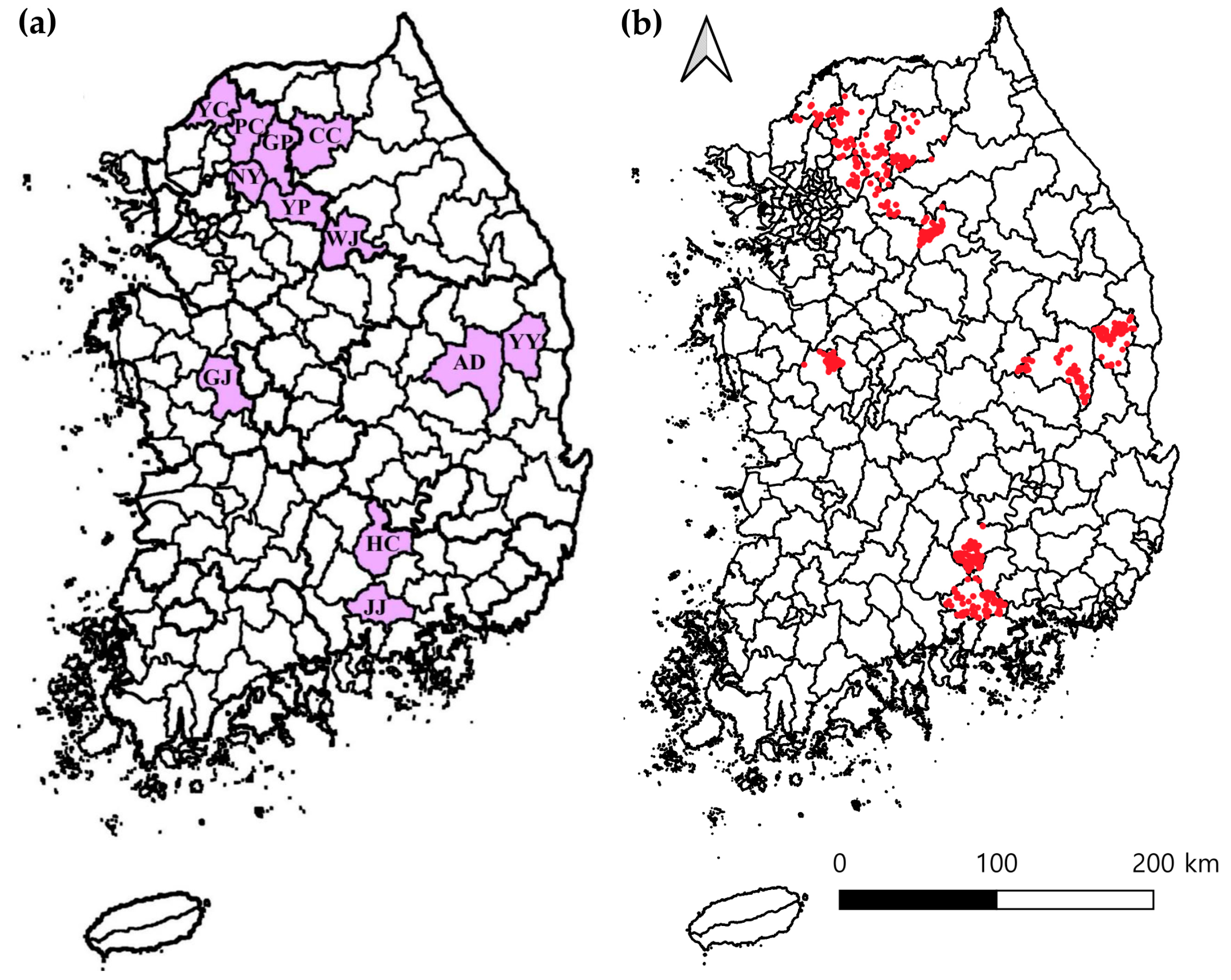
2.2. Sample Collection

Between April and November 2023, spleen tissue samples were collected from a total of 1035 Korean water deer across 12 regions of Korea. The northern region included Wonju (n = 134), Chuncheon (n = 72), Gapyeong (n = 60), Namyangju (n = 56), Yangpyeong (n = 31), Yeoncheon (n = 60), and Pocheon (n = 85). In the central region, samples were collected from Andong (n = 125), Yeongyang (n = 78), and Gongju (n = 104). The southern region included Jinju (n = 115) and Hapcheon (n = 115) (Figure 1a). Geographic coordinates were recorded for each sampled individual, and a spatial map showing all 1035 sampling locations was generated (Figure 1b). These data were collected on-site using handheld GPS devices at the time of sampling. The GPS coordinates and collection dates of all 1035 sampled individuals are provided in Supplementary Table S1.
By month, samples were obtained from 56 Korean water deer in April, 69 in May, 83 in June, 89 in July, 114 in August, 207 in September, 250 in October, and 167 in November.

2.3. Nucleic Acids Extraction and Processing

For gene extraction, spleen tissues from Korean water deer were homogenized using the Precellys CK28-R Lysing Kit (bead tube for hard tissue homogenization; Bertin Technologies, Bretonneux, France) in combination with the Precellys Evolution Homogenizer (Bertin Technologies). Approximately 10 mg of spleen tissue was used per sample. For tissue lysis, 710 µL of lysis buffer was prepared by mixing 700 µL of DLD buffer (lysis agent) with 10 µL of β-mercaptoethanol (reducing agent). The entire volume was applied to each tissue sample for nucleic acid extraction. Genomic DNA and RNA were simultaneously extracted using the Clear-S™ Quick DNA Extraction Kit (Invirustech, Gwangju, Republic of Korea), following a modified protocol optimized for spleen samples from Korean water deer. Detailed steps of the modified protocol are provided in Appendix A.

2.4. Detection of Diseases Using Real-Time PCR

Real-time PCR was employed to detect seven TBDs. Pathogen detection was carried out using commercially available real-time PCR kits in accordance with the manufacturers’ protocols. The “SB-Plex™ Tickborne System 1” (SNB, Gwangmyeong, Republic of Korea) was used to detect Anaplasma spp., Borrelia spp., and Theileria spp., targeting the 23S rDNA region for Borrelia spp. and the 16S rDNA region for both Anaplasma spp. and Theileria spp. The primers for Borrelia spp. were designed to detect a broad range of zoonotic species within the Borrelia burgdorferi sensu lato complex but were not specific to B. burgdorferi sensu stricto. The “SB-Plex™ YeV/OzV/TBEV qPCR Kit” (SNB) was used to detect YezoV, OZV, and TBEV, targeting the M segment for YezoV and OZV, and the 5′ untranslated region (UTR) for TBEV. For the detection of Babesia spp., the “AniQvet Babesia qPCR Kit“ (Koreagene, Seoul, Republic of Korea) was utilized, targeting the 18S rDNA region as per the manufacturer’s instructions.

2.5. Detection of Diseases Using Conventional PCR

Conventional PCR was performed to validate the positive findings obtained through real-time PCR and confirm the sequence identity of each detected pathogen. Species-specific primers described in previous studies were used to amplify target gene regions of Anaplasma spp. [15], Borrelia spp. [16], and Theileria/Babesia spp. [17]. For the detection of bacterial and parasitic pathogens, the AccuPower® PCR Premix kit (Bioneer, Daejeon, Republic of Korea) was utilized. For viral pathogens, amplification was performed using the AccuPower® RT-PCR Premix kit (Bioneer). To ensure the accuracy and reproducibility of PCR results, both positive and negative controls were included in all assays.

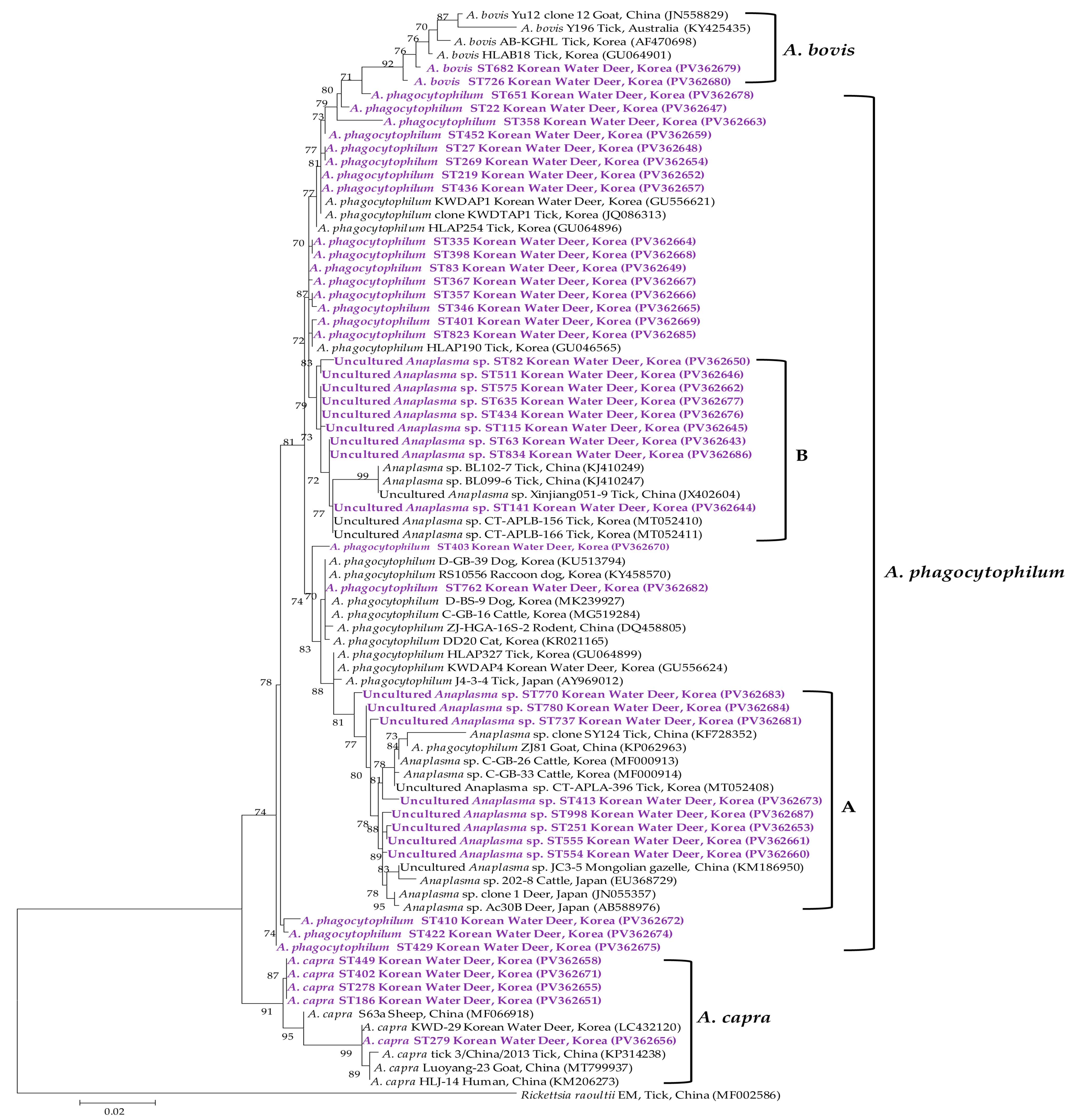
2.6. Genetic Sequencing and Phylogenetic Study

PCR amplicons confirmed as positive for the target pathogens were submitted to Macrogen (Seoul, Republic of Korea) for nucleotide sequencing along with the corresponding primers. All samples confirmed as positive were subjected to sequencing analysis, and in cases where identical sequences were obtained, a single representative sequence was selected for further analysis. The resulting sequences were then compared with reference sequences available in the NCBI GenBank database using the BLAST algorithm (BLAST+ v. 2.14.1) provided by NCBI against the nt (nucleotide collection) database to facilitate molecular genetic analysis. Sequence alignment was conducted using CLUSTAL Omega (v. 1.2.1, Bioweb, Ferndale, WA, USA), and redundant sequences were eliminated using BioEdit (v. 7.2.5). Regions exhibiting ambiguous alignments or containing gaps were excluded prior to phylogenetic analysis. Phylogenetic relationships were inferred using MEGA software (v. 6.0) [18], employing the Kimura two-parameter distance model [19] and the maximum likelihood method, with bootstrap support calculated from 1000 replicates. Only values ≥ 70% were shown on the phylogenetic trees.

2.7. Statistical Examination

All statistical analyses were performed using GraphPad Prism (v. 5.04, GraphPad Software Inc., La Jolla, CA, USA). Pearson’s chi-square test was applied to contingency tables involving more than two variables. A p-value of ≤ 0.05 was considered statistically significant, and 95% confidence intervals (CIs) were calculated for all estimates.

2.8. GenBank Accession Numbers

Anaplasma phagocytophilum sequences generated in this study were deposited in GenBank under the following accession numbers: PV362647–PV362649, PV362652, PV362654, PV362657, PV362659, PV362663–PV362670, PV362672, PV362674, PV362675, PV362678, PV362682, and PV362685. APLA sequences generated in this study were deposited in GenBank under the following accession numbers: PV362660, PV362661, PV362653, PV362673, PV362681, PV362683, PV362684, and PV362687. APLB sequences generated in this study were deposited in GenBank under the following accession numbers: PV362643, PV362644, PV362645, PV362646, PV362650, PV362662, PV362676, PV362677, and PV362686. A. bovis sequences generated in this study were deposited in GenBank under the following accession numbers: PV362679 and PV362680. A. capra sequences generated in this study were deposited in GenBank under the following accession numbers: PV362651, PV362655, PV362656, PV362658, and PV362671.
Borrelia theileri sequences generated in this study were deposited in GenBank under the following accession numbers: PV389464–PV389471.
Theileria capreoli sequences generated in this study were deposited in GenBank under the following accession numbers: PV436602–PV436606, PV436611, PV436613–PV436619, PV436622, PV436623, PV436626, PV436627, PV436629, PV436633, PV436634, and PV436636. T. cervi sequences generated in this study were deposited in GenBank under the following accession numbers: PV436624, PV436625, and PV436631. T. luwenshuni sequences generated in this study were deposited in GenBank under the following accession numbers: PV436601, PV436607-PV436610, PV436612, PV436620, PV436621, PV436628, PV436630, PV436632, PV436635, and PV436637.

3. Results

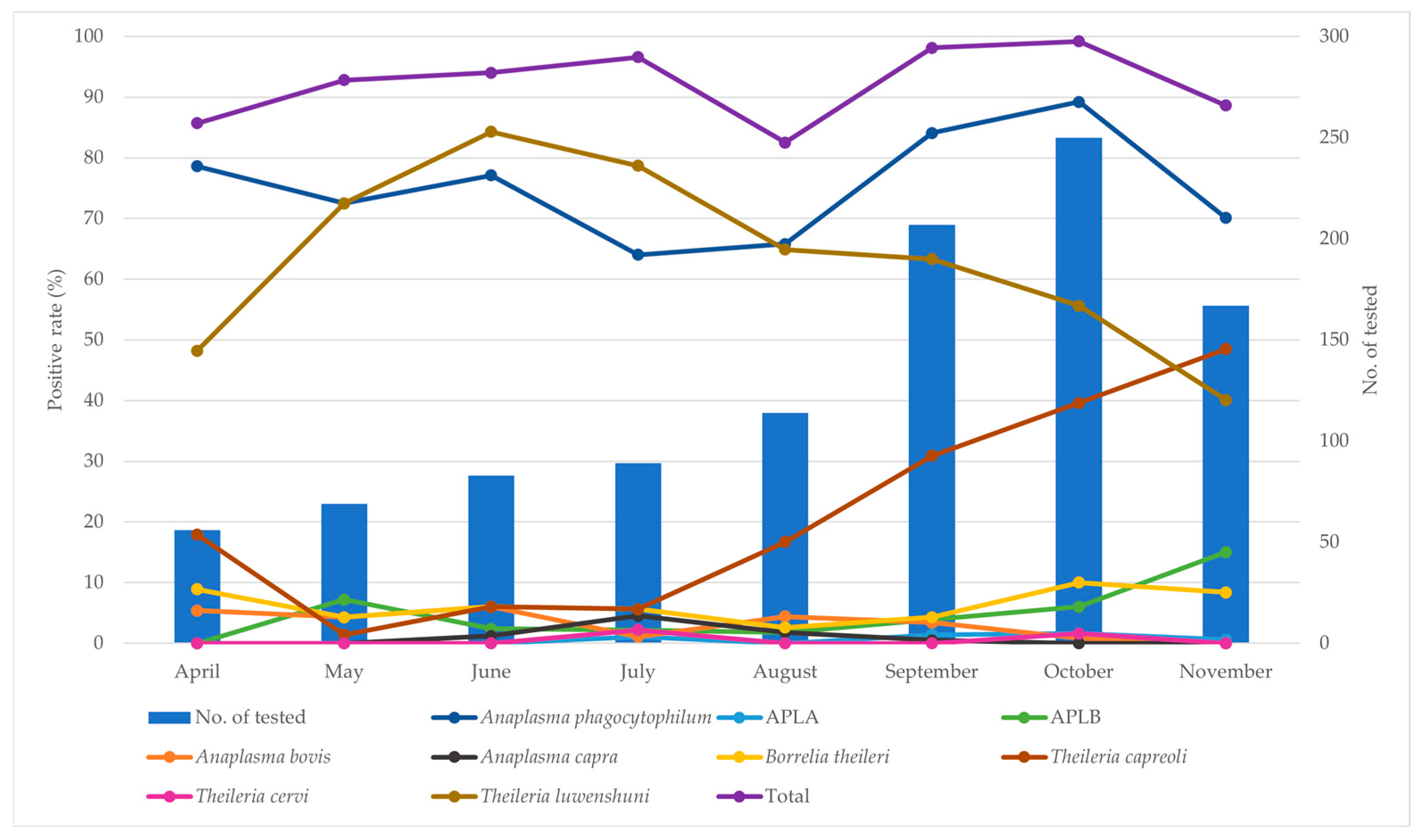
3.1. Prevalence Analysis Using Real-Time PCR

Real-time PCR analysis detected four tick-borne pathogens—Anaplasma spp., Babesia spp., Borrelia spp., and Theileria spp.—in Korean water deer samples. Anaplasma spp. and Theileria spp. showed high detection frequencies, while Babesia spp. and Borrelia spp. were identified at lower rates. No positive results were observed for TBEV, the OZ virus, or the Yezo virus in any of the 1035 samples tested. Detailed detection rates and confidence intervals are presented in Table 1.

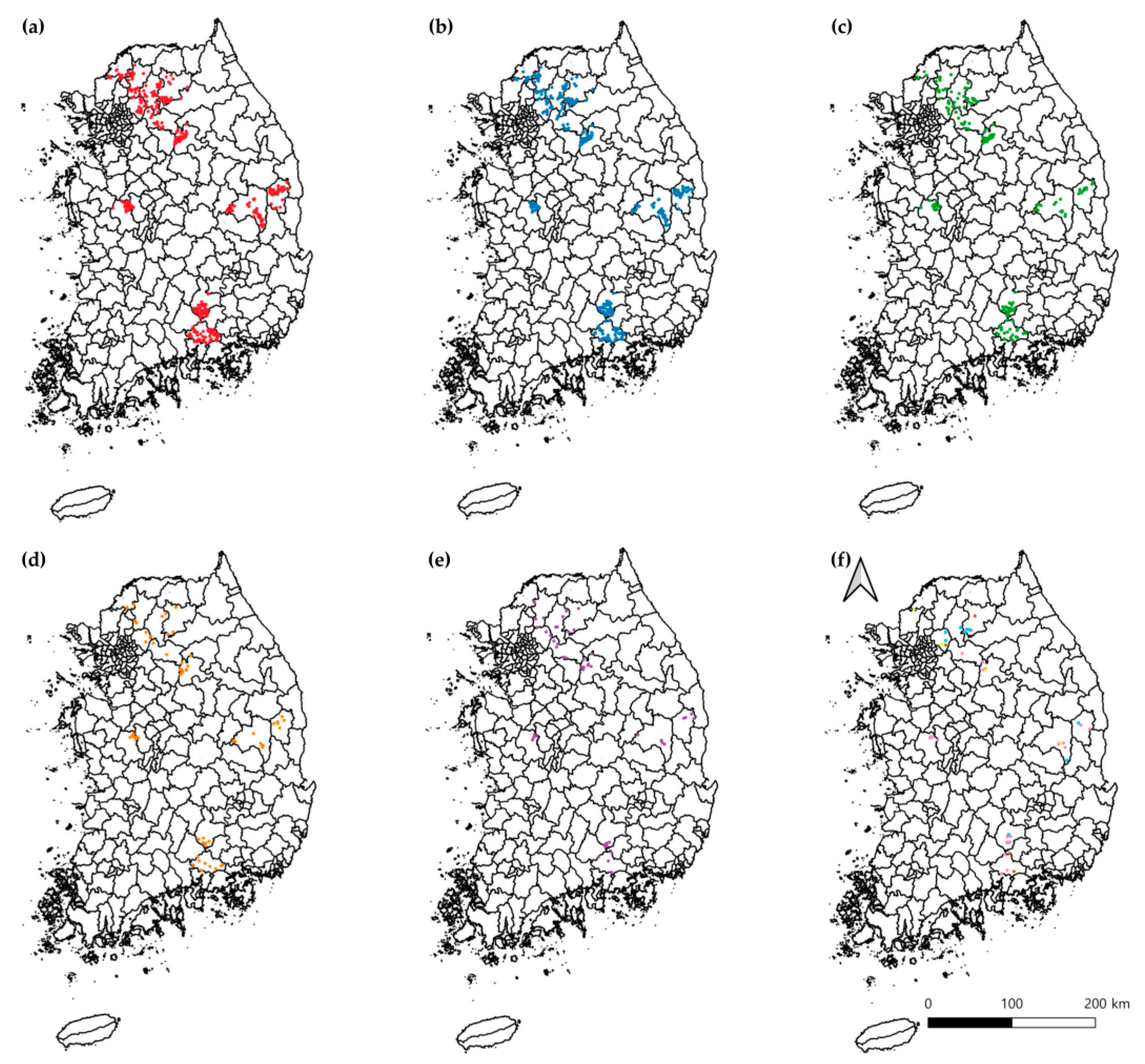
3.2. Prevalence Analysis Using Conventional PCR

Conventional PCR analysis identified 9 TBDs, with 969 out of 1035 samples testing positive for at least one pathogen, corresponding to an overall positivity rate of 93.6% (95% CI 92.1–95.1) (Table 2). Anaplasma spp. were classified into five distinct species: A. phagocytophilum (804 samples; 77.7%; 95% CI 75.1–80.2), A. phagocytophilum-like A (APLA; 9 samples; 0.9%; 95% CI 0.3–1.4), A. phagocytophilum-like B (APLB; 59 samples; 5.7%; 95% CI 4.3–7.1), A. bovis (26 samples; 2.5%; 95% CI 1.6–3.5), and A. capra (8 samples; 0.8%; 95% CI 0.2–1.3). Borrelia spp. was detected solely as B. theileri (69 samples; 6.7%; 95% CI 5.1–8.2). Although Babesia and Theileria spp. were amplified using a common primer set, only Theileria spp. were identified. These were further classified into T. capreoli (284 samples; 27.4%; 95% CI 24.7–30.2), T. cervi (6 samples; 0.6%; 95% CI 0.1–1.0), and T. luwenshuni (628 samples; 60.7%; 95% CI 57.7–63.7).
Spatial distribution maps were generated to visualize the geographic location of individuals that tested positive for each pathogen (Figure 2). A. phagocytophilum-positive samples were widely distributed across the country, particularly concentrated in the northern region, including Namyangju (Figure 2a). Similarly, a high regional density of T. luwenshuni-positive samples was observed in Chuncheon (Figure 2b). T. capreoli (Figure 2c) was spatially concentrated in Yangpyeong, while B. theileri-positive individuals were more sporadically distributed but with notable clustering in central regions (Figure 2d). APLB infections were mostly detected in northern sites, especially in Yangpyeong (Figure 2e). Due to the relatively low prevalence of APLA, A. bovis, A. capra, and T. cervi, these pathogens were grouped and are visualized collectively in Figure 2f.
By region, the highest positivity rates were observed in Namyangju (56/56; 100%) and Yangpyeong (31/31; 100%). The regional comparison revealed a statistically significant difference in positivity rates (χ2 = 92.305, df = 11, p < 0.0001) (Table 2). Pathogen-specific analyses showed the highest prevalence of A. phagocytophilum in Namyangju (89.3%; 95% CI 81.2–97.4; χ² = 36.576, df = 11, p = 0.0001) and A. capra in Chuncheon (5.6%; 95% CI 0.3–10.8; χ2 = 29.948, df = 11, p = 0.0016), both reaching statistical significance. Similarly, T. capreoli was most prevalent in Yangpyeong (54.8%; 95% CI 37.3–72.4; χ2 = 52.410, df = 11, p < 0.0001), T. cervi in Namyangju (5.4%; 95% CI 0–11.3; χ2 = 27.425, df = 11, p = 0.0400), and T. luwenshuni in Chuncheon (73.6%; 95% CI 63.4–83.8; χ2 = 21.561, df = 11, p = 0.0280), all of which also demonstrated statistically significant regional variation. These statistical results were in accordance with the spatial patterns observed in Figure 2.
By season, the overall positivity rate peaked in October (248/250; 99.2%), with a significant difference observed across seasons (χ2 = 57.998, df = 7, p < 0.0001) (Table 3 and Figure 3). Season-specific analysis indicated the highest prevalence of A. phagocytophilum in October (89.2%; 95% CI 85.4–93.0; χ2 = 49.551, df = 7, p < 0.0001), APLB in November (15.0%; 95% CI 9.6–20.4; χ2 = 38.674, df = 7, p < 0.0001), A. bovis in June (6.0%; 95% CI 0.9–11.1; χ2 = 17.251, df = 7, p = 0.0158), and A. capra in July (4.5%; 95% CI 0.2–8.8; χ2 = 22.153, df = 7, p = 0.0024), all showing statistically significant seasonal patterns. Among Theileria species, the prevalence of T. capreoli was highest in November (48.5%; 95% CI 40.9–56.1; χ2 = 130.080, df = 7, p < 0.0001), while T. luwenshuni was most prevalent in June (84.3%; 95% CI 76.5–92.2; χ2 = 72.914, df = 7, p < 0.0001), both with significant seasonal variation.

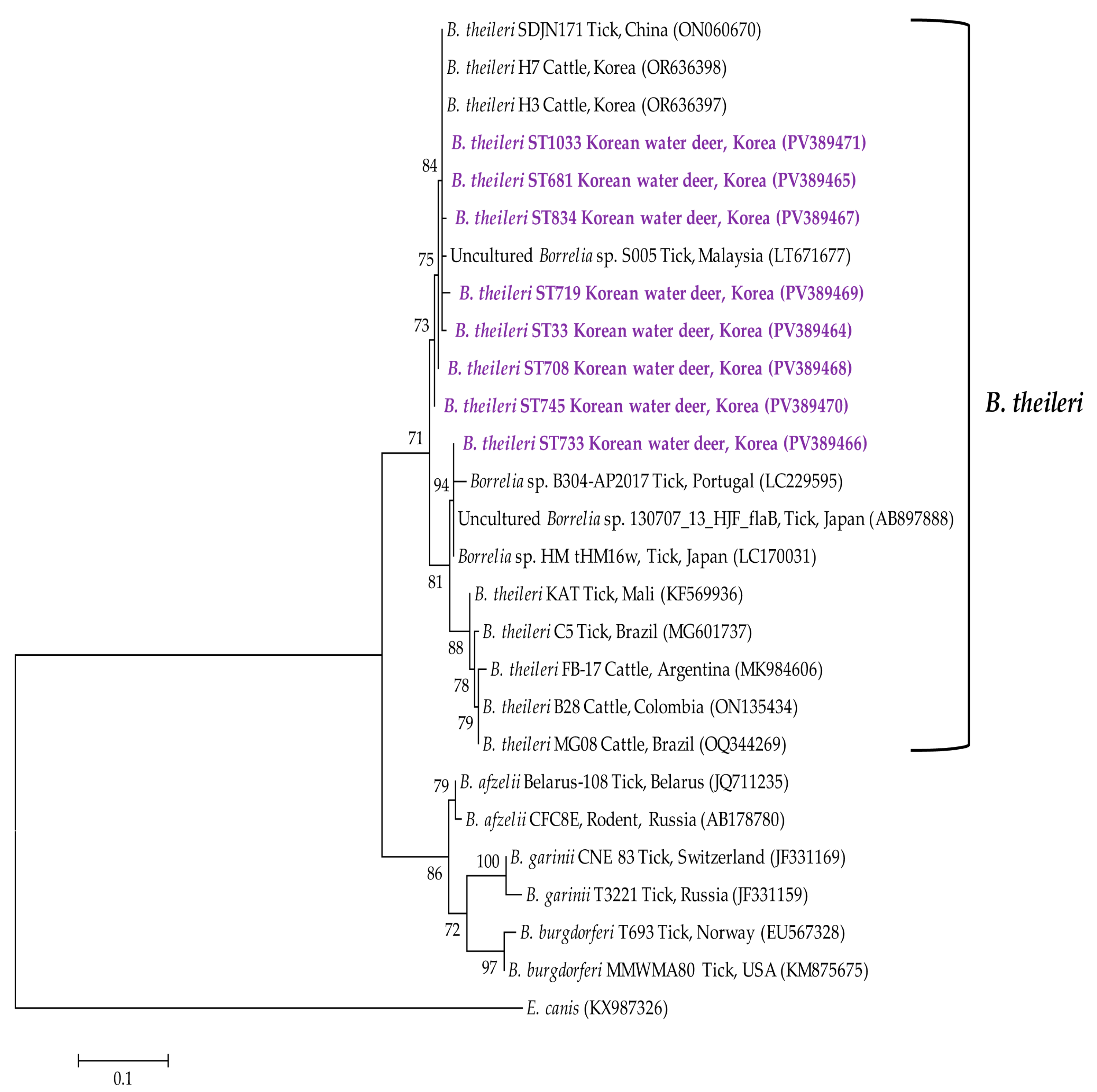
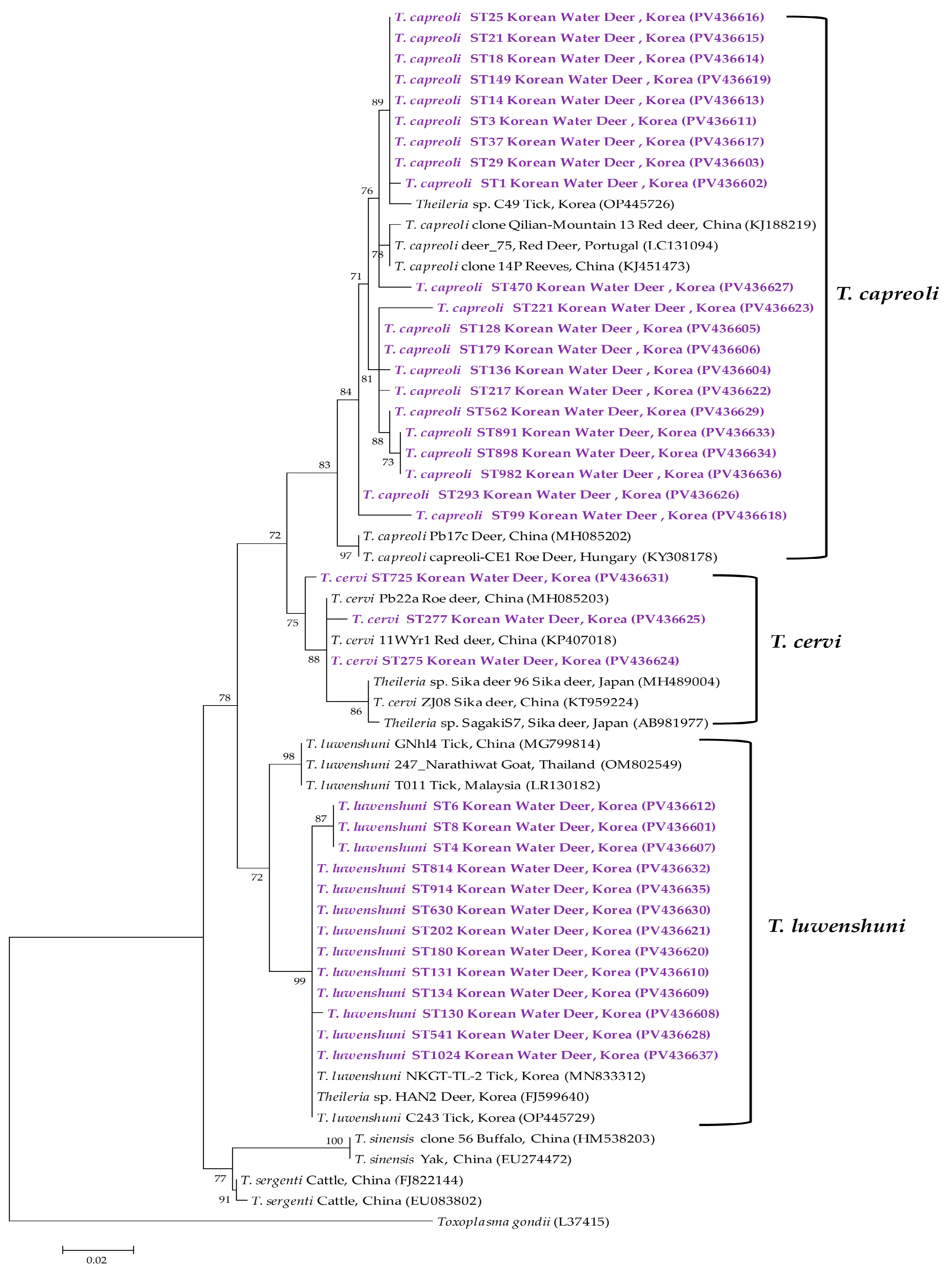
3.3. Molecular and Phylogenetic Analyses

Phylogenetic analysis of the 16S rRNA sequences of Anaplasma spp. revealed classification into A. phagocytophilum, APLA, APLB, A. bovis, and A. capra (Figure 4). The 21 representative A. phagocytophilum sequences exhibited 98.3–99.9% identity with one another and 98.3–100% identity with previously deposited A. phagocytophilum isolates in GenBank. The eight representative APLA sequences showed 98.5–99.5% identity with one another and 97.4–99.5% identity with reference APLA isolates in GenBank. Similarly, the nine representative APLB sequences demonstrated 98.9–100% identity among themselves and 97.5–100% identity with previously reported APLB isolates. The two representative A. bovis sequences displayed 98.4% identity with each other and 96.0–98.9% identity with GenBank-deposited A. bovis isolates. The five representative A. capra sequences shared 97.6–99.8% identity with one another and 97.6–100% identity with A. capra sequences in GenBank.
Phylogenetic analysis of the Borrelia spp. flagellin B gene sequence identified the organism as B. theileri (Figure 5). The eight representative B. theileri sequences exhibited 95.1–99.7% identity with each other and 95.1–100% identity with B. theileri isolates previously deposited in GenBank.
Analysis of the 18S rRNA sequences of Theileria spp. indicated classification into T. capreoli, T. cervi, and T. luwenshuni (Figure 6). The 21 representative T. capreoli sequences shared 93.1–93.6% identity with one another and 94.9–97.2% identity with corresponding GenBank isolates. The three representative T. cervi sequences exhibited 97.7–99.5% identity among themselves and 94.4–98.5% identity with GenBank T. cervi sequences. Finally, the 13 representative T. luwenshuni sequences showed 96.4–100% identity with each other and 92.6–97.8% identity with previously deposited T. luwenshuni isolates.

4. Discussion

In recent years, the spectrum of TBDs affecting both livestock and humans has expanded, with increasing attention focused on zoonotic TBDs such as anaplasmosis, babesiosis, ehrlichiosis, and Lyme borreliosis in both medical and veterinary contexts [2]. Beyond the pathogens identified in the present study, tick-borne viral infections have also garnered growing concern. For example, TBEV is considered endemic in several regions, including Siberia, the Russian Far East, China, and Japan [20]. Human and wildlife infections with OZV were reported in Japan between 2013 and 2019 [21] and YezoV was first identified in humans in Japan in 2019 [22]. Although no confirmed cases of TBEV, OZV, or YezoV have been reported in Korea—and none of these viruses were detected in this study—their documented presence in neighboring countries suggests a potential risk of future introduction into Korea.
Among the 1035 samples analyzed in this study, 969 tested positive, resulting in an overall positivity rate of 93.6%. Detection rates for all seven pathogens were consistently higher with real-time PCR than with conventional PCR, likely reflecting the superior sensitivity of real-time PCR in identifying low concentrations of pathogen DNA. This finding aligns with previous reports demonstrating that real-time PCR generally offers higher detection sensitivity compared to conventional PCR [23].
Geographically, samples collected from northern regions exhibited uniformly high positivity rates, with average detection rates exceeding 95% across nearly all surveyed sites. These sampling locations were originally selected by the NIWDC based on regions with a high incidence of human SFTS cases. Anticipating elevated SFTSV infection rates in local tick populations, the NIWDC prioritized sample collection from Korean water deer in these high-risk northern areas. Given that ticks are vectors not only of SFTSV but also of diverse TBD pathogens—and that Korean water deer are widely distributed tick hosts in Korea [5]—these animals were hypothesized to serve as important reservoirs for multiple TBDs. The consistently high positivity rates observed in deer from northern regions support this hypothesis and correspond with known regional distributions of human SFTS cases and presumed tick infection prevalence.
Seasonally, Korean water deer are distributed nationwide throughout the year and are recognized as major hosts for ticks. In this study, individuals captured in autumn were frequently found to carry heavy tick burdens. This observation is consistent with prior studies indicating a seasonal increase in tick larvae during autumn, contributing to a rise in overall tick density during this period [24]. These findings suggest a heightened likelihood of tick exposure among Korean water deer in autumn. Although tick-borne pathogens were detected in Korean water deer throughout the year, both tick burden and pathogen positivity rates peaked during the autumn months, indicating an elevated seasonal risk of transmission. This pattern is supported by monthly prevalence data, which showed the highest overall positivity rates in September (98.1%) and October (99.2%). Notably, A. phagocytophilum reached 84.1% in September and 89.2% in October. These results highlight autumn as a critical period for both tick activity and the transmission of tick-borne pathogens in Korean water deer.
Several Anaplasma species were detected in this study, highlighting the need for continued TBD surveillance in Korea. Notably, some of these pathogens remain poorly characterized with respect to their ecological and geographic distributions in wildlife hosts. Currently, only A. phagocytophilum, A. capra, A. ovis, and A. platys are recognized as zoonotic among the Anaplasma genus [25,26,27]. Of particular concern, A. phagocytophilum is a well-documented zoonotic agent that targets granulocytic white blood cells in mammalian hosts, causing HGA [28]. This pathogen has been identified in multiple tick species, and there is increasing concern about its potential emergence and transmission dynamics within Korea [29]. Supporting its established presence, A. phagocytophilum was previously detected in 18 out of 42 (42.9%) wild Korean water deer sampled in Gyeongbuk and Gangwon Provinces [30]. In the present study, this pathogen was identified in 77.7% of samples—the highest positivity rate among all detected TBD pathogens—providing compelling evidence of widespread systemic infection in Korean water deer.
Anaplasma bovis also represents a significant TBD pathogen. First identified in cattle in Brazil in 1936, it is now distributed throughout Asia, Africa, the Americas, and southern Europe [31]. Recent reports of human infections in China [32] and the United States [33] suggest an expanding geographic range and growing relevance to public health. In Korea, A. bovis has been detected in 4 of 173 tick pools (2.3%) collected from Korean water deer between 2013 and 2017 [14] and in 5 of 42 (11.9%) wild deer samples [30], indicating its presence in both vectors and hosts. In this study, A. bovis was detected in 2.5% of samples, reinforcing evidence of systemic infection in wild cervids and underscoring the need for wildlife-based surveillance to better understand its zoonotic potential.
Anaplasma capra, first identified in goats and humans with a history of tick exposure in China in 2015, was later confirmed as a novel species through phylogenetic analysis [27]. In Korea, A. capra was detected in 5 of 173 tick pools (2.9%) from Korean water deer collected between 2013 and 2017 [14], suggesting its circulation within cervid–tick ecological networks. A separate study reported its presence in 35 of 198 samples (17.8%) from Korean water deer collected between 2015 and 2018 [34], providing further evidence of systemic infection in this species. Given that A. phagocytophilum is a recognized zoonotic pathogen and A. capra has also been implicated in human infections in China, both species warrant close monitoring from a public health standpoint. In this study, A. capra was identified in 0.8% of samples, further supporting its potential infectivity in mammalian hosts and emphasizing the need for further investigation into its transmission dynamics.
Strains genetically similar to A. phagocytophilum, referred to in this study as APLA, have been identified in a variety of tick species parasitizing ruminants in Japan [35], including both cattle [36] and sika deer [37]. Phylogenetic analyses have demonstrated that these strains constitute a distinct monophyletic clade closely related to A. phagocytophilum yet exhibiting clear genetic divergence [38]. Their association with non-canonical tick vectors and the lack of clinical manifestations in infected animals further support their classification as novel Anaplasma strains, initially described in Japan [38]. In Korea, A. phagocytophilum-like strains have also been detected in domestic animals and ticks. For example, APLA was previously identified in 2.6% of cattle and was characterized by sequence divergence in the groEL gene [39], while APLB—a strain first reported in China—has been found in questing ticks from various regions of Korea [40]. However, to date, neither APLA nor APLB has been reported in Korean water deer or in any other cervid species globally. In the present study, APLA and APLB were detected in 0.9% and 5.7% of samples, respectively, representing the first global detection of these strains in this host species. These findings substantially broaden the recognized wildlife host range for both strains and underscore the importance of wildlife-based surveillance in elucidating their ecological roles and potential zoonotic risk.
Borrelia species represent another group of tick-borne pathogens that pose considerable health risks to both animals and humans. These spirochetes are generally categorized into two major groups: those associated with Lyme disease and those causing relapsing fever [41]. Lyme disease-associated Borrelia species, such as B. burgdorferi and B. afzelii, are predominantly transmitted by hard ticks. In Korea, B. afzelii was molecularly detected in ticks collected from wild Korean water deer between 2013 and 2015 [13]. The relapsing fever group has recently undergone further classification based on vector type, including soft-tick-borne species such as B. duttonii and hard-tick-borne species such as B. theileri [42]. In Korea, B. theileri has been reported in various hosts and vectors, including raccoon dogs [43], indigenous cattle [44], and ticks [45], suggesting a broader host and geographic distribution than previously recognized. The transmission dynamics of Borrelia spp., including B. theileri, are shaped not only by tick abundance but also by host–vector interactions and environmental factors. Previous Korean studies have documented a temporal lag between the peak of tick activity and the incidence of human disease. For instance, the detection of Borrelia spp. in ticks peaked in July, while human Lyme disease cases peaked in August [46]. Similarly, in a 2020 study, SFTSV detection in ticks peaked in August, whereas human cases peaked in October [24]. In our study, the highest B. theileri positivity was observed in October, potentially reflecting a similar temporal lag. Increased tick activity during the autumn season may lead to more frequent host–vector interactions, contributing to a delayed rise in pathogen detection. To our knowledge, this is the first global report of B. theileri infection in Korean water deer. These findings emphasize the role of complex host–vector–environment interactions in shaping the epidemiology of tick-borne pathogens and highlight the need for further investigation into their transmission cycles.
In our study, both Babesia and Theileria spp. tested positive by real-time PCR. However, only Theileria spp. were detected using conventional PCR, which identified three species: T. capreoli, T. cervi, and T. luwenshuni. This discrepancy may be attributable to cross-reactivity with Theileria DNA, given the high sequence similarity of the 18S rRNA gene between Babesia and Theileria, even when TaqMan probes are employed [47]. A pertinent example is the reclassification of Babesia equi as Theileria equi, based on phylogenetic analysis of the 18S rRNA gene in conjunction with morphological characteristics [48]. Therefore, the observed Babesia signal in this study may reflect cross-reactivity with Theileria spp., particularly T. capreoli, which exhibits high sequence homology with Babesia capreoli within the 18S rRNA region. To enhance species-level discrimination, the sequencing of real-time PCR amplicons or the use of alternative genetic markers with greater discriminatory power is recommended.
Theileria is a clinically and economically significant tick-borne protozoan parasite that predominantly targets the bloodstream and presents a major threat to the global livestock industry [49]. In addition to domestic animals, Theileria spp. have also been detected in wild ruminants, including roe deer (Capreolus capreolus L.), in which T. capreoli has been previously identified [50]. In Korea, T. capreoli was first reported in Korean water deer, with a prevalence of 5.6% (1/18) during the 2008–2009 period [51], and more recently in ticks at a detection rate of 25% (4/16) in 2024 [46]. However, the limited sample sizes in these studies constrain the broader applicability of their findings. In contrast, our investigation identified T. capreoli as the third most prevalent pathogen, with a positivity rate of 27.4%, underscoring the need for further research into its epidemiological significance and its potential impact on wildlife health and vector ecology.
Among Theileria species, T. cervi is recognized as a causative agent of cervine theileriosis [52], whereas the closely related Theileria sp. Thrivae has been identified as a non-pathogenic strain in sika deer in Japan [53]. A genetically similar Theileria sp. was also reported in China and designated as T. cervi [49]. Of note, T. cervi identified in white-tailed deer and elk (Cervus canadensis) in the United States and Canada [54] is genetically distinct from both Theileria sp. Thrivae and the T. cervi strains reported in Japan and China. Phylogenetic analysis of the T. cervi sequences obtained in this study demonstrated clustering with a previously reported Chinese strain (KT959224), suggesting that the isolate identified herein is genetically distinct from North American strains. In our cohort, T. cervi was the least prevalent of the three Theileria species, with a detection rate of 0.6%.
Theileria luwenshuni was initially believed to be confined to sheep and goats in China [55]. However, more recent evidence indicates its pathogenic potential, particularly in association with high mortality among small ruminants [56]. In Korea, T. luwenshuni has been detected in all 23 wild roe deer examined on Jeju Island, as well as in 34.8% of ticks collected from these animals [57]. A 2015 study also reported its presence in 10 out of 18 Korean water deer (55.6%) [58]. Consistent with these findings, T. luwenshuni was detected in 60.7% of samples in our study, making it the second most prevalent pathogen among all tick-borne agents identified. These data support the conclusion that T. luwenshuni is the most widely distributed Theileria species among wild cervids in Korea and may play a predominant role in the regional epidemiology of Theileria infections in these hosts.
Although this study focused on the molecular detection of TBDs in Korean water deer, it did not include the collection or analysis of ticks parasitizing the hosts. A previous study by our group has provided baseline information on ticks parasitizing Korean water deer, including the detection of multiple pathogens [14]. Given that tick burden can influence pathogen transmission dynamics, future studies should incorporate data on tick species composition and infestation levels.

5. Conclusions

This study presents comprehensive molecular evidence of multiple tick-borne pathogens in Korean water deer, including the first global identification of APLA, APLB, and B. theileri in this host species. The notably high prevalence of T. luwenshuni and A. phagocytophilum suggests that Korean water deer may serve as significant reservoir hosts for tick-borne diseases in Korea. These findings underscore the critical importance of wildlife-based surveillance in elucidating the ecological dynamics and zoonotic potential of emerging tick-borne pathogens. Further investigation is warranted to delineate transmission pathways, evaluate associated public health risks, and guide the development of targeted vector control strategies.

Supplementary Materials

The following supporting information can be downloaded at: https://www.mdpi.com/article/10.3390/ani15101499/s1, Table S1: GPS coordinates and collection dates of 1035 Korean water deer sampled between April and November 2023.

Author Contributions

Conceptualization, writing—original draft preparation, B.K. and S.-J.C.; methodology, investigation, Y.-J.L.; validation, formal analysis, H.S.; validation, formal analysis, S.K.; methodology, investigation, H.J.; supervision, writing—review and editing, M.-G.S.; supervision, visualization, D.K.; methodology, project administration, S.L. All authors have read and agreed to the published version of the manuscript.

Funding

This study was supported by the National Institute of Wildlife Disease Control and Prevention under a research support project.

Institutional Review Board Statement

The animal protocol used in this study was reviewed and approved according to the guidelines for ethical procedures and scientific care by the Institutional Animal Care and Use Committee of Kyungpook National University (KNU 2024-0407).

Informed Consent Statement

Not applicable.

Data Availability Statement

Data supporting the conclusions of this article are included within the article. The newly generated sequences were submitted to the GenBank database under the accession numbers PV362643–PV362687, PV389464–PV389471, and PV436601–PV436637. The datasets used and/or analyzed during the present study are available from the corresponding author upon reasonable request.

Conflicts of Interest

The authors declare no conflicts of interest.

Appendix A

Modified DNA and RNA Extraction Protocol for Korean Water Deer Spleen Tissue
(Adapted from the Clear-S™ Quick DNA Extraction Kit, InVirusTech)
This protocol was specifically optimized for the simultaneous extraction of DNA and RNA from the spleen tissue of Korean water deer. Please note that the procedure deviates from the standard Clear-S™ tissue extraction manual.
Pre-experiment Preparation:
  • Prepare 1.5 mL microcentrifuge tubes and add β-mercaptoethanol (reducing agent) at a ratio of 10 μL per 700 μL of DLD buffer (i.e., 10 μL + 700 μL per sample).
  • Add 45 mL of 100% ethanol to the WA buffer and 66 mL to the wash buffer as instructed by the kit.
  • Preheat the elution buffer (EB) to 37 °C prior to use.
  • Protocol Steps.
  • Add 710 μL of prepared lysis buffer (700 μL DLD + 10 μL β-mercaptoethanol) to ~10 mg of spleen tissue in a homogenization tube.
  • Homogenize using a tissue grinder at 3500 rpm for 30 s, pause for 10 s, and repeat twice. If homogenization is incomplete, repeat up to 1–2 additional cycles.
  • Vortex thoroughly and incubate at room temperature for 5–10 min. Then, centrifuge at 13,000 rpm for 1 min.
  • Label a pre-treatment column and collection tube. Transfer the supernatant (~710 μL) to the column and centrifuge at 13,000 rpm for 1 min.
  • Discard the upper spin column. To the remaining flow-through (~710 μL), add 710 μL of 100% ethanol (final volume: 1420 μL).
  • Attach a binding column to a new collection tube. Mix the ethanol-added lysate by pipetting 3–4 times.
  • Load 710 μL of the solution into the column and centrifuge at 13,000 rpm for 1 min. Discard the flow-through and wipe the tube rim.
  • Load the remaining 710 μL of the lysate and repeat the centrifugation step.
  • Add 700 μL of WA buffer, centrifuge at 13,000 rpm for 1 min, discard the flow-through, and reattach the column.
  • Add 500 μL of wash buffer, centrifuge at 13,000 rpm for 1 min, discard the flow-through, and repeat with another 500 μL followed by centrifugation for 3 min.
  • Transfer the column to a clean 1.5 mL tube. Add 200 μL of preheated EB buffer (37 °C) to the column and incubate at room temperature for 5 min.
  • Centrifuge at 13,000 rpm for 1 min to collect the final eluate.
  • Store nucleic acid extracts at –70 °C until analysis.

References

  1. Taylor, M.A. Emerging parasitic diseases of sheep. Vet. Parasitol. 2012, 189, 2–7. [Google Scholar] [CrossRef] [PubMed]
  2. Dantas-Torres, F.; Chomel, B.B.; Otranto, D. Ticks and tick-borne diseases: A one health perspective. Trends Parasitol. 2012, 28, 437–446. [Google Scholar] [CrossRef]
  3. Cummins, G.A.; Traub, S.J. Tick-borne diseases. In Auerbach’s Wilderness Medicine; Elsevier: Amsterdam, The Netherlands, 2016; Volume 1–2, pp. 968–993. [Google Scholar] [CrossRef]
  4. Harris, R.B.; Duckworth, J.W. The IUCN Red List of Threatened Species. [CrossRef]
  5. Kim, B.J.; Oh, D.H.; Chun, S.H.; Lee, S.D. Distribution, density, and habitat use of the Korean water deer (Hydropotes inermis argyropus) in Korea. Landsc. Ecol. Eng. 2011, 7, 291–297. [Google Scholar] [CrossRef]
  6. Severo, M.S.; Stephens, K.D.; Kotsyfakis, M.; Pedra, J.H. Anaplasma phagocytophilum: Deceptively simple or simply deceptive? Future Microbiol. 2012, 7, 719–731. [Google Scholar] [CrossRef]
  7. Qiu, Y.; Squarre, D.; Nakamura, Y.; Lau, A.C.C.; Moonga, L.C.; Kawai, N.; Ohnuma, A.; Hayashida, K.; Nakao, R.; Yamagishi, J.; et al. Evidence of Borrelia theileri in wild and domestic animals in the kafue ecosystem of Zambia. Microorganisms 2021, 9, 2405. [Google Scholar] [CrossRef]
  8. Mans, B.J.; Pienaar, R.; Latif, A.A. A review of Theileria diagnostics and epidemiology. Int. J. Parasitol. Parasites Wildl. 2015, 4, 104–118. [Google Scholar] [CrossRef]
  9. Shin, S.U.; Park, Y.J.; Ryu, J.H.; Jang, D.H.; Hwang, S.; Cho, H.C.; Park, J.; Han, J.I.; Choi, K.S. Identification of zoonotic tick-borne pathogens from Korean water deer (Hydropotes inermis argyropus). Vector Borne Zoonotic Dis. 2020, 20, 745–754. [Google Scholar] [CrossRef]
  10. Seong, G.; Han, Y.J.; Oh, S.S.; Chae, J.S.; Yu, D.H.; Park, J.; Park, B.K.; Yoo, J.G.; Choi, K.S. Detection of tick-borne pathogens in the Korean water deer (Hydropotes inermis argyropus) from Jeonbuk province, Korea. Korean J. Parasitol. 2015, 53, 653–659. [Google Scholar] [CrossRef]
  11. Lee, H.S.; Kim, J.; Son, K.; Kim, Y.; Hwang, J.; Jeong, H.; Ahn, T.Y.; Jheong, W.H. Phylogenetic analysis of severe fever with thrombocytopenia syndrome virus in Korean water deer (Hydropotes inermis argyropus) in the Republic of Korea. Ticks Tick-Borne Dis. 2020, 11, 101331. [Google Scholar] [CrossRef]
  12. Kang, J.G.; Ko, S.; Kim, H.C.; Chong, S.T.; Klein, T.A.; Chae, J.B.; Jo, Y.S.; Choi, K.S.; Yu, D.H.; Park, B.K.; et al. Prevalence of Anaplasma and Bartonella spp. in ticks collected from Korean water deer (Hydropotes inermis argyropus). Korean J. Parasitol. 2016, 54, 87–91. [Google Scholar] [CrossRef] [PubMed]
  13. VanBik, D.; Lee, S.H.; Seo, M.G.; Jeon, B.R.; Goo, Y.K.; Park, S.J.; Rhee, M.H.; Kwon, O.D.; Kim, T.H.; Geraldino, P.J.L.; et al. Borrelia species detected in ticks feeding on wild Korean water deer (Hydropotes inermis) using molecular and genotypic analyses. J. Med. Entomol. 2017, 54, 1397–1402. [Google Scholar] [CrossRef]
  14. Seo, M.G.; Kwon, O.D.; Kwak, D. Molecular detection of Rickettsia raoultii, Rickettsia tamurae, and associated pathogens from ticks parasitizing water deer (Hydropotes inermis argyropus) in South Korea. Ticks Tick Borne Dis. 2021, 12, 101712. [Google Scholar] [CrossRef]
  15. Barlough, J.E.; Madigan, J.E.; DeRock, E.; Bigornia, L. Nested polymerase chain reaction for detection of Ehrlichia equi genomic DNA in horses and ticks (Ixodes pacificus). Vet. Parasitol. 1996, 63, 319–329. [Google Scholar] [CrossRef]
  16. Lee, K.; Takano, A.; Taylor, K.; Sashika, M.; Shimozuru, M.; Konnai, S.; Kawabata, H.; Tsubota, T. A relapsing fever group Borrelia sp. similar to Borrelia lonestari found among wild sika deer (Cervus nippon yesoensis) and Haemaphysalis spp. ticks in Hokkaido, Japan. Ticks Tick Borne Dis. 2014, 5, 841–847. [Google Scholar] [CrossRef]
  17. Casati, S.; Sager, H.; Gern, L.; Piffaretti, J.C. Presence of potentially pathogenic Babesia sp. for human in Ixodes ricinus in Switzerland. Ann. Agric. Environ. Med. 2006, 13, 65–70. [Google Scholar]
  18. Tamura, K.; Stecher, G.; Peterson, D.; Filipski, A.; Kumar, S. MEGA6: Molecular evolutionary genetics analysis version 6.0. Mol. Biol. Evol. 2013, 30, 2725–2729. [Google Scholar] [CrossRef]
  19. Kimura, M. A simple method for estimating evolutionary rates of base substitutions through comparative studies of nucleotide sequences. J. Mol. Evol. 1980, 16, 111–120. [Google Scholar] [CrossRef]
  20. Im, J.H.; Baek, J.H.; Durey, A.; Kwon, H.Y.; Chung, M.H.; Lee, J.S. Geographic distribution of Tick-borne encephalitis virus complex. J. Vector Borne Dis. 2020, 57, 14–22. [Google Scholar] [CrossRef]
  21. Tran, N.T.B.; Shimoda, H.; Ishijima, K.; Yonemitsu, K.; Minami, S.; Kuroda, Y.; Tatemoto, K.; Mendoza, M.V.; Kuwata, R.; Takano, A.; et al. Zoonotic infection with Oz virus, a novel thogotovirus. Emerg. Infect. Dis. 2022, 28, 436–439. [Google Scholar] [CrossRef]
  22. Ogata, Y.; Sato, T.; Kato, K.; Kikuchi, K.; Mitsuhashi, K.; Watari, K.; Tamiya, K.; Goto, A.; Yamaguchi, H.; Hisada, R. A case of tick-borne Yezo virus infection: Concurrent detection in the patient and tick. Int. J. Infect. Dis. 2024, 143, 107038. [Google Scholar] [CrossRef] [PubMed]
  23. Dagher, H.; Donninger, H.; Hutchinson, P.; Ghildyal, R.; Bardin, P. Rhinovirus detection: Comparison of real-time and conventional PCR. J. Virol. Methods 2004, 117, 113–121. [Google Scholar] [CrossRef]
  24. Seo, M.G.; Noh, B.E.; Lee, H.S.; Kim, T.K.; Song, B.G.; Lee, H.I. Nationwide temporal and geographical distribution of tick populations and phylogenetic analysis of Severe Fever with Thrombocytopenia Syndrome virus in ticks in Korea, 2020. Microorganisms 2021, 9, 1630. [Google Scholar] [CrossRef] [PubMed]
  25. Arraga-Alvarado, C.M.; Qurollo, B.A.; Parra, O.C.; Berrueta, M.A.; Hegarty, B.C.; Breitschwerdt, E.B. Molecular evidence of Anaplasma platys infection in two women from Venezuela. Am. Soc. Trop. Med. Hyg. 2014, 91, 1161–1165. [Google Scholar] [CrossRef]
  26. García-Pérez, A.L.; Oporto, B.; Espí, A.; del Cerro, A.; Barral, M.; Povedano, I.; Barandika, J.F.; Hurtado, A. Anaplasmataceae in wild ungulates and carnivores in northern Spain. Ticks Tick-Borne Dis. 2016, 7, 264–269. [Google Scholar] [CrossRef]
  27. Li, H.; Zheng, Y.C.; Ma, L.; Jia, N.; Jiang, B.G.; Jiang, R.R.; Huo, Q.B.; Wang, Y.W.; Liu, H.B.; Chu, Y.L.; et al. Human infection with a novel tick-borne Anaplasma species in China: A surveillance study. Lancet Infect. Dis. 2015, 15, 663–670. [Google Scholar] [CrossRef]
  28. Im, J.H.; Baek, J.H.; Durey, A.; Kwon, H.Y.; Chung, M.H.; Lee, J.S. Current status of tick-borne diseases in South Korea. Vector-Borne Zoonotic Dis. 2018, 19, 225–233. [Google Scholar] [CrossRef]
  29. Kim, C.M.; Kim, M.S.; Park, M.S.; Park, J.H.; Chae, J.S. Identification of Ehrlichia chaffeensis, Anaplasma phagocytophilum, and A. bovis in Haemaphysalis longicornis and Ixodes persulcatus ticks from Korea. Vector-Borne Zoonotic Dis. 2003, 3, 17–26. [Google Scholar] [CrossRef]
  30. Lee, M.; Seo, M.G.; Lee, S.H.; Ouh, I.O.; Kim, Y.H.; Kim, J.K.; Goo, Y.K.; Rhee, M.H.; Kim, T.H.; Kwon, O.D.; et al. Molecular detection and phylogenetic analysis of tick-borne pathogens in wild Korean water deer and farmed elk in Gyeongbuk and Gangwon provinces of Korea. J. Vet. Med. Sci. 2018, 80, 1473–1478. [Google Scholar] [CrossRef]
  31. Atif, F.A. Alpha proteobacteria of genus Anaplasma (Rickettsiales: Anaplasmataceae): Epidemiology and characteristics of Anaplasma species related to veterinary and public health importance. Parasitology 2016, 143, 659–685. [Google Scholar] [CrossRef]
  32. Lu, M.; Li, F.; Liao, Y.; Shen, J.J.; Xu, J.M.; Chen, Y.Z.; Li, J.H.; Holmes, E.C.; Zhang, Y.Z. Epidemiology and diversity of Rickettsiales Bacteria in humans and animals in Jiangsu and Jiangxi provinces, China. Sci. Rep. 2019, 9, 13176. [Google Scholar] [CrossRef] [PubMed]
  33. Karpathy, S.; Kingry, L.; Pritt, B.; Berry, J.; Chilton, N.; Dergousoff, S.; Cortinas, R.; Sheldon, S.; Oatman, S.; Anacker, M.; et al. Anaplasma bovis–like infections in humans, United States, 2015–2017. Emerg. Infect. Dis. J. 2023, 29, 1904. [Google Scholar] [CrossRef]
  34. Amer, S.; Kim, S.; Yun, Y.M.; Na, K.J. Novel variants of the newly emerged Anaplasma capra from Korean water deer (Hydropotes inermis argyropus) in South Korea. Parasites Vectors 2019, 12, 365. [Google Scholar] [CrossRef]
  35. Ohashi, N.; Inayoshi, M.; Kitamura, K.; Kawamori, F.; Kawaguchi, D.; Nishimura, Y.; Naitou, H.; Hiroi, M.; Masuzawa, T. Anaplasma phagocytophilum—Infected ticks, Japan. Emerg. Infect. Dis. J. 2005, 11, 1780. [Google Scholar] [CrossRef]
  36. Yoshimoto, K.; Matsuyama, Y.; Matsuda, H.; Sakamoto, L.; Matsumoto, K.; Yokoyama, N.; Inokuma, H. Detection of Anaplasma bovis and Anaplasma phagocytophilum DNA from Haemaphysalis megaspinosa in Hokkaido, Japan. Vet. Parasitol. 2010, 168, 170–172. [Google Scholar] [CrossRef]
  37. Jilintai; Seino, N.; Hayakawa, D.; Suzuki, M.; Hata, H.; Kondo, S.; Matsumoto, K.; Yokoyama, N.; Inokuma, H. Molecular survey for Anaplasma bovis and Anaplasma phagocytophilum infection in cattle in a Pastureland where sika deer appear in Hokkaido, Japan. Jpn. J. Infect. Dis. 2009, 62, 73–75. [Google Scholar] [CrossRef]
  38. Ybañez, A.P.; Matsumoto, K.; Kishimoto, T.; Inokuma, H. Molecular analyses of a potentially novel Anaplasma species closely related to Anaplasma phagocytophilum detected in sika deer (Cervus nippon yesoensis) in Japan. Vet. Microbiol. 2012, 157, 232–236. [Google Scholar] [CrossRef]
  39. Seo, M.G.; Ouh, I.O.; Kwon, O.D.; Kwak, D. Molecular detection of Anaplasma phagocytophilum-like Anaplasma spp. and pathogenic A. phagocytophilum in cattle from South Korea. Mol. Phylogenetics Evol. 2018, 126, 23–30. [Google Scholar] [CrossRef]
  40. Seo, M.G.; Lee, H.; Alkathiri, B.; Ahn, K.; Lee, S.H.; Shin, S.; Bae, S.; Kim, K.T.; Jang, M.; Lee, S.K.; et al. Tick populations and molecular analysis of Anaplasma species in ticks from the Republic of Korea. Microorganisms 2023, 11, 820. [Google Scholar] [CrossRef]
  41. Margos, G.; Vollmer, S.A.; Ogden, N.H.; Fish, D. Population genetics, taxonomy, phylogeny and evolution of Borrelia burgdorferi sensu lato. Infect. Genet. Evol. 2011, 11, 1545–1563. [Google Scholar] [CrossRef]
  42. Trevisan, G.; Cinco, M.; Trevisini, S.; di Meo, N.; Ruscio, M.; Forgione, P.; Bonin, S. Borreliae part 2: Borrelia relapsing fever group and unclassified Borrelia. Biology 2021, 10, 1117. [Google Scholar] [CrossRef] [PubMed]
  43. Kang, J.G.; Chae, J.B.; Cho, Y.K.; Jo, Y.S.; Shin, N.S.; Lee, H.; Choi, K.S.; Yu, D.H.; Park, J.; Park, B.K.; et al. Molecular detection of Anaplasma, Bartonella, and Borrelia theileri in raccoon dogs (Nyctereutes procyonoides) in Korea. Am. Soc. Trop. Med. Hyg. 2018, 98, 1061–1068. [Google Scholar] [CrossRef] [PubMed]
  44. Hyung, H.J.; Choi, Y.S.; Park, J.; Lee, K.J.; Kang, J.G. Molecular detection of Borrelia theileri in cattle in Korea. Parasites Hosts Dis. 2024, 62, 151–156. [Google Scholar] [CrossRef]
  45. Kim, M.; Cho, S.; Park, G.; Kim, J.; Rieu, M.; Noh, K.T.; Ha, S.; Park, Q.; Kim, D.H.; Han, S.; et al. Molecular detection of, and Severe fever with thrombocytopenia syndrome virus (SFTSV) in military working dogs and ticks collected from the Republic of Korea army garrisons in Gangwon province in 2021–2022. Entomol. Res. 2024, 54, e12762. [Google Scholar] [CrossRef]
  46. Monoldorova, S.; Lee, S.; Yun, S.; Park, S.; Jeong, J.U.; Kim, J.; Lee, I.Y.; Jun, H.; Park, C.H.; Byeon, H.S.; et al. Seasonal dynamics of ticks and tick-borne pathogens in Republic of Korea. Pathogens 2024, 13, 1079. [Google Scholar] [CrossRef]
  47. Schnittger, L.; Yin, H.; Qi, B.; Gubbels, M.J.; Beyer, D.; Niemann, S.; Jongejan, F.; Ahmed, J.S. Simultaneous detection and differentiation of Theileria and Babesia parasites infecting small ruminants by reverse line blotting. Parasitol Res. 2004, 92, 189–196. [Google Scholar] [CrossRef]
  48. Mehlhorn, H.; Schein, E. Redescription of Babesia equi Laveran, 1901 as Theileria equi Mehlhorn, Schein 1998. Parasitol Res. 1998, 84, 467–475. [Google Scholar] [CrossRef]
  49. Liu, J.; Yang, J.; Guan, G.; Liu, A.; Wang, B.; Luo, J.; Yin, H. Molecular detection and identification of piroplasms in sika deer (Cervus nippon) from Jilin province, China. Parasites Vectors 2016, 9, 156. [Google Scholar] [CrossRef]
  50. Garc, J.; Aurtenetxe, O.; Barral, M.; Marco, I.; Lavin, S.; Garc, A.; Rez, A.P.; Hurtado, A. Molecular detection and characterization of piroplasms infecting cervids and chamois in Northern Spain. Parasitology 2006, 134, 391–398. [Google Scholar] [CrossRef]
  51. Han, J.I.; Jang, H.J.; Lee, S.J.; Na, K.J. High prevalence of Theileria sp. in wild Chinese Water Deer (Hydropotes inermis argyropus) in South Korea. Vet. Parasitol. 2009, 164, 311–314. [Google Scholar] [CrossRef]
  52. He, L.; Khan, M.K.; Zhang, W.J.; Zhang, Q.L.; Zhou, Y.Q.; Hu, M.; Zhao, J. Detection and identification of Theileria infection in Sika deer (Cervus nippon) in China. J. Parasitol. 2012, 98, 598–603. [Google Scholar] [CrossRef] [PubMed]
  53. Watanabe, Y.; Fukumoto, S.; Harasawa, R. Prevalence of tick-borne hemolytic microbes in free-living sika deer (Cervus nippon) captured in a deer-overcrowded area. Jpn. J. Zoo Wildl. Med. 2016, 21, 17–27. [Google Scholar] [CrossRef]
  54. Chae, J.s.; Waghela, S.D.; Craig, T.M.; Kocan, A.A.; Wagner, G.G.; Holman, P.J. Two Theileria cervi SSU rRNA gene sequence types found in isolates from white-tailed deer and elk in North America. J. Wildl. Dis. 1999, 35, 458–465. [Google Scholar] [CrossRef]
  55. Yin, H.; Schnittger, L.; Luo, J.; Seitzer, U.; Ahmed, J.S. Ovine theileriosis in China: A new look at an old story. Parasitol. Res. 2007, 101 (Suppl. S2), S191–S195. [Google Scholar] [CrossRef]
  56. Goudarzi, G.; Tavakoli, M.; Ezatpour, B.; Kooshki, H.; Hosseini-Chegeni, A. Molecular detection of Theileria ovis (Apicomplexa: Theileriidae), Anaplasma ovis (Rickettsiales: Anaplasmataceae), and Mycoplasma sp. (Tenericutes: Mycoplasmataceae) from sheep blood in western Iran. Comp. Clin. Pathol. 2019, 28, 1661–1666. [Google Scholar] [CrossRef]
  57. Moon, K.H.; Lee, S.; Choi, C.Y.; Kim, S.Y.; Kang, C.W.; Lee, K.K.; Yun, Y.M. Investigation of Theileria sp. from ticks and roe deer (Capreolus pygargus) in Jeju Island. J. Vet. Clin. 2014, 31, 6–10. [Google Scholar] [CrossRef]
  58. Han, Y.J.; Park, J.; Lee, Y.S.; Chae, J.s.; Yu, D.H.; Park, B.K.; Kim, H.C.; Choi, K.S. Molecular identification of selected tick-borne pathogens in wild deer and raccoon dogs from the Republic of Korea. Vet. Parasitol. Reg. Stud. Rep. 2017, 7, 25–31. [Google Scholar] [CrossRef]
Figure 1. Sampling locations of Korean water deer across Korea. (a) Regional distribution of the 12 collection sites, categorized into northern, central, and southern regions. The northern region includes Wonju (WJ), Chuncheon (CC), Gapyeong (GP), Namyangju (NY), Yangpyeong (YP), Yeoncheon (YC), and Pocheon (PC). The central region includes Andong (AD), Yeongyang (YY), and Gongju (GJ), while the southern region includes Jinju (JJ) and Hapcheon (HC). (b) Geospatial distribution of GPS coordinates representing all 1035 Korean water deer individuals sampled between April and November 2023. Each red dot represents a single sampling location.
Animals 15 01499 g001
Figure 2. Spatial distribution of Korean water deer individuals testing positive for tick-borne pathogens detected by conventional PCR. Each colored dot represents an individual infected with a specific pathogen. (a) Anaplasma phagocytophilum (red), (b) Theileria luwenshuni (blue), (c) Theileria capreoli (green), (d) Borrelia theileri (orange), (e) Anaplasma phagocytophilum-like B (purple), and (f) Combined distribution of A. phagocytophilum-like A (teal), A. bovis (pink), A. capra (brown), and T. cervi (yellow) due to their relatively low prevalence. Each map displays the GPS coordinates of infected individuals, providing spatial insight into the regional clustering and distribution patterns of each pathogen.
Animals 15 01499 g002
Figure 3. Temporal distribution of Korean water deer captured and the monthly infection rates of nine pathogens detected from April to November 2023 in Korea. The line graph represents pathogen infection rates. Abbreviations: APLA, Anaplasma phagocytophilum-like A; APLB, Anaplasma phagocytophilum-like B.
Animals 15 01499 g003
Figure 4. Phylogenetic tree constructed using the maximum likelihood method based on Anaplasma spp. 16S rRNA gene sequences. Sequences generated in this study are indicated in purple font. GenBank accession numbers for reference sequences are shown next to each label. Bootstrap values from 1000 replicates are shown at the respective branches, and the scale bar indicates genetic distance. Sequences labeled ‘A’ and ‘B’ represent A. phagocytophilum-like A (APLA) and B (APLB), respectively.
Animals 15 01499 g004
Figure 5. Phylogenetic tree constructed using the maximum likelihood method based on Borrelia spp. flagellin B gene sequences. Sequences generated in this study are indicated in purple font. GenBank accession numbers for reference sequences are shown next to each label. Bootstrap values from 1000 replicates are shown at the respective branches, and the scale bar indicates genetic distance.
Animals 15 01499 g005
Figure 6. Phylogenetic tree constructed using the maximum likelihood method based on Theileria spp. 18S rRNA gene sequences. Sequences generated in this study are indicated in purple font. GenBank accession numbers for reference sequences are shown next to each label. Bootstrap values from 1000 replicates are shown at the respective branches, and the scale bar indicates genetic distance.
Animals 15 01499 g006
Table 1. Pathogens detected as positive by real-time PCR in 1035 clinical samples.
PathogenPositive Samples (%)
Anaplasma spp.937 (90.5; CI 88.7–92.3)
Babesia spp.325 (31.4; CI 28.6–34.2)
Borrelia spp.173 (16.7; CI 14.4–19.0)
Theileria spp.965 (93.2; CI 91.7–94.8)
Tick-borne encephalitis virus0
OZ virus0
Yezo virus0
Abbreviations: CI, confidence intervals.
Table 2. Geographic distribution of infections identified using conventional PCR.
AreaRegionNo. of TestedNo. Positive (%)
Anaplasma phagocytophilumAPLAAPLBAnaplasma bovisAnaplasma capraBorrelia theileriTheileria capreoliTheileria cerviTheileria luwenshuniTotal
NorthernGangwonWonju134108 (80.6;
CI 73.9–87.3)
07 (5.2;
CI 1.5–9.0)
3 (2.2;
CI 0–4.7)
010 (7.5;
CI 3.0–11.9)
40 (29.9;
CI 22.1–37.6)
1 (0.7;
CI 0–2.2)
82 (61.2;
CI 52.9–69.4)
128 (95.5;
CI 92.0–99.0)
Chuncheon7249 (68.1;
CI 57.3–78.8)
3 (4.2;
CI 0–8.8)
4 (5.6;
CI 0.3–10.8)
2 (2.8;
CI 0–6.6)
4 (5.6;
CI 0.3–10.8) *
3 (4.2;
CI 0–8.8)
14 (19.4;
CI 10.3–28.6)
053 (73.6;
CI 63.4–83.8) *
70 (97.2;
CI 93.4–100)
GyeonggiGapyeong6053 (88.3;
CI 80.2–96.5)
1 (1.7;
CI 0–4.9)
4 (6.7;
CI 0.4–13.0)
002 (3.3;
CI 0–7.9)
19 (31.7;
CI 19.9–43.4)
040 (66.7;
CI 54.7–78.6)
59 (98.3;
CI 95.1–100)
Namyangju5650 (89.3;
CI 81.2–97.4) *
2 (3.6;
CI 0–8.4)
3 (5.4;
CI 0–11.3)
01 (1.8;
CI 0–5.3)
5 (8.9;
CI 1.5–16.4)
23 (41.1;
CI 28.2–54.0)
3 (5.4;
CI 0–11.3) *
30 (53.6;
CI 40.5–66.6)
56 (100) *
Yangpyeong3123 (74.2;
CI 58.8–89.6)
05 (16.1;
CI 3.2–29.1)
1 (3.2;
CI 0–9.4)
02 (6.5;
CI 0–15.1)
17 (54.8;
CI 37.3–72.4) *
014 (45.2;
CI 27.6–62.7)
31 (100) *
Yeoncheon6040 (66.7;
CI 54.7–78.6)
02 (3.3;
CI 0–7.9)
01 (1.7;
CI 0–4.9)
2 (3.3;
CI 0–7.9)
14 (23.3;
CI 12.6–34.0)
1 (1.7;
CI 0–4.9)
30 (50.0;
CI 37.3–62.7)
46 (76.7;
CI 66.0–87.4)
Pocheon8572 (84.7;
CI 77.1–92.4)
07 (8.2;
CI 2.4–14.1)
005 (5.9;
CI 0.9–10.9)
37 (43.5;
CI 33.0–54.1)
045 (52.9;
CI 42.3–63.6)
82 (96.5;
CI 92.5–100)
CentralGyeongbukAndong12596 (76.8;
CI 69.4–84.2)
1 (0.8;
CI 0–2.4)
6 (4.8;
CI 1.1–8.5)
5 (4.0;
CI 0.6–7.4)
09 (7.2;
CI 2.7–11.7)
17 (13.6;
CI 7.6–19.6)
1 (0.8;
CI 0–2.4)
86 (68.8;
CI 60.7–76.9)
117 (93.6;
CI 89.3–97.9)
Yeongyang7848 (61.5;
CI 50.7–72.3)
1 (1.3;
CI 0–3.8)
4 (5.1;
CI 0.2–10.0)
2 (2.6;
CI 0–6.1)
07 (9.0;
CI 2.6–15.3)
10 (12.8;
CI 5.4–20.2)
043 (55.1;
CI 44.1–66.2)
59 (75.6;
CI 66.1–85.2)
ChungnamGongju10475 (72.1;
CI 63.5–80.7)
06 (5.8;
CI 1.3–10.3)
4 (3.8;
CI 0.2–7.5)
08 (7.7;
CI 2.6–12.8)
27 (26.0;
CI 17.5–34.4)
060 (57.7;
CI 48.2–67.2)
95 (91.3;
CI 85.9–96.7)
SouthernGyeongnamJinju11596 (83.5;
CI 76.7–90.3)
04 (3.5;
CI 0.1–6.8)
3 (2.6;
CI 0–5.5)
2 (1.7;
CI 0–4.1)
9 (7.8;
CI 2.9–12.7)
33 (28.7;
CI 20.4–37.0)
076 (66.1;
CI 57.4–74.7)
114 (99.1;
CI 97.4–100)
Hapcheon11594 (81.7;
CI 74.7–88.8)
1 (0.9;
CI 0–2.6)
7 (6.1;
CI 1.7–10.5)
6 (5.2;
CI 1.2–9.3)
07 (6.1;
CI 1.7–10.5)
33 (28.7;
CI 20.4–37.0)
069 (60.0;
CI 51.0–69.0)
112 (97.4;
CI 94.5–100)
Total1035804 (77.7;
CI 75.1–80.2)
9 (0.9;
CI 0.3–1.4)
59 (5.7;
CI 4.3–7.1)
26 (2.5;
CI 1.6–3.5)
8 (0.8;
CI 0.2–1.3)
69 (6.7;
CI 5.1–8.2)
284 (27.4;
CI 24.7–30.2)
6 (0.6;
CI 0.1–1.0)
628 (60.7;
CI 57.7–63.7)
969 (93.6;
CI 92.1–95.1)
Abbreviations: APLA, Anaplasma phagocytophilum-like A; APLB, Anaplasma phagocytophilum-like B; CI, confidence intervals. * Significant differences in prevalence (p < 0.05).
Table 3. Temporal distribution of infections identified using conventional PCR.
Collection MonthNo. of TestedNo. Positive (%)
Anaplasma phagocytophilumAPLAAPLBAnaplasma bovisAnaplasma capraBorrelia theileriTheileria capreoliTheileria cerviTheileria luwenshuniTotal
April5644 (78.6;
CI 67.8–89.3)
003 (5.4;
CI 0–11.3)
05 (8.9;
CI 1.5–16.4)
10 (17.9;
CI 7.8–27.9)
027 (48.2;
CI 35.1–61.3)
48 (85.7;
CI 76.5–94.9)
May6950 (72.5;
CI 61.9–83.0)
05 (7.2;
CI 1.1–13.4)
3 (4.3;
CI 0–9.2)
03 (4.3;
CI 0–9.2)
1 (1.4;
CI 0–4.3)
050 (72.5;
CI 61.9–83.0)
64 (92.8;
CI 86.6–98.9)
June8364 (77.1;
CI 68.1–86.1)
02 (2.4;
CI 0–5.7)
5 (6.0;
CI 0.9–11.1) *
1 (1.2;
CI 0–3.6)
5 (6.0;
CI 0.9–11.1)
5 (6.0;
CI 0.9–11.1)
070 (84.3;
CI 76.5–92.2) *
78 (94.0;
CI 88.9–99.1)
July8957 (64.0;
CI 54.1–74.0)
1 (1.1;
CI 0–3.3)
2 (2.2;
CI 0–5.3)
1 (1.1;
CI 0–3.3)
4 (4.5;
CI 0.2–8.8) *
5 (5.6;
CI 0.8–10.4)
5 (5.6;
CI 0.8–10.4)
2 (2.2;
CI 0–5.3)
70 (78.7;
CI 70.1–87.2)
86 (96.6;
CI 92.9–100)
August11475 (65.8;
CI 57.1–74.5)
02 (1.8;
CI 0–4.2)
5 (4.4;
CI 0.6–8.1)
2 (1.8;
CI 0–4.2)
3 (2.6;
CI 0–5.6)
19 (16.7;
CI 9.8–23.5)
074 (64.9;
CI 56.2–73.7)
94 (82.5;
CI 75.5–89.4)
September207174 (84.1;
CI 79.1–89.0)
3 (1.4;
CI 0–3.1)
8 (3.9;
CI 1.2–6.5)
7 (3.4;
CI 0.9–5.8)
1 (0.5;
CI 0–1.4)
9 (4.3;
CI 1.6–7.1)
64 (30.9;
CI 24.6–37.2)
0131 (63.3;
CI 56.7–69.9)
203 (98.1;
CI 96.2–99.9)
October250223 (89.2;
CI 85.4–93.0) *
4 (1.6;
CI 0–3.2)
15 (6.0;
CI 3.1–8.9)
2 (0.8;
CI 0–1.9)
025 (10.0;
CI 6.3–13.7)
99 (39.6;
CI 33.5–45.7)
4 (1.6;
CI 0–3.2)
139 (55.6;
CI 49.4–61.8)
248 (99.2;
CI 98.1–100) *
November167117 (70.1;
CI 63.1–77.0)
1 (0.6;
CI 0–1.8)
25 (15.0;
CI 9.6–20.4) *
0014 (8.4;
CI 4.2–12.6)
81 (48.5;
CI 40.9–56.1) *
067 (40.1;
CI 32.7–47.6)
148 (88.6;
CI 83.8–93.4)
Total1035804 (77.7;
CI 75.1–80.2)
9 (0.9;
CI 0.3–1.4)
59 (5.7;
CI 4.3–7.1)
26 (2.5;
CI 1.6–3.5)
8 (0.8;
CI 0.2–1.3)
69 (6.7;
CI 5.1–8.2)
284 (27.4;
CI 24.7–30.2)
6 (0.6;
CI 0.1–1.0)
628 (60.7;
CI 57.7–63.7)
969 (93.6;
CI 92.1–95.1)
Abbreviations: APLA, Anaplasma phagocytophilum-like A; APLB, Anaplasma phagocytophilum-like B; CI, confidence intervals. * Significant differences in prevalence (p < 0.05).
Disclaimer/Publisher’s Note: The statements, opinions and data contained in all publications are solely those of the individual author(s) and contributor(s) and not of MDPI and/or the editor(s). MDPI and/or the editor(s) disclaim responsibility for any injury to people or property resulting from any ideas, methods, instructions or products referred to in the content.

