Review

Pet Wellness and Vitamin A: A Narrative Overview

Nutrition & Health Division, BASF SE, 67063 Ludwigshafen am Rhein, Germany
*
Author to whom correspondence should be addressed.
Animals 2024, 14(7), 1000; https://doi.org/10.3390/ani14071000
Submission received: 21 February 2024 / Revised: 19 March 2024 / Accepted: 20 March 2024 / Published: 25 March 2024
(This article belongs to the Section Companion Animals)

Abstract

:

Simple Summary

Vitamin A, a vital fat-soluble micronutrient, is indispensable for the health and well-being of companion animals, notably dogs and cats. Its multifaceted roles encompass crucial functions in vision, immune modulation, and reproductive health. Vitamin A is intricately involved in cellular differentiation, gene expression, and antioxidant defense mechanisms, exerting a profound influence on the overall physiological function. A deficiency in this essential vitamin can lead to a spectrum of health issues, including compromised vision, an impaired immune function, and reproductive abnormalities. A comprehensive understanding of the mechanisms involved in the absorption, cellular uptake, and metabolic pathways of vitamin A is crucial for optimizing the nutrition of companion animals. Further research on retinoids is essential to deepen our understanding and to refine dietary recommendations tailored to the unique needs of companion animals, thereby ensuring their optimal health and vitality.

Abstract

The health of companion animals, particularly dogs and cats, is significantly influenced by nutrition, with vitamins playing a crucial role. Vitamin A, in particular, is indispensable, with diverse roles ranging from vision to immune modulation and reproduction. Despite its importance, the metabolism and dietary requirements of vitamin A in companion animals remain complex and not fully understood. This review provides a comprehensive overview of the historical perspective, the digestion, the metabolism, the physiological roles, the deficiency, the excess, and the interactions with other micronutrients of vitamin A in companion animals. Additionally, it highlights future research directions and gaps in our understanding. Insights into the metabolism of vitamin A in companion animals, personalized nutrition strategies based on genetic variability, longitudinal studies tracking the status of vitamin A, and investigations into its immunomodulatory effects are crucial for optimizing pet health and wellness. Furthermore, understanding the stability and bioavailability of vitamin A in pet food formulations is essential for ensuring the provision of adequate micronutrients. Overall, this review underscores the importance of vitamin A in companion animal nutrition and the need for further research to enhance our understanding and to optimize dietary recommendations for pet health and well-being.

1. Introduction

In the important domain of companion animal health, particularly concerning species such as dogs and cats, there is an increasing emphasis on the vital role of nutrition, particularly in relation to vitamins. Veterinarians, pet owners, and researchers are progressively acknowledging the significance of these organic compounds in animal well-being and longevity, given their indispensable role in physiological functions [1,2,3,4]. Vitamins, functioning as crucial components in the complex machinery of life, serve diverse roles, with vitamin A being notably versatile, involved in vision, immune system modulation, cellular differentiation, and reproduction [5]. Noteworthily, vitamin A is considered by many veterinary nutritionists to be the most important vitamin [6]. The term “vitamin A” encompasses three chemical compounds: retinol, retinal, and retinoic acid (all-trans retinoic acid, ATRA) [7].
The profound bond between humans and their pets transcends mere ownership, evolving into a shared journey of companionship and caregiving. As caretakers of their well-being, it is crucial for owners to comprehend the fundamental nutritional elements contributing to the health of our pets [8]. The investigation of vitamin A in this context is not solely an academic endeavor; it delves into the foundational aspects of pet nutrition, exerting a substantial influence on their overall well-being [9,10].
Dietary sources serve as pathways through which pets obtain nutrients, and understanding the availability of vitamin A in various diets is crucial for ensuring optimal pet health [10,11]. The decisions about pet diets are not just about providing food; they are intentional choices that significantly impact the pets’ wellness by shaping their nutritional needs [12,13]. Vitamin A, sourced from various foods, becomes a critical consideration, whether obtained from animal products or as a precursor from plant-based sources. The presence and accessibility of vitamin A in pet diets establish the nutritional groundwork influencing a pet’s health trajectory [11,14].
It is crucial to recognize that dogs and cats, as obligate carnivores, have varying and unpredictable abilities to convert certain plant carotenoids like β-carotene into vitamin A, unlike many other animals such as poultry, livestock, and most wild animals [15]. This introduces a critical aspect into the dietary landscape of these carnivorous companions, where dependence on animal-derived or synthetic sources of vitamin A becomes pivotal for meeting their nutritional requirements [16]. This subtlety adds complexity to their nutritional needs, prompting pet owners and pet food producers to make informed choices tailored to the unique needs of pets. At its core, this review aims to answer the fundamental question: how does vitamin A contribute to the wellness of our pets? To unravel this inquiry, we embark on a scientific exploration that focuses on the following key facets:
  • Historical perspective on vitamin A in pet nutrition;
  • Digestion and metabolism of vitamin A;
  • Physiological role of vitamin A;
  • Vitamin A deficiency and excess in pets;
  • Interactions with other micronutrients;
  • Comparing vitamin A requirements: livestock vs. pets;
  • Future directions and research gaps.
By addressing these objectives, this review aims to deepen our understanding of the role of vitamin A in optimizing pet health and well-being. This knowledge can inform effective management strategies to ensure appropriate micronutrient intake and optimize dietary formulations for pet wellness.

2. Historical Perspective on Vitamin A in Pet Nutrition

The historical journey of vitamin A in pet nutrition spans over a century, marked by key discoveries and evolving dietary guidelines. From its identification as vital for growth to the current research on therapeutic uses, this history offers deep insights into scientific inquiry and nutritional advancements [17,18,19,20].
The discovery of vitamin A can be traced back to the early 20th century when researchers embarked on a quest to elucidate the causes of various nutritional deficiencies. In 1913, Elmer Vernon McCollum and Marguerite Davis conducted groundbreaking experiments on rats, revealing the existence of an essential factor vital for normal growth and health in animals [17]. This factor, later identified as vitamin A, emerged as a key player in preventing conditions such as night blindness and promoting overall well-being [21,22]. A significant stride in the practical application of vitamin A knowledge was its analytical determination in various foods and feeds [23]. The analytical determination of retinol began in the early 20th century, coinciding with the identification of vitamin A’s chemical structure by Paul Karrer in 1932 ([24]; Figure 1). Pivotal studies during this period focused on the isolation and purification of vitamin A from diverse natural sources, laying the foundation for subsequent analytical pursuits [25,26]. In 1937, Harry Holmes and Ruth Corbet isolated and crystallized vitamin A. The synthesis methods for vitamin A were later established through the research conducted by David Adriaan van Dorp and Jozef Ferdinand Arens in 1946, as well as by Otto Isler and his colleagues in 1947 [24].
These early discoveries sparked interest in understanding the dietary sources of vitamin A and its specific functions in different species, including companion animals [20]. Researchers delved into exploring the relationship between vitamin A and vision, reproduction, the immune function, and the integrity of epithelial tissues in dogs and cats [21,27].
As our understanding of vitamin A deepened, efforts were made to establish recommended dietary allowances to guide pet food formulation. The first nutritional requirements for dogs and cats were established in the 1960s and 1970s [28,29,30,31]. The National Academies of Sciences, Engineering, and Medicine (NASEM; formerly known as the National Research Council) later published updated recommendations for companion animals in 1985–1986, subsequently updated in 2006 [11,32,33]. These recommendations played a pivotal role in preventing vitamin deficiencies and associated health issues in companion animals. Mass production allowed for more consistent nutrient profiles, including vitamins, in commercial pet foods [34,35]. This era marked a shift from homemade diets to convenient, commercially prepared pet foods aimed at meeting essential nutritional requirements, including those of vitamin A.
The evolution of pet diets over the decades has influenced the formulation of vitamin A in commercial pet foods. With the transition towards commercially prepared pet foods, manufacturers have incorporated vitamin A from various sources to meet the specific needs of dogs and cats [16]. The inclusion of synthetic vitamin A, primarily in the forms of retinyl acetate, in pet food has become a standard practice to ensure optimal bioavailability and meet the requirements for this essential vitamin [23]. Consequently, the chemical synthesis of retinol has played a crucial role in nearly eliminating the risk of hypovitaminosis A in livestock, poultry, and pet nutrition, making a substantial contribution to the overall well-being of domesticated animals.
In recent years, research on vitamin A in pet nutrition has expanded beyond basic requirements to explore potential therapeutic applications. The investigations into the vitamin’s role in supporting the immune function, reducing inflammation, and promoting overall well-being have opened new avenues for enhancing the health of companion animals [36,37].
Studies in mammals have delved into the impact of vitamin A supplementation in managing specific health conditions, including dermatological issues, ocular disorders, and immune-mediated diseases [6,38,39,40,41].

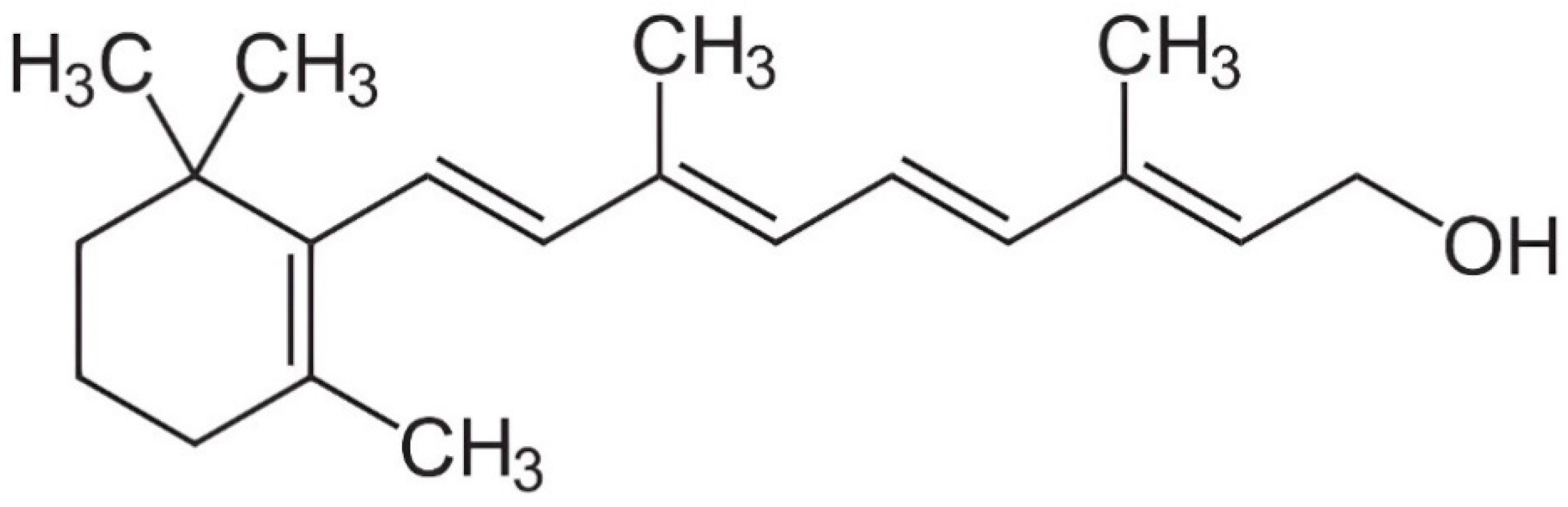
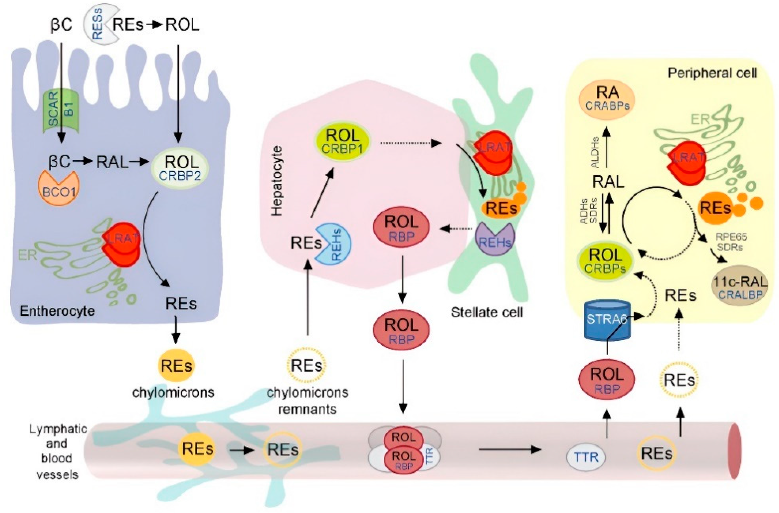
3. Digestion and Metabolism of Vitamin A

The metabolic process of vitamin A in carnivores entails a sophisticated and multi-phase mechanism involving various enzymes and pathways. Initially present as retinyl esters, such as retinyl acetate, vitamin A undergoes hydrolysis facilitated by pancreatic lipases in the small intestine [5,42]. This enzymatic activity liberates retinol, which then forms mixed micelles upon combining with other lipids and bile salts, thus promoting efficient absorption by enterocytes ([43]; Figure 2). In carnivorous animals, the absorption of preformed vitamin A primarily takes place in the small intestine, particularly the jejunum [44,45]. Within this section, the preformed vitamin A is integrated into chylomicrons, lipid-containing particles, and transported via the lymphatic system to the liver [46].
In the liver, vitamin A is stored as retinyl esters in the hepatic stellate cells of dogs and cats, available for mobilization as required [7,10,48,49]. Conversely, provitamin A carotenoids, such as β-carotene found in plant-based sources, have the potential to undergo enzymatic cleavage in the intestinal mucosa, converting into retinaldehyde and retinol in canines [43]. However, the efficiency of this conversion process varies considerably; notably, dogs, akin to many other obligate carnivores, exhibit a limited ability to convert carotenoids compared to omnivores and herbivores [15]. Substantial amounts of intact β-carotene have been observed in the bloodstream of dogs following dietary supplementation, indicating a constrained conversion within the enterocytes [50,51].
Moreover, cats may be among the least efficient converters of β-carotene to vitamin A among domesticated animals. Previously, it was believed that felines were unable to convert β-carotene to vitamin A [52,53,54]. However, recent evidence suggests that cats can convert β-carotene into retinol, albeit to a very limited extent [55,56]. Consequently, the type of diet and dietary vitamin A supplementation emerge as critical factors in maintaining optimal vitamin A levels in both domesticated canines and felines.
Ancestral vertebrates underwent a loss of biosynthetic pathways for most vitamins, prompting the evolution of specialized mechanisms, such as dedicated transport proteins [57]. These proteins facilitate the absorption of dietary vitamins from the intestine, storage tissues, and serum [58]. In instances where there is a demand for vitamin A, the stored retinyl esters in the liver are mobilized and subsequently hydrolyzed to retinol. This retinol is then transported to target tissues predominantly via the retinol-binding protein (RBP) in most mammals [59,60,61]. The RBP plays a crucial role in regulating vitamin A homeostasis, ensuring its proper distribution and delivery to target tissues while preventing potential toxicity [5,62]. Interestingly, cats and dogs exhibit a lower dependence on the RBP for transporting vitamin A in plasma compared to other mammals. Instead, they primarily transport vitamin A as retinyl esters (mostly retinyl stearate, with lesser amounts of retinyl oleate and palmitate) bound to low-density lipoprotein (LDL) and very low-density lipoprotein (VLDL), in concentrations significantly higher (10–50 times) than other mammals ([6,63,64]; Table 1). The significance of this circulating pool of retinyl esters and its impact on the tissue metabolism of vitamin A remains unclear [65]. In dogs, cats, and other carnivores, only a portion of the retinol in the blood plasma is bound to the RBP, with an average ratio of retinol to total retinyl esters ranging from 0.2 to 1 to 1:1 [64,66,67]. This discrepancy underscores the necessity for species-specific investigations to comprehend the metabolism of vitamin A. It is of note that, in addition to potential biliary excretion, carnivores such as dogs and cats have a unique ability to eliminate excess vitamin A from their bodies through urine, specifically in the form of protein-bound vitamin A and retinyl esters [67].
Upon entering a cell, the retinol binds to the intracellular retinol-binding protein (CRBP) within the cytoplasm in both dogs and cats [76,77]. The CRBP acts as a carrier protein, facilitating the intracellular transport of the retinol to specific cellular compartments where it is utilized [78]. Within the cell, the retinol undergoes various metabolic transformations to fulfill the cell’s specific requirements.
A critical pathway involves the oxidation of retinol to retinaldehyde, a crucial precursor for the synthesis of the active forms of vitamin A. In mammals, including felines and canines, this oxidation reaction is mediated by enzymes called alcohol dehydrogenases (ADHs), particularly class I ADH and class IV ADH, which convert retinol to retinaldehyde [79,80,81,82,83,84,85]. Retinaldehyde can further metabolize to produce ATRA through the enzymatic activity of retinaldehyde dehydrogenases (RALDHs) [86,87,88]. The primary isoform responsible for this conversion, as evidenced by studies in dogs, is RALDH1A2 [89].
ATRA, the biologically active derivative of vitamin A, exerts its regulatory influence on the cellular metabolism in animals by selectively binding to nuclear receptors, specifically retinoic acid receptors (RAR) and retinoid X receptors (RXR). These receptors demonstrate widespread expression across various tissues, including the liver, kidney, intestine, adipose tissue, and immune cells [90,91,92]. Upon binding, the resultant retinoic acid-receptor complex interacts with specific DNA sequences, thereby modulating the gene expression in numerous biological processes [93,94,95].

4. Physiological Roles of Vitamin A

Exploring the intricate mechanisms of vitamin A reveals its profound physiological significance. From visual perception to cellular proliferation, retinol and its active derivatives play multifaceted roles in sustaining bodily functions [5]. In this section, we delve into the dynamic interplay of vitamin A within the intricate landscape of canine and feline physiology.

4.1. Vision

One of the most well-established functions of vitamin A in pets is its indispensable role in vision [11]. It plays a significant role in eye morphogenesis (Figure 3), as well as in the structure and function of the photoreceptor cells within the retina. In dogs and cats, the visual cycle comprises a complex biochemical process that ensures uninterrupted vision across varying light conditions. It initiates with the absorption of light by the photoreceptor cells in the retina, specifically the rod and cone cells [96,97,98]. These cells harbor visual pigments, including rhodopsin, a G-protein-coupled receptor, in rod cells, and iodopsins, a photopsin, in cone cells, comprised of opsin protein and light-sensitive retinal molecule [99,100,101].
After light absorption, the retinal shifts from its active 11-cis-retinal form to the inactive all-trans-retinal [103]. To maintain vision, the all-trans-retinal must convert back to 11-cis-retinal [104], a process occurring in the retinal pigment epithelium (RPE) behind the retina’s photoreceptor cells [103,105,106]. In the visual cycle, the all-trans-retinal is reduced to all-trans retinol, then oxidized to 11-cis retinal within the RPE cells [103,107]. This regenerated 11-cis-retinal is transported back to the photoreceptor cells, where it combines with opsin to create functional visual pigments [108]. The continuous regeneration of 11-cis-retinal is vital for maintaining light sensitivity and adapting to changes in illumination [109].
Furthermore, vitamin A is imperative for maintaining the structural integrity of the eye. Adequate vitamin A levels support the differentiation and upkeep of ocular tissues such as the cornea and conjunctiva [7,110].

4.2. Immune Function

Vitamin A, acting through its metabolite ATRA, plays a critical role in regulating the immune system’s functionality in mammals. ATRA engages with the specific receptors known as RARs present within the immune cells, thereby initiating gene transcription processes [111,112]. This transcriptional activity leads to the synthesis of key proteins, such as Interferon Regulatory Factors (IRFs), which are essential for the proper function of immune cells [113,114,115,116]. These proteins contribute to regulating critical cellular processes including the differentiation, proliferation, and modulation of immune responses [117]. Ultimately, the influence of vitamin A on gene expression dynamics helps maintain a balanced and robust immune system, thereby enhancing the body’s ability to combat infections and sustain overall health [118].
Retinol and its derivatives are crucial for preserving the integrity of the mucosal surfaces found in the respiratory, gastrointestinal, and urogenital tracts, thereby acting as a protective barrier against pathogens [39]. Vitamin A plays a pivotal role in governing the differentiation and proliferation of various immune cells, including T and B lymphocytes, as well as the antigen-presenting cells (APCs) like dendritic cells ([40]; Figure 4). Due to its essential function in immune modulation, vitamin A is often referred to as “the anti-infective vitamin” [5,119]. As early as 1926, Mellanby [18], based on a comprehensive review of 330 post-mortem examinations of dogs, observed a significant correlation between bronchopneumonia, resulting from bacterial lung infections, and vitamin A deficiency, indicating a higher incidence among dogs with insufficient retinol levels.
Currently, retinol stands as one of the most extensively studied micronutrients concerning the immune function [121,122]. Moreover, vitamin A enhances mucosal immunity by stimulating the production of secretory IgA, IgM, and IgG antibodies [123,124], which are particularly crucial for defending against the pathogens at mucosal surfaces such as those in the gastrointestinal and respiratory tracts.

4.3. Growth, Cellular Differentiation, Morphogenesis, and Reproductive Health

ATRA is recognized for its pivotal involvement in fundamental cellular processes such as growth (including in bones), differentiation, and organogenesis [125,126]. Through its specific binding to the RARs located in target cells, ATRA operates as a transcriptional regulator, modulating the expression of genes crucial for cell differentiation, proliferation, morphogenesis, and tissue development [127,128,129]. This regulatory mechanism orchestrates the intricate process of cell specialization and the formation of distinct tissues and organs during embryonic development [130].
With regards to cellular proliferation and differentiation, ATRA exerts its influence by promoting the progression of cells through the cell cycle, particularly the G1 phase. This is achieved through the activation of genes supportive of cell division while concurrently suppressing genes associated with cell cycle arrest [131]. Moreover, ATRA signaling contributes significantly to morphogenesis. For instance, it plays a critical role in organizing the trunk through three key morphogenetic processes: mesoderm segmentation (including somite size and bilateral registration), axial elongation (posterior extension), and the establishment of anterior–posterior identity within individual segments (regionalization) [132].
Vitamin A and its metabolites are recognized as vital for the optimal reproductive health and development of pets [10,133,134]. In the ovaries, ATRA exerts significant effects on granulosa cell functions such as proliferation, differentiation, and steroidogenesis, thereby contributing to follicular development [135,136,137,138]. Furthermore, ATRA is implicated in the regulation of gene expression related to reproduction and enhances estrogen production, particularly estradiol (E2), which is crucial for the menstrual cycle [139,140].
Moreover, ATRA is known to modulate uterine receptivity and facilitate embryo implantation by promoting the secretion of uterine factors necessary for embryo attachment and placental development [141,142]. Additionally, ATRA plays a pivotal role in male reproduction, influencing processes such as spermatogenesis, testicular development, and sperm production and motility [143,144].

4.4. Antioxidant Properties

Oxidative damage to DNA, proteins, and lipids is widely recognized as a significant factor contributing to aging and the development of various chronic diseases in cats and dogs [145]. Vitamin A plays a vital role as a systemic antioxidant, impacting various biological processes in animals, including pets [4,146]. Retinol possesses direct antioxidant properties due to its hydrophobic polyene chains, allowing it to quench singlet oxygen and neutralize radicals [147]. However, in high oxygen levels, vitamin A can undergo auto-oxidation, though it remains effective at physiological oxygen tensions [148,149].
Palace et al. [150] elucidated how retinol acts as a chain-breaking antioxidant, halting lipid peroxidation by reacting with peroxyl radicals, thus preventing the spread of lipid peroxidation in cells and the formation of hydroperoxides. Retinol efficiently scavenges peroxyl radicals in various lipid models, such as liposomes mimicking cell membranes. Additionally, ATRA, a vitamin A metabolite, serves as a potent transcriptional regulator, influencing the expression of genes related to antioxidant processes [151]. Specifically, ATRA upregulates genes involved in the glutathione (GSH) metabolism, bolstering cellular antioxidant defenses [152,153,154]. It also enhances superoxide dismutase (SOD) activity, crucial for neutralizing superoxide radicals [155]. ATRA modulates the oxidative stress pathways by downregulating the NADPH oxidase genes, thereby reducing reactive oxygen species production [156].
Furthermore, ATRA enhances mitochondrial antioxidant activity by upregulating genes associated with biogenesis and defense mechanisms [157,158]. Specifically, Tourniaire et al. [157] found that ATRA increases the expression of genes linked to these processes, resulting in a heightened oxidative phosphorylation capacity and mitochondrial content (Figure 5). These findings suggest significant implications for managing oxidative stress. Additionally, ATRA induces autophagy, assisting cellular survival under oxidative stress conditions [159,160]. This process involves autophagosome acidification through a pathway independent of classic nuclear retinoid receptors, ultimately contributing to cellular homeostasis regulation [159].

5. Vitamin A Deficiency and Excess in Pets

Vitamin A is a fundamental micronutrient for pets, but, like many essential substances, it must be carefully regulated to avoid health complications. Both the deficiency and excess of vitamin A can have notable effects on pets’ well-being, necessitating an understanding of the causes, symptoms, and management of these conditions [161,162].

5.1. Vitamin A Deficiency

Vitamin A deficiency can arise in pets due to various factors, encompassing inadequate dietary intake, compromised absorption, or increased metabolic demands [146,163,164]. Pets consuming diets deficient in vitamin A or experiencing gastrointestinal disorders such as intestinal fat malabsorption are particularly susceptible. Furthermore, certain medical conditions or periods of rapid growth may elevate the requirement for vitamin A in dogs and cats [35,165].
One of the primary physiological roles of vitamin A involves maintaining the optimal visual function. Consequently, the felines and canines deficient in this vitamin may manifest symptoms such as nyctalopia, reduced vision in low light, conjunctivitis, xerosis with keratitis and corneal neovascularization, photophobia, mydriasis in normal lighting, delayed pupillary light reflex, progressive retinal cell degeneration, cataract formation, or even complete blindness in severe cases [19,27,146]. Additionally, common neurological manifestations of progressive vitamin A deficiency include an altered mental state, seizures, nystagmus, ataxia, kyphosis, hyperesthesia, muscle wasting, nerve degeneration, and impaired nerve conduction [32,146,166].
Hypovitaminosis A in pets also frequently leads to weight loss, bronchial epithelial metaplasia, squamous metaplasia in the salivary glands and endometrium, a dry and lackluster coat, dermatological issues, and a compromised immune function predisposed to infections [18,32,33,119,167]. Reproductive complications such as infertility or dystocia can also arise due to vitamin A deficiency in breeding animals [15,168].
The diagnosis of vitamin A deficiency in pets typically involves a comprehensive evaluation including clinical signs, dietary history, and laboratory analyses. Blood assays may reveal diminished vitamin A levels, though these findings can be inconclusive due to various factors such as the presence of different vitamin A forms in plasma, carnivores’ ability to renally excrete vitamin A, individual variabilities, age, physiological and nutritional influences, diurnal fluctuations, and sampling stress, among others [64,66,169,170,171,172,173,174,175,176]. Furthermore, it is important to recognize that veterinary practitioners often have a limited availability of the specialized instrumentation required for measuring the vitamin A levels in various tissues. Finally, veterinarians may also scrutinize the pet’s diet and overall health status to elucidate the root cause of the deficiency [9].
The treatment of vitamin A deficiency in dogs and cats involves administering vitamin A supplements orally or via injections, contingent upon the severity of the condition [6,9]. Additionally, transitioning to commercial pet foods formulated to meet nutritional requirements may be warranted if the pet was previously fed deficient homemade diets. The regular monitoring of the pet’s response and addressing any underlying health issues contributing to the deficiency are imperative for successful management [177,178]. According to Silver [6], the following examples of diseases often linked to retinol deficiency in dogs and cats can be mitigated or treated with supplements of retinoids:
Retinoid-responsive dermatoses;
Sebaceous gland disorder;
Canine icthyosis;
Solar dermatosis of dogs and cats;
Feline muzzle folliculitis;
Nyctalopia;
Xerophthalmia;
Keratoconjunctivitis sicca;
Squamos metaplasia;
Squamous cell carcinoma;
Epitheliotrophic T cell lymphoma;
Respiratory infections;
Intestinal inflammation;
Hyperkeratinization of the epithelial surfaces;
Seborrhea;
Keratoacanthoma;
Haircoat problems;
Schnauzer comedo syndrome;
Alopecia;
Increased susceptibility to infection;
Exfoliation;
Delayed wound healing;
Sebaceous adenitis;
Follicular dysplasia.

5.2. Vitamin A Excess

Hypervitaminosis A, or vitamin A overdose, can occur in canines and felines when they consume excessively high levels of vitamin A, either through their diet or via supplementation. Dogs and cats are at a higher risk of experiencing vitamin A overdose if they devour excessive amounts of raw liver from fish, swine, or cattle over extended periods, as it contains particularly high levels of this vitamin [68,161,179].
Generally, felines and canines exhibit a higher tolerance to developing hypervitaminosis A compared to other domesticated mammals and poultry [6]. This heightened tolerance can be attributed to several factors. Firstly, dogs and cats predominantly transport vitamin A as retinyl ester in their blood plasma [180]. Unlike in many mammals, elevated retinyl ester levels in plasma among carnivores do not correlate with the signs of vitamin A overdose or postprandial effects [64]. Secondly, carnivores eliminate excess vitamin A through urine in the form of protein-bound vitamin A and retinyl esters [67]. Dogs exhibit a higher urinary retinol excretion capacity compared to cats ([64,66]; Table 2), ranging, for example, from 15 to 63% of the intake in beagle dogs [180]. This efficient excretion mechanism prevents the accumulation of retinyl esters in the liver and kidney cells, leading to the manifestation of hypervitaminosis A only at extremely high intake levels of vitamin A (>90 mg of retinol/kg BW consumed over a prolonged period) [180]. While humans also excrete the metabolic products of retinol via urine [181], the urinary excretion of unmetabolized retinol or its esters in healthy non-carnivorous domesticated animals is unheard of. The ability of carnivores to rapidly excrete vitamin A and its esters via urine may be an evolutionary adaptation, allowing them to consume potentially high amounts of retinol in the wild (e.g., liver consumption).
The symptoms of hypervitaminosis A in pets vary depending on the severity and duration of the exposure. The studies investigating retinol overdose employed extraordinarily high doses of vitamin A, ranging from 3,500,000 to 15,000,000 IU per kg of diet, administered to cats consistently over a period of up to 10 months [48,182,183]. These doses exceeded the latest NASEM requirement estimate for adult cats by up to 4500 times. The dogs fed diets supplemented with 100,000 IU of vitamin A per 1000 kcal of diet for 44 weeks exhibited no signs of hypervitaminosis A or any adverse health effects [10].
The early signs of hypervitaminosis A often include gastrointestinal disturbances such as diarrhea and loss of appetite [183]. As the overdose progresses, dogs and cats may exhibit neurological symptoms such as lethargy, weakness, disorientation, and seizures, along with bone demineralization and reduced thyroxin levels in the blood plasma [68].
Preventing vitamin A overdose in pets entails feeding them a balanced diet formulated to meet their nutritional needs without excessive supplementation (Table 3). Pet owners should refrain from feeding large amounts of liver or pure vitamin A supplements without consulting a veterinarian. Regular veterinary check-ups can aid in identifying and addressing any nutritional imbalances before they adversely affect pet health.

6. Interactions with Other Micronutrients

Understanding vitamin A’s interactions with other micronutrients is vital for maintaining overall health and preventing deficiencies or overdose. Here, we explore the intricate relationships between vitamin A and several key micronutrients.

6.1. Vitamin A and Vitamin D

Vitamin A and vitamin D are integral to various physiological processes, including immune regulation, bone metabolism, and cellular differentiation [10,185,186]. The interplay between these vitamins is complex, involving their active forms binding to specific nuclear receptors. The active forms of vitamin A, such as ATRA and 9-cis retinoic acid (9-cis RA), interact with RAR and RXR, while 1,25-dihydroxyvitamin D3 (1,25(OH)2D3) binds to the vitamin D receptor (VDR) and RXR [187]. These receptors form heterodimers that bind to response elements like the vitamin D response element (VDRE) and the retinoic acid response element. The research indicates that 9-cis RA can modulate the effects of 1,25(OH)2D3, leading to diverse outcomes ranging from antagonistic to synergistic [187,188,189,190,191]. Notably, ATRA can influence the expression of the vitamin D-binding protein (DBP) complex, which is pivotal for the cellular uptake and actions of vitamin D, thus implicating vitamin A in the modulation of the vitamin D metabolism within specific cell types [187]. Moreover, vitamin A and vitamin D collaboratively regulate immune responses in the innate lymphoid cells (ILCs) ([186]; see Figure 6). However, ATRA and 1,25(OH)2D3 may also exert antagonistic effects on the expression of effector cytokines and gut-homing integrin by mammalian ILCs. The balance between these vitamins could be a key determinant in ILC activity and associated diseases, including allergic inflammation [189].

6.2. Vitamin A and Vitamin E

Vitamin A and vitamin E are both powerful antioxidants that protect cells from oxidative damage and play essential roles in maintaining skin health, vision, and the immune function [4,192]. While they have distinct antioxidant properties, they may also interact synergistically to enhance each other’s effectiveness. In experiments conducted within a unilamellar liposomal system comprised of phosphatidylcholine, there is evidence indicating that α-tocopherol enhances the antioxidant efficacy of all-trans-retinol by reducing its autooxidation [193]. This action likely occurs through the concerted scavenging of radicals, leading to the synergistic protection of the lipid system against peroxidative stress [194]. Consequently, this mechanism could potentially reduce the consumption of α-tocopherol over time. However, given that retinol exhibits relatively weak direct antioxidant properties, further investigations are warranted to accurately quantify the potential interaction between these vitamins in vivo. Moreover, vitamin E aids in the body’s utilization of vitamin A while also regulating vitamin A stores, thereby mitigating the risk of hypervitaminosis A. Nevertheless, the excessive intake of vitamin E may negatively influence the vitamin A storage levels in the body [6].

6.3. Vitamin A and C

Vitamin C is synthesized endogenously within the liver across a variety of species, including canines and felines, and is distributed widely throughout the body’s [195]. This essential micronutrient serves pivotal physiological functions in various metabolic pathways, including tissue growth and maintenance, the mitigation of oxidative stress, and the modulation of the immune system [1].
Administering vitamin A and C, either prior to or following the induction of stress in mammals, has been shown to significantly enhance the activities of crucial antioxidant enzymes such as SOD, glutathione-S-transferase, and catalase [196]. Moreover, there is a notable increase in the levels of reduced glutathione, accompanied by a decrease in lipid peroxidation. Importantly, this combined treatment demonstrates markedly superior outcomes compared to the use of either vitamin alone. Similarly, Hosseini Omshi et al. [197] reported that the supplementation of vitamins A and C in animals may offer promising effects against the imbalance between oxidants and antioxidants.

6.4. Vitamin A and Zinc

Zinc, an indispensable trace element, plays a pivotal role in numerous enzymatic reactions, immune modulation, and the process of wound healing [198]. Studies in humans and chickens, as well as ex vivo research using cultures of spleen leukocytes from dogs, have suggested the existence of a synergistic relationship between the dietary intake of zinc and the metabolism and status of vitamin A [199,200,201]. Simultaneous long-term zinc and vitamin A supplementation was shown to be associated with reduced parasitic gastrointestinal infections caused by Giardia lamblia and Ascaris lumbricoides in humans [202]. The influence of zinc extends to various facets of vitamin A metabolism, encompassing absorption, transportation, and utilization. This influence arises from the regulatory function of zinc in vitamin A transportation, primarily facilitated through the synthesis of retinol-binding protein (RBP) in the liver. Additionally, zinc serves as a crucial cofactor in the synthesis of enzymes that regulate the absorption and function of vitamin A [203]. Furthermore, zinc plays a pivotal role in the oxidative conversion of retinol to retinal, necessitating the action of zinc-dependent retinol dehydrogenase enzymes [204]. Studies indicate that in animals a deficiency in zinc consistently leads to reduced plasma vitamin A concentrations, despite a diet being nutritionally adequate in vitamin A [203]. Moreover, zinc deficiency has been shown to diminish the lymphatic absorption of retinol in animal models, a phenomenon linked to a decline in the output of lymphatic phospholipids due to compromised biliary secretion into the intestinal lumen [205]. Conversely, vitamin A is required for the absorption and utilization of zinc by improving intestinal functionality [201,204].

7. Comparing Vitamin A Requirements: Livestock vs. Pets

Variability in the requirements for vitamin A is evident across different species and even among individuals within the same species [5,7,62,206,207]. Understanding these variations is crucial for maintaining the optimal health and well-being of animals under human care. Due to extensive artificial selection, dogs have evolved into one of the most morphologically diverse vertebrate species, encompassing approximately 400 distinct breeds [208,209]. Similarly, domestic cats exhibit a wide range of breed variations and morphological traits [210]. The selective breeding of dogs and cats has primarily been motivated by considerations of aesthetics, morphology, and behavior [211].
In contrast to companion animals, modern livestock species such as swine, poultry, and cattle have undergone intense selection and breeding for traits such as rapid growth, lean meat deposition, efficient feed conversion, and high egg and milk production [5,62,206,207]. This genetic manipulation imposes additional metabolic demands and alters the requirement for essential nutrients, including vitamins. For instance, modern pig breeds exhibit an increased metabolic turnover and oxidative stress levels akin to those of endurance athletes [212]. Consequently, the demand for fat-soluble vitamins in livestock has increased significantly over the past few decades [5,62,206,207,213].
While certain dog breeds may be exceptions due to selective breeding for enhanced performance [214], the overall trend suggests that the vitamin A requirements for companion animals, particularly dogs and cats, may not have escalated to the same extent as those of livestock species. This discrepancy underscores a fundamental difference between the breeding pressures shaping the nutrient needs of pets versus those of livestock.
The specific vitamin A requirements for domesticated animals depend on various factors, including the animal’s age, weight, and reproductive status, as well as other parameters [5]. For example, pregnant and lactating pets have increased vitamin A requirements to support fetal development and milk production [35,215]. Similarly, young, growing animals require higher levels of vitamin A to support bone development and overall growth [216]. In contrast, mature animals may have lower vitamin A requirements but still need adequate amounts to maintain health and the immune function. We propose that the ideal dosage of vitamin supplementation in companion animals depends on promoting optimal growth and development, as well as achieving a balance marked by maximal physiological well-being, including immune competence, while ensuring adequate bodily reserves are maintained. Table 4 presents the minimum requirements and practical dosage recommendations for vitamin A in cats and dogs sourced from various references. The authors have published similar information for poultry, swine, and cattle elsewhere. For comparison with felines and canines, readers are referred to these peer-reviewed data [62,206,207]. Furthermore, it should be noted that NASEM estimates for vitamins represent the minimum levels necessary to prevent clinical deficiencies and may not necessarily ensure optimal health, including the immune function, and sufficient bodily reserves [217,218].
Overall, while the vitamin A requirements of livestock and pets share similarities, such as the need for adequate levels to support growth, development, and overall health, significant differences between the two arise due to variations in the goals of genetic selection.

8. Future Directions and Research Gaps

As the field of companion animal wellness progresses, there are numerous avenues for future research aimed at understanding the role of vitamin A in the health and well-being of pets. In this section, we outline potential directions for future investigation and emphasize key research gaps that require attention within the scientific community.
Although there have been notable advancements in elucidating the metabolism of vitamin A in humans and laboratory animals, our comprehension of these mechanisms in companion animals, such as dogs and cats, remains comparatively limited. This disparity in knowledge presents obstacles in effectively optimizing the dietary management of pets. To bridge this gap, it is proposed to widen usage of in vitro simulation techniques and ex vivo methodologies to investigate the metabolism of vitamin A in companion animals [220]. This approach is advocated primarily due to ethical considerations.
Furthermore, investigating rodents as potential models for elucidating the vitamin A metabolism in dogs and cats should be assessed [221,222,223]. This implies that the translational-related challenges between the models regarding vitamin A need clarification. By directing the research efforts towards uncovering the specific pathways involved in the absorption, storage, and utilization of vitamin A, potentially valuable insights can be gained. If proven to be applicable, these insights might facilitate the customization of dietary vitamin A levels to suit different breeds of dogs and cats, thereby enhancing overall pet health and wellness.
Genetic factors play a crucial role in determining the vitamin A requirements and response to supplementation [224,225]. Investigating genetic variability among different pet breeds and its influence on the metabolism and function of vitamin A could aid in tailoring personalized nutrition strategies for optimal health outcomes. Integrating genomic approaches with nutritional studies will pave the way for precision nutrition in companion animals. Nevertheless, genetic association studies remain challenging, even in human populations. Recognizing the potential challenges for similar studies in pets would provide further confidence.
Long-term studies tracking vitamin A status and its correlation with various health parameters in pets are essential for establishing causal relationships and understanding the impact of vitamin A deficiency or excess on overall health outcomes [226,227]. Longitudinal research designs, encompassing diverse populations of pets across different life stages and environmental conditions, will provide robust evidence to guide nutritional recommendations and preventive healthcare strategies.
Vitamin A is known to play a critical role in modulating the immune function and reducing the risk of infectious and chronic diseases in humans [176,228]. Similarly, investigating the immunomodulatory effects of vitamin A in pets can offer valuable insights into enhancing immune resilience and mitigating disease susceptibility. This includes reducing the incidence of respiratory infections and improving wound healing times. Specifically, exploring the translational potential of molecular studies on retinoids’ impact on the immune function in companion animals holds significant promise for animal health, a concept already supported by the research in humans [229]. Understanding how vitamin A enhances mucosal immunity, as evidenced by its stimulation of secretory IgA, IgM, and IgG antibodies [39,230], forms a crucial foundation for potential applications in veterinary practice for pets [231,232]. These antibodies play a pivotal role in defending mucosal surfaces, such as those in the gastrointestinal and respiratory tracts, against pathogens, making the findings a promising research direction for enhancing the health and well-being of companion animals [8].
Pets are frequently exposed to diverse pathogens in their environments, so enhancing their natural defenses through vitamin A interventions could lead to reduced disease susceptibility and improved overall health outcomes. Moreover, comprehending how retinoids influence gene expression related to immunity could provide insights into tailoring treatments for individual animals, thereby enhancing their well-being and quality of life [90,233]. Ultimately, the application of the molecular findings on retinoids in pets has the potential to transform veterinary medicine, offering more effective and personalized care for our beloved companions.
The stability and bioavailability of vitamin A from pet food and animal feed formulations can vary depending on factors such as the vitamin A formulation (from a commercial source), the ingredient composition, processing methods, and storage conditions [110,234,235,236,237]. Systematic assessments of vitamin A bioavailability in commercial pet foods are necessary to ensure adequate nutrient delivery and to optimize dietary formulations for pet health and wellness. Incorporating advanced analytical techniques and in vitro digestion models can facilitate the accurate determination of vitamin A bioavailability in pet food matrices.

9. Conclusions

The multifaceted role of vitamin A in the health and well-being of companion animals is undeniable. From its crucial involvement in vision, the immune function, and reproduction to its antioxidant properties, vitamin A serves as a cornerstone of optimal pet nutrition. Through historical perspectives, we have seen how our understanding of vitamin A’s importance has evolved, leading to the establishment of recommended dietary allowances and the development of commercial pet foods aimed at meeting the essential nutritional requirements.
The intricate processes of digestion, metabolism, and physiological functions elucidate the complexities of ensuring adequate vitamin A levels in pets, especially considering their unique dietary needs as obligate carnivores. While deficiencies can lead to a myriad of health issues, including impaired vision, a compromised immune function, and reproductive complications, excess intake may also carry potential concerns, underscoring the importance of balanced nutrition and careful supplementation.
Exploring interactions with other micronutrients further emphasizes the interconnectedness of dietary components in promoting overall health. Future research directions, including investigating genetic variability, long-term studies tracking vitamin A status, and exploring immunomodulatory effects, offer promising avenues for enhancing our understanding of vitamin A’s role in companion animal nutrition. Additionally, considerations of stability and bioavailability in pet food formulations underscore the importance of optimizing nutritional strategies to ensure the well-being of our beloved animal companions. By addressing these research gaps and advancing our knowledge, we can continue to improve the nutritional management of pets, ultimately enhancing their quality of life and longevity.

Author Contributions

Conceptualization, Y.S.; methodology, Y.S. and W.P.; software, Y.S.; validation, W.P.; formal analysis, W.P.; investigation, Y.S.; resources, Y.S. and W.P.; data curation, Y.S.; writing—original draft preparation, Y.S.; writing—review and editing, W.P.; visualization, Y.S.; supervision, W.P.; and project administration, Y.S. All authors have read and agreed to the published version of the manuscript.

Funding

This research received no external funding.

Institutional Review Board Statement

Not applicable.

Data Availability Statement

Not applicable.

Conflicts of Interest

Both authors of this work are affiliated with BASF, a manufacturer of vitamins and carotenoids, including vitamin A and β-carotene. Nevertheless, it is crucial to underscore that the content of this manuscript has been sourced exclusively from scientific peer-reviewed data. Our unwavering commitment lies in upholding transparency and adhering to ethical research principles.

Abbreviations

SODSuperoxide dismutase
1,25(OH)2D31,25-dihydroxyvitamin D3
11c-RAL11-cis-retinal; ADHs, alcohol dehydrogenases
ADHsAlcohol dehydrogenases
ALDHsAldehyde dehydrogenases
APCsAntigen-presenting cells
ATRAAll-trans retinoic acid
BCO1 β,β-carotene-15,15-dioxygenase
CRABPsCellular retinoic acid-binding proteins
CRALBPCellular retinaldehyde-binding protein
CRBPIntracellular retinol-binding protein
CRBP1Cellular retinol-binding protein 1
CRBP2Cellular retinol-binding protein 2
DBPVitamin D-binding protein
DNADeoxyribonucleic acid
E2Estradiol
EREndoplasmic reticulum
GSHGlutathione
IgImmunoglobulin
ILCsInnate lymphoid cells
IRFsInterferon Regulatory Factors
LDL Low-density lipoprotein
LRATLecithin:retinol acyltransferase
NADPHNicotinamide adenine dinucleotide phosphate
Pitx2 iRA/RAR-regulated transcription factor
RAAll-trans-retinoic acid
RALAll-trans-retinaldehyde
RALDHsRetinaldehyde dehydrogenases
RAR Retinoic acid receptors
RBPSerum retinol-binding protein
REHsRetinyl ester hydrolases
REsRetinyl esters
RESsRetinyl esterases
ROLAll-trans-retinol
RPERetinal pigment epithelium
RPE65Retinoid isomerase
RXRRetinoid X receptors
SCARB1Scavenger receptor class B, type I
SDRsShort-chain dehydrogenases/reductases
STRA6Retinoic acid 6
ThT helper cells
Tregregulatory T cells
TTRTransthyretin
VDRVitamin D receptor
VDREVitamin D response element
VLDLVery low-density lipoprotein
βCβ,β-carotene

References

  1. Gordon, D.S.; Rudinsky, A.J.; Guillaumin, J.; Parker, V.J.; Creighton, K.J. Vitamin C in Health and Disease: A Companion Animal Focus. Top. Companion Anim. Med. 2020, 39, 100432. [Google Scholar] [CrossRef] [PubMed]
  2. Stockman, J.; Villaverde, C.; Corbee, R.J. Calcium, Phosphorus, and Vitamin D in Dogs and Cats: Beyond the Bones. Vet. Clin. N. Am. Small Anim. Pract. 2021, 51, 623–634. [Google Scholar] [CrossRef] [PubMed]
  3. Fan, Z.; Bian, Z.; Huang, H.; Liu, T.; Ren, R.; Chen, X.; Zhang, X.; Wang, Y.; Deng, B.; Zhang, L. Dietary Strategies for Relieving Stress in Pet Dogs and Cats. Antioxidants 2023, 12, 545. [Google Scholar] [CrossRef] [PubMed]
  4. Shastak, Y.; Gordillo, A.; Pelletier, W. The relationship between vitamin A status and oxidative stress in animal production. J. Appl. Anim. Res. 2023, 51, 546–553. [Google Scholar] [CrossRef]
  5. Shastak, Y.; Pelletier, W. The role of vitamin A in non-ruminant immunology. Front. Anim. Sci. 2023, 4, 1197802. [Google Scholar] [CrossRef]
  6. Silver, R.J. Liquid A Drops. Technical Report for Veterinarian Use Only. VBS Direct Ltd. 1 Mill View Close, Bulkeley, Cheshire, SY14 8DB. 2024. Available online: https://vbsdirect.co.uk/files/Vitamin_A_Tech_Report2.pdf (accessed on 10 February 2024).
  7. Carazo, A.; Macáková, K.; Matoušová, K.; Krčmová, L.K.; Protti, M.; Mladěnka, P. Vitamin A Update: Forms, Sources, Kinetics, Detection, Function, Deficiency, Therapeutic Use and Toxicity. Nutrients 2021, 13, 1703. [Google Scholar] [CrossRef] [PubMed]
  8. Shoveller, A.K.; De Godoy, M.R.; Larsen, J.; Flickinger, E. Emerging Advancements in Canine and Feline Metabolism and Nutrition. Sci. World J. 2016, 2016, 9023781. [Google Scholar] [CrossRef]
  9. Watson, T.D. Diet and skin disease in dogs and cats. J. Nutr. 1998, 128 (Suppl. S12), 2783S–2789S. [Google Scholar] [CrossRef]
  10. Morris, P.J.; Salt, C.; Raila, J.; Brenten, T.; Kohn, B.; Schweigert, F.J.; Zentek, J. Safety evaluation of vitamin A in growing dogs. Br. J. Nutr. 2012, 108, 1800–1809. [Google Scholar] [CrossRef]
  11. NASEM (The National Academies of Sciences, Engineering, and Medicine). Nutrient Requirements of Dogs and Cats; National Academy of Science: Washington, DC, USA, 2006. [Google Scholar]
  12. Di Cerbo, A.; Morales-Medina, J.C.; Palmieri, B.; Pezzuto, F.; Cocco, R.; Flores, G.; Iannitti, T. Functional foods in pet nutrition: Focus on dogs and cats. Res. Vet. Sci. 2017, 112, 161–166. [Google Scholar] [CrossRef]
  13. Domínguez-Oliva, A.; Mota-Rojas, D.; Semendric, I.; Whittaker, A.L. The Impact of Vegan Diets on Indicators of Health in Dogs and Cats: A Systematic Review. Vet. Sci. 2023, 10, 52. [Google Scholar] [CrossRef]
  14. Baker, H.; Schor, S.M.; Murphy, B.D.; DeAngelis, B.; Feingold, S.; Frank, O. Blood vitamin and choline concentrations in healthy domestic cats, dogs, and horses. Am. J. Vet. Res. 1986, 47, 1468–1471. [Google Scholar]
  15. Green, A.S.; Fascetti, A.J. Meeting the vitamin A requirement: The efficacy and importance of β-carotene in animal species. Sci. World J. 2016, 2016, 7393620. [Google Scholar] [CrossRef]
  16. Verbrugghe, A.; Dodd, S. Plant-Based Diets for Dogs and Cats. World Small Animal Veterinary Association Congress Proceedings. 2019. Available online: https://www.vin.com/doc/?id=9382843 (accessed on 8 February 2024).
  17. McCollum, E.V.; Davis, M. The necessity of certain lipins in the diet during growth. J. Biol. Chem. 1913, 15, 167–175. [Google Scholar] [CrossRef]
  18. Mellanby, E. Diet and disease, with special reference to the teeth, lungs, and pre-natal feeding. Lancet 1926, 1, 515–519. [Google Scholar]
  19. Stimson, A.M.; Hedley, O.F. Observations on Vitamin A Deficiency in Dogs. Public Health Rep. 1933, 48, 445–449. [Google Scholar] [CrossRef]
  20. Semba, R.D. Vitamin A as “anti-infective” therapy, 1920–1940. J. Nutr. 1999, 129, 783–791. [Google Scholar] [CrossRef] [PubMed]
  21. Leitner, Z.A. The Clinical Significance Of Vitamin-A Deficiency. Br. Med. J. 1951, 1, 1110–1114. [Google Scholar] [CrossRef] [PubMed]
  22. Lanska, D.J. Chapter 29: Historical aspects of the major neurological vitamin deficiency disorders: Overview and fat-soluble vitamin A. In Handbook of Clinical Neurology; Elsevier B.V.: Amsterdam, The Netherlands, 2010; Volume 95, pp. 435–444. [Google Scholar] [CrossRef]
  23. Shastak, Y.; Pelletier, W.; Kuntz, A. Insights into Analytical Precision: Understanding the Factors Influencing Accurate Vitamin A Determination in Various Samples. Analytica 2024, 5, 54–73. [Google Scholar] [CrossRef]
  24. Semba, R.D. On the ‘Discovery’ of Vitamin A. Ann. Nutr. Metab. 2012, 61, 192–198. [Google Scholar] [CrossRef]
  25. Anonymous. Isolation of Vitamin A. Nature 1932, 129, 88. [Google Scholar] [CrossRef]
  26. Holmes, H.N.; Corbet, R.E. The Isolation of Crystalline Vitamin A1. J. Am. Chem. Soc. 1937, 59, 2042–2047. [Google Scholar] [CrossRef]
  27. Scott, P.P.; Greaves, J.P.; Scott, M.G. Nutritional blindness in the cat. Exp. Eye Res. 1964, 3, 357–364. [Google Scholar] [CrossRef]
  28. NASEM (The National Academies of Sciences, Engineering, and Medicine). Nutrient Requirements of Dogs; National Academy of Science: Washington, DC, USA, 1974. [Google Scholar]
  29. NASEM (The National Academies of Sciences, Engineering, and Medicine). Nutrient Requirements of Cats; National Academy of Science: Washington, DC, USA, 1978. [Google Scholar]
  30. Case, L.P.; Daristotle, L.; Hayek, M.G.; Raasch, M.F. Pet Foods in: Canine and Feline Nutrition; Elsevier Inc.: Maryland Heights, MO, USA, 2011; pp. 119–177. [Google Scholar] [CrossRef]
  31. PFI (Pet Food Institute). 2023. Available online: https://www.petfoodinstitute.org/about-pet-food/nutrition/history-of-pet-food/ (accessed on 23 December 2023).
  32. NASEM (The National Academies of Sciences, Engineering, and Medicine). Nutrient Requirements of Domestic Animals: Nutrient Requirement of Dog and Cats; National Academy Press: Washington, DC, USA, 1985. [Google Scholar]
  33. NASEM (The National Academies of Sciences, Engineering, and Medicine). Nutrient Requirements of Domestic Animals: Nutrient Requirement of Cats; National Academy Press: Washington, DC, USA, 1986. [Google Scholar]
  34. Dodd, S.A.S.; Shoveller, A.K.; Fascetti, A.J.; Yu, Z.Z.; Ma, D.W.L.; Verbrugghe, A. A Comparison of Key Essential Nutrients in Commercial Plant-Based Pet Foods Sold in Canada to American and European Canine and Feline Dietary Recommendations. Animals 2021, 11, 2348. [Google Scholar] [CrossRef] [PubMed]
  35. FEDIAF Scientific Advisory Board Nutritional Guidelines for Complete and Complementary Pet Food for Cats and Dogs. 2021. pp. 1–98. Available online: https://europeanpetfood.org/wp-content/uploads/2022/03/Updated-Nutritional-Guidelines.pdf (accessed on 15 June 2022).
  36. Kumar, N.; Gupta, R.C.; Nagarajan, G. Vitamin A deficiency in dogs: A review. J. Anim. Res. 2016, 6, 189–196. [Google Scholar]
  37. Stasieniuk, E.; Barbi, L.; Bomcompagni, T. Recent Advances in Nutritional Immunomodulation in Dogs and Cats. All Pet Food. 2022. Available online: https://en.allpetfood.net/entrada/recent-advances-in-nutritional-immunomodulation-in-dogs-and-cats-53655 (accessed on 1 February 2024).
  38. Lam, A.T.H.; Affolter, V.K.; Outerbridge, C.A.; Gericota, B.; White, S.D. Oral vitamin A as an adjunct treatment for canine sebaceous adenitis. Vet. Dermatol. 2011, 22, 305–311. [Google Scholar] [CrossRef]
  39. Huang, Z.; Liu, Y.; Qi, G.; Brand, D.; Zheng, S.G. Role of Vitamin A in the Immune System. J. Clin. Med. 2018, 7, 258. [Google Scholar] [CrossRef]
  40. Oliveira, L.M.; Teixeira, F.M.E.; Sato, M.N. Impact of Retinoic Acid on Immune Cells and Inflammatory Diseases. Mediat. Inflamm. 2018, 2018, 3067126. [Google Scholar] [CrossRef] [PubMed]
  41. Roche, F.C.; Harris-Tryon, T.A. Illuminating the Role of Vitamin A in Skin Innate Immunity and the Skin Microbiome: A Narrative Review. Nutrients 2021, 13, 302. [Google Scholar] [CrossRef]
  42. Van Bennekum, A.M.; Fisher, E.A.; Blaner, W.S.; Harrison, E.H. Hydrolysis of retinyl esters by pancreatic triglyceride lipase. Biochemistry 2000, 39, 4900–4906. [Google Scholar] [CrossRef]
  43. Hynd, P.I. Digestion in the mono-gastric animal. In Animal Nutrition: From Theory to Practice; Hynd, P.I., Ed.; CSIRO Publishing: Clayton South, VIC, Australia, 2019; pp. 42–63. [Google Scholar]
  44. Kaser, M.M.; Hussey, C.V.; Ellis, S.J. The Absorption of Vitamin A in Dogs Following Cholecystonephrostomy. J. Nutr. 1958, 66, 35–44. [Google Scholar] [CrossRef]
  45. Kouti, V.; Papazoglou, L.; Rallis, T. Short-Bowel Syndrome in Dogs and Cats. Compend. Contin. Educ. Pract. Vet. 2006, 28, 182–193. [Google Scholar]
  46. Wilson, D.E.; Hejazi, J.; Elstad, N.L.; Ing-Fong, C.; Gleeson, J.M.; Iverius, P.-H. Novel aspects of vitamin A metabolism in the dog: Distribution of lipoprotein retinyl esters in vitamin A-deprived and cholesterol-fed animals. Biochim. Biophys. Acta 1987, 922, 247–258. [Google Scholar] [CrossRef]
  47. Chelstowska, S.; Widjaja-Adhi, M.A.; Silvaroli, J.A.; Golczak, M. Molecular Basis for Vitamin A Uptake and Storage in Vertebrates. Nutrients 2016, 8, 676. [Google Scholar] [CrossRef]
  48. Seawright, A.A.; English, P.B.; Gartner, R.J.W. Hypervitaminosis A and deforming cervical spondylosis of the cat. J. Comp. Pathol. 1967, 77, 29–39. [Google Scholar] [CrossRef]
  49. Carmona, R.; Barrena, S.; Muñoz-Chápuli, R. Retinoids in Stellate Cells: Development, Repair, and Regeneration. J. Dev. Biol. 2019, 7, 10. [Google Scholar] [CrossRef]
  50. Chew, B.P.; Park, J.S.; Wong, T.S.; Weng, B.C.; Kim, H.W.; Byrne, K.M.; Hayek, M.G.; Reinhart, G.A. Role of dietary β-carotene in modulating cell-mediated and humoral immune responses in dogs. FASEB J. 1998, 12, A967. [Google Scholar]
  51. Chew, B.P.; Park, J.S.; Weng, B.C.; Wong, T.S.; Hayek, M.G.; Reinhart, G.A. Dietary β-carotene is taken up by blood plasma and leukocytes in dogs. J. Nutr. 2000, 130, 1788–1791. [Google Scholar] [CrossRef] [PubMed]
  52. Gershoff, S.N.; Andrus, S.B.; Hegsted, D.M.; Lentini, E.A. Vitamin A deficiency in cats. Lab. Investig. 1957, 6, 227–240. [Google Scholar] [CrossRef] [PubMed]
  53. Lakshmanan, M.R.; Chansang, H.; Olson, J.A. Purification and properties of carotene 15, 15′-dioxygenase of rabbit intestine. J. Lipid Res. 1972, 13, 477–482. [Google Scholar] [CrossRef] [PubMed]
  54. Schweigert, F.J.; Raila, J.; Wichert, B.; Kienzle, E. Cats absorb beta-carotene, but it is not converted to vitamin A. J. Nutr. 2002, 132 (Suppl. S2), 1610S–1612S. [Google Scholar] [CrossRef]
  55. Chew, B.P.; Park, J.S. Carotenoid action on the immune response. J. Nutr. 2004, 134, 257S–261S. [Google Scholar] [CrossRef]
  56. Green, A.S.; Tang, G.; Lango, J.; Klasing, K.C.; Fascetti, A.J. Domestic cats convert [2H8]-β-carotene to [2H4]-retinol following a single oral dose. J. Anim. Physiol. Anim. Nutr. 2012, 96, 681–692. [Google Scholar] [CrossRef] [PubMed]
  57. White, H.B., III; Merrill, A.H., Jr. Riboflavin-Binding Proteins. Annu. Rev. Nutr. 1988, 8, 279–299. [Google Scholar] [CrossRef] [PubMed]
  58. M’Clelland, D.A. The Refolding of Riboflavin Binding Protein. Ph.D. Thesis, University of Stirling, Stirling, UK, 1996. [Google Scholar]
  59. van Hoek, I.; Meyer, E.; Duchateau, L.; Peremans, K.; Smets, P.; Daminet, S. Retinol-binding protein in serum and urine of hyperthyroid cats before and after treatment with radioiodine. J. Vet. Intern. Med. 2009, 23, 1031–1037. [Google Scholar] [CrossRef]
  60. Raila, J.; Brunnberg, L.; Schweigert, F.J.; Kohn, B. Influence of kidney function on urinary excretion of albumin and retinol-binding protein in dogs with naturally occurring renal disease. Am. J. Vet. Res. 2010, 71, 1387–1394. [Google Scholar] [CrossRef] [PubMed]
  61. Steinhoff, J.S.; Lass, A.; Schupp, M. Retinoid homeostasis and beyond: How retinol binding protein 4 contributes to health and disease. Nutrients 2022, 14, 1236. [Google Scholar] [CrossRef] [PubMed]
  62. Shastak, Y.; Pelletier, W. Vitamin A supply in swine production: A review of current science and practical considerations. Appl. Anim. Sci. 2023, 39, 289–305. [Google Scholar] [CrossRef]
  63. Schweigert, F.J.; Ryder, O.A.; Rambeck, W.A.; Zucker, H. The majority of vitamin A is transported as retinyl esters in the blood of most carnivores. Comp. Biochem. Physiol. A Comp. Physiol. 1990, 95, 573–578. [Google Scholar] [CrossRef] [PubMed]
  64. Raila, J.; Buchholz, I.; Aupperle, H.; Raila, G.; Schoon, H.A.; Schweigert, F.J. The distribution of vitamin A and retinol-binding protein in the blood plasma, urine, liver and kidneys of carnivores. Vet. Res. 2000, 31, 541–551. [Google Scholar] [CrossRef]
  65. Barko, P.C.; Williams, D.A. Serum concentrations of lipid-soluble vitamins in dogs with exocrine pancreatic insufficiency treated with pancreatic enzymes. J. Vet. Intern. Med. 2018, 32, 1600–1608. [Google Scholar] [CrossRef]
  66. Raila, J.; Mathews, U.; Schweigert, F.J. Plasma transport and tissue distribution of beta-carotene, vitamin A and retinol-binding protein in domestic cats. Comp. Biochem. Physiol. A Mol. Integr. Physiol. 2001, 130, 849–856. [Google Scholar] [CrossRef]
  67. Kolb, E.; Seehawer, J. Verwertung, Stoffwechsel, Bedeutung und Anwendung des Vitamins A bei Hund und Katze. Prakt. Tierarzt 2001, 82, 98–106. Available online: https://sd9915b4744868808.jimcontent.com/download/version/1478279129/module/10685394693/name/Vitamin%20A%20bei%20Hund%20und%20Katze.pdf (accessed on 10 February 2024).
  68. Kolb, E. Vitamin A. In Verwertung und Anwendung von Vitaminen bei Haustieren; Hoffmann-LaRoche AG: Grenzach-Wyhlen, Germany, 1998; pp. 35–45. [Google Scholar]
  69. Schweigert, F.J. Insensitivity of dogs to the effects of nonspecific bound vitamin A in plasma. Int. J. Vitam. Nutr. Res. 1988, 58, 23–25. [Google Scholar] [PubMed]
  70. Raila, J.; Radon, R.; Trüpschuch, A.; Schweigert, F.J. Retinol and retinyl ester responses in the blood plasma and urine of dogs after a single oral dose of vitamin A. J. Nutr. 2002, 132 (Suppl. S2), 1673S–1675S. [Google Scholar] [CrossRef]
  71. Stowe, H.D.; Lawler, D.F.; Kealy, R.D. Antioxidant status of pair-fed labrador retrievers is affected by diet restriction and aging. J. Nutr. 2006, 136, 1844–1848. [Google Scholar] [CrossRef] [PubMed]
  72. Puls, R. Vitamin Levels in Animal Health. Diagnostic Data; Sherpa International: Clearbrook, BC, Canada, 1994; p. 184. [Google Scholar]
  73. Olsen, K.; Suri, D.J.; Davis, C.; Sheftel, J.; Nishimoto, K.; Yamaoka, Y.; Toya, Y.; Welham, N.V.; Tanumihardjo, S.A. Serum retinyl esters are positively correlated with analyzed total liver vitamin A reserves collected from US adults at time of death. Am. J. Clin. Nutr. 2018, 108, 997–1005. [Google Scholar] [CrossRef]
  74. O’Byrne, S.M.; Blaner, W.S. Retinol and retinyl esters: Biochemistry and physiology. J. Lipid Res. 2013, 54, 1731–1743. [Google Scholar] [CrossRef]
  75. Penniston, K.L.; Weng, N.; Binkley, N.; Tanumihardjo, S.A. Serum retinyl esters are not elevated in postmenopausal women with and without osteoporosis whose preformed vitamin A intakes are high. Am. J. Clin. Nutr. 2006, 84, 1350–1356. [Google Scholar] [CrossRef]
  76. Chytil, F.; Ong, D.E. Cellular retinoid-binding proteins In The Retinoids; Sporn, M.B., Roberts, A.B., Goodman, D.S., Eds.; Academic Press: New York, NY, USA, 1984; Volume 2, pp. 89–123. [Google Scholar]
  77. Sasaki, N.; Ishibashi, M.; Soeta, S. Molecular characterization and tissue distribution of feline retinol-binding protein 4. J. Vet. Med. Sci. 2013, 75, 1383–1387. [Google Scholar] [CrossRef] [PubMed]
  78. Napoli, J.L. Functions of Intracellular Retinoid Binding-Proteins. Subcell. Biochem. 2016, 81, 21–76. [Google Scholar] [CrossRef]
  79. Levene, R.; Horton, G.; Einaugler, R. Retinal Vessel Dehydrogenase Enzymes. Arch. Ophthalmol. 1964, 72, 99–103. [Google Scholar] [CrossRef]
  80. Hoffmann, I.; Ang, H.L.; Duester, G. Alcohol dehydrogenases in Xenopus development: Conserved expression of ADH1 and ADH4 in epithelial retinoid target tissues. Dev. Dyn. 1998, 213, 261–270. [Google Scholar] [CrossRef]
  81. Connally, H.E.; Hamar, D.W.; Thrall, M.A. Inhibition of canine and feline alcohol dehydrogenase activity by fomepizole. Am. J. Vet. Res. 2000, 61, 450–455. [Google Scholar] [CrossRef]
  82. Kumar, S.; Sandell, L.L.; Trainor, P.A.; Koentgen, F.; Duester, G. Alcohol and aldehyde dehydrogenases: Retinoid metabolic effects in mouse knockout models. Biochim. Biophys. Acta 2012, 1821, 198–205. [Google Scholar] [CrossRef]
  83. Bchini, R.; Vasiliou, V.; Branlant, G.; Talfournier, F.; Rahuel-Clermont, S. Retinoic acid biosynthesis catalyzed by retinal dehydrogenases relies on a rate-limiting conformational transition associated with substrate recognition. Chem. Biol. Interact. 2013, 202, 78–84. [Google Scholar] [CrossRef]
  84. Kedishvili, N.Y. Enzymology of retinoic acid biosynthesis and degradation. J. Lipid Res. 2013, 54, 1744–1760. [Google Scholar] [CrossRef] [PubMed]
  85. Fujihara, M.; Yamamizu, K.; Comizzoli, P.; Wildt, D.E.; Songsasen, N. Retinoic acid promotes in vitro follicle activation in the cat ovary by regulating expression of matrix metalloproteinase 9. PLoS ONE. 2018, 13, e0202759. [Google Scholar] [CrossRef] [PubMed]
  86. Yang, K.; Adin, C.; Shen, Q.; Lee, L.J.; Yu, L.; Fadda, P.; Samogyi, A.; Ham, K.; Xu, L.; Gilor, C.; et al. Aldehyde dehydrogenase 1 a1 regulates energy metabolism in adipocytes from different species. Xenotransplantation 2017, 24, e12318. [Google Scholar] [CrossRef]
  87. Harper, A.R.; Le, A.T.; Mather, T.; Burgett, A.; Berry, W.; Summers, J.A. Design, synthesis, and ex vivo evaluation of a selective inhibitor for retinaldehyde dehydrogenase enzymes. Bioorganic Med. Chem. 2018, 26, 5766–5779. [Google Scholar] [CrossRef] [PubMed]
  88. Cho, K.; Lee, S.-M.; Heo, J.; Kwon, Y.M.; Chung, D.; Yu, W.-J.; Bae, S.S.; Choi, G.; Lee, D.-S.; Kim, Y. Retinaldehyde Dehydrogenase Inhibition-Related Adverse Outcome Pathway: Potential Risk of Retinoic Acid Synthesis Inhibition during Embryogenesis. Toxins 2021, 13, 739. [Google Scholar] [CrossRef] [PubMed]
  89. Kasimanickam, V.R. Expression of retinoic acid-metabolizing enzymes, ALDH1A1, ALDH1A2, ALDH1A3, CYP26A1, CYP26B1 and CYP26C1 in canine testis during post-natal development. Reprod. Domest. Anim. 2016, 51, 901–909. [Google Scholar] [CrossRef]
  90. Balmer, J.E.; Blomhoff, R. Gene expression regulation by retinoic acid. J. Lipid Res. 2002, 43, 1773–1808. [Google Scholar] [CrossRef] [PubMed]
  91. Brandebourg, T.D.; Hu, C.Y. Regulation of differentiating pig preadipocytes by retinoic acid. J. Anim. Sci. 2005, 83, 98–107. [Google Scholar] [CrossRef]
  92. de Mello Souza, C.H.; Valli, V.E.; Kitchell, B.E. Detection of retinoid receptors in non-neoplastic canine lymph nodes and in lymphoma. Can. Vet. J. 2014, 55, 1219–1224. [Google Scholar]
  93. Hoffmann, B.; Lehmann, J.M.; Zhang, X.K.; Hermann, T.; Husmann, M.; Graupner, G.; Pfahl, M. A retinoic acid receptor-specific element controls the retinoic acid receptor-beta promoter. Mol. Endocrinol. 1990, 4, 1727–1736. [Google Scholar] [CrossRef] [PubMed]
  94. Rastinejad, F.; Wagner, T.; Zhao, Q.; Khorasanizadeh, S. Structure of the RXR-RAR DNA-binding complex on the retinoic acid response element DR1. EMBO J. 2000, 19, 1045–1054. [Google Scholar] [CrossRef]
  95. Huang, P.; Chandra, V.; Rastinejad, F. Retinoic acid actions through mammalian nuclear receptors. Chem. Rev. 2014, 114, 233–254. [Google Scholar] [CrossRef]
  96. Wolf, G. The visual cycle of the cone photoreceptors of the retina. Nutr. Rev. 2004, 62 Pt 1, 283–286. [Google Scholar] [CrossRef]
  97. Wang, J.S.; Kefalov, V.J. The cone-specific visual cycle. Prog. Retin. Eye Res. 2011, 30, 115–128. [Google Scholar] [CrossRef]
  98. Byosiere, S.E.; Chouinard, P.A.; Howell, T.J.; Bennett, P.C. What do dogs (Canis familiaris) see? A review of vision in dogs and implications for cognition research. Psychon. Bull. Rev. 2018, 25, 1798–1813. [Google Scholar] [CrossRef]
  99. Kemp, C.M.; Jacobson, S.G.; Borruat, F.X.; Chaitin, M.H. Rhodopsin levels and retinal function in cats during recovery from vitamin A deficiency. Exp. Eye Res. 1989, 49, 49–65. [Google Scholar] [CrossRef] [PubMed]
  100. Miyazono, S.; Isayama, T.; Delori, F.C.; Makino, C.L. Vitamin A activates rhodopsin and sensitizes it to ultraviolet light. Vis. Neurosci. 2011, 28, 485–497. [Google Scholar] [CrossRef] [PubMed]
  101. Palczewski, K. Chemistry and biology of the initial steps in vision: The Friedenwald lecture. Investig. Ophthalmol. Vis. Sci. 2014, 55, 6651–6672. [Google Scholar] [CrossRef] [PubMed]
  102. Clagett-Dame, M.; Knutson, D. Vitamin A in Reproduction and Development. Nutrients 2011, 3, 385–428. [Google Scholar] [CrossRef]
  103. Kono, M.; Goletz, P.W.; Crouch, R.K. 11-cis- and all-trans-retinols can activate rod opsin: Rational design of the visual cycle. Biochemistry 2008, 47, 7567–7571. [Google Scholar] [CrossRef]
  104. Kiser, P.D. Retinal pigment epithelium 65 kDa protein (RPE65): An update. Prog. Retin. Eye Res. 2022, 88, 101013. [Google Scholar] [CrossRef] [PubMed]
  105. Stecher, H.; Gelb, M.H.; Saari, J.C.; Palczewski, K. Preferential release of 11-cis-retinol from retinal pigment epithelial cells in the presence of cellular retinaldehyde-binding protein. J. Biol. Chem. 1999, 274, 8577–8585. [Google Scholar] [CrossRef]
  106. Parker, R.O.; Crouch, R.K. Retinol dehydrogenases (RDHs) in the visual cycle. Exp. Eye Res. 2010, 91, 788–792. [Google Scholar] [CrossRef]
  107. Strauss, O. The retinal pigment epithelium in visual function. Physiol. Rev. 2005, 85, 845–881. [Google Scholar] [CrossRef]
  108. Daruwalla, A.; Choi, E.H.; Palczewski, K.; Kiser, P.D. Structural biology of 11-cis-retinaldehyde production in the classical visual cycle. Biochem. J. 2018, 475, 3171–3188. [Google Scholar] [CrossRef] [PubMed]
  109. Palczewski, K.; Kiser, P.D. Shedding new light on the generation of the visual chromophore. Proc. Natl. Acad. Sci. USA 2020, 117, 19629–19638. [Google Scholar] [CrossRef]
  110. Shastak, Y.; Pelletier, W. Nutritional Balance Matters: Assessing the Ramifications of Vitamin A Deficiency on Poultry Health and Productivity. Poultry 2023, 2, 493–515. [Google Scholar] [CrossRef]
  111. Dawson, H.D.; Collins, G.; Pyle, R.; Key, M.; Taub, D.D. The Retinoic Acid Receptor-alpha mediates human T-cell activation and Th2 cytokine and chemokine production. BMC Immunol. 2008, 9, 16. [Google Scholar] [CrossRef]
  112. Mora, J.R.; Iwata, M.; von Andrian, U.H. Vitamin effects on the immune system: Vitamins A and D take centre stage. Nat. Rev. Immunol. 2008, 8, 685–698. [Google Scholar] [CrossRef]
  113. Matikainen, S.; Ronni, T.; Hurme, M.; Pine, R.; Julkunen, I. Retinoic acid activates interferon regulatory factor-1 gene expression in myeloid cells. Blood 1996, 88, 114–123. [Google Scholar] [CrossRef]
  114. Al Tanoury, Z.; Piskunov, A.; Rochette-Egly, C. Vitamin A and retinoid signaling: Genomic and nongenomic effects. J. Lipid Res. 2013, 54, 1761–1775. [Google Scholar] [CrossRef] [PubMed]
  115. Hao, X.; Chen, H.; Li, Y.; Chen, B.; Liang, W.; Xiao, X.; Zhou, P.; Li, S. Molecular characterization and antiviral effects of canine interferon regulatory factor 1 (CaIRF1). BMC Vet. Res. 2022, 18, 440. [Google Scholar] [CrossRef]
  116. Liu, Y.; Liu, X.; Kang, H.; Hu, X.; Liu, J.; Tian, J.; Qu, L. Identification of Feline Interferon Regulatory Factor 1 as an Efficient Antiviral Factor against the Replication of Feline Calicivirus and Other Feline Viruses. Biomed. Res. Int. 2018, 2018, 2739830. [Google Scholar] [CrossRef]
  117. Jefferies, C.A. Regulating IRFs in IFN Driven Disease. Front. Immunol. 2019, 10, 325. [Google Scholar] [CrossRef]
  118. Tourkochristou, E.; Triantos, C.; Mouzaki, A. The Influence of Nutritional Factors on Immunological Outcomes. Front. Immunol. 2021, 12, 665968. [Google Scholar] [CrossRef] [PubMed]
  119. Green, H.N.; Mellanby, E. Vitamin A as an anti-infective agent. Brit. Med. J. 1928, 2, 691. [Google Scholar] [CrossRef] [PubMed]
  120. Bono, M.R.; Tejon, G.; Flores-Santibañez, F.; Fernandez, D.; Rosemblatt, M.; Sauma, D. Retinoic Acid as a Modulator of T Cell Immunity. Nutrients 2016, 8, 349. [Google Scholar] [CrossRef] [PubMed]
  121. Villamor, E.; Fawzi, W. Effects of Vitamin A supplementation on immune responses and correlation with clinical outcomes. Clin. Microbiol. Rev. 2005, 18, 446–464. [Google Scholar] [CrossRef] [PubMed]
  122. Gürbüz, M.; Aktaç, S. Understanding the role of Vitamin A and its precursors in the immune system. Nutr. Clin. Metab. 2022, 36, 89–98. [Google Scholar] [CrossRef]
  123. Stephensen, C.B. Vitamin A, infection, and immune function. Annu. Rev. Nutr. 2001, 21, 167–192. [Google Scholar] [CrossRef] [PubMed]
  124. Hu, Y.; Zhang, L.; Zhang, Y.; Xiong, H.; Wang, F.; Wang, Y.; Lu, Z. Effects of starch and gelatin encapsulated Vitamin A on growth performance, immune status and antioxidant capacity in weaned piglets. Anim. Nutr. 2020, 6, 130–133. [Google Scholar] [CrossRef] [PubMed]
  125. Ross, S.A.; McCaffery, P.Y.; Drager, U.C.; de Luca, L.M. Retinoids in embryonal development. Physiol. Rev. 2000, 80, 1021–1054. [Google Scholar] [CrossRef]
  126. Kam, R.K.; Deng, Y.; Chen, Y.; Zhao, H. Retinoic acid synthesis and functions in early embryonic development. Cell Biosci. 2012, 2, 11. [Google Scholar] [CrossRef]
  127. Duong, V.; Rochette-Egly, C. The molecular physiology of nuclear retinoic acid receptors. From health to disease. Biochim. Biophys. Acta 2011, 1812, 1023–1031. [Google Scholar] [CrossRef]
  128. Dubey, A.; Rose, R.E.; Jones, D.R.; Saint-Jeannet, J.P. Generating retinoic acid gradients by local degradation during craniofacial development: One cell’s cue is another cell’s poison. Genesis 2018, 56, e23091. [Google Scholar] [CrossRef]
  129. Brown, G. Retinoic acid receptor regulation of decision-making for cell differentiation. Front. Cell Dev. Biol. 2023, 11, 1182204. [Google Scholar] [CrossRef]
  130. Duester, G. Retinoic acid synthesis and signaling during early organogenesis. Cell 2008, 134, 921–931. [Google Scholar] [CrossRef] [PubMed]
  131. Qiu, J.; Nordling, S.; Vasavada, H.H.; Butcher, E.C.; Hirschi, K.K. Retinoic Acid Promotes Endothelial Cell Cycle Early G1 State to Enable Human Hemogenic Endothelial Cell Specification. Cell Rep. 2020, 33, 108465. [Google Scholar] [CrossRef]
  132. Organisation for Economic Co-operation and Development (OECD). Detailed Review Paper on the Retinoid System. OECD Series on Testing and Assessment, Nr.343, Paris. 2021. Available online: https://one.oecd.org/document/ENV/CBC/MONO(2021)20/en/pdf (accessed on 11 February 2024).
  133. Gross, K.L.; Wedekind, K.J.; Kirk, C.A.; Cowell, C.S.; Schoenherr, W.D.; Richardson, D.C. Effect of dietary vitamin A on reproductive performance and immune response of dogs. Am. J. Vet. Res. 1990, 51, 1423–1430. [Google Scholar]
  134. Werner, A.H.; Power, H.T. Retinoids in veterinary dermatology. Clin. Dermatol. 1994, 12, 579–586. [Google Scholar] [CrossRef] [PubMed]
  135. Pauli, S.A.; Session, D.R.; Shang, W.; Easley, K.; Wieser, F.; Taylor, R.N.; Pierzchalski, K.; Napoli, J.L.; Kane, M.A.; Sidell, N. Analysis of follicular fluid retinoids in women undergoing in vitro fertilization: Retinoic acid influences embryo quality and is reduced in women with endometriosis. Reprod. Sci. 2013, 20, 1116–1124. [Google Scholar] [CrossRef] [PubMed]
  136. Demczuk, M.; Huang, H.; White, C.; Kipp, J.L. Retinoic acid regulates calcium signaling to promote mouse ovarian granulosa cell proliferation. Biol. Reprod. 2016, 95, 70. [Google Scholar] [CrossRef]
  137. Suwa, H.; Kishi, H.; Imai, F.; Nakao, K.; Hirakawa, T.; Minegishi, T. Retinoic acid enhances progesterone production via the cAMP/PKA signaling pathway in immature rat granulosa cells. Biochem. Biophys. Rep. 2016, 8, 62–67. [Google Scholar] [CrossRef]
  138. Jiang, Y.; Li, C.; Chen, L.; Wang, F.; Zhou, X. Potential role of retinoids in ovarian physiology and pathogenesis of polycystic ovary syndrome. Clin. Chim. Acta 2017, 469, 87–93. [Google Scholar] [CrossRef]
  139. Munetsuna, E.; Hojo, Y.; Hattori, M.; Ishii, H.; Kawato, S.; Ishida, A.; Kominami, S.A.; Yamazaki, T. Retinoic acid stimulates 17β-estradiol and testosterone synthesis in rat hippocampal slice cultures. Endocrinology 2009, 150, 4260–4269. [Google Scholar] [CrossRef]
  140. Damdimopoulou, P.; Chiang, C.; Flaws, J.A. Retinoic acid signaling in ovarian folliculogenesis and steroidogenesis. Reprod. Toxicol. 2019, 87, 32–41. [Google Scholar] [CrossRef]
  141. Han, B.C.; Xia, H.F.; Sun, J.; Yang, Y.; Peng, J.P. Retinoic acid-metabolizing enzyme cytochrome P450 26a1 (cyp26a1) is essential for implantation: Functional study of its role in early pregnancy. J. Cell. Physiol. 2010, 223, 471–479. [Google Scholar] [CrossRef]
  142. Yin, Y.; Haller, M.E.; Chadchan, S.B.; Kommagani, R.; Ma, L. Signaling through retinoic acid receptors is essential for mammalian uterine receptivity and decidualization. JCI Insight 2021, 6, e150254. [Google Scholar] [CrossRef]
  143. Crespo, D.; Assis, L.H.C.; van de Kant, H.J.G.; de Waard, S.; Safian, D.; Lemos, M.S.; Bogerd, J.; Schulz, R.W. Endocrine and local signaling interact to regulate spermatogenesis in zebrafish: Follicle-stimulating hormone, retinoic acid and androgens. Development 2019, 146, dev178665. [Google Scholar] [CrossRef]
  144. Griswold, M.D. Cellular and molecular basis for the action of retinoic acid in spermatogenesis. J. Mol. Endocrinol. 2022, 69, T51–T57. [Google Scholar] [CrossRef]
  145. Jewell, D.E.; Toll, P.W.; Wedekind, K.J.; Zicker, S.C. Effect of increasing dietary antioxidants on concentrations of vitamin E and total alkenals in serum of dogs and cats. Vet. Ther. 2000, 1, 264–272. [Google Scholar]
  146. Espadas, I.; Ricci, E.; McConnell, F.; Sanchez-Masian, D. MRI, CT and histopathological findings in a cat with hypovitaminosis A. Vet. Rec. Case Rep. 2017, 5, e000467. [Google Scholar] [CrossRef]
  147. Landete, J.M. Dietary intake of natural antioxidants: Vitamins and polyphenols. Crit. Rev. Food Sci. Nutr. 2013, 53, 706–721. [Google Scholar] [CrossRef] [PubMed]
  148. Krinsky, N.I.; Johnson, E.J. Carotenoid actions and their relation to health and disease. Mol. Asp. Med. 2005, 26, 459–516. [Google Scholar] [CrossRef] [PubMed]
  149. Dao, D.Q.; Ngo, T.C.; Thong, N.M.; Nam, P.C. Is vitamin A an antioxidant or a pro-oxidant? J. Phys. Chem. B 2017, 121, 9348–9357. [Google Scholar] [CrossRef]
  150. Palace, V.P.; Khaper, N.; Qin, Q.; Singal, P.K. Antioxidant potentials of vitamin A and carotenoids and their relevance to heart disease. Free Radic. Biol. Med. 1999, 26, 746–761. [Google Scholar] [CrossRef] [PubMed]
  151. Blaner, W.S.; Shmarakov, I.O.; Traber, M.G. Vitamin A and vitamin E: Will the real antioxidant please stand up? Annu. Rev. Nutr. 2021, 41, 105–131. [Google Scholar] [CrossRef] [PubMed]
  152. Park, U.-H.; Han, H.S.; Um, E.; An, X.-H.; Kim, E.-J.; Um, S.J. Redox regulation of transcriptional activity of retinoic acid receptor by thioredoxin glutathione reductase (TGR). Biochem. Biophys. Res. Commun. 2009, 390, 241–246. [Google Scholar] [CrossRef] [PubMed]
  153. El Haddad, M.; Jean, E.; Turki, A.; Hugon, G.; Vernus, B.; Bonnieu, A.; Passerieux, E.; Hamade, A.; Mercier, J.; Laoudj-Chenivesse, D.; et al. Glutathione peroxidase 3, a new retinoid target gene, is crucial for human skeletal muscle precursor cell survival. J. Cell Sci. 2012, 125, 6147–6156. [Google Scholar] [CrossRef] [PubMed]
  154. Brigelius-Flohé, R.; Flohé, L. Regulatory phenomena in the glutathione peroxidase superfamily. Antioxid. Redox Signal. 2020, 33, 498–516. [Google Scholar] [CrossRef] [PubMed]
  155. Rao, J.; Zhang, C.; Wang, P.; Lu, L.; Zhang, F. All-trans retinoic acid alleviates hepatic ischemia/reperfusion injury by enhancing manganese superoxide dismutase in rats. Biol. Pharm. Bull. 2010, 33, 869–875. [Google Scholar] [CrossRef]
  156. Azzam, M.M.; Hussein, A.M.; Marghani, B.H.; Barakat, N.M.; Khedr, M.M.M.; Heakel, N.A. Retinoic acid potentiates the therapeutic efficiency of bone marrow-derived mesenchymal stem cells (BM-MSCs) against cisplatin-induced hepatotoxicity in rats. Sci. Pharm. 2022, 90, 58. [Google Scholar] [CrossRef]
  157. Tourniaire, F.; Musinovic, H.; Gouranton, E.; Astier, J.; Marcotorchino, J.; Arreguin, A.; Bernot, D.; Palou, A.; Bonet, M.L.; Ribot, J.; et al. All-trans retinoic acid induces oxidative phosphorylation and mitochondria biogenesis in adipocytes. J. Lipid Res. 2015, 56, 1100–1109. [Google Scholar] [CrossRef]
  158. Tripathy, S.; Chapman, J.D.; Han, C.Y.; Hogarth, C.A.; Arnold, S.L.; Onken, J.; Kent, T.; Goodlett, D.R.; Isoherranen, N. All-trans-retinoic acid enhances mitochondrial function in models of human liver. Mol. Pharmacol. 2016, 89, 560–574. [Google Scholar] [CrossRef]
  159. Rajawat, Y.; Hilioti, Z.; Bossis, I. Autophagy: A target for retinoic acids. Autophagy 2010, 6, 1224–1226. [Google Scholar] [CrossRef] [PubMed]
  160. Choi, A.M.; Ryter, S.W.; Levine, B. Autophagy in human health and disease. N. Engl. J. Med. 2013, 368, 1845–1846. [Google Scholar] [CrossRef]
  161. Polizopoulou, Z.S.; Kazakos, G.; Patsikas, M.N.; Roubies, N. Hypervitaminosis A in the cat: A case report and review of the literature. J. Feline Med. Surg. 2005, 7, 363–368. [Google Scholar] [CrossRef] [PubMed]
  162. Guerra, J.M.; Daniel, A.G.; Aloia, T.P.; de Siqueira, A.; Fukushima, A.R.; Simões, D.M.N.; Reche-Júior, A.; Cogliati, B. Hypervitaminosis A-induced hepatic fibrosis in a cat. Educ. Eval. Policy Anal. 2014, 16, 243–248. [Google Scholar] [CrossRef]
  163. Ralli, E.P.; Pariente, A.; Flaum, G.; Waterhouse, A. A study of vitamin A deficiency in normal and depancreatized dogs. Am. J. Physiol.-Leg. Content 1933, 103, 458–467. [Google Scholar] [CrossRef]
  164. Crimm, P.D.; Short, D.M. Vitamin a deficiency in the dog. Am. J. Physiol.-Leg. Content 1937, 118, 477–482. [Google Scholar] [CrossRef]
  165. Wennogle, S.A.; Priestnall, S.L.; Suárez-Bonnet, A.; Webb, C.B. Comparison of clinical, clinicopathologic, and histologic variables in dogs with chronic inflammatory enteropathy and low or normal serum 25-hydroxycholecalciferol concentrations. J. Vet. Intern. Med. 2019, 33, 1995–2004. [Google Scholar] [CrossRef]
  166. Olcott, H.S. Vitamin A Deficiency in the Dog. Proc. Soc. Exp. Biol. Med. 1933, 30, 767. [Google Scholar] [CrossRef]
  167. Ihrke, P.J.; Goldschmidt, M.H. Vitamin A-responsive dermatosis in the dog. J. Am. Vet. Med. Assoc. 1983, 182, 687–690. [Google Scholar]
  168. Verstegen, J.; Dhaliwal, G.; Verstegen-Onclin, K. Canine and feline pregnancy loss due to viral and non-infectious causes: A review. Theriogenology 2008, 70, 304–319. [Google Scholar] [CrossRef]
  169. Glover, J. Factors affecting vitamin A transport in animals and man. Proc. Nutr. Soc. 1983, 42, 19–30. [Google Scholar] [CrossRef] [PubMed]
  170. Olson, J.A. Serum levels of vitamin A and carotenoids as reflectors of nutritional status. J. Natl. Cancer Inst. 1984, 73, 1439–1444. [Google Scholar] [PubMed]
  171. Schweigert, F.J.; Thomann, E.; Zucker, H. Vitamin A in the urine of carnivores. Int. J. Vitam. Nutr. Res. 1991, 61, 110–113. [Google Scholar]
  172. Faure, H.; Preziosi, P.; Roussel, A.M.; Bertrais, S.; Galan, P.; Hercberg, S.; Favier, A. Factors influencing blood concentration of retinol, α-tocopherol, vitamin C, and β-carotene in the French participants of the SU.VI.MAX trial. Eur. J. Clin. Nutr. 2006, 60, 706–717. [Google Scholar] [CrossRef] [PubMed]
  173. Ross, A.; Harrison, E. Vitamin A: Nutritional aspects of retinoids and carotenoids. In Handbook of Vitamins, 4th ed.; Zempleni, J., Rucker, R.B., Suttie, J.W., McCormick, D.B., Eds.; CRC Press: Boca Raton, FL, USA, 2007; pp. 1–39. [Google Scholar]
  174. Shastak, Y.; Witzig, M.; Hartung, K.; Bessei, W.; Rodehutscord, M. Comparison and evaluation of bone measurements for the assessment of mineral phosphorus sources in broilers. Poult. Sci. 2012, 91, 2210–2220. [Google Scholar] [CrossRef]
  175. Shastak, Y.; Rodehutscord, M. Determination and estimation of phosphorus availability in growing poultry and their historical development. World’s Poult. Sci. J. 2013, 69, 569–586. [Google Scholar] [CrossRef]
  176. Huang, J.; Weinstein, S.J.; Yu, K.; Männistö, S.; Albanes, D. Association between serum retinol and overall and cause-specific mortality in a 30-year prospective cohort study. Nat. Commun. 2021, 12, 6418. [Google Scholar] [CrossRef] [PubMed]
  177. Fascetti, A.J. Nutritional management and disease prevention in healthy dogs and cats. R. Bras. Zootec. 2010, 39, 42–51. [Google Scholar] [CrossRef]
  178. Cline, M.G.; Burns, K.M.; Coe, J.B.; Downing, R.; Durzi, T.; Murphy, M.; Parker, V. 2021 AAHA Nutrition and Weight Management Guidelines for Dogs and Cats. J. Am. Anim. Hosp. Assoc. 2021, 57, 153–174. [Google Scholar] [CrossRef]
  179. Clark, L. Hypervitaminosis A: A review. Aust. Vet. J. 1971, 47, 568–571. [Google Scholar] [CrossRef]
  180. Kolb, E. Vitamin A. In Der Gehalt an Vitaminen im Blut, im Blutplasma, in Geweben und in der Milch von Haustieren: Bedeutung für Gesundheit und Diagnostik; Hoffmann-La Roche AG: Grenzach-Wyhlen, Germany, 1999; pp. 32–42. [Google Scholar]
  181. EFSA (European Food Safety Authority). Scientific Opinion on Dietary Reference Values for vitamin A. EFSA J. 2015, 13, 4028. [Google Scholar] [CrossRef]
  182. Seawright, A.A.; Hrdlicka, J. Pathogenetic factors in tooth loss in young cats on a high daily oral intake of vitamin A. Aust. Vet. J. 1974, 50, 133–141. [Google Scholar] [CrossRef] [PubMed]
  183. NASEM (The National Academies of Sciences, Engineering, and Medicine). Vitamin Tolerance of Animals; National Academy Press: Washington, DC, USA, 1987. [Google Scholar]
  184. AAFCO (Association of American Feed Control Officials). 2014. AAFCO Dog and Cat Food Nutrient Profiles, Champaign, Illinois, USA. Available online: https://www.aafco.org/wp-content/uploads/2023/01/Model_Bills_and_Regulations_Agenda_Midyear_2015_Final_Attachment_A.__Proposed_revisions_to_AAFCO_Nutrient_Profiles_PFC_Final_070214.pdf (accessed on 18 February 2024).
  185. Zafalon, R.V.A.; Ruberti, B.; Rentas, M.F.; Amaral, A.R.; Vendramini, T.H.A.; Chacar, F.C.; Kogika, M.M.; Brunetto, M.A. The Role of Vitamin D in Small Animal Bone Metabolism. Metabolites 2020, 10, 496. [Google Scholar] [CrossRef] [PubMed]
  186. Džopalić, T.; Božić-Nedeljković, B.; Jurišić, V. The role of vitamin A and vitamin D in modulation of the immune response with a focus on innate lymphoid cells. Cent. Eur. J. Immunol. 2021, 46, 264–269. [Google Scholar] [CrossRef] [PubMed]
  187. Guilland, J.-C. Les interactions entre les vitamines A, D, E et K: Synergie et/ou competition. OCL 2011, 18, 59–67. [Google Scholar] [CrossRef]
  188. MacDonald, P.N.; Dowd, D.R.; Nakajima, S.; Galligan, M.A.; Reeder, M.C.; Haussler, C.A.; Ozato, K.; Haussler, M.R. Retinoid X receptors stimulate and 9-cis retinoic acid inhibits 1,25-dihydroxyvitamin D3-activated expression of the rat osteocalcin gene. Mol. Cell. Biol. 1993, 13, 5907–5917. [Google Scholar] [CrossRef] [PubMed]
  189. Ruiter, B.; Patil, S.U.; Shreffler, W.G. Vitamins A and D have antagonistic effects on expression of effector cytokines and gut-homing integrin in human innate lymphoid cells. Clin. Exp. Allergy 2015, 45, 1214–1225. [Google Scholar] [CrossRef] [PubMed]
  190. Cheng, Y.H.; Chiang, E.P.; Syu, J.N.; Chao, C.Y.; Lin, H.Y.; Lin, C.C.; Yang, M.D.; Tsai, S.Y.; Tang, F.Y. Treatment of 13-cis retinoic acid and 1,25-dihydroxyvitamin D3 inhibits TNF-alpha-mediated expression of MMP-9 protein and cell invasion through the suppression of JNK pathway and microRNA 221 in human pancreatic adenocarcinoma cancer cells. PLoS ONE 2021, 16, e0247550. [Google Scholar] [CrossRef]
  191. Treptow, S.; Grün, J.; Scholz, J.; Radbruch, A.; Heine, G.; Worm, M. 9-cis Retinoic acid and 1.25-dihydroxyvitamin D3 drive differentiation into IgA+ secreting plasmablasts in human naïve B cells. Eur. J. Immunol. 2021, 51, 125–137. [Google Scholar] [CrossRef]
  192. Shastak, Y.; Obermueller-Jevic, U.; Pelletier, W. A Century of Vitamin E: Early Milestones and Future Directions in Animal Nutrition. Agriculture 2023, 13, 1526. [Google Scholar] [CrossRef]
  193. Tesoriere, L.; Bongiorno, A.; Pintaudi, A.M.; D’Anna, R.; D’Arpa, D.; Livrea, M.A. Synergistic interactions between vitamin A and vitamin E against lipid peroxidation in phosphatidylcholine liposomes. Arch. Biochem. Biophys. 1996, 326, 57–63. [Google Scholar] [CrossRef]
  194. Tesoriere, L.; Ciaccio, M.; Bongiorno, A.; Riccio, A.; Pintaudi, A.M.; Livrea, M.A. Antioxidant activity of all-trans-retinol in homogeneous solution and in phosphatidylcholine liposomes. Arch. Biochem. Biophys. 1993, 307, 217–223. [Google Scholar] [CrossRef]
  195. De Tullio, M.C. The Mystery of Vitamin C. Nat. Educ. 2010, 3, 48. [Google Scholar]
  196. Kanter, M.; Coskun, O.; Armutcu, F.; Uz, Y.H.; Kizilay, G. Protective effects of vitamin C, alone or in combination with vitamin A, on endotoxin-induced oxidative renal tissue damage in rats. Tohoku J. Exp. Med. 2005, 206, 155–162. [Google Scholar] [CrossRef] [PubMed]
  197. Hosseini Omshi, F.S.; Abbasalipourkabir, R.; Abbasalipourkabir, M.; Nabyan, S.; Bashiri, A.; Ghafourikhosroshahi, A. Effect of vitamin A and vitamin C on attenuation of ivermectin-induced toxicity in male Wistar rats. Environ. Sci. Pollut. Res. Int. 2018, 25, 29408–29417. [Google Scholar] [CrossRef] [PubMed]
  198. Lin, P.H.; Sermersheim, M.; Li, H.; Lee, P.H.U.; Steinberg, S.M.; Ma, J. Zinc in Wound Healing Modulation. Nutrients 2017, 10, 16. [Google Scholar] [CrossRef]
  199. Rahman, M.M.; Wahed, M.A.; Fuchs, G.J.; Baqui, A.H.; Alvarez, J.O. Synergistic Effect of Zinc and Vitamin A on the Biochemical Indexes of Vitamin A Nutrition in Children. Am. J. Clin. Nutr. 2002, 75, 92–98. [Google Scholar] [CrossRef] [PubMed]
  200. Hernandez, F.M.O.; Santos, M.O.; Venturin, G.L.; Bragato, J.P.; Rebech, G.T.; Melo, L.M.; Costa, S.F.; de Freitas, J.H.; Siqueira, C.E.; Morais, D.A.; et al. Vitamins A and D and Zinc Affect the Leshmanicidal Activity of Canine Spleen Leukocytes. Animals 2021, 11, 2556. [Google Scholar] [CrossRef]
  201. Jackson, C.; Kolba, N.; Tako, E. Assessing the Interactions between Zinc and Vitamin A on Intestinal Functionality, Morphology, and the Microbiome In Vivo (Gallus gallus). Nutrients 2023, 15, 2754. [Google Scholar] [CrossRef]
  202. Long, K.Z.; Rosado, J.L.; Montoya, Y.; Solano, M.D.L.; Hertzmark, E.; DuPont, H.L.; Santos, J.I. Effect of Vitamin A and Zinc Supplementation on Gastrointestinal Parasitic Infections among Mexican Children. Pediatrics 2007, 120, e846–e855. [Google Scholar] [CrossRef]
  203. Smith, J.C., Jr. The vitamin A-zinc connection: A review. Ann. N. Y. Acad. Sci. 1980, 355, 62–75. [Google Scholar] [CrossRef] [PubMed]
  204. Christian, P.; West, K.P., Jr. Interactions between zinc and vitamin A: An update. Am. J. Clin. Nutr. 1998, 68 (Suppl. S2), 435S–441S. [Google Scholar] [CrossRef] [PubMed]
  205. Ahn, J.; Koo, S.I. Effects of zinc and essetial fatty acid deficiencies on the lymphatic absorption of vitamin A and secretion of phospholipids. J. Nutr. Biochem. 1995, 6, 595–603. [Google Scholar] [CrossRef]
  206. Shastak, Y.; Pelletier, W. Balancing Vitamin A Supply for Cattle: A Review of the Current Knowledge. In Advances in Animal Science and Zoology 21; Nova Science Publishers, Inc.: Hauppauge, NY, USA, 2023; Chapter 2; Available online: https://novapublishers.com/wp-content/uploads/2023/11/Advances-in-Animal-Science-and-Zoology.-Volume-21-Chapter-2.pdf (accessed on 14 December 2023).
  207. Shastak, Y.; Pelletier, W. Delving into Vitamin A Supplementation in Poultry Nutrition: Current Knowledge, Functional Effects, and Practical Implications. Worlds Poult. Sci. J. 2023, 80, 109–131. [Google Scholar] [CrossRef]
  208. Shearin, A.L.; Ostrander, E.A. Canine morphology: Hunting for genes and tracking mutations. PLoS Biol. 2010, 8, e1000310. [Google Scholar] [CrossRef] [PubMed]
  209. Streitberger, K.; Schweizer, M.; Kropatsch, R.; Dekomien, G.; Distl, O.; Fischer, M.S.; Epplen, J.T.; Hertwig, S.T. Rapid genetic diversification within dog breeds as evidenced by a case study on Schnauzers. Anim. Genet. 2012, 43, 577–586. [Google Scholar] [CrossRef]
  210. Finka, L.R.; Luna, S.P.L.; Mills, D.S.; Farnworth, M.J. The Application of Geometric Morphometrics to Explore Potential Impacts of Anthropocentric Selection on Animals’ Ability to Communicate via the Face: The Domestic Cat as a Case Study. Front. Vet. Sci. 2020, 7, 606848. [Google Scholar] [CrossRef]
  211. Friedrich, J.; Talenti, A.; Arvelius, P.; Strandberg, E.; Haskell, M.J.; Wiener, P. Unravelling selection signatures in a single dog breed suggests recent selection for morphological and behavioral traits. Adv. Genet. 2020, 1, e10024. [Google Scholar] [CrossRef]
  212. Lauridsen, M. Vitamin E—A Key Nutritional Factor for Health and Growth; VII Latin American Animal Nutrition Congress—CLANA: Cancun, Mexico, 2016. [Google Scholar]
  213. Sacakli, P.; Ramay, M.S.; Harijaona, J.A.; Shastak, Y.; Pelletier, W.; Gordillo, A.; Calik, A. 2024. Does the latest NASEM vitamin A requirement estimate support optimal growth in broiler chickens? In Proceedings of the 16th European Poultry Congress, Valencia, Spain, 24–28 June 2024. [Google Scholar]
  214. Kim, J.; Williams, F.J.; Dreger, D.L.; Plassais, J.; Davis, B.W.; Parker, H.G.; Ostrander, E.A. Genetic selection of athletic success in sport-hunting dogs. Proc. Natl. Acad. Sci. USA 2018, 115, E7212–E7221. [Google Scholar] [CrossRef]
  215. Meyer, H.; Heckötter, E. Futterwerttabellen für Hunde und Katzen; Schlüter: Hannover, Germany, 1986; pp. 9–12. ISBN 3877060854. [Google Scholar]
  216. McDowell, L.R. (Ed.) Vitamin A. In Vitamins in Animal and Human Nutrition; Iowa State University Press: Ames, IA, USA, 2000; pp. 5–90. [Google Scholar] [CrossRef]
  217. McDowell, L.R. (Ed.) Vitamin Supplementation. In Vitamins in Animal Nutrition: Comparative Aspects to Human Nutrition, 1st ed.; Academic Press Inc.: Iowa, IA, USA, 1989; pp. 422–431. [Google Scholar] [CrossRef]
  218. Kırkpınar, F.; Açıkgöz, Z. Feeding. In Animal Husbandry and Nutrition; Yücel, B., Taşkin, T., Eds.; IntechOpen Limited: London, UK, 2018; pp. 97–114. [Google Scholar] [CrossRef]
  219. AWT (Arbeitsgemeinschaft für Wirkstoffe in der Tierernährung e.V.). Vitamins in Animal Nutrition: Vitamin E; AgriMedia: Eisenberg, Germany, 2002; p. 50. ISBN 3-86037-167-3. [Google Scholar]
  220. Xu, Y.; Shrestha, N.; Préat, V.; Beloqui, A. An overview of in vitro, ex vivo and in vivo models for studying the transport of drugs across intestinal barriers. Adv. Drug Deliv. Rev. 2021, 175, 113795. [Google Scholar] [CrossRef]
  221. King, A.J. The use of animal models in diabetes research. Br. J. Pharmacol. 2012, 166, 877–894. [Google Scholar] [CrossRef] [PubMed]
  222. Moro, C.A.; Hanna-Rose, W. Animal Model Contributions to Congenital Metabolic Disease. Adv. Exp. Med. Biol. 2020, 1236, 225–244. [Google Scholar] [CrossRef] [PubMed]
  223. Campion, S.; Inselman, A.; Hayes, B.; Casiraghi, C.; Joseph, D.; Facchinetti, F.; Salomone, F.; Schmitt, G.; Hui, J.; Davis-Bruno, K.; et al. The benefits, limitations and opportunities of preclinical models for neonatal drug development. Dis. Model. Mech. 2022, 15, dmm049065. [Google Scholar] [CrossRef] [PubMed]
  224. Borel, P.; Desmarchelier, C. Genetic Variations Associated with Vitamin A Status and Vitamin A Bioavailability. Nutrients 2017, 9, 246. [Google Scholar] [CrossRef] [PubMed]
  225. Suzuki, M.; Tomita, M. Genetic Variations of Vitamin A-Absorption and Storage-Related Genes, and Their Potential Contribution to Vitamin A Deficiency Risks Among Different Ethnic Groups. Front. Nutr. 2022, 9, 861619. [Google Scholar] [CrossRef] [PubMed]
  226. Tanumihardjo, S.A. Vitamin A: Biomarkers of nutrition for development. Am. J. Clin. Nutr. 2011, 94, 658S–665S. [Google Scholar] [CrossRef] [PubMed]
  227. Tanumihardjo, S.A.; Russell, R.M.; Stephensen, C.B.; Gannon, B.M.; Craft, N.E.; Haskell, M.J.; Lietz, G.; Schulze, K.; Raiten, D.J. Biomarkers of Nutrition for Development (BOND)-Vitamin A Review. J. Nutr. 2016, 146, 1816S–1848S. [Google Scholar] [CrossRef] [PubMed]
  228. Amimo, J.O.; Michael, H.; Chepngeno, J.; Raev, S.A.; Saif, L.J.; Vlasova, A.N. Immune Impairment Associated with Vitamin A Deficiency: Insights from Clinical Studies and Animal Model Research. Nutrients 2022, 14, 5038. [Google Scholar] [CrossRef]
  229. Carratù, M.R.; Marasco, C.; Mangialardi, G.; Vacca, A. Retinoids: Novel immunomodulators and tumour-suppressive agents? Br. J. Pharmacol. 2012, 167, 483–492. [Google Scholar] [CrossRef]
  230. Bos, A.; van Egmond, M.; Mebius, R. The role of retinoic acid in the production of immunoglobulin A. Mucosal Immunol. 2022, 15, 562–572. [Google Scholar] [CrossRef]
  231. Stokes, C.; Waly, N. Mucosal defence along the gastrointestinal tract of cats and dogs. Vet. Res. 2006, 37, 281–293. [Google Scholar] [CrossRef] [PubMed]
  232. Allenspach, K. Clinical immunology and immunopathology of the canine and feline intestine. Vet. Clin. N. Am. Small Anim. Pract. 2011, 41, 345–360. [Google Scholar] [CrossRef] [PubMed]
  233. Petersen-Jones, S.M.; Komáromy, A.M. Canine and Feline Models of Inherited Retinal Diseases. Cold Spring Harb. Perspect. Med. 2024, 14, a041286. [Google Scholar] [CrossRef] [PubMed]
  234. Mooney, A. Stability of Essential Nutrients in Pet Food Manufacturing and Storage. Master’s Thesis, Kansas State University, Manhattan, KS, USA, 2016. Available online: https://core.ac.uk/download/pdf/33382101.pdf (accessed on 16 February 2024).
  235. Belay, T.; Lambrakis, L.; Goodgame, S.; Price, A.; Kersey, J.; Shields, R. 392 Vitamin Stability in wet pet food Formulation and Production perspective. J. Anim. Sci. 2018, 96 (Suppl. S3), 155–156. [Google Scholar] [CrossRef]
  236. Shastak, Y.; Pelletier, W. A cross-species perspective: β-carotene supplementation effects in poultry, swine, and cattle—A review. J.Appl. Anim. Res. 2024, 52, 2321957. [Google Scholar] [CrossRef]
  237. Galli, G.M.; Andretta, I.; Martinez, N.; Wernick, B.; Shastak, Y.; Gordillo, A.; Gobi, J. Stability of vitamin A at critical points in pet-feed manufacturing and during premix storage. Front. Vet. Sci. 2024, 11, 1309754. [Google Scholar] [CrossRef]
Figure 1. Structural formula for all-trans-retinol.
Animals 14 01000 g001
Figure 2. Diagram of the current model of vitamin A absorption, transport, and storage. Retinoid metabolism can be classified into three major processes—intestinal uptake, hepatic storage, and tissue-specific metabolism that are interconnected via lymphatic and blood vitamin A transport [47]. “Although each of these steps is characterized by a set of specialized proteins, lecithin:retinol acyltransferase (LRAT) plays a pivotal role in each of them. The abbreviations used are the following: 11c-RAL, 11-cis-retinal; ADHs, alcohol dehydrogenases; ALDHs, aldehyde dehydrogenases; βC, β,β-carotene; BCO1, β,β-carotene-15,15-dioxygenase; CRABPs, cellular retinoic acid-binding proteins; CRALBP, cellular retinaldehyde-binding protein; CRBP1, cellular retinol-binding protein 1; CRBP2, cellular retinol-binding protein 2; ER, endoplasmic reticulum; LRAT, lecithin:retinol acyltransferase; RA, all-trans-retinoic acid; RAL, all-trans-retinaldehyde; REHs, retinyl ester hydrolases; REs, retinyl esters; RESs, retinyl esterases; RPE65, retinoid isomerase; ROL, all-trans-retinol; RBP, serum retinol-binding protein; SCARB1, scavenger receptor class B, type I; SDRs, short-chain dehydrogenases/reductases; STRA6, stimulated by retinoic acid 6; TTR, transthyretin”.
Animals 14 01000 g002
Figure 3. Schematic showing the proposed sites of RA (all-trans-retinoic acid; ATRA) function during eye morphogenesis (left) and differentiation (right) [102]. At the early stages of the eye development, the RA generated by RALDH1 and RALDH3 acts as a paracrine signal binding to the RARs located in the perioptic mesenchyme to support the anterior eye segment development and the closure of the optic fissure. Pitx2 is a RA/RAR-regulated transcription factor that is required both for anterior eye segment morphogenesis, as well as the closure of the optic fissure. At the later stages of development, the RA promotes the differentiation of the neural retina. The mechanism is unclear but could involve either a paracrine effect of the RA outside of the neural retina or a direct effect on the cells within the retina itself. https://creativecommons.org/licenses/by/3.0/ (accessed on 3 January 2024).
Animals 14 01000 g003
Figure 4. ATRA (all-trans-retinoic acid) as a modulator of T cell immunity [120].
Animals 14 01000 g004
Figure 5. Oxygen consumption in adipocytes exposed to 2 µM of ATRA (all-trans-retinoic acid) was measured using Clarke’s electrode (adapted from Tourniaire et al. [157]). Control refers to control cells, which received the vehicle (dimethyl sulfoxide). Data are the mean  ±  SEM of three independent cultures per treatment condition. The assessment compared ATRA-treated cells to untreated cells and measured their oxygen consumption rates to determine if ATRA-induced gene expression changes altered cellular metabolism. ATRA increased oxygen consumption by 15% (* p  <  0.05).
Animals 14 01000 g005
Figure 6. Immunomodulatory capacity of vitamin A and D [186]. Th = T helper cells: Th1 (they primarily produce cytokines such as interferon-gamma (IFN-γ) and interleukin-2 (IL-2) and are involved in cell-mediated immunity), Th17 (they produce cytokines such as interleukin-17 (IL-17) and interleukin-22 (IL-22) and are involved in the defense against extracellular pathogens), and Th2 (they produce cytokines such as interleukin-4 (IL-4), interleukin-5 (IL-5), and interleukin-13 (IL-13) and are involved in humoral immunity and allergic responses); and Treg = regulatory T cells (they express the transcription factor Foxp3 and play a crucial role in immune tolerance and regulation).
Animals 14 01000 g006
Table 1. Blood plasma or serum concentrations of retinol and retinyl esters in dogs, cats, and humans.
SpeciesRetinolRetinyl StearateRetinyl Palmitate + OleateTotal Retinyl EstersReference
Dogs0.3–1.0 mg/L0.8–1.0 mg/L0.5–0.7 mg/L1.3–1.7 mg/L[68] 1
0.6–0.8 mg/L0.9–0.1 mg/L0.6–0.7 mg/L1.5–1.7 mg/L[69] 1
0.9–1.3 mg/L0.23–0.45 mg/L0.3–0.4 mg/L0.53–0.85 mg/L[64] 1
2.3–4.1 µmol/L3.5–10.6 µmol/L1.4–6.2 µmol/L4.9–16.8 µmol/L[70] 1
2.7–3.4 µmol/LNot measured4.5–12.0 * µmol/L-[71] 2
642 ng/mol916 ng/mol609 ng/mol1525 ng/mol[63] 1
Cats0.2–1.6 mg/L0.3–0.4 mg/L0.1–0.2 mg/L0.4–0.6 mg/L[72] 1
0.24 mg/L0.4 mg/L0.3 mg/L0.7 mg/L[66] 1
366–533 nmol/L247–327 nmol/L162–203 nmol/L409–530 nmol/L[54] 1
213 ng/mol323 ng/mol165 ng/mol488 ng/mol[63] 1
Humans0.065–3.14 μmol/L--0.00–0.11 μmol/L[73] 2
2.0–4.0 μmol/L--0.1–0.2 μmol/L[74] 1
2.1–2.4 μmol/L--0.054–0.056 μmol/L[75] 2
* Only retinyl palmitate was measured; 1 blood plasma; and 2 blood serum.
Table 2. Retinol and retinyl ester concentrations in the urine of domestic cats and dogs (mean ± SD).
SpeciesTotal Vitamin ARetinolRetinyl StearateRetinyl Palmitate/OleateReference
Dogs0.44 ± 0.55 µg/mL0.22 ± 0.04 µg/mL0.006 ± 0.008 µg/mL0.21 ± 0.39 µg/mL[64]
0.6 ± 0.4 mg/LNot indicatedNot indicatedNot indicated[180]
580 ng/mLNot measuredNot measuredNot measured[54]
Cats22 ± 21.0 ng/mLNot detected15 ± 13.6 ng/mL9 ± 7.6 ng/mL/11 ± 17.5 ng/mL[66]
131 µg/dLNot indicatedNot indicatedNot indicated[180]
Table 3. Upper safe levels of vitamin A in dogs and cats as provided by the various literature sources.
SourceDogsCats
[183]33,330 IU/kg of diet100,000 IU/kg of diet
[11]Puppies and breeding bitches: 50,000 IU/kg of diet
Adult: 210,000 IU/kg of diet
330,000 IU/kg of diet
[10]100,000 IU/1000 kcal of diet-
[184]250,000 IU/kg of diet (DM basis)333,300 IU/kg of diet (DM basis)
[35]400,000 IU/kg of diet (DM basis)Adult and growth: 400,000 IU/kg of diet (DM basis)
Reproduction: 333,333 IU/kg of diet (DM basis)
Table 4. Minimum requirement estimates (NASEM), minimum recommended levels (FEDIAF), minimum levels (AAFCO), and recommendations for optimum supply (AWT) of vitamin A in dog and cat food (IU/kg of diet; dry matter basis).
SourceDogsCats
GrowingReproductionAdultGrowingReproductionAdult
[33]---3333 *5999 *-
[32]3336 *-----
[11]3533 *3533 *3533 *3333 *6666 *3333 *
[35]5000 *5000 *6060–7020 *3333–4444 *9000 *9000 *
[184]5000 *5000 *5000 *6668 *6668 *3332 *
[219] 8000–12,000 **8000–12,000 **8000–12,000 **15,000–25,000 **15,000–25,000 **15,000–25,000 **
* Minimum; and ** Recommendations for optimum supply.
Disclaimer/Publisher’s Note: The statements, opinions and data contained in all publications are solely those of the individual author(s) and contributor(s) and not of MDPI and/or the editor(s). MDPI and/or the editor(s) disclaim responsibility for any injury to people or property resulting from any ideas, methods, instructions or products referred to in the content.

