Article

Epidemiology and Molecular Characterization of Zoonotic Gastrointestinal Protozoal Infection in Zoo Animals in China

1
National Animal Protozoa Laboratory, College of Veterinary Medicine, China Agricultural University, Beijing 100193, China
2
National Natural History Museum of China, Beijing 100050, China
3
College of Life Science, Hebei Normal University, Shijiazhuang 050024, China
4
Beijing Key Laboratory of Captive Wildlife Technologies, Beijing Zoo, Beijing 100044, China
5
Key Laboratory of Animal Epidemiology of the Ministry of Agriculture and Rural Affairs, College of Veterinary Medicine, China Agricultural University, Beijing 100193, China
*
Author to whom correspondence should be addressed.
These authors contributed equally to this work.
Animals 2024, 14(6), 853; https://doi.org/10.3390/ani14060853
Submission received: 7 January 2024 / Revised: 20 February 2024 / Accepted: 8 March 2024 / Published: 10 March 2024
(This article belongs to the Section Zoo Animals)

Abstract

:

Simple Summary

Zoo visitors frequently interact with animals, heightening the potential for the transmission of zoonotic parasitic diseases between humans and animals. This study aimed to assess the prevalence of zoonotic gastrointestinal protozoa in animals from five cities in China, elucidating the species and infection rates of these parasites. The findings revealed a high incidence of intestinal parasitic protozoal infections in zoo animals, with the identified zoonotic species and genotypes including Cryptosporidium spp., Giardia duodenalis, Enterocytozoon bieneusi, and Blastocystis spp. The imperative to prevent and control parasitic diseases in zoos extends beyond the realm of protection and management; it holds significant public health implications.

Abstract

Zoo animals, harboring zoonotic gastrointestinal protozoal diseases, pose potential hazards to the safety of visitors and animal keepers. This study involved the collection and examination of 400 fresh fecal samples from 68 animal species, obtained from five zoos. The aim of this study was to determine the occurrence, genetic characteristics, and zoonotic potential of common gastrointestinal protists. PCR or nested PCR analysis was conducted on these samples to detect four specific parasites: Cryptosporidium spp., Giardia duodenalis, Enterocytozoon bieneusi, and Blastocystis spp. The overall prevalence of Cryptosporidium spp was 0.5% (2/400), G. duodenalis was 6.0% (24/400), Blastocystis spp. was 24.5% (98/400), and E. bieneusi was 13.5% (54/400). G. duodenalis, Blastocystis spp., and E. bieneusi were detected in all of the zoos, exhibiting various zoonotic genotypes or subtypes. G. duodenalis-positive samples exhibited three assemblages (D, E, and B). Blastocystis spp. subtypes (ST1, ST2, ST3, ST4, ST5, ST8, ST10, ST13, and ST14) and one unknown subtype (ST) were identified. A total of 12 genotypes of E. bieneusi were identified, including SC02, BEB6, Type IV, pigEBITS 7, Peru8, PtEb IX, D, CD9, EbpC, SCBB1, CM4, and CM7. Moreover, significant differences in the positive rates among different zoos were observed (p < 0.01). The findings indicate that zoo animals in China are affected by a range of intestinal protozoa infections. Emphasizing molecular identification for specific parasite species or genotypes is crucial for a better understanding of the zoonotic risk. Preventing and controlling parasitic diseases in zoos is not only vital for zoo protection and management but also holds significant public health implications.

1. Introduction

The confined living environment of captive animals in zoos, distinct from their natural habits, renders them more susceptible to diseases due to limited activity space and high population density [1]. Pathogen infections in those animals not only adversely affect their health and well-being but also pose potential threats to zoo staff. Furthermore, the escalating number of visitors to wildlife parks heightens the risk of direct or indirect contact between tourists and wild animals. This contributes significantly to the widespread occurrence of zoonotic gastrointestinal parasite infections among zoo animals. Therefore, a comprehensive understanding of the health and welfare of wildlife in zoos is crucial. This understanding serves not only the purpose of conservation and management but also is imperative for safeguarding public health [2].
As a consequence of regular deworming and the implementation of hygienic measures, helminth infections are infrequent. However, certain protozoan parasites, such as Cryptosporidium spp., G. duodenalis, E. bieneusi, and Blastocystis spp., are commonly reported and recognized as significant contributors to gastro-enteritis [3]. These common gastrointestinal protozoa have a global distribution and have the potential to infect various hosts, including humans, livestock, companion animals, and wildlife, primarily through the fecal–oral route. Additionally, transmission can occur through the ingestion of contaminated food and water [4]. Symptoms in immunocompetent hosts are generally mild and self-limiting, whereas immunocompromised or deficient hosts may experience severe chronic diarrhea, malnutrition, and even face the risk of death [5].
Several studies have consistently reported the prevalence of Cryptosporidium spp. and Giardia spp. in eastern Europe. Cryptosporidium spp. and G. duodenalis have been ranked as the sixth and eleventh most important foodborne parasites globally, respectively [6]. Although fatalities caused by G. duodenalis are uncommon, fatal cases have been reported in chinchillas and birds [7]. Additionally, multiple investigations into Cryptosporidium spp. have demonstrated that C. andersoni is the fourth major species infecting humans, alongside the commonly known C. hominis, C. parvum, and C. meleagridis species [8]. One study described a healthy man who was found to be infected with a new microsporidium species [9]. While the source and mode of
Transmission for this microsporidium infection remain uncertain, it is hypothesized that humans or animals infected with microsporidium may be the likely sources of infection. At the same time, another report identified E. bieneusi in 11 adults (13.25%) and 23 children (13.61%) suffering from diarrhea [10]. Blastocystis sp., a controversial unicellular protist, has been documented to inhabit the gastrointestinal tract of humans and various animal species around the world [11]. Likewise, a study conducted in a rural community in Nepal revealed the presence of Blastocystis sp. subtype 4 in humans, the animals they raised, and the rivers they regularly frequented [12].
Polymerase chain reaction (PCR) is the most modern practical technology in diagnosis, and compared with classical techniques, it has been shown to be more rapid, with results obtained in a few hours, and also more reliable [13]. In addition, PCR can overcome the interference caused by the persistent presence of parasite antigens and antibodies, which can be applied to clinical diagnosis and epidemiological investigation. At the same time, PCR can also be used for genotyping based on a stable marker, DNA, and is not dependent on gene expression. The ability to distinguish between genomes is important in several disciplines of microbiological research, for example, in studies on population genetics and microbial epidemiology [14].
Considering that humans and animals are in constant interaction with their environment, epidemiological studies of those four zoonotic protozoans are indispensable. However, the genetic diversity and potential for the zoonotic transmission of these species of intestinal parasitic protozoa among captive wildlife remain largely unexplored. Therefore, the primary objective of this study was to evaluate the prevalence of zoonotic intestinal protozoa in zoo animals and to investigate the presence of specific species and subtypes within zoos in China.

2. Materials and Methods

2.1. Specimen Collection

A total of 400 fecal samples were randomly collected from 68 species of animals in Guiyang city (n = 49), Beijing city (n = 101), Shijiazhuang city (n = 69), Tangshan city (n = 66), and Xingtai city (n = 115) in China from September 2020 to November 2021. The animals were classified into eight categories, including 84 fecal samples from 14 primate species, 171 fecal samples from 22 artiodactyla species, 37 fecal samples from 5 perissodactyla species, 6 fecal samples from 1 proboscidean species, 5 fecal samples from 1 marsupial species, 2 fecal samples from 2 avian species, 89 fecal samples from 21 carnivora species, and 6 fecal samples from 2 rodent species. Upon collection, the samples were immediately placed in dry, clean, and labeled self-sealing bags. Subsequently, they were transported to the National Protozoa Laboratory, China Agricultural University, and stored at 4 °C until laboratory examination.

2.2. Genomic DNA Extraction

Genomic DNA was extracted from approximately 200 mg of fecal samples using the TIANamp Stool DNA Kit (Tigen, Beijing, China, TIANamp Stool DNA Kit) according to the manufacturer’s instructions. The elution buffer (50 μL) was used to elute the DNA, which was subsequently stored at −20 °C for PCR amplification.

2.3. PCR Amplification

The 400 samples were identified by nested PCR for Cryptosporidium spp., G. duodenalis, and E. bieneusi, as well as common PCR for Blastocystis spp. The small subunit ribosomal RNA gene (SSU rRNA) was amplified to identify Cryptosporidium spp. and Blastocystis spp. For E. bieneusi identification, the internal transcribed spacer (ITS) sequence was amplified, while the triose-phosphate isomerase (tpi) and β-giardin (bg) gene were amplified for G. duodenalis identification. Primer sequences for the four intestinal protozoa species are shown in Table 1.
The first-round template of nested PCR was the extracted DNA, with a reaction system volume of 20 μL. The second-round template consisted of the product from the first round of PCR, diluted 10 times, with a reaction system volume of 25 μL. The amplified PCR products were analyzed using 1.5% agarose gel electrophoresis and visualized by staining with Golden View. A gel imaging analysis system and UV light were used to observe the electrophoresis results.

2.4. Sequencing and Phylogenetic Analysis

The positive PCR products were sent to Beijing Ruiboxingke Company for sequence analysis. The obtained sequences were then compared with published GenBank sequences using the freely available Basic Local Alignment Search Tool (BLAST) provided by the National Center for Biotechnology Information (NCBI) (https://blast.ncbi.nlm.nih.gov/Blast.cgi, accessed on 2 June 2021). The sequence alignment with the downloaded reference sequences was analyzed using Clustal X 2.13 software to determine the species/genotype of intestinal protozoa.
The ITS sequence obtained in this study, along with a reference sequence of E. bieneusi, was used to construct the phylogenetic tree using Mega6.0 software. The neighbor-joining (NJ) method and the Tamura–Nei model was selected as the appropriate model to analyze the phylogenetic relationship. To assess the reliability analysis of the evolutionary tree, bootstrap analysis with 1000 replicates was performed. The reference sequences necessary for constructing the evolutionary tree were obtained from the previously reported literature and downloaded from GenBank.

2.5. Statistical Analysis

The chi-square test was used to calculate the differences using SPSS 20.0 software. A statistical significance level of p < 0.01 was considered to indicate a significant difference.

3. Results

3.1. Prevalence and Species Distribution of Cryptosporidium spp.

The prevalence and species distribution of Cryptosporidium spp. were investigated in a sample of 400 zoo animals collected from five different zoos in China. Among the collected samples, two individuals tested positive for Cryptosporidium spp., resulting in an overall positive rate of 0.5% (2/400). The positive samples originated from a camel in Guiyang and an argali in Beijing. Subsequent analysis identified the Cryptosporidium spp. present in these positive samples as C. andersoni and C. ubiquitum.

3.2. Prevalence and Genotype Distribution of G. duodenalis

Among the 400 samples included in this study, PCR examination detected the presence of G. duodenalis in 6.0% of the samples (24/400). The highest positive rate was observed in Guiyang Wildlife Park at 16.3% (8/49), while the lowest positive rate was observed in Xingtai Zoo at 0.9% (1/115). The other three zoos exhibited positive rates of 9.9% (10/101) in Beijing, 4.5% (3/66) in Tangshan, and 2.9% (2/69) in Shijiazhuang. The study found statistically significant differences in the positive rates of G. duodenalis among the various zoos (p < 0.01). Table 2 presents the distribution of G. duodenalis detection across different zoos. Out of the 24 samples that tested positive for G. duodenalis, three different genotypes were identified: assemblage D, E, and B. Based on the tpi gene, a total of 14 positive samples of G. duodenalis were identified, with assemblage E (n = 2) and B (n = 12) being the predominant genotypes. Based on the bg gene, 17 positive samples of G. duodenalis were found, including assemblage E (n = 12), B (n = 4), and D (n = 1). According to Table 3, the prevalence of single infections (91.7%, 22/24) was higher than that of mixed infections (8.3%, 2/24) among zoo animals. Among the 24 positive samples, 11 samples were infected with assemblage E, including giraffes (n = 2), a milk goat (n = 1), Siberian ibex (n = 2), an argali (n = 1), roe deer (n = 2), an addax (n = 1), a yak (n = 1), and a ring-tailed lemur (n = 1). Furthermore, 10 samples demonstrated single infections with assemblage B, which consisted of ring-tailed lemurs (n = 8) and chimpanzees (n = 2). Only one sample from a masked civet (n = 1) exhibited a single infection with assemblage D. Additionally, mixed infections with both assemblage E and B were observed in only two samples from a milk goat (n = 1) and brown bear (n = 1) (p < 0.05). In addition, the highest prevalence of G. duodenalis infection was found in primates (36.7%), while the lowest prevalence was found in carnivores (22.2%). These findings suggest the importance of prioritizing prevention and control measures for G. duodenalis within primate habitats.

3.3. Prevalence and Gene Subtype Distribution of Blastocystis spp.

A total of 98 samples tested positive for the presence of Blastocystis spp. through PCR analysis, resulting in an overall positive rate of 24.5%. Among the zoos, Beijing had the highest prevalence of Blastocystis spp. with a rate of 35.6% (36/101), while Tangshan Zoo had the lowest prevalence at 10.6% (7/66). The other three zoos had the following prevalence rates: Guiyang with 24.5% (12/49), Shijiazhuang with 20.3% (14/69), and Xingtai with 25.2% (29/115). There was a significant difference in the prevalence of Blastocystis spp. infections among the different zoos (p < 0.01).
Blastocystis spp. was detected in a total of 68 animals from six different animal groups: proboscidean, artiodactyla, perissodactyla, primate, carnivora, and avian species. However, no detection of Blastocystis spp. was found in rodents or marsupials. Among the animal groups, the infection rates for Blastocystis spp. were as follows: artiodactyla, 35.7% (61/171); perissodactyla, 5.4% (2/37); avian, 50.0% (1/2); carnivore, 3.4% (3/89); primate, 35.7% (30/84); and proboscidean, 16.7% (1/6). There were significant differences in the prevalence of Blastocystis spp. infections among the six animal groups. Based on the data provided, the data suggest that enhancing control measures for Blastocystis spp. is imperative in the feeding and management practices of artiodactyla. Table 4 provides an overview of the detection of Blastocystis spp. in various animals.
By conducting sequence alignment, a total of nine known Blastocystis spp. subtypes (ST1, ST2, ST3, ST4, ST5, ST8, ST10, ST13, and ST14) and one unknown subtype (ST) were identified by sequence alignment. Among these subtypes, the most prevalent was ST10, found in 31 out of 98 positive samples. ST10 was detected in 14 different species, making it the dominant subtype. Table 5 provides the distribution of the different Blastocystis spp. subtypes among the animals.

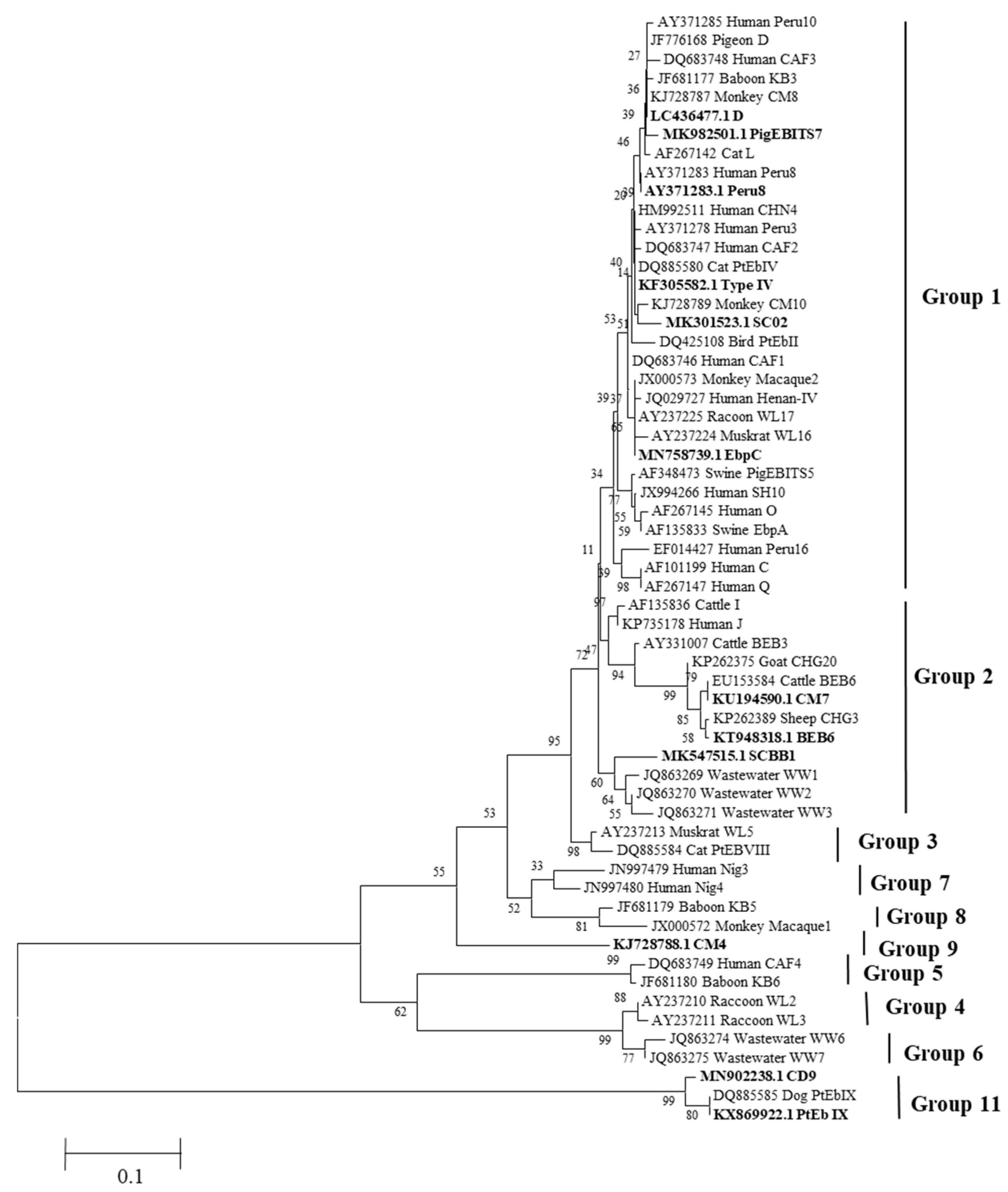
3.4. Prevalence and Genotype Distribution of E. bieneusi

The ITS sequence of E. bieneusi was detected through PCR analysis, and out of the 400 samples tested, 54 were positive, resulting in an overall infection rate of 13.5% (54/400). Xingtai Zoo had the highest positive rate of E. bieneusi at 25.4% (29/115), while Tangshan Zoo had the lowest positive rate at 6.1% (4/66). The infection rates in Guiyang, Beijing, and Shijiazhuang were 12.2% (6/49), 6.9% (7/101), and 11.6% (8/69), respectively. Among these, the infection rate of carnivores was 17.8%, and the infection rate of artiodactyls was 15.8%, ranking first and second, respectively. Hence, attention should be directed towards controlling E. bieneusi in the residential areas of these two animal groups.
The study found a significant difference in the positive rate of E. bieneusi among the different zoos (p < 0.01). A total of 12 genotypes were identified in the 54 E. bieneusi-positive samples. These genotypes included SC02 (n = 1) found in a panda; BEB6 (n = 8) found in milk goats, giraffes, addax and sika deer; Type IV (n = 7) found in Suri alpacas (Lama glama), roe deer, gibbons, ring-tailed lemurs, bush pigs, alpacas (Vicugna pacos), and red deer; pigEBITS 7 (n = 2) found in a gibbon and Siberian ibex; Peru8 (n = 2) found in a river deer and masked civet; PtEb IX (n = 3) found in gray wolves; D (n = 11) found in colobus monkeys (Colobus polykomos), squirrels, African lions (Panthera leo), masked civets, camels, yaks, fallow deer, sika deer, red deer, and gray wolves; D9 (n = 15) found in yaks, fallow deer, sika deer, big-eared sheep, red deer, wild horses, fragrance pigs, Amur tigers (Panthera tigris ssp. altaica), and gray wolves; EbpC (n = 1) found in a brown bear; SCBB1 (n = 2) found in black bears; CM4 (n = 1) found in a squirrel; CM7 (n = 1) found in an argali.
The obtained sequences of the 12 E. bieneusi genotypes were utilized to construct neighbor-joining phylogenetic trees (Figure 1). The phylogenetic analysis revealed the classification of the genotypes into different groups based on their genetic relatedness. Genotype D, PigEBITS7, Peru8, Type IV, SC02, and EbpC were classified into group 1, which suggests potential for zoonotic transmission. CM7, BEB6, and SCBB1 were categorized into group 2, indicating a potentially zoonotic nature. CM4 was assigned to group 9, while CD9 and PtEb IX were placed in group 11 based on the phylogenetic analysis.

4. Discussion

In the current study conducted on Chinese zoo animals, the prevalence rates of Cryptosporidium spp., G. duodenalis, E. bieneusi, and Blastocystis spp. were observed. The overall infection rates are presented in Table 6, along with the identification of various zoonotic genotypes. It is strongly advised that both breeders and visitors exercise caution during interactions with these animals, particularly when there is a possibility of contact with their feces and wastewater. This precaution is crucial for preventing the transmission of zoonotic parasitic diseases.
Advances in molecular biology detection technology have provided significant advantages in comprehending the classification and population genetic characteristics of various intestinal protozoa. Cryptosporidium spp., which exhibit a broad host range, have been reported in animals from various zoos in China [19]. In this study, two samples tested positive for Cryptosporidium spp. were identified, including C. andersoni found in a camel and C. ubiquitum found in an argali. C. andersoni was previously considered to primarily infect domestic animals with certain host specificity, but it has been identified in various animal hosts as well as humans [20]. Similarly, C. ubiquitum has been identified in argali and even in drinking water, suggesting it serves as a potential source of Cryptosporidium spp infection in humans [21].
Currently, six species of Giardia have been identified, including G. duodenalis, G. agilis, G. ardeae, G. muris, G. microti, and G. psittaci. Among these, G. duodenalis is the most commonly reported species in humans and animals. G. duodenalis is considered as a complex species and has been further categorized into eight assemblages (A–H) based on genetic analysis [22]. Assemblages A and B have a wide host range and are responsible for human infections [23]. The remaining six assemblages usually occur in specific animal hosts, although assemblages C, D, E, and F have been occasionally detected in human cases as well [24]. In total, 27 parasite species and over 70 host-adapted genotypes have been identified [25]. By PCR amplification of the tpi and bg genes of G. duodenalis, positive results were observed in 11 species from three animal groups. Three assemblages, E, B, and D, were identified among these positive samples. The detection of assemblage B in ring-tailed lemurs and chimpanzees confirms previous reports of its prevalence in primates [26]. Additionally, a mixed infection of assemblage E and B was found in milk goats and brown bears. Utilizing multiple genetic loci for the detection of G. duodenalis can improve the sensitivity of the diagnosis of mixed infections, with clusters of G. duodenalis being more commonly seen in some developing countries [27]. The identification and characterization of different Giardia species and assemblages are crucial for understanding their epidemiology, host range, and potential for zoonotic transmission. By studying the genetic diversity of Giardia, it becomes possible to unravel the complex interactions between the parasite, animals, and humans and design appropriate control measures to prevent the spread of infection.
E. bieneusi is classified as a microsporidium species within the fungi group, but it is often studied alongside other zoonotic protozoa. E. bieneusi is the most frequently diagnosed microsporidium species and accounts for more than 90% of human microsporidiosis cases [28]. While E. bieneusi infection in wild animals has been extensively reported worldwide, there have been relatively few studies on its occurrence in zoo animals [29]. A total of 12 genotypes of E. bieneusi were identified in this study. Previous reports have shown that genotypes within group 1 (D, PigEBITS7, Peru8, Type IV, SC02, and EbpC) have been detected in humans and various animal groups, indicating low host specificity and high potential for cross-species transmission [30,31]. Additionally, emerging evidence indicates that genotypes within group 2 also possess zoonotic potential. Genotypes I, J, BEB4, and BEB6 from group 2 have been found to infect humans [32]. The four genotypes, SC02, SCBB1, CM4, and PtEb IX, that we detected were found to be the dominant genotypes among pandas, black bears, squirrels, and gray wolves, respectively. The zoonotic genotypes D, PigEBITS7, Peru8, Type IV, SC02, and EbpC were identified in a variety of animals [33,34,35,36], suggesting that zoo animals may be important sources of human E. bieneusi infections and should be closely monitored and controlled.
Out of the 400 samples tested, a total of 98 (24.5%) were found to be positive for Blastocystis spp. The prevalence of Blastocystis spp. varied across different animal species: it was 35.7% in artiodactyla and primate species, 50% in avian species, 16.7% in proboscidean species, 5.4% in perissodactyla species, and 3.4% in carnivora species. No Blastocystis spp. were detected in rodents or marsupials. Another study conducted in two zoos in France reported a total positive rate of 32.2% for Blastocystis spp. [36], while a higher infection rate of 40.2% was reported in 295 wild animals in Qinling Mountains, China [37]. Recent molecular epidemiological studies have revealed potential modes of transmission for Blastocystis spp., including human-to-human, foodborne, waterborne, and zoonotic routes. Notably, the World Health Organization classified Blastocystis sp. as a waterborne pathogen, implying a potential public health concern [11]. Blastocystis spp. comprise 17 subtypes (ST1-ST17), with ST1-ST9 capable of infecting both humans and a variety of animals [38]. In China, Blastocystis spp. infection is primarily caused by ST1-ST3 subtypes in humans, while ST10-ST17 subtypes predominantly affect animals. This infection has been reported in more than 12 provinces/municipalities in humans and over 25 different animal hosts, showing significant variability in prevalence across geographic regions [10]. Nine Blastocystis sp. subtypes, including ST1-ST5, ST8, ST10, ST13, ST14, and one unidentified subtype (Unknown ST), were identified across 14 different animal species. Among those subtypes, ST1-ST5 and ST8 have previously been reported to infect humans. Specifically, ST1-ST4 and ST8 are predominantly found in primates, while ST5 is primarily detected in ungulates, which is consistent with previous studies [39,40]. ST3 merged as the dominant subtype in human infections, and ST1 also exhibited significant presence among human infections.
In general, the prevalence of Blastocystis spp. infections was found to be the highest, while Cryptosporidium spp. had the lowest prevalence. The variation in infection rates of those protozoans among studies can be attributed to several factors, including geographical location, climatic conditions, sampling season, animal species, sample size, and most importantly, the zoo policies related to feeding and hygiene management. For example, our study highlights the unique living conditions at Guiyang Wild Animal Park, which employs a semi-free-range breeding mode, unlike the other four zoos. This arrangement fosters increased interaction among animals. Our findings reveal that the Cryptosporidium spp. and G. duodenalis infection rates at Guiyang Wild Animal Park are notably higher compared to the other locations. Similarly, the prevalence of E. bieneusi and Blastocystis spp. infection is also considerable, ranking second and third, respectively. These results suggest a potential correlation between zoo living patterns and parasite infection rates. The presence of these parasites can be attributed to their relatively simple lifecycle, which does not involve intermediate hosts and allows for immediate infectivity upon excretion. Additionally, their low infective dose and short prepatent period contribute to their ease of transmission.
Our data showed the presence and diversity of significant opportunistic protozoa in zoo animals. Furthermore, we employed both the saturated saline floating method and centrifugal precipitation method to enhance the detection of oocysts for microscopic examination and identified the presence of nematodes oocysts.
However, the origin, the route of the transmission, and the significance of these pathogens for both animal and public health remain unknown. Addressing these lingering questions requires a more comprehensive approach, including the repeated sampling of individuals, the quantification of protozoa, and the correlation of the results with the actual health parameters of the sampled individuals. Gathering all these data is critical for the development of well-managed ecotourism and research practices that prioritize minimal impact on animal health. By monitoring and addressing the presence of these parasites in zoo animals, we can effectively mitigate the risks associated with zoonotic transmission, thereby promoting both animal welfare and public health. Specific measures include the regular cleaning of animals’ habitats, ensuring access to clean drinking water, and implementing routine deworming protocols. Therefore, it is crucial to prioritize ongoing surveillance and diagnostic measures for the detection and management of zoonotic parasites in zoo settings.

5. Conclusions

Our study provides valuable insights into the prevalence and genotype distribution of Cryptosporidium spp., G. duodenalis, E. bieneusi, and Blastocystis spp. infections among zoo animals in five zoos in China. Those protozoa were detected in all the zoos studied. The detection of multiple zoonotic genotypes in animals indicates the potential for zoonotic transmission to humans. These findings emphasize the importance of the regular testing of animals residing in zoo facilities in order to diagnose and prevent the spread of zoonotic parasites.

Author Contributions

J.L., L.M. and Q.L. were responsible for the study’s conception. D.A., T.J. (Tingting Jiang), C.Z., L.M. and T.J. (Ting Jia) were involved in the sample collection, detection, and molecular characterization. D.A., Y.P. and T.J. (Tingting Jiang). performed the analysis of result. D.A. and Z.Z. contributed to provision of experimental materials. D.A., T.J. (Tingting Jiang) and J.L. were involved in the drafting and revision of the manuscript. All authors have read and agreed to the published version of the manuscript.

Funding

This study was supported by the National Natural Science Foundation of China (32273092, 31972700) and Hebei Natural Science Foundation (H202105002).

Institutional Review Board Statement

Not applicable.

Informed Consent Statement

Not applicable.

Data Availability Statement

The sequences that support the findings of this study are openly available in the GenBank database at https://www.ncbi.nlm.nih.gov/nucleotide/ (accessed on 3 June 2021).

Conflicts of Interest

The authors declare no conflicts of interest.

References

  1. Bengis, R.G.; Leighton, F.A.; Fischer, J.R.; Artois, M.; Mörner, T.; Tate, C.M. The role of wildlife in emerging and re-emerging zoonoses. Rev. Sci. Tech. 2004, 23, 497–511. [Google Scholar]
  2. Karim, M.R.; Li, J.; Rume, F.I.; Sumon, S.M.R.; Selim, A.S.M.; Hoda, N.; Zhang, L. Occurrence and molecular characterization of Cryptosporidium spp. and Giardia duodenalis among captive mammals in the Bangladesh National Zoo. Parasitol. Int. 2021, 84, 102414. [Google Scholar] [CrossRef] [PubMed]
  3. Levecke, B.; Dorny, P.; Geurden, T.; Vercammen, F.; Vercruysse, J. Gastrointestinal protozoa in non-human primates of four zoological gardens in Belgium. Vet. Parasitol. 2007, 148, 236–246. [Google Scholar] [CrossRef] [PubMed]
  4. Pielok, Ł.A.; Kłudkowska, M.; Frąckowiak, K.; Stefaniak, J. Parasitic infections among patients hospitalized in the Tropical and Parasitic Clinic of Poznan University of Medical Sciences, Poland between 2015 and 2018. Is there a relationship between protozoa infection and gastrointestinal symptoms? Prz. Gastroenterol. 2022, 17, 310–315. [Google Scholar] [CrossRef] [PubMed]
  5. Thompson, R.C.; Smith, A. Zoonotic enteric protozoa. Vet. Parasitol. 2011, 182, 70–78. [Google Scholar] [CrossRef] [PubMed]
  6. Plutzer, J.; Lassen, B.; Jokelainen, P.; Djurković-Djaković, O.; Kucsera, I. Review of Cryptosporidium and Giardia in the eastern part of Europe, 2016. Euro Surveill 2018, 23, 16–00825. [Google Scholar] [CrossRef] [PubMed]
  7. Upcroft, J.A.; McDonnell, P.A.; Gallagher, A.N.; Chen, N.; Upcroft, P. Lethal Giardia from a wild-caught sulphur-crested cockatoo (Cacatua galerita) established in vitro chronically infects mice. Parasitology 1997, 114, 407–412. [Google Scholar] [CrossRef] [PubMed]
  8. Liu, H.; Shen, Y.; Yin, J.; Yuan, Z.; Jiang, Y.; Xu, Y.; Pan, W.; Hu, Y.; Cao, J. Prevalence and genetic characterization of Cryptosporidium, Enterocytozoon, Giardia and Cyclospora in diarrheal outpatients in China. BMC Infect. Dis. 2014, 14, 25. [Google Scholar] [CrossRef] [PubMed]
  9. Suankratay, C.; Thiansukhon, E.; Nilaratanakul, V.; Putaporntip, C.; Jongwutiwes, S. Disseminated infection caused by novel species of Microsporidium. Thailand. Emerg. Infect. Dis. 2012, 18, 302–304. [Google Scholar] [CrossRef]
  10. Deng, L.; Chai, Y.; Zhou, Z.; Liu, H.; Zhong, Z.; Hu, Y.; Fu, H.; Yue, C.; Peng, G. Epidemiology of Blastocystis sp. infection in China: A systematic review. Parasite 2019, 26, 41. [Google Scholar] [CrossRef]
  11. Rauff-Adedotun, A.A.; Meor Termizi, F.H.; Shaari, N.; Lee, I.L. The Coexistence of Blastocystis spp. in Humans, Animals and Environmental Sources from 2010–2021 in Asia. Biology 2021, 10, 990. [Google Scholar] [CrossRef]
  12. Lee, L.I.; Chye, T.T.; Karmacharya, B.M.; Govind, S.K. Blastocystis sp.: Waterborne zoonotic organism, a possibility? Parasit. Vectors 2012, 5, 130. [Google Scholar] [CrossRef]
  13. Ahsani, M.R.; Mohammad Abadi, M.R.; Shamsodini Bafti, M.; Ezatkhah, M.; Hasani Derakhshan, M.; Esmailzadeh Koshkooeh, A. Application of triplex PCR technique in identification of clostridium perfringens b, c and d types. Iran J. Pharm. Res. 2010, 2, 185–190. [Google Scholar]
  14. Mohammadabadi, M.R.; Omarovich Shaikhaev, G.; Efimovna Sulimova, G.; Rahman, O.; Mozafari, M.R. Detection of bovine leukemia virus proviral DNA in Yaroslavsl, Mongolian and black pied cattle by PCR. Cell Mol. Biol. 2004, 9, 766–768. [Google Scholar]
  15. Xiao, L.; Singh, A.; Limor, J.; Graczyk, T.K.; Gradus, S.; Lal, A. Molecular characterization of Cryptosporidium oocysts in samples of raw surface water and wastewater. Appl. Environ. Microbiol. 2001, 67, 1097–1101. [Google Scholar] [CrossRef] [PubMed]
  16. Zhang, X.; Wang, L.; Lan, X.; Dan, J.; Ren, Z.; Cao, S.; Shen, L.; Deng, J.; Zuo, Z.; Yu, S.; et al. Occurrence and multilocus genotyping of Giardia duodenalis in captive non-human primates from 12 zoos in China. PLoS ONE 2020, 15, e0228673. [Google Scholar] [CrossRef] [PubMed]
  17. Scicluna, S.M.; Tawari, B.; Clark, C.G. DNA barcoding of Blastocystis. Protist 2006, 157, 77–85. [Google Scholar] [CrossRef] [PubMed]
  18. Buckholt, M.A.; Lee, J.H.; Tzipori, S. Prevalence of Enterocytozoon bieneusi in swine: An 18-month survey at a slaughterhouse in Massachusetts. Appl. Environ. Microbiol. 2002, 68, 2595–2599. [Google Scholar] [CrossRef] [PubMed]
  19. Karim, M.R.; Zhang, S.; Jian, F.; Li, J.; Zhou, C.; Zhang, L.; Sun, M.; Yang, G.; Zou, F.; Dong, H.; et al. Multilocus typing of Cryptosporidium spp. and Giardia duodenalis from non-human primates in China. Int. J. Parasitol. 2014, 44, 1039–1047. [Google Scholar] [CrossRef]
  20. Feng, Y.; Ryan, U.M.; Xiao, L. Genetic Diversity and Population Structure of Cryptosporidium. Trends Parasitol. 2018, 34, 997–1011. [Google Scholar] [CrossRef]
  21. Li, N.; Xiao, L.; Alderisio, K.; Elwin, K.; Cebelinski, E.; Chalmers, R.; Santin, M.; Fayer, R.; Kvac, M.; Ryan, U.; et al. Subtyping Cryptosporidium ubiquitum, a zoonotic pathogen emerging in humans. Emerg. Infect. Dis. 2014, 20, 217–224. [Google Scholar] [CrossRef]
  22. Zou, Y.; Li, X.D.; Meng, Y.M.; Wang, X.L.; Wang, H.N.; Zhu, X.Q. Prevalence and multilocus genotyping of Giardia duodenalis in zoo animals in three cities in China. Parasitol. Res. 2022, 121, 2359–2366. [Google Scholar] [CrossRef]
  23. Cai, W.; Ryan, U.; Xiao, L.; Feng, Y. Zoonotic giardiasis: An update. Parasitol. Res. 2021, 120, 4199–4218. [Google Scholar] [CrossRef]
  24. Feng, Y.; Xiao, L. Zoonotic potential and molecular epidemiology of Giardia species and Giardiasis. Clin. Microbiol. Rev. 2011, 24, 110–140. [Google Scholar] [CrossRef]
  25. Li, J.; Qi, M.; Chang, Y.; Wang, R.; Li, T.; Dong, H.; Zhang, L. Molecular Characterization of Cryptosporidium spp., Giardia duodenalis, and Enterocytozoon bieneusi in Captive Wildlife at Zhengzhou Zoo, China. J. Eukaryot. Microbiol. 2015, 62, 833–839. [Google Scholar] [CrossRef] [PubMed]
  26. Karim, M.R.; Wang, R.; Yu, F.; Li, T.; Dong, H.; Li, D.; Zhang, L.; Li, J.; Jian, F.; Zhang, S.; et al. Multi-locus analysis of Giardia duodenalis from nonhuman primates kept in zoos in China: Geographical segregation and host-adaptation of assemblage B isolates. Infect. Genet. Evol. 2015, 30, 82–88. [Google Scholar] [CrossRef]
  27. Ryan, U.; Cacciò, S.M. Zoonotic potential of Giardia. Int. J. Parasitol. 2013, 43, 943–956. [Google Scholar] [CrossRef]
  28. Wang, S.S.; Wang, R.J.; Fan, X.C.; Liu, T.L.; Zhang, L.X.; Zhao, G.H. Prevalence and genotypes of Enterocytozoon bieneusi in China. Acta Trop. 2018, 183, 142–152. [Google Scholar] [CrossRef]
  29. Santín, M.; Fayer, R. Microsporidiosis: Enterocytozoon bieneusi in domesticated and wild animals. Res. Vet. Sci. 2011, 90, 363–371. [Google Scholar] [CrossRef] [PubMed]
  30. Li, W.; Xiao, L. Ecological and public health significance of Enterocytozoon bieneusi. One Health 2020, 12, 100209. [Google Scholar] [CrossRef] [PubMed]
  31. Li, W.; Feng, Y.; Santin, M. Host Specificity of Enterocytozoon bieneusi and Public Health Implications. Trends Parasitol. 2019, 35, 436–451. [Google Scholar] [CrossRef] [PubMed]
  32. Zhang, K.; Zheng, S.; Wang, Y.; Wang, K.; Wang, Y.; Gazizova, A.; Han, K.; Yu, F.; Chen, Y.; Zhang, L. Occurrence and molecular characterization of Cryptosporidium spp., Giardia duodenalis, Enterocytozoon bieneusi, and Blastocystis sp. in captive wild animals in zoos in Henan, China. BMC Vet. Res. 2021, 17, 332. [Google Scholar] [CrossRef] [PubMed]
  33. Li, W.; Deng, L.; Yu, X.; Zhong, Z.; Wang, Q.; Liu, X.; Niu, L.; Xie, N.; Deng, J.; Lei, S.; et al. Multilocus genotypes and broad host-range of Enterocytozoon bieneusi in captive wildlife at zoological gardens in China. Parasit. Vectors 2016, 9, 395. [Google Scholar] [CrossRef] [PubMed]
  34. Karim, M.R.; Dong, H.; Li, T.; Yu, F.; Li, D.; Zhang, L.; Li, J.; Wang, R.; Li, S.; Li, X.; et al. Predomination and new genotypes of Enterocytozoon bieneusi in captive nonhuman primates in zoos in China: High genetic diversity and zoonotic significance. PLoS ONE 2015, 10, e0117991. [Google Scholar] [CrossRef] [PubMed]
  35. Huang, X.; Zhou, Z.; Liu, H.; Deng, L.; Bi, B.; Chai, Y.; Zhong, Z.; Hu, Y.; Fu, H.; Peng, G. New genotypes and molecular characterization of Enterocytozoon bieneusi in captive black bears in China. Int. J. Parasitol. Parasites Wildl. 2019, 10, 1–5. [Google Scholar] [CrossRef]
  36. Cian, A.; El Safadi, D.; Osman, M.; Moriniere, R.; Gantois, N.; Benamrouz-Vanneste, S.; Delgado-Viscogliosi, P.; Guyot, K.; Li, L.L.; Monchy, S.; et al. Molecular Epidemiology of Blastocystis sp. in Various Animal Groups from Two French Zoos and Evaluation of Potential Zoonotic Risk. PLoS ONE 2017, 12, e0169659. [Google Scholar] [CrossRef] [PubMed]
  37. Song, J.K.; Yin, Y.L.; Yuan, Y.J.; Tang, H.; Ren, G.J.; Zhang, H.J.; Li, Z.X.; Zhang, Y.M.; Zhao, G.H. First genotyping of Blastocystis sp. in dairy, meat, and cashmere goats in northwestern China. Acta Trop. 2017, 176, 277–282. [Google Scholar] [CrossRef] [PubMed]
  38. Alfellani, M.A.; Taner-Mulla, D.; Jacob, A.S.; Imeede, C.A.; Yoshikawa, H.; Stensvold, C.R.; Clark, C.G. Genetic diversity of blastocystis in livestock and zoo animals. Protist 2013, 164, 497–509. [Google Scholar] [CrossRef]
  39. Stensvold, C.R.; Clark, C.G. Current status of Blastocystis: A personal view. Parasitol. Int. 2016, 65, 763–771. [Google Scholar] [CrossRef]
  40. Böhm-Gloning, B.; Knobloch, J.; Walderich, B. Five subgroups of Blastocystis hominis from symptomatic and asymptomatic patients revealed by restriction site analysis of PCR-amplified 16S-like rDNA. Trop. Med. Int. Health 1997, 2, 771–778. [Google Scholar] [CrossRef]
Figure 1. Phylogenetic relationships for E. bieneusi ITS sequence from the present study. Numbers on the branches are percent bootstrapping values from 1000 replicates.
Animals 14 00853 g001
Table 1. Primers for four intestinal protozoa.
PathogenGene LocusPrime Sequence (5′-3′)Expected Product Size (bp)Annealing Temperature (°C)References
Cryptosporidium spp.SSU rRNASSU-F2: TTCTAGAGCTAATACATGC~132555[15]
SSU-R2: CCCATTTCCTTCGAAACAGGA
SSU-F3: GGAAGGGTTGTATTTATTAGATAAAG~84055
SSU-F4: CTCATAAGGTGCTGAAGGAGTA
G. duodenalistpiAL3543: AAATIATGCCTGCTCGTCG~60555[16]
AL3546: CAAACCTTITCCGCAAACC
AL3544: CCCTTCATCGGIGGTAACTT~53055
AL3545: GTGGCCACCACICCCGTGCC
bgG7: AAGCCCGACGACCTCACCCGCAGTGC75355
G759: GAGGCCGCCCTGGATCTTCGAGACGAC
B-F: GAA CGA ACG AGA TCG AGG TCCG51155
B-R: CTCGACGAGCTTCGTGTT
Blastocystis spp.SSU rRNARD5: ATCTGGTTGATCCTGCCAGT~60058[17]
BhRDr: GAGCTTTTTAACTGCAACAACG
E. bieneusiITSEBITS3: GATGGTCATAGGGATGAAGAGCTT~43557[18]
EBITS4: TATGCTTAAGTCCAGGGAG
EBITS1: AGGGATGAAGAGCTTCGGCTCTG~39255
EBITS2.4: AGTGATCCTGTATTAGGGATATT
Table 2. Genotype distribution of G. duodenalis in different zoos.
LocationSample SizeNo. of Positive SamplesType of AssemblageGenotype
tpi (n)bg (n)
Guiyang498E (2), B (4)E (5)E (4), B (2), E + B (2)
Beijing10110B (5)E (5), B (3)E (5), B (5)
Shijiazhuang692B (1)E (1), B (1)E (1), B (1)
Tangshan663B (2)E (1)E (1), B (2)
Xingtai1151-D (1)D (1)
Total40024E (2) B (12)E (12), B (4), D (1)E (11), B (10), E + B (2), D (1)
Table 3. Distribution of G. duodenalis genotype in animals.
Groups of AnimalsAnimal Species (Common Name/Scientific Name)Sample SizeNo. of Positive SamplesType of Assemblage
ArtiodactylaGiraffe/Giraffa camelopardalis52E (2)
Milk goat/Capra hircus L.32E (1), E + B (1)
Siberian ibex/Capra sibirica112E (2)
Argali/Ovis ammon151E (1)
Roe deer/Capreolus pygargus42E (2)
Addax/Addax nasomaculatus31E (1)
Yak/Bos grunniens domesticus51E (1)
CarnivoraMasked civet/Paguma larvata31D (1)
Brown bear/Ursus arctos61E + B (1)
PrimateRing-tailed lemur/Lemur catta219E (1), B (8)
Chimpanzee/Pan troglodytes92B (2)
Table 4. Infection rates of Blastocystis spp. in animals.
Groups of AnimalsAnimal Species (Common Name/Scientific Name)Sample SizeNo. of Positive SamplesPositive Rate (%)
ProboscideanAsian elephant/Pan troglodytes6116.7
ArtiodactylaCamel/Camelus bactrianus10330.0
Milk goat/Capra hircus L.3133.3
Giraffe/Giraffa camelopardalis5240.0
Siberian ibex/Capra sibirica11545.5
Ammotragus/Ammotragus lervia33100.0
Argali/Ovis ammon15640.0
Roe deer/Capreolus pygargus4250.0
River deer/Hydropotes inermis11981.8
Addax/Addax nasomaculatus33100.0
Giant eland/Tragelaphus derbianus7342.9
Alpaca/Vicugna pacos24312.5
Red deer/Cervus canadensis18738.9
Yak/Bos grunniens domesticus5360.0
Fallow deer/Dama dama13430.8
Blue sheep/Pseudois nayaur5360.0
Sika deer/Cervus nippon18211.1
Big-eared sheep/Capra hircus6233.3
PerissodactylaZebra/Equus quagga2328.7
PrimatesGolden monkey/
Rhinopithecus
5240.0
Mandrill/Mandrillus sphinx9777.8
Ring-tailed lemur/
Lemur catta
211152.4
Gibbon/Hylobatidae10220.0
Baboon/Papio11100.0
Spider monkey/Ateles11100.0
Black langur/
Trachypithecus francoisi
2150.0
Chimp/Pan troglodytes9111.1
Patas monkey/Erythrocebus patas3133.3
Macaque/Macaca mulatta10330.0
CarnivoraOcelot/Prionailurus bengalensis1317.7
Manchurian tiger
/Panthera tigris ssp. altaica
6116.7
Gray wolf/Canis lupus1616.3
AvianCassowary/Casuarius unappendiculatus11100.0
Table 5. Subtype distribution of Blastocystis spp. in animals.
Gene Subtypes
(Population Size)
Host (Population Size)
ST1 (16)Mandrill (7), ring-tailed lemur (4), cassowary (1), gibbon (1), spider monkey (1), chimp (1), patas monkey (1)
ST2 (6)Ring-tailed lemur (1), baboon (1), gibbon (1), macaque (3)
ST3 (3)Asian elephant (1), patas monkey (1), gray wolf (1)
ST4 (3)Ring-tailed lemur (3)
ST5 (21)Milk goat (1), Siberian ibex (1), Ammotragus (1), argali (2), river deer (3), ocelot (1), ring-tailed lemur (1), zebra (2), yak (3), fallow deer (1), blue sheep (3), red deer (1)
ST8 (3)Camel (1), golden monkey (1), ring-tailed lemur (1)
ST10 (31)Camel (2), giraffe (2), Siberian ibex (1), Ammotragus (1), argali (4), roe deer (1), river deer (1), addax (3), giant eland (3), fallow deer (3), alpaca (1), sika deer (2), big-eared sheep (2), red deer (5)
ST13 (2)Golden monkey (1), black langur (1)
ST14 (12)Siberian ibex (3), Ammotragus (1), roe deer (1), river deer (5), alpaca (2)
Unknown ST (1)Red deer (1)
Table 6. Occurrence of parasites among various animals in different zoos.
LocationsSample SizeInfection Rate (%) (Positive Samples/Total Samples)
TotalCryptosporidium spp.E. bieneusiG. duodenalisBlastocystis spp.
Guiyang4955.1 (27/49)2.0 (1/49)12.2 (6/49)16.3 (8/49)24.5 (12/49)
Beijing10153.5 (54/101)1.0 (1/101)6.9 (7/101)9.9 (10/101)35.6 (36/101)
Shijiazhuang6936.2 (25/69)0 (0/69)11.6 (8/69)2.9 (2/69)20.3 (14/69)
Tangshan6619.8 (13/66)0 (0/66)6.1 (4/66)4.5 (3/66)10.6 (7/66)
Xingtai11551.3 (59/115)0 (0/115)25.4 (29/115)0.9 (1/115)25.2 (29/115)
Total40044.5 (178/400)0.5 (2/400)13.5 (54/400)6 (24/400)24.5 (98/400)
Disclaimer/Publisher’s Note: The statements, opinions and data contained in all publications are solely those of the individual author(s) and contributor(s) and not of MDPI and/or the editor(s). MDPI and/or the editor(s) disclaim responsibility for any injury to people or property resulting from any ideas, methods, instructions or products referred to in the content.

