Article

Evaluation of Immunodiagnostic Performances of Neospora caninum Peroxiredoxin 2 (NcPrx2), Microneme 4 (NcMIC4), and Surface Antigen 1 (NcSAG1) Recombinant Proteins for Bovine Neosporosis

by
Ruenruetai Udonsom
1,
Poom Adisakwattana
2,
Supaluk Popruk
1,
Onrapak Reamtong
3,
Charoonluk Jirapattharasate
4,
Tipparat Thiangtrongjit
3,
Sarinya Rerkyusuke
5,
Aran Chanlun
5,
Tanjila Hasan
6,7,
Manas Kotepui
8,
Sukhontha Siri
9,
Yoshifumi Nishikawa
7,* and
Aongart Mahittikorn
1,*
1
Department of Protozoology, Faculty of Tropical Medicine, Mahidol University, Bangkok 10400, Thailand
2
Department of Helminthology, Faculty of Tropical Medicine, Mahidol University, Bangkok 10400, Thailand
3
Department of Molecular Tropical Medicine and Genetics, Faculty of Tropical Medicine, Mahidol University, Bangkok 10400, Thailand
4
Department of Pre-Clinic and Animal Science, Faculty of Veterinary Science, Mahidol University, Nakhon Pathom 73170, Thailand
5
Division of Livestock Medicine, Faculty of Veterinary Medicine, Khon Kaen University, Khon Kaen 40002, Thailand
6
Department of Medicine and Surgery, Faculty of Veterinary Medicine, Chattogram Veterinary and Animal Sciences University, Chattogram 4225, Bangladesh
7
National Research Center for Protozoan Diseases, Obihiro University of Agriculture and Veterinary Medicine, Obihiro 080-8555, Japan
8
Medical Technology, School of Allied Health Sciences, Walailak University, Nakhon Si Thammarat 80160, Thailand
9
Department of Epidemiology, Faculty of Public Health, Mahidol University, Bangkok 10400, Thailand
*
Authors to whom correspondence should be addressed.
Animals 2024, 14(4), 531; https://doi.org/10.3390/ani14040531
Submission received: 25 December 2023 / Revised: 22 January 2024 / Accepted: 3 February 2024 / Published: 6 February 2024
(This article belongs to the Section Cattle)

Abstract

:

Simple Summary

Bovine neosporosis is a widespread and economically impactful disease affecting the dairy and meat industries globally. With no effective drug or vaccine available, the disease’s control relies on accurate detection methods. This study assessed the performance of three Neospora caninum proteins—NcPrx2, NcMIC4, and NcSAG1—as immunodiagnostic tools for identifying IgG antibodies against N. caninum. Comparisons were made with the indirect fluorescent antibody test (IFAT). The findings indicate that NcSAG1 exhibited the highest sensitivity and specificity, followed by NcMIC4. In contrast, NcPrx2 demonstrated lower sensitivity and specificity compared with IFAT. This study highlights that NcSAG1 is helpful as antigen marker and that NcPrx2 and NcMIC4 have potential in immunodiagnosis for detecting N. caninum infections in field samples. These findings could contribute to improved treatment management, surveillance, and risk assessment of neosporosis in livestock in the future.

Abstract

Bovine neosporosis is among the main causes of abortion in cattle worldwide, causing serious economic losses in the beef and dairy industries. A highly sensitive and specific diagnostic method for the assessment of the epidemiology of the disease, as well as it surveillance and management, is imperative, due to the absence of an effective treatment or vaccine against neosporosis. In the present study, the immunodiagnostic performance of Neospora caninum peroxiredoxin 2 (NcPrx2), microneme 4 (NcMIC4), and surface antigen 1 (NcSAG1) to detect IgG antibodies against N. caninum in cattle were evaluated and compared with that of the indirect fluorescent antibody test (IFAT). The results revealed that NcSAG1 had the highest sensitivity and specificity, with values of 88.4% and 80.7%, respectively, followed by NcPrx2, with a high sensitivity of 87.0% but a low specificity of 67.0%, whereas NcMIC4 showed sensitivity and specificity of 84.1% and 78.9%, respectively, when compared with IFAT. A high degree of agreement was observed for NcSAG1 (k = 0.713) recombinant protein, showing the highest diagnostic capability, followed by NcMIC4 (k = 0.64) and NcPrx2 (k = 0.558). The present study demonstrates that NcSAG1 is helpful as an antigen marker and also demonstrates the potential immunodiagnostic capabilities of NcPrx2 and NcMIC4, which could serve as alternative diagnostic markers for detecting N. caninum infection in cattle. These markers may find utility in future treatment management, surveillance, and risk assessment of neosporosis in livestock or other animal host species. Further research should be directed toward understanding the in vivo immune response differences resulting from immunization with both recombinant proteins.

1. Introduction

Neosporosis, caused by Neospora caninum, is an economically important disease in cattle. This infectious disease is associated with various reproductive complications, such as abortion, stillbirth, and the delivery of weakened calves [1,2]. The prevalence of N. caninum is widespread on a global scale, making it one of the predominant causes of bovine abortion and resulting in economic losses in both beef and dairy industries worldwide [3]. Vertical transmission is the most important route of parasite transmission and plays a pivotal role in sustaining the infection within the cattle population [4,5]. Neosporosis often presents as asymptomatic, although it can also manifest symptoms or cause abortions. However, the efficiency of transplacental transmission of N. caninum in cattle has been estimated to range from 44 to >95% [6,7], and the percentage of abortions is higher in seropositive dams than in seronegative dams [8,9,10]. To date, there is no effective chemotherapeutic treatment or vaccine for the prevention of neosporosis. Consequently, the management of the disease hinges on the utilization of accurate detection tests, influencing the formulation of control strategies [11].
Several serological methods, including the indirect fluorescent antibody test (IFAT), enzyme-linked immunosorbent assays (ELISAs), immunoblot, and direct agglutination tests, are available, and these tests are used to detect specific antibodies against N. caninum in cattle [12]. Among these assays, IFAT is considered the reference technique for detecting antibodies to N. caninum [13]. However, using intact tachyzoites as antigens in the IFAT method may detect cross-reactivity with antibodies against other members of the phylum Apicomplexa, including the protozoan Toxoplasma gondii [14]. Besides IFAT, another effective diagnostic method utilized for detecting N. caninum infections in a large number of animals is the indirect ELISA (iELISA), which employs recombinant proteins from N. caninum. This approach has been reported to have considerable sensitivity and excellent diagnostic accuracy [15,16].
Immunoproteomics is a powerful tool for identifying potential immunogenic antigens against N. caninum infection [17,18,19]. In a previous study, N. caninum species-specific antigens were identified using bovine infected sera through immunoproteomic analysis. Based on two-dimensional electrophoresis immunoblotting, 14 different antigenic proteins were specific to N. caninum. Among these, peroxiredoxin 2 (Prx2) and microneme 4 (MIC4) exhibited high immunoreactivity specificity against N. caninum infection [20]. In the pursuit of a reliable diagnostic method and vaccine development, various N. caninum recombinant proteins have been investigated as potential target antigens and vaccine candidates.
N. caninum peroxiredoxin (NcPrx) was recently reported to have peroxidase and antioxidant functions. The recombinant NcPrx (rNcPrx) protein exhibited a strong reaction with the polyclonal anti-rNcPrx serum, as observed with native NcPrx [21]. However, the antigenicity of NcPrx remains unknown. In T. gondii, recombinant proteins TgPrx1 and TgPrx3 induced protection against T. gondii-infected mice and were evaluated as potential vaccine candidates against toxoplasmosis [22,23]. Additionally, Leishmania peroxidoxin 1 (LdPxn1) elicited a robust CD4+ T cell response, resulting in partial protection against cutaneous leishmaniasis in immunized mice [24].
N. caninum microneme 4 (NcMIC4) is an associated protein found within the micronemes, and it is released by the parasite as a soluble component during host cell entry [25]. Experiments involving recombinant NcMIC4 have demonstrated a significant increase in protective immunity against neosporosis in a mouse model [26]. For T. gondii, immunization of mice with TgMIC1 and TgMIC4 triggers a protective immune response against T. gondii infections [27]. Additionally, a combination of TgMIC1/TgMIC4/TgMIC6 recombinant proteins significantly heightens an effective immune response, leading to a reduction in the mortality rate of mice [28].
The surface antigen 1 (NcSAG1) has been recognized as one of the most immunogenic and efficacious antigens for diagnosing bovine neosporosis [29]. Recently, the NcSAG1-based iELISA was developed, demonstrating remarkable antigenic properties and facilitating the assessment of Neospora-induced abortions in cattle [30]. Moreover, the utilization of NcSAG1 in an immunochromatographic assay (ICT) was consistently effective in detecting anti-N. caninum antibodies in cattle field samples [31].
The number of N. caninum proteins that have been investigated as diagnostic targets is limited. Therefore, the present study aimed to comprehensively assess the following three N. caninum recombinant antigens: NcPrx2, NcMIC4, and NcSAG1. Our study objective was to evaluate their serodiagnostic performance and compare their efficacy with that of IFAT, an established reference method for diagnosing neosporosis in cattle.

2. Materials and Methods

2.1. Preparation of N. caninum Tachyzoites

African green monkey kidney (Vero) cells were cultured in Dulbecco’s modified Eagle medium (Cytiva HyClone™, South Logan, UT, USA), supplemented with 8% fetal bovine serum (FBS), L-glutamine (2 mM/mL), penicillin–streptomycin (100 U/mL of penicillin and 100 μg/mL of streptomycin), and amphotericin B (0.25 µg/mL), in a humidified atmosphere with 5% CO2 at 37 °C until a confluent monolayer of cells was achieved. Subsequently, the cells were transferred to a maintenance medium containing 2% FBS and inoculated with N. caninum (Nc-1) tachyzoites. The culture was then incubated at 37 °C with 5% CO2 for 24 h. A serum-free cell culture medium (without FBS) was introduced and maintained until the tachyzoites were harvested at approximately 48 h. The purification of tachyzoites involved passing them through a 27-gauge needle and a 5 µm filter. The parasites were washed twice with phosphate-buffered saline (PBS), counted, and then utilized for the IFAT.

2.2. Production of GST-Fused Recombinant Proteins NcPrx2, NcMIC4, and NcSAG1

The recombinant NcSAG1 protein was expressed and purified following previously described protocols [30]. Similar to NcSAG1, recombinant NcPrx2 and NcMIC4 proteins were expressed in Escherichia coli BL21 (DE3) under optimal conditions. The target sequences were polymerase chain reaction (PCR)-amplified using specific primers containing suitable restriction enzyme sites, and the resulting proteins were expressed as glutathione S-transferase (GST) fusions (New England BioLabs Inc., Ipswich, MA, USA). Briefly, the PCR tests were performed with N. caninum cDNA (Nc-1 strain) as the template. The truncated lengths of the NcPrx2 (NCLIV_053640) and NcMIC4 (NCLIV_002940) genes were generated through PCR using specific primers, which were as follows: NcPrx2_Forward, 5′-TGGATCCCCGGAATTAATGAGTCACCCCCATGAT-3′, and reverse, 5′-GATGCGGCCGCTCGACTAAGCCGAAGGATCTGG-3′; and NcMIC4_ Forward, 5′-TGGATCCCCGGAATTAATGACTATAGGTGGTGACG-3′, and reverse, 5′-GATGCGGCCGCTCGATTATGCGTCTTCCTCTTCAA-3′; respectively. The PCR products were purified from agarose gels and cloned into the pGEX-4T-1 expression vector, which was treated with EcoR1 and Xho1 restriction enzymes. Successful insertion was confirmed by DNA sequencing. The resulting recombinant plasmids were introduced into E. coli BL21 (DE3) cells for protein expression. Bacterial cells that were transformed with NcPrx2 and NcMIC4 recombinant plasmids were cultured in liquid LB media supplemented with 50 µg/mL ampicillin and incubated at 37 °C for 14 h. The bacterial cultures were then transferred to fresh liquid LB media and incubated until an optical density of 0.5–0.7 was reached. NcPrx2 and NcMIC4 were induced in the culture with 0.1 mM isopropyl β-D-1-thiogalactopyranoside and incubated at 37 °C for 2 and 4 h, respectively. The bacterial cells were harvested, then centrifuged at 7180× g at 4 °C for 20 min. The pellets were suspended in a sonication buffer [50 mM Tris-HCl, pH 8, 50 mM NaCl, 1 mM ethylenediaminetetraacetic acid (EDTA), and 1 mM dithiothreitol] along with lysozyme with a final concentration of 500 µg/mL and incubated on ice for 30 min. The suspension was sonicated and Triton X-100 (10%) in PBS was added, followed by incubation on ice for 20 min and subsequent centrifugation at 7180× g at 4 °C for 20 min. The resulting supernatant was applied to Glutathione Sepharose 4B beads (GE Healthcare Life Sciences, Buckinghamshire, UK) as per the manufacturer’s instructions. Specifically, the supernatant was incubated with washed beads at 4 °C overnight with gentle rotation. GST fusion proteins were eluted with an elution buffer (100 mM Tris-HCl, pH 8.0, containing 100 mM NaCl, 5 mM EDTA, and 25 mM reduced glutathione powder; MP Biomedicals GmbH., Eschwege, Germany) and incubated at 4 °C overnight with mild rotation before centrifugation at 800× g, 4 °C for 3 min. The purified recombinant proteins were analyzed through Sodium Dodecyl Sulphate-Polyacrylamide Gel Electrophoresis (SDS-PAGE) and stained with Coomassie Brilliant Blue R250 (MP Biomedicals Inc., Illkirch-Graffenstaden, France). Protein concentrations were quantified using a bicinchoninic acid protein assay kit (Thermo Fisher Scientific, Inc., Rockford, IL, USA). Finally, NcPrx2, NcMIC4, and NcSAG1 recombinant proteins were identified and confirmed via liquid chromatography-mass spectrometry (LC-MS/MS).

2.3. Protein Identification by Mass Spectrometry

Each protein band section was excised from 1-D SDS-PAGE and de-stained overnight at 4 °C with 50% acetonitrile (ACN; Sigma-Aldrich, St. Louis, MO, USA) in 50 mM ammonium bicarbonate (Merck, Rahway, NJ, USA). The protein samples were reduced using 4 mM Dithiothreitol (DTT) in 50 mM ammonium bicarbonate, and this reduction process occurred at a temperature of 60 °C for 15 min. Following reduction, the proteins were alkylated with 250 mM iodoacetamide at room temperature for 30 min in the absence of light. The reaction was quenched using 4 mM DTT in 50 mM ammonium bicarbonate for 5 min. Subsequently, all solutions were removed, and the gel band was dehydrated using acetonitrile. The protein samples were digested with trypsin proteomics grade (Sigma-Aldrich, St. Louis, MO, USA) overnight at 37 °C in 50 mM ammonium bicarbonate. The resulting digested peptides were extracted using acetonitrile and then dried in a vacuum centrifuge. The dried tryptic peptides were analyzed using an UltiMate 3000 nano-liquid chromatography (nano-LC) system (Dionex, Surrey, UK). The mass spectra from both mass spectrometry (MS) and tandem mass spectrometry (MS/MS) covered mass ranges of m/z 400–2000 and m/z 50–1500, respectively. A mascot generic file (.mgf) was generated using data analysis 4.1 software (Bruker Daltonics, Billerica, MA, USA). For merging the .mgf files and protein identification, Mascot Daemon version 2.3.2 (Matrix Science, London, UK) was employed. Protein identification was executed using the Mascot algorithm search on the National Center for Biotechnology Information database (https://www.ncbi.nlm.nih.gov/) (accessed on 9 June 2023) and Toxoplasma informatics resource (http://www.ToxoDB.org) (accessed on 9 June 2023) protein databases.

2.4. Serum Samples

2.4.1. Control Sera

NcPrx2, NcMIC4, and NcSAG1 recombinant proteins were assessed using sera from mice experimentally infected with parasites and sera from infected bovines. The infected mouse sera were obtained from female ICR mice aged 6–8 weeks provided by the National Laboratory Animal Center, Mahidol University, Salaya Campus, Thailand. The mice were housed at the Laboratory Animal Science Unit, Faculty of Tropical Medicine, Mahidol University. Intraperitoneal inoculation of 1 × 10⁵ N. caninum or 1 × 10³ T. gondii tachyzoites was performed [30]. Blood samples were collected from the mice on day 0 and at 8 weeks post-inoculation. The infected and uninfected bovine sera, confirmed by IFAT and iELISA against NcSAG1, were provided by the National Research Center for Protozoan Diseases, Obihiro University of Agriculture, and Veterinary Medicine, Obihiro, Hokkaido, Japan [32]. Bovine sera infected with other protozoan parasites, including T. gondii (2 samples), C. parvum (2 samples), Babesia bovis (2 samples), and Babesia bigemina (2 samples), which were confirmed by at least two serology tests, were also included for specificity testing [20].

2.4.2. Field Dairy Cattle Serum Samples

Altogether, 247 dairy cattle serum samples from 8 cattle farms were collected from randomly selected herds from smallholder dairy farmers in Khon Kaen Province, Thailand. Khon Kaen is a city located in the northeastern region of Thailand and is one of the four major cities in the region. It lies approximately 450 km northeast of Bangkok (Figure 1). According to the records from Thailand’s Regional Livestock Office in 2021, Khon Kaen Province has 38,652 dairy cattle. The majority of cattle in this area are raised for milk production (Regional Livestock Office 3, Thailand, 2021). The cattle were confirmed to be clinically healthy and belonged to the Holstein-Friesian cross-breed. These cattle were uniformly accommodated in conventional open structures, covering a wide age range from 1 to 14 years. Their dietary regimen consisted of a combination of roughage and concentrate, with grazing opportunities allowed during the rainy season. Remarkably, no cases of abortion were recorded by the farmers in association with this cattle cohort. It is noteworthy that dogs were consistently present on all farms, and neighboring dogs had the potential to interact with the cattle. Blood samples were collected from either the jugular or caudal vein. These samples were promptly transferred into 10 mL vacuum blood tubes. All samples were placed in a styrofoam box, kept cool with ice packs, and transported to the laboratory. Subsequently, the sera were separated and stored at −20 °C until examination. Our study experiments involving mice and field cattle samples were approved by the Animal Care and Use Committee of the Faculty of Tropical Medicine-Animal Care and Use Committee (FTM-ACUC), Mahidol University, Bangkok, Thailand (Approval No. FTM-ACUC 027/2020).

2.5. Indirect ELISA (iELISA)

In our study, 96-well microtiter plates (Nunc, Roskilde, Denmark) were coated with purified recombinant protein at 4 °C overnight. The final concentrations were 0.1 μM for NcMIC4 and NcSAG1, and 0.5 μM for NcPrx2, in a 50 mM carbonate-bicarbonate buffer (pH 9.6). The plates were washed once with PBS containing 0.05% Tween 20 (PBS-T) and then blocked with 3% skimmed milk in PBS (PBS-SM) for 1 h at 37 °C. Following the blocking step, the plates were washed again with PBS-T, and 50 μL of the test sera (each sample in duplicate) diluted at 1:200 was added to the wells. The plates were incubated at 37 °C for 1 h. After washing, a secondary antibody, peroxidase-conjugated anti-mouse or bovine IgG (Invitrogen, Rockford, IL, USA), diluted at 1:4000 in PBS-SM, was added to the wells and incubated at 37 °C for 1 h. The plates were washed six times, and then 100 μL of 3,3′, 5,5′-tetramethylbenzidine substrate (Thermo Fisher Scientific, Frederick, MD, USA) was added to each well. The reaction was stopped by adding 100 μL of 1-M HCl. The absorbance at 450 nm was measured using the SUNRISE microplate reader (Tecan Austria GmbH, Grödig, Austria). The cut-off value was determined as the mean value of the standard N. caninum-negative control sera plus five times the standard deviation.

2.6. IFAT

Field cattle serum samples were evaluated for N. caninum infection using IFAT as the standard test and compared with iELISA utilizing NcPrx2, NcMIC4, and NcSAG1 recombinant proteins. The IFAT procedures were performed as described previously [33]. Briefly, killed N. caninum whole tachyzoites were fixed on printed microscope slides (Epredia™, Portsmouth, NH, USA). The serum samples were analyzed at a dilution of 1:200 [33,34]. The diluted sera were incubated on antigen-coated slides at 37 °C for 1 h in a moist chamber. Subsequently, the slides were washed with a rinse buffer (VMRD, Inc., Pullman, WA, USA). Fluorescein-labeled antibody to bovine IgG (SeraCare, Milford, MA, USA) was added and incubated in a moist chamber for 1 h. After washing, the slides were mounted in buffered glycerol and examined under a fluorescent microscope (ZEISS Axio Imager M2, Göttingen, Germany). Positive samples were identified by the presence of complete peripheral fluorescein around the N. caninum tachyzoites, whereas a negative result was indicated by the absence of fluorescence signal (Figure S1).

2.7. Statistical Analysis

The diagnostic performance of the iELISA assays, including the standard cut-off point, sensitivity, and specificity, was determined by receiver operating characteristic (ROC) analysis using PASW Statistics 18.0 (Chicago, IL, USA: SPSS Inc.; 2009). Test agreement (Kappa values; k) with 95% confidence intervals (95% CI) was calculated using VassarStats: Website for Statistical Computation (http://vassarstats.net) (accessed on 2 November 2023). The strength of agreement was categorized based on the kappa values as follows: fair (0.21–0.40), moderate (0.41–0.60), substantial (0.61–0.80), and almost perfect (0.81–1.00) [35].

3. Results

3.1. Identification of NcPrx2, NcMIC4, and NcSAG1 Recombinant Proteins

The analysis of NcPrx2, NcMIC4, and NcSAG1 recombinant proteins using 12% SDS-PAGE revealed estimated molecular weights of 47.2, 86.1, and 55 kDa, respectively (Figure S2). Additionally, through further identification by mass spectrometry, all the proteins exhibited correct amino acid sequences (Supplementary Materials S1–S3).

3.2. Assessment of Recombinant Proteins Using Mouse and Bovine Infected Sera

The diagnostic performance of NcPrx2, NcMIC4, and NcSAG1-based iELISA for N. caninum infection was assessed using sera from experimentally infected mice and cattle. The highest reactivity was detected with NcSAG1, followed by NcMIC4 and NcPrx2, in the sera obtained from mice experimentally infected with N. caninum (Figure S3). No cross-reactivity was observed in the sera from T. gondii-infected animals. When using the known bovine sera infected and uninfected with N. caninum, strong and high signal reactivity was observed for all recombinant antigens with N. caninum-infected sera, whereas low signal reactivity was observed in other protozoan-infected bovine sera (Figure S4).

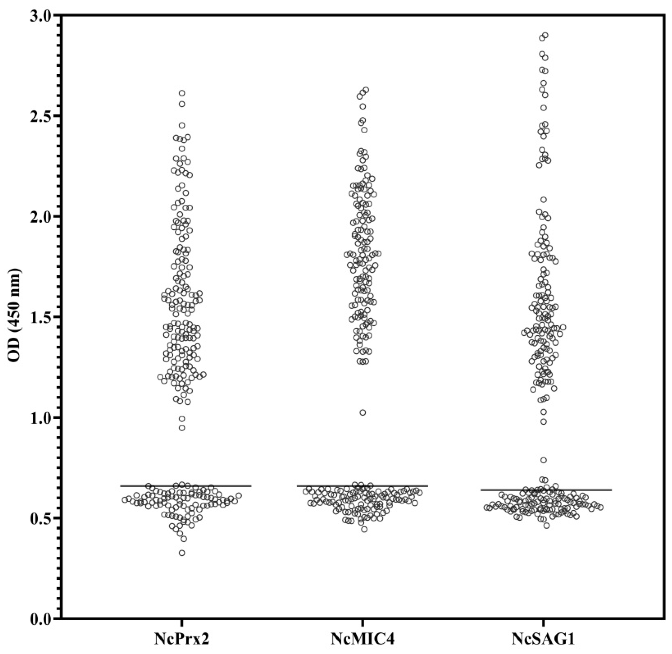
3.3. Comparison of ELISAs Using NcPrx2, NcMIC4, and NcSAG1 with IFAT in Field Cattle Serum Samples

We evaluated the antigenic efficacy of three recombinant antigens in detecting N. caninum infection in cattle using iELISA and compared the results with those of the standard IFAT. Among the 247 tested sera, 62.7% of the samples were positive for NcPrx2, 55.8% for NcMIC4, 55.4% for NcSAG1, and 55.8% by IFAT (Table 1).
Receiver Operating Characteristics (ROC) analysis was conducted to assess the performance of indirect ELISA with recombinant proteins and field cattle sera. The area under the curve (AUC) values for NcPrx2, NcMIC4, and NcSAG1 were 0.808, 0.790, and 0.854, respectively (Figure 2). Based on the ROC curve analysis, the optimal cut-off points were determined as 0.66 for NcPrx2 and NcMIC4, and 0.64 for NcSAG1. Samples were classified as positive when their values exceeded the respective cut-off points (Figure 3). Among these, NcSAG1 exhibited the highest sensitivity and specificity at 88.4% and 80.7%, respectively. NcPrx2 showed a high sensitivity of 87.0% but a lower specificity of 67.0%, while NcMIC4 displayed sensitivity and specificity of 84.1% and 78.9%, respectively (Table 2 and Table 3). Notably, substantial agreement was observed for NcSAG1 and NcMIC4, with kappa values of 0.713 and 0.64, respectively. In contrast, NcPrx2 demonstrated moderate agreement with a kappa value of 0.558 (Table 3).

4. Discussion

Numerous serological methods for detecting bovine neosporosis have been reported [12]. Nonetheless, enhancing diagnostic precision remains imperative to enhance diagnostic sensitivity and specificity, considering the potential for serologic cross-reactivity among infections caused by related protozoan parasites in cattle [36]. In the present study, we expressed, and assessed the performance of three recombinant antigens from N. caninum for diagnosing bovine neosporosis. The NcSAG1-based iELISA exhibited substantial agreement, demonstrating a commendable sensitivity of 88.4% and specificity of 80.7% when compared with the values of IFAT. For NcMIC4-based iELISA and IFAT, the sensitivity, and specificity were 84.1% and 78.9%, respectively. In contrast, the NcPrx2-based iELISA and IFAT demonstrated a high sensitivity but low specificity, with values of 87.0% and 67.0%, respectively. Notably, NcSAG1 proves to be an effective serodiagnostic tool for both bovine and canine neosporosis [37,38,39] Moreover, it has displayed high sensitivity and specificity in cattle and ovine hosts [40,41]. Furthermore, our study results are consistent with the findings of a previous study in which NcSAG1-based iELISA, and ICT displayed heightened sensitivity, specificity, and substantial agreement, indicating remarkable efficacy in detecting anti-N. caninum antibodies in field samples from Japan [31]. This consistency with our results reinforces the effectiveness of NcSAG1-based methods for antibody detection in Thailand.
NcMIC4 plays a crucial role in the initial stages of host cell adhesion and invasion [25]. A recent immunoproteomics study highlighted the highly specific immunogenic nature of NcMIC4, recognized by bovine-infected sera, suggesting its potential for diagnostic use and vaccine development [20]. However, while NcMIC4 exhibited high sensitivity, specificity, and substantial agreement in detecting N. caninum antibodies in field cattle, its efficacy is somewhat lower compared with NcSAG1 in the present study. Further research is required to explore the potential of NcMIC4 as a vaccine candidate against N. caninum infections.
Peroxidoxins or peroxiredoxins (Prxs) have been identified as a group of antioxidants present in both eukaryotes and prokaryotes [42]. However, despite the previous findings indicating significant immunoreactivity of NcPrx2 against N. caninum, suggesting its potential as a marker for immunodiagnostic development [20], its sensitivity and specificity for detecting N. caninum antibodies in cattle field samples are limited.
In this study, the performance of recombinant protein-based iELISA was compared with a standard IFAT. IFAT has been accepted as reference method for N. caninum serology [4,43,44,45] and has been a valuable standard for the development of other serological techniques [12,46,47]. Hence, we adopted IFAT as the reference test in this study to assess the diagnostic performance of the iELISA utilizing recombinant proteins. The cut-off titer in IFAT varies among laboratories, ranging from 1:100 to 1:640 for adult cattle and from 1:16 to 1:80 for detecting N. caninum antibodies in fetal sera [46,48]. A recommended cut-off value in IFAT for detecting N. caninum antibodies in adult cattle is 1:200 dilution [49,50], while lower cut-off values of 1:16–1:25 have been suggested for fetal fluids [48]. Additionally, using a high dilution cut-off titer in IFAT is considered appropriate to minimize cross-reactivity with related parasites, such as T. gondii, in serum samples [51,52]. Despite its widespread use for the detection of specific anti-N. caninum antibodies in cattle, IFAT is a labor-intensive technique, unsuitable for large-scale investigations, and its interpretation can be subjective [16,53]. The immunoblot has demonstrated high sensitivity and specificity for detecting N. caninum antibodies in cattle [53,54,55]. However, it is only performed in a limited number of laboratories as a routine tool for screening cattle sera due to its labor-intensive nature, specialized equipment requirements, and time-consuming process [53,56].
In contrast to an earlier study conducted in the same region, the overall prevalence of N. caninum infection was approximately four times higher (13% compared with >55% in this study) [57]. The differences in the prevalence pattern may be due to differences in the N. caninum purified recombinant antigen-based indirect ELISA used in this study, whereas the previous study was based on an ELISA utilizing parasite proteins incorporated into immunostimulating complexes as antigens. This substantial increase in prevalence underscores the widespread distribution of N. caninum infection within the dairy cattle population across the study areas. Furthermore, the prevalence of N. caninum in the present study surpassed values reported in other regions, such as 46.9% in dairy cattle from the northern area [58], 15% in dairy cattle, and 20.8% in beef cattle in the western Provinces of Thailand [39,59]. The increased prevalence observed in the current study compared with the earlier study could be attributed to the disparities in sampling locations, climatic conditions, study design, duration, and the utilization of diverse diagnostic tests or varying cut-off values [60]. In serological tests, various factors can lead to result variability, including differences in serological assays, titer and absorbance values, antigen composition and concentration, cut-off points and conjugated characteristics, as well as the sensitivity and specificity of the tests. This variability can also extend to the agreement between individual tests [4,12,46,48,50]. In addition, low levels of N. caninum antibodies in the serum can contribute to variations in results, both between different tests and different laboratories using the same diagnostic test [49]. Previous studies have described the variability in results when different antigens are used in serological tests, affecting the detection of antibodies directed at various epitopes of N. caninum [49,61]. The variation in test results in this study likely stems from the use of different antigen preparations or compositions. For instance, IFAT is prepared from whole N. caninum tachyzoites, while indirect ELISA employs recombinant proteins that carry distinct immunodominant epitopes. Additionally, the random sampling strategy may influence the differences in results observed in this study. As revealed by kappa statistical analysis, NcSAG1-iELISA, when compared with IFAT, demonstrated the highest level of agreement. This finding is valuable for the detection and diagnosis of N. caninum in cattle.
Currently, there is no available N. caninum vaccine or treatment for cattle or dogs. Control options for N. caninum infection in cattle farms have been modeled to reduce the infection [62]. Unfortunately, data collection was performed during the COVID-19 pandemic, which restricted authorized veterinarians’ farm access solely for animal health monitoring. Consequently, comprehensive information concerning the risk factors associated with N. caninum infection remained unavailable for this study. As evidenced by previous research reported in 2007, vertical transmission emerged as the primary route of N. caninum infection in this particular region [57]. Our study suggests that horizontal infection may play a major role in N. caninum infection in cattle in the studied area as the cattle were healthy and no cases of abortion were recorded by the farmers. This is also supported by the finding that seropositivity tends to increase with the age of cattle and the presence of dogs on all farms. At a practical level in this situation, domesticated and neighboring dogs play an important role, potentially increasing cattle exposure to N. caninum through contamination of feed with excreted oocysts in their feces [12]. The presence and number of dogs were found to be correlated with a high prevalence of N. caninum antibodies in dairy cattle [63,64]. A study by Inpankaew et al. (2014) reported that the high seroprevalence of N. caninum in dairy cattle was associated with the presence of farm dogs in herds in the northern provinces of Thailand [58]. In the current study, dogs were close to all the cattle farms, and consequently, the risk of exposure to the infection was increased. Therefore, reducing horizontal transmission of N. caninum in dairy herds is a useful goal in controlling the infection. To achieve this, preventing stray or neighboring dogs from entering the farm area, as well as protecting feedstuffs and drinking water from contamination with canine feces, is crucial [65]. Additionally, feeding dogs with commercial food and keeping them away from cattle are effective measures to decrease N. caninum infection in dairy herds. Further study is needed to investigate the relationship between the presence of dogs on dairy farms and the prevalence of N. caninum infection in Thailand.
On the other hand, the age of the animal is demonstrated to be relatively significant for N. caninum infection. Guimarães et al. (2004) reported that cows aged 24 months or over showed an increased risk of N. caninum infection [66]. Other reports also indicate that older cattle exhibit higher seropositivity to N. caninum, suggesting a greater possibility of horizontal transmission of the infection [67,68,69]. In this study, where cattle ranged in age from 1 to 14 years, the aforementioned findings suggest that the increasing age of cattle is also a significant factor for N. caninum infection. Although vertical transmission is the principal route of infection in cattle [70,71] and cannot be ruled out, recent research demonstrated a high prevalence of N. caninum in bovine placenta, indicating vertical transmission as the main route for N. caninum infection in Phayao Province, Northern Thailand [72]. A profitable control strategy to reduce infection is to avoid retaining or breeding heifers from seropositive cows and to inseminate all seropositive dams using beef semen [65]. Culling seropositive animals from the herd is also useful in reducing the infection rate [73].
The ROC curve analysis reveals good performance by NcSAG1, followed in decreasing order by NcMIC4 and NcPrx2, in detecting N. caninum-specific IgG in cattle sera. However, NcMIC4 and NcPrx2, while showing high sensitivity, exhibit lower specificity, which may make them less feasible for screening purposes. Nevertheless, NcMIC4 demonstrates a substantial level of agreement, and this study provides preliminary insight into the diagnostic efficacy of NcMIC4 and NcPrx2 for N. caninum antibody detection. Further studies are needed to explore the potential immunodiagnostic performance of both antigens in other animal host species for N. caninum infection. Consequently, a more comprehensive examination is warranted to assess the in vivo immune responses induced by immunizing mice with both recombinant proteins. Moreover, a comparison between the recombinant protein-based iELISA and the standard IFAT, immunoblot or commercial serological diagnostic kits in cattle or other animal host species should be conducted concurrently using substantial sample sizes. This will contribute to a comprehensive evaluation of the diagnostic performance and validity of these markers in a larger context.

5. Conclusions

Notably, NcSAG1 demonstrated the highest diagnostic potential, and the results highlight its superior effectiveness for diagnosing neosporosis in cattle compared with IFAT. Our study offers preliminary insights into the antigenicity of NcPrx2 and NcMIC4, which showed high sensitivity but low specificity compared with NcSAG1. Therefore, NcSAG1 is helpful as an antigen marker for detecting antibodies to N. caninum in cattle. However, NcPrx2 and NcMIC4 could serve as alternative candidates. Furthermore, our study data revealed a significant prevalence of N. caninum infection among dairy cattle in the studied regions.

Supplementary Materials

The following supporting information can be downloaded at https://www.mdpi.com/article/10.3390/ani14040531/s1. Figure S1: N. caninum probed with anti-N. caninum cattle serum, followed by fluorescent-labeled anti-bovine IgG; Figure S2: Coomassie blue stained SDS-PAGE of the recombinant proteins; Figure S3: Evaluation of NcPrx2, NcMIC4 and NcSAG1 tested with mice experimentally infected with N. caninum or T. gondii and uninfected sera by indirect ELISA; Figure S4: Evaluation of NcPrx2, NcMIC4 and NcSAG1 tested with known bovine infected and uninfected sera by indirect ELISA; Supplementary Material S1: Amino acid sequences of NcPrx2 identified by mass spectrometry; Supplementary Material S2: Amino acid sequences of NcMIC4 identified by mass spectrometry; Supplementary Material S3: Amino acid sequences of NcSAG1 identified by mass spectrometry.

Author Contributions

Conceptualization, P.A., Y.N. and A.M.; methodology, R.U., P.A., O.R., T.T., T.H., Y.N. and A.M.; software, M.K. and S.S.; validation, P.A., S.P., T.T. and A.M.; formal analysis, R.U., P.A., S.P., C.J. and Y.N.; investigation, R.U.; resources, P.A., S.P., O.R., C.J., S.R., A.C., Y.N. and A.M.; data curation, R.U.; writing—original draft preparation, R.U. and A.M.; writing—review and editing, P.A., S.R., A.C. and Y.N.; visualization, R.U. and A.M.; supervision, P.A, S.P., O.R., C.J., Y.N. and A.M.; project administration, Y.N. and A.M.; funding acquisition, Y.N. and A.M. All authors have read and agreed to the published version of the manuscript.

Funding

This research was funded by the Thailand Research Fund (TRF), the National Research Council of Thailand (NRCT), the Thailand Science Research and Innovation (TSRI) through the Royal Golden Jubilee Ph.D. programme [RGJ, Grant Number PHD/0067/2561]; the Faculty of Tropical Medicine, Mahidol University, Thailand; and the Ministry of Education, Culture, Sports, Science and Technology KAKENHI, Japan [grant number 23K18071, 21H02353, 20K21359, 18H0233501 (Y.N.)].

Institutional Review Board Statement

Our study experiments involving mice and field cattle samples were approved by the Animal Care and Use Committee of the Faculty of Tropical Medicine-Animal Care and Use Committee (FTM-ACUC), Mahidol University, Bangkok, Thailand (Approval No. FTM-ACUC 027/2020) (Approval date 25 August 2020).

Informed Consent Statement

Not applicable.

Data Availability Statement

All data relating to the present study are available in this manuscript and Supplementary Files.

Acknowledgments

We would like to thank the farm owners who allowed and assisted us with the sample collection. Additionally, we acknowledge Oiko Buzon Tacusalme from the Office of Research Services, Faculty of Tropical Medicine, Mahidol University, for supporting us during the manuscript preparation.

Conflicts of Interest

The authors declare no conflicts of interest. The financial supporter had no role in the design of the study; in the collection, analyses, or interpretation of data; in the writing of the manuscript; or in the decision to publish the results.

References

  1. Dubey, J.P.; Schares, G.; Ortega-Mora, L.M. Epidemiology and control of neosporosis and Neospora caninum. Clin. Microbiol. Rev. 2007, 20, 323–367. [Google Scholar] [CrossRef] [PubMed]
  2. Innes, E.A.; Bartley, P.M.; Maley, S.W.; Wright, S.E.; Buxton, D. Comparative host-parasite relationships in ovine toxoplasmosis and bovine neosporosis and strategies for vaccination. Vaccine 2007, 25, 5495–5503. [Google Scholar] [CrossRef] [PubMed]
  3. Reichel, M.P.; Ayanegui-Alcérreca, M.A.; Gondim, L.F.; Ellis, J.T. What is the global economic impact of Neospora caninum in cattle–the billion dollar question. Int. J. Parasitol. 2013, 43, 133–142. [Google Scholar] [CrossRef] [PubMed]
  4. Dubey, J.P. Review of Neospora caninum and neosporosis in animals. Korean J. Parasitol. 2003, 41, 1–16. [Google Scholar] [CrossRef] [PubMed]
  5. Horcajo, P.; Regidor-Cerrillo, J.; Aguado-Martínez, A.; Hemphill, A.; Ortega-Mora, L. Vaccines for bovine neosporosis: Current status and key aspects for development. Parasite Immunol. 2016, 38, 709–723. [Google Scholar] [CrossRef] [PubMed]
  6. Davison, H.C.; Otter, A.; Trees, A.J. Significance of Neospora caninum in British dairy cattle determined by estimation of seroprevalence in normally calving cattle and aborting cattle. Int. J. Parasitol. 1999, 29, 1189–1194. [Google Scholar] [CrossRef] [PubMed]
  7. Bergeron, N.; Fecteau, G.; Pare, J.; Martineau, R.; Villeneuve, A. Vertical and horizontal transmission of Neospora caninum in dairy herds in Quebec. Can. Vet. J. 2000, 41, 464–467. [Google Scholar] [PubMed]
  8. Mazuz, M.L.; Fish, L.; Reznikov, D.; Wolkomirsky, R.; Leibovitz, B.; Savitzky, I.; Golenser, J.; Shkap, V. Neosporosis in naturally infected pregnant dairy cattle. Vet. Parasitol. 2014, 205, 85–91. [Google Scholar] [CrossRef]
  9. Spilovska, S.; Reiterova, K.; Antolova, D. Neospora caninum—Associated Abortions in Slovak Dairy Farm. Iran. J. Parasitol. 2015, 10, 96–101. [Google Scholar]
  10. Lefkaditis, M.; Mpairamoglou, R.; Sossidou, A.; Spanoudis, K.; Tsakiroglou, M. Neospora caninum, A potential cause of reproductive failure in dairy cows from Northern Greece. Vet. Parasitol. Reg. Stud. Rep. 2020, 19, 100365. [Google Scholar] [CrossRef]
  11. Wang, X.; Song, X.; Yang, J.; Liu, Q.; Liu, J. Characterization of Neospora caninum microneme protein 26 and its potential use as a diagnostic marker for neosporosis in cattle. Front. Vet. Sci. 2020, 7, 357. [Google Scholar] [CrossRef]
  12. Dubey, J.P.; Schares, G. Diagnosis of bovine neosporosis. Vet. Parasitol. 2006, 140, 1–34. [Google Scholar] [CrossRef] [PubMed]
  13. Ghalmi, F.; China, B.; Jenkins, M.; Azzag, N.; Losson, B. Comparison of different serological methods to detect antibodies specific to Neospora caninum in bovine and canine sera. J. Vet. Diagn. Investig. 2014, 26, 136–140. [Google Scholar] [CrossRef] [PubMed]
  14. Dong, J.; Otsuki, T.; Kato, T.; Park, E.Y. Development of a diagnostic method for neosporosis in cattle using recombinant Neospora caninum proteins. BMC Biotechnol. 2012, 12, 19. [Google Scholar] [CrossRef]
  15. Sinnott, F.A.; Monte, L.G.; Collares, T.F.; Silveira, R.M.; Borsuk, S. Review on the immunological and molecular diagnosis of neosporosis (years 2011–2016). Vet. Parasitol. 2017, 239, 19–25. [Google Scholar] [CrossRef] [PubMed]
  16. Novoa, M.B.; Valentini, B.S.; Sarli, M.; Torioni-de-Echaide, S.M.; Primo, M.E.; Echaide, I.E. Evaluation of a competitive inhibition ELISA based on the recombinant protein tSAG1 to detect anti-Neospora caninum antibodies in cattle. J. Vet. Diagn. Invest. 2020, 32, 401–408. [Google Scholar] [CrossRef]
  17. Lee, E.G.; Kim, J.H.; Shin, Y.S.; Shin, G.W.; Kim, Y.H.; Kim, G.S.; Kim, D.Y.; Jung, T.S.; Suh, M.D. Two-dimensional gel electrophoresis and immunoblot analysis of Neospora caninum tachyzoites. J. Vet. Sci. 2004, 5, 139–145. [Google Scholar] [CrossRef]
  18. Shin, Y.S.; Lee, E.G.; Shin, G.W.; Kim, Y.R.; Lee, E.Y.; Kim, J.H.; Jang, H.; Gershwin, L.J.; Kim, D.Y.; Kim, Y.H.; et al. Identification of antigenic proteins from Neospora caninum recognized by bovine immunoglobulins M, E, A and G using immunoproteomics. Proteomics 2004, 4, 3600–3609. [Google Scholar] [CrossRef]
  19. Lee, E.G.; Kim, J.H.; Shin, Y.S.; Shin, G.W.; Suh, M.D.; Kim, D.Y.; Kim, Y.H.; Kim, G.S.; Jung, T.S. Establishment of a two-dimensional electrophoresis map for Neospora caninum tachyzoites by proteomics. Proteomics 2003, 3, 2339–2350. [Google Scholar] [CrossRef]
  20. Udonsom, R.; Reamtong, O.; Adisakwattana, P.; Popruk, S.; Jirapattharasate, C.; Nishikawa, Y.; Inpankaew, T.; Toompong, J.; Kotepui, M.; Mahittikorn, A. Immunoproteomics to identify species-specific antigens in Neospora caninum recognised by infected bovine sera. Parasite 2022, 29, 60. [Google Scholar] [CrossRef]
  21. Venancio-Brochi, J.C.; Pereira, L.M.; Baroni, L.; Abreu-Filho, P.G.; Yatsuda, A.P. Characterization of the Neospora caninum peroxiredoxin: A novel peroxidase and antioxidant enzyme. Parasitol. Res. 2022, 121, 1735–1748. [Google Scholar] [CrossRef]
  22. Fereig, R.M.; Kuroda, Y.; Terkawi, M.A.; Mahmoud, M.E.; Nishikawa, Y. Immunization with Toxoplasma gondii peroxiredoxin 1 induces protective immunity against toxoplasmosis in mice. PLoS ONE 2017, 12, e0176324. [Google Scholar] [CrossRef]
  23. Fereig, R.M.; Nishikawa, Y. Peroxiredoxin 3 promotes IL-12 production from macrophages and partially protects mice against infection with Toxoplasma gondii. Parasitol. Int. 2016, 65, 741–748. [Google Scholar] [CrossRef] [PubMed]
  24. Bayih, A.G.; Daifalla, N.S.; Gedamu, L. DNA-protein immunization using Leishmania peroxidoxin-1 induces a strong CD4+ T cell response and partially protects mice from cutaneous leishmaniasis: Role of fusion murine granulocyte-macrophage colony-stimulating factor DNA adjuvant. PLoS Negl. Trop. Dis. 2014, 8, e3391. [Google Scholar] [CrossRef] [PubMed]
  25. Keller, N.; Riesen, M.; Naguleswaran, A.; Vonlaufen, N.; Stettler, R.; Leepin, A.; Wastling, J.M.; Hemphill, A. Identification and characterization of a Neospora caninum microneme-associated protein (NcMIC4) that exhibits unique lactose-binding properties. Infect. Immun. 2004, 72, 4791–4800. [Google Scholar] [CrossRef] [PubMed]
  26. Srinivasan, S.; Mueller, J.; Suana, A.; Hemphill, A. Vaccination with microneme protein NcMIC4 increases mortality in mice inoculated with Neospora caninum. J. Parasitol. 2007, 93, 1046–1055. [Google Scholar] [CrossRef] [PubMed]
  27. Lourenco, E.V.; Bernardes, E.S.; Silva, N.M.; Mineo, J.R.; Panunto-Castelo, A.; Roque-Barreira, M.C. Immunization with MIC1 and MIC4 induces protective immunity against Toxoplasma gondii. Microbes Infect. 2006, 8, 1244–1251. [Google Scholar] [CrossRef] [PubMed]
  28. Pinzan, C.F.; Sardinha-Silva, A.; Almeida, F.; Lai, L.; Lopes, C.D.; Lourenco, E.V.; Panunto-Castelo, A.; Matthews, S.; Roque-Barreira, M.C. Vaccination with Recombinant Microneme Proteins Confers Protection against Experimental Toxoplasmosis in Mice. PLoS ONE 2015, 10, e0143087. [Google Scholar] [CrossRef]
  29. Takashima, Y.; Takasu, M.; Yanagimoto, I.; Hattori, N.; Batanova, T.; Nishikawa, Y.; Kitoh, K. Prevalence and dynamics of antibodies against NcSAG1 and NcGRA7 antigens of Neospora caninum in cattle during the gestation period. J. Vet. Med. Sci. 2013, 75, 1413–1418. [Google Scholar] [CrossRef] [PubMed]
  30. Abdelbaky, H.H.; Nishimura, M.; Shimoda, N.; Hiasa, J.; Fereig, R.M.; Tokimitsu, H.; Inokuma, H.; Nishikawa, Y. Evaluation of Neospora caninum serodiagnostic antigens for bovine neosporosis. Parasitol. Int. 2020, 75, 102045. [Google Scholar] [CrossRef]
  31. Fereig, R.M.; Abdelbaky, H.H.; Nishikawa, Y. Comparative Evaluation of Four Potent Neospora caninum Diagnostic Antigens Using Immunochromatographic Assay for Detection of Specific Antibody in Cattle. Microorganisms 2021, 9, 2133. [Google Scholar] [CrossRef] [PubMed]
  32. Nishimura, M.; Kohara, J.; Kuroda, Y.; Hiasa, J.; Tanaka, S.; Muroi, Y.; Kojima, N.; Furuoka, H.; Nishikawa, Y. Oligomannose-coated liposome-entrapped dense granule protein 7 induces protective immune response to Neospora caninum in cattle. Vaccine 2013, 31, 3528–3535. [Google Scholar] [CrossRef] [PubMed]
  33. Wiengcharoen, J.; Thompson, R.C.; Nakthong, C.; Rattanakorn, P.; Sukthana, Y. Transplacental transmission in cattle: Is Toxoplasma gondii less potent than Neospora caninum? Parasitol. Res. 2011, 108, 1235–1241. [Google Scholar] [CrossRef] [PubMed]
  34. Wiengcharoen, J.; Nakthong, C.; Mitchaothai, J.; Udonsom, R.; Sukthana, Y. Toxoplasmosis and neosporosis among beef cattle slaughtered for food in Western Thailand. Southeast. Asian J. Trop. Med. Public. Health 2012, 43, 1087. [Google Scholar] [PubMed]
  35. McHugh, M.L. Interrater reliability: The kappa statistic. Biochem Med. (Zagreb) 2012, 22, 276–282. [Google Scholar] [CrossRef] [PubMed]
  36. Gondim, L.F.; Mineo, J.R.; Schares, G. Importance of serological cross-reactivity among Toxoplasma gondii, Hammondia spp., Neospora spp., Sarcocystis spp. and Besnoitia besnoiti. Parasitology 2017, 144, 851–868. [Google Scholar] [CrossRef]
  37. Chahan, B.; Gaturaga, I.; Huang, X.; Liao, M.; Fukumoto, S.; Hirata, H.; Nishikawa, Y.; Suzuki, H.; Sugimoto, C.; Nagasawa, H.; et al. Serodiagnosis of Neospora caninum infection in cattle by enzyme-linked immunosorbent assay with recombinant truncated NcSAG1. Vet. Parasitol. 2003, 118, 177–185. [Google Scholar] [CrossRef]
  38. Kubota, N.; Sakata, Y.; Miyazaki, N.; Itamoto, K.; Bannai, H.; Nishikawa, Y.; Xuan, X.; Inokuma, H. Serological survey of Neospora caninum infection among dogs in Japan through species-specific ELISA. J. Vet. Med. Sci. 2008, 70, 869–872. [Google Scholar] [CrossRef]
  39. Udonsom, R.; Sukthana, Y.; Nishikawa, Y.; Fereig, R.M.; Jirapattharasate, C. Current situation of Neospora caninum and Toxoplasma gondii infection among beef cattle in Kanchanaburi, Ratchaburi and Nakhon Patom provinces, Thailand. Thai J. Vet. Med. 2018, 48, 403–409. [Google Scholar] [CrossRef]
  40. Howe, D.K.; Tang, K.; Conrad, P.A.; Sverlow, K.; Dubey, J.P.; Sibley, L.D. Sensitive and specific identification of Neospora caninum infection of cattle based on detection of serum antibodies to recombinant Ncp29. Clin. Diagn. Lab. Immunol. 2002, 9, 611–615. [Google Scholar] [CrossRef]
  41. Alves Sinnott, F.; da Silva Leal, K.; de Oliveira Silva, M.T.; Barros de Pinho, R.; Pappen, F.; da Rosa Farias, N.A.; Bedoya Llano, H.A.; Pereira Garcia Melo, D.; Borsuk, S. An indirect ELISA for Neosporosis: Associating recombinant Neospora caninum proteins NcSRS2 and NcSAG1. Vet. Parasitol. 2020, 281, 109101. [Google Scholar] [CrossRef]
  42. McGonigle, S.; Dalton, J.P.; James, E.R. Peroxidoxins: A new antioxidant family. Parasitol. Today 1998, 14, 139–145. [Google Scholar] [CrossRef] [PubMed]
  43. Dubey, J.P.; Lindsay, D.S. A review of Neospora caninum and neosporosis. Vet. Parasitol. 1996, 67, 1–59. [Google Scholar] [CrossRef] [PubMed]
  44. Dubey, J.P. Neosporosis in cattle: Biology and economic impact. J. Am. Vet. Med. Assoc. 1999, 214, 1160–1163. [Google Scholar] [CrossRef]
  45. Ghalmi, F.; China, B.; Ghalmi, A.; Hammitouche, D.; Losson, B. Study of the risk factors associated with Neospora caninum seroprevalence in Algerian cattle populations. Res. Vet. Sci. 2012, 93, 655–661. [Google Scholar] [CrossRef] [PubMed]
  46. Bjorkman, C.; Uggla, A. Serological diagnosis of Neospora caninum infection. Int. J. Parasitol. 1999, 29, 1497–1507. [Google Scholar] [CrossRef]
  47. Atkinson, R.; Harper, P.; Reichel, M.; Ellis, J. Progress in the serodiagnosis of Neospora caninum infections of cattle. Parasitol. Today 2000, 16, 110–114. [Google Scholar] [CrossRef]
  48. Alvarez-Garcia, G.; Collantes-Fernandez, E.; Costas, E.; Rebordosa, X.; Ortega-Mora, L.M. Influence of age and purpose for testing on the cut-off selection of serological methods in bovine neosporosis. Vet. Res. 2003, 34, 341–352. [Google Scholar] [CrossRef]
  49. Dubey, J.P.; Jenkins, M.C.; Adams, D.S.; McAllister, M.M.; Anderson-Sprecher, R.; Baszler, T.V.; Kwok, O.C.; Lally, N.C.; Bjorkman, C.; Uggla, A. Antibody responses of cows during an outbreak of neosporosis evaluated by indirect fluorescent antibody test and different enzyme-linked immunosorbent assays. J. Parasitol. 1997, 83, 1063–1069. [Google Scholar] [CrossRef]
  50. von Blumroder, D.; Schares, G.; Norton, R.; Williams, D.J.; Esteban-Redondo, I.; Wright, S.; Bjorkman, C.; Frossling, J.; Risco-Castillo, V.; Fernandez-Garcia, A.; et al. Comparison and standardisation of serological methods for the diagnosis of Neospora caninum infection in bovines. Vet. Parasitol. 2004, 120, 11–22. [Google Scholar] [CrossRef]
  51. Silva, D.A.; Lobato, J.; Mineo, T.W.; Mineo, J.R. Evaluation of serological tests for the diagnosis of Neospora caninum infection in dogs: Optimization of cut off titers and inhibition studies of cross-reactivity with Toxoplasma gondii. Vet. Parasitol. 2007, 143, 234–244. [Google Scholar] [CrossRef] [PubMed]
  52. Benetti, A.H.; Schein, F.B.; dos Santos, T.R.; Toniollo, G.H.; da Costa, A.J.; Mineo, J.R.; Lobato, J.; de Oliveira Silva, D.A.; Gennari, S.M. [Inquiry of antibodies anti-Neospora caninum in dairy cattle, dogs and rural workers of the south-west region of Mato Grosso State]. Rev. Bras. Parasitol. Vet. 2009, 18 (Suppl. S1), 29–33. [Google Scholar] [CrossRef] [PubMed]
  53. Campero, L.M.; Minke, L.; More, G.; Rambeaud, M.; Bacigalupe, D.; Moore, D.P.; Hecker, Y.; Campero, C.M.; Schares, G.; Venturini, M.C. Evaluation and comparison of serological methods for the detection of bovine neosporosis in Argentina. Rev. Argent. Microbiol. 2015, 47, 295–301. [Google Scholar] [CrossRef] [PubMed]
  54. Sondgen, P.; Peters, M.; Barwald, A.; Wurm, R.; Holling, F.; Conraths, F.J.; Schares, G. Bovine neosporosis: Immunoblot improves foetal serology. Vet. Parasitol. 2001, 102, 279–290. [Google Scholar] [CrossRef]
  55. Staubli, D.; Nunez, S.; Sager, H.; Schares, G.; Gottstein, B. Neospora caninum immunoblotting improves serodiagnosis of bovine neosporosis. Parasitol. Res. 2006, 99, 648–658. [Google Scholar] [CrossRef]
  56. Ortega-Mora, L.; Fernández-García, A.; Gómez-Bautista, M. Diagnosis of bovine neosporosis: Recent advances and perspectives. Acta Parasitol. 2006, 51, 1–14. [Google Scholar] [CrossRef]
  57. Chanlun, A.; Emanuelson, U.; Frossling, J.; Aiumlamai, S.; Bjorkman, C. A longitudinal study of seroprevalence and seroconversion of Neospora caninum infection in dairy cattle in northeast Thailand. Vet. Parasitol. 2007, 146, 242–248. [Google Scholar] [CrossRef]
  58. Inpankaew, T.; Jittapalapong, S.; Mitchell, T.J.; Sununta, C.; Igarashi, I.; Xuan, X. Seroprevalence of Neospora caninum infection in dairy cows in Northern provinces, Thailand. Acta Parasitol. 2014, 59, 305–309. [Google Scholar] [CrossRef]
  59. Arunvipas, P.; Inpankaew, T.; Jittapalapong, S. Seroprevalence and risk factors of Neospora caninum infection among dairy cows in the Western Provinces of Nakhon Pathom, Ratchaburi and Kanchanaburi, Thailand. Agric. Nat. Resour. 2012, 46, 64–70. [Google Scholar]
  60. Schares, G.; Rauser, M.; Zimmer, K.; Peters, M.; Wurm, R.; Dubey, J.P.; de Graaf, D.C.; Edelhofer, R.; Mertens, C.; Hess, G.; et al. Serological differences in Neospora caninum-associated epidemic and endemic abortions. J. Parasitol. 1999, 85, 688–694. [Google Scholar] [CrossRef]
  61. Jenkins, M.C.; Caver, J.A.; Bjorkman, C.; Anderson, T.C.; Romand, S.; Vinyard, B.; Uggla, A.; Thulliez, P.; Dubey, J.P. Serological investigation of an outbreak of Neospora caninum-associated abortion in a dairy herd in southeastern United States. Vet. Parasitol. 2000, 94, 17–26. [Google Scholar] [CrossRef] [PubMed]
  62. Reichel, M.P.; Ellis, J.T. If control of Neospora caninum infection is technically feasible does it make economic sense? Vet. Parasitol. 2006, 142, 23–34. [Google Scholar] [CrossRef] [PubMed]
  63. Wouda, W.; Bartels, C.J.; Moen, A.R. Characteristics of Neospora caninum-associated abortion storms in diary herds in The Netherlands (1995 to 1997). Theriogenology 1999, 52, 233–245. [Google Scholar] [CrossRef]
  64. Asmare, K.; Regassa, F.; Robertson, L.J.; Skjerve, E. Seroprevalence of Neospora caninum and associated risk factors in intensive or semi-intensively managed dairy and breeding cattle of Ethiopia. Vet. Parasitol. 2013, 193, 85–94. [Google Scholar] [CrossRef] [PubMed]
  65. McAllister, M.M. Diagnosis and Control of Bovine Neosporosis. Vet. Clin. N. Am. Food Anim. Pract. 2016, 32, 443–463. [Google Scholar] [CrossRef]
  66. Guimarães, J.S., Jr.; Souza, S.L.; Bergamaschi, D.P.; Gennari, S.M. Prevalence of Neospora caninum antibodies and factors associated with their presence in dairy cattle of the north of Paraná state, Brazil. Vet. Parasitol. 2004, 124, 1–8. [Google Scholar] [CrossRef]
  67. Fávero, J.F.; Da Silva, A.S.; Campigotto, G.; Machado, G.; Daniel de Barros, L.; Garcia, J.L.; Vogel, F.F.; Mendes, R.E.; Stefani, L.M. Risk factors for Neospora caninum infection in dairy cattle and their possible cause-effect relation for disease. Microb. Pathog. 2017, 110, 202–207. [Google Scholar] [CrossRef]
  68. Manca, R.; Ciccarese, G.; Scaltrito, D.; Chirizzi, D. Detection of Anti-Neospora caninum Antibodies on Dairy Cattle Farms in Southern Italy. Vet. Sci. 2022, 9, 87. [Google Scholar] [CrossRef]
  69. Selim, A.; Alshammari, A.; Gattan, H.S.; Marzok, M.; Salem, M.; Al-Jabr, O.A. Neospora caninum infection in dairy cattle in Egypt: A serosurvey and associated risk factors. Sci. Rep. 2023, 13, 15489. [Google Scholar] [CrossRef]
  70. Anderson, M.L.; Andrianarivo, A.G.; Conrad, P.A. Neosporosis in cattle. Anim. Reprod. Sci. 2000, 60–61, 417–431. [Google Scholar] [CrossRef]
  71. Bartels, C.J.; Huinink, I.; Beiboer, M.L.; van Schaik, G.; Wouda, W.; Dijkstra, T.; Stegeman, A. Quantification of vertical and horizontal transmission of Neospora caninum infection in Dutch dairy herds. Vet. Parasitol. 2007, 148, 83–92. [Google Scholar] [CrossRef] [PubMed]
  72. Japa, O.; Nuangmek, A.; Prakhammin, K.; Flynn, R.J. Prevalence of vertically transmitted Neospora caninum amongst beef cattle in Phayao, Thailand. Parasitol. Int. 2019, 70, 98–101. [Google Scholar] [CrossRef] [PubMed]
  73. Hall, C.A.; Reichel, M.P.; Ellis, J.T. Neospora abortions in dairy cattle: Diagnosis, mode of transmission and control. Vet. Parasitol. 2005, 128, 231–241. [Google Scholar] [CrossRef] [PubMed]
Figure 1. A map indicating the sampling sites within Khon Kaen Province, Thailand. The marker indicates the specific area under investigation in this study.
Animals 14 00531 g001
Figure 2. Receiver Operating Characteristics (ROC) analysis for the indirect ELISA with NcPrx2, NcMIC4 and NcSAG1 proteins using field cattle sera.
Animals 14 00531 g002
Figure 3. Evaluation of N. caninum-derived peroxiredoxin 2 (NcPrx2), microneme 4 (NcMIC4), and surface antigen 1 (NcSAG1) recombinant antigens using field cattle serum samples (n = 247), as an optimal cut-off determined by the Receiver Operating Characteristic (ROC). Each bar represents the cut-off values of each recombinant protein based indirect ELISA.
Animals 14 00531 g003
Table 1. Detection of anti-N. caninum IgG in field cattle specimens from eight farms in Khon Kaen Province, Thailand using NcPrx2-, NcMIC4-, and NcSAG1-based indirect ELISA and IFAT (n = 247).
FarmNo. of Dairy CattleNo. of Positive (%)
IFATNcPrx2NcMIC4NcSAG1
16132 (52.4)36 (59.0)34 (55.7)27 (44.2)
276 (85.7)4 (57.1)6 (85.7)6 (85.7)
32914 (48.2)15 (51.7)16 (55.1)14 (48.2)
42915 (51.7)20 (68.9)16 (55.1)19 (65.5)
53120 (64.5)14 (45.1)21 (67.7)21 (67.7)
63621 (58.3)28 (77.7)16 (44.4)25 (69.4)
73017 (56.6)21 (70.0)14 (46.6)14 (46.6)
82413 (54.1)17 (70.8)15 (62.5)11 (45.8)
Total247138 (55.8)155 (62.7)138 (55.8)137(55.4)
Abbreviations: NcPrx2, N. caninum-derived peroxiredoxin 2; NcMIC4, N. caninum microneme 4; NcSAG1, N. caninum surface antigen 1; ELISA, enzyme-linked immunosorbent assay; IFAT, indirect fluorescent antibody test.
Table 2. Comparison of N. caninum IgG detection rate in field cattle serum samples using NcPrx2-, NcMIC4-, and NcSAG1-based indirect ELISA, with that of IFAT as the reference test (n = 247).
IFATNcPrx2NcMIC4NcSAG1
(+)(−)(+)(−)(+)(−)
(+)120181162212018
(−)357422871792
Total15592138109137110
Abbreviations: NcPrx2, N. caninum-derived peroxiredoxin 2; NcMIC4, N. caninum microneme 4; NcSAG1, N. caninum surface antigen 1; ELISA, enzyme-linked immunosorbent assay; IFAT, indirect fluorescent antibody test.
Table 3. Sensitivity and specificity of indirect ELISAs using recombinant proteins for the detection of specific N. caninum IgG antibodies, with IFAT as the reference test.
ParametersNcPrx2NcMIC4NcSAG1
Sensitivity (%)87.084.188.4
Specificity (%)67.078.980.7
Kappa value0.5580.640.713
Abbreviations: NcPrx2, N. caninum-derived peroxiredoxin 2; NcMIC4, N. caninum microneme 4; NcSAG1, N. caninum surface antigen 1; ELISA, enzyme-linked immunosorbent assay; IFAT, indirect fluorescent antibody test.
Disclaimer/Publisher’s Note: The statements, opinions and data contained in all publications are solely those of the individual author(s) and contributor(s) and not of MDPI and/or the editor(s). MDPI and/or the editor(s) disclaim responsibility for any injury to people or property resulting from any ideas, methods, instructions or products referred to in the content.

