Article

Effects of High-Concentrate-Induced SARA on Antioxidant Capacity, Immune Levels and Rumen Microbiota and Function in Goats

College of Animal Science and Technology, Hunan Agricultural University, Changsha 410125, China
*
Author to whom correspondence should be addressed.
Animals 2024, 14(2), 263; https://doi.org/10.3390/ani14020263
Submission received: 10 December 2023 / Revised: 9 January 2024 / Accepted: 12 January 2024 / Published: 15 January 2024

Abstract

:

Simple Summary

Our study elucidated the specific alterations in rumen microbiota associated with subacute ruminal acidosis (SARA) triggered by a high-concentrate diet. It demonstrated that SARA can lead to a decrease in microbial richness and diversity in the rumen of goats. SARA could potentially hinder the growth of rumen bacterial communities. Within the context of SARA, the antioxidant capacity of goats diminishes, while inflammatory cytokines increase, potentially raising the risk of health issues in growing goats. Understanding this is crucial for predicting changes in goat microbiota and improving the efficiency and well-being of ruminants through nutritional approaches such as dietary interventions.

Abstract

This study aims to explore the antioxidant, immune, and enzyme metabolism aspects in goats experiencing subacute ruminal acidosis (SARA). Furthermore, we seek to elucidate the relationship between the symbiotic microbiota of goats and their metabolic function. Sixteen goats were equally divided into two groups and fed a normal-concentrate diet (NC, 55% concentrate) or a high-concentrate diet (HC, 90% concentrate) for five weeks. We found that the HC diet reduced the total antioxidant capacity (T-AOC) (p = 0.022) and increased interleukin-1β (IL-1β) (p = 0.015), interleukin-4 (IL-4) (p = 0.008) and interleukin-6 (IL-6) (p = 0.002) concentration of goats. Simultaneously, the HC diet significantly increased the concentrations of alkaline phosphatase (ALP) and amylase (AMY) in the blood and rumen fluid of goats (p < 0.05). Microbial analysis in the rumen of goats revealed that the HC diet decreased bacterial richness and diversity, as evidenced by the changed observed species, Chao 1, PD whole tree and Shannon when compared to the NC diet (p < 0.01). The proportion of Proteobacteria increased while that of Spirochaetes and Fibrobacteres significantly decreased with the HC diet (p < 0.05). The Christensenellaceae_R-7_group and Ruminococcaceae_UCG-010 in rumen was notably decreased when a diet was switched from 55% concentrate diet to 90% concentrate diet (p < 0.05). Additionally, microbial functional potentials deduced that the HC diet significantly increased the abundance of the citrate cycle (TCA cycle) (ko00020) associated with carbohydrate metabolism (p = 0.028). Furthermore, the HC diet significantly increased the glutathione metabolism (ko00480) associated with the metabolism of other amino acids (p = 0.008). Our findings suggested that SARA reduced the total antioxidant capacity and increased levels of inflammatory factors in goats, as well as decreased rumen bacterial species and abundance.

1. Introduction

In recent years, in pursuit of enhanced production efficiency, intensive farming has witnessed the widespread adoption of high-concentrate diets, primarily reliant on cereal starch as the primary energy source. Unfortunately, this extensive use has led to an upsurge in the prevalence of metabolic diseases. SARA is a prevalent gastrointestinal ailment in the field of ruminant farming. It is caused by excessive feeding of highly fermentable carbohydrates and insufficient dietary crude fiber [1]. A distinctive feature of SARA is the prolonged period of low rumen pH, with the rate of absorption and metabolism of rumen VFA serving as a pivotal determinant of rumen pH [2]. During the initial stages of SARA, typical symptoms are often absent, making diagnosis challenging. The primary diagnostic criterion for SARA is a rumen fluid pH persisting between 5.2 and 5.6 for over 3 h [3]. Consequences of SARA encompass reduced feed intake, diminished fiber digestibility, lower milk fat content, heightened occurrence of diarrhea, hoof inflammation, liver abscesses, elevated bacterial endotoxin levels, and increased acute phase protein during inflammation [4]. Owing to the scarcity of high-quality roughage resources in China, often characterized by subpar quality, producers find themselves compelled to heavily rely on starch-rich grain concentrates to fulfill the nutritional demands of animals, all in the pursuit of elevated production performance. Consequently, animals suffer from inadequate consumption of high-quality fiber, leading to the onset of acidosis, particularly prevalent in high-yield dairy cows and intensively fattened cattle and sheep. The emergence of SARA has emerged as a significant impediment to the production of cattle and sheep in China.
Research has demonstrated that in response to acid stress, microorganisms possess the ability to mitigate cell damage by modulating the dynamic equilibrium of intracellular pH (pHi), inducing the expression of stress proteins, and regulating cell membrane morphology and function [5]. Nevertheless, if stress surpasses the self-regulating capacity of microorganisms, intracellular protons and acid ions accumulate gradually, leading to a rapid decline in pHi. This, in turn, causes damage to cell membranes and internal macromolecular structures, disrupting normal cell growth and metabolism, and, in severe cases, resulting in microbial mortality [6,7,8]. The alterations in physical and chemical properties, reduced proliferation, and potential mortality of rumen microorganisms under acid stress constitute significant factors influencing rumen fermentation and shifts in microbial flora. Mertens [9] defined peNDF as: neutral detergent fiber (NDF) in the diet that can promote rumen liquid and solid phase stratification and affect the chewing of ruminants. Previous studies have shown that increasing the peNDF content of the diet increased the average rumen pH and is an effective means of preventing SARA [10,11].
As sequencing technology continues to advance, we have the opportunity to delve deeper into the influence of dietary changes on the gut microbiome of animals [12,13,14,15]. Maintaining relatively high hay feeding and a stable bacterial composition is imperative for the well-being of ruminants. SARA, on the other hand, leads to significant alterations in both rumen and intestinal microbiota [16,17,18]. Currently, research into the gut microbiota of both animals and humans is advancing, unveiling ever-expanding functions of the gut microbiota. Simultaneously, there is a continuous emergence of developments in gut microbiota preparations [19,20]. This experiment aims to construct a subacute rumen acidosis model in goats and study the changes in antioxidant capacity, immune levels, and microbial community structure and composition under sub healthy rumen conditions, enriching the survival and reproduction patterns of rumen bacteria under diseases, and providing a basis for the development of gut microbiota preparations for ruminants.

2. Materials and Methods

2.1. Animals and Treatment

This study was approved by the Animal Care and Use Committee of Hunan Agricultural University (HAU201408). To conduct this study, we chose sixteen Liuyang black goats at random. These goats were all six months old and had an average weight of 15.3 ± 1.67 kg. The goats were subsequently separated into two categories and given either a regular-concentrate meal (NC, with a ratio of 55% concentrate to 45% forage) or a concentrated diet (HC, with a ratio of 90% concentrate to 10% forage). Before the diets were formulated, the paddy straw was chopped to approximately 2 cm in length. The dry matter intake in the two groups was 572 g/d (NC) and 602 g/d (HC), respectively. Table 1 provided a comprehensive breakdown of the diets’ composition and nutritional value. The experimental period spanned 35 days, including a 7-day diet adaptation phase. The diets were evenly distributed to the goats at approximately 08:00 and 18:00 h, respectively. Each goat was housed in its individual enclosure.

2.2. Sample Collection

Upon completion of the 35-day feeding period, plasma samples were collected into tubes using the methodology described in a prior study [21], and subsequently stored at −20 °C for later analysis. Rumen epithelial samples were extracted and preserved in 10% neutral formalin to facilitate histomorphology examinations. Six animals were randomly chosen from each group and humanely euthanized. Rumen fluid was collected, promptly frozen at −80 °C, and later utilized for genomic DNA isolation and the assessment of enzyme metabolism indicators. The pH values in rumen samples of two groups in SARA model were the same before feeding in the morning (0 h), but it was declined in the HC group at 3 h or 6 h after feeding. During the time period from 3 h after the feeding until the sampling at 6 h, the average ruminal pH was below 5.7 in the HC group and remained significantly lower in NC goats [22]. Rumen segments from each goat were gathered to observe the morphology of the rumen epithelium.

2.3. Measurement of Enzyme Metabolism, Antioxidant and Immune Indicators

The T-AOC, NO, IFN-γ, HIS, IL-1β, IL-2, IL-4, and IL-6 in the blood were detected using a corresponding enzyme-linked immunosorbent assay (ELISA) kit (Jiangsu Yutong Biological Technology Co., Ltd. Yancheng, China). The blood and rumen fluid metabolites including ALT (alanine aminotransferase), AST (aspartate aminotransferase), ALP (alkaline phosphatase), and AMY (amylase) were detected with the commercial assay kits (Roche Diagnostics (Shanghai) Ltd., Shanghai, China; and determined using an Automatic Biochemistry analyzer (Cobas c 311, Roche, Shanghai, China), and strictly following the manufacturer’s instructions, respectively.

2.4. Histological Examination

Morphological observation involved the collection of rumen epithelial segments from every goat. The rumen epithelial tissues were dehydrated, embedded, and subsequently sliced into approximately 5 mm-thick sections using a microtome. Afterwards, they were dyed with hematoxylin and eosin. Images of the rumen epithelium’s structure were taken using a light microscope (Pannoramic DESK, P-MIDI, P250, 3DHISTECH, Budapest, Hungary) that had a computer-assisted morphometric system.

2.5. DNA Extraction and PCR Amplification

DNA extraction from rumen fluid was carried out using the E.Z.N.A.® soil DNA Kit (Omega Bio-Tek, Norcross, GA, USA) and verified through 1% agarose gel electrophoresis [23]. The V3–V4 hypervariable regions of the bacterial 16S-rDNA gene were subsequently amplified using universal primers 338F (5′-ACTCCTACGGGAGGCAGCAG-3′) and 806R (5′-GGACTACHVGGGTWTCTAAT-3′). PCR reactions were conducted in a total volume of 20 μL, comprising 1 × FastPfu Buffer, 250 μM dNTPs, 0.1 μM of each primer, 1 U FastPfu Polymerase (Beijing TransGen Biotech, Beijing, China), and 10 ng of template DNA. The PCR protocol consisted of an initial denaturation at 95 °C for 2 min, followed by 30 cycles of denaturation at 95 °C for 30 s, annealing at 55 °C for 30 s, elongation at 72 °C for 30 s, and a final extension at 72 °C for 5 min. Subsequently, the amplification products were confirmed using 2% agarose gel electrophoresis. Purified amplicons were equimolarly pooled and subjected to paired-end sequencing (2 × 300) on an Illumina MiSeq platform (Allwegene, Beijing, China) following standard protocols [24].

2.6. Bacterial Data Processing and Function Predication

The FASTQ files were processed using QIIME (version 1.17) by demultiplexing and quality filtering. The following criteria were applied: (i) Reads with a length of 300 bp were truncated at positions where the average quality score dropped below 20 within a 10 bp sliding window. Reads shorter than 50 bp after truncation were discarded. (ii) Stringent processing involved precise barcode matching, allowing for a maximum of two nucleotide mismatches in primer matching, and excluding reads containing ambiguous characters. (iii) Assembly of sequences was performed only for those with overlaps longer than 10 bp in their overlapping regions. The unassembled reads were eliminated. OTUs were clustered using UPARSE (version 7.1) with a 97% similarity cutoff, and chimeric sequences were identified and removed using UCHIME (https://www.drive5.com/uchime/uchime_download.html, accessed on 8 January 2024). A rarefaction analysis was performed using Mothur v.1.21.1 to evaluate diversity indices including observed_species, PD_whole_tree, and Shannon. Primer 6 software (Primer-E Ltd., London, UK) was used for conducting hierarchical clustering analysis, and Canoco 4.5 was employed for performing Principal Component Analysis (PCA). The bioinformatics tool PICRUSt was used to predict the functional potentials of metagenomes by reconstructing unobserved states through a phylogenetic investigation of communities, relying on data from the 16S rRNA gene [25]. The OTU table was utilized in PICRUSt to predict functional genes, making use of the Kyoto Encyclopedia of Genes and Genomes (KEGG) database.

2.7. Statistical Analysis

All of the experimental data in the tables and figures are presented as the mean ± SD and the differences between groups were assessed by the independent-samples t test using SPSS (IBM SPSS 21.0, Chicago, IL, USA). The data of 16S rRNA sequencing were analyzed on the allwegene platform. Statistical significance was set at p < 0.05 and tendencies at 0.05 ≤ p ≤ 0.10.

3. Results

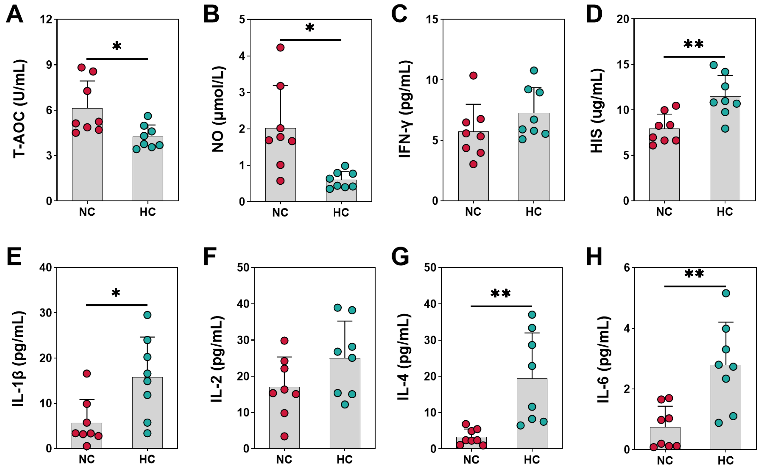
3.1. Antioxidant and Immune Indicators of Growing Goats Fed Different Diets

As shown in Figure 1A, the T-AOC was decreased dramatically in plasma by feeding the HC diet to goats compared to the NC diet (p = 0.022). Compared with the NC group, the data also showed that the NO level of goats was improved notably by the HC diet (p = 0.011) (Figure 1B). And diets supplemented with the HC diet has no significant effect on the IFN-γ (Figure 1C), there were no statistical differences between the groups (p = 0.185). The HC diet raised the HIS concentration of goats remarkably (p = 0.003) (Figure 1D). Results for plasma IL-1β was significantly boosted for the HC group compared to the NC group (p = 0.015) (Figure 1E). The level of IL-4 (p = 0.008) and IL-6 (p = 0.002) in plasma increased significantly when dietary concentrate ratio increased from 55% to 90% (Figure 1G,H). Compared with the NC group, plasma concentration of IL-2 in the HC group was not significantly different compared to the NC group (p = 0.110) (Figure 1F).

3.2. The Enzyme Metabolism Indicators of Growing Goats Fed Different Diets

The results shown in Figure 2A demonstrated that the ALT concentration in plasma was increased by the HC diet (p = 0.025). And the AST concentration in plasma was also increased by the HC diet compared to the NC diet (p = 0.051) (Figure 2B). The ALP concentration in plasma was notably increased when a diet administered to steers was switched from 55% concentrate diet to 90% concentrate diet (p < 0.001) (Figure 2C). Results for plasma AMY concentration were significantly boosted for the HC group compared to the NC group (p = 0.015) (Figure 2D), where ALT (p < 0.001) and AST (p = 0.025) concentrations in rumen were increased in this experimental model by increasing the grain percentage of the diet to induce SARA (Figure 2E,F). And the HC diet elevated significantly the ALP (p < 0.001) and AMY (p < 0.001) in rumen compared to the NC diet fed to growing goats (Figure 2G,H). The rumen epithelium from the NC group showed an intact structure while the rumen epithelium of the HC group showed an indication of sloughing in the stratum corneum (Figure 2I–L).

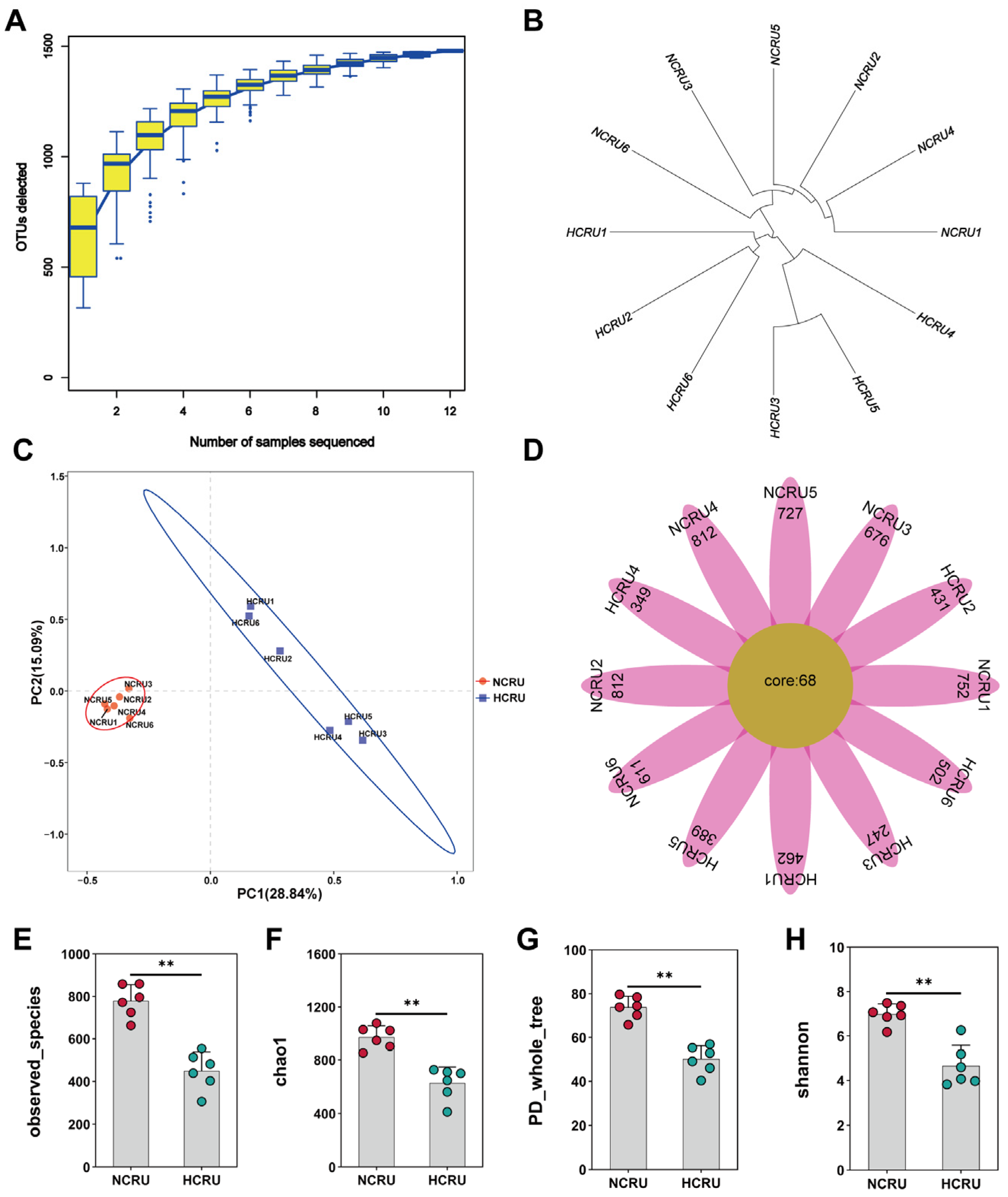
3.3. Rumen Microflora of Growing Goats Fed Different Diets

The increase in species as the sample size increases was described using species accumulation curves (Figure 3A). The findings indicated the frequency at which novel OTUs (novel species) emerge through ongoing sampling. In a specific interval, when the sample size grows, a steep rise in the curve suggests the presence of numerous species in the community. Conversely, a flattening curve indicates that the species in this habitat do not substantially increase as the sample size increases. Our results indicated that sampling is sufficient for microbiological data analysis. Based on the analysis of hierarchical clustering, the branch length indicates the distance between samples, and samples with greater similarity tend to cluster closer together from Figure 3B. Based on the evidence, it can be verified that the rumen samples from goats that were given regular and high-concentrate diets were grouped distinctly. The PCoA revealed distinct separation between rumen samples in the NC group and those in the HC group, as demonstrated in Figure 3C. The flower plot represents a sample with each petal, with the core number in the middle representing the number of OTUs common to all samples, and the number on the petal representing the unique number of OTUs for that sample. it can be proven that the HC diet reduces the number of OTUs in the rumen compared to the NC diet fed to growing goats (Figure 3D). The observed species was markedly lower in the HC group than in the NC group (p < 0.05) (Figure 3E). In the rumen fluid, the Chao 1, PD whole tree, and the Shannon index notably decreased (p < 0.01 for Chao 1 and PD whole tree, p < 0.05 for the Shannon index) when the goats’ diet was changed from 55% concentrate to 90% concentrate (Figure 3F–H). Hence, the HC diet declined the rumen bacteria richness and diversity.

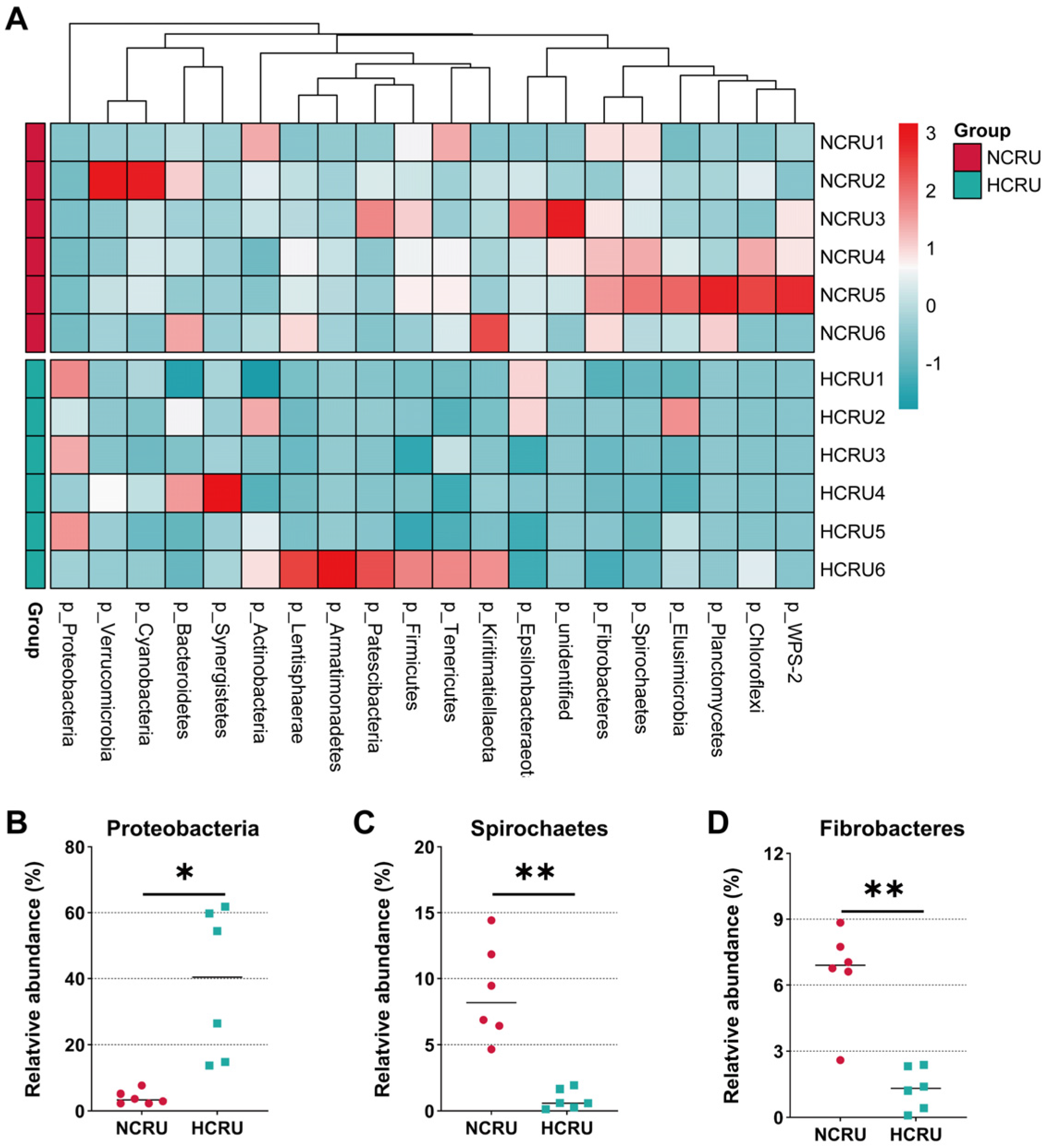
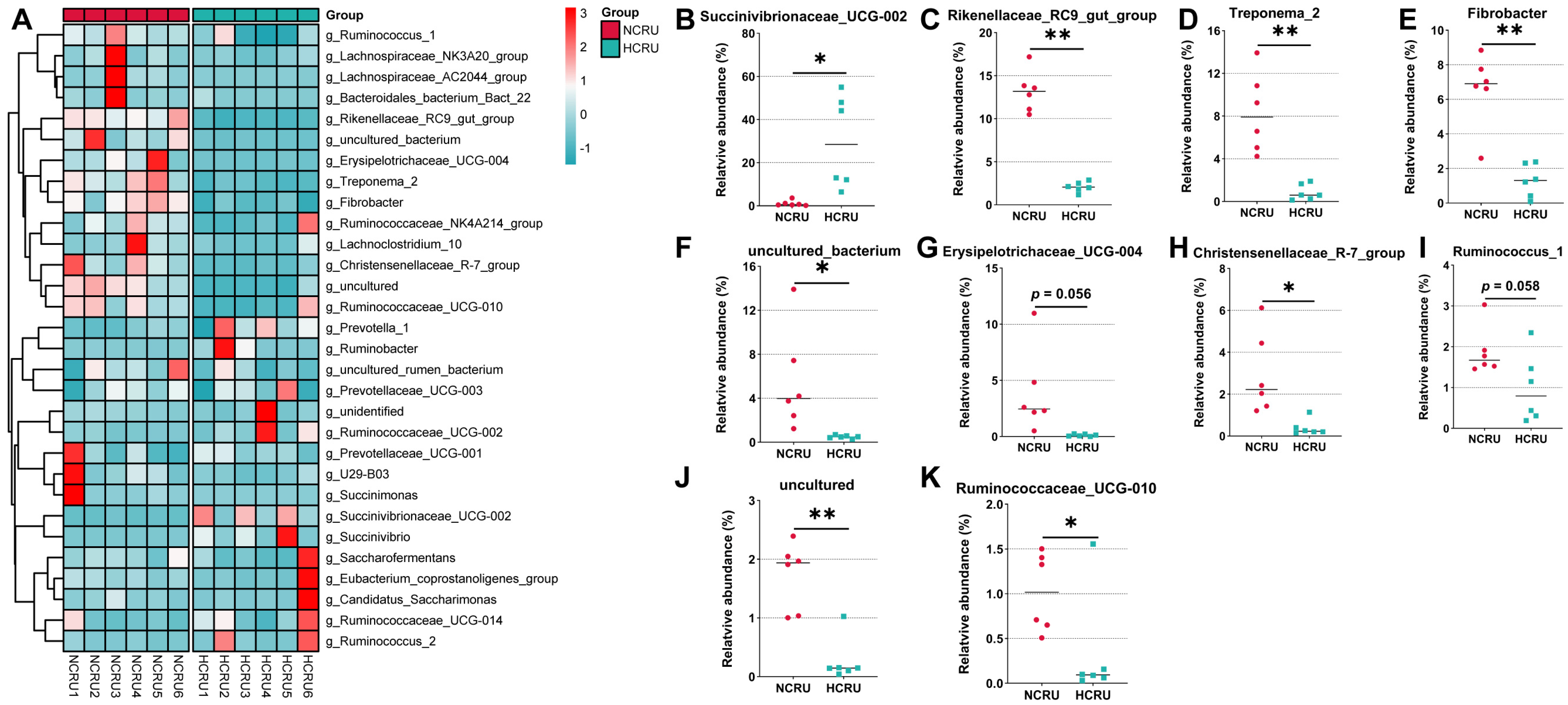
3.4. Rumen Bacterial Community Structure of Growing Goats Fed Different Diets

Bacteroidetes, Proteobacteria and Firmicutes were dominant phyla in the rumen of goats, accounting for more than 87% of the total rumen bacterial community (Figure 4A). Bacteroidetes and Proteobacteria accounted for a relative abundance of 45.9% and 21.3%, respectively, followed by Firmicutes, at 20.7%. When the diet of concentrate proportion increased from 55% to 90%, the abundance of Proteobacteria was increased significantly in the HCRU group than the NCRU group (p < 0.05) (Figure 4B). However, the proportion of Spirochaetes decreased dramatically in goats when the diet administered to goats was switched from 55% concentrate diet to 90% concentrate diet (p < 0.01) (Figure 4C). At the same time, the HC diet significantly dropped the abundance of Fibrobacteres (p < 0.01) (Figure 4D).
Within the bacterial population, the top 30 genera were identified across all samples (Figure 5A)-the genus Succinivibrionaceae_UCG-002 was the most abundant genera, accounting for 15.4%, followed by Prevotella_1, Rikenellaceae_RC9_gut_group, Treponema_2, and Fibrobacter as the predominant genera of rumen in the NCRU and HCRU groups. Among the genera, the percentage abundance of Succinivibrionaceae_UCG-002 was greater in the rumen of the HCRU group when compared to that in the NCRU group (p < 0.05) (Figure 5B), and Erysipelotrichaceae_UCG-004 tended to be lower by the HC diet (p = 0.056) (Figure 5G). In contrast, the relative abundance of Rikenellaceae_RC9_gut_group, Treponema_2 and Fibrobacter was markedly decreased in the rumen of the HCRU group than in the NCRU group (p < 0.01) (Figure 5C–E), and Ruminococcus_1 tended to decrease by the HC diet (p = 0.058) (Figure 5I). The Christensenellaceae_R-7_group and Ruminococcaceae_UCG-010 in rumen was notably decreased when a diet administered to steers was switched from 55% concentrate diet to 90% concentrate diet (p < 0.05) (Figure 5H,K).

3.5. Function Prediction of Rumen Bacterial Community Using PICRUSt

Functional potentials of rumen microbiota were predicted using 16S rRNA marker gene sequences through PICRUSt against KEGG pathways. As showed in Figure 6, within the 20 most abundant level 2 KEGG pathways, ten, amino acid metabolism, carbohydrate metabolism, energy metabolism, the metabolism of cofactors and vitamins, nucleotide metabolism, glycan biosynthesis and metabolism, lipid metabolism, enzyme families, the metabolism of terpenoids and polyketides, and the metabolism of other amino acids, were associated with metabolism; and one, membrane transport, was associated with environmental information processing; four, including replication and repair, translation, folding, sorting and degradation, and transcription were associated with genetic information processing; one, cell motility was associated with cellular processes.
In total, the related amino acid metabolism, carbohydrate metabolism and the metabolism of other amino acids individual pathways were predicted, and a comparison of different pathways (as shown in Table 2) revealed that the HC diet influenced the functional potentials of rumen microbiota. Specifically, compared to the NC group, the HC diet tended to decrease the abundance of histidine metabolism (ko00340) (p = 0.081), and the HC diet significantly decreased the abundance of lysine degradation (ko00310), tryptophan metabolism (ko00380), valine, leucine and isoleucine degradation (ko00280), and selenocompound metabolism (ko00450) (p < 0.05). In contrast, the HC diet significantly increased the abundance of the citrate cycle (TCA cycle) (ko00020) associated with carbohydrate metabolism (p = 0.028). Furthermore, the HC diet significantly increased the glutathione metabolism (ko00480) associated with the metabolism of other amino acids (p = 0.008).

4. Discussion

The diet’s formulation and composition can potentially impact the growth performance and antioxidant capacity of animals, as stated in references [26,27,28]. Zhang et al. showed that SARA elevated systematic oxidative status and enhanced autophagy in the liver, and suppressed SIRT1 and FOXA2 may mediate enhanced oxidative damage and autophagy in the livers of dairy cows fed a HC diet [29]. Cows with SARA often develop complications or other diseases with physiologic immunosuppression and inflammation [30]. Our current study has verified that the HC diet could diminish the antioxidant capacity of goats, underscoring the potential adverse consequences of indiscriminately promoting weight gain in these animals. Dietary patterns can impact not only body weight but also bone density [31]. In addition to antioxidant capacity, anti-inflammatory and immunomodulatory properties are equally vital for the well-being of the host [32]. Cytokines, produced and released by immune cells that are activated, constitute a category of biologically active substances. Immune cells interact and mutually regulate each other, playing crucial parts in inflammation, immune responses, tissue healing, and hematopoietic functions. Promoting the proliferation of immune cells are several noteworthy immune mediators, namely IL-2, IL-4, IL-6, IL-10, and IL-12 [33,34,35]. Feeding goats with a high-concentrate (HC) diet resulted in lower nitric oxide (NO) production compared to the normal group. Typically, increased NO levels are associated with an enhanced pro-inflammatory response to lipopolysaccharides (LPS) [36], which correlates with elevated concentrations of various interleukins. Prior research has demonstrated that elevated IL-6 levels may enhance the permeability of the rumen epithelium and diminish its barrier function [37]. Furthermore, our research validated that the nitric oxide (NO) [38] mediated the control of IL-2 and IL-6 production in these goats through a negative feedback mechanism. These findings suggest that feeding goats with a high-concentrate (HC) diet during a state of subacute rumen acidosis (SARA) results in elevated plasma NO levels, which subsequently suppresses the production of cytokines, notably IL-6.
The activity of animal digestive enzymes [39] can be significantly influenced by the composition of the diet. The digestive enzymes produced by rumen microorganisms in ruminants help to break down and utilize the natural polymers found in their feed [40]. The high-carbohydrate diet resulted in an increase in AMY activity, in line with previous studies suggesting that higher intake of starch leads to elevated AMY activity in the contents of the pancreas and small intestine [41,42]. ALT and AST activity are reliable markers of liver function, released into the bloodstream when the liver is compromised. Typically, serum AST activity remains low under normal conditions but significantly elevates in the presence of cell damage. Increased AST and ALT values are indicative of varying degrees of liver impairment [43]. The HC group displayed elevated AST and ALT values in contrast to the NC group, implying that the HC diet may lead to liver damage in goats. ALP is an enzyme widely distributed in tissues and organs, including the liver, bones, and small intestine. It facilitates the hydrolysis of phosphate into phosphoric acid under alkaline conditions. Various forms of ALP, including small intestine ALP and bone-type ALP, contribute to the regulation of intestinal function, as well as calcium and phosphorus metabolism [44,45]. DiLorenzo and McCarthy, among others, have provided evidence suggesting that AMY may potentially accelerate the rate of ruminal carbohydrate degradation [46,47]. Dietary nutrients have the capacity to impact the morphology of small intestinal tissue and the digestive function of animals [48]. An imbalance in the interplay between gut microbiota and other factors can disrupt the homeostasis of the intestinal mucosa [49]. The intestinal epithelial barrier, acting as the first line of defense between the luminal environment and the host, can result in severe inflammation or other intestinal diseases if compromised [50]. This study additionally furnishes evidence that diets can impact the morphology of the rumen epithelium. Elevated enzyme levels (ALT, AST, and ALP) indicate potential damage or alterations in membrane permeability [51]. As a result, feeding goats an HC diet may have detrimental effects on their intestinal epithelium.
For this research, we employed 16S rRNA sequencing to investigate the bacteria present in rumen fluid. Our focus was on observing the changes in the composition and structure of rumen flora as goats progressed from a state of good health to one of acidosis. Our goal was to explore the reproduction patterns of rumen bacteria during disease and enhance the understanding of rumen microecology. Species accumulation curves are useful instruments for analyzing the species makeup of samples and forecasting species abundance. In biodiversity and community surveys, they are extensively used to assess the adequacy of sample size and estimate the diversity of species [52]. Thus, species accumulation curves not only assess sample size sufficiency but also predict species richness when the sample size is adequate. Utilizing the beta diversity distance matrix for hierarchical clustering analysis [53], the tree structure was constructed using the UPGMA algorithm, which helped with visual analysis. Our research validates the appropriateness of sequencing rumen samples from goats subjected to the NC and HC diets, revealing distinct clustering patterns for the two treatment groups. Rumen environmental imbalances often result in shifts in rumen flora. For example, Kim et al. induced subacute rumen acidosis (SARA) through a high-grain diet, which led to decreased rumen flora diversity and a reduction in the relative abundance of Prevotella [54]. Our study reveals that goats fed a HC diet display lower bacterial richness and diversity compared to those fed the NC diet, as evident from reductions in observed species, Shannon, Chao 1, and PD whole tree indices. This observation highlights the notably detrimental effect of the HC diet on the biodiversity of the goat rumen ecosystem.
Modifying dietary formulations can lead to shifts in the microbial composition of the animal gut and impact digestive function [55,56]. Zhao et al. indicated the importance of bacterial sphingolipids in maintaining hindgut symbiosis and homeostasis. Dietary supplementation with citrus flavonoid extract can decrease systemic inflammation by maintaining hindgut microbiota homeostasis and regulating sphingolipid metabolism in dairy cows fed a high-starch diet [57]. Environmental acid stress has a substantial impact on the growth and reproduction of microorganisms during microbial fermentation [58,59]. Previous studies have reported that Bacteroides is a major anaerobic genus in the rumen [60], and high-concentrate diets have been associated with a decrease in Bacteroides populations [61,62]. Prevotella, a member of the Bacteroides genus, is the predominant genus of rumen bacteria [63]. Prevotella is known for its high activity in protein degradation and its ability to utilize starch and pectin [60]. Surprisingly, our findings revealed no notable alteration in the prevalence of Prevotella in the rumen prior to and following SARA, which contradicts earlier research. Considering our experimental design, it is hypothesized that Prevotella may rapidly multiply when provided with ample starch substrates in a short timeframe, maintaining a stable relative abundance. The health and function of the rumen greatly depend on the diversity of microorganisms. During our investigation, we noticed a decrease in the ratios of the Rikenellaceae RC9 intestinal cluster, Treponema 2, Fibrobacter, and Christensenellaceae R-7 cluster when exposed to high-calorie feeding circumstances. It is worth mentioning that Treponema and Fibrobacter are acknowledged for their role in breaking down fibers [64,65]. Numerous studies on SARA have extensively documented the abundance of bacteria that break down starch and the decrease in bacteria that break down fiber [66,67]. Our current research aligns with these findings, confirming disruptions in rumen digestion and metabolic capabilities in the HC-fed goats. Succinivibrionaceae UCG-002 is a member of the Succinivibrionaceae family and has been associated with ruminal fatty acid metabolism [68]. As for the Rikenellaceae RC9 gut cluster, the Rikenellaceae family is a fairly recent taxonomic categorization, and there is a significant requirement for additional information regarding its metabolic role, despite the research conducted by Su et al.suggested a single isolate that produces acetate from the Rikenellaceae family [69]. Rikenellaceae RC9 was able to alter the digestibility of dietary NDF and ADF as well as the VFA concentration, as shown by Zhao et al. [70]. The Christensenellaceae R-7 cluster is most closely associated with Christensenella minuta [71], a species capable of generating acetate and butyrate through glucose [72]. In summary, these findings indicated that the HC goats may have impaired rumen digestion and metabolic capabilities.
Rumen bacteria play a crucial role in maintaining ruminant health [73,74,75]. In our study, we used PICRUSt to assess the potential functions of bacteria associated with digestion in the rumen. The clear disparities in anticipated functional pathways between the NC and HC groups suggest that alterations in bacterial populations can influence the functional potential of the rumen bacterial community. Within the HC group, we noticed a decline in pathways associated with the metabolism of amino acids, such as histidine, lysine, tryptophan, valine, leucine, and isoleucine breakdown. This indicates possible detrimental impacts on the metabolism of amino acids in the host resulting from prolonged consumption of an HC diet. Additionally, we noted a relative increase in pathways associated with the tricarboxylic acid (TCA) cycle and carbohydrate metabolism in the HC group, suggesting that the rumen microbiomes of HC-fed goats may have an enhanced capacity for energy extraction. Overall, these predicted findings suggest that long-term HC diet feeding may impact the bacterial function of the rumen microbiome in goats. However, it is important to note that while PICRUSt is a well-validated predictive tool, true metagenomic shotgun sequencing is necessary for a more comprehensive examination of the effects of HC diets on rumen-associated bacteria in goats.

5. Conclusions

In summary, SARA could alter the total antioxidant capacity and inflammatory factors levels of goats, and affect enzyme metabolism in their blood and rumen. Through our research, we have gained a deeper comprehension of the microorganisms present in the rumen, the presence of SARA reduced the number and variety of bacteria in the rumen and may affect the development and performance of bacteria in the rumen.

Author Contributions

Conceptualization, L.Y.; methodology, S.F.; formal analysis, S.F.; data curation, M.Z., A.R., H.M. and D.L.; writing—original draft preparation, S.F.; writing—review and editing, L.Y.; visualization, S.F.; supervision, L.Y.; project administration, L.Y. All authors have read and agreed to the published version of the manuscript.

Funding

This research project was funded by the Hunan Provincial Innovation Foundation for Postgraduate (Grant No. CX2017B368).

Institutional Review Board Statement

This study was conducted according to the guidelines of the Declaration of Helsinki, and approved by the Institutional Review Board of the Animal Care and Use Committee of Hunan Agricultural University.

Informed Consent Statement

Not applicable for studies not involving humans.

Data Availability Statement

The data presented in this study are available on request from the corresponding author. The availability of the data is restricted to investigators based in academic institutions.

Conflicts of Interest

The authors declare no conflict of interest.

References

  1. Gozho, G.N.; Plaizier, J.C.; Krause, D.O.; Kennedy, A.D.; Wittenberg, K.M. Subacute ruminal acidosis induces ruminal lipopolysaccharide endotoxin release and triggers an inflammatory response. J. Dairy Sci. 2005, 88, 1399–1403. [Google Scholar] [CrossRef] [PubMed]
  2. Gao, X.; Oba, M. Characteristics of dairy cows with a greater or lower risk of subacute ruminal acidosis: Volatile fatty acid absorption, rumen digestion, and expression of genes in rumen epithelial cells. J. Dairy Sci. 2016, 99, 8733–8745. [Google Scholar] [CrossRef] [PubMed]
  3. Abdela, N. Sub-acute Ruminal Acidosis (SARA) and its Consequence in Dairy Cattle: A Review of Past and Recent Research at Global Prospective. Achiev. Life Sci. 2016, 10, 187–196. [Google Scholar] [CrossRef]
  4. Kleen, J.L.; Hooijer, G.A.; Rehage, J.; Noordhuizen, J.P. Subacute ruminal acidosis (SARA): A review. J. Vet. Med. A Physiol. Pathol. Clin. Med. 2003, 50, 406–414. [Google Scholar] [CrossRef] [PubMed]
  5. Bearson, S.; Bearson, B.; Foster, J.W. Acid stress responses in enterobacteria. FEMS Microbiol. Lett. 1997, 147, 173–180. [Google Scholar] [CrossRef]
  6. Guan, N.; Liu, L. Microbial response to acid stress: Mechanisms and applications. Appl. Microbiol. Biotechnol. 2020, 104, 51–65. [Google Scholar] [CrossRef]
  7. Monteiro, H.F.; Faciola, A.P. Ruminal acidosis, bacterial changes, and lipopolysaccharides. J. Anim. Sci. 2020, 98, skaa248. [Google Scholar] [CrossRef]
  8. Guan, N.Z.; Li, J.H.; Shin, H.D.; Du, G.C.; Chen, J.; Liu, L. Microbial response to environmental stresses: From fundamental mechanisms to practical applications. Appl. Microbiol. Biotechnol. 2017, 101, 3991–4008. [Google Scholar] [CrossRef]
  9. Mertens, D.R. Creating a system for meeting the fiber requirements of dairy cows. J. Dairy Sci. 1997, 80, 1463–1481. [Google Scholar] [CrossRef]
  10. Li, C.; Beauchemin, K.A.; Yang, W. Feeding diets varying in forage proportion and particle length to lactating dairy cows: I. Effects on ruminal pH and fermentation, microbial protein synthesis, digestibility, and milk production. J. Dairy Sci. 2020, 103, 4340–4354. [Google Scholar] [CrossRef]
  11. Yang, W.Z.; Beauchemin, K.A. Increasing physically effective fiber content of dairy cow diets through forage proportion versus forage chop length: Chewing and ruminal pH. J. Dairy Sci. 2009, 92, 1603–1615. [Google Scholar] [CrossRef] [PubMed]
  12. He, B.; Zhu, R.; Yang, H.; Lu, Q.; Wang, W.; Song, L.; Sun, X.; Zhang, G.; Li, S.; Yang, J.; et al. Assessing the Impact of Data Preprocessing on Analyzing Next Generation Sequencing Data. Front. Bioeng. Biotechnol. 2020, 8, 817. [Google Scholar] [CrossRef]
  13. Wang, K.; Ma, J.; Li, Y.; Han, Q.; Yin, Z.; Zhou, M.; Luo, M.; Chen, J.; Xia, S. Effects of essential oil extracted from Artemisia argyi leaf on lipid metabolism and gut microbiota in high-fat diet-fed mice. Front. Nutr. 2022, 9, 1024722. [Google Scholar] [CrossRef] [PubMed]
  14. Liu, S.; Wang, K.J.; Lin, S.Z.; Zhang, Z.D.; Cheng, M.; Hu, S.S.; Hu, H.J.; Xiang, J.; Chen, F.M.; Li, G.H.; et al. Comparison of the Effects between Tannins Extracted from Different Natural Plants on Growth Performance, Antioxidant Capacity, Immunity, and Intestinal Flora of Broiler Chickens. Antioxidants 2023, 12, 441. [Google Scholar] [CrossRef] [PubMed]
  15. Chen, F.M.; Wang, Y.S.; Wang, K.J.; Chen, J.Y.; Jin, K.; Peng, K.Q.; Chen, X.; Liu, Z.M.; Ouyang, J.; Wang, Y.; et al. Effects of Litsea cubeba essential oil on growth performance, blood antioxidation, immune function, apparent digestibility of nutrients, and fecal microflora of pigs. Front. Pharmacol. 2023, 14, 1166022. [Google Scholar] [CrossRef]
  16. Zhang, R.Y.; Ye, H.M.; Liu, J.H.; Mao, S.Y. High-grain diets altered rumen fermentation and epithelial bacterial community and resulted in rumen epithelial injuries of goats. Appl. Microbiol. Biotechnol. 2017, 101, 6981–6992. [Google Scholar] [CrossRef]
  17. Wang, K.; Ren, A.; Zheng, M.; Jiao, J.; Yan, Q.; Zhou, C.; Tan, Z. Diet with a High Proportion of Rice Alters Profiles and Potential Function of Digesta-Associated Microbiota in the Ileum of Goats. Animals 2020, 10, 1261. [Google Scholar] [CrossRef]
  18. Wang, K.J.; Yan, Q.X.; Ren, A.; Zheng, M.L.; Zhang, P.H.; Tan, Z.L.; Zhou, C.S. Novel Linkages between Bacterial Composition of Hindgut and Host Metabolic Responses to SARA Induced by High-Paddy Diet in Young Goats. Front. Vet. Sci. 2022, 8, 791482. [Google Scholar] [CrossRef]
  19. Wu, F.; Guo, X.; Zhang, J.; Zhang, M.; Ou, Z.; Peng, Y. Phascolarctobacterium faecium abundant colonization in human gastrointestinal tract. Exp. Ther. Med. 2017, 14, 3122–3126. [Google Scholar] [CrossRef]
  20. Wang, H.; Hu, C.; Cheng, C.; Cui, J.; Ji, Y.; Hao, X.; Li, Q.; Ren, W.; Deng, B.; Yin, Y.; et al. Unraveling the association of fecal microbiota and oxidative stress with stillbirth rate of sows. Theriogenology 2019, 136, 131–137. [Google Scholar] [CrossRef]
  21. Yin, L.; Li, J.; Wang, H.; Yi, Z.; Wang, L.; Zhang, S.; Li, X.; Wang, Q.; Li, J.; Yang, H.; et al. Effects of vitamin B6 on the growth performance, intestinal morphology, and gene expression in weaned piglets that are fed a low-protein diet. J. Anim. Sci. 2020, 98, skaa022. [Google Scholar] [CrossRef] [PubMed]
  22. Wang, K.J.; Zheng, M.L.; Ren, A.; Zhou, C.S.; Yan, Q.X.; Tan, Z.L.; Zhang, P.; Yi, K. Effects of high rice diet on growth performance, nutrients apparent digestibility, nitrogen metabolism, blood parameters and rumen fermentation in growing goats. Kafkas Univ. Vet. Fak. Derg. 2019, 25, 749–755. [Google Scholar] [CrossRef]
  23. Wang, K.J.; Peng, X.M.; Yang, A.Q.; Huang, Y.Q.; Tan, Y.X.; Qian, Y.J.; Lv, F.F.; Si, H.B. Effects of Diets with Different Protein Levels on Lipid Metabolism and Gut Microbes in the Host of Different Genders. Front. Nutr. 2022, 9, 940217. [Google Scholar] [CrossRef] [PubMed]
  24. Wang, K.J.; Peng, X.M.; Lv, F.F.; Zheng, M.L.; Long, D.L.; Mao, H.X.; Si, H.B.; Zhang, P.H. Microbiome-Metabolites Analysis Reveals Unhealthy Alterations in the Gut Microbiota but Improved Meat Quality with a High-Rice Diet Challenge in a Small Ruminant Model. Animals 2021, 11, 2306. [Google Scholar] [CrossRef]
  25. Langille, M.G.; Zaneveld, J.; Caporaso, J.G.; McDonald, D.; Knights, D.; Reyes, J.A.; Clemente, J.C.; Burkepile, D.E.; Vega Thurber, R.L.; Knight, R.; et al. Predictive functional profiling of microbial communities using 16S rRNA marker gene sequences. Nat. Biotechnol. 2013, 31, 814–821. [Google Scholar] [CrossRef] [PubMed]
  26. Chen, F.M.; He, J.Y.; Wang, X.; Lv, T.; Liu, C.J.; Liao, L.P.; Li, Z.B.; Zhou, J.; He, B.S.; Qiu, H.J.; et al. Effect of Dietary Ramie Powder at Various Levels on the Growth Performance, Meat Quality, Serum Biochemical Indices and Antioxidative Capacity of Yanling White Geese. Animals 2022, 12, 2045. [Google Scholar] [CrossRef]
  27. Ma, W.J.; Wei, S.S.; Peng, W.J.; Sun, T.L.; Huang, J.H.; Yu, R.; Zhang, B.K.; Li, W.Q. Antioxidant Effect of Polygonatum sibiricum Polysaccharides in D-Galactose-Induced Heart Aging Mice. Biomed. Res. Int. 2021, 2021, 6688855. [Google Scholar] [CrossRef]
  28. Chen, J.Y.; Chen, F.M.; Peng, S.M.; Ou, Y.J.; He, B.S.; Li, Y.H.; Lin, Q. Effects of Artemisia argyi Powder on Egg Quality, Antioxidant Capacity, and Intestinal Development of Roman Laying Hens. Front. Physiol. 2022, 13, 902568. [Google Scholar] [CrossRef]
  29. Zhang, H.; Xue, Y.; Xie, W.; Wang, Y.; Ma, N.; Chang, G.; Shen, X. Subacute ruminal acidosis downregulates FOXA2, changes oxidative status, and induces autophagy in the livers of dairy cows fed a high-concentrate diet. J. Dairy Sci. 2023, 106, 2007–2018. [Google Scholar] [CrossRef]
  30. Sato, S. Subacute ruminal acidosis (SARA) challenge, ruminal condition and cellular immunity in cattle. Jpn. J. Vet. Res. 2015, 63, S25–S36. [Google Scholar]
  31. Chen, Y.; Xiang, J.; Wang, Z.; Xiao, Y.; Zhang, D.; Chen, X.; Li, H.; Liu, M.; Zhang, Q. Associations of Bone Mineral Density with Lean Mass, Fat Mass, and Dietary Patterns in Postmenopausal Chinese Women: A 2-Year Prospective Study. PLoS ONE 2015, 10, e0137097. [Google Scholar] [CrossRef] [PubMed]
  32. Li, J.; Yin, L.; Wang, L.; Li, J.; Huang, P.; Yang, H.; Yin, Y. Effects of vitamin B6 on growth, diarrhea rate, intestinal morphology, function, and inflammatory factors expression in a high-protein diet fed to weaned piglets. J. Anim. Sci. 2019, 97, 4865–4874. [Google Scholar] [CrossRef]
  33. Zhu, J.F.; Paul, W.E. Peripheral CD4+ T-cell differentiation regulated by networks of cytokines and transcription factors. Immunol. Rev. 2010, 238, 247–262. [Google Scholar] [CrossRef] [PubMed]
  34. Sun, X.; Gao, R.L.; Xiong, Y.K.; Huang, Q.C.; Xu, M. Antitumor and immunomodulatory effects of a water-soluble polysaccharide from Lilii Bulbus in mice. Carbohydr. Polym. 2014, 102, 543–549. [Google Scholar] [CrossRef] [PubMed]
  35. Alexander, A.F.; Kelsey, I.; Forbes, H.; Miller-Jensen, K. Single-cell secretion analysis reveals a dual role for IL-10 in restraining and resolving the TLR4-induced inflammatory response. Cell Rep. 2021, 36, 109728. [Google Scholar] [CrossRef]
  36. He, Z.; Tan, Z.; Sun, Z.; Beauchemin, K.A.; Tang, S.; Zhou, C.; Han, X.; Wang, M.; Wu, D. Unchanged interleukin 6 level of protein and energy restricted goats during late gestation: The role of elevated blood nitric oxide. J. Endocrinol. 2012, 213, 59–65. [Google Scholar] [CrossRef] [PubMed]
  37. Trabi, E.B.; Seddik, H.E.; Xie, F.; Wang, X.F.; Liu, J.H.; Mao, S.Y. Effect of pelleted high-grain total mixed ration on rumen morphology, epithelium-associated microbiota and gene expression of proinflammatory cytokines and tight junction proteins in Hu sheep. Anim. Feed Sci. Technol. 2020, 263, 114453. [Google Scholar] [CrossRef]
  38. Connelly, L.; Palacios-Callender, M.; Ameixa, C.; Moncada, S.; Hobbs, A.J. Biphasic regulation of NF-kappa B activity underlies the pro- and anti-inflammatory actions of nitric oxide. J. Immunol. 2001, 166, 3873–3881. [Google Scholar] [CrossRef]
  39. Deng, Q.; Shao, Y.; Wang, Q.; Li, J.; Li, Y.; Ding, X.; Huang, P.; Yin, J.; Yang, H.; Yin, Y. Effects and interaction of dietary electrolyte balance and citric acid on growth performance, intestinal histomorphology, digestive enzyme activity and nutrient transporters expression of weaned piglets. J. Anim. Physiol. Anim. Nutr. 2021, 105, 272–285. [Google Scholar] [CrossRef]
  40. Plaizier, J.C.; Khafipour, E.; Li, S.; Gozho, G.N.; Krause, D.O. Subacute ruminal acidosis (SARA), endotoxins and health consequences. Anim. Feed Sci. Technol. 2012, 172, 9–21. [Google Scholar] [CrossRef]
  41. Wang, X.B.; Ogawa, T.; Suda, S.; Taniguchi, K.; Uike, H.; Kumagai, H.; Mitani, K. Effects of nutritional level on digestive enzyme activities in the pancreas and small intestine of calves slaughtered at same body weight. Asian-Australas J. Anim. Sci. 1998, 11, 375–380. [Google Scholar] [CrossRef]
  42. Owsley, W.F.; Orr, D.E.; Tribble, L.F. Effects of age and diet on the development of the pancreas and the synthesis and secretion of pancreatic enzymes in the young pig. J. Anim. Sci. 1986, 63, 497–504. [Google Scholar] [CrossRef] [PubMed]
  43. Guo, W.Z.; Ke, Y.T.; Shang, Y.Z.; Wang, J.H.; Wen, Y.Q.; Wei, J.T. Effects of different crude protein levels of formulated pellet diets on growth performance and blood biochemical parameters of Matou goats. Chin. J. Anim. Sci. 2019, 55, 108–110, 125. [Google Scholar]
  44. Wang, Z.W.; Mou, X.L.; Yang, G.W.; Li, J.K.; Liu, D.S. Effects of dietary nutritional levels on calcium, phosphorus concentrations and ALK activity in serum and calcium, phosphorus contents in tibia for geese. China Poult. 2009, 31, 16–20. [Google Scholar]
  45. Zhang, C.L.; Meng, H.J.; Zhu, S.X.; Wu, L.; Liu, Z. Effect of alkaline phosphatase on intestinal health of weaning piglets. Swine Prod. 2017, 6, 13–16. [Google Scholar]
  46. DiLorenzo, N.; Smith, D.R.; Quinn, M.J.; May, M.L.; Ponce, C.H.; Steinberg, W.; Engstrom, M.A.; Galyean, M.L. Effects of grain processing and supplementation with exogenous amylase on nutrient digestibility in feedlot diets. Livest. Sci. 2011, 137, 178–184. [Google Scholar] [CrossRef]
  47. McCarthy, M.M.; Engstrom, M.A.; Azem, E.; Gressley, T.F. The effect of an exogenous amylase on performance and total-tract digestibility in lactating dairy cows fed a high-byproduct diet. J. Dairy Sci. 2013, 96, 3075–3084. [Google Scholar] [CrossRef]
  48. Chen, C.; Wang, Z.; Li, J.; Li, Y.; Huang, P.; Ding, X.; Yin, J.; He, S.; Yang, H.; Yin, Y. Dietary vitamin E affects small intestinal histomorphology, digestive enzyme activity, and the expression of nutrient transporters by inhibiting proliferation of intestinal epithelial cells within jejunum in weaned piglets. J. Anim. Sci. 2019, 97, 1212–1221. [Google Scholar] [CrossRef]
  49. Luo, W.W.; Tian, L.; Tan, B.; Shen, Z.H.; Xiao, M.W.; Wu, S.; Meng, X.R.; Wu, X.; Wang, X.Y. Update: Innate Lymphoid Cells in Inflammatory Bowel Disease. Dig. Dis. Sci. 2022, 67, 56–66. [Google Scholar] [CrossRef]
  50. Wu, J.; He, C.; Bu, J.; Luo, Y.; Yang, S.; Ye, C.; Yu, S.; He, B.; Yin, Y.; Yang, X. Betaine attenuates LPS-induced downregulation of Occludin and Claudin-1 and restores intestinal barrier function. BMC Vet. Res. 2020, 16, 75. [Google Scholar] [CrossRef]
  51. Rashid, S.; Irshadullah, M. Evaluation of antioxidant and oxidant status of goats (Capra aegagrus hircus) naturally infected with Haemonchus contortus. J. Helminthol. 2020, 94, e36. [Google Scholar] [CrossRef] [PubMed]
  52. Maughan, H.; Wang, P.W.; Diaz Caballero, J.; Fung, P.; Gong, Y.; Donaldson, S.L.; Yuan, L.; Keshavjee, S.; Zhang, Y.; Yau, Y.C.; et al. Analysis of the cystic fibrosis lung microbiota via serial Illumina sequencing of bacterial 16S rRNA hypervariable regions. PLoS ONE 2012, 7, e45791. [Google Scholar] [CrossRef] [PubMed]
  53. Jiang, X.T.; Peng, X.; Deng, G.H.; Sheng, H.F.; Wang, Y.; Zhou, H.W.; Tam, N.F. Illumina sequencing of 16S rRNA tag revealed spatial variations of bacterial communities in a mangrove wetland. Microb. Ecol. 2013, 66, 96–104. [Google Scholar] [CrossRef] [PubMed]
  54. Kim, Y.H.; Nagata, R.; Ohkubo, A.; Ohtani, N.; Kushibiki, S.; Ichijo, T.; Sato, S. Changes in ruminal and reticular pH and bacterial communities in Holstein cattle fed a high-grain diet. BMC Vet. Res. 2018, 14, 310. [Google Scholar] [CrossRef] [PubMed]
  55. Ji, F.J.; Wang, L.X.; Yang, H.S.; Hu, A.; Yin, Y.L. Review: The roles and functions of glutamine on intestinal health and performance of weaning pigs. Animal 2019, 13, 2727–2735. [Google Scholar] [CrossRef]
  56. Wang, K.; Zhou, M.; Gong, X.; Zhou, Y.; Chen, J.; Ma, J.; Zhang, P. Starch–protein interaction effects on lipid metabolism and gut microbes in host. Front. Nutr. 2022, 9, 1018026. [Google Scholar] [CrossRef]
  57. Zhao, Y.; Yu, S.; Zhao, H.; Li, L.; Li, Y.; Liu, M.; Jiang, L. Integrated multi-omics analysis reveals the positive leverage of citrus flavonoids on hindgut microbiota and host homeostasis by modulating sphingolipid metabolism in mid-lactation dairy cows consuming a high-starch diet. Microbiome 2023, 11, 236. [Google Scholar] [CrossRef]
  58. Akkermans, S.; Logist, F.; Van Impe, J.F. An interaction model for the combined effect of temperature, pH and water activity on the growth rate of E. coli K12. Food Res. Int. 2018, 106, 1123–1131. [Google Scholar] [CrossRef]
  59. Ren, X.D.; Chen, X.S.; Tang, L.; Zeng, X.; Wang, L.; Mao, Z.G. Physiological mechanism of the overproduction of epsilon-poly-L-lysine by acidic pH shock in fed-batch fermentation. Bioprocess Biosyst. Eng. 2015, 38, 2085–2094. [Google Scholar] [CrossRef]
  60. Li, Y.Q.; Xi, Y.M.; Zeng, H.F.; Zhang, L.; Chen, M.J.; Han, Z.Y. Diversity Changes of Bacterial Community in Rumen of Dairy Cows in Vitro Fermentation Analyzed by 16S rRNA Sequencing Technology. Chin. J. Anim. Nutr. 2018, 30, 4059–4070. [Google Scholar] [CrossRef]
  61. Nagaraja, T.G.; Titgemeyer, E.C. Ruminal acidosis in beef cattle: The current microbiological and nutritional outlook. J. Dairy Sci. 2007, 90 (Suppl. 1), E17–E38. [Google Scholar] [CrossRef]
  62. Chiquette, J.; Allison, M.J.; Rasmussen, M.A. Prevotella bryantii 25A used as a probiotic in early-lactation dairy cows: Effect on ruminal fermentation characteristics, milk production, and milk composition. J. Dairy Sci. 2008, 91, 3536–3543. [Google Scholar] [CrossRef] [PubMed]
  63. Bekele, A.Z.; Koike, S.; Kobayashi, Y. Genetic diversity and diet specificity of ruminal Prevotella revealed by 16S rRNA gene-based analysis. FEMS Microbiol. Lett. 2010, 305, 49–57. [Google Scholar] [CrossRef]
  64. Bekele, A.Z.; Koike, S.; Kobayashi, Y. Phylogenetic diversity and dietary association of rumen Treponema revealed using group-specific 16S rRNA gene-based analysis. FEMS Microbiol. Lett. 2011, 316, 51–60. [Google Scholar] [CrossRef] [PubMed]
  65. Ransom-Jones, E.; Jones, D.L.; McCarthy, A.J.; McDonald, J.E. The Fibrobacteres: An Important Phylum of Cellulose-Degrading Bacteria. Microb. Ecol. 2012, 63, 267–281. [Google Scholar] [CrossRef] [PubMed]
  66. Plaizier, J.C.; Li, S.C.; Danscher, A.M.; Derakshani, H.; Andersen, P.H.; Khafipour, E. Changes in Microbiota in Rumen Digesta and Feces Due to a Grain-Based Subacute Ruminal Acidosis (SARA) Challenge. Microb. Ecol. 2017, 74, 485–495. [Google Scholar] [CrossRef] [PubMed]
  67. Mickdam, E.; Khiaosa-ard, R.; Metzler-Zebeli, B.U.; Klevenhusen, F.; Chizzola, R.; Zebeli, Q. Rumen microbial abundance and fermentation profile during severe subacute ruminal acidosis and its modulation by plant derived alkaloids in vitro. Anaerobe 2016, 39, 4–13. [Google Scholar] [CrossRef]
  68. Wang, X.; Martin, G.B.; Wen, Q.; Liu, S.; Li, Y.; Shi, B.; Guo, X.; Zhao, Y.; Guo, Y.; Yan, S. Palm oil protects alpha-linolenic acid from rumen biohydrogenation and muscle oxidation in cashmere goat kids. J. Anim. Sci. Biotechnol. 2020, 11, 100. [Google Scholar] [CrossRef]
  69. Su, X.L.; Tian, Q.; Zhang, J.; Yuan, X.Z.; Shi, X.S.; Guo, R.B.; Qiu, Y.L. Acetobacteroides hydrogenigenes gen. nov., sp. nov., an anaerobic hydrogen-producing bacterium in the family Rikenellaceae isolated from a reed swamp. Int. J. Syst. Evol. Microbiol. 2014, 64, 2986–2991. [Google Scholar] [CrossRef]
  70. Zhao, Y.; Xie, B.; Gao, J.; Zhao, G. Dietary supplementation with sodium sulfate improves rumen fermentation, fiber digestibility, and the plasma metabolome through modulation of rumen bacterial communities in steers. Appl. Environ. Microbiol. 2020, 86, e01412-20. [Google Scholar] [CrossRef]
  71. Seshadri, R.; Leahy, S.C.; Attwood, G.T.; Teh, K.H.; Lambie, S.C.; Cookson, A.L.; Eloe-Fadrosh, E.A.; Pavlopoulos, G.A.; Hadjithomas, M.; Varghese, N.J.; et al. Cultivation and sequencing of rumen microbiome members from the Hungate1000 Collection. Nat. Biotechnol. 2018, 36, 359–367. [Google Scholar] [CrossRef] [PubMed]
  72. Holman, D.B.; Gzyl, K.E. A meta-analysis of the bovine gastrointestinal tract microbiota. FEMS Microbiol. Ecol. 2019, 95, fiz072. [Google Scholar] [CrossRef] [PubMed]
  73. Chen, X.; Su, X.; Li, J.; Yang, Y.; Wang, P.; Yan, F.; Yao, J.; Wu, S. Real-time monitoring of ruminal microbiota reveals their roles in dairy goats during subacute ruminal acidosis. NPJ Biofilms Microbiomes 2021, 7, 45. [Google Scholar] [CrossRef] [PubMed]
  74. Fu, Y.; He, Y.; Xiang, K.; Zhao, C.; He, Z.; Qiu, M.; Hu, X.; Zhang, N. The role of rumen microbiota and its metabolites in subacute ruminal acidosis (SARA)-induced inflammatory diseases of ruminants. Microorganisms 2022, 10, 1495. [Google Scholar] [CrossRef]
  75. Plaizier, J.C.; Mulligan, F.J.; Neville, E.W.; Guan, L.L.; Steele, M.A.; Penner, G.B. Invited review: Effect of subacute ruminal acidosis on gut health of dairy cows. J. Dairy Sci. 2022, 105, 7141–7160. [Google Scholar] [CrossRef]
Figure 1. The antioxidant and immune indicators of growing goats fed normal-concentrate diet (NC) and high-concentrate diet (HC). (A) T-AOC, total antioxidant capacity; (B) NO, Nitric Oxide; (C) IFN-γ, interferon-γ; (D) HIS, Histamine; (E) IL-1β, interleukin-1β; (F) IL-2, interleukin-2; (G) IL-4, interleukin-4; (H) IL-6, interleukin-6. (*) 0.01 < p < 0.05; (**) p < 0.01.
Animals 14 00263 g001
Figure 2. The enzyme metabolism indicators of growing goats fed normal-concentrate diet (NC) and high-concentrate diet (HC). (AD) The enzyme metabolism indicators in plasma. (EH) The enzyme metabolism indicators in rumen. The representative original images (I,J) and light micrographs (K,L) of ruminal epithelium in goats fed the NC and HC diets are shown. (*) 0.01 < p < 0.05; (**) p < 0.01.
Animals 14 00263 g002
Figure 3. Rumen microflora of growing goats fed normal-concentrate diet (NC) and high-concentrate diet (HC). (A) Species accumulation curves. (B) Hierarchical clustering (NCRU1–NCRU6 and HCRU1–HCRU6 are rumen samples of goats fed with 55% or 90% concentrate, separately). (C) Principal component analysis (PCA) of rumen bacterial community. (D) Flower plot. (EH) Alpha diversity indices of rumen bacterial community. (**) p < 0.01.
Animals 14 00263 g003
Figure 4. (A) Heatmap based on hierarchical clustering solution at phylum level (Bray-Curtis distance metric and complete clustering method) in rumen for goats fed a normal diet (NCRU) or a high-concentrate diet (HCRU), shown across columns. (BD) Differential bacteria in the rumen at the phylum level. (*) 0.01 < p < 0.05; (**) p < 0.01.
Animals 14 00263 g004
Figure 5. (A) Heatmap based on hierarchical clustering solution at genus level (Bray-Curtis distance metric and complete clustering method) in rumen for goats fed a normal diet (NCRU) or a high-concentrate diet (HCRU), shown across rows. (BK) Differential bacteria in the rumen at the genus level. (*) 0.01 < p < 0.05; (**) p < 0.01.
Animals 14 00263 g005
Figure 6. Top 20 predicted metagenomic functions at level 2 of the KEGG (Kyoto Encyclopedia of Genes and Genomes) pathways in rumen bacteria of goats fed the NC and HC diets.
Animals 14 00263 g006
Table 1. Ingredients and nutrient levels of the experimental diets (air-dried basis).
ItemNC 1HC 2
Ingredients composition (%)
Forage
Rice straw45.010.0
Concentrate
Rice with shell33.254.3
Soybean meal9.6015.7
Wheat bran6.009.80
Fat powder3.205.20
Calcium carbonate0.500.80
Calcium bicarbonate1.101.80
Sodium chloride0.601.00
Premix 31.001.40
Nutrient levels 4, % of DM
Crude protein13.517.6
Crude ash9.349.12
Crude fat4.186.01
Neutral detergent fiber49.838.4
Acid detergent fiber36.59.51
Starch26.5738.57
1 NC: normal-concentrate diet. 2 HC: high-concentrate diet. 3 Premix composition per kg diet: 68 mg FeSO4·H2O, 44 mg CuSO4·5H2O, 411 µg CoCl2·6H2O, 1.70 mg KIO3, 211 mg MnSO4·H2O, 126 mg ZnSO4·H2O, 56 µg Na2SeO3, 462 mg MgSO4·7H2O, 737 IU vitamin A, 8.29 mg vitamin E, 5.1 g carrier zeolite powder. 4 Nutrient levels were measured values.
Table 2. KEGG pathways that showed different abundances between ruminal digesta microbiota of NC- and HC-diet goats.
Level 2Level 3Pathway IDNC 1HC 2p-Value
Amino acid metabolismHistidine metabolismko003400.69 ± 0.030.65 ± 0.050.081
Lysine degradationko003100.16 ± 0.020.13 ± 0.020.030
Tryptophan metabolismko003800.19 ± 0.010.16 ± 0.020.003
Valine, leucine and isoleucine degradationko002800.29 ± 0.020.25 ± 0.020.006
Carbohydrate metabolismCitrate cycle (TCA cycle)ko000200.73 ± 0.050.80 ± 0.040.028
Metabolism of other amino acidsGlutathione metabolismko004800.17 ± 0.0040.24 ± 0.050.008
Selenocompound metabolismko004500.38 ± 0.010.34 ± 0.020.002
1 NC: normal-concentrate diet; 2 HC: high-concentrate diet.
Disclaimer/Publisher’s Note: The statements, opinions and data contained in all publications are solely those of the individual author(s) and contributor(s) and not of MDPI and/or the editor(s). MDPI and/or the editor(s) disclaim responsibility for any injury to people or property resulting from any ideas, methods, instructions or products referred to in the content.

