Communication

A Health Status Update of Myocastor coypus in Northern Italy

by
Arturo Nicoletti
,
Paola Pregel
,
Laura Starvaggi Cucuzza
,
Enrico Bollo
and
Frine Eleonora Scaglione
*
Department of Veterinary Sciences, University of Turin, 10095 Grugliasco, TO, Italy
*
Author to whom correspondence should be addressed.
Animals 2024, 14(2), 245; https://doi.org/10.3390/ani14020245
Submission received: 3 November 2023 / Revised: 28 December 2023 / Accepted: 9 January 2024 / Published: 12 January 2024
(This article belongs to the Special Issue Wildlife Diseases: Pathology and Diagnostic Investigation)

Abstract

:

Simple Summary

Nutria (Myocastor coypus) is a pest animal present in Africa, Europe, North America and Asia that causes agricultural and ecological damages. Moreover, it has to be considered as a potential risk for public health. A health survey on a population of coypu of Northwest Italy has been carried out to evaluate its zoonotic risks. None of the animals were positive for Hepatitis E virus, Encephalomyocarditis virus, Francisella or Neospora caninum, whereas two animals tested positive for Toxoplasma gondii. Moreover, a high prevalence of histological lesions has been found. Coypu can act as a host for several pathogens, including important agents for human and animal health, and surveillance is necessary to fully understand the biological role and the importance of coypu as a disease reservoir in our country.

Abstract

Myocastor coypus is a pest animal present in Africa, Europe, North America and Asia that causes agricultural and ecological damages. Moreover, it has to be considered as a potential risk for public health. Forty-four coypus from the “Parco Naturale La Mandria” (Piedmont region, Northwest Italy) have been analysed. A complete necropsy and a whole histological evaluation of the liver, kidney and lung have been carried out on all the animals. Moreover, the positivity to Hepatitis E Virus (HEV), Encephalomyocarditis virus (EMCV), Francisella spp., Toxoplasma gondii and Neospora caninum have been investigated. None of the animal were positive for HEV, EMCV, Francisella spp. or Neospora caninum. Two animals tested positive for Toxoplasma gondii. A high presence of histological lesions has been identified in different organs, suggesting that lesions could be induced by different pathogens. As previously reported, coypu can act as a host for several pathogens, including important agents for human and animal health, and surveillance is necessary to fully understand the biological role and the importance of coypu as a disease reservoir in our country.

1. Introduction

The nutria, or coypu (Myocastor coypus), is a semiaquatic herbivorous rodent, originally native of South America, present currently in large feral populations in Africa, Europe, North America and Asia [1,2]. In Europe, the coypu was introduced principally for meat and fur production. In Italy the first introduction for fur farming dates back to 1928 by the National Institute of Rabbit husbandry in Alessandria (Piedmont, Northwest Italy) [3].
Coypu colonization of the natural environment is primarily due to intentional release actions and, minimally, to animals escaped from fur farms. During the following decades, coypu population, density and distribution have dramatically increased and, thanks to its ecological plasticity, even suboptimal habitats were colonized [4]. Due to its negative ecological impact, the coypu is currently considered a pest in the areas of introduction [5]. This animal usually lives next to water courses, in wetlands, riparian zones and coastlands [6]. Thereby, its burrowing behaviour undermines the banks of rivers, canals and dykes, whereas its feeding activity reduces plants’ biodiversity and cover, altering the water’s flowing speed and increasing the erosion of the banks [7].
Moreover, the damage to the habitat can negatively affect the reproduction of fishes, birds and invertebrates [8]. Coypu can also cause agricultural damages by feeding on crops, resulting in a great economic impact [9,10]. Due to the typical aspect of coypu ecology, this species is subjected to population control programs. Nevertheless, the population is still expanding [4,11].
Furthermore, the coypu can be infected by several pathogens and parasites. Some of them can be transmissible to humans and other animals. Several investigations focused on agents that might cause epizootics in wild populations and livestock, as well as in humans [5,12,13].
In this context, this research aims to evaluate the health status of the coypu in Northwest Italy and the presence of pathologies through gross necropsy examinations, histological and biomolecular investigations. The study focuses on viral, bacterial and parasitological pathogens, to investigate potential public health risks linked to coypu diffusion and to improve our knowledge about diseases affecting this species in the analysed area.

2. Materials and Methods

2.1. Animals and Samples Collection

Following the adoption of a regional animal containment programme (according to the D.G.R no. 74–6702 [08/03/2007] and subsequent amendments), 44 coypus from the “Parco Naturale La Mandria” (Piedmont region, Northwest Italy, 45°8′7″ N, 7°37′31″ E) were trapped with baited cage traps or shot. Trapped animals were euthanized with CO2, according to Italian National Bioethics Committee guidelines and to law no. 157 (02/11/1992) and subsequent amendments. The sample included 25 males and 19 females. For each animal, biometric features as weight (kg) and foot length (cm) were collected. All animals were subjected to a standard necropsy procedure.
Samples of liver, kidney, lung and both eyeballs were fixed in 10% phosphate-buffered formalin for histological analysis and age determination.
Based on previous study in the same area [12], samples of liver, lung, heart and central nervous system (CNS) were collected and frozen at −20 °C for virological, bacteriological and parasitological investigations.

2.2. Age Estimation

Due to the impossibility of distinguishing between juveniles, adults and elderly by aspect, morphometric measures or dentition, the age was determined in 33 out of the 44 animals by dry eye-lens weight, according to the protocol proposed by Gosling and colleagues. In detail, the eyes were removed as soon after death as possible and placed in 10% buffered formalin. Eyes were kept in formalin for at least 1 month to allow the lens to harden. The lenses were extracted, and extraneous tissue was removed by rolling on absorbent paper and careful scraping with a blunt seeker. Any damaged lenses were discarded. Pairs of lenses were placed in a small crucible and dried in a forced draught at 80 °C for 22 h. Lenses were weighed on an analytical balance to an accuracy of 0.1 mg. The equation used to determine the age in months is log10 (age + 4.34 months) = 0.511 + 0.013 (mean lens weight) [14]. Based on the study of Pagnoni and Santolini [15], the animals were divided into two groups: juveniles (<8 months) and adults (8–12 months).

2.3. Histological Analysis

The fixed samples were routinely processed and paraffin embedded. Three μm sections were stained with haematoxylin and eosin (HE) and with von Kossa staining, if calcium salts precipitations in the tissue were suspected.

2.4. Virological Analysis

The total RNA was extracted from liver and heart samples using TRIzol® Reagent (Invitrogen™, ThermoFisher Scientific, Waltham, MA, USA), according to the manufacturer’s instructions. RNA extracted from liver was tested for the presence of Hepatitis Virus E (HEV) according to the protocol of Jothikumar [16]. Samples were considered negative for Ct (cycle threshold) values >38, doubtful if Ct was comprised between 36 and 38 and positive for Ct < 36. RNA extracted from heart was retrotranscribed through the High-Capacity cDNA Reverse Transcription Kit (Applied Biosystems™, ThermoFisher Scientific), and the obtained cDNAs were tested for the presence of the encephalomyocarditis virus (EMCV) using the protocol by Vanderhallen [17].

2.5. Bacteriological Analysis

The genomic DNA was extracted from liver samples using the QIAamp DNA Mini Kit (Qiagen, Hilden, Germany) according to the manufacturer’s protocol. The extracted templates were amplified using a primer set specific for Francisella spp. [18]. Specimens collected from 39 lungs were plated onto 5% sheep Blood Agar and Gassner Agar and incubated for 24 h at 37 °C. Bacterial isolates were identified to species level by means of an automated system using Vitek® 2 Compact (bioMérieux, Inc., Durham, NC, USA), a testing system that combines an automated platform with an expansive database of clinically significant organisms.

2.6. Parasitological Analysis

Total genomic DNA was extracted from 25 mg of CNS homogenate, using the commercial kit NucleoSpin® Tissue (Macherey-Nagel, Düren, Germany). The extracted templates were tested for Toxoplasma gondii [19] and Neospora caninum [20]. PCR was performed as previously reported [21].

2.7. Statistical Analysis

Statistical analyses were performed using GraphPad Prism (version 10.0.1, GraphPad Software, La Jolla, CA, USA). Fisher’s exact test was performed to determine non-random associations between sex and the recurrent pathologies detected in lung and kidney. A p value < 0.05 was considered statistically significant.

3. Results

3.1. Age Estimation

Out of 33 coypus considered for age determination, 32 were juveniles and only 1 was an adult (Table 1).

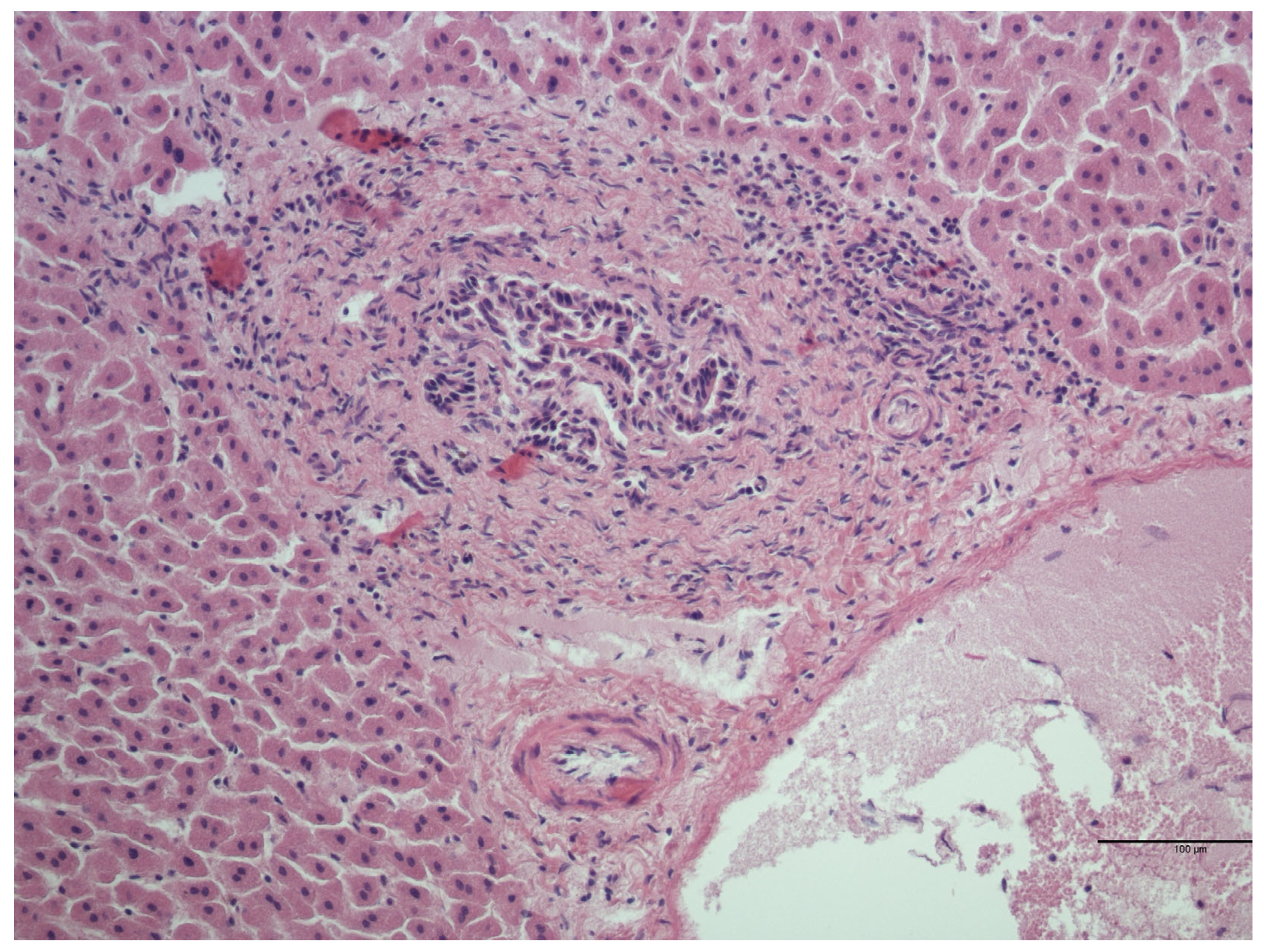
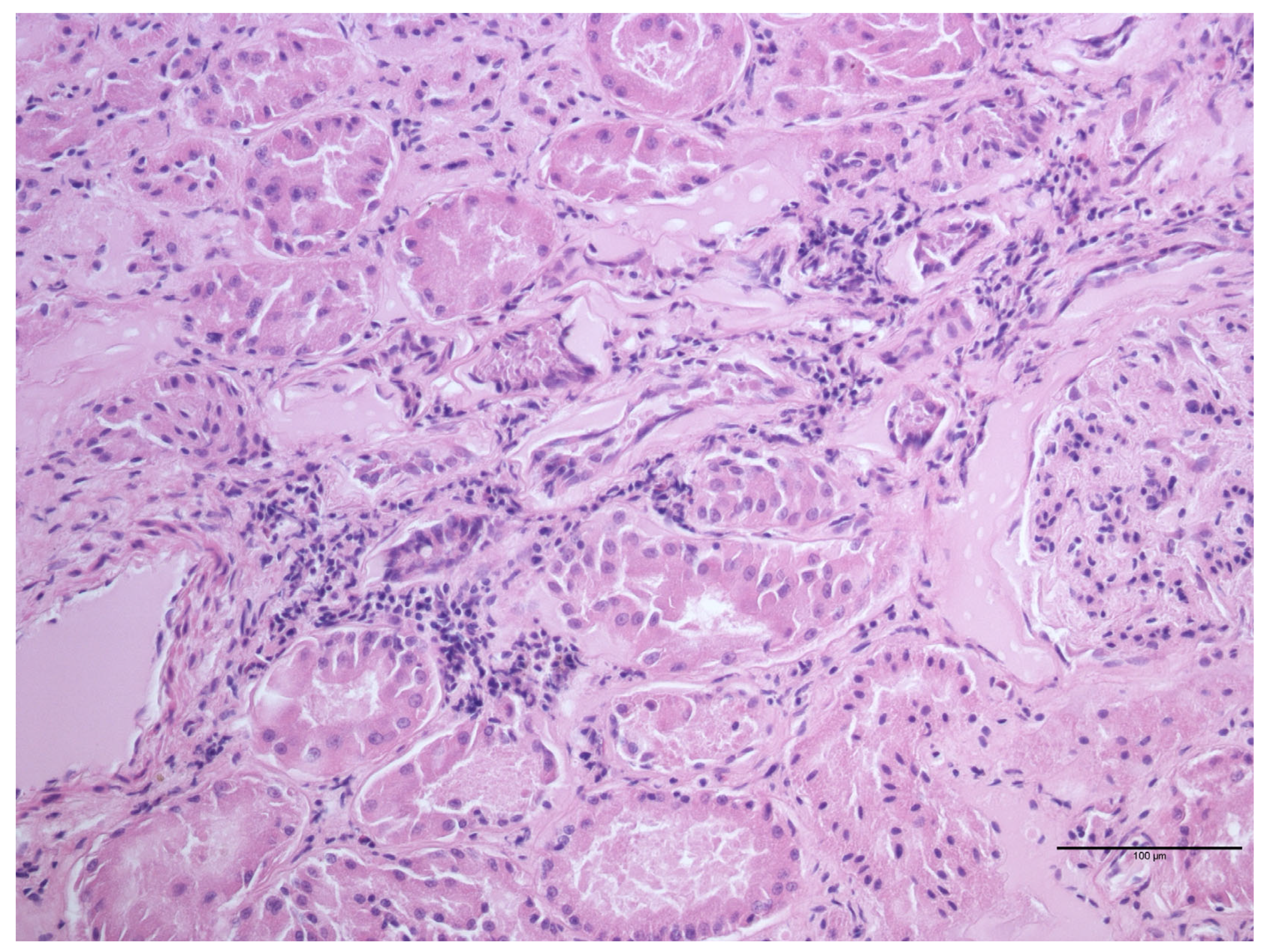
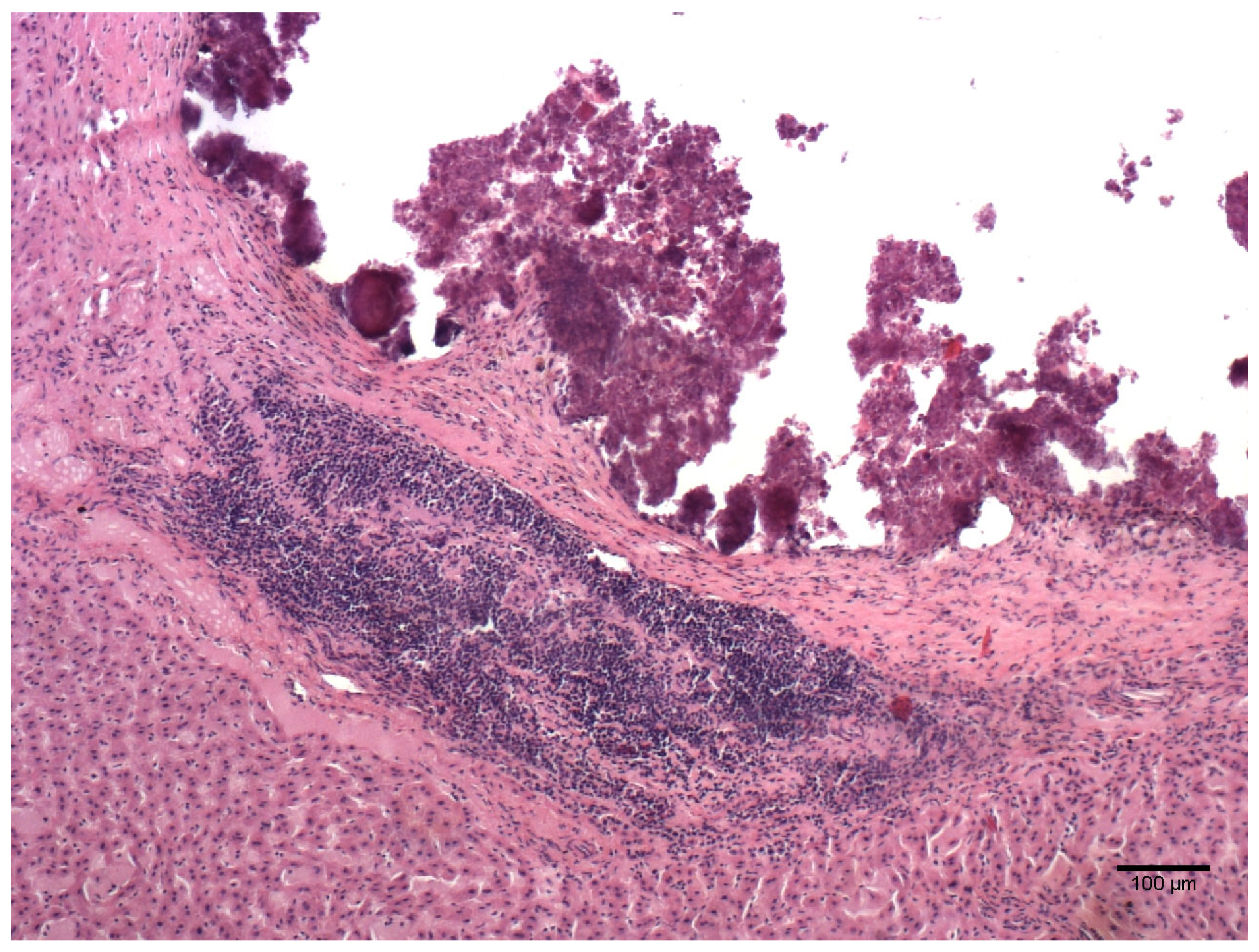
3.2. Histological Analysis

Out of 44 examined livers, 25 (56.8%) had no detectable microscopic lesions, whereas 18 (40.9%) showed one or more concurrent microscopic lesions (Table 2, Figure 1 and Figure 2). One sample (2.3%) was autolytic and impossible to evaluate.
The evaluation of the kidneys showed 21 (47.7%) samples without lesions and 23 (52.3%) affected from one or more detectable microscopic lesions (Table 3, Figure 3). No statistically significant association between lymphocytic interstitial nephritis and sex or age was detected.
Each of the 44 examined lungs showed the presence of concomitant different lesions (Table 4, Figure 4). No statistically significant association was found between perivascular lymphocytic infiltrate or BALT activation and sex or age.

3.3. Bacteriological, Virological and Parasitological Analyses

None of the Samples Tested Positive for HEV or EMCV

None of the tested animals were positive for Francisella spp., while 25 out of 39 analysed lungs (64.1%) were positive in the bacteriological analysis. Six of them (15.4%) revealed polymicrobial infection. The bacterial species identification of the other 19 positive samples is listed in Table 5. Due to the conservation methods of the lung samples, it has to be considered that an underestimation of bacterial identification is possible.
Two out of the thirty-five (5.7%) analysed animals were positive for T. gondii, whereas N. caninum infection has never been detected.

4. Discussion

All of the considered lungs had microscopical lesions. However, out of the 44 samples, only in 19 samples was it possible to isolate bacterial pathogens. Fifteen out of the nineteen (89.5%) analysed lungs showed the presence of lymphocytic infiltrates. Nine of them also showed the presence of lymphocytic infiltrate in the kidney and five of them in the liver. The other three individuals showed inflammatory lesions only in the liver.
As previously reported by Bollo and colleagues [12], cold and high humidity could be a predisposing factor for the high prevalence of pneumonia in wild coypu.
Histologically, kidneys mostly showed interstitial lymphocytic infiltrate (45.45%). This prevalence is higher than the one reported in another study conducted in the same region (10.1%) [12], and it could be caused by infection or immune-mediated diseases, such as Leptospirosis, as previously reported in other populations of coypu serologically investigated [12,22,23,24].
Livers showed microscopical lesions, with inflammatory infiltrations, mostly lymphocytic. The aetiology of those lesions can be related to inflammatory and degenerative processes, differently from the results obtained by Bollo and colleagues [12].
The investigation for EMCV’s RNA was negative. In previous works, seropositivity against EMCV was found in Argentina [13] and Italy [12], whereas it was not detected in the USA [25].
PCR for Francisella spp. gave negative results. Similar results have been serologically obtained in two different studies made in Louisiana (USA) and Argentina, in which no antibodies against F. tularensis were found [13,25].
All the samples analysed for HEV and Neospora caninum were negative and, to the authors’ best knowledge, positivity in this species has never been demonstrated.
Worthy of interest, two animals were positive for Toxoplasma gondii, which can infect all warm-blooded animals and is the aetiological agent of a major zoonosis. In the present research, one of the two animals found to be positive for T. gondii showed an interstitial lymphocytic nephritis and a lymphocytic perivascular and parenchymal infiltrate in the liver, even if, normally, animals challenged with Toxoplasma do not develop referable clinical signs [5,12]. The other positive animal showed no microscopical findings. The prevalence of Toxoplasma in the examined area (4.5%) is very low compared to Nardoni and colleagues’ study (59.4%) [5]. A coypu infected with Toxoplasma gondii is a potential contagion source for other scavengers and can be used in the analysed area for monitoring the quantity of oocysts [5]. Human infection during pregnancy may be extremely dangerous for the foetus, and this parasite in immunocompromised patients can cause a life-threatening encephalitis [26]. New findings suggest that Toxoplasma can actually cause changes in memory, learning, behaviour and anxiety [27].
Currently, the coypu is mostly raised in South America for fur production. Meat consumption of the coypu is considered a by-product, but there are new studies that are considering coypu meat as a new novel and exotic food [28]. In this scenario, the importance of coypu infections with Toxoplasma and other pathogens such as HEV acquires even more relevance.

5. Conclusions

In conclusion, the coypu is an acknowledged threat for both the environment and animals. It can act as a host for several pathogens, including important agents for human and animal health [5,9,12]. Therefore, further investigation for viral, bacterial and parasitic surveillance are necessary, to fully understand the biological role and the importance of coypu as a disease reservoir in our country.

Author Contributions

Conceptualization, A.N. and F.E.S.; methodology, L.S.C.; software, P.P.; validation, E.B.; formal analysis, A.N. and L.S.C.; investigation, F.E.S., A.N. and L.S.C.; writing—original draft preparation, A.N., F.E.S. and P.P.; writing—review and editing, F.E.S. and P.P.; supervision, E.B. All authors have read and agreed to the published version of the manuscript.

Funding

This research received no external funding.

Institutional Review Board Statement

Not applicable.

Informed Consent Statement

Not applicable.

Data Availability Statement

Data are contained within the article.

Conflicts of Interest

The authors declare no conflicts of interest.

References

  1. Carter, J.; Leonard, B.P. A Review of the Literature on the Worldwide Distribution, Spread of, and Efforts to Eradicate the Coypu (Myocastor Coypus). Wildl. Soc. Bull. 2002, 30, 162–175. [Google Scholar]
  2. Pedruzzi, L.; Schertler, A.; Giuntini, S.; Leggiero, I.; Mori, E. An Update on the Distribution of the Coypu, Myocastor Coypus, in Asia and Africa through Published Literature, Citizen-Science and Online Platforms. Mamm. Biol. 2022, 102, 109–118. [Google Scholar] [CrossRef]
  3. Adriani, S.; Bonanni, M.; Amici, A. Study on the Presence and Perception of Coypu (Myocastor coypus Molina, 1782) in Three Areas of Lazio Region (Italy). Jul.-Kühn-Arch. 2011, 432, 49–50. [Google Scholar] [CrossRef]
  4. Panzacchi, M.; Cocchi, R.; Genovesi, P.; Bertolino, S. Population Control of Coypu Myocastor Coypus in Italy Compared to Eradication in UK: A Cost-Benefit Analysis. Wildl. Biol. 2007, 13, 159–171. [Google Scholar] [CrossRef]
  5. Nardoni, S.; Angelici, M.C.; Mugnaini, L.; Mancianti, F. Prevalence of Toxoplasma Gondii Infection in Myocastor Coypus in a Protected Italian Wetland. Parasites Vectors 2011, 4, 240. [Google Scholar] [CrossRef]
  6. Robert, H.; Lafontaine, R.M.; Beudels-Jamar, R.C.; Delsinne, T.; Baiwy, E. Risk Analysis of the Coypu Myocastor Coypus (Molina, 1792). Risk Analysis Report of Non-Native Organisms in Belgium; Royal Belgian Institute of Natural Sciences: Brussels, Belgium, 2013. [Google Scholar] [CrossRef]
  7. Kang, W.; Kim, G.; Park, Y. Habitat Suitability and Connectivity Modeling Predict Genetic Population Structure and Priority Control Areas for Invasive Nutria (Myocastor Coypus) in a Temperate River Basin. PLoS ONE 2022, 17, e0279082. [Google Scholar] [CrossRef] [PubMed]
  8. GISD. Available online: http://www.iucngisd.org/gisd/ (accessed on 26 October 2023).
  9. Barrat, J.; Richomme, C.; Moinet, M. The Accidental Release of Exotic Species from Breeding Colonies and Zoological Collections: -EN- -FR- Lâcher Accidentel d’espèces Exotiques à Partir de Colonies d’élevage et de Collections Zoologiques-ES-Liberación Accidental de Especies Exóticas de Colonias Reproductoras y Colecciones Zoológicas. Rev. Sci. Tech. OIE 2010, 29, 113–122. [Google Scholar] [CrossRef]
  10. Wang, S.; Deng, T.; Zhang, J.; Li, Y. Global Economic Costs of Mammal Invasions. Sci. Total Environ. 2023, 857, 159479. [Google Scholar] [CrossRef]
  11. Funzione Specializzata Tutela della Flora e della Fauna Ufficio Tecnico Gestionale. Piano di Contenimento della Nutria (Myocastor coypus) sul Territorio della citta’ Metropolitana di Torino Anni 2022–2026; Funzione Specializzata Tutela della Flora e della Fauna Ufficio Tecnico Gestionale: Turin, Italy, 2021. [Google Scholar]
  12. Bollo, E.; Pregel, P.; Gennero, S.; Pizzoni, E.; Rosati, S.; Nebbia, P.; Biolatti, B. Health Status of a Population of Nutria (Myocastor Coypus) Living in a Protected Area in Italy. Res. Vet. Sci. 2003, 75, 21–25. [Google Scholar] [CrossRef]
  13. Martino, P.E.; Stanchi, N.O.; Silvestrini, M.; Brihuega, B.; Samartino, L.; Parrado, E. Seroprevalence for Selected Pathogens of Zoonotic Importance in Wild Nutria (Myocastor Coypus). Eur. J. Wildl. Res. 2014, 60, 551–554. [Google Scholar] [CrossRef]
  14. Gosling, L.M.; Huson, L.W.; Addison, G.C. Age Estimation of Coypus (Myocastor Coypus) from Eye Lens Weight. J. Appl. Ecol. 1980, 17, 641. [Google Scholar] [CrossRef]
  15. Pagnoni, G.A.; Santolini, R. Struttura di popolazione di nutria (Myocastor coypus) in un’area agricola della Pianura Padana Orientale. Studi Trent. Sci. Nat. 2011, 88, 45–52. [Google Scholar]
  16. Jothikumar, N.; Cromeans, T.L.; Robertson, B.H.; Meng, X.J.; Hill, V.R. A Broadly Reactive One-Step Real-Time RT-PCR Assay for Rapid and Sensitive Detection of Hepatitis E Virus. J. Virol. Methods 2006, 131, 65–71. [Google Scholar] [CrossRef]
  17. Vanderhallen, H.; Koenen, F. Identification of Encephalomyocarditis Virus in Clinical Samples by Reverse Transcription-PCR Followed by Genetic Typing Using Sequence Analysis. J. Clin. Microbiol. 1998, 36, 3463–3467. [Google Scholar] [CrossRef] [PubMed]
  18. Forsman, M.; Sandstrom, G.; Sjostedt, A. Analysis of 16S Ribosomal DNA Sequences of Francisella Strains and Utilization for Determination of the Phylogeny of the Genus and for Identification of Strains by PCR. Int. J. Syst. Bacteriol. 1994, 44, 38–46. [Google Scholar] [CrossRef] [PubMed]
  19. Homan, W.L.; Vercammen, M.; Braekeleer, J.D.; Verschueren, H. Identication of a 200- to 300-Fold Repetitive 529 Bp DNA Fragment in Toxoplasma Gondii, and Its Use for Diagnostic and Quantitative PCRp. Int. J. Parasitol. 2000, 30, 69–75. [Google Scholar] [CrossRef] [PubMed]
  20. Romano, A.; Trisciuoglio, A.; Grande, D.; Ferroglio, E. Comparison of Two PCR Protocols for the Detection of Neospora Caninum DNA in Rodents. Vet. Parasitol. 2009, 159, 159–161. [Google Scholar] [CrossRef]
  21. Ferroglio, E.; Bosio, F.; Trisciuoglio, A.; Zanet, S. Toxoplasma Gondii in Sympatric Wild Herbivores and Carnivores: Epidemiology of Infection in the Western Alps. Parasit. Vectors 2014, 7, 196. [Google Scholar] [CrossRef]
  22. Zanzani, S.A.; Cerbo, A.D.; Gazzonis, A.L.; Epis, S.; Invernizzi, A.; Tagliabue, S.; Manfredi, M.T. Parasitic and Bacterial Infections of Myocastor Coypus in a Metropolitan Area of Northwestern Italy. J. Wildl. Dis. 2016, 52, 126–130. [Google Scholar] [CrossRef]
  23. Ayral, F.; Kodjo, A.; Guédon, G.; Boué, F.; Richomme, C. Muskrats Are Greater Carriers of Pathogenic Leptospira than Coypus in Ecosystems with Temperate Climates. PLoS ONE 2020, 15, e0228577. [Google Scholar] [CrossRef]
  24. Mazzotta, E.; Bellinati, L.; Bertasio, C.; Boniotti, M.B.; Lucchese, L.; Ceglie, L.; Martignago, F.; Leopardi, S.; Natale, A. Synanthropic and Wild Animals as Sentinels of Zoonotic Agents: A Study of Leptospira Genotypes Circulating in Northeastern Italy. Int. J. Environ. Res. Public Health 2023, 20, 3783. [Google Scholar] [CrossRef] [PubMed]
  25. Howerth, E.W.; Reeves, A.J.; McElveen, M.R.; Austin, F.W. Survey for Selected Diseases in Nutria (Myocastor Coypus) from Louisiana. J. Wildl. Dis. 1994, 30, 450–453. [Google Scholar] [CrossRef] [PubMed]
  26. Montoya, J.G.; Liesenfeld, O. Toxoplasmosis. Lancet 2004, 363, 1965–1976. [Google Scholar] [CrossRef] [PubMed]
  27. Tyebji, S.; Seizova, S.; Hannan, A.J.; Tonkin, C.J. Toxoplasmosis: A Pathway to Neuropsychiatric Disorders. Neurosci. Biobehav. Rev. 2019, 96, 72–92. [Google Scholar] [CrossRef]
  28. Saadoun, A.; Cabrera, M.C. A Review of Productive Parameters, Nutritive Value and Technological Characteristics of Farmed Nutria Meat (Myocastor Coypus). Meat Sci. 2019, 148, 137–149. [Google Scholar] [CrossRef]
Figure 1. Myocastor coypus liver: granuloma with necrotic area surrounded by connective tissue and lymphocytic infiltrate (HE).
Animals 14 00245 g001
Figure 2. Myocastor coypus liver: mild activation of the periportal lymphoid tissue (HE).
Animals 14 00245 g002
Figure 3. Myocastor coypus kidney: mild interstitial lymphoid nephritis (HE).
Animals 14 00245 g003
Figure 4. Myocastor coypus lung: severe parenchymal lymphocytic infiltrate (HE).
Animals 14 00245 g004
Table 1. Mean weight of the dry eye lens (mg), standard deviation, age in months and age class in tested coypus. NA: not applicable.
Coypus IDMean Weight (mg)Standard Deviation (mg)Age (Months)Age Class
1NANANANA
2NANANANA
3NANANANA
4NANANANA
5NANANANA
611.54.95<1Juvenile
77.54.95<1Juvenile
861.41<1Juvenile
971.41<1Juvenile
105.50.71<1Juvenile
11NANANANA
12NANANANA
1335.59.195.0Juvenile
1434.52.124.8Juvenile
1546NA8.5Adult
1644NA7.8Juvenile
1736NA5.2Juvenile
1827.54.953.0Juvenile
19212.831.7Juvenile
20385.665.8Juvenile
21447.077.8Juvenile
2224.50.712.4Juvenile
2362.83<1Juvenile
2413.52.120.5Juvenile
2513.54.950.5Juvenile
26131.410.4Juvenile
2764.24<1Juvenile
2812.52.120.4Juvenile
2915.52.120.8Juvenile
3010.50.710.1Juvenile
319.54.950.0Juvenile
3254.24<1Juvenile
33205.661.6Juvenile
34100.000.0Juvenile
35212.831.7Juvenile
36282.833.2Juvenile
377.50.71<1Juvenile
38140.000.6Juvenile
39NANANANA
4024.50.712.4Juvenile
41NANANANA
4213.52.120.5Juvenile
43NANANANA
44NANANANA
Table 2. Frequency and characteristics of the histological lesions detected in liver samples.
LesionNumber of Samples Positive for the Lesion/Total Number of Microscopical Lesions (%)
Periportal lymphoid tissue activation8/18 (44.4%)
Parenchymal lymphocytic infiltrate7/18 (38.9%)
Perivascular lymphocytic infiltrate3/18 (16.7%)
Macrophage infiltration1/18 (5.6%)
Multifocal granuloma1/18 (5.6%)
Table 3. Frequency and characteristics of the histological lesions detected in kidney samples.
LesionNumber of Samples Positive for the Lesion/Total Number of Microscopical Lesions (%)
Interstitial lymphocytic nephritis20/23 (87.0%)
Urine crystals2/23 (8.7%)
Perivascular lymphocytic infiltrate1/23 (4.3%)
Cyst with focal lymphocytic infiltrate1/23 (4.3%)
Interstitial lymphocytic and eosinophilic nephritis 1/23 (4.3%)
Lymphocytic infiltrate into perirenal fat1/23 (4.3%)
Table 4. Frequency and characteristics of the histological lesions detected in lung samples.
LesionNumber of Samples Positive for the Lesion/Total Number of Microscopical Lesions (%)
Emphysema44/44 (100%)
Oedema36/44 (81.8%)
Parenchymal lymphocytic infiltrate36/44 (81.8%)
Perivascular lymphocytic infiltrate32/44 (72.7%)
BALT activation27/44 (61.4%)
Alveolar thickening 12/44 (27.3%)
Atelectasis9/44 (20.5%)
Parenchymal lymphocytic and eosinophilic infiltrate2/44 (4.5%)
Lymphocytic bronchitis2/44 (4.5%)
Parenchymal neutrophilic infiltrate 1/44 (2.3%)
Focal haemorrhages1/44 (2.3%)
Table 5. Isolated bacteria.
Isolated BacteriaNumber of Samples Positive/Total Number of Analysed Lungs (%)
Enterococcus spp.4/19 (21.0%)
Enterococcus hirae2/19 (10.5%)
Pseudomonas fluorescens2/19 (10.5%)
Nocardia spp.2/19 (10.5%)
Enterococcus durans1/19 (5.3%)
Pseudomonas mendocina1/19 (5.3%)
Achromobacter xylosoxidans1/19 (5.3%)
Brevibacillus laterosporus1/19 (5.3%)
Corynebacterium propinquum1/19 (5.3%)
Corynebacterium pseudodiphthericum1/19 (5.3%)
Ochrobactrum anthropi1/19 (5.3%)
Streptococcus aginosus1/19 (5.3%)
Non-identifiable1/19 (5.3%)
Disclaimer/Publisher’s Note: The statements, opinions and data contained in all publications are solely those of the individual author(s) and contributor(s) and not of MDPI and/or the editor(s). MDPI and/or the editor(s) disclaim responsibility for any injury to people or property resulting from any ideas, methods, instructions or products referred to in the content.

