1. Introduction
According to large-scale statistics on domestic dogs in Japan in 2023, 6.84 million dogs are kept as pets and 9.1% of households in Japan share their lives with dogs [
1]. Moreover, it is noteworthy that the total number of human children under the age of 15 was approximately 14.17 million, while the number of domesticated dogs and cats in 2023 was 15.91 million, exceeding the number of human children [
2]. Against this background, domestic dogs and cats are welcomed as family members just like humans, and are sometimes raised with love in the same way as human children. Reflecting this trend, despite the recent economic downturn in Japan due to the weak yen and other factors, domestic dogs’ expenses, including medical expenses, have increased year on year [
3], and the number of contracts with pet insurance companies that cover medical expenses has also increased in the same manner [
4]. The above findings indicate that a society is developing in which dogs and cats are respected to a level similar to that of humans, receiving medical care while ensuring that their welfare is looked after.
The medical care and welfare of domestic dogs somewhat lag behind that of humans but are slowly developing day by day and keeping pace with human medical advances. For example, drug treatment options for canine osteoarthritis have evolved over the past decade to keep pace with human medicine [
5]. Advances have also been made in the treatment of various canine cancers, which have improved life expectancy [
6,
7,
8]. While canine medicine has evolved, similar to human medicine, there are very few published papers on the development of testing and treatment methods for mitochondrial diseases in dogs.
Canine mitochondrial diseases are caused either congenitally or acquired due to mutations or defects in nuclear DNA, which encodes mitochondria-related genes, or in mitochondrial DNA [
9]. Mitochondrial damage resulting from such mutations can severely affect organs that are heavily dependent on oxidative metabolism, especially the brain, skeletal and cardiac muscles, sensory organs, and kidneys [
9]. Symptoms of mitochondrial disorders, also known as mitochondrial myopathy, include muscle weakness, gait disturbance, spontaneous pain, and a variety of other symptoms [
9]. In addition, it has been shown that canine cancers are associated with mutations in the mtDNA-encoded genetic and D-loop regions [
9]. Because these mutations in mtDNA present with a wide variety of symptoms, including mild to severe symptoms, a differential diagnosis is incredibly difficult and requires the development of reliable molecular tests [
9].
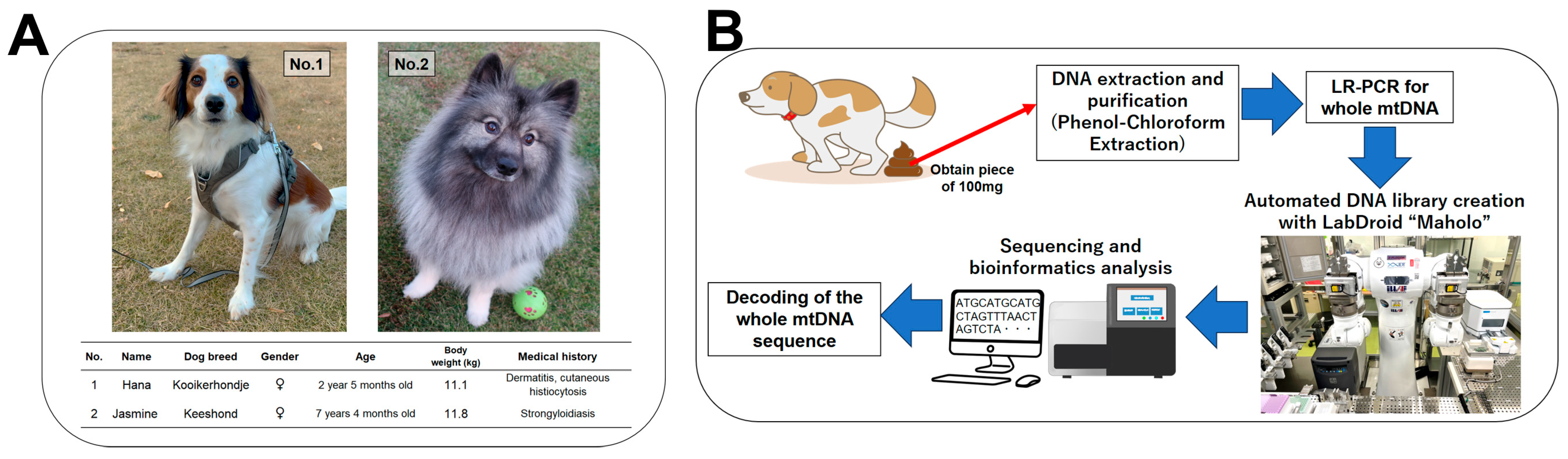
As a matter of course, in order to develop a treatment strategy for canine mitochondrial diseases, it is first necessary to establish a test method to aid in making an accurate differential diagnosis. In order to make a diagnosis, it is essential to examine the full-length integrity and mutations of mtDNA. Therefore, we previously established a method for resequencing the full length of mitochondrial DNA from canine oral mucosal DNA using a next-generation sequencer (NGS) to accurately decode sequences and mutations [
10]. This method may be of great help in the differential diagnosis of canine mitochondrial diseases. It could also be applied to identify individual dogs because the mammalian mitochondrial genome is maternally inherited. Nevertheless, it is recognized that many dogs show a lack of willingness to have their oral mucosal DNA collected. Moreover, the biological samples that can be collected from dogs are not limited to oral mucosal DNA. Various types of samples can be collected, including blood, skin tissue, urine, hair follicles, residual tissue after surgery, feces, etc. Therefore, we focused our attention on feces, which can be easily collected in the least invasive manner as a lightweight solid material.
Feces are a mixture of undigested food residues, intestinal bacteria, cholesterol, and fats [
11,
12]. They also contain cellular debris detached from the intestinal mucosa and dead white blood cells [
11,
13]. In addition, mammalian cell mtDNA copy numbers have been shown to be present in hundreds to thousands of copies per cell, although this depends on the specific tissue [
14,
15]. In other words, canine feces may contain high levels of mitochondrial DNA. In light of the above, it could be hypothesized that the whole mtDNA of dogs can be sequenced and decoded using DNA extracted and purified from feces. Therefore, the purpose of the present study was to establish a method to decode the whole mtDNA sequence using DNA obtained from canine feces.
As a result of the development of our method, the whole mtDNA sequence from the feces samples was successfully decoded with high accuracy. It is anticipated that this method will be utilized for the individual identification of dogs and the differentiation of mitochondrial diseases, which may prove advantageous for the future medical care and welfare of domestic dogs.
4. Discussion
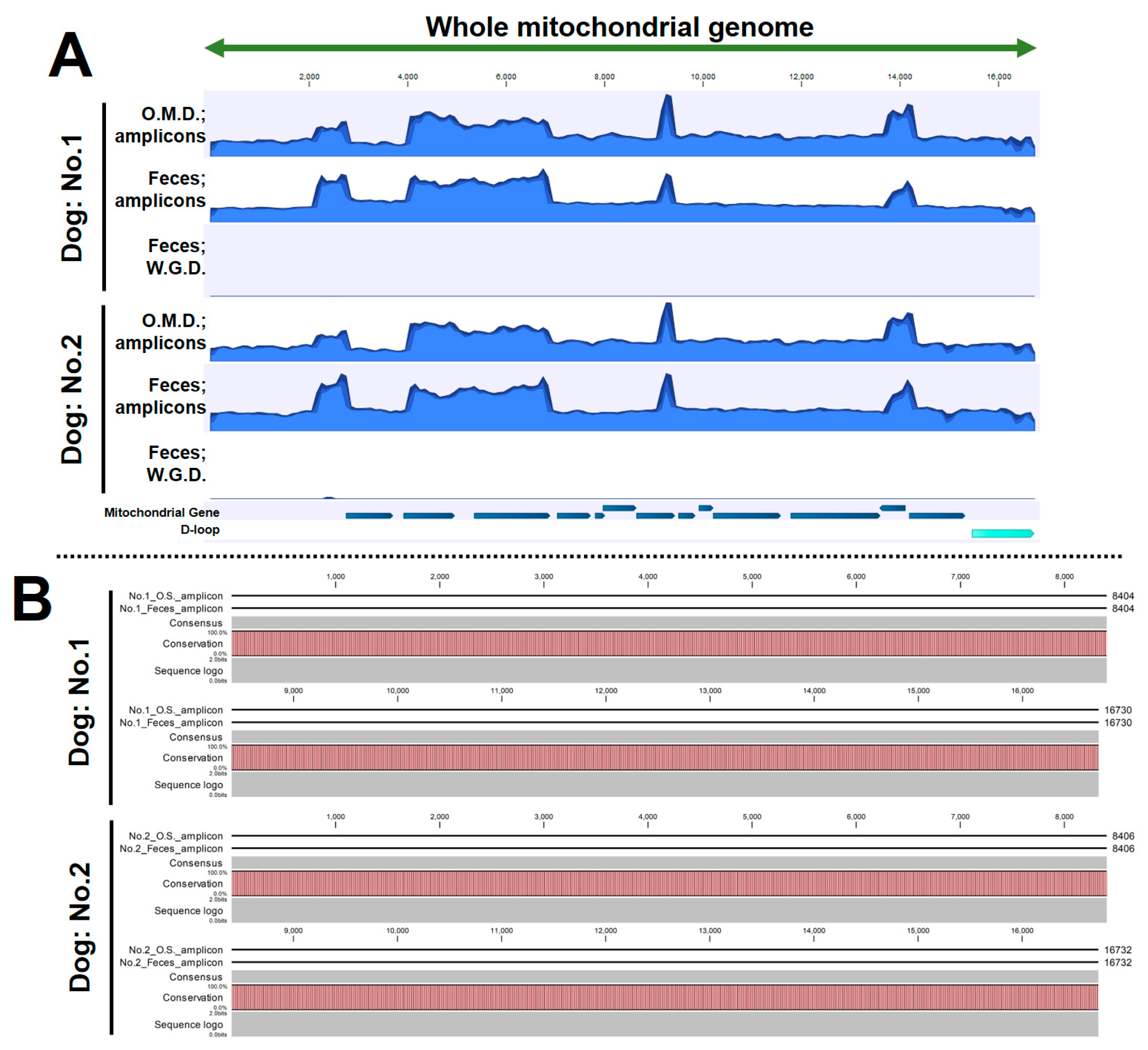
The objective of the present study was to develop a method for determining the whole mtDNA sequence of domestic dogs using the least invasive method available. In addition, we used our previously successful sequencing data of whole mtDNA by using oral mucosal DNA [
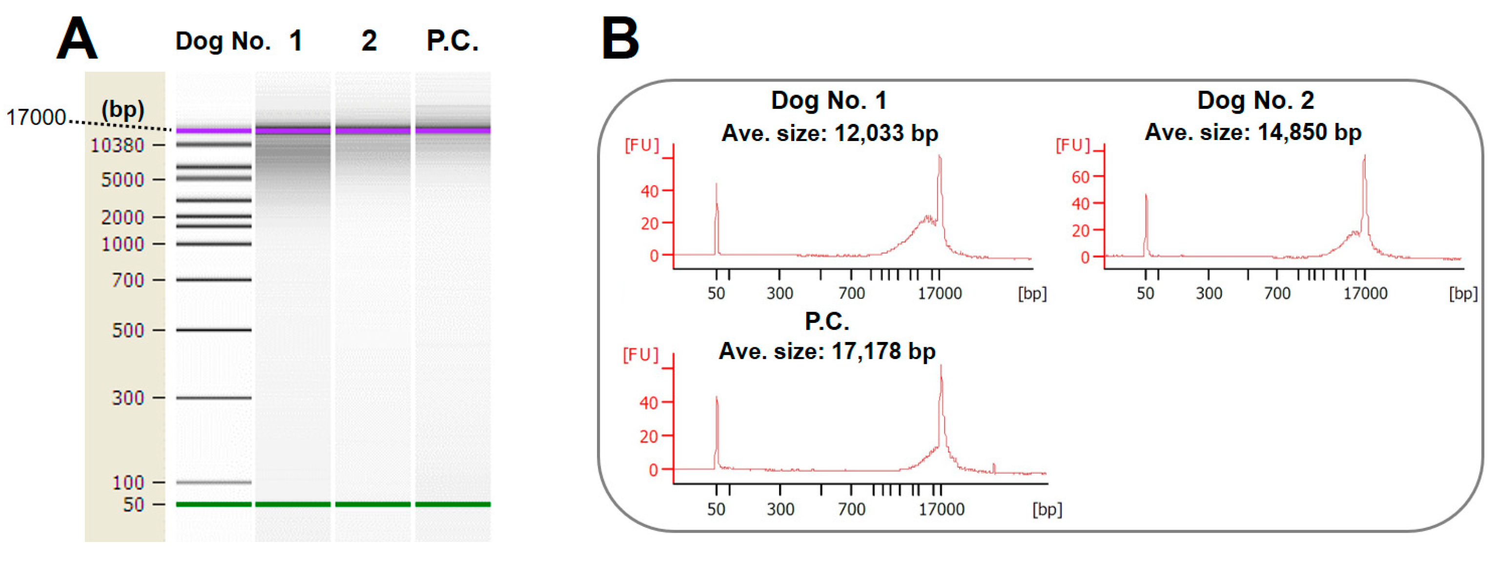
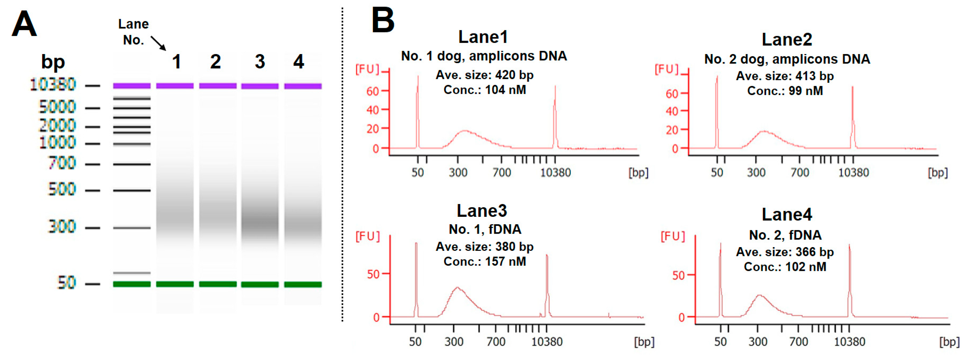
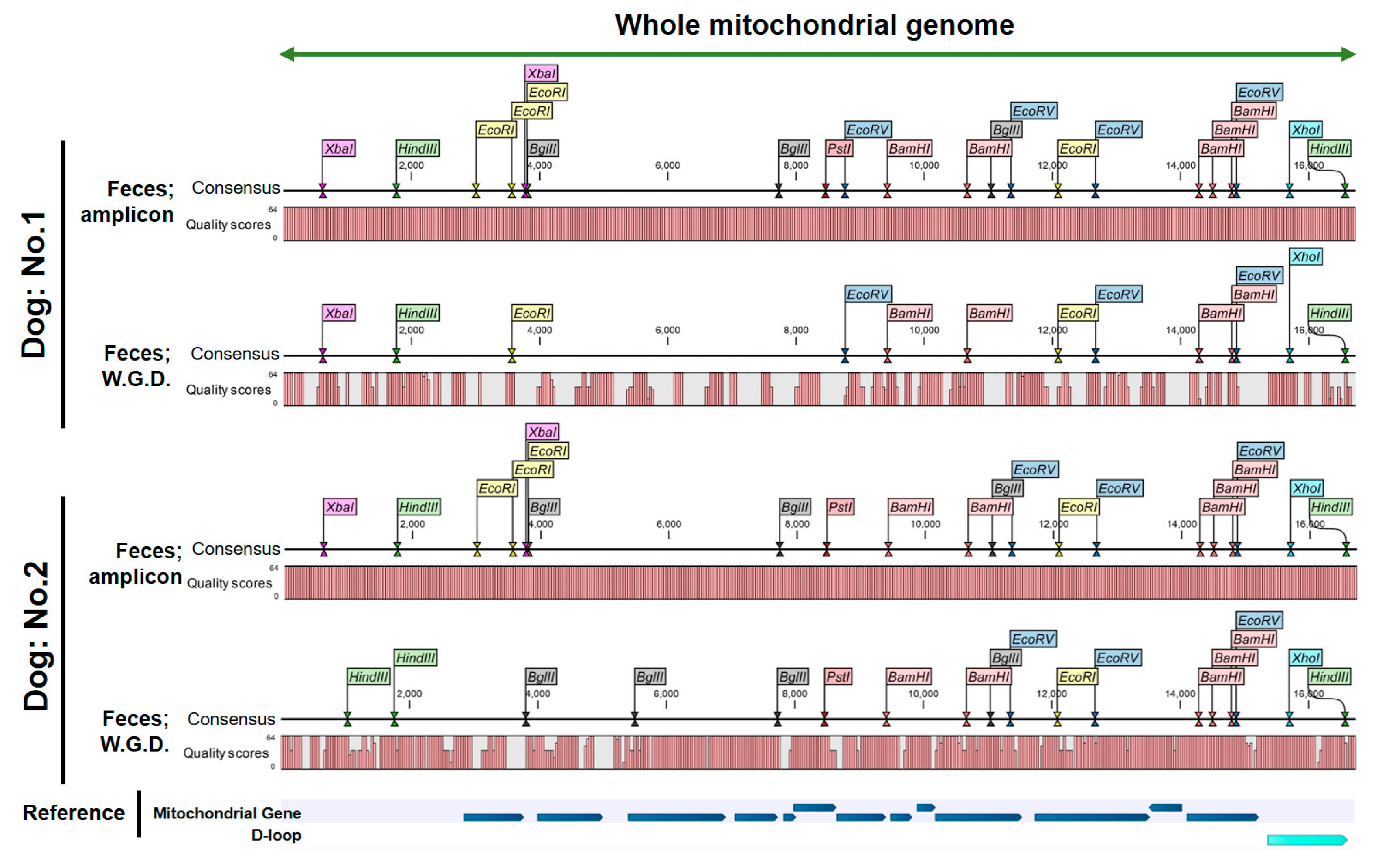
10] and compared them to the data from the fecal samples in the present study to confirm their accuracy. As a result, we succeeded in obtaining whole-length mitochondrial DNA amplicons divided into four parts from fecal DNA. Library preparation using the amplicons was automated using an experimental robot, and we were able to obtain a library that can be adapted to next-generation sequencers. Through bioinformatics analysis using the data obtained from sequencing, we were able to accurately determine the mtDNA sequences derived from the feces of the two dogs. Furthermore, this mtDNA sequence was a perfect match to the sequence derived from the oral mucosal DNA. Taken together, these results indicate that the whole mtDNA sequence can be determined from fecal samples using the method established in the study presented herein. Feces represent the least invasive form of sampling and their acquisition does not cause stress to the dog. Therefore, in the future, the use of this method could lead to advancements in the medical care and welfare of dogs.
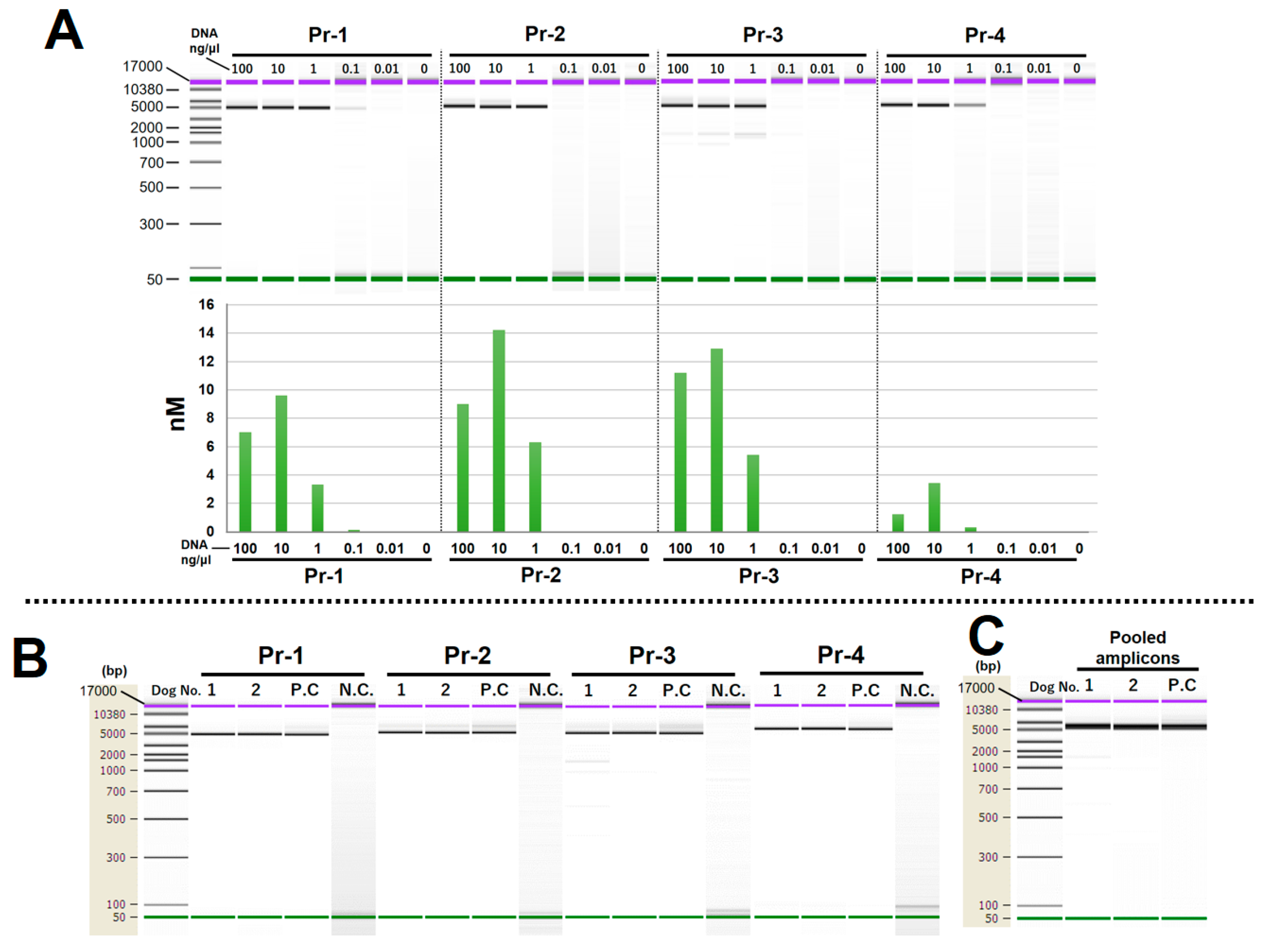
The use of this method yields four PCR amplicons containing the whole mtDNA sequence (including four duplicated regions), which can be sequenced using a next-generation sequencer. This principle is similar to our previously established method for whole mtDNA sequencing using oral mucosal DNA [
10]. However, we found one difference: in PCR using oral mucosal DNA, the highest amplification efficiency was observed when the template DNA concentration was 1 ng/µL (using 2 ng DNA/PCR reaction) [
10]. Conversely, when fecal DNA was used as a template in the present study, the highest amplification efficiency was achieved at a concentration of 10 ng/µL (using 20 ng DNA/PCR reaction). This difference is likely due to the high mix of DNA from different species in the fecal material, which may comprise food residue and bacteria. Bacteria in fecal solids, excluding water, are estimated to account for 30% to 50% of fecal material [
11,
13]. Therefore, depending on the type of specimen, the template concentration should be optimized. In addition, although the two dogs in the present study were healthy on the day of the experiment, if the subject dogs in a study suffer from intestinal conditions/diseases such as diarrhea, constipation, inflammatory diseases, etc., the percentage of intestinal bacteria may vary substantially. If researchers use this method, they would need to first consider the optimal template DNA concentration to adapt to this situation.
The establishment of a non-invasive method of sequencing mtDNA from feces has the potential to promote the understanding and management of mitochondrial diseases in domestic dogs. Multiple mitochondrial diseases, such as myopathies, sensory ataxic neuropathy, cardiomyopathy, and arrhythmia, have been reported in domestic dogs and clinical symptoms are heterogeneous, causing difficulties in diagnosis. Many pathogenic mtDNA mutations have been identified in dogs; thus, it is possible to diagnose mitochondrial diseases through genetic evaluation [
18]. To date, mitochondrial diseases are rarely found in domestic dogs. However, the significant health impact and frequency of human mitochondrial mutations combined with the genetic similarity between humans and dogs suggest that mitochondrial DNA alterations could also have a significant health impact on domestic dogs and the prevalence of mitochondrial diseases in dogs could be underestimated due to insufficient investigation. Thus, it would be beneficial to perform mitochondrial genetic analyses in veterinary clinics to better understand the genetic causes of diseases, consequently revealing the frequency of mitochondrial diseases. mtDNA genetic tests can also be used to discover genetic defects at an early stage in order to provide appropriate care and facilitate selective breeding programs for domestic dogs. Mitochondrial genetics are inherited entirely from the maternal line; therefore, mtDNA analysis can be performed prior to breeding to ensure the health of descendants.
From another perspective, the application of this method indicates the possibility of analyzing the DNA of other types of dogs using canine fecal samples, and it may be possible to study the epidemiology of stray dogs through fecal DNA analysis. The increase in the number of stray dogs is a public health concern in many countries. This concern emanates from the fact that stray dogs and cats usually carry infectious diseases such as parvovirus, distemper, and rabies, which can be transmitted to humans through human contact [
19,
20]. In parallel with controlling stray dog populations, monitoring the health epidemiology of stray dogs in the community by collecting fecal samples from such dogs and collecting fecal DNA could be a potential measure. Analysis of the diverse DNA present in fecal samples could aid in the identification of the genetic material of viral, bacterial, and parasitic pathogens and identify infections that these animals may have. In this way, zoonotic diseases in stray dogs in public spaces can be monitored, and prevention and control measures can be rapidly implemented. Such measures would represent a means of not only protecting human health, but also improving the welfare of this venerable friend of man, the animal. Fecal DNA analysis across different geographic regions is also useful for epidemiological surveillance, revealing patterns, trends, and emerging threats, and allowing for a timely response. Fecal DNA analysis thus has the potential to be part of a comprehensive strategy to protect the health and welfare of both humans and domestic and stray dogs. Therefore, in addition to mtDNA analysis, as employed in the present study, the authors of future studies will be able to reveal in depth the usefulness of fecal samples by employing NGS to quantify the presence and proportion of DNA in samples obtained from various species.
It is recognized that the mitochondrial genome in mammalian cells is inherited from the maternal line [
21]. The complete mitochondrial DNA sequences of a mother and her offspring exhibit a perfect match across their entire length, with the exception of instances where genetic disease is present. This finding was also corroborated in our previous research [
10]. Consequently, the method used in the present study is not only capable of identifying individual dogs, but also tracing the blood relatives of dogs over many generations. Moreover, even in the absence of DNA sequence reference data, it is feasible to ascertain the presence of blood relatives if the DNA of the mother or siblings is available. In light of these considerations, it may also be feasible to address challenges at breeding facilities. For instance, at dog breeding facilities, there are instances where the maternal lineage of a puppy is uncertain, such as with mistaken puppies. In such cases, this method can be employed to ascertain the maternal lineage non-invasively. With such potential applications in mind, the method may prove beneficial in enhancing animal welfare in the future.
There is an interesting report on canine feces testing: in 2024, in Bolzano, northern Italy, the city’s police ordered all local dog owners to have their dogs’ fecal DNA tested in order to tackle the issue of dog feces left on the streets [
22]. This process facilitated the identification and fining of owners who had not picked up their dogs’ feces. The DNA test used in the above study appears to involve the use of a method to detect short tandem repeats (STRs); however, our method of decoding the entire mtDNA may also be applicable. Each test appears to have its advantages and disadvantages. Our method involves the use of NGS, which facilitates the sequencing of the mtDNA of hundreds of dogs at a time and enables high throughput. Furthermore, even if the DNA sequence of the target dog is not registered, it is possible to trace the blood relationship, which may lead to the identification of the owner. However, if the number of samples is small (e.g., less than 10), the cost per sample increases significantly. In addition, advanced skills in NGS and bioinformatics analysis are required. STR-targeted tests are simple, and the results can be analyzed at low cost even with a small number of samples; however, they cannot identify dogs if their DNA sequences have not been registered. Thus, although each test has its advantages and disadvantages, using the two tests in different situations would help to obtain accurate evidence and achieve identification of the owner.
Another unique feature of the present study is the successful preparation of automated amplicon DNA libraries. DNA library preparation, when performed by human handlers, requires a high degree of skill and long hours of work. Therefore, there are significant barriers for beginners to perform this experiment. To solve this problem and compile the DNA library with simple operations, we used an experimental robot, LabDroid “Maholo” [
23]. Our study likely represents the first attempt to use LabDroid “Maholo” in veterinary medicine and animal welfare. The above robot was created by a Japanese venture company [
23] and specializes in molecular biology, biochemistry, and cell biology experiments. In recent years, the robot has been able to automatically create pluripotent stem cell- (iPSC) [
24], and iPSC-derived retinal pigment epithelial (iPSC-RPE) cells [
25,
26], and the LabDroid innovation is currently being conducted in Japan. In addition, the robot’s user-friendly graphical interface allows researchers to create their own experimental programs from scratch. We successfully used this functionality to create an original amplicon DNA library. Experiments using the robot have also resulted in a significant reduction in operator time and thus a significant reduction in researcher labor. We hope to utilize this robot in the future for veterinary tests that require complex processes, thereby contributing to improvements in veterinary medicine and animal welfare.
In recent times, SNP (single nucleotide polymorphism) chips have been made available by major companies for the purpose of deciphering the genotype profiles of dogs. This method is capable of deciphering specific bases within the whole genome, thereby enabling the identification of individuals and their respective breeds. It is also likely to prove useful in the diagnosis of the mitochondrial diseases. Conversely, as this method is designed for the specific deciphering of bases, it is unable to accommodate unknown mutations. Pathological mutations or defects in mammalian mtDNA that involve amino acid substitutions are thought to occur randomly due to ROS (reactive oxygen species) [
27,
28]. As the mutations are not limited to specific bases, it is necessary to decode the whole mtDNA for detailed differential diagnosis. In consideration of the pathomechanism, it would be recognized that our method is superior to the SNP chips. The comprehensive nature of our method allows for the examination of the whole length of mtDNA, facilitating the acquisition of detailed sequence information encompassing not only the regions encoding enzymes associated with oxidative phosphorylation (OXPHOS) but also the non-coding regions. This comprehensive approach may prove advantageous in addressing the potential limitations of the SNIP chip. However, it may be somewhat inferior in terms of cost and ease of experimentation. Nevertheless, it is desirable to implement an examination method that matches the symptoms of the case, and this method would be a valuable contribution to the differential diagnosis of canine mitochondrial disease.
It should be noted that this study is not without limitations. This study employed fresh fecal samples that could be processed within one hour of defecation. Also, given the limited size of our dog community, we were finally able to use fecal samples from only two dogs. Ideally, it would have been preferable to analyze more than 10 dogs. However, when the results of the oral mucosal DNA and fecal DNA were compared, it was observed that the entire mtDNA sequence was identical. Additionally, in the experiment where LR-PCR was not performed (equivalent to whole fecal genome sequencing), no accurate sequence was obtained, and it was established as a negative control. In conclusion, despite the limitations imposed by the small sample size, the results were promising, and the negative control was also successful. Therefore, it can be reasonably asserted that the scientific validity of the results is assured. It remains unclear, however, whether the same method can be applied to degraded samples. For example, it is unclear whether the same results as in this study can be obtained when using feces that have been left at room temperature, in the hot sun, or in high humidity for a long period of time. This is a topic that we intend to pursue further in future research.


 ,
, 