

Anatomical Description of Loggerhead Turtle (Caretta caretta) and Green Iguana (Iguana iguana) Skull by Three-Dimensional Computed Tomography Reconstruction and Maximum Intensity Projection Images

 , , ,
, , ,  and
and 
Abstract
:Simple Summary
Abstract
1. Introduction
2. Materials and Methods
2.1. Animals
2.2. CT Technique
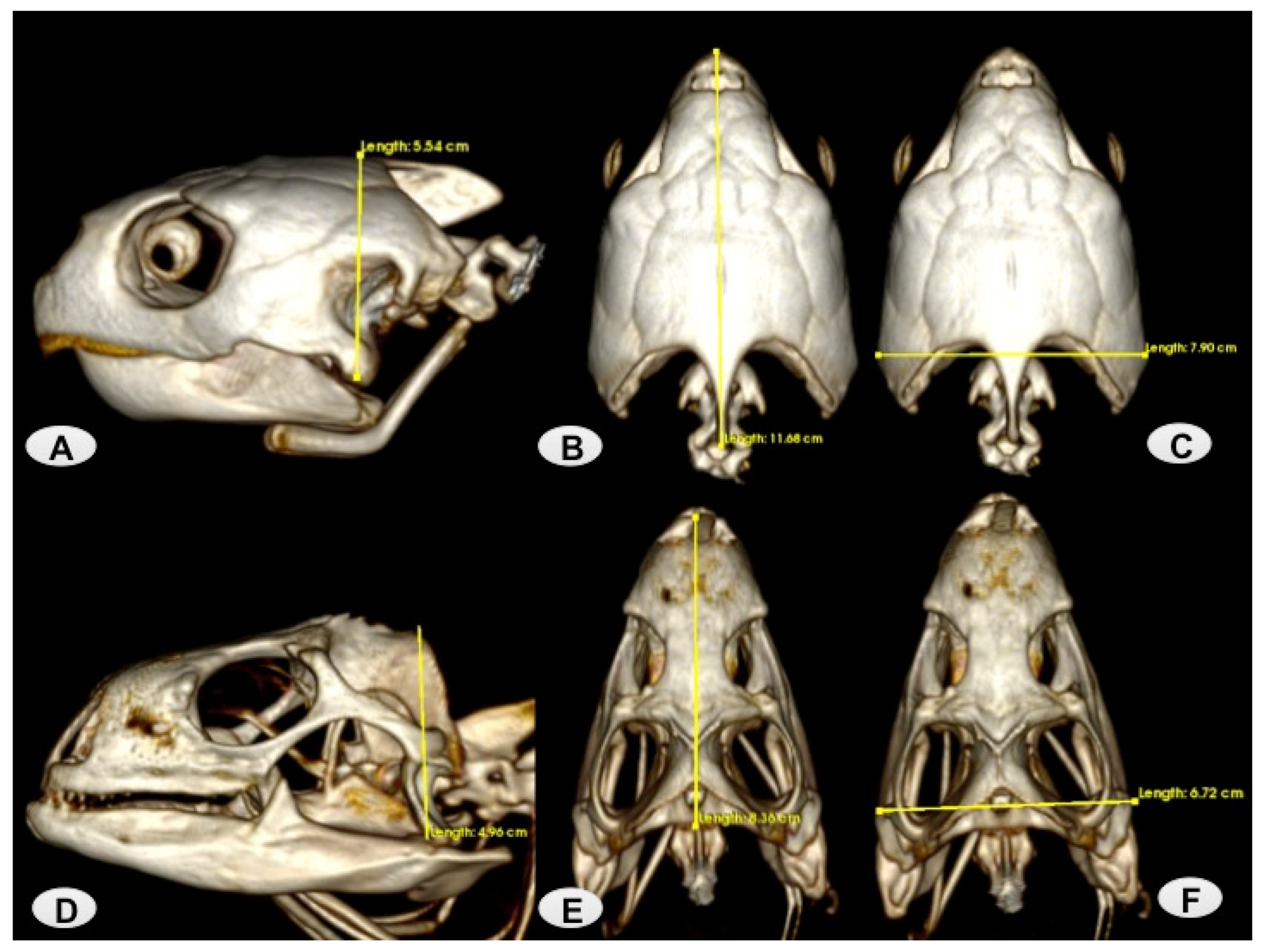
3. Results
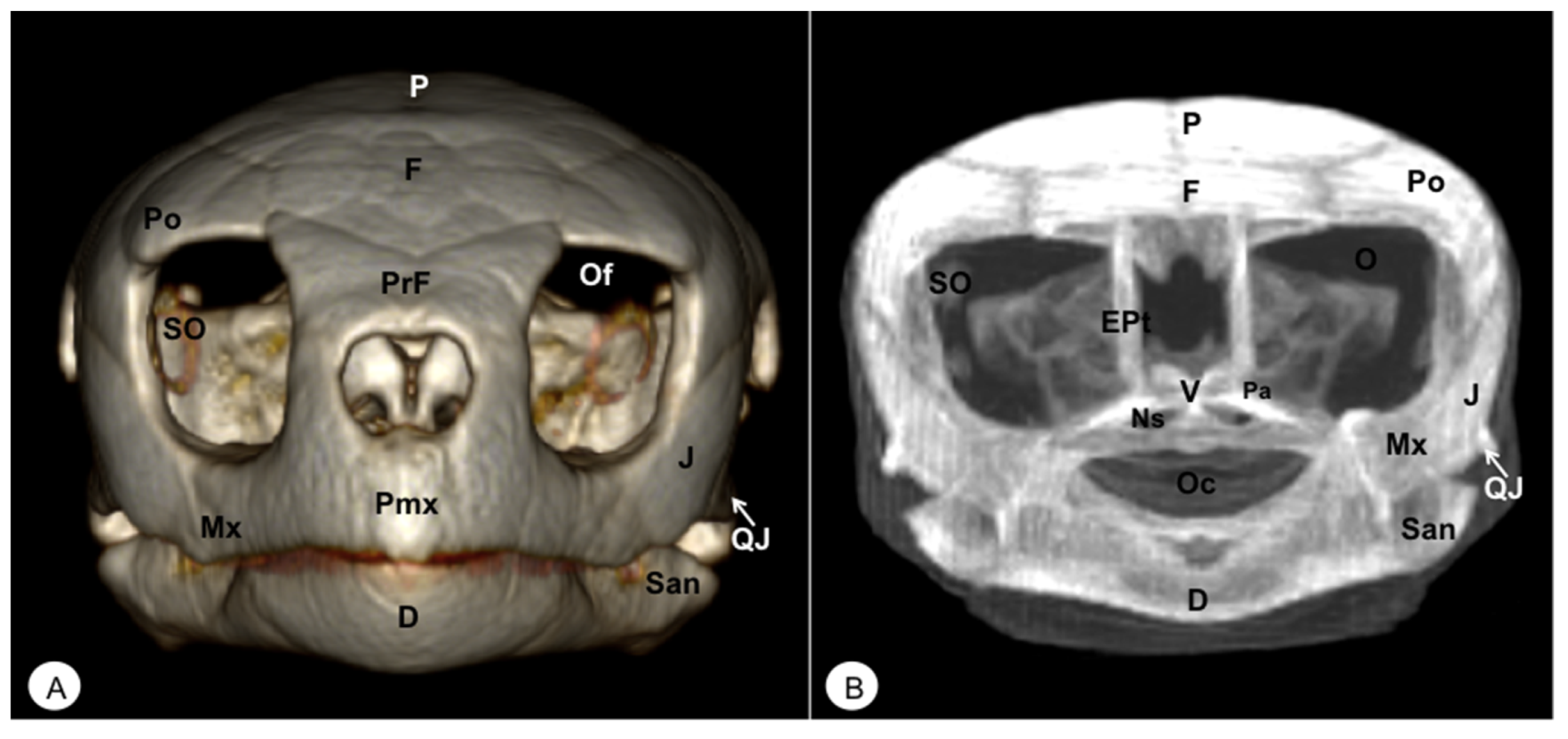
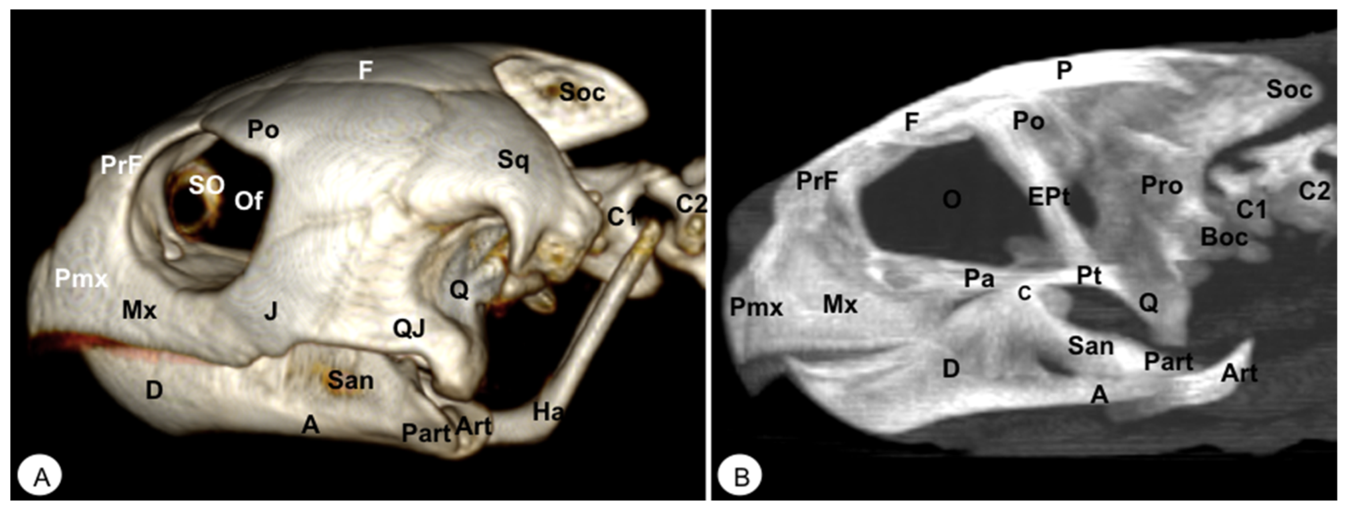
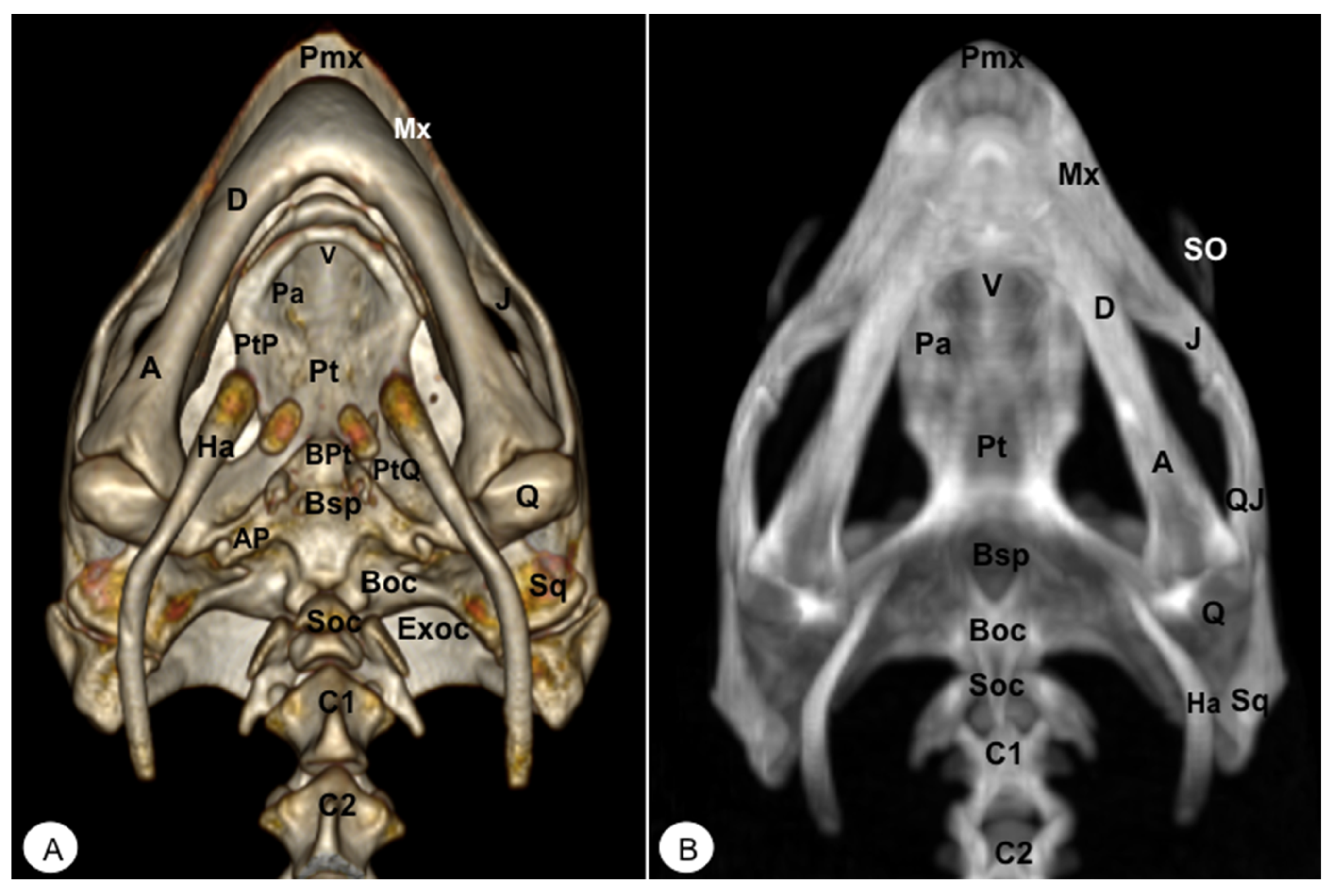
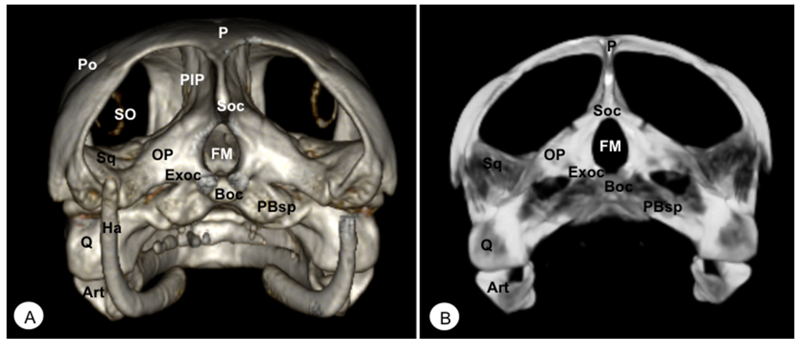
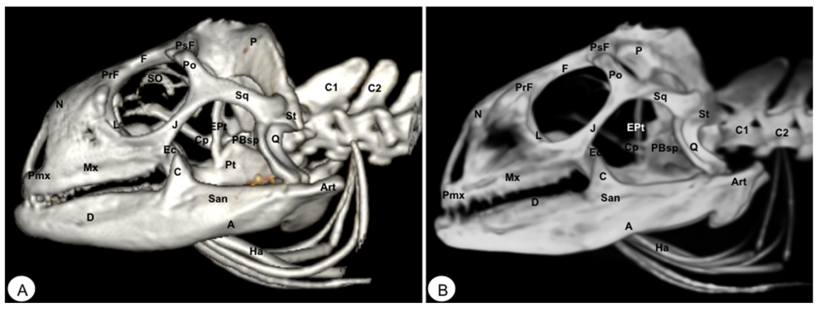
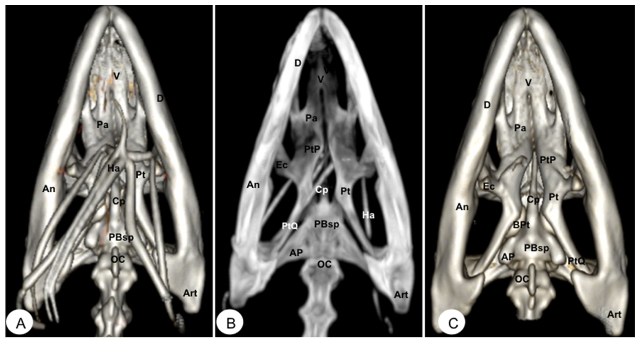
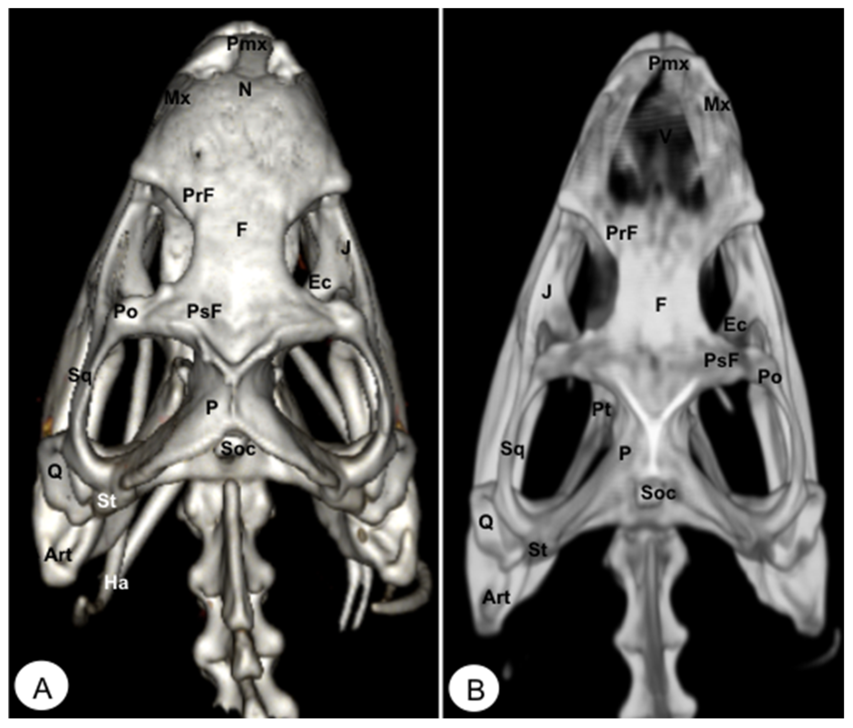
3.1. Dermatocranium
3.1.1. PREMAXILLA (Os Premaxillare)
3.1.2. MAXILLA (Os Maxillare)
3.1.3. NASAL (Os Nasale)
3.1.4. VOMER
3.1.5. PALATINE (Os Palatinum)
3.1.6. PREFRONTAL-FRONTAL
3.1.7. POSTFRONTAL-POSTORBITAL
3.1.8. PARIETAL (Os Parietale)
3.1.9. JUGAL (Os Jugale)
3.1.10. QUADRATE (Os Quadratum)
3.1.11. QUADRATOJUGAL (Os Quadratojugale)
3.1.12. SQUAMOSAL (Os Squamosum)
3.1.13. PTERYGOID (Os Pterygoideum)
3.1.14. EPIPTERYGOID
3.2. Neurocranium
3.2.1. PARABASISPHENOID-BASISPHENOID (Os Basisphenoidale)
3.2.2. BASIOCCIPITAL (Os Basioccipitale)
3.2.3. SUPRAOCCIPITAL (Os Supraoccipitale)
3.2.4. EXOCCIPITAL (Os Exoccipitale)
3.3. Mandible
3.3.1. DENTARY (Os Dentale)
3.3.2. ANGULAR (Os Angulare)
3.3.3. SURANGULAR (Os Surangulare)
3.3.4. CORONOID (Os Coronoideum)
3.3.5. PREARTICULAR (Os Prearticulare)
3.3.6. ARTICULAR (Os Articulare)
3.3.7. HYOID APPARATUS (Os Hyoideum)
4. Discussion
Author Contributions
Funding
Institutional Review Board Statement
Informed Consent Statement
Data Availability Statement
Acknowledgments
Conflicts of Interest
References
- Kuszyk, B.S.; Heath, D.G.; Bliss, D.F.; Fishman, E.K. Skeletal 3-D CT: Advantages of Volume Rendering over Surface Rendering. Skeletal Radiol. 1996, 25, 207–214. [Google Scholar] [CrossRef] [PubMed]
- Hughes, J.R.; Johnson, V.S.; Genain, M.A. CT Characteristics of Primary Splenic Torsion in Eight Dogs. Vet. Radiol. Ultrasound 2020, 61, 261–268. [Google Scholar] [CrossRef] [PubMed]
- Rowe, S.P.; Fishman, E.K. Three-Dimensional Computed Tomography Cinematic Rendering of Mandibular Odontogenic Myxofibroma. Oral Surg. Oral Med. Oral Pathol. Oral Radiol. 2019, 128, e122–e125. [Google Scholar] [CrossRef]
- Dianna, D.; Cody, P. AAPM/RSNA Physics Tutorial for Residents: Topics in CT. Radiographics 2002, 22, 14. [Google Scholar] [CrossRef]
- Fishman, E.K.; Ney, D.R.; Heath, D.G.; Corl, F.M.; Horton, K.M.; Johnson, P.T. Volume Rendering versus Maximum Intensity Projection in CT Angiography: What Works Best, When, and Why. Radiographics 2006, 26, 905–922. [Google Scholar] [CrossRef] [PubMed]
- Calhoun, P.S.; Kuszyk, B.S.; Heath, D.G.; Carley, J.C.; Fishman, E.K. Three-Dimensional Volume Rendering of Spiral CT Data: Theory and Method. Radiographics 1999, 19, 745–764. [Google Scholar] [CrossRef]
- Prokop, M.; Shin, H.O.; Schanz, A.; Schaefer-Prokop, C.M. Use of Maximum Intensity Projections in CT Angiography: A Basic Review. Radiographics 1997, 17, 433–451. [Google Scholar] [CrossRef]
- Thrall, D. Textbook of Veterinary Diagnostic Radiology; Elsevier: St. Louis, MO, USA, 2012; ISBN 9780323482479. [Google Scholar]
- Ho, J.L.; Konda, A.; Rahman, J.; Harris, E.; Korn, R.; Sabir, A.; Bawany, B.; Gulati, R.; Harris, G.J.; Boswell, W.D.; et al. Comparative Analysis of Three-Dimensional Volume Rendering and Maximum Intensity Projection for Preoperative Planning in Liver Cancer. Eur. J. Radiol. Open 2020, 7, 100259. [Google Scholar] [CrossRef]
- Li, W.J.; Chu, Z.G.; Zhang, Y.; Li, Q.; Zheng, Y.N.; Lv, F.J. Effect of Slab Thickness on the Detection of Pulmonary Nodules by Use of CT Maximum and Minimum Intensity Projection. Am. J. Roentgenol. 2019, 213, 562–567. [Google Scholar] [CrossRef]
- Bertolini, G.; Rolla, E.C.; Zotti, A.; Caldin, M. Three-Dimensional Multislice Helical Computed Tomography Techniques for Canine Extra-Hepatic Portosystemic Shunt Assessment. Vet. Radiol. Ultrasound 2006, 47, 439–443. [Google Scholar] [CrossRef]
- Secrest, S.; Bugbee, A.; Waller, K.; Jiménez, D.A. Comparison of Transverse Computed Tomographic Excretory Urography Images and Maximum Intensity Projection Images for Diagnosing Ectopic Ureters in Dogs. Vet. Radiol. Ultrasound 2017, 58, 163–168. [Google Scholar] [CrossRef] [PubMed]
- Ribas, L.M.; Massad, M.R.R.; Pinto, A.C.B.C.F.; Heng, H.G.; Tremori, T.M.; Reis, S.T.J.; Baroni, C.O.; Massad, E.; Rocha, N.S. Post-Mortem CT vs Necropsy in Feline Medicine. J. Feline Med. Surg. 2020, 22, 1206–1213. [Google Scholar] [CrossRef] [PubMed]
- Jeong, Y.; Lim, C.; Oh, S.; Jung, J.; Chang, J.; Yoon, J.; Choi, M. Three-Dimensional CT Angiography of the Canine Hepatic Vasculature. J. Vet. Sci. 2008, 9, 407–413. [Google Scholar] [CrossRef]
- Jha, D.K.; Khera, P.; Bhaskar, S.; Garg, M. Three-Dimensional Volume Rendering: An Underutilized Tool in Neurosurgery. World Neurosurg. 2019, 130, 485–492. [Google Scholar] [CrossRef] [PubMed]
- Pérez, S.; Encinoso, M.; Corbera, J.A.; Morales, M.; Arencibia, A.; González-Rodríguez, E.; Déniz, S.; Melián, C.; Suárez-Bonnet, A.; Jaber, J.R. Cranial Structure of Varanus komodoensis as Revealed by Computed-Tomographic Imaging. Animals 2021, 11, 1078. [Google Scholar] [CrossRef] [PubMed]
- Arencibia, A.; Melián, A.; Orós, J. Anatomic Interactive Atlas of the Loggerhead Sea Turtle (Caretta Caretta) Head. Animals 2021, 11, 1–13. [Google Scholar] [CrossRef]
- Pérez, S.; Encinoso, M.; Morales, M.; Arencibia, A.; Suárez-Bonnet, A.; González-Rodríguez, E.; Jaber, J.R. Comparative evaluation of the Komodo dragon (Varanus komodoensis) and the Green iguana (Iguana iguana) skull by three dimensional computed tomographic reconstruction. Slov. Vet. Res. 2021, 58, 111–116. [Google Scholar] [CrossRef]
- Hernández Morales, C.; Peloso, P.L.V.; Bolívar García, W.; Daza, J.D. Skull Morphology of the Lizard Ptychoglossus Vallensis (Squamata: Alopoglossidae) With Comments on the Variation Within Gymnophthalmoidea. Anat. Rec. 2019, 302, 1074–1092. [Google Scholar] [CrossRef]
- Roscito, J.G.; Rodrigues, M.T. Comparative Cranial Osteology of Fossorial Lizards from the Tribe Gymnophthalmini (Squamata, Gymnophthalmidae). J. Morphol. 2010, 271, 1352–1365. [Google Scholar] [CrossRef]
- Banzato, T.; Russo, E.; Di Toma, A.; Palmisano, G.; Zotti, A. Anatomic imaging of the Boa constrictor head: A comparison between radiography, computed tomography and cadaver anatomy. Am. J. Vet. Res. 2011, 72, 1592–1599. [Google Scholar] [CrossRef]
- Banzato, T.; Selleri, P.; Veladiano, I.A.; Martin, A.; Zanetti, E.; Zotti, A. Comparative evaluation of the cadaveric, radiographic and computed tomographic anatomy of the heads of green iguana (Iguana iguana), common tegu (Tupinambis merianae) and bearded dragon (Pogona vitticeps). BMC Vet. Res. 2012, 11, 53. [Google Scholar] [CrossRef] [PubMed]
- Jones, M.E.H.; Werneburg, I.; Curtis, N.; Penrose, R.; O’Higginns, P.; Fagan, M.; Evans, S. The head and neck anatomy of sea turtles (Cryptodira: Chelonioidea) and skull shape in Testudines. PLoS ONE 2012, 7, e47852. [Google Scholar] [CrossRef] [PubMed]
- Sasai, H.; Iwai, H.; Fujita, D.; Seto, E.; Izumi, Y. The use of micro-computed tomography in the diagnosis of dental and oral diseases in rabbits. BMC Vet. Res. 2014, 10, 209. [Google Scholar] [CrossRef] [PubMed]
- Spoor, F.; Jeffery, N.; Zonneveld, F. Using diagnostic radiology in human evolutionary studies. J. Anat. 2000, 197, 61–76. [Google Scholar] [CrossRef]
- Capello, V. Disorders of the oral cavity. Vet. Clin. N. Am. Exot. Anim. Pract. 2016, 19, 669–998. [Google Scholar] [CrossRef]
- Herrel, A.; O’Reilly, J.C.; Richmond, A.M. Evolution of bite performance in turtles. J. Evol. Biol. 2002, 15, 1083–1094. [Google Scholar] [CrossRef]
- Chatterji, R.M.; Hutchinson, M.N.; Jones, M.E.H. Redescription of the skull of the Australian flatback sea turtle, Natator depressus, provides new morphological evidence for phylogenetic relationships among sea turtles (Chelonioidea). Zool. J. Linn. Soc. 2021, 191, 1090–1113. [Google Scholar] [CrossRef]
- Wernerburg, I.; Maier, W. Diverging Development of Akinetic Skulls in Cryptodire and Pleurodire Turtles: An Ontogenetic and Phylogenetic Study Ingmar. Vertebr. Zool. 2019, 69, 113–143. [Google Scholar]
- Avens, L.; Taylor, J.C.; Goshe, L.R.; Jones, T.T.; Hastings, M. Use of Skeletochronological Analysis to Estimate the Age of Leatherback Sea Turtles Dermochelys Coriacea in the Western North Atlantic. Endanger. Species Res. 2009, 8, 165–177. [Google Scholar] [CrossRef]
- Abdala, F.; Lobo, F.; Scrocchi, G. Patterns of ossification in the skeleton of Liolaemus quilmes (Iguania: Tropiduridae). Amphibia-Reptilia 1997, 18, 75–83. [Google Scholar]
- Lions, M.L.; Álvarez, B.B. Desarrollo del esqueleto de Tropidurus etheridgei (Iguania: Tropiduridae). Rev. Esp. Herpetol. 1998, 12, 7–18. [Google Scholar]
- Evans, S.E. The skull of lizards and tuatara. In The Skull of Lepidosauria; Biology of the Reptilia; Gans, C., Ed.; Society for the Study of Amphibians and Reptiles: Ithaca, NY, USA, 2008; Volume 20, pp. 1–347. [Google Scholar]
- Holliday, C.; Witmer, L.M. Archosaur Adductor Chamber Evolution: Integration of Musculoskeletal and Topological Criteria in Jaw Muscle Homology. J. Morphol. 2007, 268, 457–484. [Google Scholar] [CrossRef] [PubMed]


Disclaimer/Publisher’s Note: The statements, opinions and data contained in all publications are solely those of the individual author(s) and contributor(s) and not of MDPI and/or the editor(s). MDPI and/or the editor(s) disclaim responsibility for any injury to people or property resulting from any ideas, methods, instructions or products referred to in the content. |
© 2023 by the authors. Licensee MDPI, Basel, Switzerland. This article is an open access article distributed under the terms and conditions of the Creative Commons Attribution (CC BY) license (https://creativecommons.org/licenses/by/4.0/).
Share and Cite
Mohamad, J.R.J.; González-Rodríguez, E.; Arencibia, A.; Déniz, S.; Carrascosa, C.; Encinoso, M. Anatomical Description of Loggerhead Turtle (Caretta caretta) and Green Iguana (Iguana iguana) Skull by Three-Dimensional Computed Tomography Reconstruction and Maximum Intensity Projection Images. Animals 2023, 13, 621. https://doi.org/10.3390/ani13040621
Mohamad JRJ, González-Rodríguez E, Arencibia A, Déniz S, Carrascosa C, Encinoso M. Anatomical Description of Loggerhead Turtle (Caretta caretta) and Green Iguana (Iguana iguana) Skull by Three-Dimensional Computed Tomography Reconstruction and Maximum Intensity Projection Images. Animals. 2023; 13(4):621. https://doi.org/10.3390/ani13040621
Chicago/Turabian StyleMohamad, Jose Raduan Jaber, Eligia González-Rodríguez, Alberto Arencibia, Soraya Déniz, Conrado Carrascosa, and Mario Encinoso. 2023. "Anatomical Description of Loggerhead Turtle (Caretta caretta) and Green Iguana (Iguana iguana) Skull by Three-Dimensional Computed Tomography Reconstruction and Maximum Intensity Projection Images" Animals 13, no. 4: 621. https://doi.org/10.3390/ani13040621
APA StyleMohamad, J. R. J., González-Rodríguez, E., Arencibia, A., Déniz, S., Carrascosa, C., & Encinoso, M. (2023). Anatomical Description of Loggerhead Turtle (Caretta caretta) and Green Iguana (Iguana iguana) Skull by Three-Dimensional Computed Tomography Reconstruction and Maximum Intensity Projection Images. Animals, 13(4), 621. https://doi.org/10.3390/ani13040621