Article

Molecular Characterization of Cryptosporidium spp., Giardia duodenalis, Enterocytozoon bieneusi and Escherichia coli in Dairy Goat Kids with Diarrhea in Partial Regions of Shaanxi Province, China

1
College of Veterinary Medicine, Northwest A&F University, Yangling 712100, China
2
Engineering Research Center of Efficient New Vaccines for Animals, Ministry of Education, Yangling 712100, China
3
Key Laboratory of Ruminant Disease Prevention and Control (West), Ministry of Agriculture and Rural Affairs, Yangling 712100, China
4
Engineering Research Center of Efficient New Vaccines for Animals, Universities of Shaanxi Province, Yangling 712100, China
*
Author to whom correspondence should be addressed.
These authors contributed equally to this work.
Animals 2023, 13(18), 2922; https://doi.org/10.3390/ani13182922
Submission received: 9 August 2023 / Revised: 7 September 2023 / Accepted: 12 September 2023 / Published: 14 September 2023

Abstract

:

Simple Summary

Cryptosporidium spp., Giardia duodenalis, Enterocytozoon bieneusi and Escherichia coli are major zoonotic pathogens causing diarrhea in humans and various animals. Knowledge of the distribution and genetic diversity of pathogens can shed a new light on the prevention and control of diseases. This study investigated the colonization frequency and genetic make-up of Cryptosporidium spp., G. duodenalis, E. bieneusi and E. coli in dairy goat kids with diarrhea in partial regions of Shaanxi Province. The frequent occurrence of zoonotic species/genotypes/subtypes/pathotypes of these four pathogens in the present study indicated the potential for zoonotic transmission between humans and animals.

Abstract

Cryptosporidium spp., Giardia duodenalis, Enterocytozoon bieneusi and Escherichia coli are important diarrheal pathogens threatening the health of humans and various animals. Goats, especially pre-weaned goat kids, that carry these pathogens are important reservoirs related to human infection. In the present study, PCR-based sequencing techniques were applied to characterize Cryptosporidium spp., G. duodenalis, E. bieneusi and E. coli in 202 fecal samples of diarrheal kids for Guanzhong dairy goats from five locations in Shaanxi Province. The positive rates of Cryptosporidium spp., G. duodenalis, E. bieneusi and E. coli were 37.6% (76/202), 16.3% (33/202), 55.4% (112/202) and 78.7% (159/202) in these goat kids, respectively. Co-infection of two to four pathogens was found in 114 of 202 fecal samples. Significant differences (p < 0.001) in the positive rates of Cryptosporidium spp. and G. duodenalis were found among locations and age groups. Furthermore, two Cryptosporidium species (C. parvum and C. xiaoi), two G. duodenalis assemblages (E and A), nine E. bieneusi genotypes (CHG3, CHG1, BEB6, CHG5, CHG2, SX1, CHG28, COS-II and CD6) and two E. coli pathotypes (EPEC and EHEC) were identified. As for Cryptosporidium, two (IIdA19G1 and IIdA19G2) and two (XXIIIa and XXIIIg) subtypes were recognized in samples positive for C. parvum and C. xiaoi, respectively. A phylogenetic analysis based on the ITS locus of E. bieneusi indicated that all nine genotypes of E. bieneusi identified in this study belonged to the group 2. Four virulence factors (ehxA, eae, stx2 and stx1) of EPEC and EHEC were found in E. coli strains. Collectively, this study explored the colonization frequency of Cryptosporidium spp., G. duodenalis, E. bieneusi and E. coli in diarrheal kids of Guanzhong dairy goats in Shaanxi Province and expanded our understanding of the genetic composition and zoonotic potential of these pathogens in goats.

1. Introduction

Cryptosporidium spp., Giardia duodenalis, Enterocytozoon bieneusi and Escherichia coli are important zoonotic pathogens closely related with the occurrence of diarrhea, significantly endangering the health of humans and various animals [1,2,3,4,5]. These pathogens can be transmitted between humans and animals via several routes, e.g., consumption of contaminated food/water and direct contact with infected persons/animals [6,7,8,9]. Although Cryptosporidium spp., G. duodenalis, E. bieneusi and E. coli are usually reported as opportunistic pathogens, they have caused hundreds of outbreaks of diarrhea in humans and animals, resulting in considerable economic losses for human health and the animal-breeding industry [10,11,12,13,14]. In addition to the public health significance, infections of these pathogens can also cause growth retardation in children and immune-compromised individuals [15,16,17,18].
Advances in the occurrence and molecular make-up of pathogens shed a novel light on the prevention and treatment of diseases. Recently, over 40 species and 100 genotypes of Cryptosporidium have been found in humans and various animals based on a molecular analysis targeting the small subunit ribosomal RNA gene (SSU rRNA) [19]. Anywhere from one to four Cryptosporidium species/genotypes have been recognized in one host species, e.g., the occurrence of Cryptosporidium hominis and C. parvum in humans, C. canis in dogs, and C. parvum, C. bovis, C. ryanae and C. andersoni in cattle [20]. Among those identified species and genotypes, some of them, e.g., C. parvum, C. hominis, C. meleagridis, C. felis, C. canis and C. ubiquitum, are zoonotic, threatening the health of both humans and animals [20]. Further subtyping analyses targeting the 60-kDa glycoprotein gene (gp60) contribute to the understanding of transmission and tracing studies of Cryptosporidium spp. [10,21,22,23]. Based on the genetic diversity of the beta-giardin gene (bg), glutamate dehydrogenase gene (gdh) and triose phosphate isomerase gene (tpi), a total of eight assemblages (A–H) were identified in G. duodenalis [3]. Of them, a wide host range was found for assemblages A and B, which can infect humans and various mammals. Assemblages C and D are mainly found in canines. Assemblage E is commonly recognized in artiodactyls. Assemblages F, G and H are frequently identified in felines, rodents and marine mammals, respectively [3,10,24]. As for E. bieneusi, 11 genetic groups (groups 1–11) with divergent host specificity have been identified based on PCR sequencing of the internal transcribed spacer (ITS) gene [6,25]. Most genotypes in the group 1, e.g., Type IV, EbpC, D and Peru6, are frequently identified in humans and various animals, indicating the zoonotic importance of this group [6]. Although genotypes from the group 2 were reported to be ruminant-specific at the beginning, an expanding host range for more and more genotypes (e.g., BEB4, BEB6 and I) reflects increasing zoonotic potential within this group [12,26,27]. Host specificities of genotypes are frequently found in the groups 3–11, indicated by the unique occurrence of PtEb VIII and WL6 in cats and rodents, respectively [6]. Escherichia coli is one of the most common bacteria in animals and humans with important significance, especially for pathogenic E. coli. Pathogenic E. coli can produce toxins and is classified into two groups, namely intestinal (IPEC) and extra-intestinal (ExPEC) pathogenic E. coli, based on divergent virulence factors (e.g., toxins and adhesin) [28]. Further, IPEC can be sub-classified into enteropathogenic (EPEC), enterotoxigenic (ETEC), enteroaggregative (EAEC), enteroinvasive (EIEC) and enterohemorrhagic (EHEC) E. coli [29], causing diarrhea, inflammation or even death in infected children and young animals [30].
As one of the most important livestock in China, goats, which can carry Cryptosporidium spp., G. duodenalis, E. bieneusi and E. coli, are important reservoirs related to human infections, threatening the health of humans and development of the breeding industry [31,32,33,34]. The Guanzhong dairy goat is a famous and excellent breed of dairy goat in China and is mainly distributed in Shaanxi Province. However, little is known of the occurrence of these pathogens in Guanzhong dairy goats. This study investigates the positive rates and genetic composition of Cryptosporidium spp., G. duodenalis, E. bieneusi and E. coli in diarrheal kids of Guanzhong dairy goats, and the results expand our understanding of the distribution and zoonotic potential of these diarrhea-related pathogens in goats.

2. Materials and Methods
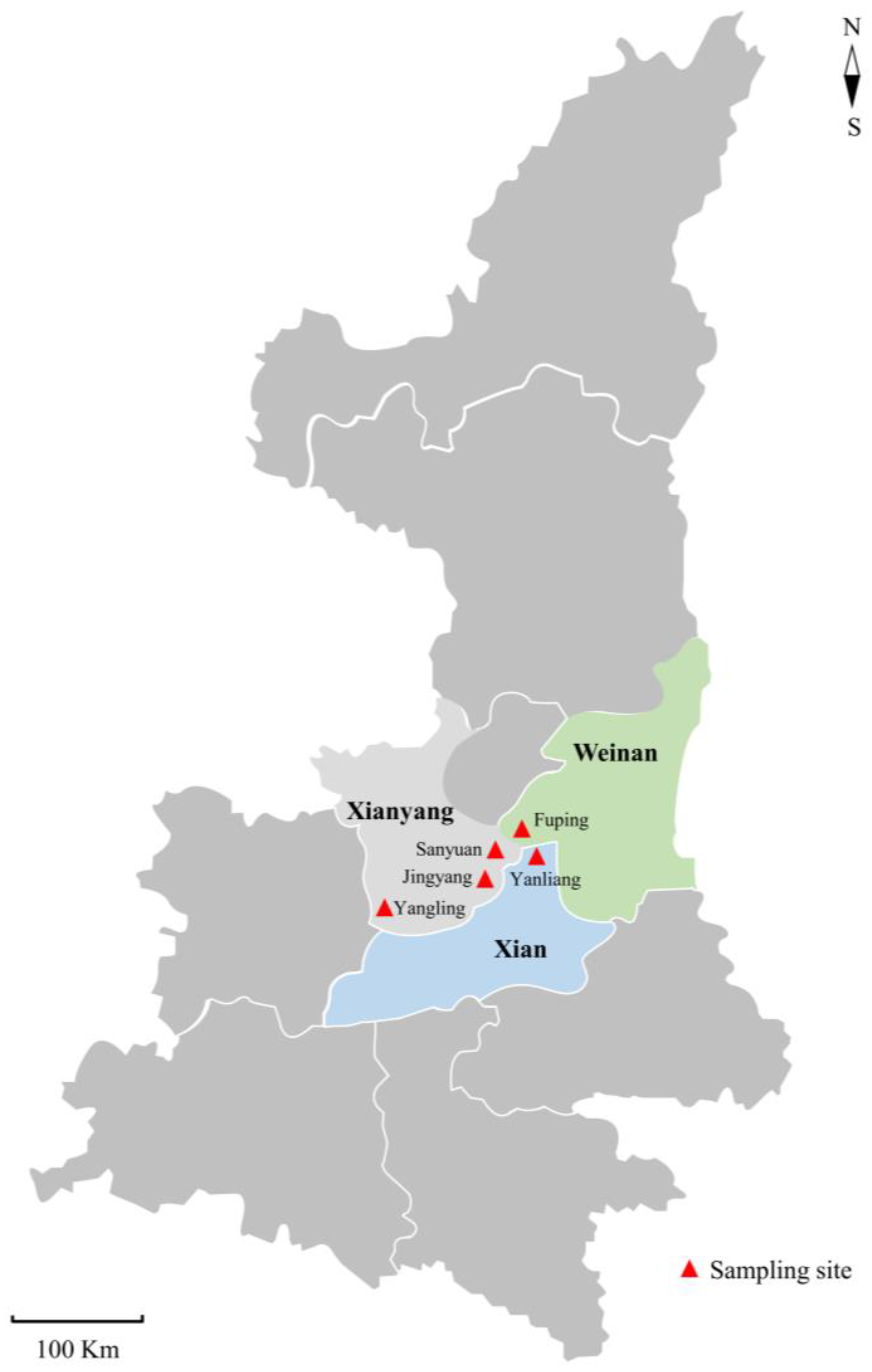
2.1. Samples

From February 2022 to May 2023, a total of 202 fecal samples were collected from diarrheal Guanzhong dairy goat kids in five locations of Shaanxi Province (Figure 1), containing 47, 102 and 53 fecal samples from goat kids aged <2 weeks, 2–4 weeks and 4–12 weeks, respectively. All samples were directly collected from the rectum of goat kids using sterile cotton swabs (HUNAUT, Qingdao, Shandong, China), placed into separate 50 mL centrifuge tubes (Thermo Fisher Scientific, Waltham, MA, USA), marked with sampling date, city name, location and age, and transported to the Parasitology Lab of Northwest A&F University for examination under cool conditions as soon as possible.

2.2. Genomic DNA Extraction

Genomic DNA was isolated from fecal samples using an E.Z.N.A. Stool DNA kit (Omega, Norcross, GA, USA) as per the manufacturer’s instructions. All gDNA samples were kept at −20 °C until use.

2.3. Detection, Genotyping and Subtyping of Cryptosporidium spp., G. duodenalis and E. bieneusi

A nested PCR-based sequencing technique targeting the SSU rRNA gene (~830 bp) was used to investigate the positive rates and species compositions of Cryptosporidium spp. in goat kids, as previously reported [35]. Then, C. parvum and C. xiaoi identified in the present study were subtyped using two nested PCR-based sequencing techniques of the gp60 gene [21,36].
The positive rates and assemblages of G. duodenalis were investigated by applying three nested PCR-based sequencing assays targeting the tpi gene (~530 bp) [37], bg gene (~511 bp) [38] and gdh gene (~599 bp) [39].
A nested PCR-based sequencing technique targeting the ITS gene locus (~392 bp) was used to determine the positive rates and genotypes of E. bieneusi in fecal samples, as previously reported [40].

2.4. Sequence Analysis

All positive secondary PCR products were sent to Sangon Biotech (Shanghai, China) for sequencing in both directions. The obtained sequences were assembled, edited and aligned using ChromasPro V1.33 (www.technelysium.com.au/ChromasPro.html (accessed on 15 June 2023)), BioEdit V7.04 (www.mbio.ncsu.edu/BioEdit/bioedit.html (accessed on 15 June 2023)) and ClustalX V2.1 (www.clustal.org/ (accessed on 15 June 2023)), respectively. To assess the relationships of Cryptosporidium spp., G. duodenalis and E. bieneusi found in the present study, three phylogenetic trees were constructed with the maximum likelihood (ML) method, a general time-reversible model and a bootstrap evaluation of 1000 replicates using MEGA V6.0 [41].

2.5. Bacterial Isolation and Identification

Escherichia coli strains were isolated from fecal samples using screening on MacConkey agar (Solarbio, Beijing, China), as previously reported [42]. Subsequently, the isolated strains were confirmed to be E. coli using a PCR-based sequencing assay targeting the 16S rRNA gene [43]. The E. coli strains of each sample were suspended in 20% glycerol and stored at −80 °C for further analysis.

2.6. Virulence Factor Determination of E. coli Strains

For each E. coli strain, a single colony of fresh bacterial culture on LB solid medium (Sangon Biotech, Shanghai, China) was selected and re-suspended in 150 µL ddH2O and then used as a DNA template in PCRs. Eight virulence genes for five pathotypes (EPEC, EHEC, ETEC, EAEC and EIEC) were detected in all the strains using two multiple PCRs, one for aggR, lt and st, and the other for ipaH, eaeA, ehxA, stx1 and stx2 [44,45,46].

2.7. Statistical Analysis

Differences in the positive rates of Cryptosporidium spp., G. duodenalis, E. bieneusi and E. coli in dairy goat kids with diarrhea among locations and age groups were analyzed using the χ2 test in SPSS V18.0 (IBM, Armonk, NY, USA). Significant differences were identified if the p value was less than 0.05.

2.8. Nucleotide Sequence Accession Numbers

Representative nucleotide sequences generated in this study are available in GenBankTM with the accession numbers of OR220596-OR220604 for E. bieneusi, OR229417-OR229423 and OR232183-OR232196 for Cryptosporidium spp., OR230698-OR230699 for E. coli and OR237889-OR237906 for G. duodenalis.

3. Results

3.1. Occurrence of Cryptosporidium Species and Subtypes in Dairy Goat Kids with Diarrhea

Of 202 fecal samples detected in the present study, 76 (37.6%) were positive for Cryptosporidium spp. in dairy goat kids with diarrhea (Table 1). A significant difference among the positive rates of Cryptosporidium was confirmed among the five locations (χ2 = 31.602; df = 4; p < 0.001), with the highest in Fuping (50.0%, 65/130), followed by Yangling (40.0%, 8/20), Yanliang (10.5%, 2/19), Jingyang (10.0%, 1/10) and Sanyuan (0%, 0/23). Significant differences in the positive rates of Cryptosporidium were also found among three farms in Fuping (χ2 = 6.333; df = 2; p = 0.042). Additionally, a significant difference in positive rates was also identified among three age groups (χ2 = 29.520; df = 2; p < 0.001), with the highest in goat kids aged 2–4 weeks (52.0%, 53/102), followed by <2 weeks (40.4%, 19/47) and 4–12 weeks (7.5%, 4/53) (Table 1).
A sequence analysis of the SSU rRNA gene indicated the existence of two Cryptosporidium species in goat kids in the present study, namely C. parvum (n = 41) and C. xiaoi (n = 35) (Table 1 and Figure 2). Both C. parvum and C. xiaoi were found in goat kids in Fuping and Yangling, while only one species was recognized in Yanliang (C. parvum) and Jingyang (C. xiaoi). Meanwhile, both C. parvum and C. xiaoi were found in goat kids in all three age groups (Table 1).
Further sequence analysis based on the gp60 gene showed subtype diversity within C. parvum and C. xiaoi in goat kids in the present study. As for C. parvum, 38 positive samples were successfully subtyped, with IIdA19G1 (n = 37) being the dominant one, followed by IIdA19G2 (n = 1). Meanwhile, two subtypes, namely XXIIIa (n = 22) and XXIIIg (n = 9), were found in 35 C. xiaoi-positive samples, with the mixed infection of XXIIIa and XXIIIg in two samples.

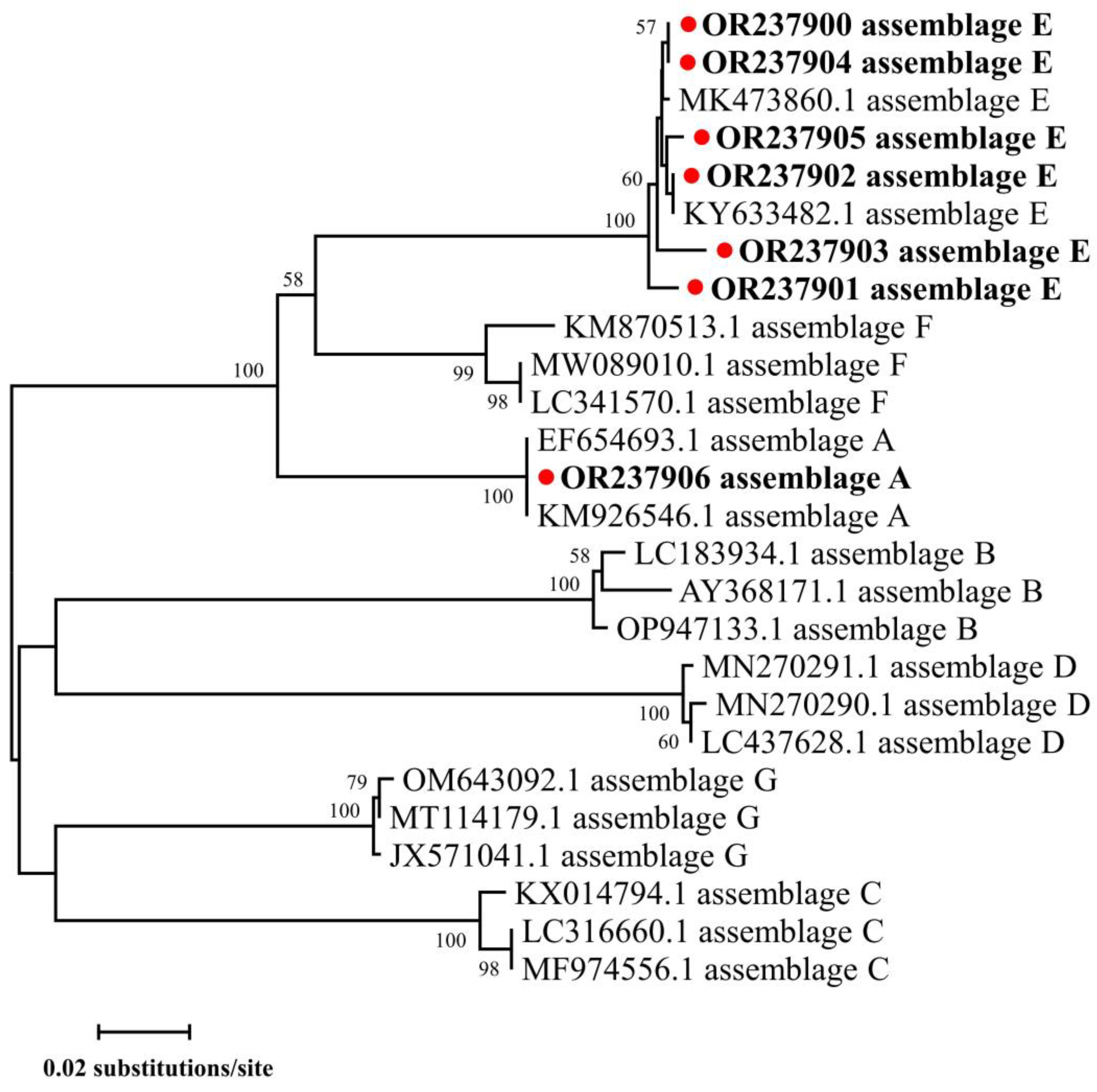
3.2. Occurrence of G. duodenalis Genotypes in Dairy Goat Kids with Diarrhea

The PCR analysis indicated the occurrence of G. duodenalis in 16.3% (33/202) of fecal samples, with the positive rates of 16.3% (33/202), 7.9% (16/202) and 14.9% (30/202) at the gene loci gdh, tpi and bg, respectively (Table 2). A significant difference in the positive rates of G. duodenalis was found among five locations (χ2 = 20.064; df = 4; p < 0.001) and three age groups (χ2 = 20.093; df = 2; p < 0.001), with the highest positive rates in goat kids from Jingyang (60.0%, 6/10) and aged 4–12 weeks (35.8%, 19/53). Meanwhile, a significant difference in the positive rates of G. duodenalis was also identified among three farms in Fuping (χ2 = 11.555; df = 2; p = 0.003). The sequence alignment of the gene loci gdh, tpi and bg and the phylogenetic analysis of the tpi gene locus indicated assemblages E and A in those samples positive for G. duodenalis (Table 2 and Figure 3). The most commonly identified genotype was assemblage E (n = 28), followed by assemblage A (n = 5). Both assemblages E and A were found in goat kids in Fuping, while only assemblage E was found in the other four regions. Meanwhile, both assemblages E and A were found in goat kids aged both <2 and 2–4 weeks, but only assemblage E was found in animals aged 4–12 weeks.

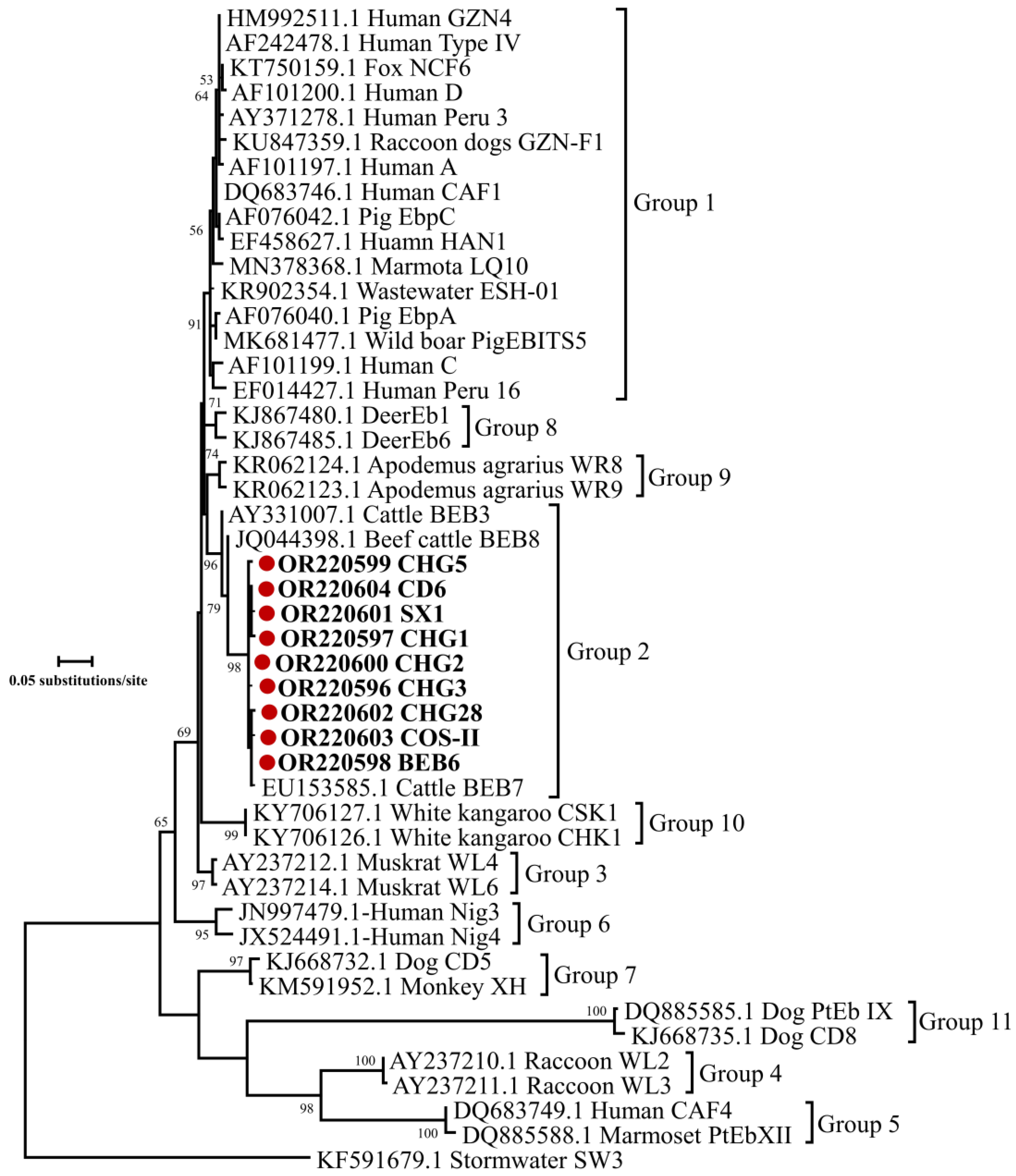
3.3. Occurrence of E. bieneusi Genotypes in Dairy Goat Kids with Diarrhea

Of the 202 fecal samples examined in the present study, 112 (55.4%) samples were positive for E. bieneusi in dairy goat kids with diarrhea (Table 3). The highest positive rate was found in goat kids from Yanliang (68.4%, 13/19), followed by Fuping (56.9%, 74/130), Sanyuan (56.5%, 13/23), Yangling (50.0%, 10/20) and Jingyang (20%, 2/10). Meanwhile, the positive rates were 60.8% (62/102), 54.7% (29/53) and 44.7% (21/47) in animals aged 2–4 weeks, 4–12 weeks and <2 weeks, respectively. Although the positive rates of E. bieneusi varied among locations (χ2 = 6.747; df = 4; p = 0.150) and age groups (χ2 = 3.393; df = 2; p = 0.183), no significant differences were found. Additionally, a significant difference in the positive rates of E. bieneusi was identified among three farms in Fuping (χ2 = 8.470; df = 2; p = 0.014).
A sequence analysis of the ITS gene of E. bieneusi identified nine known genotypes in the 112 sequences in the present study, namely CHG3, CHG1, BEB6, CHG5, CHG2, SX1, CHG28, COS-II and CD6 (Table 3). Among these identified genotypes, CHG3 was the most common genotype found in 51.8% (58/112) of goat kids, followed by CHG1 (25.0%, 28/112), BEB6 (7.1%, 8/112), CHG5 (6.3%, 7/112), CHG2 (3.6%, 4/112), SX1 (3.6%, 4/112), CHG28 (0.9%, 1/112), COS-II (0.9%, 1/112) and CD6 (0.9%, 1/112). Differences in genotype diversity were found among three locations, with nine (CHG3, CHG1, BEB6, CHG2, CHG5, SX1, CHG28, COS-II and CD6), four (CHG3, CHG5, SX1 and CHG2), three (CHG1, BEB6 and CHG3), two (CHG3 and CHG5) and two (CHG3 and BEB6) genotypes in Fuping, Yanliang, Yangling, Jingyang and Sanyuan, respectively. Meanwhile, seven (CHG1, CHG3, CHG5, CHG28, COS-II, SX1 and BEB6), seven (CHG3, CHG1, BEB6, CHG2, SX1, CHG5 and CD6) and four (CHG3, CHG1, CHG5 and BEB6) genotypes were identified in animals aged <2 weeks, 2–4 weeks and 4–12 weeks, respectively.
Further phylogenetic analysis based on the ITS gene of E. bieneusi showed that all nine genotypes (CHG3, CHG1, BEB6, CHG2, CHG5, SX1, CHG28, COS-II and CD6) belonged to the group 2, with increasing zoonotic potential (Figure 4). Since there were no variations among the sequences of each genotype, one representative sequence of each genotype was included in the phylogenetic analysis in Figure 4.

3.4. Occurrence of E. coli Pathotypes in Dairy Goat Kids with Diarrhea

The PCR-based sequencing analysis targeting the 16S rRNA gene indicated that 78.7% (159/202) of fecal samples were positive for E. coli (Table 4). Although the positive rates of E. coli varied among locations (χ2 = 5.499; df = 4; p = 0.240) and age groups (χ2 = 0.164; df = 2; p = 0.921), no significant differences were identified. In addition, no significant differences in the positive rates of E. coli were identified among three farms in Fuping (χ2 = 4.214; df = 2; p = 0.122). To further understand the pathotypes of E. coli in the present study, a total of eight virulence factors (eae, ehxA, stx2, stx1, lt, st, aggR and ipaH) representing EPEC, EHEC, ETEC, EAEC and EIEC E. coli were examined (Table 4). Four of eight virulence genes were identified in 159 strains in this study, namely eae (66.7%, 106/159), ehxA (36.5%, 58/159), stx2 (11.9%, 19/159) and stx1 (9.4%, 15/159). However, no strains were positive for the gene loci lt, st, aggR and ipaH (Table 4). Meanwhile, most strains carried anywhere from one to four virulence genes of EPEC and EHEC.

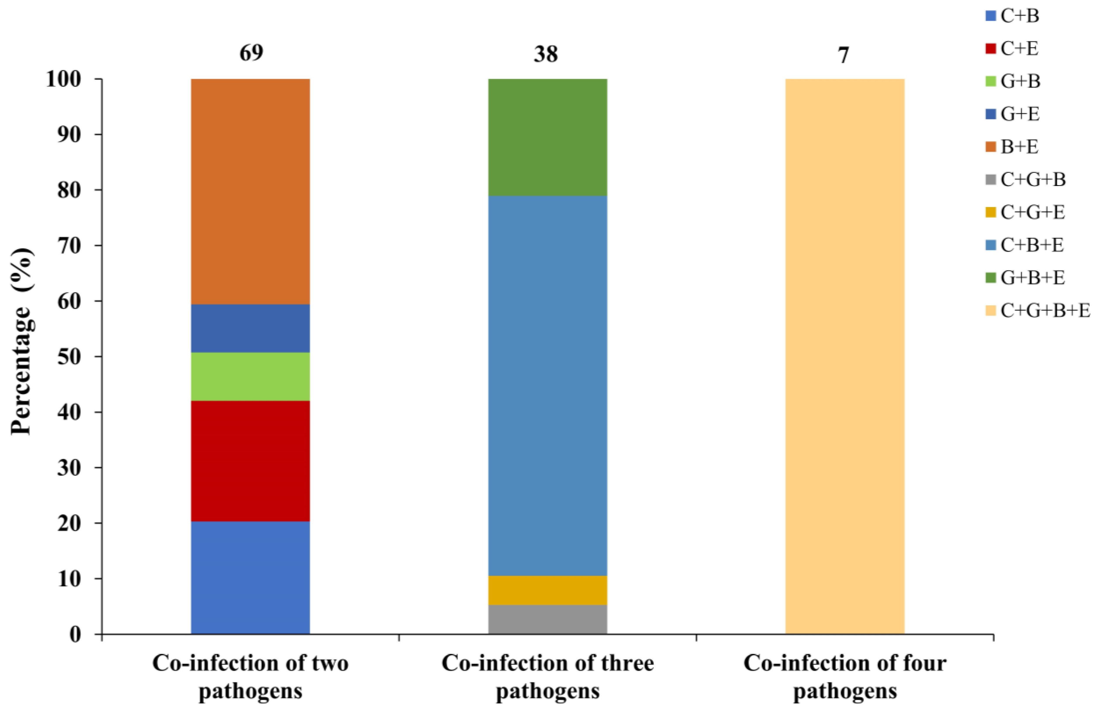
3.5. Co-Infection of Pathogens for Cryptosporidium spp., G. duodenalis, E. bieneusi and E. coli in Dairy Goat Kids with Diarrhea

Co-infection of pathogens was found in 114 of 202 (56.4%) fecal samples, with 69, 38 and 7 samples positive for two, three and four pathogens, respectively (Figure 5). As for co-infection of two pathogens, a total of five types were identified, with the co-infection of E. bieneusi and E. coli being the most common one. As for co-infection of three pathogens, four types were found, with the co-infection of Cryptosporidium spp., E. bieneusi and E. coli being the dominant one.

4. Discussion

Cryptosporidium spp., G. duodenalis, E. bieneusi and E. coli are common and important zoonotic diarrheal pathogens greatly endangering the health of humans and animals, especially children and young animals [1,2,3,4]. The present study investigated the colonization frequency and genetic composition of Cryptosporidium spp., G. duodenalis, E. bieneusi and E. coli in diarrheal kids of Guanzhong dairy goats, which could expand our knowledge on the occurrence and distribution of these four pathogens in goats.
Cryptosporidium spp. are commonly found in sheep and goats in China. In the present study, the positive rate of Cryptosporidium spp. was 37.6% (76/202) in diarrheal kids of Guanzhong dairy goats from Shaanxi, which is similar to that seen in meat-producing goats (34.0%, 49/144), but higher than that in Saanen dairy goats (14.5%, 25/170) and northern Shaanxi white cashmere goats (9.5%, 30/315) in Shaanxi [32]. In addition, higher positive rates of Cryptosporidium spp. have been reported in sheep and goats in Ningxia (45.5%, 55/121), Henan (55.4%, 92/166) and Anhui (66.9%, 87/130) [47,48], while lower positive rates have been found in other provinces in China [49], such as Chongqing (5.3%, 16/301) [50], Heilongjiang (0.4%, 2/489) [51], Gansu (4.5%, 8/177) [52] and Xinjiang (0.9%, 3/318) [53]. The differences in the positive rates of Cryptosporidium spp. in sheep and goats are possibly caused by discrepancies in animal species and breeds, detection methods, sampling sizes, geographic regions and management practices.
Cryptosporidium infection in dairy goat kids was related to the age. In the present study, Cryptosporidium spp. were frequently found in all age groups, with the positive rates of 40.4% (19/47), 52.0% (53/102) and 7.5% (4/53) for goat kids aged <2, 2–4 and 4–12 weeks, respectively, indicating lower positive rates of animals aged >4 weeks compared with <4 weeks. Similar results have also been reported in sheep in Qinghai [54] and goats in Henan, Anhui and Qinghai [47], reflecting that immunity to Cryptosporidium in sheep and goats possibly increases with age. However, contrary results have been found in ewes and lambs in Inner Mongolia [55] and sheep in Henan, Anhui and Qinghai [47], reflected by higher positive rates in older animals compared with younger ones.
The sequences analysis indicated two Cryptosporidium species (C. parvum and C. xiaoi) in goat kids. Cryptosporidium parvum is a zoonotic species with a wide host range, including humans, cattle, sheep and goats, dogs, cats and mice [20]. Although C. xiaoi is the most common species in sheep and goats in previous reports [20,32,55], the present study found that C. parvum contributed to over half of the cases, indicating that potentially zoonotic strains of Cryptosporidium circulated on the investigated farms. A further subtyping analysis showed the existence of subtypes IIdA19G1 and IIdA19G2 in C. parvum-positive samples, with the former being the dominant one, which was also found in previous studies [56]. IIdA19G1 has also previously been reported in humans and animals [57,58,59,60], reflecting potential zoonotic transmission between humans and animals of this subtype. Cryptosporidium xiaoi was the other common species in the present study, which has also been widely reported in sheep and goats [31,32,53,54,56]. A further subtyping analysis based on the gp60 gene locus found two potential goat-adapted subtypes (XXIIIa and XXIIIg) in C. xiaoi-positive samples, which was in accordance with a previous report [36].
Assemblages E and A were two G. duodenalis genotypes in goat kid samples in the present study, with the former being the major one, which is similar to studies in sheep and goats from Henan, Heilongjiang, Yunnan, Shaanxi and Qinghai [32,61,62,63,64,65]. Assemblage E has been widely reported in artiodactyls, such as cattle, sheep, pigs, goats and alpacas [24,66]. For a period of time, assemblage E was recognized as one animal-adapted genotype, but a report of this genotype in humans indicated its zoonotic potential [67]. Assemblage A is a zoonotic genotype found in humans, livestock, cats, dogs, beavers, guinea pigs and other primates and has also frequently been reported in sheep and goats [61,62,68,69].
A sequence analysis based on the ITS gene locus of 112 E. bieneusi isolates revealed nine known genotypes (CHG3, CHG1, BEB6, CHG5, CHG2, SX1, CHG28, COS-II and CD6) in goat kids in the present study, and these genotypes have been previously reported in sheep and goats [32,70,71,72]. Among the identified genotypes in this study, CHG3 was the most common genotype, which was also found in goats from Henan, Yunnan, Anhui, Chongqing and Shaanxi, as well as in sheep in Henan [71]. BEB6 is a zoonotic genotype with a wide host range, including humans [26], non-human primates [73], bovines [74], deer [75], takins [76], alpacas [77], cats [78] and birds [79]. Further phylogenetic analysis revealed that all nine genotypes belonged to the group 2. Although genotypes in the group 2 were reported to be ruminant-adapted at the beginning, more and more zoonotic genotypes identified in this group reflect an increasing risk of causing zoonotic infection between humans and animals [25,80].
A high positive rate of E. coli was commonly found in the present study, which is in accordance with previous reports in goats [42,81]. Further pathotype analysis based on the eight virulence genes of these E. coli strains indicated the existence of EPEC and EHEC, with the former being the dominant one in goat kids, while no strains were identified to be positive for virulence genes of ETEC, EAEC and EIEC. However, divergent pathotypes were reported in sheep, reflected by the dominance of EAEC and EHEC [42].
Co-infection of these diarrhea-related pathogens was found in goats in the present study, which has also been reported in sheep and goats in previous studies [32,52,53]. Previous studies have reported the co-infections of two pathogens (Cryptosporidium spp. and G. duodenalis, G. duodenalis and E. bieneusi, and Cryptosporidium spp. and E. bieneusi) and three pathogens (Cryptosporidium spp., G. duodenalis and E. bieneusi) in sheep and goats [32,52,53]. Although the present study did not find the co-infection of Cryptosporidium spp. and G. duodenalis, the co-infection of E. coli with Cryptosporidium spp., G. duodenalis and E. bieneusi was identified for the first time in sheep and goats.

5. Conclusions

This study explored the colonization frequency and genetic make-up of Cryptosporidium spp., G. duodenalis, E. bieneusi and E. coli in diarrheal kids of Guanzhong dairy goats from partial regions of Shaanxi Province. The findings in the present study indicated high positive rates and zoonotic species/genotypes/subtypes/pathotypes of these four diarrhea-related pathogens in goat kids. Considering the zoonotic potential of Cryptosporidium spp., G. duodenalis, E. bieneusi and E. coli in goat kids in this study, interventions are needed to prevent the cross-transmission of these diarrhea-related pathogens between animals and humans.

Author Contributions

Conceptualization, G.Z.; methodology, X.Y. and J.W.; software and formal analysis, X.Y. and J.W.; validation and investigation, X.Y. and J.W.; data curation, S.H., Y.F. and J.S.; writing—original draft preparation, X.Y.; writing—review and editing, X.Y. and G.Z.; visualization, X.Y. and J.W.; supervision, G.Z., Y.F. and J.S.; project administration, G.Z. All authors have read and agreed to the published version of the manuscript.

Funding

This project was supported by the Key Research and Development Program of Shaanxi Province (2022NY-097), the National Natural Science Foundation of China (32072890) and the Scientific Research Foundation of the Northwest A&F University (2452021058; 2452022158).

Institutional Review Board Statement

This study was conducted under the approval and instructions of the ethics committee of Northwest A&F University (DY2022052, approved on 15 January 2022).

Informed Consent Statement

Not applicable.

Data Availability Statement

Data are contained within the article.

Conflicts of Interest

The authors declare no conflict of interest.

References

  1. Yang, X.; Guo, Y.; Xiao, L.; Feng, Y. Molecular epidemiology of human cryptosporidiosis in low- and middle-income countries. Clin. Microbiol. Rev. 2021, 34, e00087-19. [Google Scholar] [CrossRef]
  2. Li, W.; Feng, Y.; Xiao, L. Enterocytozoon bieneusi. Trends Parasitol. 2022, 38, 95–96. [Google Scholar] [CrossRef] [PubMed]
  3. Feng, Y.; Xiao, L. Zoonotic potential and molecular epidemiology of Giardia species and giardiasis. Clin. Microbiol. Rev. 2011, 24, 110–140. [Google Scholar] [CrossRef] [PubMed]
  4. Ferens, W.A.; Hovde, C.J. Escherichia coli O157:H7: Animal reservoir and sources of human infection. Foodborne Pathog. Dis. 2011, 8, 465–487. [Google Scholar] [CrossRef]
  5. Zhu, G.; Yin, J.; Cuny, G.D. Current status and challenges in drug discovery against the globally important zoonotic cryptosporidiosis. Anim. Dis. 2021, 1, 3. [Google Scholar] [CrossRef]
  6. Li, W.; Liu, X.; Gu, Y.; Liu, J.; Luo, J. Prevalence of Cryptosporidium, Giardia, Blastocystis, and trichomonads in domestic cats in East China. J. Vet. Med. Sci. 2019, 81, 890–896. [Google Scholar] [CrossRef] [PubMed]
  7. Rojas-López, L.; Marques, R.C.; Svärd, S.G. Giardia duodenalis. Trends Parasitol. 2022, 38, 605–606. [Google Scholar] [CrossRef] [PubMed]
  8. Ortega, Y.R.; Cama, V.A. Foodborne transmission. In Cryptosporidium and Cryptosporidiosis, 2nd ed.; Fayer, R., Xiao, L., Eds.; CRC Press: Boca Raton, FL, USA, 2007; pp. 289–304. [Google Scholar]
  9. Hwang, S.B.; Chelliah, R.; Kang, J.E.; Rubab, M.; Banan-MwineDaliri, E.; Elahi, F.; Oh, D.H. Role of recent therapeutic applications and the infection strategies of Shiga toxin-producing Escherichia coli. Front. Cell. Infect. Microbiol. 2021, 11, 614963. [Google Scholar] [CrossRef]
  10. Xiao, L.; Feng, Y. Molecular epidemiologic tools for waterborne pathogens Cryptosporidium spp. and Giardia duodenalis. Food Waterborne Parasitol. 2017, 8–9, 14–32. [Google Scholar] [CrossRef]
  11. Cui, Z.; Wang, R.; Huang, J.; Wang, H.; Zhao, J.; Luo, N.; Li, J.; Zhang, Z.; Zhang, L. Cryptosporidiosis caused by Cryptosporidium parvum subtype IIdA15G1 at a dairy farm in Northwestern China. Parasit. Vectors 2014, 7, 529. [Google Scholar] [CrossRef]
  12. Li, N.; Wang, R.; Cai, M.; Jiang, W.; Feng, Y.; Xiao, L. Outbreak of cryptosporidiosis due to Cryptosporidium parvum subtype IIdA19G1 in neonatal calves on a dairy farm in China. Int. J. Parasitol. 2019, 49, 569–577. [Google Scholar] [CrossRef] [PubMed]
  13. Matos, O.; Lobo, M.L.; Xiao, L. Epidemiology of Enterocytozoon bieneusi infection in Humans. J. Parasitol. Res. 2012, 2012, 981424. [Google Scholar] [CrossRef] [PubMed]
  14. Hoffmann, M.; Fischer, M.A.; Neumann, B.; Kiesewetter, K.; Hoffmann, I.; Werner, G.; Pfeifer, Y.; Lübbert, C. Carbapenemase-producing Gram-negative bacteria in hospital wastewater, wastewater treatment plants and surface waters in a metropolitan area in Germany, 2020. Sci. Total Environ. 2023, 890, 164179. [Google Scholar] [CrossRef] [PubMed]
  15. Kotloff, K.L.; Nasrin, D.; Blackwelder, W.C.; Wu, Y.; Farag, T.; Panchalingham, S.; Sow, S.O.; Sur, D.; Zaidi, A.K.M.; Faruque, A.S.G.; et al. The incidence, aetiology, and adverse clinical consequences of less severe diarrhoeal episodes among infants and children residing in low-income and middle-income countries: A 12-month case-control study as a follow-on to the Global Enteric Multicenter Study (GEMS). Lancet Glob. Health 2019, 7, e568–e584. [Google Scholar]
  16. Agholi, M.; Hatam, G.R.; Motazedian, M.H. HIV/AIDS-associated opportunistic protozoal diarrhea. AIDS Res. Hum. Retroviruses 2013, 29, 35–41. [Google Scholar] [CrossRef]
  17. Wang, L.; Zhang, H.; Zhao, X.; Zhang, L.; Zhang, G.; Guo, M.; Liu, L.; Feng, Y.; Xiao, L. Zoonotic Cryptosporidium species and Enterocytozoon bieneusi genotypes in HIV-positive patients on antiretroviral therapy. J. Clin. Microbiol. 2013, 51, 557–563. [Google Scholar] [CrossRef]
  18. Rogawski, E.T.; Liu, J.; Platts-Mills, J.A.; Kabir, F.; Lertsethtakarn, P.; Siguas, M.; Khan, S.S.; Praharaj, I.; Murei, A.; Nshama, R.; et al. Use of quantitative molecular diagnostic methods to investigate the effect of enteropathogen infections on linear growth in children in low-resource settings: Longitudinal analysis of results from the MAL-ED cohort study. Lancet Glob. Health 2018, 6, e1319–e1328. [Google Scholar] [CrossRef]
  19. Ryan, U.; Feng, Y.; Fayer, R.; Xiao, L. Taxonomy and molecular epidemiology of Cryptosporidium and Giardia—A 50 year perspective (1971–2021). Int. J. Parasitol. 2021, 51, 1099–1119. [Google Scholar] [CrossRef]
  20. Feng, Y.; Ryan, U.M.; Xiao, L. Genetic diversity and population structure of Cryptosporidium. Trends Parasitol. 2018, 34, 997–1011. [Google Scholar] [CrossRef]
  21. Sulaiman, I.M.; Hira, P.R.; Zhou, L.; Al-Ali, F.M.; Al-Shelahi, F.A.; Shweiki, H.M.; Iqbal, J.; Khalid, N.; Xiao, L. Unique endemicity of cryptosporidiosis in children in Kuwait. J. Clin. Microbiol. 2005, 43, 2805–2809. [Google Scholar] [CrossRef]
  22. Jiang, W.; Roellig, D.M.; Guo, Y.; Li, N.; Feng, Y.; Xiao, L. Development of a subtyping tool for zoonotic pathogen Cryptosporidium canis. J. Clin. Microbiol. 2021, 59, e02474-20. [Google Scholar] [CrossRef] [PubMed]
  23. Stensvold, C.R.; Beser, J.; Axén, C.; Lebbad, M. High applicability of a novel method for gp60-based subtyping of Cryptosporidium meleagridis. J. Clin. Microbiol. 2014, 52, 2311–2319. [Google Scholar] [CrossRef] [PubMed]
  24. Cai, W.; Ryan, U.; Xiao, L.; Feng, Y. Zoonotic giardiasis: An update. Parasitol. Res. 2021, 120, 4199–4218. [Google Scholar] [CrossRef] [PubMed]
  25. Li, W.; Feng, Y.; Zhang, L.; Xiao, L. Potential impacts of host specificity on zoonotic or interspecies transmission of Enterocytozoon bieneusi. Infect. Genet. Evol. 2019, 75, 104033. [Google Scholar] [CrossRef] [PubMed]
  26. Wang, L.; Xiao, L.; Duan, L.; Ye, J.; Guo, Y.; Guo, M.; Liu, L.; Feng, Y. Concurrent infections of Giardia duodenalis, Enterocytozoon bieneusi, and Clostridium difficile in children during a cryptosporidiosis outbreak in a pediatric hospital in China. PLoS Negl. Trop. Dis. 2013, 7, e2437. [Google Scholar] [CrossRef] [PubMed]
  27. Zhang, X.; Wang, Z.; Su, Y.; Liang, X.; Sun, X.; Peng, S.; Lu, H.; Jiang, N.; Yin, J.; Xiang, M.; et al. Identification and genotyping of Enterocytozoon bieneusi in China. J. Clin. Microbiol. 2011, 49, 2006–2008. [Google Scholar] [CrossRef]
  28. Habouria, H.; Pokharel, P.; Maris, S.; Garénaux, A.; Bessaiah, H.; Houle, S.; Veyrier, F.J.; Guyomard-Rabenirina, S.; Talarmin, A.; Dozois, C.M. Three new serine-protease autotransporters of Enterobacteriaceae (SPATEs) from extra-intestinal pathogenic Escherichia coli and combined role of SPATEs for cytotoxicity and colonization of the mouse kidney. Virulence 2019, 10, 568–587. [Google Scholar] [CrossRef]
  29. Guimarães, A.C.; Meireles, L.M.; Lemos, M.F.; Guimarães, M.C.C.; Endringer, D.C.; Fronza, M.; Scherer, R. Antibacterial activity of terpenes and terpenoids present in essential oils. Molecules 2019, 24, 2471. [Google Scholar] [CrossRef]
  30. Malberg Tetzschner, A.M.; Johnson, J.R.; Johnston, B.D.; Lund, O.; Scheutz, F. In silico genotyping of Escherichia coli isolates for extraintestinal virulence genes by use of whole-genome sequencing data. J. Clin. Microbiol. 2020, 58, e01269-20. [Google Scholar] [CrossRef]
  31. Mi, R.; Wang, X.; Huang, Y.; Zhou, P.; Liu, Y.; Chen, Y.; Chen, J.; Zhu, W.; Chen, Z. Prevalence and molecular characterization of Cryptosporidium in goats across four provincial level areas in China. PLoS ONE 2014, 9, e111164. [Google Scholar] [CrossRef]
  32. Peng, X.Q.; Tian, G.R.; Ren, G.J.; Yu, Z.Q.; Lok, J.B.; Zhang, L.X.; Wang, X.T.; Song, J.K.; Zhao, G.H. Infection rate of Giardia duodenalis, Cryptosporidium spp. and Enterocytozoon bieneusi in cashmere, dairy and meat goats in China. Infect. Genet. Evol. 2016, 41, 26–31. [Google Scholar] [CrossRef]
  33. Wang, R.; Li, G.; Cui, B.; Huang, J.; Cui, Z.; Zhang, S.; Dong, H.; Yue, D.; Zhang, L.; Ning, C.; et al. Prevalence, molecular characterization and zoonotic potential of Cryptosporidium spp. in goats in Henan and Chongqing, China. Exp. Parasitol. 2014, 142, 11–16. [Google Scholar] [CrossRef]
  34. Zhong, Z.; Tu, R.; Ou, H.; Yan, G.; Dan, J.; Xiao, Q.; Wang, Y.; Cao, S.; Shen, L.; Deng, J.; et al. Occurrence and genetic characterization of Giardia duodenalis and Cryptosporidium spp. from adult goats in Sichuan Province, China. PLoS ONE 2018, 13, e0199325. [Google Scholar] [CrossRef]
  35. Xiao, L.; Escalante, L.; Yang, C.; Sulaiman, I.; Escalante, A.A.; Montali, R.J.; Fayer, R.; Lal, A.A. Phylogenetic analysis of Cryptosporidium parasites based on the small-subunit rRNA gene locus. Appl. Environ. Microbiol. 1999, 65, 1578–1583. [Google Scholar] [CrossRef] [PubMed]
  36. Fan, Y.; Huang, X.; Guo, S.; Yang, F.; Yang, X.; Guo, Y.; Feng, Y.; Xiao, L.; Li, N. Subtyping Cryptosporidium xiaoi, a common pathogen in sheep and goats. Pathogens 2021, 10, 800. [Google Scholar] [CrossRef] [PubMed]
  37. Sulaiman, I.M.; Fayer, R.; Bern, C.; Gilman, R.H.; Trout, J.M.; Schantz, P.M.; Das, P.; Lal, A.A.; Xiao, L. Triosephosphate isomerase gene characterization and potential zoonotic transmission of Giardia duodenalis. Emerg. Infect. Dis. 2003, 9, 1444–1452. [Google Scholar] [CrossRef] [PubMed]
  38. Lalle, M.; Pozio, E.; Capelli, G.; Bruschi, F.; Crotti, D.; Cacciò, S.M. Genetic heterogeneity at the beta-giardin locus among human and animal isolates of Giardia duodenalis and identification of potentially zoonotic subgenotypes. Int. J. Parasitol. 2005, 35, 207–213. [Google Scholar] [CrossRef]
  39. Cacciò, S.M.; Beck, R.; Lalle, M.; Marinculic, A.; Pozio, E. Multilocus genotyping of Giardia duodenalis reveals striking differences between assemblages A and B. Int. J. Parasitol. 2008, 38, 1523–1531. [Google Scholar] [CrossRef]
  40. Sulaiman, I.M.; Fayer, R.; Lal, A.A.; Trout, J.M.; Schaefer, F.W.; Xiao, L. Molecular characterization of microsporidia indicates that wild mammals Harbor host-adapted Enterocytozoon spp. as well as human-pathogenic Enterocytozoon bieneusi. Appl. Environ. Microbiol. 2003, 69, 4495–4501. [Google Scholar] [CrossRef]
  41. Tamura, K.; Stecher, G.; Peterson, D.; Filipski, A.; Kumar, S. MEGA6: Molecular evolutionary genetics analysis version 6.0. Mol. Biol. Evol. 2013, 30, 2725–2729. [Google Scholar] [CrossRef]
  42. Zhao, X.; Lv, Y.; Adam, F.E.A.; Xie, Q.; Wang, B.; Bai, X.; Wang, X.; Shan, H.; Wang, X.; Liu, H.; et al. Comparison of antimicrobial resistance, virulence genes, phylogroups, and biofilm formation of Escherichia coli isolated from intensive farming and free-range sheep. Front. Microbiol. 2021, 12, 699927. [Google Scholar] [CrossRef]
  43. Greisen, K.; Loeffelholz, M.; Purohit, A.; Leong, D. PCR primers and probes for the 16S rRNA gene of most species of pathogenic bacteria, including bacteria found in cerebrospinal fluid. J. Clin. Microbiol. 1994, 32, 335–351. [Google Scholar] [CrossRef]
  44. López-Saucedo, C.; Cerna, J.F.; Villegas-Sepulveda, N.; Thompson, R.; Velazquez, F.R.; Torres, J.; Tarr, P.I.; Estrada-García, T. Single multiplex polymerase chain reaction to detect diverse loci associated with diarrheagenic Escherichia coli. Emerg. Infect. Dis. 2003, 9, 127–131. [Google Scholar] [CrossRef]
  45. Chapman, T.A.; Wu, X.Y.; Barchia, I.; Bettelheim, K.A.; Driesen, S.; Trott, D.; Wilson, M.; Chin, J.J. Comparison of virulence gene profiles of Escherichia coli strains isolated from healthy and diarrheic swine. Appl. Environ. Microbiol. 2006, 72, 4782–4795. [Google Scholar] [CrossRef] [PubMed]
  46. Toma, C.; Lu, Y.; Higa, N.; Nakasone, N.; Chinen, I.; Baschkier, A.; Rivas, M.; Iwanaga, M. Multiplex PCR assay for identification of human diarrheagenic Escherichia coli. J. Clin. Microbiol. 2003, 41, 2669–2671. [Google Scholar] [CrossRef] [PubMed]
  47. Huang, X. Molecular Epidemiological Investigation of Cryptosporidium spp. in Sheep and Goats in Midwest China and Development of a Subtyping Tool for Cryptosporidium xiaoi. Master’s Thesis, South China Agricultural University, Guangzhou, China, 27 August 2020. (In Chinese). [Google Scholar]
  48. Mi, R.; Wang, X.; Huang, Y.; Mu, G.; Zhang, Y.; Jia, H.; Zhang, X.; Yang, H.; Wang, X.; Han, X.; et al. Sheep as a potential source of zoonotic cryptosporidiosis in China. Appl. Environ. Microbiol. 2018, 84, e00868-18. [Google Scholar] [CrossRef] [PubMed]
  49. Yang, X.Y.; Gong, Q.L.; Zhao, B.; Cai, Y.N.; Zhao, Q. Prevalence of Cryptosporidium infection in sheep and goat flocks in China during 2010–2019: A systematic review and meta-analysis. Vector Borne Zoonotic Dis. 2021, 21, 692–706. [Google Scholar] [CrossRef]
  50. Chen, J.; Shen, K.; Ren, H.; Gao, L.; Ning, C. Investigation of goats Cryptosporidium infection in some breeding farms of Chongqing. Chin. J. Vet. Med. 2012, 48, 15–17. (In Chinese) [Google Scholar]
  51. Jiang, Y. Genotyping of Three Enteric Protozoans in Sheep from Heilongjiang Province and Assessment of Their Zoonotic Potential. Master’s Thesis, Northeast Agricultural University, Harbin, China, 14 June 2016. (In Chinese). [Google Scholar]
  52. Wu, Y.; Chang, Y.; Chen, Y.; Zhang, X.; Li, D.; Zheng, S.; Wang, L.; Li, J.; Ning, C.; Zhang, L. Occurrence and molecular characterization of Cryptosporidium spp., Giardia duodenalis, and Enterocytozoon bieneusi from Tibetan sheep in Gansu, China. Infect. Genet. Evol. 2018, 64, 46–51. [Google Scholar] [CrossRef]
  53. Qi, M.; Zhang, Z.; Zhao, A.; Jing, B.; Guan, G.; Luo, J.; Zhang, L. Distribution and molecular characterization of Cryptosporidium spp., Giardia duodenalis, and Enterocytozoon bieneusi amongst grazing adult sheep in Xinjiang, China. Parasitol. Int. 2019, 71, 80–86. [Google Scholar] [CrossRef]
  54. Li, P.; Cai, J.; Cai, M.; Wu, W.; Li, C.; Lei, M.; Xu, H.; Feng, L.; Ma, J.; Feng, Y.; et al. Distribution of Cryptosporidium species in Tibetan sheep and yaks in Qinghai, China. Vet. Parasitol. 2016, 215, 58–62. [Google Scholar] [CrossRef] [PubMed]
  55. Ye, J.; Xiao, L.; Wang, Y.; Wang, L.; Amer, S.; Roellig, D.M.; Guo, Y.; Feng, Y. Periparturient transmission of Cryptosporidium xiaoi from ewes to lambs. Vet. Parasitol. 2013, 197, 627–633. [Google Scholar] [CrossRef] [PubMed]
  56. Quílez, J.; Torres, E.; Chalmers, R.M.; Hadfield, S.J.; Del Cacho, E.; Sánchez-Acedo, C. Cryptosporidium genotypes and subtypes in lambs and goat kids in Spain. Appl. Environ. Microbiol. 2008, 74, 6026–6031. [Google Scholar] [CrossRef]
  57. Yu, F.; Li, D.; Chang, Y.; Wu, Y.; Guo, Z.; Jia, L.; Xu, J.; Li, J.; Qi, M.; Wang, R.; et al. Molecular characterization of three intestinal protozoans in hospitalized children with different disease backgrounds in Zhengzhou, central China. Parasit. Vectors 2019, 12, 543. [Google Scholar] [CrossRef] [PubMed]
  58. Huang, J.; Zhang, Z.; Zhang, Y.; Yang, Y.; Zhao, J.; Wang, R.; Jian, F.; Ning, C.; Zhang, W.; Zhang, L. Prevalence and molecular characterization of Cryptosporidium spp. and Giardia duodenalis in deer in Henan and Jilin, China. Parasit. Vectors 2018, 11, 239. [Google Scholar] [CrossRef] [PubMed]
  59. Wang, Y.; Zhang, B.; Li, J.; Yu, S.; Zhang, N.; Liu, S.; Zhang, Y.; Li, J.; Ma, N.; Cai, Y.; et al. Development of a quantitative real-time PCR assay for detection of Cryptosporidium spp. infection and threatening caused by Cryptosporidium parvum subtype IIdA19G1 in diarrhea calves from Northeastern China. Vector Borne Zoonotic Dis. 2021, 21, 179–190. [Google Scholar] [CrossRef] [PubMed]
  60. Jian, F.; Liu, A.; Wang, R.; Zhang, S.; Qi, M.; Zhao, W.; Shi, Y.; Wang, J.; Wei, J.; Zhang, L.; et al. Common occurrence of Cryptosporidium hominis in horses and donkeys. Infect. Genet. Evol. 2016, 43, 261–266. [Google Scholar] [CrossRef]
  61. Wang, H.; Qi, M.; Zhang, K.; Li, J.; Huang, J.; Ning, C.; Zhang, L. Prevalence and genotyping of Giardia duodenalis isolated from sheep in Henan Province, central China. Infect. Genet. Evol. 2016, 39, 330–335. [Google Scholar] [CrossRef]
  62. Zhang, W.; Zhang, X.; Wang, R.; Liu, A.; Shen, Y.; Ling, H.; Cao, J.; Yang, F.; Zhang, X.; Zhang, L. Genetic characterizations of Giardia duodenalis in sheep and goats in Heilongjiang Province, China and possibility of zoonotic transmission. PLoS Negl. Trop. Dis. 2012, 6, e1826. [Google Scholar] [CrossRef]
  63. Yang, F.; Ma, L.; Gou, J.M.; Yao, H.Z.; Ren, M.; Yang, B.K.; Lin, Q. Seasonal distribution of Cryptosporidium spp., Giardia duodenalis and Enterocytozoon bieneusi in Tibetan sheep in Qinghai, China. Parasit. Vectors 2022, 15, 394. [Google Scholar] [CrossRef]
  64. Chen, D.; Zou, Y.; Li, Z.; Wang, S.S.; Xie, S.C.; Shi, L.Q.; Zou, F.C.; Yang, J.F.; Zhao, G.H.; Zhu, X.Q. Occurrence and multilocus genotyping of Giardia duodenalis in black-boned sheep and goats in southwestern China. Parasit. Vectors 2019, 12, 102. [Google Scholar] [CrossRef] [PubMed]
  65. Yin, Y.L.; Zhang, H.J.; Yuan, Y.J.; Tang, H.; Chen, D.; Jing, S.; Wu, H.X.; Wang, S.S.; Zhao, G.H. Prevalence and multi-locus genotyping of Giardia duodenalis from goats in Shaanxi province, northwestern China. Acta Trop. 2018, 182, 202–206. [Google Scholar] [CrossRef] [PubMed]
  66. Ryan, U.; Cacciò, S.M. Zoonotic potential of Giardia. Int. J. Parasitol. 2013, 43, 943–956. [Google Scholar] [CrossRef] [PubMed]
  67. Zahedi, A.; Field, D.; Ryan, U. Molecular typing of Giardia duodenalis in humans in Queensland—First report of assemblage E. Parasitology 2017, 144, 1154–1161. [Google Scholar] [CrossRef]
  68. Ye, J.; Xiao, L.; Wang, Y.; Guo, Y.; Roellig, D.M.; Feng, Y. Dominance of Giardia duodenalis assemblage A and Enterocytozoon bieneusi genotype BEB6 in sheep in Inner Mongolia, China. Vet. Parasitol. 2015, 210, 235–239. [Google Scholar] [CrossRef]
  69. Berrilli, F.; D’Alfonso, R.; Giangaspero, A.; Marangi, M.; Brandonisio, O.; Kaboré, Y.; Glé, C.; Cianfanelli, C.; Lauro, R.; Di Cave, D. Giardia duodenalis genotypes and Cryptosporidium species in humans and domestic animals in Côte d’Ivoire: Occurrence and evidence for environmental contamination. Trans. R. Soc. Trop. Med. Hyg. 2012, 106, 191–195. [Google Scholar] [CrossRef]
  70. Li, W.C.; Wang, K.; Gu, Y.F. Detection and genotyping study of Enterocytozoon bieneusi in sheep and goats in East-central China. Acta Parasitol. 2019, 64, 44–50. [Google Scholar] [CrossRef]
  71. Shi, K.; Li, M.; Wang, X.; Li, J.; Karim, M.R.; Wang, R.; Zhang, L.; Jian, F.; Ning, C. Molecular survey of Enterocytozoon bieneusi in sheep and goats in China. Parasit. Vectors 2016, 9, 23. [Google Scholar] [CrossRef]
  72. Xie, S.C.; Zou, Y.; Li, Z.; Yang, J.F.; Zhu, X.Q.; Zou, F.C. Molecular detection and genotyping of Enterocytozoon bieneusi in black goats (Capra hircus) in Yunnan Province, Southwestern China. Animals 2021, 11, 3387. [Google Scholar] [CrossRef]
  73. Karim, M.R.; Wang, R.; Dong, H.; Zhang, L.; Li, J.; Zhang, S.; Rume, F.I.; Qi, M.; Jian, F.; Sun, M.; et al. Genetic polymorphism and zoonotic potential of Enterocytozoon bieneusi from nonhuman primates in China. Appl. Environ. Microbiol. 2014, 80, 1893–1898. [Google Scholar] [CrossRef]
  74. Fayer, R.; Santín, M.; Trout, J.M. Enterocytozoon bieneusi in mature dairy cattle on farms in the eastern United States. Parasitol. Res. 2007, 102, 15–20. [Google Scholar] [CrossRef] [PubMed]
  75. Zhao, W.; Zhang, W.; Wang, R.; Liu, W.; Liu, A.; Yang, D.; Yang, F.; Karim, M.R.; Zhang, L. Enterocytozoon bieneusi in sika deer (Cervus nippon) and red deer (Cervus elaphus): Deer specificity and zoonotic potential of ITS genotypes. Parasitol. Res. 2014, 113, 4243–4250. [Google Scholar] [CrossRef] [PubMed]
  76. Zhao, G.H.; Du, S.Z.; Wang, H.B.; Hu, X.F.; Deng, M.J.; Yu, S.K.; Zhang, L.X.; Zhu, X.Q. First report of zoonotic Cryptosporidium spp., Giardia intestinalis and Enterocytozoon bieneusi in golden takins (Budorcas taxicolor bedfordi). Infect. Genet. Evol. 2015, 34, 394–401. [Google Scholar] [CrossRef] [PubMed]
  77. Li, W.; Deng, L.; Yu, X.; Zhong, Z.; Wang, Q.; Liu, X.; Niu, L.; Xie, N.; Deng, J.; Lei, S.; et al. Multilocus genotypes and broad host-range of Enterocytozoon bieneusi in captive wildlife at zoological gardens in China. Parasit. Vectors 2016, 9, 395. [Google Scholar] [CrossRef] [PubMed]
  78. Karim, M.R.; Dong, H.; Yu, F.; Jian, F.; Zhang, L.; Wang, R.; Zhang, S.; Rume, F.I.; Ning, C.; Xiao, L. Genetic diversity in Enterocytozoon bieneusi isolates from dogs and cats in China: Host specificity and public health implications. J. Clin. Microbiol. 2014, 52, 3297–3302. [Google Scholar] [CrossRef]
  79. Zhao, W.; Yu, S.; Yang, Z.; Zhang, Y.; Zhang, L.; Wang, R.; Zhang, W.; Yang, F.; Liu, A. Genotyping of Enterocytozoon bieneusi (Microsporidia) isolated from various birds in China. Infect. Genet. Evol. 2016, 40, 151–154. [Google Scholar] [CrossRef]
  80. Li, W.; Feng, Y.; Santin, M. Host specificity of Enterocytozoon bieneusi and public health implications. Trends Parasitol. 2019, 35, 436–451. [Google Scholar] [CrossRef]
  81. Yang, X.; Liu, Q.; Bai, X.; Hu, B.; Jiang, D.; Jiao, H.; Lu, L.; Fan, R.; Hou, P.; Matussek, A.; et al. High prevalence and persistence of Escherichia coli strains producing Shiga toxin subtype 2k in goat herds. Microbiol. Spectr. 2022, 10, e0157122. [Google Scholar] [CrossRef]
Figure 1. Geographical distribution of sampling sites of dairy goat kids with diarrhea in partial regions of Shaanxi Province in the present study.
Animals 13 02922 g001
Figure 2. Phylogenetic relationships of Cryptosporidium spp. from sheep and goats based on the SSU rRNA gene by maximum likelihood analysis using general time-reversible model. Red-filled circles before the bold sample names represent species identified in the present study. Bootstrap values over 50 are presented at the nodes. Eimeria tenella (AF026388.1) is used as the outgroup. Representative sequences of each sequence type in this study are included in the phylogenetic analysis.
Animals 13 02922 g002
Figure 3. Phylogenetic relationships of Giardia duodenalis based on the tpi gene by maximum likelihood analysis using general time-reversible model. Red-filled circles before the bold sample names represent assemblages identified in the present study. Bootstrap values over 50 are presented at the nodes. Representative sequences of each sequence type in this study are included in the phylogenetic analysis.
Animals 13 02922 g003
Figure 4. Phylogenetic relationships of representative sequences for the ITS genotypes of E. bieneusi identified in this study with reference sequences by maximum likelihood analysis using general time-reversible model. Red-filled circles before the bold sample names represent genotypes identified in the present study. Bootstrap values over 50 are presented at the nodes. Genotype SW3 from stormwater (KF591679.1) is used as the outgroup.
Animals 13 02922 g004
Figure 5. Percentage (%) of infection types within co-infections of two, three and four pathogens. C+B/C+E/G+B/G+E/B+E/C+G+B/C+G+E/C+B+E/G+B+E/C+G+B+E represent co-infection of Cryptosporidium spp. and E. bieneusi/Cryptosporidium spp. and E. coli/G. duodenalis and E. bieneusi/G. duodenalis and E. coli/E. bieneusi and E. coli/Cryptosporidium spp., G. duodenalis and E. bieneusi/Cryptosporidium spp., G. duodenalis and E. coli/Cryptosporidium spp., E. bieneusi and E. coli/G. duodenalis, E. bieneusi and E. coli/Cryptosporidium spp., G. duodenalis, E. bieneusi and E. coli, respectively. Numbers above bars denote sample size (n).
Animals 13 02922 g005
Table 1. Occurrence and distribution of Cryptosporidium species and subtypes in dairy goat kids with diarrhea in partial regions of Shaanxi Province.
Factor No. ExaminedNo. Positive (%)Species (No.)Subtype (No.)
Location
  FupingFarm 15423 (42.6)C. parvum (18)
C. xiaoi (5)
IIdA19G1 (16)
XXIIIg (4), XXIIIa (1)
Farm 24621 (45.7)C. parvum (14)
C. xiaoi (7)
IIdA19G1 (13)
XXIIIg (3), XXIIIa (3)
Farm 33021 (70.0)C. xiaoi (21)XXIIIg (1), XXIIIa (18), XXIIIa + XXIIIg (2)
  Sub-total 13065 (50.0)C. parvum (32)
C. xiaoi (33)
IIdA19G1 (29)
XXIIIa (22), XXIIIg (8), XXIIIa + XXIIIg (2)
  YanliangFarm 4192 (10.5)C. parvum (2)IIdA19G1 (2)
  YanglingFarm 5208 (40.0)C. parvum (7)
C. xiaoi (1)
IIdA19G1 (6), IIdA19G2 (1)
NA
  JingyangFarm 6101 (10)C. xiaoi (1)XXIIIg (1)
  SanyuanFarm 7230 (0)
Age (weeks)
  <2 4719 (40.4)C. parvum (14)
C. xiaoi (5)
IIdA19G1 (12), IIdA19G2 (1)
XXIIIg (5)
  2–4 10253 (52.0)C. parvum (24)
C. xiaoi (29)
IIdA19G1 (22)
XXIIIa (22), XXIIIg (4), XXIIIa + XXIIIg (2)
  4–12 534 (7.5)C. parvum (3)
C. xiaoi (1)
IIdA19G1 (3)
NA
Total 20276 (37.6)C. parvum (41)
C. xiaoi (35)
IIdA19G1 (37), IIdA19G2 (1)
XXIIIa (22), XXIIIg (9), XXIIIa + XXIIIg (2)
NA: not available.
Table 2. Occurrence of Giardia duodenalis assemblages in dairy goat kids with diarrhea in partial regions of Shaanxi Province.
Factor No. ExaminedNo. Positive (%)Assemblage (No.)
gdhtpibggdhtpibg
Location
  FupingFarm 1545 (9.3)3 (5.6)4 (7.4)A (2), E (3)A (2), E (1)A (1), E (3)
Farm 2461 (2.2)0 (0)1 (2.2)A (1)A (1)
Farm 3308 (26.7)8 (26.7)8 (26.7)A (2), E (6)A (2), E (6)A (2), E (6)
  Sub-total 13014 (10.8)11 (8.5)13 (10.0)E (9), A (5) E (7), A (4)E (9), A (4)
  YanliangFarm 4192 (10.5)0 (0)2 (10.5)E (2)E (2)
  YanglingFarm 5205 (25.0)5 (25.0)5 (25.0)E (5)E (5)E (5)
  JingyangFarm 6106 (60.0)0 (0)5 (50.0)E (6)E (5)
  SanyuanFarm 7236 (26.1)0 (0)5 (21.7)E (6)E (5)
Age (weeks)
  <2 475 (10.6)3 (6.4)4 (8.5)E (3), A (2) E (1), A (2) E (3), A (1)
  2–4 1029 (8.8)8 (7.8)9 (8.8)E (6), A (3) E (6), A (2) E (6), A (3)
  4–12 5319 (35.8)5 (9.4)17 (32.1)E (19)E (5)E (17)
Total 20233 (16.3)16 (7.9)30 (14.9)E (28), A (5)E (12), A (4)E (26), A (4)
Table 3. Occurrence of Enterocytozoon bieneusi genotypes in dairy goat kids with diarrhea in partial regions of Shaanxi Province.
Factor No. ExaminedNo. Positive (%)Genotype (No.)
Location
  FupingFarm 15427 (50.0)CHG1 (16), CHG2 (1), CHG3 (4), CHG28 (1), BEB6 (3), COS-II (1), SX1 (1)
Farm 24623 (50.0)CHG1 (5), CHG2 (1), CHG3 (13), CHG5 (2), BEB6 (1), CD6 (1)
Farm 33024 (80.0)CHG2 (1), CHG3 (20), BEB6 (1), SX1 (2)
  Sub-total 13074 (56.9)CHG3 (37), CHG1(21), BEB6 (5), CHG2 (3), CHG5 (2), SX1 (3), CHG28 (1), COS-II (1), CD6 (1)
  YanliangFarm 41913 (68.4)CHG3 (7), CHG5 (4), SX1 (1), CHG2 (1)
  YanglingFarm 52010 (50.0)CHG1 (7), BEB6(2), CHG3(1)
  JingyangFarm 6102 (20.0)CHG3 (1), CHG5 (1)
  SanyuanFarm 72313 (56.5)CHG3 (12), BEB6 (1)
Age (weeks)
  <2 4721 (44.7)CHG1 (13), CHG3 (3), CHG5 (1), CHG28 (1), COS-II (1), SX1 (1), BEB6 (1)
  2–4 10262 (60.8)CHG3 (38), CHG1 (10), BEB6 (4), CHG2 (4), SX1 (3), CHG5 (2), CD6 (1)
  4–12 5329 (54.7)CHG3 (17), CHG1 (5), CHG5 (4), BEB6 (3)
Total 202112 (55.4)CHG3 (58), CHG1 (28), BEB6 (8), CHG5 (7), CHG2 (4), SX1 (4), CHG28 (1), COS-II (1), CD6 (1)
Table 4. Occurrence of Escherichia coli pathotypes in dairy goat kids with diarrhea in partial regions of Shaanxi Province.
Factor No. ExaminedNo. Positive (%)Pathotype and Virulence Gene (No.)
EPECEHECETECEAECEIEC
ehxAeaestx2stx1ltstaggRipaH
Location
  Fuping
Farm 15438 (70.4)31 33 000000
Farm 24635 (76.1)5 16 2 6 0000
Farm 33027 (90.0)2 21 01 0000
  Sub-total 130100 (76.9)38 70 2 70000
  YanliangFarm 42013 (65.0)01000000
  YanglingFarm 51917 (89.5)9 10 620000
  JingyangFarm 6109 (90.0)4 9 020000
  SanyuanFarm 72320 (87.0)7 16 11 4 0000
Age (weeks)
  <2 4736 (76.6)2628000000
  2–4 10281 (79.4)19488 9 0000
  4–12 5342 (79.2)13 3011 6 0000
Total 202159 (78.7)58106 1915 0000
Disclaimer/Publisher’s Note: The statements, opinions and data contained in all publications are solely those of the individual author(s) and contributor(s) and not of MDPI and/or the editor(s). MDPI and/or the editor(s) disclaim responsibility for any injury to people or property resulting from any ideas, methods, instructions or products referred to in the content.

