Simple Summary
Tick-borne diseases adversely impact bovine health, leading to huge financial losses for farmers. Timely and accurate disease diagnosis is key in managing bovine herds, particularly in production animals such as dairy cattle. In the present study, we molecularly detected tick-borne pathogens (TBPs) in dairy cattle from selected provinces in the northern and western parts of Thailand. Specifically, common TBP infection was widespread in the sampled animals. Two detection tools were also compared for pathogen detection. Furthermore, cattle positive for tick-borne infections had notably lower hematocrit values. The results of this study emphasize the importance of regular tick-borne surveillance and its possible clinical impact on economically important dairy cattle in Thailand.
Abstract
Tick-borne diseases (TBDs) massively impact bovine production. In endemic countries, animals are often subclinically infected, showing no signs of the illness. Anemia is a hallmark of TBDs, but there is inadequate information on its presence in infected Thai cattle. In the present study, 265 cattle from four provinces in Thailand were surveyed to identify tick-borne pathogens (TBPs) and to evaluate the changes in the packed cell volume (PCV) values associated with detection. Microscopy and polymerase chain reaction (PCR) were also compared for TBP detection. Babesia/Theileria/Hepatozoon was detected in 33.58% (89/265) of the cattle samples. Specifically, Babesia bovis (9/265), B. bigemina (12/265), Theileria orientalis (62/265), and Anaplasma marginale (50/265) were identified using species-specific assays. Significant decreases in the mean PCV levels were observed in cattle that were positive for at least one TBP (p < 0.001), Babesia/Theileria/Hepatozoon (p < 0.001), T. orientalis (p < 0.001), and A. marginale (p = 0.049). The results of PCR and microscopy for the detection of TBPs suggested slight and fair agreement between the two detection tools. The present findings contribute to a better understanding of TBDs in the field and shall facilitate the formulation of effective control for TBDs in Thailand.
1. Introduction
Dairy farming is an essential aspect of livestock raising, providing a stable economic enterprise scheme for smallholder farmers and a source of nutritious dairy products. In Southeast Asian countries such as Thailand, the dairy industry is largely composed of dairy farms that operate on a small scale [1]. The growth potential of dairy farming is well recognized; thus, initiatives to improve productivity, such as breed improvement programs, have been carried out. However, alongside the advancement of the dairy sector, constraints have continuously hampered its development. One of the glaring problems encountered by dairy farms using genetically improved stocks for dairy farming is the relatively greater susceptibility to infectious diseases compared to native stocks, specifically diseases that are carried by ticks and other vectors. Tick-borne diseases (TBDs) have been a major threat in the global cattle-raising industry, forcing farmers to shoulder US$20–30 billion in losses annually [2,3].
The most common bovine TBDs in Thailand are theileriosis, babesiosis, and anaplasmosis. Theileriosis is among the most serious TBDs. Economically notable species affecting bovines include the Theileria orientalis group, T. annulata, and T. parva, the known agents of oriental theileriosis, tropical theileriosis, and East Coast Fever, respectively [4]. Of these, T. orientalis has been confirmed to be present in Thailand [5,6,7]. T. orientalis infection in cattle may cause pyrexia, anemia, icterus, weakness, pallor, lethargy, abortion, and in severe outbreaks death [4]. Babesiosis is a major TBD affecting bovines, especially those in tropical and subtropical areas of the world. Babesia bovis and B. bigemina are the two most important species, as they account for the majority of bovine babesiosis clinical cases. Other species infecting cattle include B. divergens and B. ovata, which are prevalent in Europe and East Asia [8], respectively, and the recently discovered B. naoakii [9]. In endemically stable countries, clinical cases of bovine babesiosis are rare and usually display as fever, anemia, and hemoglobinuria, and in the case of B. bovis, nervous and respiratory signs lead to fatalities. In Thailand, several Babesia species have been detected in cattle using molecular techniques [6,10,11,12,13]. Anaplasmosis is a disease resulting from Anaplasma spp. infection, including A. marginale. This pathogen is transmitted not only by ticks but also through mechanical (e.g., biting flies, lice, and contaminated fomites) and transplacental means [14]. Infected cattle show signs of inappetence, weakness, anorexia, pyrexia, anemia, paleness, icterus, and reproductive problems such as abortion and infertility [14,15]. After initial infection, cattle are persistently infected for life and act as carriers of the pathogen [16].
The abovementioned parasites invade the host red blood cells (RBCs), and their proliferation involves the destruction of RBCs. Subsequently, a cardinal sign of these TBDs is anemia, which is usually seen in severe clinical cases of theileriosis, babesiosis, and anaplasmosis. The impact of anemia on animals such as cattle is substantial, as it can reduce meat and milk production [17]. and in severe cases, it can lead to mortalities within herds. Although the risk of clinical cases of TBDs is low in endemic areas [18], the possible emergence of pathogenic isolates of TBPs can devastate dairy herds in Thailand.
Prompt and accurate identification of agents is vital in the control of TBDs in the field. Traditional diagnostic tools (microscopy of blood smears, inoculation, in vitro culture) and immunological methods (status of exposure through the detection of specific antibodies) are usually lengthy and laborious. Moreover, these tools have a variety of sensitivity (particularly in low-parasitemia/bacteremia animals) and specificity issues (differential identification of agents), often making the diagnosis imprecise [19]. The use of the nucleic-acid-based platform, such as isothermal and polymerase chain reaction (PCR) assays, has addressed these issues due to its improved analytical and diagnostic sensitivity and specificity.
This study aimed to identify TBPs infecting dairy cattle in selected provinces in northern and western Thailand and evaluate the association of TBP detection with the packed cell volume (PCV) of animals. In addition, the agreement between two tools in detecting TBPs, namely, microscopy and PCR, was compared.
2. Materials and Methods
2.1. Ethical Statements
The farm owners were briefed and informed on the purpose of the study prior to the collection of samples. The animals were handled and restrained with utmost care to minimize discomfort. Procedures employed in the survey adhered to the animal welfare laws in Thailand and the ethical guidelines for the use of animal samples implemented by Obihiro University of Agriculture and Veterinary Medicine (animal experiment approval 22-23; DNA experiment approval 20-07, 20-08; approval date: 1 April 2023)
2.2. Background of Farms and Sampled Cattle
A total of 20 farms located in 4 provinces, namely, Lampang (N = 10), Lamphun (N = 8), Nakhon Pathom (N = 1), and Kanchanaburi (N = 1), were surveyed in the present study (Table 1). The herd sizes in the surveyed farms ranged from 4 to 93 head of Holstein Friesian or crossbred (Holstein Friesian and native) cattle. Eleven farms were medium-scale dairy farms (20 to 50 head), while 5 and 4 farms were large-scale (>50 head) and small-scale farms (<20 head), respectively (Table 1). Data on the management practices relating to parasite and tick controls were only available for 18 farms from Lampang and Lamphun. Lampang farmers free-grazed their livestock, whereas Lamphun farmers practiced stall feeding. Of the 10 farms in Lampang, all farmers were aware of the impact of TBDs, and 70% of them implemented tick control measures. On the other hand, only 3 of the Lamphun farms were aware of TBDs, and only half of them practiced tick countermeasures (Table 1).

Table 1.
Background of dairy cattle farms surveyed in the current study.
2.3. Study Site and Blood Sample Collection
From September to December 2017, a total of 265 Holstein cattle from 20 dairy farms were the subjects of the current study. Farms were selected based on their willingness to take part in the study and as suggested by local collaborators. Animals, however, were randomly selected. These cattle were all clinically healthy with no obvious signs of disease. Approximately 10% of the animal population of the farm was used as the basis for the sample number per sampling site. The samples were from the provinces of Lampang (n = 84), Lamphun (n = 51), Nakhon Pathom (n = 70), and Kanchanaburi (n = 60) (Figure 1). Whole blood samples (5 mL) were aseptically collected in ethylenediaminetetraacetic acid-anticoagulated vacutainers. The samples were kept at 4 °C until further processing.
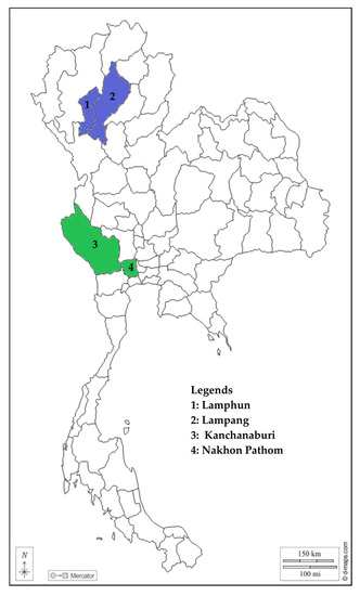
Figure 1.
Map of Thailand with provinces (shaded) where the surveyed farms are located. Purple-shaded provinces are from the northern part, while green-shaded provinces are from the western part. The map was adopted and modified from d-maps.com (accessed on 3 September 2023).
2.4. Hematologic Analysis and Microscopic Examination
The PCV values of the samples were analyzed using an automatic hematologic analyzer. Thin blood smears were prepared in glass slides, air-dried, fixed with methanol, and stained with 20% Giemsa in 1 × phosphate-buffered saline solution for 30 to 45 min. Slides were observed using a light microscope under an oil-immersed 100× magnification lens (Nikon Eclipse, Tokyo, Japan). Common bovine blood parasites (Babesia, Theileria, and Anaplasma) were searched for in at least 1000 erythrocytes.
2.5. Blood Sample Processing and PCR Tests of DNA Samples
Approximately 200 µL of whole blood was used for isolating the genomic DNA (gDNA) using the column-based QIAamp® Mini Blood Kit following the manufacturer’s instructions (Qiagen, Hilden, Germany). The samples were eluted to a final volume of 100 µL. The extracted DNA samples were stored at −30 °C until the molecular screening.
A PCR test was performed to molecularly identify samples positive for BTH (Babesia, Theileria, and Hepatozoon) 18S rRNA gene [20]. Then, subsequent piroplasma identification was also conducted using species-specific assays for B. bigemina [21], B. bovis [22], and T. orientalis [23], with gene targets, namely, rhoptry-associated protein-1a (RAP-1a), spherical body protein-2 (SBP-2), and major piroplasm surface protein (MPSP), respectively. In addition, A. marginale was detected using a species-specific, nested assay targeting the heat-shock chaperon gene (groEL) [24]. Details on the primer sets and assays used are listed in Table S1. The PCR was run at a final volume setup of 10 µL, comprising 1 × ThermoPol® reaction buffer (New England Biolabs, Ipswich, MA, USA), 2 μM each of dNTP in a solution mix, 0.25 U Taq DNA polymerase (New England Biolabs), 1.5 μL gDNA sample, 2 μM of each primer, and 6.85 μL double-distilled water. For the nested assays, 1.5 μL of PCR product obtained from the first round of amplification was used for the second-round reaction. The thermocycling conditions from the referenced studies were followed, except for the annealing and extension temperatures and duration, which were modified based on the provided conditions in the Taq polymerase kit (New England Biolabs). Controls were run alongside the samples to ensure the absence of contamination. gDNA of Babesia (B. bigemina Argentina strain and B. bovis Texas strain), and bovine DNA samples that were confirmed positive for T. orientalis and A. marginale by sequencing were used as positive controls in the assays, while the negative control was double-distilled water. PCR products were gel-electrophoresed on a 1.5% agarose in TAE buffer solution, stained with ethidium bromide (Nacalai Tesque, Kyoto, Japan), then viewed under a UV transilluminator (Atto, Tokyo, Japan). Positive samples were confirmed with the presence of a band similar to that of the corresponding positive control samples.
2.6. Statistical Analyses
The various analyses were performed using GraphPad Prism 8.0.2 (GraphPad Software, San Diego, CA, USA) and Microsoft Excel. Pearson’s chi-squared test (χ2) was run to determine the association between PCR positivity and sample location. The datasets for mean PCV values were tested for normality prior to running an unpaired Student’s t-test or a Mann–Whitney U test to evaluate the changes between positive and negative samples. The significance threshold for p values was set at <0.05. The degree of agreement between microscopy and PCR results was determined based on Cohen’s kappa [25]. The kappa values were interpreted as: no agreement (<0), slight agreement (0–0.20), fair agreement (0.21–0.40), moderate agreement (0.41–0.60), substantial agreement (0.61–0.80), and almost perfect agreement (0.81–1.00) [25].
3. Results
3.1. Tick-borne Pathogen Detection by PCR
Generic PCR screening showed that 33.58% (89/265) were positive for BTH, with the highest detection rate observed in Lampang (63.10%; 53/84) and Nakhon Pathom (32.86%; 23/70) (Table 2). Species-specific assays revealed that B. bigemina, B. bovis, T. orientalis, and A. marginale were detected in 4.53%, 3.40%, 23.40%, and 18.87% of cattle samples, respectively. Compared with other provinces, samples from Lampang had significantly higher detection rates for all the detected pathogens (Table 2). B. bigemina and B. bovis were detected in three provinces, while T. orientalis and A. marginale were detected in all surveyed provinces. In addition, all screened pathogens were present in cattle samples from Lampang and Nakhon Pathom provinces. Based on the results of the species-specific PCR assays, 74 samples were positive for only 1 TBP, while 26 were positive for 2 or more TBPs: 20, 5, and 1 sample/s had dual, triple, and quadruple infections, respectively (Table 3). Specifically, coinfection with T. orientalis and A. marginale (n = 13) was recorded as the most common combination of TBPs in the coinfected samples.
Table 2.
Detection rates for tick-borne pathogens in cattle samples from dairy farms in northern and western Thailand.
Table 3.
Infection types of positive samples based on species-specific PCR results.
3.2. Agreement between PCR and Microscopic Examination Results
Table 4 shows the results obtained by comparing PCR and microscopy methods for TBP detection. The microscopic examination searched for Babesia, Theileria, and Anaplasma in 176 samples only. Therefore, we excluded the samples without microscopy data for analysis. For piroplasma, the PCR targeted the BTH 18S rRNA gene (Babesia/Theileria/Hepatozoon), while the microscopy data were the pooled results for Babesia and Theileria. Based on Cohen’s kappa index, piroplasma (κ = 0.20) and Babesia (κ = 0.11) showed slight agreement between PCR and microscopy results, while Theileira and Anaplasma both recorded a κ value of 0.21, indicating a fair agreement between the two detection tools (Table 4).
Table 4.
Agreement analysis between PCR and microscopic examination results.
3.3. PCV Alterations Associated with Tick-Borne Pathogen Positivity
As anemia is one of the hallmarks of TBDs, we analyzed if PCR-positive samples had altered PCV values compared with the PCR-negative samples (Table 5). We excluded in the analysis the PCR results with fewer than 15 values per group (i.e., data on B. bigemina and B. bovis) and one clotted sample that had an incorrect reading to avoid biases. The mean PCV of samples positive for at least one TBP (27.59 ± 5.21) was significantly lower than the mean PCV of uninfected samples (30.54 ± 4.52). Similar trends were observed in the mean PCV of BTH 18S rRNA-positive (27.18 ± 5.39) and T. orientalis-positive (26.90 ± 5.44) samples, which were significantly decreased compared with those of PCR-negative samples (30.11 ± 4.62 and 29.82 ± 4.76, respectively). The mean PCV of A. marginale-positive (27.84 ± 4.92) samples was notably lower than that of the uninfected samples (29.43 ± 5.07).
Table 5.
Changes in the packed cell volume of PCR-positive and PCR-negative cattle samples in this study.
4. Discussion
The adverse impact of TBDs on bovine health, mainly on dairy herds, has led to not only financial losses but more importantly to opportunity costs that have hampered the improvement of the growing dairy industry in countries like Thailand. Therefore, uncovering tick-borne infections in the field, as well as assessing their clinical impact, is vital in formulating solutions to these diseases.
Herein, we report the detection of selected TBPs in dairy bovine samples from Thailand. The agents of tick fever, B. bovis, B. bigemina, and A. marginale, were identified. Thailand has long been considered an endemic country for tick fever (bovine babesiosis and anaplasmosis), with reported herd mortality rates of 0.5% and losses, mainly from mortalities, estimated at US$1.8 million in 1999 [27]. The predominant species of Babesia in cattle in Thailand are B. bovis and B. bigemina, the etiologic agents of bovine babesiosis. Both were observed at relatively lower rates (4.53% and 3.40%, respectively) than A. marginale (18.87%). The relatively high detection rates of T. orientalis in the current survey imply that this parasite is the predominant TBP among bovine herds in the sampled areas. The presently recorded T. orientalis overall detection rate (23.40%) was lower than previous surveys that reported 30.1–41.54% across Thailand [5,6,28]. The bovine infectious anemia outbreaks associated with the geographical spread of pathogenic T. orientalis have been documented in countries such as Australia, New Zealand, and recently, the USA [29,30,31,32]. As such, the widespread presence of T. orientalis in cattle, as demonstrated in this survey, signifies an enormous threat to the Thai dairy industry. Therefore, monitoring of T. orientalis, specifically the pathogenic Ikeda and Chitose types, should be conducted regularly, and infected animals should be provided proper treatment and management.
A majority of the positive samples were singly infected, while several samples were found to be positive for more than one TBP. A possible explanation for the single-TBP-positive samples is the competitive exclusion between various TBPs in cattle [33]. On the other hand, in endemic areas, coinfection of TBPs is a common occurrence, especially in cases where the pathogens are transmitted by the same vectors [34]. B. bovis, B. bigemina, and A. marginale have the same biological vector, the Rhipicephalus spp. tick, which is the predominant cattle tick in Thailand [35]. In this study, the coinfection combination of T. orientalis and A. marginale was the most frequently recorded. Remarkably, these two pathogens were the only noted combination not to exhibit competitive exclusion or negative interference within cattle [33]. A broader investigation is required to obtain absolute confirmation regarding this observation.
We demonstrated the detection of TBPs in dairy cattle from four provinces in northern and western (central) Thailand and recorded higher detection rates in samples from Lampang province. Bovine tick-borne infections have been extensively studied in northern and western (central) parts of Thailand in the past, where researchers reported the aforementioned pathogens in cattle [6,10,11,12,28,36,37], indicating their endemicity in the localities where they were detected. Studies on bovine TBDs have been performed in Lampang previously. B. bovis (8.33%) and B. bigemina (10.71%) detection in Lampang in this survey were lower than those reported by Cao et al. [10] and Simking et al. [11] but higher than those observed by Koonyosying et al. [28] and Yoshinari et al. [36]. Current T. orientalis (32.14%) and A. marginale (25%) detection were higher and lower than the detection rates of a previous Lampang survey of 21.05% and 85.53%, respectively [28]. On the other hand, we observed higher TBP detection rates than a report from Lamphun and lower than those from Nakhon Pathom [28]. The variances in the reports of TBPs may be attributed to different factors, such as the profiles of the animals (age, sex, breed, etc.) and external aspects, such as environmental factors (microclimate, tick abundance, and management practices of farmers) [38]. Worth noting in this study is the practice of free grazing in Lampang, where the highest rates were recorded. Grazing allows a longer exposure time to questing ticks in the pastures, increasing the risk of ectoparasite infestation and TBDs [6]. This may explain the higher rates in Lampang than in other provinces.
Anemia is one of the major indicators of TBDs. In this study, the detection of TBPs, specifically samples positive for BTH, T. orientalis, and A. marginale, was associated with significantly lower PCV values. This contradicts the results of previous Thai studies where the differences between the PCV values of cattle positive and negative for T. orientalis were trivial [5,28]. T. orientalis-infected cattle may show normal hematocrit values (24–46%) to as low as 8% [30,39], indicating that positive animals present varying hematologic indices. In cases of BTH- and A. marginale-infected cattle, the PCV values markedly decreased as well, similar to the findings of an investigation on cattle in India [40]. The significantly lower PCV values observed in infected cattle were within the lower normal ranges [26]. Although no overt disease was observed in the TBP-positive cattle in the present survey, there may be an impact on milk yield. Still, the impact of this finding on bovine productivity should not be ignored, as lower hematocrit caused by tick-borne infections can significantly reduce milk production in dairy animals [17,38,41]. This is a point to investigate in future studies.
We also assessed two diagnostic tools for the detection of TBPs: microscopic identification (MI) and PCR assays. Cohen’s kappa indices obtained in this study resulted in slight and fair agreement between the two tools, suggesting that microscopy may not be an adequate alternative to PCR in the detection of TBPs. Although traditionally used, MI of TBD agents is appropriate during the acute stage of the infections, when parasites are abundant in the bloodstream [42]. Although MI is the most convenient detection tool, it is not effective in detecting carrier or subclinical cattle due to the very low parasitemia [43,44,45]. Moreover, MI is highly reliant on the skill of the microscopist, leading to varied results and poor diagnosis [42]. On the other hand, the PCR test is said to be a thousand times more sensitive than microscopy; thus, it is currently the preferred pathogen identification tool for TBPs [43]. PCR tests and other nucleic acid-based tests in general can be adapted to a variety of conditions and highly specific targets, making them more reliable and accurate pathogen identification tools [46]. Laboratory reagents and equipment that are required to perform the tests can be costly for resource-constrained places, but the increasing accessibility to nucleic acid-based tests has contributed to lowering the cost of PCR tests. Therefore, PCR may soon make traditional detection tools, such as MI, obsolete.
There are several limitations to the present study. First, the study only focused on the northern and central/western parts of Thailand. It would have been more interesting if data from other parts, such as east and southern Thailand, were available, as this would have provided a broader insight into the impact of TBD on dairy animals across Thailand. Another limitation is the lack of molecular characterization of the detected TBPs, specifically for BTH-positive samples that were negative in the species-specific PCRs. In this study, the authors focused on reported piroplasma in Thailand, i.e., those that were screened using species-specific PCRs. Confirmation through sequencing is recommended in future studies. Finally, the status of the animals pertaining to other anemia-causing infections was not confirmed. Hence, this must be considered in interpreting the findings on PCV.
5. Conclusions
In this investigation, various TBPs, namely, B. bovis, B. bigemina, T. orientalis, and A. marginale, were detected in dairy cattle samples from the northern and western provinces of Thailand. Notably, T. orientalis and A. marginale were prevalent in the surveyed bovine herds. TBP-positive samples had significantly altered PCV values compared with those of the non-infected cattle. Moreover, the comparison between microscopy and PCR yielded a slight and fair agreement, indicating that results obtained by microscopy may not suitably replace TBP detection by PCR. Further studies on the impact of TBDs on the production and reproduction of Thai dairy cattle are warranted.
Supplementary Materials
The following supporting material can be downloaded at https://www.mdpi.com/article/10.3390/ani13182844/s1. Table S1. PCR assays used in the current study.
Author Contributions
P.F.A.M. and E.M.G. contributed equally to this work. Conceptualization, P.F.A.M., E.M.G. and X.X.; methodology, P.F.A.M., E.M.G., M.A.T., B.B., R.N., S.T., K.K. and T.I.; investigation, P.F.A.M. and E.M.G.; formal analysis, P.F.A.M., E.M.G., M.A.T. and B.B.; visualization, E.M.G.; validation, X.X.; writing—original draft preparation, P.F.A.M. and E.M.G.; writing—review and editing, P.F.A.M., E.M.G., K.K., T.I. and X.X.; supervision, X.X.; project administration, X.X.; funding acquisition, X.X. All authors have read and agreed to the published version of the manuscript.
Funding
Paul Franck Adjou Moumouni and Eloiza May Galon were supported by research grants and fellowship from the Japan Society for the Promotion of Science (JSPS) for Young Scientists (17F17105 and 20J20134, respectively). This study was supported by a Grant-in-Aid for Scientific Research (26304036, 18KK0188) from the Ministry of Education, Culture, Sports, Science and Technology (MEXT), Japan, and a grant from the JSPS Core-to-Core Program, Japan.
Institutional Review Board Statement
Procedures employed in the survey adhered to the animal welfare laws in Thailand and the ethical guidelines for the use of animal samples implemented by Obihiro University of Agriculture and Veterinary Medicine (animal experiment approval 22-23; DNA experiment approval 20-07, 20-08; approval date: 1 April 2023).
Informed Consent Statement
Informed consent was obtained from the farm owners involved in the study.
Data Availability Statement
The datasets generated during and/or analyzed during the current study are available from the corresponding author upon reasonable request.
Acknowledgments
The authors extend thanks to the cattle farmers in Lampang, Lamphun, Nakhon Pathom, and Kanchanaburi, Thailand for their participation and cooperation during the sample collection and survey.
Conflicts of Interest
The authors declare no conflict of interest.
References
- Pattamanont, P.; Nutdechanan, J.; Vangtal, A.; Maneetup, C.; Ajariyakhajorn, K. Towards Sustainability of the Dairy Industry in Thailand. FFTC Agricultural Policy Platform. Available online: https://ap.fftc.org.tw/article/3090 (accessed on 13 June 2023).
- Lew-Tabor, A.E.; Rodriguez Valle, M. A review of reverse vaccinology approaches for the development of vaccines against ticks and tick borne diseases. Ticks Tick-Borne Dis. 2016, 7, 573–585. [Google Scholar] [CrossRef] [PubMed]
- Strydom, T.; Lavan, R.P.; Torres, S.; Heaney, K. The economic impact of parasitism from nematodes, trematodes and ticks on beef cattle production. Animals 2023, 13, 1599. [Google Scholar] [CrossRef] [PubMed]
- Kiara, H.; Steinaa, L.; Nene, V.; Svitek, N. Theileria in ruminants. In Parasitic Protozoa of Farm Animals and Pets; Florin-Christensen, M., Schnittger, L., Eds.; Springer International Publishing: Cham, Switzerland, 2018; pp. 187–213. ISBN 978-3-319-70131-8. [Google Scholar]
- Rakwong, P.; Keawchana, N.; Ngasaman, R.; Kamyingkird, K. Theileria infection in bullfighting cattle in Thailand. Vet. World 2022, 15, 2917–2921. [Google Scholar] [CrossRef]
- Jirapattharasate, C.; Adjou Moumouni, P.F.; Cao, S.; Iguchi, A.; Liu, M.; Wang, G.; Zhou, M.; Vudriko, P.; Changbunjong, T.; Sungpradit, S.; et al. Molecular epidemiology of bovine Babesia spp. and Theileria orientalis parasites in beef cattle from northern and northeastern Thailand. Parasitol. Int. 2016, 65, 62–69. [Google Scholar] [CrossRef]
- Udonsom, R.; Mahittikorn, A.; Jirapattharasate, C. Molecular detection and genetic diversity of tick-borne pathogens in goats from the southern part of Thailand. Pathogens 2022, 11, 477. [Google Scholar] [CrossRef] [PubMed]
- Schnittger, L.; Ganzinelli, S.; Bhoora, R.; Omondi, D.; Nijhof, A.M.; Florin-Christensen, M. The Piroplasmida Babesia, Cytauxzoon, and Theileria in farm and companion animals: Species compilation, molecular phylogeny, and evolutionary insights. Parasitol. Res. 2022, 121, 1207–1245. [Google Scholar] [CrossRef]
- Sivakumar, T.; Tuvshintulga, B.; Otgonsuren, D.; Batmagnai, E.; Ahedor, B.; Kothalawala, H.; Vimalakumar, S.C.; Silva, S.S.P.; Yamagishi, J.; Yokoyama, N. Phylogenetic analyses of the mitochondrial, plastid, and nuclear genes of Babesia sp. Mymensingh and its naming as Babesia naoakii n. sp. Parasit. Vectors 2022, 15, 299. [Google Scholar] [CrossRef]
- Cao, S.; Aboge, G.O.; Terkawi, M.A.; Yu, L.; Kamyingkird, K.; Luo, Y.; Li, Y.; Goo, Y.-K.; Yamagishi, J.; Nishikawa, Y.; et al. Molecular detection and identification of Babesia bovis and Babesia bigemina in cattle in northern Thailand. Parasitol. Res. 2012, 111, 1259–1266. [Google Scholar] [CrossRef]
- Simking, P.; Saengow, S.; Bangphoomi, K.; Sarataphan, N.; Wongnarkpet, S.; Inpankaew, T.; Jittapalapong, S.; Munkhjargal, T.; Sivakumar, T.; Yokoyama, N.; et al. The molecular prevalence and MSA-2b gene-based genetic diversity of Babesia bovis in dairy cattle in Thailand. Vet. Parasitol. 2013, 197, 642–648. [Google Scholar] [CrossRef]
- Jirapattharasate, C.; Adjou Moumouni, P.F.; Cao, S.; Iguchi, A.; Liu, M.; Wang, G.; Zhou, M.; Vudriko, P.; Efstratiou, A.; Changbunjong, T.; et al. Molecular detection and genetic diversity of bovine Babesia spp., Theileria orientalis, and Anaplasma marginale in beef cattle in Thailand. Parasitol. Res. 2017, 116, 751–762. [Google Scholar] [CrossRef]
- Simking, P.; Yatbantoong, N.; Saetiew, N.; Saengow, S.; Yodsri, W.; Chaiyarat, R.; Wongnarkpet, S.; Jittapalapong, S. Prevalence and risk factors of Babesia infections in cattle trespassing natural forest areas in Salakpra Wildlife Sanctuary, Kanchanaburi province. J. Trop. Med. Parasitol. 2014, 37, 10–19. [Google Scholar]
- Aubry, P.; Geale, D.W. A review of bovine anaplasmosis. Transbound. Emerg. Dis. 2011, 58, 1–30. [Google Scholar] [CrossRef] [PubMed]
- Kocan, K.M.; de la Fuente, J.; Blouin, E.F.; Coetzee, J.F.; Ewing, S.A. The natural history of Anaplasma marginale. Vet. Parasitol. 2010, 167, 95–107. [Google Scholar] [CrossRef]
- Curtis, A.K.; Kleinhenz, M.D.; Anantatat, T.; Martin, M.S.; Magnin, G.C.; Coetzee, J.F.; Reif, K.E. Failure to eliminate persistent Anaplasma marginale infection from cattle using labeled doses of chlortetracycline and oxytetracycline antimicrobials. Vet. Sci. 2021, 8, 283. [Google Scholar] [CrossRef]
- Perera, P.K.; Gasser, R.B.; Firestone, S.M.; Anderson, G.A.; Malmo, J.; Davis, G.; Beggs, D.S.; Jabbar, A. Oriental theileriosis in dairy cows causes a significant milk production loss. Parasit. Vectors 2014, 7, 73. [Google Scholar] [CrossRef]
- Jonsson, N.N.; Bock, R.E.; Jorgensen, W.K.; Morton, J.M.; Stear, M.J. Is endemic stability of tick-borne disease in cattle a useful concept? Trends Parasitol. 2012, 28, 85–89. [Google Scholar] [CrossRef] [PubMed]
- Mans, B.J.; Pienaar, R.; Latif, A.A. A review of Theileria diagnostics and epidemiology. Int. J. Parasitol. Parasites Wildl. 2015, 4, 104–118. [Google Scholar] [CrossRef]
- Masatani, T.; Hayashi, K.; Andoh, M.; Tateno, M.; Endo, Y.; Asada, M.; Kusakisako, K.; Tanaka, T.; Gokuden, M.; Hozumi, N.; et al. Detection and molecular characterization of Babesia, Theileria, and Hepatozoon Species in hard ticks collected from Kagoshima, the southern region in Japan. Ticks Tick-Borne Dis. 2017, 8, 581–587. [Google Scholar] [CrossRef]
- Terkawi, M.A.; Huyen, N.X.; Shinuo, C.; Inpankaew, T.; Maklon, K.; Aboulaila, M.; Ueno, A.; Goo, Y.-K.; Yokoyama, N.; Jittapalapong, S.; et al. Molecular and serological prevalence of Babesia bovis and Babesia bigemina in water buffaloes in the northeast region of Thailand. Vet. Parasitol. 2011, 178, 201–207. [Google Scholar] [CrossRef]
- AbouLaila, M.; Yokoyama, N.; Igarashi, I. Development and evaluation of a nested PCR based on spherical body protein 2 gene for the diagnosis of Babesia bovis infection. Vet. Parasitol. 2010, 169, 45–50. [Google Scholar] [CrossRef]
- Ota, N.; Mizuno, D.; Kuboki, N.; Igarashi, I.; Nakamura, Y.; Yamashina, H.; Hanzaike, T.; Fujii, K.; Onoe, S.; Hata, H.; et al. Epidemiological survey of Theileria orientalis infection in grazing cattle in the eastern part of Hokkaido, Japan. J. Vet. Med. Sci. 2009, 71, 937–944. [Google Scholar] [CrossRef] [PubMed]
- Ybañez, A.P.; Sivakumar, T.; Ybañez, R.H.D.; Ratilla, J.C.; Perez, Z.O.; Gabotero, S.R.; Hakimi, H.; Kawazu, S.; Matsumoto, K.; Yokoyama, N.; et al. First molecular characterization of Anaplasma marginale in cattle and Rhipicephalus (Boophilus) microplus ticks in Cebu, Philippines. J. Vet. Med. Sci. 2013, 75, 27–36. [Google Scholar] [CrossRef] [PubMed]
- McHugh, M.L. Interrater reliability: The kappa statistic. Biochem. Med. 2012, 22, 276–282. [Google Scholar] [CrossRef]
- Jackson, P.G.G.; Cockcroft, P.D. Clinical Examination of Farm Animals; Blackwell Science: Oxford, UK, 2002; ISBN 978-0-470-75242-5. [Google Scholar]
- McLeod, R.; Kristjanson, P. Final Report of Joint ESYS/ILRI/ACIAR Tick Cost Project—Economic Impact of Ticks and Tick-Borne Diseases to Livestock in Africa, Asia and Australia; International Livestock Research Institute: Nairobi, Kenya, 1999. [Google Scholar]
- Koonyosying, P.; Rittipornlertrak, A.; Chomjit, P.; Sangkakam, K.; Muenthaisong, A.; Nambooppha, B.; Srisawat, W.; Apinda, N.; Singhla, T.; Sthitmatee, N. Incidence of hemoparasitic infections in cattle from central and northern Thailand. PeerJ 2022, 10, e13835. [Google Scholar] [CrossRef] [PubMed]
- Lawrence, K.; Gedye, K.; McFadden, A.; Pulford, D.; Heath, A.; Pomroy, W. Review of the New Zealand Theileria orientalis Ikeda type epidemic and epidemiological research since 2012. Pathogens 2021, 10, 1346. [Google Scholar] [CrossRef] [PubMed]
- Watts, J.; Playford, M.; Hickey, K. Theileria orientalis: A review. N. Z. Vet. J. 2016, 64, 3–9. [Google Scholar] [CrossRef]
- Jenkins, C. Bovine theileriosis in Australia: A decade of disease. Microbiol. Aust. 2018, 39, 215. [Google Scholar] [CrossRef]
- Oakes, V.J.; Yabsley, M.J.; Schwartz, D.; LeRoith, T.; Bissett, C.; Broaddus, C.; Schlater, J.L.; Todd, S.M.; Boes, K.M.; Brookhart, M.; et al. Theileria orientalis Ikeda genotype in cattle, Virginia, USA. Emerg. Infect. Dis. 2019, 25, 1653–1659. [Google Scholar] [CrossRef]
- Dib, L.; Bitam, I.; Tahri, M.; Bensouilah, M.; De Meeûs, T. Competitive exclusion between piroplasmosis and anaplasmosis agents within cattle. PLoS Pathog. 2008, 4, e7. [Google Scholar] [CrossRef]
- Hofmann-Lehmann, R.; Meli, M.L.; Dreher, U.M.; Gönczi, E.; Deplazes, P.; Braun, U.; Engels, M.; Schüpbach, J.; Jörger, K.; Thoma, R.; et al. Concurrent infections with vector-borne pathogens associated with fatal hemolytic anemia in a cattle herd in Switzerland. J. Clin. Microbiol. 2004, 42, 3775–3780. [Google Scholar] [CrossRef]
- Tan, L.P.; Hamdan, R.H.; Hassan, B.N.H.; Reduan, M.F.H.; Okene, I.A.-A.; Loong, S.K.; Khoo, J.J.; Samsuddin, A.S.; Lee, S.H. Rhipicephalus tick: A contextual review for Southeast Asia. Pathogens 2021, 10, 821. [Google Scholar] [CrossRef] [PubMed]
- Yoshinari, T.; Sivakumar, T.; Asada, M.; Battsetseg, B.; Huang, X.; Lan, D.T.B.; Inpankaew, T.; Ybañez, A.P.; Alhassan, A.; Thekisoe, O.M.M.; et al. A PCR based survey of Babesia ovata in cattle from various Asian, African and South American Countries. J. Vet. Med. Sci. 2013, 75, 211–214. [Google Scholar] [CrossRef] [PubMed]
- Rittipornlertrak, A.; Nambooppha, B.; Simking, P.; Punyapornwithaya, V.; Tiwananthagorn, S.; Jittapalapong, S.; Chung, Y.-T.; Sthitmatee, N. Low levels of genetic diversity associated with evidence of negative selection on the Babesia bovis apical membrane antigen 1 from parasite populations in Thailand. Infect. Genet. Evol. 2017, 54, 447–454. [Google Scholar] [CrossRef] [PubMed]
- Betancur Hurtado, O.J.; Giraldo-Ríos, C. Economic and health impact of the ticks in production animals. In Ticks and Tick-Borne Pathogens; Abubakar, M., Perera, P.K., Eds.; IntechOpen: London, UK, 2019; ISBN 978-1-78985-765-8. [Google Scholar]
- Sivakumar, T.; Ikehara, Y.; Igarashi, I.; Inokuma, H.; Yokoyama, N. Dynamics of erythrocyte indices in relation to anemia development in Theileria orientalis-infected cattle. J. Protozool. Res. 2017, 27, 23–33. [Google Scholar]
- Kaur, R.; Yadav, A.; Rafiqi, S.I.; Godara, R.; Sudan, V.; Chakraborty, D.; Katoch, R. Epidemiology, haematology and molecular characterization of haemoprotozoon and rickettsial organisms causing infections in cattle of Jammu region, north India. BMC Vet. Res. 2021, 17, 219. [Google Scholar] [CrossRef]
- Ayadi, O.; Gharbi, M.; Benchikh Elfegoun, M.C. Milk losses due to bovine tropical theileriosis (Theileria annulata infection) in Algeria. Asian Pac. J. Trop. Biomed. 2016, 6, 801–802. [Google Scholar] [CrossRef]
- Alvarez, J.A.; Rojas, C.; Figueroa, J.V. Diagnostic tools for the identification of Babesia sp. in persistently infected cattle. Pathogens 2019, 8, 143. [Google Scholar] [CrossRef]
- World Organisation for Animal Health. Chapter 3.4.2. Bovine babesiosis. In Manual of Diagnostic Tests and Vaccines for Terrestrial Animals, 12th ed.; World Organisation for Animal Health: Paris, France, 2023. [Google Scholar]
- World Organisation for Animal Health. Chapter 3.4.15. Theileriosis. In Manual of Diagnostic Tests and Vaccines for Terrestrial Animals, 12th ed.; World Organisation for Animal Health: Paris, France, 2023. [Google Scholar]
- World Organisation for Animal Health. Chapter 3.4.1. Bovine anaplasmosis. In Manual of Diagnostic Tests and Vaccines for Terrestrial Animals, 12th ed.; World Organisation for Animal Health: Paris, France, 2023. [Google Scholar]
- Garcia, K.; Weakley, M.; Do, T.; Mir, S. Current and future molecular diagnostics of tick-borne diseases in cattle. Vet. Sci. 2022, 9, 241. [Google Scholar] [CrossRef]
Disclaimer/Publisher’s Note: The statements, opinions and data contained in all publications are solely those of the individual author(s) and contributor(s) and not of MDPI and/or the editor(s). MDPI and/or the editor(s) disclaim responsibility for any injury to people or property resulting from any ideas, methods, instructions or products referred to in the content. |
© 2023 by the authors. Licensee MDPI, Basel, Switzerland. This article is an open access article distributed under the terms and conditions of the Creative Commons Attribution (CC BY) license (https://creativecommons.org/licenses/by/4.0/).