Article

Thoroughbred Racehorses in Hong Kong Require Vitamin D Supplementation to Mitigate the Risk of Low Vitamin D Status

1
Royal (Dick) School of Veterinary Studies and The Roslin Institute, University of Edinburgh, Easter Bush Campus, Roslin EH25 9RG, UK
2
The Hong Kong Jockey Club Equine Welfare Research Foundation, Sha Tin Racecourse, New Territories, Hong Kong SAR, China
3
Kentucky Equine Research, Versailles, KY 40383, USA
*
Author to whom correspondence should be addressed.
Animals 2023, 13(13), 2145; https://doi.org/10.3390/ani13132145
Submission received: 31 May 2023 / Revised: 20 June 2023 / Accepted: 27 June 2023 / Published: 29 June 2023
(This article belongs to the Section Equids)

Abstract

:

Simple Summary

Vitamin D biology in equids is unique and poorly understood. Naturally managed (grazing) horses rely on dietary vitamin D2 (ergocalciferol) to provide adequate vitamin D, because endogenous ultraviolet radiation-mediated synthesis of vitamin D3 (cholecalciferol) is ineffective in this species. To test the hypothesis that the management of stabled, non-grazing racehorses is a risk factor for low vitamin D status, the vitamin D status of non-grazing Thoroughbred racehorses in Hong Kong (HK) and grazing Thoroughbred racehorses in the United Kingdom (UK) was compared. The HK horses had lower serum concentrations of 25-hydroxyvitamin D2 (25OHD2) and total 25-hydroxyvitamin D (total 25OHD: the index of vitamin D status), reflecting reduced dietary vitamin D2 intake. These data indicate that HK racehorses required dietary vitamin D3 supplementation to maintain adequate vitamin D status. The inverse relationship between the serum concentrations of 25OHD2 and 25OHD3, previously identified in humans, was observed for the first time in horses, indicating that further study is needed to determine the optimal form of dietary vitamin D supplementation for Thoroughbred racehorses.

Abstract

There is a paucity of data relating to the vitamin D status of racehorses. We hypothesised that the management of racehorses in Hong Kong (HK) predisposes to low vitamin D status unless they receive dietary supplementation. Serum concentrations of 25-hydroxyvitamin D2 (25OHD2), 25-hydroxyvitamin D3 (25OHD3) and total 25-hydroxyvitamin D (total 25OHD) for 79 non-grazing HK racehorses were compared with those for 22 racehorses training in the United Kingdom (UK) that grazed for ≥1 h/d, and for which published data exists. A nested group of 41 HK horses was sampled twice to determine the effect of the duration in HK on vitamin D status. The HK horses had significantly lower serum concentrations of total 25OHD and 25OHD2 than the UK horses; 25OHD2 was undetectable in 15/79 HK sera and serum concentrations of 25OHD2 declined with the duration in HK. The main determinants of vitamin D status were assessed using linear regression; the retained variables were the 25OHD3 concentration and the duration in HK. The inverse relationship between the serum concentrations of 25OHD2 and 25OHD3, previously identified in humans, was observed for the first time in horses. In conclusion, HK racehorses have low serum 25OHD2 and total 25OHD concentrations and rely on D3 supplementation to maintain adequate vitamin D status. Further study is required to determine the optimal form of dietary vitamin D supplementation for Thoroughbred racehorses.

1. Introduction

Vitamin D has important roles in calcium homeostasis, bone health and modulation of immune and inflammatory responses through endocrine and paracrine effects in many body tissues [1,2,3]. Vitamin D exists in two forms, namely vitamin D2 (D2: ergocalciferol), which is produced by fungi growing on plants [4,5], and vitamin D3 (D3: cholecalciferol) which, in some species, can be synthesised within the skin through the action of ultraviolet B (UVB) radiation on 7-dehydrocholesterol [6]. D2 and D3 may also be derived from dietary supplementation. To become physiologically active, these compounds must undergo a first hydroxylation in the liver. The serum concentrations of the resultant 25-hydroxyvitamin D2 (25OHD2) and 25-hydroxyvitamin D3 (25OHD3), and the sum of 25OHD2 and 25OHD3 (total 25OHD), are used as indicators of the vitamin D status in humans and horses [7,8]. The 25OHD compounds undergo a final hydroxylation in the kidneys to produce the biologically active moieties 1,25-dihydroxyvitamin D2 and 1,25-dihydroxyvitamin D3.
Horses appear to have a unique vitamin D biology. In the absence of vitamin D supplementation, they rely on 25OHD2 derived from UVB irradiated dietary forage because they synthesise no or negligible quantities of D3 in their skin through the action of solar UVB radiation on 7-dehydrocholesterol [8,9,10]. Consistent with this, D3 could not be detected in equine skin following experimental UVB exposure [11]. Furthermore, serum 25OHD3 concentrations remained low or undetectable throughout the year in healthy non-supplemented grazing horses in New Zealand, the United Kingdom (UK) and Thailand (respectively, 40° S, 56° N and 15° N) [8,9,10].
Hymøller and Jensen [1] concluded their article entitled ‘We know next to nothing about vitamin D in horses’ by emphasising the need to assess vitamin D requirements of horses under different management conditions and for horses of different breeds, ages and genders. In particular, there is a paucity of data relating to vitamin D status in racehorses. The present study assessed the vitamin D status of racehorses in training in Hong Kong (HK) to investigate the hypothesis that the horses’ management and athletic activity predispose them to having low vitamin D status unless they receive appropriate dietary vitamin D supplementation. HK horses are stabled with limited UVB exposure, have no access to grazing and are fed restricted amounts of forage and commercial feed containing various amounts of supplemental D3. HK horses undergo strenuous athletic activity, which has been reported to reduce vitamin D metabolites in healthy horses [10,12]. Additionally, reductions in serum 25OHD concentrations have been reported in horses with inflammatory diseases, including mild-moderate equine asthma, which is prevalent in racehorses [13,14].
A cross-sectional study was used to determine the vitamin D status of non-grazing HK racehorses (n = 79). Data were compared with those for racehorses training in the UK that grazed for ≥1 h/d, and with those reported in previous equine studies. We hypothesised that the UK horses would have higher serum 25OHD2 concentrations than the HK horses given that grazing provides a natural source of dietary D2 [4,8]. Additionally, a nested group of HK horses was sampled twice to determine the effect of the duration in HK on vitamin D status. The effects of country of origin, duration in HK, premises, age and gender on the vitamin D status of HK horses were also assessed.

2. Materials and Methods

2.1. Horses

The 79 HK Thoroughbred horses comprised 5 colts, 69 geldings and 5 females. At the time of the first (or unique) sampling, mean age was 4.3 years (range 2.4–7.9). Moreover, to investigate the effect of duration in HK on vitamin D status, a nested group of 41 HK horses was sampled at two time points, a median of 62 days (range 20–152) apart. HK horses were imported from New Zealand (n = 28), Australia (n = 28), the UK (n = 12), Ireland (n = 5), France (n = 4) and USA (n = 2). Duration in HK before the first or unique sampling ranged from 2 weeks to 4.7 years (median 0.8 years, Q1–Q3 14 days–1.7 years). HK horses were stabled with 19 different trainers in a single complex in an urban environment. Trainers all shared the same training resources although they employed different management practices and fed the horses different diets. Horses effectively had no access to grazing and typically ≤ 30 min sunlight exposure while training and during a similar time of in-hand walking daily. They were fed approximately 2–2.5% bw as dry matter (DM), including restricted amounts (likely averaging < 1% bw as DM) of forage, which was typically timothy (Phleum pratense) hay imported from North America, with some receiving chaff and/or haylage imported from New Zealand. They also received 6–7 kg/d of commercial feeds supplemented with 1100–2100 IU/kg (as fed) of D3, which provides in excess of the current minimum vitamin D requirement of 6.6 IU/kg BW/d [15]. Food was stored in air-conditioned stores, which likely retarded fungal growth on forage, possibly reducing the forage D2 content. Some horses also received 1 kg/d freshly cut grass. The UK horses (n = 22) had a mean age of 7 years (range 3–11) and comprised 2 females and 20 geldings. They were stabled on a single premises in Scotland, were in training and were fed 8–10 kg/d haylage and a commercial concentrate feed providing 6600–8800 IU/d D3, which exceeds the current minimum requirement of 6.6 IU/kg/d [15]. They grazed on pasture for ≥1 h/d, wearing a rug.

2.2. Serum Samples

Venous blood was collected from all horses and transferred into plain vacutainers. Blood samples were residues of samples taken for clinical purposes as part of the horses’ routine veterinary management with trainer consent and ethical approval from the HKJC Ethical Review Committee and the Edinburgh School of Veterinary Medicine Ethical Review Committee. Serum was harvested and frozen at −20 °C or −80 °C prior to analysis. Serum from HK horses was shipped on dry ice to the UK for analysis.

2.3. Vitamin D Analyses

Serum concentrations of 25OHD2, 25OHD3 and total 25OHD were quantified by a commercial laboratory (Biolab Medical Unit, London, UK) using high-performance liquid chromatography with UV detection using reagents supplied by Chromsystems Instruments and Chemicals GMBH (Heimburgstrasse 3, 81243 Munchen, Germany). The lower limit of detection for 25OHD2 and 25OHD3 was 0.5 nmol/L. For statistical analysis, values for 25OHD2 that were below this detection limit were recorded as 0.49 nmol/L.

2.4. Statistical Analysis

Most data were non-normally distributed. Intergroup paired comparisons (e.g., 22 UK horses versus 79 HK horses) were made using the Mann–Whitney test. Multiple-group comparisons (e.g., country of origin) were made using the Kruskal–Wallis test. Intragroup comparisons (e.g., first versus second sampling) were made using the Wilcoxon rank test. For the regression analysis, non-normally distributed data were ln transformed, although 25OHD2 remained non-normally distributed after this transformation. Multivariate linear regression was used to assess the effect of duration in HK, country of origin, stable, age and gender on vitamin D status. In the univariate model, only variables that had p < 0.2 were put forward into the final model. Statistical analyses were conducted with SPSS 26 (IBM).

3. Results

3.1. Intergroup Comparisons

There was no significant inter-group age difference. Descriptive statistics for serum concentrations of 25OHD2, 25OHD3 and total 25OHD for the UK and the HK horses are presented in Table 1 and Figure 1. The UK horses had significantly higher concentrations of 25OHD2 and total 25OHD (both p < 0.001; Mann–Whitney) (Table 1 and Figure 1). The 25OHD2 was detectable in all the UK horse sera but was below the limit of detection for 15/79 (19%) of the HK horse sera.

3.2. Intragroup Comparisons

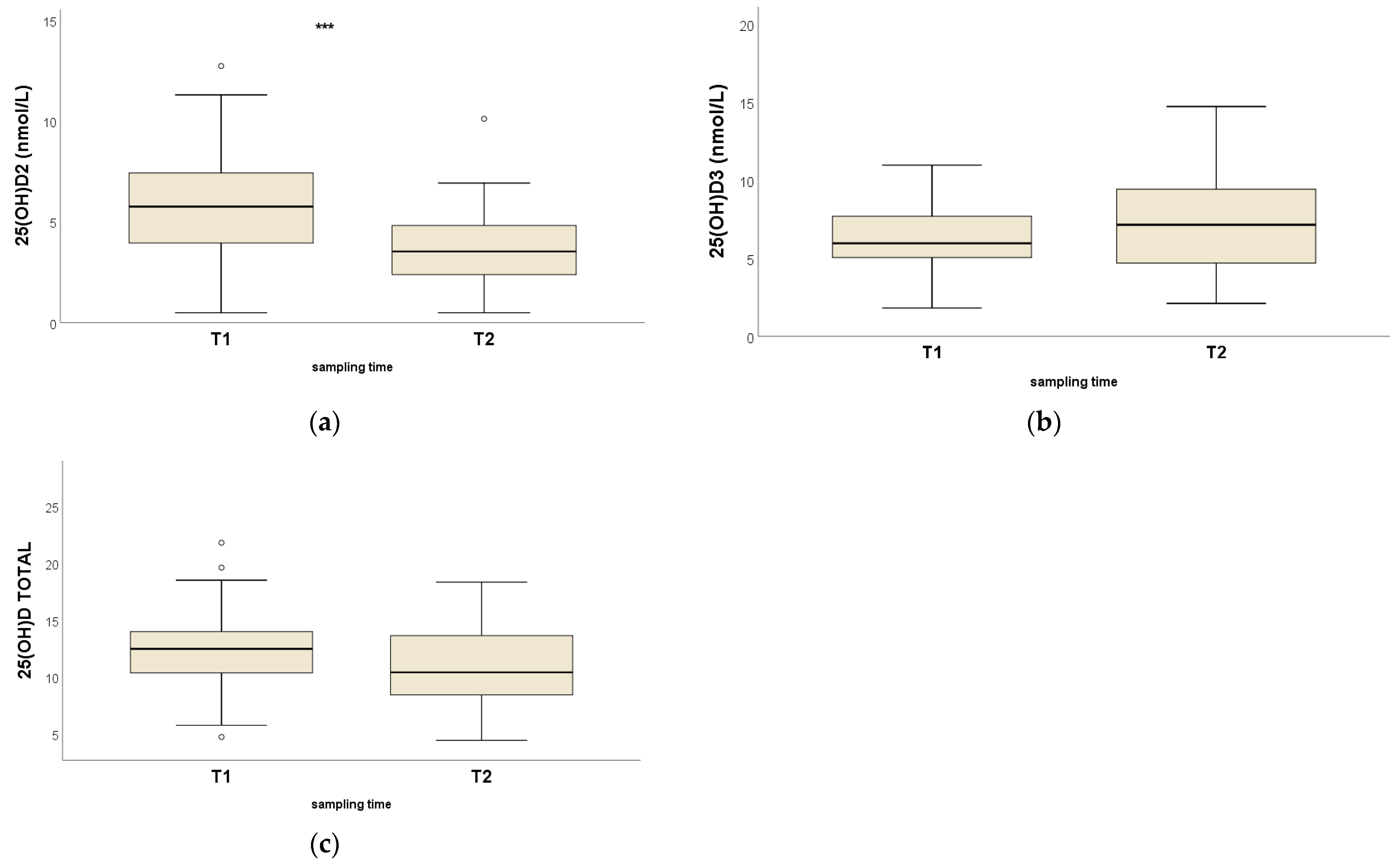
Serum concentrations of 25OHD2 for the HK horses decreased significantly between the first and second sampling points (p < 0.001; Wilcoxon rank test), while 25OHD3 (p = 0.12) and total 25OHD (p = 0.35) concentrations were not significantly different (Table 2 and Figure 2).

3.3. Univariate and Multivariate Linear Regression

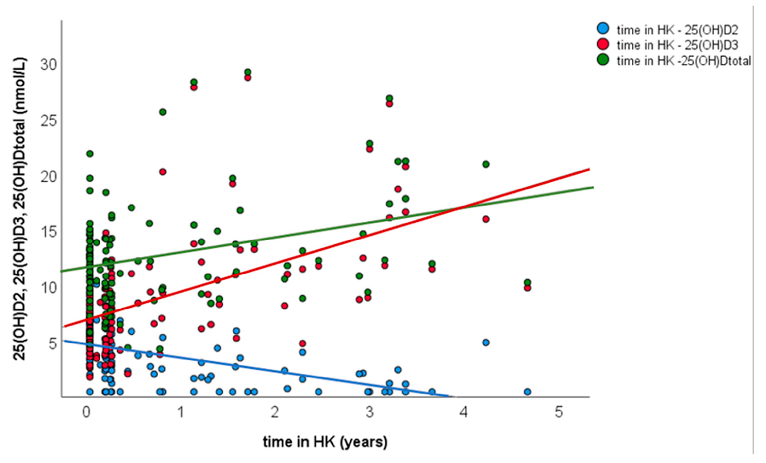
For univariate linear regression, 25OHD concentrations were transformed to a logarithmic scale (ln) and ln total 25OHD concentration of the first samples for the 79 HK horses used as outcome, and, as predictors, duration in HK, ln25OHD2 concentration, ln25OHD3 concentration, trainer and country of origin. Only variables with p < 0.2 in the univariate analysis, namely duration in HK and ln25OHD3 concentration, were put forward into the final multivariate analysis (Table 3). Ln25OHD3 and duration in HK were retained in the final model (p < 0.01), with 72% of the variability in total 25OHD concentrations being explained by the 25OHD3 concentration and the duration in HK (R2 = 0.722) (Table 3). There was a positive relationship between the duration in HK and the 25OHD3 concentration (p < 0.01, β = 0.985), and a negative relationship between the duration in HK and the 25OHD2 concentration (p < 0.01, β = −0.330) (Figure 3).

3.4. Correlation of 25OHD2, 25OHD3 and Total 25OHD

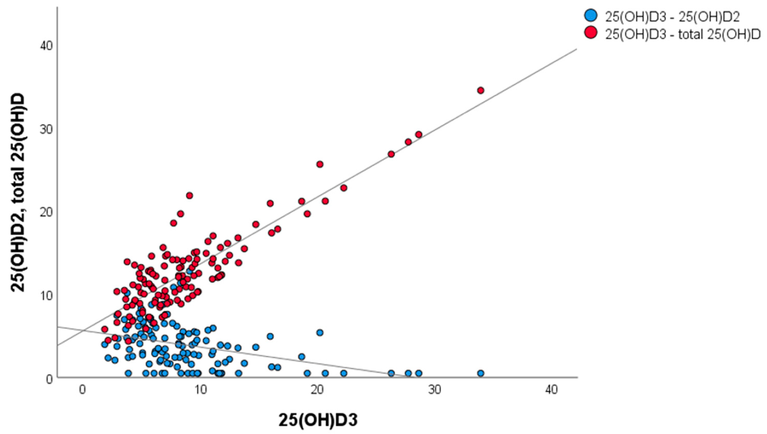
When data for all 120 HK samples were considered (Figure 4), there was a positive relationship between total 25OHD and 25OHD3 concentrations (β = 0.800, p < 0.01), and a negative relationship between 25OHD3 and 25OHD2 concentrations (β = −0.453, p < 0.01).

4. Discussion

4.1. Vitamin D Status of UK and HK Thoroughbred Racehorses

This study contributes to our knowledge regarding the vitamin D status of Thoroughbred racehorses in training in HK and in the UK. Serum concentrations of total 25OHD for HK (median 12.5, range 4.3–29.1 nmol/L) and UK (15.8, range 9.9–26.8 nmol/L) horses were largely consistent with previous reports that healthy horses from a wide range of breeds, ages, management systems and latitudes, and in different seasons, have total 25OHD concentrations < 25 nmol/L [1,8,9,16,17,18]. These values are considerably lower than those for other species, reflecting the peculiarities in equid vitamin D biology [16,19,20,21,22]. Consistent with published equine data [summarised by 2], a wide range of total 25OHD concentrations was observed in the HK and UK horses, despite each group having fairly similar management practices. Some horses, predominantly in the HK group, had very low total 25OHD concentrations, with the lowest being 4.3 nmol/L. While the threshold serum concentration of total 25OHD which reflects adequacy of vitamin D for maintenance of optimal health and athletic performance is currently unknown, it is possible that those horses with the lowest total 25OHD concentrations may have had inadequate vitamin D status and may have benefitted from additional vitamin D supplementation. The main limitation of this study was the inability, for practical and logistical reasons, to quantify accurately dietary D2 (forage and grazing) and D3 (supplementation) intakes for all of the individual horses in the study.
HK horses had significantly lower serum concentrations of total 25OHD than UK horses. This was attributable largely to their very low serum concentrations of 25OHD2; indeed, 25OHD2 was undetectable (<0.5 nmol/L) in 15/79 (19%) of the HK horse sera. Furthermore, there was a significant temporal reduction in 25OHD2 concentrations in the nested group of HK horses that were sampled on two occasions and a negative statistical association between the 25OHD2 concentrations and the duration in HK. As 25OHD2 is derived from D2, which is produced by fungi growing on UVB irradiated forages [5], the low 25OHD2 concentrations in HK horses likely reflect the absence of grazing and restricted intake of sun-cured forage. Consistent with this hypothesis, serum concentrations of 25OHD2 for HK horses were lower than previously reported for grazing and forage fed horses [8,9,17]. The low 25OHD2 and total 25OHD concentrations in the HK horses may also reflect the effects of intense athletic activity, and/or lower airway inflammation, which is prevalent in Thoroughbred racehorses [10,12,13,14].
This study highlights the importance of dietary D3 supplementation in compensating for inadequate D2 intake and maintaining adequate vitamin D status in HK racehorses. Considering the fact that HK horses have very low or undetected 25OHD2 concentrations, it is logical that in the multivariate analysis of factors affecting vitamin D status (total 25OHD), the only variables retained in the final model were the 25OHD3 concentration and the duration in HK.

4.2. In Racehorses, Serum 25OHD3 Is Probably Derived Solely from Dietary D3 Supplementation

It is highly probable that the 25OHD3 detected in the HK and UK horse sera was derived solely from dietary D3 supplementation, as reported in other equine studies [8,9,17]. Endogenous UVB-mediated 25OHD3 synthesis can be ruled out because a recent ex vivo study demonstrated that, unlike ovine skin biopsies, equine skin biopsies produce negligible quantities of vitamin D3 when exposed to the wavelengths of UVB light that induce non-enzymatic conversion of 7-dehydrocholesterol to vitamin D3 in other species [11]. Consequently, serum 25OHD3 concentrations remained low or undetectable throughout the year in healthy non-supplemented grazing horses in New Zealand, the United Kingdom (UK) and Thailand (respectively, 40° S, 56° N and 15° N) [8,9,10]. The horse’s inability to endogenously synthesise vitamin D3 contrasts with other domestic herbivores (sheep, cattle and goats) that exhibit effective endogenous vitamin D3 synthesis [23,24,25]. Furthermore, the horses in this study had only limited UVB exposure at the time of sampling. Most (89%) of the HK samples were collected during autumn–winter (November to February), when the local UV index (https://www.hko.gov.hk/en/wxinfo/uvinfo/uvinfo.html data gathered by HK observatory website accessed on the 2 April 2023) was below that required for endogenous 25OHD3 synthesis in other species [26]. Similarly, UVB radiation is insufficient for endogenous 25OHD3 synthesis in UK horses given the latitude (56° N) and the month (March) of sampling for the UK horses.

4.3. Vitamin D2 or D3?

This study identified, for the first time, an inverse relationship between equine serum concentrations of 25OHD2 and 25OHD3. This has been reported in humans, whereby oral D2 or D3 supplementation resulted in a linear reduction in serum concentrations of 25OHD3 and 25OHD2, respectively [27,28,29]. The mechanism underlying this relationship is unclear but may reflect competition between 25OHD2 and 25OHD3 for interaction with transport binding proteins and metabolising enzymes [27]. It has been proposed that the additional methyl group on carbon 24 of D2 influences its affinity for binding proteins and vitamin D receptors, which are critical steps in the activation of the final di-hydroxylated form (1,25(OH)D2). Moreover, in the kidneys, an additional hydroxylation of carbon 24 occurs, generating 1,24,25(OH)D3. While 1,24,25(OH)D2 is no longer active, 1,24,25(OH)D3 still maintains biological activity and can bind the vitamin D receptor [30]. These differences are likely to account for the superior potency of D3 in some species. It has been proposed that a dynamic balance exists between 25OHD2 and 25OHD3 metabolites that enables organisms to adapt and maintain adequate vitamin D status despite variability in dietary D2 and D3 intakes and sun exposure [27]. Given that knowledge of vitamin D metabolism in the horse is limited, and due to the fundamental species-specific differences we have highlighted in this study, further study is warranted to determine whether D3 does indeed have superior potency in the horse.
While this study suggests that non-grazing HK racehorses should receive vitamin D supplementation to maintain an adequate vitamin D status for optimal health and athletic performance, further study is required to determine the optimal dose and form of supplementation. In the present study, D3 supplementation provided an apparently adequate vitamin D status for most HK racehorses. The HK racehorses included in this study were fed large amounts of concentrate feeds (6–7 kg /horse/d), which provides 6600–14700 IU/d of D3. If the average racehorse bodyweight is 500–550 kg, this amount of concentrate alone should provide 12–28 IU/kg BW/d of D3. This exceeds the requirements recommended by the National Research Council (NRC) on Nutrients Requirements of Horses [15] (i.e., 6.6 IU/kg for maintenance requirements and 14.7 IU/kg for growing horses of 19–24 months of age), but is well within the upper safety limit of 44 IU/kg/d. It should be noted that the NRC recommendations were extrapolated from an in vivo experimental study conducted in 1979 [31] where circulating vitamin D metabolites were not measured. In light of more recent advances in equine research, up to date and evidence-based guidelines are much needed to better inform daily dietary requirements for vitamin D for sedentary and athletic horses. In humans, oral daily supplementation of 1000–4000 IU/d vitamin D (either D2 or D3) is sufficient to maintain serum concentrations of 25(OH)D above the threshold for deficiency, which is 75 nmol/L [30]. Interestingly, while on average the horses included in this study received much higher amounts of supplemental D3, their total 25(OH)D concentrations did not exceed 29.1 nmol/L. Several factors, such as the molecular form of vitamin D supplementation (i.e., D2 vs. D3, hydroxylated vs. non-hydroxylated), the food matrix (oil vs. powder), composition of the meal and individual characteristics (e.g., disease state), are likely to influence bioavailability of this nutrient [32]. To the authors’ knowledge, none of these factors has been investigated in the horse. Given that horses do not synthesise relevant amounts of endogenous 25OHD3, vitamin D2 supplementation may be a more natural approach to efficiently and safely provide supplemental vitamin D to horses, since naturally managed horses rely mostly on 25OHD2 derived from dietary forage. Furthermore, D3 supplementation potentially has a negative impact on serum 25OHD2 concentrations, given the inverse relationship identified herein between the serum concentrations of 25OHD2 and 25OHD3. While toxicity has been induced in horses using experimental administration of high doses of D2 or D3 [33,34], there are insufficient data to inform the relative safety of supplemental D2 and D3 and guide recommendations for supplementation. When the relative toxicity of equimolar high doses (33,000 IU/kg) of D2 or D3 was compared using only two horses, the D3 supplemented horse had a more profound increase in serum concentrations of vitamin D metabolites, and more marked clinical abnormalities and tissue calcification [34]. Further work is required to determine whether supplementation with D2, the source of vitamin D in naturally managed horses, is a more appropriate and safer form for horses than D3.

4.4. The Role of Vitamin D in Equine Musculoskeletal Health and Athletic Performance

The vitamin D status of the racehorses in this study may have been influenced by their high intensity exercise regimens, since experimental high intensity exercise causes a reduction in equine serum 25(OH)D concentrations brought about by unknown mechanisms [10]. Similarly, human elite athletes, especially those exercising for prolonged periods indoors, are at risk of having vitamin D deficiency/insufficiency [35]. The importance of this pleiotropic hormone in skeletal muscle physiology and athletic performance has been widely investigated in other species but not in the horse. Human beings with vitamin D deficiency disorders including rickets and osteomalacia not only suffer skeletal pathologies, but also have skeletal muscle atrophy, myopathies, reduced muscle strength, prolonged time to peak muscle contraction and prolonged time to muscle relaxation [36,37]. Vitamin D is also involved in maintaining adequate calcium availability to support myofiber contraction, and has some local effects via the vitamin D receptors expressed on skeletal muscle cells [10,38,39]. Vitamin D is also thought to stimulate muscle regeneration and to optimise muscle cell metabolism, especially in fast twitch type II fibres, which could be relevant to racing Thoroughbred horses. In some studies of human athletes, vitamin D supplementation increased muscle strength, particularly in the lower limb muscles [40] and in those individuals with the lowest serum concentrations of 25(OH)D [35,41,42,43]. The effect of vitamin D status on racehorse athletic performance clearly warrants investigation. Finally, there is a growing body of evidence demonstrating the beneficial role of vitamin D supplementation on skeletal health and its protective effect against skeletal injuries in human athletes [43]. The incidence of non-catastrophic and catastrophic fractures in HK racehorses is 2.2 per 1000 racing starts and 0.6 per 1000 racing starts, respectively [44]. While vitamin D deficiency and reduced bone density appear to be risk factors for stress fractures in human athletes [45], increased bone density is observed at the most common sites of fracture in equine athletes suggesting that there may be inter-species differences in the mechanisms underlying fractures in human and equine athletes [46,47]. Furthermore, vitamin D appears to have a less important role in regulating calcium metabolism in horses when compared with humans, given that equine intestinal calcium absorption is poorly regulated and urinary calcium excretion is very efficient [21]. The unique nature of vitamin D biology in equids means that caution must be used when making inter-species comparisons regarding the role of vitamin D in bone metabolism and pathology. Investigation of the effect of vitamin D status on bone metabolism and pathology in racehorses is, therefore, clearly warranted.

5. Conclusions

Stabled, non-grazing Thoroughbred racehorses should receive vitamin D supplementation to maintain an adequate vitamin D status, because management and occupational factors predispose them to low serum concentrations of 25OHD2 and total 25OHD. Further work is required to determine the optimal dose and form of vitamin D supplementation for racehorses. While variable levels of D3 supplementation yielded an apparently adequate vitamin D status in most of the Thoroughbred racehorses in this study, the inverse relationship identified between 25OHD2 and 25OHD3 raises the possibility that oral D3 supplementation has a negative impact on serum concentrations of 25OHD2. D2 supplementation may have advantages because it is the main source of vitamin D in naturally managed horses. Further study is also required to determine the threshold concentrations of serum vitamin D metabolites which indicate deficiency, and whether vitamin D supplementation can improve racehorse health and athletic performance.

Supplementary Materials

The following supporting information can be downloaded at: https://www.mdpi.com/article/10.3390/ani13132145/s1, Table S1: Full study data set.

Author Contributions

Conceptualization, B.C.M. and C.M.R.; methodology, B.C.M., E.C.-G., J.M., A.L. and C.M.R.; formal analysis, B.C.M., M.C.M.D. and J.P.; data curation, B.C.M. and M.C.M.D.; writing—original draft preparation, B.C.M.; writing—review and editing, all authors; project administration, B.C.M. and C.M.R.; funding acquisition, B.C.M. and C.M.R. All authors have read and agreed to the published version of the manuscript.

Funding

This research was funded by the Fiona and Ian Russell Seed Corn Fund for Companion Animal Research and the Hong Kong Jockey Club.

Institutional Review Board Statement

The animal study protocol was conducted in accordance with the Declaration of Helsinki and approved by the Hong Kong Jockey Club Ethical Review Committee and the Edinburgh School of Veterinary Medicine Ethical Review Committee (124.19, 19 September 2019).

Informed Consent Statement

Not applicable.

Data Availability Statement

All study data are available in Table S1.

Acknowledgments

The authors acknowledge the support of the horses’ trainers and carers.

Conflicts of Interest

The authors declare no conflict of interest.

References

  1. Hymøller, L.; Jensen, S.K. We Know Next to Nothing About Vitamin D in Horses! J. Equine Vet. Sci. 2015, 35, 785–792. [Google Scholar] [CrossRef]
  2. Yin, K.; Agrawal, D.K. Vitamin D and inflammatory diseases. J. Inflamm. Res. 2014, 7, 69–87. [Google Scholar] [PubMed] [Green Version]
  3. Rosen, C.J.; Adams, J.S.; Bikle, D.D.; Black, D.M.; Demay, M.B.; Manson, J.E.; Murad, M.H.; Kovacs, C.S. The Nonskeletal Effects of Vitamin D: An Endocrine Society Scientific Statement. Endocr. Rev. 2012, 33, 456–492. [Google Scholar] [CrossRef] [PubMed] [Green Version]
  4. Jäpelt, R.B.; Didion, T.; Smedsgaard, J.; Jakobsen, J. Seasonal variation of provitamin D2 and vitamin D2 in perennial ryegrass (Lolium perenne L.). J. Agric. Food Chem. 2011, 59, 10907–10912. [Google Scholar] [CrossRef]
  5. Jäpelt, R.B.; Jakobsen, J. Vitamin D in plants: A review of occurrence, analysis, and biosynthesis. Front. Plant Sci. 2013, 4, 136. [Google Scholar] [CrossRef] [Green Version]
  6. Hurst, E.A.; Homer, N.Z.; Mellanby, R.J. Vitamin D Metabolism and Profiling in Veterinary Species. Metabolites 2020, 10, 371. [Google Scholar] [CrossRef]
  7. Toribio, R.E. Disorders of calcium and phosphate metabolism in horses. Vet. Clin. N. Am. Equine Pract. 2011, 27, 129–147. [Google Scholar] [CrossRef]
  8. Azarpeykan, S.; Dittmer, K.; Gee, E.; Marshall, J.; Wallace, J.; Elder, P.; Acke, E.; Thompson, K. Influence of blanketing and season on vitamin D and parathyroid hormone, calcium, phosphorus, and magnesium concentrations in horses in New Zealand. Domest. Anim. Endocrinol. 2016, 56, 75–84. [Google Scholar] [CrossRef]
  9. Dosi, M.C.; McGorum, B.C.; Kirton, R.D.; Cillán-García, E.; Mellanby, R.J.; Keen, J.A.; Hurst, E.A.; Morgan, R.A. The effect of season, management and endocrinopathies on vitamin D status in horses. Equine Vet. J. 2023, 55, 672–680. [Google Scholar] [CrossRef]
  10. Puangthong, C.; Sukhong, P.; Saengnual, P.; Srikuea, R.; Chanda, M. A single bout of high-intensity exercise modulates the expression of vitamin D receptor and vitamin D-metabolising enzymes in horse skeletal muscle. Equine Vet. J. 2021, 53, 796–805. [Google Scholar] [CrossRef]
  11. Azarpeykan, S.; Gee, E.K.; Thompson, K.G.; Dittmer, K.E. Undetectable vitamin D(3) in equine skin irradiated with ultraviolet light. J. Equine Sci. 2022, 33, 45–49. [Google Scholar] [CrossRef] [PubMed]
  12. Ceylan, E.; Dede, S.; Deger, Y.; Yoruk, I. Investigation of the Effects of Carrying Heavy Load on Prooxidation/ Antioxidant Status and Vitamin D3 in Healthy Horses. Asian J. Anim. Vet. Adv. 2009, 4, 41–46. [Google Scholar] [CrossRef] [Green Version]
  13. Karagianni, A.E.; Kurian, D.; Cillán-Garcia, E.; Eaton, S.L.; Wishart, T.M.; Pirie, R.S. Training associated alterations in equine respiratory immunity using a multiomics comparative approach. Sci. Rep. 2022, 12, 427. [Google Scholar] [CrossRef] [PubMed]
  14. Bedenice, D.H.-t.A.; Rozanski, E.; Mazan, M. The relationship between serum 25-hydroxyvitamin D levels and inflammatory airway disease in horses. In Proceedings of the ACVIM Congress Proceedings, Indianapolis, IN, USA, 3–6 June 2015. [Google Scholar]
  15. NRC (National Research Council). Nutrient Requirements of Horses, 6th revised ed.; The National Academies Press: Washington DC, USA, 2007. [Google Scholar]
  16. Breidenbach, A.; Schlumbohm, C.; Harmeyer, J. Peculiarities of vitamin D and of the calcium and phosphate homeostatic system in horses. Vet. Res. 1998, 29, 173–186. [Google Scholar]
  17. Azarpeykan, S.; Dittmer, K.E.; Gee, E.K.; Marshall, J.C.; Elder, P.; Acke, E.; Thompson, K.G. Circadian rhythm of calciotropic hormones, serum calcium, phosphorus and magnesium during the shortest and longest days of the year in horses in New Zealand. J. Anim. Physiol. Anim. Nutr. 2016, 100, 1058–1066. [Google Scholar] [CrossRef]
  18. Mäenpää, P.H.; Koskinen, T.; Koskinen, E. Serum profiles of vitamins A, E and D in mares and foals during different seasons. J. Anim. Sci. 1988, 66, 1418–1423. [Google Scholar] [CrossRef] [Green Version]
  19. Pozza, M.E.; Kaewsakhorn, T.; Trinarong, C.; Inpanbutr, N.; Toribio, R.E. Serum vitamin D, calcium, and phosphorus concentrations in ponies, horses and foals from the United States and Thailand. Vet. J. 2014, 199, 451–456. [Google Scholar] [CrossRef]
  20. Hymøller, L.; Jensen, S.K. 25-Hydroxycholecalciferol status in plasma is linearly correlated to daily summer pasture time in cattle at 56° N. Br. J. Nutr. 2012, 108, 666–671. [Google Scholar] [CrossRef] [Green Version]
  21. Rourke, K.; Coe, S.; Kohn, C.; Rosol, T.; Mendoza, F.; Toribio, R. Cloning, comparative sequence analysis and mRNA expression of calcium-transporting genes in horses. Gen. Comp. Endocrinol. 2010, 167, 6–10. [Google Scholar] [CrossRef]
  22. Azarpeykan, S.; Dittmer, K.E.; Marshall, J.C.; Perera, K.C.; Gee, E.K.; Acke, E.; Thompson, K.G. Evaluation and comparison of vitamin D responsive gene expression in ovine, canine and equine kidney. PLoS ONE 2016, 11, e0162598. [Google Scholar] [CrossRef] [Green Version]
  23. Kohler, M.; Leiber, F.; Willems, H.; Merbold, L.; Liesegang, A. Influence of altitude on vitamin D and bone metabolism of lactating sheep and goats. J. Anim. Sci. 2013, 91, 5259–5268. [Google Scholar] [CrossRef] [Green Version]
  24. Kovács, S.; Wilkens, M.R.; Liesegang, A. Influence of UVB exposure on the vitamin D status and calcium homoeostasis of growing sheep and goats. J. Anim. Physiol. Anim. Nutr. 2015, 99 (Suppl. S1), 1–12. [Google Scholar] [CrossRef] [Green Version]
  25. Hymøller, L.; Jensen, S.K. Vitamin D(3) synthesis in the entire skin surface of dairy cows despite hair coverage. J. Dairy Sci. 2010, 93, 2025–2029. [Google Scholar] [CrossRef] [PubMed] [Green Version]
  26. Engelsen, O.; Brustad, M.; Aksnes, L.; Lund, E. Daily duration of vitamin D synthesis in human skin with relation to latitude, total ozone, altitude, ground cover, aerosols and cloud thickness. Photochem. Photobiol. 2005, 81, 1287–1290. [Google Scholar] [CrossRef]
  27. Hammami, M.M.; Abuhdeeb, K.; Hammami, S.; Yusuf, A. Vitamin-D2 treatment-associated decrease in 25(OH)D3 level is a reciprocal phenomenon: A randomized controlled trial. BMC Endocr. Disord. 2019, 19, 8. [Google Scholar] [CrossRef] [Green Version]
  28. Hammami, M.M.; Yusuf, A. Differential effects of vitamin D2 and D3 supplements on 25-hydroxyvitamin D level are dose, sex, and time dependent: A randomized controlled trial. BMC Endocr. Disord. 2017, 17, 12. [Google Scholar] [CrossRef] [PubMed] [Green Version]
  29. Houghton, L.A.; Vieth, R. The case against ergocalciferol (vitamin D2) as a vitamin supplement. Am. J. Clin. Nutr. 2006, 84, 694–697. [Google Scholar] [CrossRef] [Green Version]
  30. Tripkovic, L.; Lambert, H.; Hart, K.; Smith, C.; Bucca, G.; Penson, S.; Chope, G.; Hypponen, E.; Berry, J.; Vieth, R.; et al. Comparison of vitamin D2 and vitamin D3 supplementation in raising serum 25-hydroxyvitamin D status: A systematic review and meta-analysis. Am. J. Clin. Nutr. 2012, 95, 1357–1364. [Google Scholar] [CrossRef] [PubMed] [Green Version]
  31. El Shorafa, W.M. Effect of vitamin D and sunlight on growth and bone development of young ponies. J. Anim. Sci. 1979, 48, 882–886. [Google Scholar] [CrossRef] [Green Version]
  32. Borel, P.; Caillaud, D.; Cano, N.J. Vitamin D bioavailability: State of the art. Crit. Rev. Food Sci. Nutr. 2015, 55, 1193–1205. [Google Scholar] [CrossRef]
  33. Harmeyer, J.; Schlumbohm, C. Effects of pharmacological doses of Vitamin D3 on mineral balance and profiles of plasma Vitamin D3 metabolites in horses. J. Steroid Biochem. Mol. Biol. 2004, 89–90, 595–600. [Google Scholar] [CrossRef] [PubMed]
  34. Harrington, D.D.; Page, E.H. Acute vitamin D3 toxicosis in horses: Case reports and experimental studies of the comparative toxicity of vitamins D2 and D3. J. Am. Vet. Med. Assoc. 1983, 182, 1358–1369. [Google Scholar] [PubMed]
  35. de la Puente Yagüe, M.; Yurrita, L.C.; Cabañas, M.J.C.; Cenzual, M.A.C. Role of Vitamin D in Athletes and Their Performance: Current Concepts and New Trends. Nutrients 2020, 12, 579. [Google Scholar] [CrossRef] [Green Version]
  36. Bartoszewska, M.; Kamboj, M.; Patel, D.R. Vitamin D, Muscle Function, and Exercise Performance. Pediatr. Clin. N. Am. 2010, 57, 849–861. [Google Scholar] [CrossRef] [PubMed]
  37. Rodman, J.S.; Baker, T. Changes in the kinetics of muscle contraction in vitamin D-depleted rats. Kidney Int. 1978, 13, 189–193. [Google Scholar] [CrossRef] [PubMed] [Green Version]
  38. Girgis, C.M.; Mokbel, N.; Cha, K.M.; Houweling, P.J.; Abboud, M.; Fraser, D.R.; Mason, R.S.; Clifton-Bligh, R.J.; Gunton, J.E. The vitamin D receptor (VDR) is expressed in skeletal muscle of male mice and modulates 25-hydroxyvitamin D (25OHD) uptake in myofibers. Endocrinology 2014, 155, 3227–3237. [Google Scholar] [CrossRef] [Green Version]
  39. Bischoff, H.A.; Borchers, M.; Gudat, F.; Duermueller, U.; Theiler, R.; Stähelin, H.B.; Dick, W. In situ detection of 1,25-dihydroxyvitamin D3 receptor in human skeletal muscle tissue. Histochem. J. 2001, 33, 19–24. [Google Scholar] [CrossRef]
  40. Zhang, L.; Quan, M.; Cao, Z.-B. Effect of vitamin D supplementation on upper and lower limb muscle strength and muscle power in athletes: A meta-analysis. PLoS ONE 2019, 14, e0215826. [Google Scholar] [CrossRef] [Green Version]
  41. Yoon, S.; Kwon, O.; Kim, J. Vitamin D in athletes: Focus on physical performance and musculoskeletal injuries. Phys. Act. Nutr. 2021, 25, 20–25. [Google Scholar] [CrossRef]
  42. Wyon, M.A.; Koutedakis, Y.; Wolman, R.; Nevill, A.M.; Allen, N. The influence of winter vitamin D supplementation on muscle function and injury occurrence in elite ballet dancers: A controlled study. J. Sci. Med. Sport 2014, 17, 8–12. [Google Scholar] [CrossRef]
  43. Mendes, M.M.; Botelho, P.B.; Ribeiro, H. Vitamin D and musculoskeletal health: Outstanding aspects to be considered in the light of current evidence. Endocr. Connect. 2022, 11, e210596. [Google Scholar] [CrossRef] [PubMed]
  44. Sun, T.C.; Riggs, C.M.; Cogger, N.; Wright, J.; Al-Alawneh, J.I. Noncatastrophic and catastrophic fractures in racing Thoroughbreds at the Hong Kong Jockey Club. Equine Vet. J. 2019, 51, 77–82. [Google Scholar] [CrossRef] [PubMed]
  45. Moreira, C.A.; Bilezikian, J.P. Stress Fractures: Concepts and Therapeutics. J. Clin. Endocrinol. Metab. 2016, 102, 525–534. [Google Scholar] [CrossRef] [PubMed] [Green Version]
  46. Riggs, C.M.; Boyde, A. Effect of exercise on bone density in distal regions of the equine third metacarpal bone in 2-year-old thoroughbreds. Equine Vet. J. 1999, 30, 555–560. [Google Scholar] [CrossRef] [PubMed]
  47. Loughridge, A.B.; Hess, A.M.; Parkin, T.D.; Kawcak, C.E. Qualitative assessment of bone density at the distal articulating surface of the third metacarpal in Thoroughbred racehorses with and without condylar fracture. Equine Vet. J. 2017, 49, 172–177. [Google Scholar] [CrossRef]
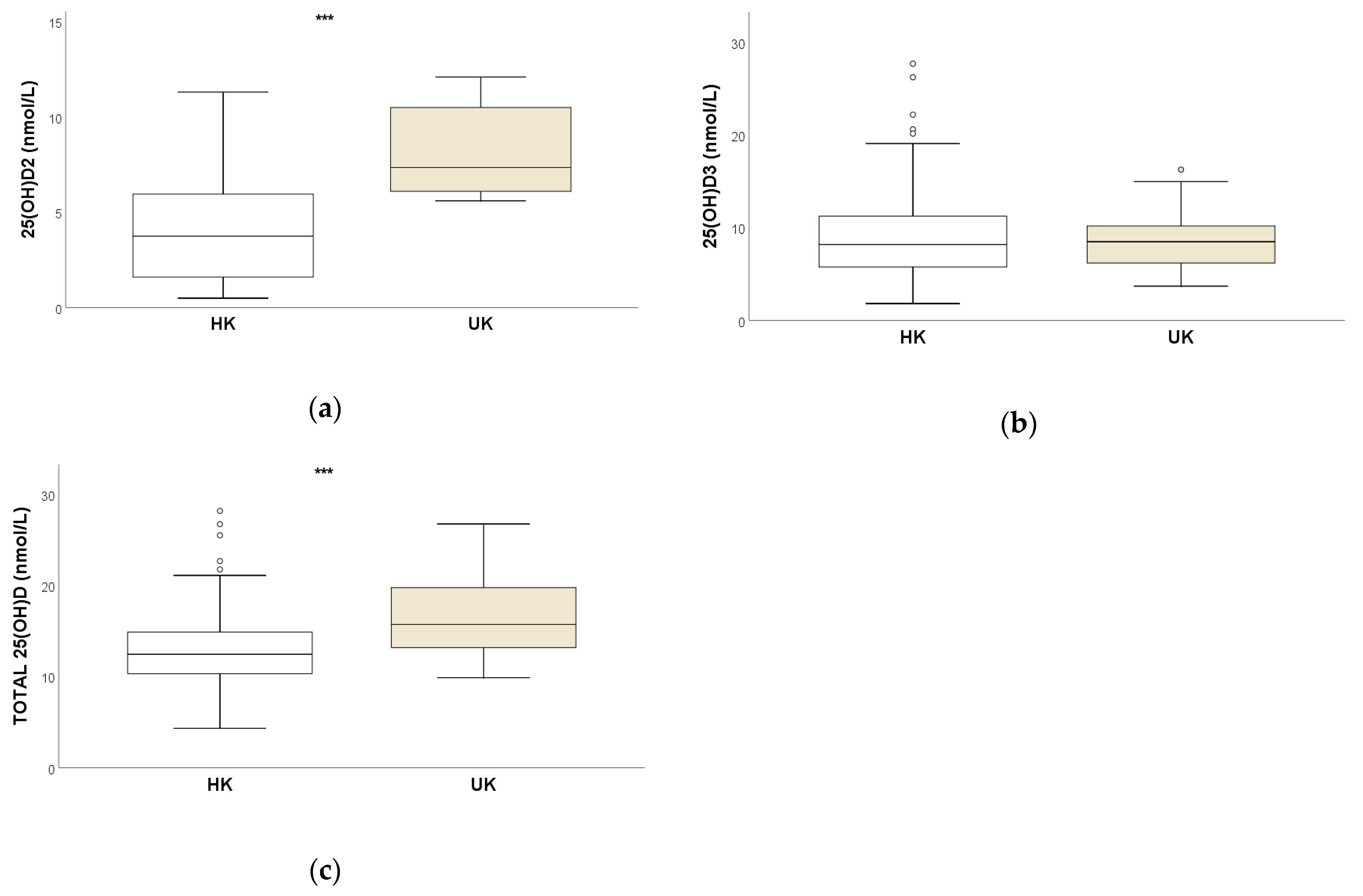
Figure 1. Box and whisker plots comparing serum concentrations (nmol/L) of (a) 25OHD2, (b) 25OHD3 and (c) total 25OHD for HK (n = 79) and UK (n = 22) horses. *** Significant inter-group difference (p < 0.001).
Animals 13 02145 g001
Figure 2. Box and whisker plots for serum concentrations (nmol/L) of (a) 25OHD2, (b) 25OHD3 and (c) total 25OHD for HK racehorses (n = 41) at two different time points. *** Significant intragroup difference (p < 0.001).
Animals 13 02145 g002
Figure 3. Scatter plot representing the association between duration in HK (years) and serum concentrations (nmol/L) of 25OHD2 (blue), 25OHD3 (red) and total 25OHD (green) for HK horses (n = 79).
Animals 13 02145 g003
Figure 4. Scatter plot for serum concentrations (nmol/L) of 25OHD3 (x axis) and 25OHD2 and total 25OHD (both on y axis) for HK horses (n = 120 samples), highlighting the negative relationship between 25OHD3 and 25OHD2 concentrations (blue), and positive relationship between 25OHD3 and total 25OHD concentrations (red).
Animals 13 02145 g004
Table 1. Serum concentrations of 25OHD2, 25OHD3 and total 25OHD (nmol/L) at the first sampling for 79 HK racehorses and for 22 UK racehorses. A Significant inter-group difference (p < 0.001).
HK UK
nmol/L25(OH)D225(OH)D325(OH)Dtot25(OH)D225(OH)D325(OH)Dtot
Median3.4 A8.012.5 A7.4 A8.515.8 A
Mean ± SD4.0 ± 2.979.4 ± 5.6113.4 ± 5.018.1 ± 2.208.6 ± 3.2216.7 ± 3.51
Range<0.5–12.71.8–28.64.3–29.15.6–12.13.7–16.39.9–26.8
Q1–Q31.6–6.05.7–11.510.3–15.06.1–10.66.2–10.213.2–19.7
Table 2. Serum concentrations of 25OHD2, 25OHD3 and total 25OHD (nmol/L) for HK racehorses (n = 41) at two different time points. A Significant intragroup difference (p < 0.001).
First SampleSecond Sample
25(OH)D2 AMean ± SD5.9 ± 2.63.6 ± 2.1
Median (range)5.8 (0.5–12.8)3.5 (0.5–10.1)
Q1–Q34.0–7.42.4–4.8
25(OH)D3Mean ± SD6.4 ± 2.17.8 ± 5.2
Median (range)6.0 (11.0–1.8)7.2 (2.1–33.9)
Q1–Q35.1–7.74.7–9.4
25(OH)DtotMean ± SD12.3 ± 3.511.4 ± 4.9
Median (range)12.510.4 (4.4–34.4)
Q1–Q310.4–14.08.5–13.6
Table 3. Coefficients calculated for the variables retained in the multivariate linear regression having as outcome total 25(OH)D.
Variables RetainedRegression Coefficient (β)Standard Error
Ln25(OH)D30.9850.024
LnDuration in HK−0.3300.056
intercept1.2200.107
p value<0.001
Pearson’s coefficient0.827
Variables were transformed into a logarithmic scale.
Disclaimer/Publisher’s Note: The statements, opinions and data contained in all publications are solely those of the individual author(s) and contributor(s) and not of MDPI and/or the editor(s). MDPI and/or the editor(s) disclaim responsibility for any injury to people or property resulting from any ideas, methods, instructions or products referred to in the content.