Article

The Complete Mitogenome of Toxocara vitulorum: Novel In-Sights into the Phylogenetics in Toxocaridae

1
Department of Parasitology, College of Veterinary Medicine, Sichuan Agricultural University, Wenjiang 611130, China
2
Department of Food Technology and Science, College of Food Science, Shanghai Ocean University, Shanghai 201306, China
3
Key Laboratory of Animal Disease and Human Health of Sichuan Province, College of Veterinary Medicine, Sichuan Agricultural University, Chengdu 611130, China
4
Animal Nutrition Institute, Sichuan Agricultural University, Chengdu 611130, China
*
Authors to whom correspondence should be addressed.
These authors contributed equally to this work.
Animals 2022, 12(24), 3546; https://doi.org/10.3390/ani12243546
Submission received: 11 October 2022 / Revised: 9 December 2022 / Accepted: 13 December 2022 / Published: 15 December 2022

Abstract

:

Simple Summary

Toxocara vitulorum is a common parasitic nematode of cattle and buffalos, and represents a severe health threat and economic burden on both populations. Poor understanding the epidemiology, population genetics, and molecular ecology of this nematode negative affects the prevention and control of T. vitulorum, and the mitogenome can yield a foundation for studying these areas. Herein, the first whole mitogenome of T. vitulorum was sequenced and characterized with bioinformatic pipeline analyses. Comparative mitogenomics and phylogenies defined a novel sister-species relationship of T. vitulorum with the congeneric species in the Toxocaridae, and supported nad4 and nad6 genes as new genetic markers for phylogenetic and evolutionary studies of the Ascaridida species. These results should contribute to a better understanding of phylogenetic relationships of Toxocaridae species, and also provide the resource of markers for population genetics, systematics, and epidemiology of this bovine nematode.

Abstract

Toxocara vitulorum (Ascaridida: Nematoda) is one of the most common intestinal nematodes of cattle and buffalos and, therefore, represents a serious threat to their populations worldwide. Despite its significance in veterinary health the epidemiology, population genetics, and molecular ecology of this nematode remain poorly understood. The mitogenome can yield a foundation for studying these areas and assist in the surveillance and control of T. vitulorum. Herein, the first whole mitogenome of T. vitulorum was sequenced utilizing Illumina technology and characterized with bioinformatic pipeline analyses. The entire genome of T. vitulorum was 15,045 bp in length and contained 12 protein-coding genes (PCGs), 22 transfer RNAs (tRNAs), and two ribosomal RNAs (rRNAs). The gene arrangement (GA) of T. vitulorum was similar to those of other Toxocara species under GA3. The whole genome showed significant levels of AT and GC skew. Comparative mitogenomics including sequence identities, Ka/Ks, and sliding window analysis, indicated a purifying selection of 12 PCGs with cox1 and nad6 having the lowest and highest evolutionary rate, respectively. Whole amino acid sequence-based phylogenetic analysis supported a novel sister-species relationship of T. vitulorum with the congeneric species Toxocara canis, Toxocara cati, and Toxocara malaysiensis in the family Toxocaridae. Further, 12 (PCGs) single gene-based phylogenies suggested that nad4 and nad6 genes shared same topological trees with that of the whole genome, suggesting that these genes were suitable as novel genetic markers for phylogenetic and evolutionary studies of Ascaridida species. This complete mitogenome of T. vitulorum refined phylogenetic relationships in Toxocaridae and provided the resource of markers for population genetics, systematics, and epidemiology of this bovine nematode.

1. Introduction

Toxocara vitulorum is one of the most common intestinal nematodes of cattle and buffalos, and poses a serious threat to younger populations by influencing feed efficiency and performance in the humid tropics [1]. T. vitulorum adult parasites live in the small intestine of the calves, and mate and reproduce unembryonated eggs in the surroundings [2,3]. Adult bovines become infected by ingestion of the infective second-stage (L2)-contained eggs. Then, the L2 larvae hatch and migrate to muscles, liver, kidneys, and other viscera for development of hypobiotic larvae [4]. These arrested larvae are activated and re-migrate to the mammary gland and the utero around the time of parturition, so calves can be infected with T. vitulorum through amniotic fluid or colostrum and milk from infected mothers [5]. Alternatively, calves can also be infected by ingestion of infective eggs from the surrounding [6,7]. The larvae mature in the small intestine of calves and shed eggs in the feces. Clinical symptoms in calves include anemia, diarrhea, weight loss, and obstruction of the small intestine, even death [8]. Moreover, farmers who drink raw cattle or buffaloes milk are likely to infect T. vitulorum and suffer visceral larval migraines (VLM) [9]. Although T. vitulorum is widespread throughout the world, it is frequently found in tropical and subtropical areas, especially those with high temperatures [10]. For example, the prevalence of T. vitulorum was 17.6% in northeast Florida [8], 22.6% in LAO PDR [11], 28.4% in Egypt [9], and 37.5% in Pakistan [12]; in contrast, sporadic infection reports of T. vitulorum were documented in Germany [13], Netherlands [14], the UK [15], and Belgium [16].
Customarily, T. vitulorum has been identified and classified based on its morphological features and particular host preference [17]. However, the taxonomic investigation of T. vitulorum was questioned due to significant limitations in traditional methods for the accurate identification and differentiation from other Toxocara species, particularly at the larval or egg stages [18]. Since molecular characterization techniques can be used to establish the genetic makeup and phylogenetic relationships with high sensitivity and specificity [19], it is important and necessary to create precise identification techniques for identification and differentiation of T. vitulorum. In fact, some molecular markers, for instance, the genes ribosomal protein 18 (18S), large subunit 28S region (28S), first internal transcribed spacer (ITS-1), and second internal transcribed spacer region (ITS-2), have been used to distinguish T. vitulorum from other related species [20,21,22,23]. Additionally, the mitochondrial (mt) DNA has also attracted more and more attention and proven more suitable than the ribosomal sequences for species identification due to its matrilineal inheritance, lack of recombination, and rapid rate of evolution [24,25,26,27]. For example, mt genes atp6, nad1, and cox1 were also used to identify T. vitulorum [9,23,28,29]. Nevertheless, compared with single gene or partial genetic genes, analysis based on the full mitogenome provides particularly effective information in the distinctiveness of species and interspecies sequence variability and therefore could yield novel opinions into evolutionary and phylogenetic-based analyses of nematode parasites. However, no complete mitogenome of T. vitulorum has been reported to date [30], in the present study, the complete mitogenome of T. vitulorum was sequenced and assembled by Illumina technology for the first time, which enabled the entire mitochondrial genome-based phylogenetic research and refined the phylogenetic relationships of T. vitulorum in the whole Toxocara and of the genus Toxocara in the order Ascaridida.

2. Materials and Methods

2.1. Parasite

In September 2021, several yak calves died of diarrhea, anorexia, and dehydration in Hongyuan, Sichuan Province of China. After postmortem examinations, a large number of adult nematodes were found in the small intestines. These nematode specimens (n = 50) were subsequently brought to Sichuan Agricultural University (Sichuan, China) for species identification and characterization. After washing in physiological saline, all nematodes were preliminarily identified as T. vitulorum based on morphologies (Figure S1), as described elsewhere [5]. Then, these worms were frozen in 50% (v/v) ethanol and kept at −20 °C for molecular study.

2.2. DNA Isolation

Total genomic DNA was extracted from a small portion (>1.5 cm) of one specimen using the Genomic DNA Extraction Kit Ver. 3.0 (TaKaRa, Shigo, Japan). PCR amplification targeting the ITS2 sequence of rDNA was used to confirm the identity of this worm by comparison with that previously reported for T. vitulorum (GenBank accession number EU189085.1). Once again, this nematode was recognized as T. vitulorum with >99.5% sequence identity.

2.3. Genome Assembly and Annotation

By BerryGenomics Company (Beijing, China), an Illumina TruSeq library was created using genomic DNA, with an average insert size of 480 bp, and sequenced on a full runoff (500 cycles) and paired-end (250 bp reads) on the Illumina Hiseq2500 platform. In total, 3.5 Gb of clean data were obtained after filtering short and low-quality reads with poly-Ns (>15 bp Ns) or >75 bp bases with quality score ≤ 3. These clean reads were assembled using IDBA-UD [31] with the parameters (similarity threshold 98% and minimum and maximum K values of 80 and 240 bp, respectively). To check the accuracy of the assembly, clean reads were mapped into the acquired mitogenome sequences using Geneious 10.1.3 [32]. At the same time, this assembled mitogenome was also validated by PCR amplifications using eight overlapping segments (ranging in length from 1.55 kb to 2.88 kb), that were designed on the basis of the alignments of the relatively conserved regions of the available Toxocara mitogenomes. The corresponding primer sequences are shown in Table S1. PCR reactions were performed in a 25 μL reaction volume containing 2 μL genomic DNA, 2×TransTaq® HiFi PCR SuperMix (TaKaRa, Japan), 0.1 μM of each primer and ddH2O. PCR conditions were 4 min denaturation at 94 °C, followed by 35 cycles of 40 s at 94 °C, 45 s at 46~54 °C and 2~4 min at 68 °C according to the Tm values and the product lengths, with a final extension at 68 °C for 10 min. After agarose gel detections, all target amplicons were sequenced either directly or following sub-cloning into the pMD19-T vector (TaKaRa, Japan). Each amplicon was sequenced three times to ensure maximum accuracy. The final version of T. vitulorum mitogenome was also annotated by manual alignments with published full mitogenome sequences of the Toxocara canis, Toxocara cati, and Toxocara malaysiensis [33] using the Clustal_X v.2.1 and online BLAST searcher through the NCBI website [34]. Transfer RNAs (tRNAs) and their secondary structures were predicted by using the online tRNAscan-SE Search Server [35]. MacVector v. 9.5 [36] was used to create the circular map. The complete mitogenome of T. vitulorum was deposited in GenBank under the accession number: OP466744.

2.4. Sequence Analysis

Primer v5.0 software [37] was used to infer and analyze the amino acid sequences of protein-coding genes (PCGs) based on the Invertebrate Mitochondrial Code, and MEGA 6.0 [38] was used to examine the codon use profiles. Further, the nucleotide skewness of T. vitulorum and their comparisons with 30 other nematodes in order Ascaridida (Table 1) were performed using the formulas [39]: AT skew = (A − T)/(A + T) and GC skew = (G − C)/(G + C). The nucleotide as well as amino acid sequences of each PCG and the concatenated PCGs obtained from aforementioned species (including T. vitulorum) were aligned using the MEGA 6.0 software. Based on pairwise alignments, the DNASTAR v15.3 program [40] was used to compute the identity percentages of nucleotide and amino acid sequences, and the synonymous (Ks) and non-synonymous (Ka) substitution rates among them were determined using DnaSP 6.12.03 [41]. Additionally, a sliding window analysis of 200 bp and a step size of 20 bp was also performed in DnaSP 6.12.03 to calculate the nucleotide divergence Pi among the mitogenomes of the order Ascaridida.

2.5. Phylogenetic Analysis

Phylogenies were inferred from either the concatenated amino acid sequence dataset of 12 PCGs or single amino acid dataset of each PCG of the T. vitulorum mitogenome. During the procedures, amino acid sequences were aligned with those of 26 other related nematodes (Table 1) using ClustalX and T-Coffee 7.81 [58], and the ambiguous regions (59 aa for the concatenated 12 PCGs and 0–32 aa for the single PCG) in the alignments removed using GBLOCKS 20.03 b [59]. Phylogenetic analyses were performed with the Maximum Parsimony (MP), Maximum likelihood (ML), and Bayesian inference (BI), using the Seuratoidea species C. robustus as the outgroup in each analysis. The MP analysis were constructed using PAUP* [60], and either the concatenated 12 PCGs dataset or multi-alignments of the amino acid sequence for each PCG were analyzed using equally weighted parsimony and heuristic searches with a tree-bisection-reconnection (TBR) branch-swapping. 1000 replicates of Wagner trees were chosen and five trees per replication were sampled, followed by the harvest of the optimal tree using Kishino-Hasegawa. Bootstrap resampling with 1000 replications was calculated for each nodal support. For ML analyses, the topological trees were constructed using PHYML 3.0 [61] under the LG +C4 + F + I (concatenated dataset of 12 PCGs) and LG + F + I (single PCG dataset) models of amino acid substitution selected with ProtTestn [62]. ML analyses were carried out using an ML + rapid bootstrap algorithm with 1000 replicates for concatenated datasets or 100,000 replicates for single gene dataset. In order to harvest a stable result, partitioned and non-partitioned ML analyses were repeated 20 times with different seeds. For BI analyses, Bayesian information criterion was used to select the optimal evolutionary models for mitogenomic data using ModelFinder [63]. The BI computations were achieved using MrBayes 3.2.7 [64], with four independent Markov chain, running for 2,000,000 (concatenated dataset of 12 PCGs) and 1,000,000 (single PCG dataset) metropolises coupled Monte Carlo generations, sampling a tree every 200 and 1000 generations. When the average standard deviation (SD) of the split frequencies reduced to below 0.01, the first 25% trees were discarded as ‘‘burn-in’’ and the remaining were used to compute Bayesian posterior probabilities (PPs). The evolutionary distance was estimated using the MrBayes order (aamodelpr = mixed) with default parameters. A consensus tree was obtained and visualized using Treeview X [65].

3. Results and Discussion

3.1. General Feature of T. vitulorum Mitogenome

The whole nucleotide sequence of mitogenome of T. vitulorum was determined to be 15,045 bp in size (Figure 1). It contained 36 genes, consisting of 12 PCGs (one subunit of the adenosine triphosphatase synthase, atp6; three subunits of cytochrome c oxidase, cox1-3; one subunit of cytochrome c-ubiquinol oxidoreductase, cytb; and seven subunits of nicotinamide dehydrogenase, nad1-6 and nad4L), 22 tRNAs (including two coding for leucine and two coding for serine), and 2 ribosomal RNAs (rRNAs) (small [rrnS] and large [rrnL] subunits), but lack an atp8 gene (Table 2), like other chromadorean nematode mitogenomes reported thus far [66]. All the genes were on the same strand and transcribed in the same direction, same as all previously characterized chromadorean nematodes [48]. The gene orders of the mitogenome followed the gene arrangement (GA3) and were similar to those of the other Toxocara members [56,67]. The mitogenome of the T. vitulorum contained seven overlapping regions, only one of them was 10 bp in length and located across the neighboring cox2 and trnH (His) genes, others were all 1 bp in length located between trnT (Thr) and nad1 genes, nad2 and trnI (Ile) genes, trnQ (Gln) and trnF (Phe) genes, trnD (Asp) and trnG (Gly) genes, nad6 and nad4L genes, trnW (Trp) and trnE (Glu) genes, respectively. Eleven intergenic spacers ranged in length from 1 to 1701 bp including one AT-rich region (also known as the control region) and one non-coding region (NCR), which were located between the trnS2 and trnN (Asn), and between cox1 and nad4, respectively, similar with other Toxocara species [33].
Among the mitogenomes of species in Toxocara, the PCGs accounted for the largest proportion, comprising 68.42~73.99% of the total size, followed by the RNA regions, accounting for 19.33~20.67% of the mitogenomes. Intergenic regions were the smallest parts of the mitogenomes, accounting for only 0.83~1.05% of the whole mitogenomes, showing that the mitogenomes of those organisms had rather compact structures (Figure S2). T. vitulorum was about 0.7~1.0 kb longer than the mitogenomes of other Toxocara species, in which the AT-rich region contributed the vast majority of expansion of the mitogenome. However, compared to other Toxocara species, the proportion of PCG regions was reduced in T. vitulorum. It may relate to an increased length in AT-rich region of T. vitulorum.

3.2. Nucleotide Composition and Codon Usage

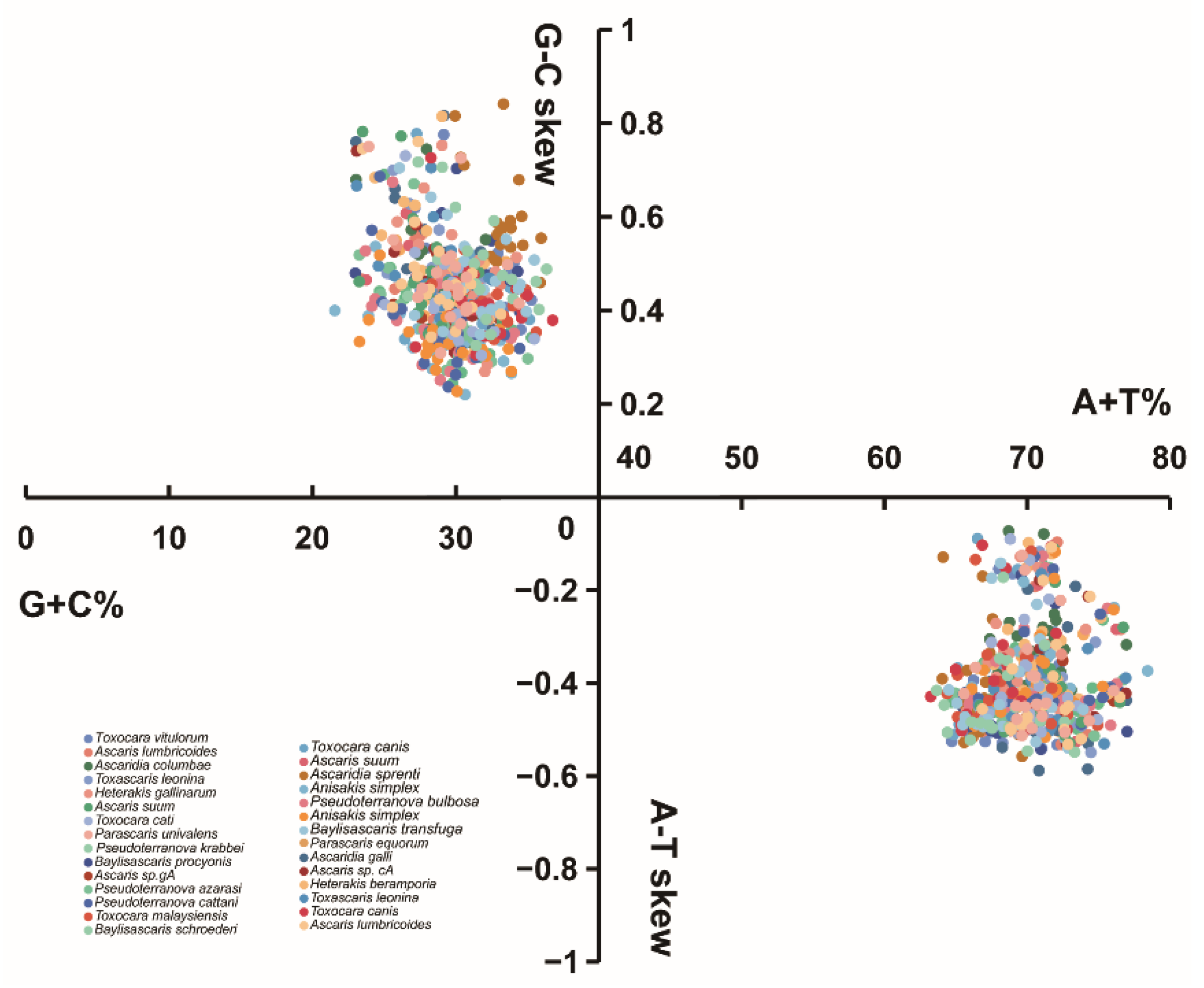
The overall nucleotide composition of the T. vitulorum mitogenome was 47.93% T, 8.34% C, 22.02% A, and 21.72% G, which came to the conclusion that T was the predominate nucleotide and C was the least favored. For the mitogenome of T. vitulorum, the concatenated PCGs had an A + T content of 66.93%, tRNAs had a value of 68.98%, rRNAs had a value of 72.22%, and the mitogenome as a whole had a value of 69.94%. Of the whole mitogenome, T. vitulorum was moderately G-skewed (GC-skew = 0.445) and T-skewed (AT-skew = −0.370), these values were similar to the skewness of T. canis, T. cati, and T. malaysiensis (Table S2), and among them, the mitogenome of T. vitulorum had the strongest bias towards T and G. Four parameters (AT-skew, GC-skew, A + T content, and G + C content) were used to make Figure 2 to compare nucleotide compositions of T. vitulorum with other nematodes. All nematode mitogenomes calculated in the figure, their AT-skews were negative, while GC-skews were positive, and they were rich in A and T, while the A + T contents of whole genomes ranged from 63.24% (T. canis) to 78.45% (A. simplex), and the G + C content were between 21.56% (A. simplex) and 36.76% (T. canis). Evidence from the investigations on the spontaneous deamination process of C and A during mammal mitogenome replication seemed to make a prediction that the strand compositional asymmetry could be attributed to the asymmetrical directional mutation pressure resulting from mitogenome replications of those nematodes including T. vitulorum [68,69].
For the codon usage, the preferred nucleotide usage at the third codon position of the Toxocara species PCGs reflected the total nucleotide content of the mitogenomes. At that position, the most frequently used nucleotide was T, whereas the least frequently use was C. We also calculated relative synonymous codon usage (RSCU) and codon counts in the mitogenome of T. vitulorum (Figure 3). It was evident that the most frequently used codon was UUG (RSCU = 3.87), followed by CGU (RSCU = 3.23) and CCU (RSCU = 3.10). The most frequently used amino acid was Phe (Count = 488), Leu (UUR) (Count = 442) and Val (Count = 392). The most frequently used start codon for the T. vitulorum is TTG (six genes; cox1, cox3, cytb, nad1, nad3, and nad6), followed by ATT (three genes; atp6, nad2, and nad4L), and GTG (nad4) and GTT (cox2) and ATG (nad5). Eight of the PCGs were predicted to use TAG (cox1, cox2, cox3, and nad4) or TAA (cytb, nad1, nad3, and nad5) as the termination codons, while the remaining genes (nad2, nad4L, nad5, and atp6) were deduced to end with an incomplete codon, such as T or TA.

3.3. tRNAs and rRNAs

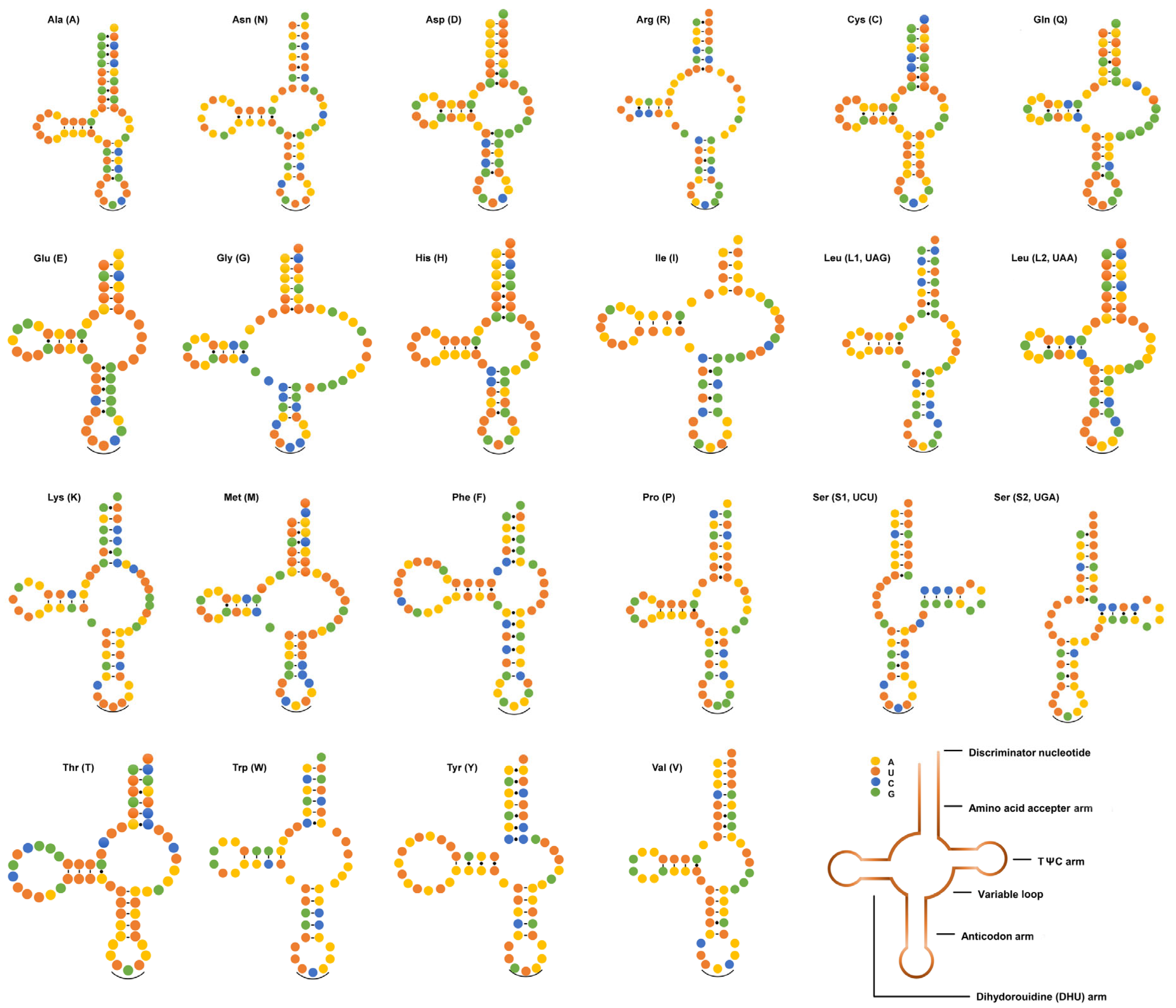
The mitogenome of T. vitulorum contained 22 tRNA genes, with lengths ranging from 52 bp (trnS [UGA]) to 72 bp (trnT) (see Table 2). The 22 tRNA secondary structures estimated (Figure 4) differed significantly from the typical cloverleaf-like structures reported in other metazoan mitogenomes but were identical to those of all other chromadorean nematodes investigated so far [57,70,71,72,73,74,75], with the exception of T. spiralis [75]. In addition, 22 anticodons (Table 1) of the T. vitulorum were as same as other species in the same genus [45,57]. For the mitogenome of T. vitulorum, the rrnS gene was 688 bp in length and the rrnL gene was 954 bp in length (Table 2). The rrnL was found between trnH and nad3, and rrnS was located between trnE and trnS2. The four PCGs in the mitogenome (nad3, nad5, nad6, and nad4L) kept two genes apart from one another (Figure 1). Their lengths were comparable to those of the homologous genes found in other nematodes, which ranged from 924 to 979 bp for rrnL and 684 to 703 bp for rrnS [72] (Table S3). The A + T percentage of the T. vitulorum rrnS gene was 68.02%, and the rrnL gene had a higher A + T content (72.22%).

3.4. Variability and Informativeness of PCGs

The majority of the PCGs (atp6, cytb, cox3, nad1, nad3-5, and nad4L) of T. vitulorum mitogenome were identical to those of other Toxocara species in length (Table S3). Nevertheless, in order to further understand the evolution and divergence of T. vitulorum mitogenome, the sequence differences of the PCGs among Ascaridoidea species were analyzed in detail (Figure 5). The nucleotide sequence identities for each of the 12 PCGs of T. vitulorum ranged 60.4–91%. In addition, the amino acid sequence identities ranged 45.5–97.9%. Sequence identities of PCGs among the genus Toxocara were higher than others, followed by the genus Toxascaris and Ascaris, in contrast, the lowest sequence identities of PCGs were observed among the genus Heterakis. The highest nucleotide sequence and amino acid sequence identities were both observed in cox1 with a darker background color (Figure 5), while the lowest nucleotide sequence identities were observed and the lowest amino acid sequence identities were observed in nad2. The data indicated that cox1 (nucleotide sequence identity as 87.2% on average, amino acid sequence identity as 93.4% on average) was the most conserved gene, and the least conserved mitochondrial genes were nad6 (nucleotide sequence identity as 76.6% on average) and nad2 (amino acid sequence identity as 71.45% on average).
Sliding window analysis was also employed in the PCGs of T. vitulorum and other Ascaridida species (Figure 6), to analyze the diversities within and between mitogenomes. It exhibited a highly variable nucleotide diversity among the concatenated 12 PCGs sequences of the mitogenomes, with Pi values for the 200 bp windows ranging from 0.137 to 0.334, and the Pi value of the whole concatenated alignment was 0.2988. The genes (cox2, cox3, cytb, atp6, nad1, nad2, nad3, nad4, nad4L, nad5, and nad6) all had relatively high nucleotide diversity of 0.306, 0.329, 0.329, 0.315, 0.318, 0.328, 0.315, 0.339, 0.325, 0.329, and 0.334, respectively, while gene cox1 had comparatively low nucleotide diversity of 0.137. Interestingly, across genes with high sequence variability, the genes with pronounced peaks and troughs of Pi seemed to have higher sequence variability than others, particularly nad4 and nad6. It appeared that cox1 was the least variable PCG, on the contrary, nad6 was one of the most variable PCGs, the conclusions were strikingly in line with pairwise comparisons between the PCG nucleotide and amino acid sequences from T. vitulorum mitogenome and those from the other reported Ascaridida nematodes. However, it appeared that nad4 also one of most variable PCG while it was nad2 observed in the identity analysis.
Cox1 was the least variable and slowly evolving mitogenome gene, according to pairwise comparisons and sliding window analysis of mitogenomes among Ascaridida mitogenomes. Further, the other PCGs exhibited significant nucleotide diversity and were rapidly developing genes, making them potential better molecular markers for species and population-level genetics investigations of T. vitulorum, particularly for diagnostics or detection involving cross contamination or other Toxocara species, since they could provide higher resolution for phylogenetically related taxonomic populations.

3.5. Substitution Ratios

The Ka/Ks ratio could be used to measure the selective pressure that has been placed on PCGs during evolution. For the purpose of comprehending the dynamics of molecular sequence evolution, estimation of Ka and Ks substitution rates for the 12 PCGs was necessary [76]. The ratio is a good predictor that when Ka/Ks = 1, it indicates neutral mutation, when Ka/Ks > 1, it indicates positive or diversifying selection, and when Ka/Ks < 1, it indicates negative or purifying selection [77,78]. Ka, Ks, and the Ka/Ks ratio were calculated for 12 PCGs from T. vitulorum and 28 other nematode isolates (Figure 7). Of all 12 PCGs, the Ka/Ks ratios were <1, indicating that these genes underwent purifying or negative selection during their evolution. The Ka/Ks ratio analysis showed that cox1 (0.121), cox2 (0.121), and cox3 (0.116) were evolving under a strong purifying selection, whereas atp6 (0.340), nad2 (0.334), and nad6 (0.317) were evolving under comparatively relaxed mutational constraints.

3.6. Phylogenetic Analysis

All three phylogenetic analyses (MP/ML/BI) using the concatenated amino acid data (Figure 8A) supported the phylogenetic relationships of Ascaris, Ascaridia, Baylisascaris, Parascaris, Porrocaecum, and Toxascaris (family Ascarididae); Toxocara (family Toxocaridae); Anisakis, Contracaecum, and Ophidascaris (family Anisakidae); Ascaridia (family Ascaridiidae); and Cucullanus (family Cucullanidae) in the order Ascaridida, and each genus was treated as a monophyletic sister group with high statistical supports (all statistical values ≥95 or 0.95). It was noteworthy that in Figure 8A, the three identical trees showed that T. malaysiensis, T. cati, and T. canis were more closely related to each other than to T. vitulorum, which supported a sister-species relationship of T. vitulorum with these three species in the family Toxocaridae. This finding was consistent with previous phylogenetic analyses based on the nuclear ITS-1 and ITS-2 and mitochondrial cox1 genes [23,24,79], but in contrast with the findings of Wickramasinghe et al. [30] and Li et al. [80], in which T. vitulorum was determined to more closely related to T. malaysiensis/T. cati than to T. canis and Toxocara was determined to more close related to Porrocaecum than to Ascaris/Baylisascaris/Parascaris/Toxascaris on the basis of several concatenated genes (e.g., 18S, 28S, cox2, and rrnS). It’s speculated that this might be attributed to use of incomplete or limited loci information. Perhaps, studies using mitogenome data from more large-scale species or isolates of Toxocara as well as taxa within the families Toxocaridae and Ascarididae worldwide are required to ascertain the phylogenetic relationships of T. vitulorum in the whole Toxocara and of the genera Porrocaecum and Toxocara in the superfamily Ascaridoidea of the order Ascaridida. Furthermore, the species of the family Toxocaridae were determined to be more closely related to species of the family Ascarididae than to species of the families Anisakidae, Ascaridiidae, and Cucullanidae, which was consistent with results of previous morphological and molecular studies [33,42,45,46,48,50,51,54,67,70,75,81,82,83,84] and also demonstrated the phylogenetic stability of the paraphyletic relationships characterized in the present study.
Subsequently, phylogenetic analyses were carried out using single protein-coding gene with an aim to screen out optimal genetic markers for phylogeny and molecular diagnostics in the order Ascaridida. As shown in Figure 8B, it appeared that although most genes showed different topologies, the phylogenetic positions of the genera Ascaridia, Toxocara, and Cucullanus were always stable in cox1-, cox2-, cox3-, cytb-, nad1-, nad2-, nad3-, nad4-, nad5-, and nad6-based phylogenetic analyses, as reported in other studies [45,55,56,85]. Further, compared to other 11 PCGs, the nad4 and nad6 genes shared the same topology as the genome-level phylogenetic analysis, suggesting that nad4 and nad6 among PCGs might be the best genetic markers and therefore could be instead of the mitogenome for molecular diagnostic, systematic, and evolutionary biological studies of this parasite and other related species in Ascaridida. Of course, the marker effectiveness of the nad4 and nad6 genes remains further validated when more additional Ascaridida mitogenomes become available, especially from the genus Toxocara, although concatenated mtDNA datasets are still the best molecular marker choice for evolutionary and phylogenetic studies of Ascaridida nematodes [33,42,45,46,48,50,51,54,67,70,75,81,82].

4. Conclusions

In this study, the complete mitogenome sequence of T. vitulorum was firstly determined and subjected to analyses and comparisons with other related species. Comparative genomics suggested that among the PCGs cox1 was the most conserved gene, whilst nad6 was the most varied gene. Genome- and individual gene-based phylogenies supported a sister-species relationship of T. vitulorum with the congeneric T. canis, T. cati, and T. malaysiensis. In further phylogenetic comparisons, the nad4 and nad6 genes were identified as novel genetic markers for phylogenetic and evolutionary studies of Ascaridida species. The results should contribute to the study of molecular diagnosis, prevention, and control of this parasitic nematode in future.

Supplementary Materials

The following supporting information can be downloaded at: https://www.mdpi.com/article/10.3390/ani12243546/s1, Figure S1: Morphological photomicrographs of Toxocara vitulorum adults. (A) Total view of T. vitulorum adults, (B) Cephalic extremity with three lips and dentiform ridges, and (C) Caudal extremity; Figure S2: The proportion of the mitogenomes comprised by PCGs, tRNAs, rRNAs, and AT-rich and intergenic regions for Toxocara species; Table S1: List of the eight primer pairs for PCR amplification and their positions in the mitogenome of Toxocara vitulorum; Table S2: Nucleotide compositions of different genomic regions in Toxocara vitulorum and other nematodes reported within Ascaridida; Table S3: Length of each PCG in Toxocara vitulorum and other nematodes reported within Ascaridida.

Author Contributions

Conceptualization, Y.X., Z.Z. and X.Z.; methodology, Y.X.; software, L.W. and Y.C.; validation, Y.X., Z.Z. and X.Z.; formal analysis, Z.W. (Zhao Wang), P.Z., Z.H., X.H., Z.W. (Zhisheng Wang) and X.Z.; investigation, Y.X., L.W. and Y.C.; resources, Z.Z. and Z.W. (Zhisheng Wang); data curation, L.W., Y.C., X.Z., Z.W. (Zhao Wang) and P.Z.; writing—original draft preparation, L.W. and Y.X.; writing—review and editing, Y.X., Z.Z. and X.Z.; visualization, Y.X., L.W. and Y.C.; supervision, Y.X.; funding acquisition, Y.X., Z.Z. and Z.W. (Zhisheng Wang). All authors have read and agreed to the published version of the manuscript.

Funding

This work was supported by the National Natural Science Foundation of China (grant no. 32273028 to Y.X.) and the China Agriculture Research System of MOF and MARA (Beef/yaks; grant no. CARS-37 to Z.Z.).

Institutional Review Board Statement

This study was approved by the Animal Ethics Committee of Sichuan Agricultural University (China; approval no. SYXK 2014-187), and all procedures including the autopsy were performed in strict accordance with the Guide for the Care and Use of Laboratory Animals (National Research Council, Bethesda, MD, USA) and the recommendations in the ARRIVE guidelines (https://www.nc3rs.org.uk/arrive-guidelines).

Informed Consent Statement

Not applicable.

Data Availability Statement

Molecular data have been deposited to GenBank with the following accession number: OP466744.

Conflicts of Interest

The funders had no role in the design of the study.

References

  1. Dorny, P.; Devleesschauwer, B.; Stoliaroff, V.; Sothy, M.; Chea, R.; Chea, B.; Sourloing, H.; Samuth, S.; Kong, S.; Nguong, K.; et al. Prevalence and Associated Risk Factors of Toxocara vitulorum Infections in Buffalo and Cattle Calves in Three Provinces of Central Cambodia. Korean J. Parasitol. 2015, 53, 197–200. [Google Scholar] [CrossRef] [PubMed] [Green Version]
  2. Chelladurai, J.J.; Bader, C.; Snobl, T.; Magstadt, D.; Cooper, V.; Brewer, M.T. Toxocara vitulorum infection in a cohort of beef calves in Iowa. Vet. Parasitol. 2015, 214, 96–99. [Google Scholar] [CrossRef] [PubMed]
  3. Dewair, A.; Bessat, M. Molecular and microscopic detection of natural and experimental infections of Toxocara vitulorum in bovine milk. PLoS ONE 2020, 15, e0233453. [Google Scholar] [CrossRef] [PubMed]
  4. Warren, E.G. Observations on the Migration and Development of Toxocara vitulorum in Natural and Experimental Hosts. Int. J. Parasitol. 1971, 1, 85–99. [Google Scholar] [CrossRef]
  5. Noriyuki, T.; Fujita, J. Morphological Observation of Toxocara vitulorum Found in Japanese Calves. J. Vet. Med. Sci. 1991, 53, 409–413. [Google Scholar] [CrossRef] [Green Version]
  6. Woodbury, M.R.; Copeland, S.; Wagner, B.; Fernando, C.; Hill, J.E.; Clemence, C. Toxocara vitulorum in a Bison (Bison Bison) Herd from Western Canada. Can. Vet. J. 2012, 53, 791–794. [Google Scholar]
  7. Holland, C.; Smith, H.V. (Eds.) Toxocara: The Enigmatic Parasite; CABI Pub: Wallingford, UK; Cambridge, MA, USA, 2006; ISBN 978-1-84593-026-4. [Google Scholar]
  8. Sarre, C.; Pardon, B.; Valgaeren, B.; Hende, D.V.; Steen, L. Intestinal Obstruction by Toxocara vitulorum in a Calf. Vlaams Diergeneeskd. Tijdschr. 2014, 83, 299–305. [Google Scholar] [CrossRef]
  9. Mahdy, O.A.; Mousa, W.M.; Abdel-Maogood, S.Z.; Nader, S.M.; Abdel-Radi, S. Molecular Characterization and Phylogenetic Analysis of Toxocara Species in Dogs, Cattle and Buffalo in Egypt. Helminthologia 2020, 57, 83–90. [Google Scholar] [CrossRef]
  10. Tamire, M.; Bedore, B. Study on Prevalence of Toxocara vitulorum in Bovine of Senkale Faris Peasant Association of Ambo Districts, West Shewa Zone, Ethiopia. Am. J. Epidemiol. Public Health 2019, 3, 1–6. [Google Scholar]
  11. Rast, L.; Lee, S.; Nampanya, S.; Toribio, J.A.L.M.L.; Khounsy, S.; Windsor, P.A. Prevalence and Clinical Impact of Toxocara vitulorum in Cattle and Buffalo Calves in Northern Lao PDR. Trop. Anim. Health Prod. 2013, 45, 539–546. [Google Scholar] [CrossRef]
  12. Raza, M.A.; Murtaza, S.; Ayaz, M.M.; Akhtar, S.; Arshad, H.M.; Basit, A.; Bachaya, H.A.; Ali, M.; Khan, M.I. Toxocara vitulorum Infestation and Associated Risk Factors in Cattle and Buffalo at Multan District, Pakistan. Sci. Int. 2013, 5, 291–294. [Google Scholar]
  13. Venjakob, P.L.; Thiele, G.; Clausen, P.H.; Nijhof, A.M. Toxocara vitulorum Infection in German Beef Cattle. Parasitol. Res. 2017, 116, 1085–1088. [Google Scholar] [CrossRef] [PubMed]
  14. Borgsteede, F.H.M.; Holzhauer, M.; Herder, F.L.; Veldhuis-Wolterbeek, E.G.; Hegeman, C. Toxocara vitulorum in Suckling Calves in The Netherlands. Res. Vet. Sci. 2012, 92, 254–256. [Google Scholar] [CrossRef]
  15. Jones, J.R.; Mitchell, E.S.E.; Redman, E.; Gilleard, J.S. Toxocara vitulorum Infection in a Cattle Herd in the UK. Vet. Rec. 2009, 164, 171–172. [Google Scholar] [CrossRef] [PubMed]
  16. Goossens, E.; Dorny, P.; Vervaecke, H.; Roden, C.; Vercammen, F.; Vercruysse, J. Toxocara vitulorum in American Bison (Bison Bison) Calves. Vet. Rec. 2007, 160, 556–557. [Google Scholar] [CrossRef]
  17. Roberts, J.A.; Fernando, S.T.; Sivanathan, S. Toxocara vitulorum in the Milk of Buffalo (Bubalus Bubalis) Cows. Res. Vet. Sci. 1990, 49, 289–291. [Google Scholar] [CrossRef] [PubMed]
  18. Zhu, X.Q.; Gasser, R.B.; Chilton, N.B.; Jacobs, D.E. Molecular Approaches for Studying Ascaridoid Nematodes with Zoonotic Potential, with an Emphasis on Toxocara Species. J. Helminthol. 2001, 75, 101–108. [Google Scholar]
  19. Chen, J.; Zhou, D.H.; Nisbet, A.J.; Xu, M.J.; Huang, S.Y.; Li, M.W.; Wang, C.R.; Zhu, X.Q. Advances in Molecular Identification, Taxonomy, Genetic Variation and Diagnosis of Toxocara spp. Infect. Genet. Evol. 2012, 12, 1344–1348. [Google Scholar] [CrossRef]
  20. Kim, H.C.; Hong, E.J.; Ryu, S.Y.; Park, J.; Cho, J.G.; Yu, D.H.; Chae, J.S.; Choi, K.S.; Park, B.K. Morphological and Molecular Characterization of Toxocara apodemi (Nematoda: Ascarididae) from Striped Field Mice, Apodemus agrarius, in Korea. Korean J. Parasitol. 2020, 58, 403–411. [Google Scholar] [CrossRef]
  21. Robbins, K.M.; Ye, W.; Fletcher, O.J. Identification of Ascaridia numidae in guinea fowl (Numida meleagris) and association with elevated mortality. Avian Dis. 2011, 55, 151–154. [Google Scholar] [CrossRef]
  22. AbouLaila, M.; Igarashi, M.; ElKhatam, A.; Menshawy, S. Gastrointestinal nematodes from buffalo in Minoufiya Governorate, Egypt with special reference to Bunostomum phlebotomum. Vet. Parasitol. Reg. Stud. Rep. 2022, 27, 100673. [Google Scholar] [CrossRef] [PubMed]
  23. Wickramasinghe, S.; Yatawara, L.; Rajapakse, R.P.V.J.; Agatsuma, T. Toxocara canis and Toxocara vitulorum: Molecular Char-acterization, Discrimination, and Phylogenetic Analysis Based on Mitochondrial (ATP Synthase Subunit 6 and 12S) and Nuclear Ribosomal (ITS-2 and 28S) Genes. Parasitol. Res. 2009, 104, 1425–1430. [Google Scholar] [CrossRef] [PubMed]
  24. Kern, E.; Kim, T.; Park, J.K. The Mitochondrial Genome in Nematode Phylogenetics. Front. Ecol. Evol. 2020, 8, 250. [Google Scholar] [CrossRef]
  25. Mikaeili, F.; Mirhendi, H.; Mohebali, M.; Hosseini, M.; Sharbatkhori, M.; Zarei, Z.; Kia, E.B. Sequence Variation in Mitochondrial Cox1 and Nad1 Genes of Ascaridoid Nematodes in Cats and Dogs from Iran. J. Helminthol. 2015, 89, 496–501. [Google Scholar] [CrossRef]
  26. Yuan, S.; Xia, Y.; Zheng, Y.; Zeng, X. Next-Generation Sequencing of Mixed Genomic DNA Allows Efficient Assembly of Rearranged Mitochondrial Genomes in Amolops chunganensis and Quasipaa boulengeri. PeerJ 2016, 4, e2786. [Google Scholar] [CrossRef] [Green Version]
  27. Barr, C.M.; Neiman, M.; Taylor, D.R. Inheritance and Recombination of Mitochondrial Genomes in Plants, Fungi and Animals. New Phytol. 2005, 168, 39–50. [Google Scholar] [CrossRef]
  28. Oguz, B. Genetic Characterization of Toxocara vitulorum in Turkey by Mitochondrial Gene Markers (Cox1). Acta Sci. Vet. 2019, 46, 6. [Google Scholar] [CrossRef]
  29. Li, K.; Lan, Y.; Luo, H.; Zhang, H.; Liu, D.; Zhang, L.; Gui, R.; Wang, L.; Shahzad, M.; Sizhu, S.; et al. Prevalence, Associated Risk Factors, and Phylogenetic Analysis of Toxocara vitulorum Infection in Yaks on the Qinghai Tibetan Plateau, China. Korean J. Parasitol. 2016, 54, 645–652. [Google Scholar] [CrossRef] [Green Version]
  30. Wickramasinghe, S.; Yatawara, L.; Rajapakse, R.P.V.J.; Agatsuma, T. Toxocara vitulorum (Ascaridida: Nematoda): Mitochondrial Gene Content, Arrangement and Composition Compared with Other Toxocara Species. Mol. Biochem. Parasitol. 2009, 166, 89–92. [Google Scholar] [CrossRef]
  31. Peng, Y.; Leung, H.C.M.; Yiu, S.M.; Chin, F.Y.L. IDBA-UD: A De Novo Assembler for Single-Cell and Metagenomic Sequencing Data with Highly Uneven Depth. Bioinformatics 2012, 28, 1420–1428. [Google Scholar] [CrossRef] [Green Version]
  32. Kearse, M.; Moir, R.; Wilson, A.; Stones-Havas, S.; Cheung, M.; Sturrock, S.; Buxton, S.; Cooper, A.; Markowitz, S.; Duran, C.; et al. Geneious Basic: An Integrated and Extendable Desktop Software Platform for the Organization and Analysis of Sequence Data. Bioinformatics 2012, 28, 1647–1649. [Google Scholar] [CrossRef] [PubMed] [Green Version]
  33. Li, M.W.; Lin, R.Q.; Song, H.Q.; Wu, X.Y.; Zhu, X.Q. The Complete Mitochondrial Genomes for Three Toxocara Species of Human and Animal Health Significance. BMC Genom. 2008, 9, 224. [Google Scholar] [CrossRef] [PubMed] [Green Version]
  34. Altschul, S.F.; Madden, T.L.; Schäffer, A.A.; Zhang, J.; Zhang, Z.; Miller, W.; Lipman, D.J. Gapped BLAST and PSI-BLAST: A New Generation of Protein Database Search Programs. Nucleic Acids Res. 1997, 25, 3389–3402. [Google Scholar] [CrossRef] [PubMed] [Green Version]
  35. Lowe, T.M.; Chan, P.P. TRNAscan-SE On-Line: Integrating Search and Context for Analysis of Transfer RNA Genes. Nucleic Acids Res. 2016, 44, W54–W57. [Google Scholar] [CrossRef] [PubMed]
  36. Rastogi, P.A. MacVector: Integrated Sequence Analysis for the Macintosh. In Bioinformatics Methods and Protocols; Humana Press: Totowa, NJ, USA, 1999; Volume 132, pp. 47–69. ISBN 978-1-59259-192-3. [Google Scholar]
  37. Lalitha, S. Primer Premier 5. Biotech. Softw. Internet Rep. 2000, 1, 270–272. [Google Scholar] [CrossRef]
  38. Tamura, K.; Stecher, G.; Peterson, D.; Filipski, A.; Kumar, S. MEGA6: Molecular Evolutionary Genetics Analysis Version 6.0. Mol. Biol. Evol. 2013, 30, 2725–2729. [Google Scholar] [CrossRef] [Green Version]
  39. Perna, N.T.; Kocher, T.D. Patterns of Nucleotide Composition at Fourfold Degenerate Sites of Animal Mitochondrial Genomes. J. Mol. Evol. 1995, 41, 353–358. [Google Scholar] [CrossRef]
  40. Burland, T.G. DNASTAR’s Lasergene Sequence Analysis Software. In Bioinformatics Methods and Protocols; Humana Press: Totowa, NJ, USA, 1999; Volume 132, pp. 71–91. ISBN 978-1-59259-192-3. [Google Scholar]
  41. Rozas, J.; Ferrer-Mata, A.; Sánchez-DelBarrio, J.C.; Guirao-Rico, S.; Librado, P.; Ramos-Onsins, S.E.; Sánchez-Gracia, A. DnaSP 6: DNA Sequence Polymorphism Analysis of Large Data Sets. Mol. Biol. Evol. 2017, 34, 3299–3302. [Google Scholar] [CrossRef]
  42. Han, L.; Yang, Y.; Li, H.; Zhou, X.; Zhou, M.; Liu, T.; Lu, Y.; Wang, Q.; Yang, S.; Shi, M.; et al. Gene Rearrangements in the Mitochondrial Genome of Ten Ascaris Species and Phylogenetic Implications for Ascaridoidea and Heterakoidea Families. Int. J. Biol. Macromol. 2022, 221, 1394–1403. [Google Scholar] [CrossRef]
  43. Kim, K.H.; Eom, K.S.; Park, J.K. The Complete Mitochondrial Genome of Anisakis simplex (Ascaridida: Nematoda) and Phylogenetic Implications. Int. J. Parasitol. 2006, 36, 319–328. [Google Scholar] [CrossRef]
  44. Mohandas, N.; Jabbar, A.; Podolska, M.; Zhu, X.Q.; Littlewood, D.T.J.; Jex, A.R.; Gasser, R.B. Mitochondrial Genomes of Anisakis simplex and Contracaecum osculatum (Sensu Stricto)—Comparisons with Selected Nematodes. Infect. Genet. Evol. 2014, 21, 452–462. [Google Scholar] [CrossRef] [PubMed]
  45. Liu, G.H.; Shao, R.; Li, J.Y.; Zhou, D.H.; Li, H.; Zhu, X.Q. The Complete Mitochondrial Genomes of Three Parasitic Nematodes of Birds: A Unique Gene Order and Insights into Nematode Phylogeny. BMC Genom. 2013, 14, 414. [Google Scholar] [CrossRef] [PubMed] [Green Version]
  46. Liu, G.H.; Wu, C.Y.; Song, H.Q.; Wei, S.J.; Xu, M.J.; Lin, R.Q.; Zhao, G.H.; Huang, S.Y.; Zhu, X.Q. Comparative Analyses of the Complete Mitochondrial Genomes of Ascaris lumbricoides and Ascaris suum from Humans and Pigs. Gene 2012, 492, 110–116. [Google Scholar] [CrossRef] [PubMed]
  47. Park, Y.C.; Kim, W.; Park, J.K. The Complete Mitochondrial Genome of Human Parasitic Roundworm, Ascaris lumbricoides. Mitochondrial DNA 2011, 22, 91–93. [Google Scholar] [CrossRef] [PubMed]
  48. Xie, Y.; Niu, L.; Zhao, B.; Wang, Q.; Nong, X.; Chen, L.; Zhou, X.; Gu, X.; Wang, S.; Peng, X.; et al. Complete Mitochondrial Genomes of Chimpanzee- and Gibbon-Derived Ascaris Isolated from a Zoological Garden in Southwest China. PLoS ONE 2013, 8, e82795. [Google Scholar] [CrossRef] [Green Version]
  49. Wolstenholme, D.R.; Okimoto, R.; Macfarlane, J.L. Nucleotide Correlations That Suggest Tertiary Interactions in the TV-Replacement Loop-Containing Mitochondrial TRNAs of the Nematodes, Caenorhabditis elegans and Ascaris suum. Nucleic Acids Res. 1994, 22, 4300–4306. [Google Scholar] [CrossRef] [Green Version]
  50. Xie, Y.; Zhang, Z.; Wang, C.; Lan, J.; Li, Y.; Chen, Z.; Fu, Y.; Nie, H.; Yan, N.; Gu, X.; et al. Complete Mitochondrial Genomes of Baylisascaris schroederi, Baylisascaris ailuri and Baylisascaris transfuga from Giant Panda, Red Panda and Polar Bear. Gene 2011, 482, 59–67. [Google Scholar] [CrossRef]
  51. Xie, Y.; Zhang, Z.; Niu, L.; Wang, Q.; Wang, C.; Lan, J.; Deng, J.; Fu, Y.; Nie, H.; Yan, N.; et al. The Mitochondrial Genome of Baylisascaris procyonis. PLoS ONE 2011, 6, e27066. [Google Scholar] [CrossRef] [Green Version]
  52. Park, J.K.; Sultana, T.; Lee, S.H.; Kang, S.; Kim, H.K.; Min, G.S.; Eom, K.S.; Nadler, S.A. Monophyly of Clade III Nematodes Is Not Supported by Phylogenetic Analysis of Complete Mitochondrial Genome Sequences. BMC Genom. 2011, 12, 392. [Google Scholar] [CrossRef] [Green Version]
  53. Wang, B.J.; Gu, X.B.; Yang, G.Y.; Wang, T.; Lai, W.M.; Zhong, Z.J.; Liu, G.H. Mitochondrial Genomes of Heterakis gallinae and Heterakis beramporia Support That They Belong to the Infraorder Ascaridomorpha. Infect. Genet. Evol. 2016, 40, 228–235. [Google Scholar] [CrossRef]
  54. Jabbar, A.; Littlewood, D.T.J.; Mohandas, N.; Briscoe, A.G.; Foster, P.G.; Müller, F.; von Samson-Himmelstjerna, G.; Jex, A.R.; Gasser, R.B. The Mitochondrial Genome of Parascaris univalens--Implications for a “Forgotten” Parasite. Parasites Vectors 2014, 7, 428. [Google Scholar] [CrossRef] [PubMed] [Green Version]
  55. Liu, S.S.; Liu, G.H.; Zhu, X.Q.; Weng, Y.B. The Complete Mitochondrial Genome of Pseudoterranova azarasi and Comparative Analysis with Other Anisakid Nematodes. Infect. Genet. Evol. 2015, 33, 293–298. [Google Scholar] [CrossRef] [PubMed]
  56. Liu, G.H.; Zhou, D.H.; Zhao, L.; Xiong, R.C.; Liang, J.Y.; Zhu, X.Q. The Complete Mitochondrial Genome of Toxascaris leonina: Comparison with Other Closely Related Species and Phylogenetic Implications. Infect. Genet. Evol. 2014, 21, 329–333. [Google Scholar] [CrossRef] [PubMed]
  57. Jex, A.R.; Waeschenbach, A.; Littlewood, D.T.J.; Hu, M.; Gasser, R.B. The Mitochondrial Genome of Toxocara canis. PLoS Negl. Trop. Dis. 2008, 2, e273. [Google Scholar] [CrossRef]
  58. Notredame, C.; Higgins, D.G.; Heringa, J. T-Coffee: A Novel Method for Fast and Accurate Multiple Sequence Alignment. J. Mol. Biol. 2000, 302, 205–217. [Google Scholar] [CrossRef] [Green Version]
  59. Castresana, J. Selection of Conserved Blocks from Multiple Alignments for Their Use in Phylogenetic Analysis. Mol. Biol. Evol. 2000, 17, 540–552. [Google Scholar] [CrossRef] [Green Version]
  60. Goloboff, P.A.; Catalano, S.A.; Torres, A. Parsimony Analysis of Phylogenomic Datasets (II): Evaluation of PAUP*, MEGA and MPBoot. Cladistics 2022, 38, 126–146. [Google Scholar] [CrossRef]
  61. Guindon, S.; Gascuel, O. A Simple, Fast, and Accurate Algorithm to Estimate Large Phylogenies by Maximum Likelihood. Syst. Biol. 2003, 52, 696–704. [Google Scholar] [CrossRef] [Green Version]
  62. Darriba, D.; Taboada, G.L.; Doallo, R.; Posada, D. ProtTest 3: Fast Selection of Best-Fit Models of Protein Evolution. Bioinformatics 2011, 27, 1164–1165. [Google Scholar] [CrossRef] [Green Version]
  63. Kalyaanamoorthy, S.; Minh, B.Q.; Wong, T.K.F.; von Haeseler, A.; Jermiin, L.S. ModelFinder: Fast Model Selection for Accurate Phylogenetic Estimates. Nat. Methods 2017, 14, 587–589. [Google Scholar] [CrossRef] [Green Version]
  64. Ronquist, F.; Teslenko, M.; van der Mark, P.; Ayres, D.L.; Darling, A.; Höhna, S.; Larget, B.; Liu, L.; Suchard, M.A.; Huelsenbeck, J.P. MrBayes 3.2: Efficient Bayesian Phylogenetic Inference and Model Choice across a Large Model Space. Syst. Biol. 2012, 61, 539–542. [Google Scholar] [CrossRef] [PubMed] [Green Version]
  65. Page, R.D.M. Visualizing Phylogenetic Trees Using TreeView. Curr. Protoc. Bioinform. 2003, 1, 6-2. [Google Scholar] [CrossRef] [PubMed]
  66. Lin, R.Q.; Liu, G.H.; Zhang, Y.; D’Amelio, S.; Zhou, D.H.; Yuan, Z.G.; Zou, F.C.; Song, H.Q.; Zhu, X.Q. Contracaecum rudolphii B: Gene Content, Arrangement and Composition of Its Complete Mitochondrial Genome Compared with Anisakis simplex s.l. Exp. Parasitol. 2012, 130, 135–140. [Google Scholar] [CrossRef] [PubMed]
  67. Meng, X.; Xie, Y.; Gu, X.; Zheng, Y.; Liu, Y.; Li, Y.; Wang, L.; Zhou, X.; Zuo, Z.; Yang, G. Sequencing and Analysis of the Complete Mitochondrial Genome of Dog Roundworm Toxocara canis (Nematoda: Toxocaridae) from USA. Mitochondrial DNA B Resour. 2019, 4, 2999–3001. [Google Scholar] [CrossRef]
  68. Reyes, A.; Gissi, C.; Pesole, G.; Saccone, C. Asymmetrical Directional Mutation Pressure in the Mitochondrial Genome of Mammals. Mol. Biol. Evol. 1998, 15, 957–966. [Google Scholar] [CrossRef] [Green Version]
  69. Saccone, C.; De Giorgi, C.; Gissi, C.; Pesole, G.; Reyes, A. Evolutionary Genomics in Metazoa: The Mitochondrial DNA as a Model System. Gene 1999, 238, 195–209. [Google Scholar] [CrossRef]
  70. Okimoto, R.; Macfarlane, J.L.; Clary, D.O.; Wolstenholme, D.R. The Mitochondrial Genomes of Two Nematodes, Caenorhabditis elegans and Ascaris suum. Genetics 1992, 130, 471–498. [Google Scholar] [CrossRef]
  71. Keddie, E.M.; Higazi, T.; Unnasch, T.R. The Mitochondrial Genome of Onchocerca volvulus: Sequence, Structure and Phylogenetic Analysis. Mol. Biochem. Parasitol. 1998, 95, 111–127. [Google Scholar] [CrossRef]
  72. Hu, M.; Chilton, N.B.; Gasser, R.B. The Mitochondrial Genomes of the Human Hookworms, Ancylostoma duodenale and Necator americanus (Nematoda: Secernentea). Int. J. Parasitol. 2002, 32, 145–158. [Google Scholar] [CrossRef]
  73. Hu, M.; Chilton, N.B.; Gasser, R.B. The Mitochondrial Genome of Strongyloides Stercoralis (Nematoda)—Idiosyncratic Gene Order and Evolutionary Implications. Int. J. Parasitol. 2003, 33, 1393–1408. [Google Scholar] [CrossRef]
  74. Hu, M.; Gasser, R.B.; Abs El-Osta, Y.G.; Chilton, N.B. Structure and Organization of the Mitochondrial Genome of the Canine Heartworm, Dirofilaria Immitis. Parasitology 2003, 127, 37–51. [Google Scholar] [CrossRef] [PubMed]
  75. Jex, A.R.; Hu, M.; Littlewood, D.T.J.; Waeschenbach, A.; Gasser, R.B. Using 454 Technology for Long-PCR Based Sequencing of the Complete Mitochondrial Genome from Single Haemonchus contortus (Nematoda). BMC Genom. 2008, 9, 11. [Google Scholar] [CrossRef] [PubMed] [Green Version]
  76. Yang, Z.; Nielsen, R. Estimating Synonymous and Nonsynonymous Substitution Rates under Realistic Evolutionary Models. Mol. Biol. Evol. 2000, 17, 32–43. [Google Scholar] [CrossRef] [Green Version]
  77. Liu, G.H.; Gasser, R.B.; Otranto, D.; Xu, M.J.; Shen, J.L.; Mohandas, N.; Zhou, D.H.; Zhu, X.Q. Mitochondrial Genome of the Eyeworm, Thelazia callipaeda (Nematoda: Spirurida), as the First Representative from the Family Thelaziidae. PLoS Negl. Trop. Dis. 2013, 7, e2029. [Google Scholar] [CrossRef]
  78. Hurst, L.D. The Ka/Ks Ratio: Diagnosing the Form of Sequence Evolution. Trends Genet. 2002, 18, 486. [Google Scholar] [CrossRef] [PubMed]
  79. Sultan, K.; Omar, M.; Desouky, A.Y.; El-Seify, M.A. Molecular and Phylogenetic Study on Toxocara vitulorum from Cattle in the Mid-Delta of Egypt. J. Parasit. Dis. 2015, 39, 584–587. [Google Scholar] [CrossRef] [Green Version]
  80. Li, L.; Lü, L.; Nadler, S.A.; Gibson, D.I.; Zhang, L.P.; Chen, H.X.; Zhao, W.T.; Guo, Y.N. Molecular Phylogeny and Dating Reveal a Terrestrial Origin in the Early Carboniferous for Ascaridoid Nematodes. Syst. Biol. 2018, 67, 888–900. [Google Scholar] [CrossRef] [Green Version]
  81. Gao, J.F.; Zhang, X.X.; Wang, X.X.; Li, Q.; Li, Y.; Xu, W.W.; Gao, Y.; Wang, C.R. According to Mitochondrial DNA Evidence, Parascaris equorum and Parascaris univalens May Represent the Same Species. J. Helminthol. 2019, 93, 383–388. [Google Scholar] [CrossRef]
  82. Zhao, Q.; Abuzeid, A.M.I.; He, L.; Zhuang, T.; Li, X.; Liu, J.; Zhu, S.; Chen, X.; Li, G. The Mitochondrial Genome Sequence Analysis of Ophidascaris baylisi from the Burmese Python (Python Molurus Bivittatus). Parasitol. Int. 2021, 85, 102434. [Google Scholar] [CrossRef]
  83. Implications for Morphological Evolution and Classification. Mol. Phylogenet. Evol. 1998, 10, 221–236. [CrossRef]
  84. Hartwich, G. Keys to genera of the Ascaridoidea. In CIH Keys to the Nematode Parasites of Vertebrates; Anderson, R.C., Chabaud, A.G., Willmott, S., Eds.; Commonwealth Agricultural Bureaux, Farnham Royal: Wallingford, UK, 1974; Volume 2, pp. 1–14. [Google Scholar]
  85. Liu, G.H.; Nadler, S.A.; Liu, S.S.; Podolska, M.; D’Amelio, S.; Shao, R.; Gasser, R.B.; Zhu, X.Q. Mitochondrial Phylogenomics Yields Strongly Supported Hypotheses for Ascaridomorph Nematodes. Sci. Rep. 2016, 6, 39248. [Google Scholar] [CrossRef] [PubMed]
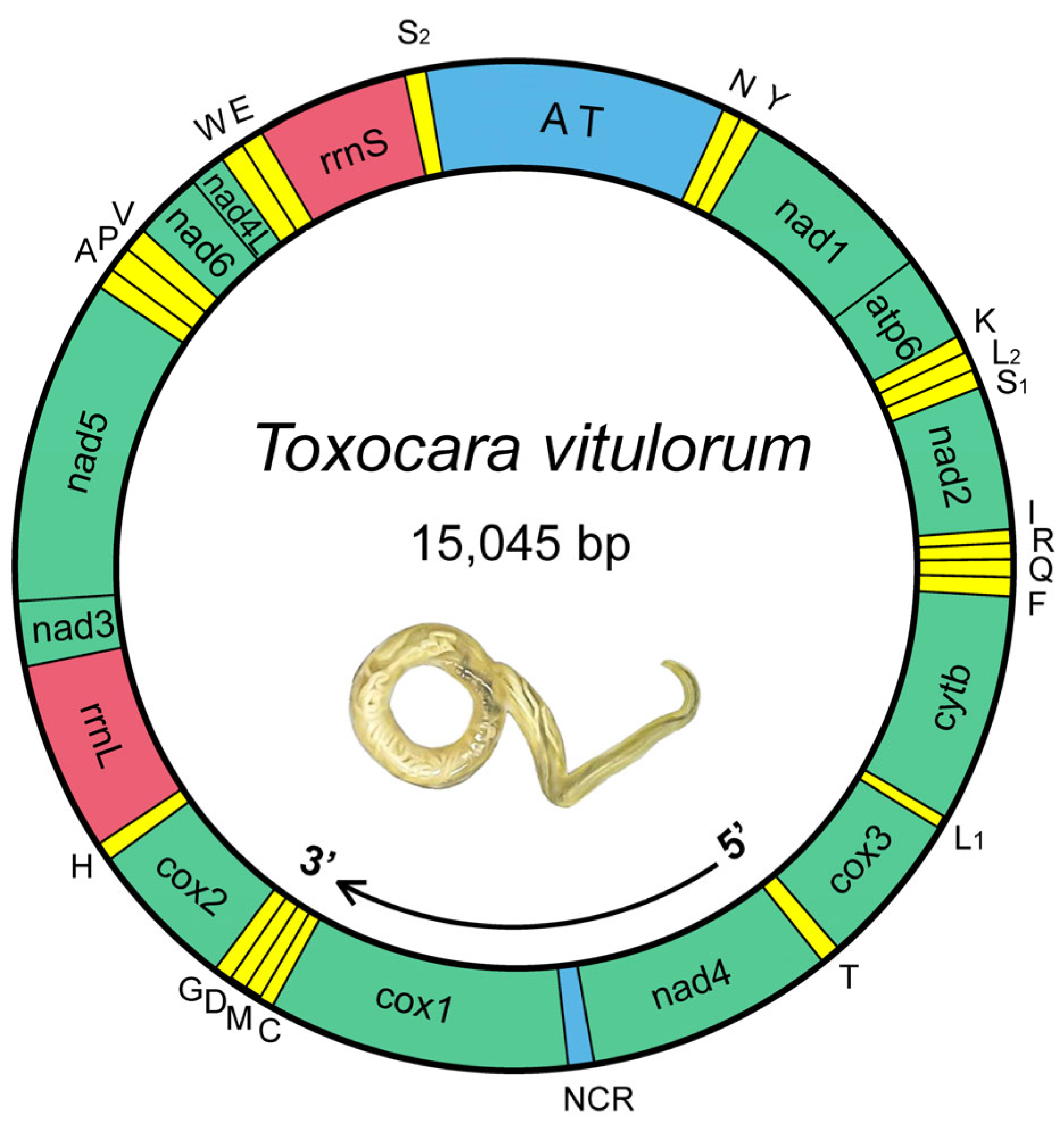
Figure 1. Circular map for T. vitulorum mitogenome. 36 genes follow standard nomenclature, including 22 tRNAs, which are denoted by one letter in accordance with the IPUC-IUB single-letter amino acid codes. The two leucine genes were differed by L1 and L2, and the two serine genes by S1 and S2. All the genes are present on the same strand and transcribed in the same direction (5′–3′), as indicated by the arrow. NCR indicates the non-coding region. AT indicates the AT-rich region.
Animals 12 03546 g001
Figure 2. AT-skew vs. GC-skew of the different genetic positions of 29 mitogenomes. The X axis provides the A + T/G + C values, while the Y axis provides the skew values. For each species, the entire mitogenome, concatenated PCGs, concatenated tRNAs, concatenated rRNAs, the 1st, 2nd, 3rd, and 12 PCGs were separate calculated.
Animals 12 03546 g002
Figure 3. The RSCU and codon numbers in T. vitulorum mitogenome. Different color bars represent the RSCU of each codon; the codon numbers are indicated by the orange line graph (right Y axis scale).
Animals 12 03546 g003
Figure 4. Predicted secondary structures of 22 tRNAs in T. vitulorum mitogenome. The tRNAs are labeled with their corresponding IPUC-IUB single-letter amino acid codes. Dashes (−) denote Watson–Crick bonds, dots (·) indicate mispaired nucleotide bonds, and (‿) indicate tRNA anticodons.
Animals 12 03546 g004
Figure 5. Pair-wise comparison of the nucleotide (left) and amino acid (right) sequence identities for PCGs. Colors from purple (high identity) to white (low identity) indicate the different identities of nucleotide sequences, and colors from green (high identity) to white (low identity) indicate the different identities of amino acid sequences.
Animals 12 03546 g005
Figure 6. Sliding window analysis of the concatenated alignments of PCGs. The black line represents the value of the nucleotide diversity and are calculated using the parameters: window size = 200 bp and step size = 20 bp. Gene names, boundaries, and average nucleotide diversity values are indicated above the graph. Colors from yellow (high diversity) to white (low diversity) indicate the different nucleotide diversities.
Animals 12 03546 g006
Figure 7. Evolutionary rates of PCGs between T. vitulorum and 28 other Rhabditida mitogenomes. The rates of non-synonymous substitutions (Ka) and synonymous substitutions (Ks) and the ratio of Ka/Ks are calculated for each PCG.
Animals 12 03546 g007
Figure 8. Phylogenetic relationships among Ascaridoidea nematodes. The phylogeny is inferred using the MP, ML, and BI methods of 27 Ascaridida species for which complete mitogenome sequences are available, using the Seuratoidea species (C. robustus) as the outgroup. (A) Phylogenetic tree is based on concatenated amino acid sequences of 12 PCGs. (B) Twelve topological phenograms are based on each PCG, and the tree topology consistent with the trees in A are marked with red line boxes. The black circle sign represents the species sequenced in this study. The numbers along the branches in A and B indicate bootstrap values/posterior probabilities (<95% or 0.95 supports not shown) resulting from different analyses in the order MP/ML/BI.
Animals 12 03546 g008
Table 1. Summary of the mitogenome information included in this study.
SpeciesHostCountryGenBank No.References
Anisakis pegreffiiScomber japonicusJapanLC222461[42]
Anisakis simplex (s.l)Conger myriasterKoreaNC_007934[43]
Anisakis simplex (s.s)Clupea harengusfromPolanadKC965056[44]
Ascaridia columbaePigeonChinaJX624729[45]
Ascaridia galliChickenChinaJX624728[45]
Ascaridia sp. GHL-2013ParrotChinaJX624730[45]
Ascaris lumbricoidesHumanChinaHQ704900[46]
Ascaris lumbricoidesHumanKoreaNC_016198 [47]
Ascaris sp. (cA.)Hylobates hoolockChinaKC839987[48]
Ascaris sp. (gA.)Pan troglodytesChinaKC839986[48]
Ascaris suumPigChinaHQ704901[46]
Ascaris suumPigUSANC_001327[49]
Baylisascaris ailuriAilurus fulgensChinaHQ671080[50]
Baylisascaris procyonisProcyon lotorChinaJF951366[51]
Baylisascaris schroederiAiluropoda melanleucaChinaNC_015927[50]
Baylisascaris transfugaUrsus maritimusChinaNC_015924 [50]
Contracaecum ogmorhiniSealAustraliaKU558725Unpublished
Contracaecum osculatumClupea harengusAustraliaNC_024037[44]
Contracaecum rudolphiiCormorantChinaNC_014870Unpublished
Cucullanus robustusConger myriasterUSANC_016128[52]
Heterakis beramporiaChickenChinaKU529972[53]
Heterakis gallinarumChickenChinaKU529973 [53]
Ophidascaris baylisiPython bivittatusChinaMW880927Unpublished
Ophidascaris sp.Elaphe carinataChinaMK106624Unpublished
Parascaris equorumHorseChinaNC_036427Unpublished
Parascaris univalensHorseChinaNC_024884[54]
Pseudoterranova azarasiEumetopias jubataJapanKR052144[55]
Pseudoterranova cattaniOtaria byroniaChileKU558721Unpublished
Pseudoterranova krabbeiHalichoerus grypusNorwayKU558724Unpublished
Pseudoterranova bulbosaErignathus barbatusCanadaKU558720Unpublished
Porrocaecum sp.CraneChinaCNP0003131[42]
Toxascaris leonineDogAustraliaKC902750[56]
Toxascaris leonineCheetahChinaMK516267Unpublished
Toxocara canisFoxAustraliaEU730761[57]
Toxocara canisDogChinaNC_010690[33]
Toxocara catiCatChinaNC_010773[33]
Toxocara malaysiensisCatChinaNC_010527[33]
Table 2. The structure and organization of the mitogenome of T. vitulorum.
GeneLocationLengthStart CodonStop CodonAnticodonIntergenic
Region
trnN1–5959 GUU0
trnY60–12061 GUA−1
nad1120–992873TTGTAA 1
atp6994–1591598ATTT 0
trnK1592–165261 UUU0
trnL2 1653–170755 UAA0
trnS11708–176053 UCU0
nad21761–2605845ATTTA −1
trnI2605–266662 GAU0
trnR2667–272155 ACG1
trnQ2723–277755 UUG−1
trnF2777–283761 GAA0
cytb2838–39441107TTGTAA 6
trnL13951–400555 UAG0
cox34006–4773768TTGTAG 0
trnT4774–484572 UGU0
nad44846–60751230GTGTAG 109
cox16185–77651581TTGTAG 2
trnC7768–782255 GCA8
trnM7831–789060 CAU1
trnD7892–794756 GUC−1
trnG7947–800256 UCC0
cox28003–8719717GTTTAG −10
trnH8710–876556 GUG0
rrnL8766–9719954 0
nad39720–10055336TTGTAA 1
nad510057–116381582ATGT 0
trnA11639–1169355 UGC2
trnP11696–1175055 UGG0
trnV11751–1180656 UAC0
nad611807–12241435TTGTAA −1
nad4l12241–12472232ATTT 0
trnW12473–1253361 UCA−1
trnE12533–1259159 UUC0
rrnS12592–13279688 13
trnS213293–1334452 UGA1701
Publisher’s Note: MDPI stays neutral with regard to jurisdictional claims in published maps and institutional affiliations.