Article

Effects of Sulfamethoxazole on Fertilization and Embryo Development in the Arbacia lixula Sea Urchin

1
Dipartimento di Scienze e Tecnologie Biologiche, Chimiche e Farmaceutiche (STEBICEF), Università di Palermo, 90128 Palermo, Italy
2
Istituto Zooprofilattico Sperimentale Della Sicilia A. Mirri, 90129 Palermo, Italy
*
Authors to whom correspondence should be addressed.
These authors contributed equally to this work.
Animals 2022, 12(18), 2483; https://doi.org/10.3390/ani12182483
Submission received: 22 August 2022 / Revised: 8 September 2022 / Accepted: 14 September 2022 / Published: 19 September 2022
(This article belongs to the Section Aquatic Animals)

Abstract

:

Simple Summary

Drugs released into the aquatic environment create serious problems for the organisms that live there. For this reason, the present study investigates the in vitro effects of the antibiotic sulfamethoxazole, widely found in wastewater, on the fertilization and development of the Arbacia lixula sea urchin. The results showed a significant reduction in the percentage of fertilized oocytes at the highest drug concentrations, together with an increase in anomalies and delays in the development of the embryo. Therefore, the data obtained suggest urgent intervention on the release of these drugs in order to prevent important alterations in the species’ development and to preserve biodiversity.

Abstract

To date, drugs released into the aquatic environment are a real problem, and among antibiotics, sulfamethoxazole is the one most widely found in wastewater; thus, the evaluation of its toxicity on marine organisms is very important. This study, for the first time, investigates the in vitro effects of 4 concentrations of sulfamethoxazole (0.05 mg/L, 0.5 mg/L, 5 mg/L, 50 mg/L) on the fertilization and development of the sea urchin Arbacia lixula. The gametes were exposed to drugs in three different stages: simultaneously with, prior to, and post-fertilization. The results show a significant reduction in the percentage of fertilized oocytes at the highest drug concentrations. Moreover, an increase in anomalies and delays in embryo development following the treatment with the drug was demonstrated. Therefore, the data suggest that this antibiotic can alter the development of marine organisms, making it urgent to act to reduce their release and to determine the concentration range with the greatest impact.

1. Introduction

To date, different types of pollutants are released into aquatic environments, negatively affecting the lives of the aquatic organisms that inhabit them [1,2,3]. These organisms are very important as a resource of bioactive molecules as food, and in the study of immune responses under stress conditions and human disease [4,5,6,7,8]. The use of drugs is widespread in the treatment of human, animal, and plant diseases, and the problem of pharmaceutical waste being released into aquatic environments is becoming urgent [9]. After their use, the remaining drugs are usually disposed of in an inadequate manner via the sinks, toilets, and drains of hospitals and pharmaceutical factories, often in the form of active compounds that have not been metabolized, thus maintaining their original chemical structures and biological functions [10,11,12,13,14]. Conventional wastewater treatment plants often cannot effectively remove these pharmaceutical compounds (PCs) since their elimination is influenced by the characteristics of drugs, the techniques used, and the environmental conditions [15,16]. This leads to the frequent detection of PCs in wastewater, and their concentrations generally range from ng/L to μg/L [17,18,19]. It is known that their persistence in wastewater can negatively affect the ecosystem as these compounds are potentially toxic for non-target organisms, including marine animals [20,21]. In fact, several studies in the literature highlight the negative effects of drugs on different aquatic species, including oxidative stress, metabolic and immunological alterations, and reductions in the success of fertilization and the development of organisms [3,22,23,24,25,26,27]. Fertilization, in particular, is a critical phase of the lifecycle that ensures the preservation of a species, and the presence of pharmaceutical compounds can seriously interfere with this process, affecting the production and quality of the gametes. Indeed, De Oliveira et al. [23] reported on the adverse effects of 4 common pharmaceuticals (acetaminophen (1.2–9.0 mg/L), chlorpromazine (0.50–3.14 mg/L), diclofenac sodium (52.0–155.3 mg/L), and propranolol (4.0–11.9 mg/L) on the reproduction of Daphnia magna after analyzing the mobility of the gametes and the quantity of offspring. The authors demonstrated the dose-dependent immobility of gametes in the presence of all of the tested drugs, as well as a significant decrease in the production of offspring caused by chlorpromazine and propranolol. Similarly, another study on Mytilus galloprovincialis showed that diclofenac (100 μg/L) causes the down-modulation of tyrosine metabolism and the up-modulation of tryptophan metabolism, which are involved in the production and release of gametes [28]. Among these pharmaceuticals products, and specifically among the classes of antibiotics, sulfonamides—synthetic antimicrobial molecules whose structure mimes that of para-aminobenzoic acid (PABA)—are widespread [29,30]. One of the most common and diffuse drugs belonging to this category is sulfamethoxazole (SMX), a competitive inhibitor of the enzyme dihydropteroate synthase, which prevents the formation of dihydropteroic acid, a precursor of folic acid, which is required for bacterial growth. It is widely used in the treatment of several bacterial infections in humans, and it is effective against both Gram-positive and Gram-negative bacteria [31]. The average concentrations of the drug that are detected in wastewater range from ng/L to μg/L [32,33], and several studies have demonstrated its negative effects (i.e., oxidative stress, the suppression of the immune response, inflammation, the inhibition of acetylcholinesterase activity, the alteration of osmotic regulation, and the alteration of energy metabolism) in marine vertebrates and invertebrates [34,35,36,37]. However, other authors have recently reported that the concentrations of this drug discarded by the livestock industry and in aquaculture wastewater can rise to the level of mg/L [38,39]. Focusing on invertebrates, it is known that they play an important role as a source of bioactive molecules with antioxidant, anti-inflammatory, and anti-tumor effects [4,5,40,41,42], and they also represent useful bioindicators for the study of environmental conditions [43,44,45,46,47,48]. Among invertebrates, echinoderms possess peculiar characteristics which make them particularly suitable for applications in the ecotoxicological field, such as their wide distribution, the ease of sampling/maintaining them, the ease of collecting their gametes, their availability throughout the year, their external fertilization mechanism, their rapid development, and their sensitivity to a wide spectrum of pollutants [49]. Furthermore, their lifecycle stages, such as fertilization and embryonic division, are particularly sensitive to environmental conditions and are therefore increasingly used in the ecotoxicological approach to assess the quality of an environment [50,51]. In light of all of this, given the importance of these invertebrates as bioindicators, and considering the SMX concentrations reported by other authors [32,33,38,39], our study aimed to investigate for the first time the in vitro effects of exposure to sulfamethoxazole at different concentrations and at three different stages (simultaneously with, prior to, and post-fertilization) on Arbacia lixula gametes and embryos. Our goal was to evaluate whether the drug negatively affects the gametes and embryos and whether this depends on the concentration. In fact, the obtained results could provide useful information for the regulation of sulfamethoxazole releases into water, thus protecting the species that live there.

2. Materials and Methods

2.1. Experimental Animals

A total of 54 specimens of Arbacia lixula (16 ± 1 g) were collected in the spring from the rocky seabed of Mongerbino (Gulf of Palermo) and housed in the aquarium of the University of Palermo (STEBICEF Department). The animals were maintained for 2 weeks, respecting the photoperiod in different tanks (with the same conditions) containing aerated, filtered, refrigerated seawater (16 ± 1 °C), at a salinity level of 38‰ and with oxygenation of 8 mg/L. During this period, the animals were fed with invertebrate food (Azoo, Taikong Corp., New Taipei, Taiwan) until 24 h prior to the beginning of the experiment (which were performed according to the guidelines of the OECD, 2012 [52]).

2.2. Gamete Collection

The emission of the gametes from each individual was induced by the intracelomic injection of 1 mL of 0.5 M KCl through the peristomial membrane [53,54,55]. The eggs and spermatozoa were collected and washed 3 times in filtered seawater (FSW: 0.5 M NaCl, 8 mM KCl, 30 mM Na2SO4, and 2 mM NaHCO3, with a pH 8.1, and pasteurized at 60 °C); the vitality and the number of the gametes was assessed using a Neubauer chamber [56].

2.3. Experimental Plan

SMX was dissolved in the FSW to perform three different experiments:
-
The simultaneous exposure (SE) of the gametes, which were simultaneously combined in multiwell plates with the experimental solutions. The effects were observed on the first embryonic division after 4 h. The percentages of unfertilized eggs and the regular divisions were evaluated;
-
The pre-exposure (PE), which was achieved by pre-treating the eggs and spermatozoa separately for 2 h before fertilization. The percentages of unfertilized eggs and the regular divisions were evaluated; and
-
The post-fertilization exposure (PFE), in which previously fertilized eggs were subsequently exposed to differing drug concentrations for 2 h. The effects were observed starting at 4 h post-exposure. The percentages of irregular, regular, and delayed divisions were evaluated.
All experiments were performed in polystyrene, 6-well plates with SMX solutions at final concentrations of 0.05 mg/L, 0.5 mg/L, 5 mg/L, and 50 mg/L (with a 10 mL final volume for each well). Embryotoxicity was evaluated using eggs and sperm at a 1:8 ratio (1 × 10⁶/mL eggs and 8 × 10⁶/mL sperm). Each experiment (SE, PE, and PFE) was repeated 3 times using 6 individuals for replication (18 in total: 9 males and 9 females). For all experiments and for all experimental points, a total of 100 unfertilized eggs (including regular, irregular, and delayed divisions) were counted, and the data were expressed as the number in each individual category.

2.4. Statistical Analysis

All of the results obtained were grouped by experimental condition (simultaneous exposure, preventive exposure, and post-fertilization exposure) and concentration of sulfamethoxazole treatment (0.05, 0.5, 5, 50 mg/L) comprising the control group for the statistical analysis. The assumption of the normality of distribution and homogeneity of variance were verified before the analysis. One-way ANOVA and non-parametric Kruskal–Wallis tests were carried out to verify significant differences in unfertilized eggs, regular divisions, and delayed divisions (only in the PFE analysis) between data groups. In the case of significant differences, parametric (Tukey test) and non-parametric (Kruskal–Wallis post-hoc test) multiple comparison tests were performed. The statistical analysis was performed with R 3.2.2 software (R Development Core Team) using the R Commander environment.

3. Results

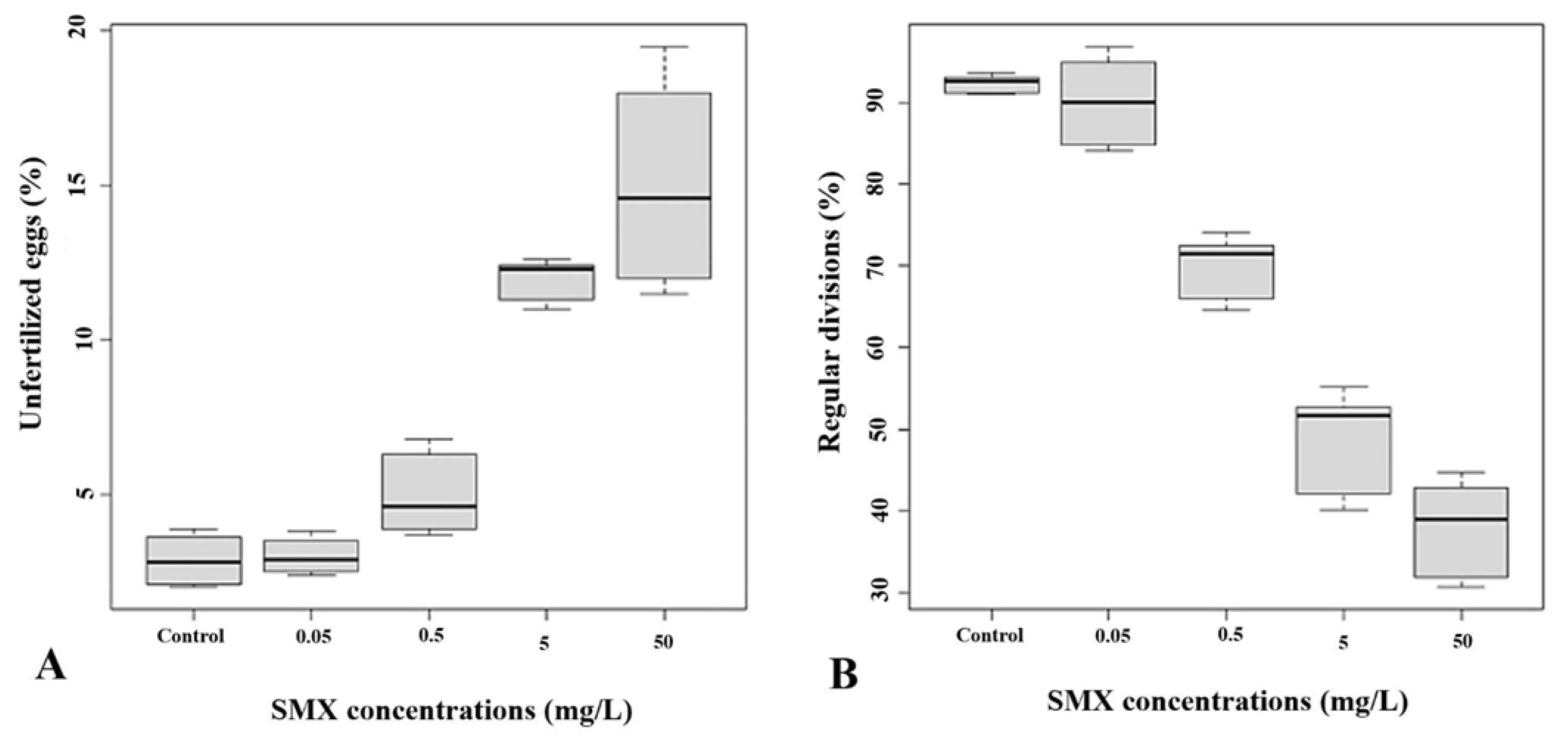
3.1. Simultaneous Exposure Effects

The simultaneous exposure (SE) of the gametes to the experimental solutions of the drug showed significant increases in the percentage of unfertilized eggs compared to the controls (2.89 ± 0.76%) only at concentrations of 5 mg/L (12 ± 0.61%) and 50 mg/L (15.10 ± 3%) (Df = 4; F value = 120.8; p < 2 × 10 − 16) (Figure 1A).
Regarding the percentage of regular divisions, the results showed a dose-dependent trend. As shown in Figure 1B, compared to the control (92.29 ± 1%), a significant decrease was observed after exposure to 0.5 mg/L, 5 mg/L, and 50 mg/L of SMX (70.11 ± 3.59%, 49.04 ± 5.81%, and 37.99 ± 5.56% respectively).

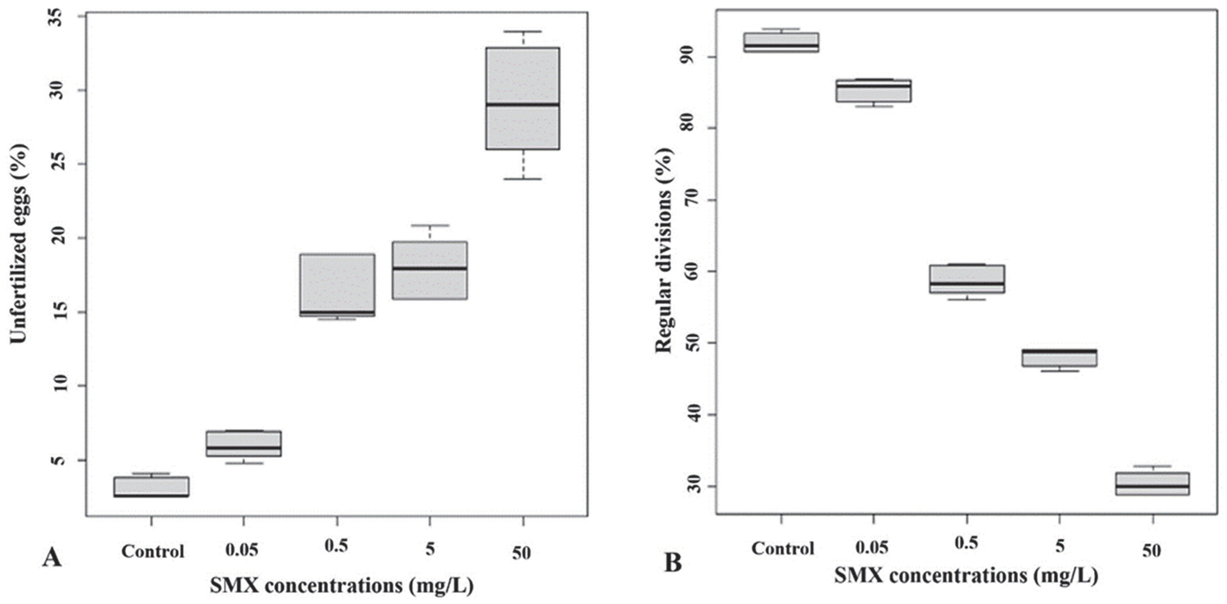
3.2. Preventive Exposure Effects

In the second set of experiments, the gametes were pre-treated for 2 h before fertilization with the four different concentrations of SMX. The exposure of the gametes to the drug showed a dose-dependent effect on fertilization (Kruskal–Wallis chi-square = 49.21, df = 5, p-value = 0.000). The multiple comparison after the Kruskal–Wallis test showed that the percentages of the unfertilized eggs resulted significantly higher as compared to the controls (3.06 ± 0.69%) only at two SMX concentrations, i.e., 5 mg/L and 50 mg/L (18.02 ± 1.9% and 29.21 ± 3.69%, respectively; p < 0.05) (Figure 2A). The results of the effects on the regular division of embryos demonstrated a significant reduction in the percentage of regular divisions compared to the control samples (mean square = 6358; F value = 302.7; p = 2 ×10 −16) at all experimental SMX concentrations (Figure 2B).

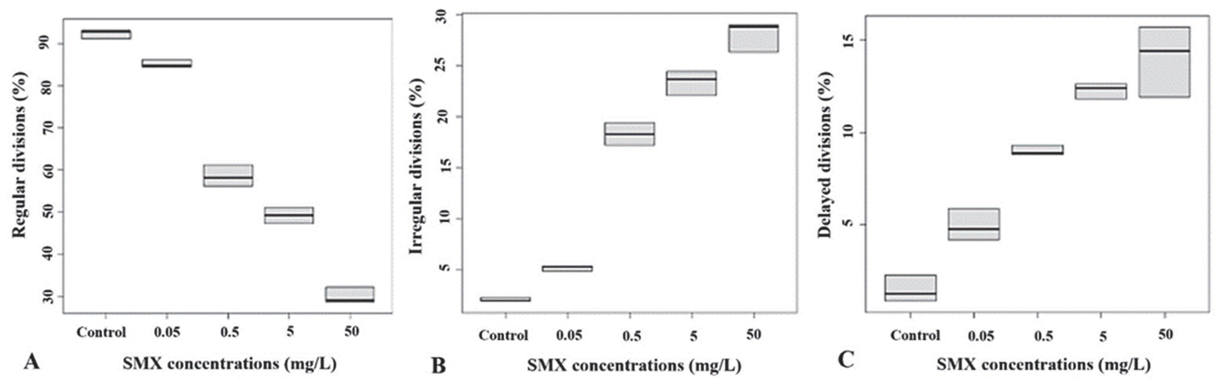
3.3. Post-Fertilization Exposure Effects

The toxic effects of the exposure to SMX were also evaluated by incubating the embryos at 2 h post-fertilization. Regarding the percentage of regular and irregular divisions, a significant decrease was observed only at two SMX experimental concentrations (5 mg/L and 50 mg/L) compared to the controls (Kruskal–Wallis chi-square = 51.687, df = 5, p-value = 6.25 × 10⁻¹⁰). In particular, a significant decrease in the percentage of regular divisions (49.3 ± 1.65% and 30 ± 1.66%, respectively) compared to the controls (92.40 ± 0.98%, Figure 3A) and a significant increase in the percentage of irregular divisions (23.43 ± 1.06% and 28.13 ± 1.30%, respectively); Kruskal–Wallis chi-square = 42.429, df = 4, p-value = 1.36 × 10⁻⁸) with respect to the controls (1.97 ± 0.18%, Figure 3B) were observed. On the other hand, the results showed a significant increase in the percentage of delayed divisions of the embryos only at 0.5 mg/L (9 ± 0.23%), 5 mg/L (12.27 ± 0.36%), and 50 mg/L (14 ± 1.67%), as compared to the controls (1.5 ± 0.62%, p < 0.05), as showed in Figure 3C.

4. Discussion

The growing presence of drugs in the aquatic environment [57], imposes the need to analyze and better understand their impact on aquatic organisms, with particular attention to their reproduction, a critical and important phase of the lifecycle [58]. In fact, in the aquatic environment, most animal species reproduce via external fertilization through the release of gametes into the surrounding environment. When pollutants, including drugs, are poured into the aquatic environment, the gametes are exposed to these substances, which alters their quality and fertilization processes [58]. As reported by Duan et al. [38], although SMX concentrations range from only ng/L to μg/L in natural water, the discharge of livestock and aquaculture wastewater can cause the elevation of SMX levels (54.83 mg/L, [39]). For this reason, with information about the toxicity of this drug being scarce and unclear [38], our study aimed to analyze for the first time its in vitro effects on the gametes and fertilization of A. lixula in particular so as to evaluate whether the negative effects occur during the gametes’ simultaneous exposure or during pre- or post-fertilization exposure.
The obtained results show that treatments with different concentrations of SMX negatively affect in a dose-dependent manner the percentage of both unfertilized eggs and the regular divisions of embryos, highlighting the negative effects of the drug on the gametes. In fact, it is known that pollutants present in the aquatic environment can alter the motility of spermatozoa or interfere with the chemotactic mechanisms (spermatozoa’s attraction towards eggs via chemotactic substances) with a consequent reduction in fertility [59]. Moreover, the negative effects observed on the regular divisions could be due to changes in the expressions of genes involved in detoxification, growth, development, and reproduction, or due to the actions of the drug as an endocrine disruptor [60,61]. In light of the results obtained from the simultaneous exposure experiment, we wanted to understand whether the effects of the drug were negative in the case of pre-treating the eggs and spermatozoa for 2 h before fertilization with the different experimental SMX solutions. In this case, as in the case of the simultaneous exposure of the gametes, significant dose-dependent increases in the levels of unfertilized eggs and significant dose-dependent decreases in the levels of regular divisions were observed. To date, no one has analyzed the effects of the pre-exposure to sulfonamides on the gametes of A. lixula sea urchins. However, similar results have been reported in studies in which anti-inflammatory drugs, another category of drugs widely detected in wastewater, were tested [62]. In addition, in this case, in our experiment, we could speculate that the pre-exposure of gametes to SMX influences sperm motility, thereby affecting the percentage of fertilized eggs. This is confirmed by Zanuri et al. [63], who investigated the effects of two anti-inflammatory drugs (diclofenac and ibuprofen) on the fertilization of the echinoderms Asterias rubens and Psammechinus miliaris after pre-incubation of the gametes with the drugs. As discussed above, the reduction in the quality of the gametes, in terms of sperm motility; the inability to fertilize the egg; and the damage to the genetic material could be the main causes of the decreased fertilization rate. Moreover, exposure to the drug could also alter or inhibit the normal mechanisms necessary for fertilization, such as polyspermy prevention and cortical granule exocytosis [64,65], inevitably leading to a reduction in normal embryonic development and subsequently to a reduction in the regular division of embryos.
Once we had evaluated the effects of SMX on the gametes, the third set of experiments in this study allowed us to investigate the consequences of post-fertilization exposure to the different concentrations of SMX on the fertilized eggs. Toxic effects were observed at the highest SMX concentrations, with a significant decrease in the percentage of regular division and an increase in the percentage of irregular and delayed divisions. There is currently no similar evidence in the literature on invertebrates, and particularly on echinoderms, which tests the effects of sulfonamides after fertilization. However, comparable results on the embryos of Mellita quinquiesperforata were obtained with anticancer drugs, as shown by the recent study by Mello et al. [66]. Moreover, evidence in the literature reports the anticancer effects of sulfonamides, which exert their action through cell cycle arrest [67,68]. Probably, the concentrations of SMX tested in our study could conceivably block DNA replication, the cell cycle, and cell proliferation. The delays recorded in the development of the embryos could also be caused by the dose-dependent increases in the accumulation of ROS species and the up-regulation of inflammatory genes, which are responsible for alterations and damage at the biochemical and cellular levels [34,69,70]. Furthermore, alterations in the normal development of fertilized eggs, due to interference with the mitotic apparatus could happen, in agreement with a previous study which evaluated the effect of the drug diamino diphenyl sulfone on the embryonic development of the sea urchin Lytechinus variegatus [71]. However, there is also conflicting evidence in the literature showing that treatment with 50 mg/L of SMX does not induce obvious alterations during the development of P. lividus embryos [72]. In view of this, it is evident that the effects of SMX could be species-specific and could depend on exposure concentrations and combinations with other drugs. It would therefore be important to perform further experiments that clarify the cellular and molecular mechanisms by which SMX negatively affects the embryonic development of A. lixula.

5. Conclusions

Given the increasingly abundant and disproportionate use of antibiotics that inevitably are poured into wastewater and then into the aquatic environment, it is necessary to investigate their potential toxicological effects. Our analysis on the consequences of the exposure of the gametes and embryos of Arbacia lixula to increasing concentrations of the antibiotic sulfamethoxazole (SMX) has shown for the first time in this species the manner in which the evaluated drug affects the reproduction of the sea urchin, causing adverse effects during the simultaneous exposure as well as the pre- and post-fertilization exposure of gametes. This has made it possible to identify that the drug acts negatively from the beginning on the gametes when released into the aquatic environment, reducing the possibility of fertilization. Subsequently, the drug can further negatively affect embryonic development, causing, in the end, negative effects on the fitness of this species. In fact, our results show that the fertilization rate was reduced in a dose-dependent manner, and the fertilized eggs showed alterations in cell division, morphological anomalies, and degeneration. The stages of reproduction are extremely important for animals as they ensure the continuity of the species, and for this reason, such a negative impact of SMX on the fertilization and development processes could lead to a serious reduction in their fitness, endangering the survival of the species and disturbing the entire ecosystem. The choice to use different SMX concentrations in this study allowed us to highlight its toxicity and to determine which concentrations cause the greatest negative effects, and consequently the level of concentration that should not be exceeded. All of this can therefore provide the basis for the proper monitoring and improvement of procedures and technologies suitable for limiting the release of drugs into the aquatic environment.

Author Contributions

Conceptualization, M.V., M.C. and V.L.; methodology, M.M., G.C. and P.B.; validation, G.C. and M.V.; investigation, M.C. and P.B.; resources, M.V. and V.F.; data curation, M.M., V.L., G.C. and P.B.; writing—original draft preparation, V.L., M.C. and M.M.; writing—review and editing, C.L. and A.V.; supervision, M.V., C.L., V.F. and A.V.; funding acquisition, M.V. and V.F. All authors have read and agreed to the published version of the manuscript.

Funding

This work was supported by the Fondo Finalizzato alla Ricerca di Ateneo (FFR, 2018–2021 with FFR-D15-180028) and FISHPATHNET, Fondo FEAMP 2014/2020.

Institutional Review Board Statement

Ethical review and approval were waived for this study performed on a marine invertebrate other than cephalopods (shrimp), which do not require Ethical review or approval according to EU directive 2010/63/EU (22 September 2010).

Informed Consent Statement

Not applicable.

Data Availability Statement

The data presented in this study are available on request from the corresponding authors ([email protected]; [email protected]).

Acknowledgments

We express our thanks to Giampaolo Badalamenti for his collaboration in the management of the animals used in the experiments.

Conflicts of Interest

The authors declare no conflict of interest.

References

  1. Patel, N.; Khan, M.D.; Shahane, S.; Rai, D.; Chauhan, D.; Kant, C.; Chaudhary, V.K. Emerging Pollutants in Aquatic Environment: Source, Effect, and Challenges in Biomonitoring and Bioremediation—A Review. Pollution 2020, 6, 99–113. [Google Scholar] [CrossRef]
  2. Mauro, M.; Pérez-Arjona, I.; Perez, E.J.B.; Ceraulo, M.; Bou-Cabo, M.; Benson, T.; Espinosa, V.; Beltrame, F.; Mazzola, S.; Vazzana, M.; et al. The effect of low frequency noise on the behaviour of juvenile Sparus aurata. J. Acoust. Soc. Am. 2020, 147, 3795–3807. [Google Scholar] [CrossRef] [PubMed]
  3. Mauro, M.; Lazzara, V.; Arizza, V.; Luparello, C.; Ferrantelli, V.; Cammilleri, G.; Inguglia, L.; Vazzana, M. Human Drug Pollution in the Aquatic System: The Biochemical Responses of Danio rerio Adults. Biology 2021, 10, 1064. [Google Scholar] [CrossRef] [PubMed]
  4. Luparello, C.; Mauro, M.; Lazzara, V.; Vazzana, M. Collective Locomotion of Human Cells, Wound Healing and Their Control by Extracts and Isolated Compounds from Marine Invertebrates. Molecules 2020, 25, E2471. [Google Scholar] [CrossRef] [PubMed]
  5. Luparello, C.; Mauro, M.; Arizza, V.; Vazzana, M. Histone Deacetylase Inhibitors from Marine Invertebrates. Biology 2020, 9, E429. [Google Scholar] [CrossRef] [PubMed]
  6. Inguglia, L.; Chiaramonte, M.; Di Stefano, V.; Schillaci, D.; Cammilleri, G.; Pantano, L.; Mauro, M.; Vazzana, M.; Ferrantelli, V.; Nicolosi, R.; et al. Salmo salar fish waste oil: Fatty acids composition and antibacterial activity. PeerJ 2020, 8, e9299. [Google Scholar] [CrossRef]
  7. Mauro, M.; Queiroz, V.; Arizza, V.; Campobello, D.; Custódio, M.R.; Chiaramonte, M.; Vazzana, M. Humoral responses during wound healing in Holothuria tubulosa (Gmelin, 1788). Comp. Biochem. Physiol. Part B Biochem. Mol. Biol. 2021, 253, 110550. [Google Scholar] [CrossRef]
  8. Vizzini, A.; Bonura, A.; La Paglia, L.; Fiannaca, A.; La Rosa, M.; Urso, A.; Mauro, M.; Vazzana, M.; Arizza, V. Transcriptomic Analyses Reveal 2 and 4 Family Members of Cytochromes P450 (CYP) Involved in LPS Inflammatory Response in Pharynx of Ciona robusta. Int. J. Mol. Sci. 2021, 22, 11141. [Google Scholar] [CrossRef]
  9. Branchet, P.; Arpin-Pont, L.; Piram, A.; Boissery, P.; Wong-Wah-Chung, P.; Doumenq, P. Pharmaceuticals in the marine environment: What are the present challenges in their monitoring? Sci. Total Environ. 2021, 766, 142644. [Google Scholar] [CrossRef]
  10. Faleye, A.C.; Adegoke, A.A.; Ramluckan, K.; Bux, F.; Stenström, T.A. Identification of antibiotics in wastewater: Current state of extraction protocol and future perspectives. J. Water Health 2017, 15, 982–1003. [Google Scholar] [CrossRef] [Green Version]
  11. Madikizela, L.M.; Tavengwa, N.T.; Chimuka, L. Status of pharmaceuticals in African water bodies: Occurrence, removal and analytical methods. J. Environ. Manag. 2017, 193, 211–220. [Google Scholar] [CrossRef] [PubMed]
  12. Yang, C.-Y.; Wang, Q.-H.; Hao, R.-J.; Liao, Y.-S.; Du, X.-D.; Deng, Y.-W. Effects of replacing microalgae with an artificial diet on pearl production traits and mineralization-related gene expression in pearl oyster Pinctada fucata martensii. Aquac. Res. 2017, 48, 5331–5337. [Google Scholar] [CrossRef]
  13. Zhong, Y.; Chen, Z.-F.; Dai, X.; Liu, S.-S.; Zheng, G.; Zhu, X.; Liu, S.; Yin, Y.; Liu, G.; Cai, Z. Investigation of the interaction between the fate of antibiotics in aquafarms and their level in the environment. J. Environ. Manag. 2018, 207, 219–229. [Google Scholar] [CrossRef] [PubMed]
  14. Aydin, S.; Aydin, M.E.; Ulvi, A.; Kilic, H. Antibiotics in hospital effluents: Occurrence, contribution to urban wastewater, removal in a wastewater treatment plant, and environmental risk assessment. Environ. Sci. Pollut. Res. 2019, 26, 544–558. [Google Scholar] [CrossRef]
  15. Le-Minh, N.; Khan, S.J.; Drewes, J.E.; Stuetz, R.M. Fate of antibiotics during municipal water recycling treatment processes. Water Res. 2010, 44, 4295–4323. [Google Scholar] [CrossRef]
  16. Gracia-Lor, E.; Sancho, J.V.; Serrano, R.; Hernández, F. Occurrence and removal of pharmaceuticals in wastewater treatment plants at the Spanish Mediterranean area of Valencia. Chemosphere 2012, 87, 453–462. [Google Scholar] [CrossRef]
  17. Carvalho, I.T.; Santos, L. Antibiotics in the aquatic environments: A review of the European scenario. Environ. Int. 2016, 94, 736–757. [Google Scholar] [CrossRef]
  18. Ekpeghere, K.I.; Sim, W.-J.; Lee, H.-J.; Oh, J.-E. Occurrence and distribution of carbamazepine, nicotine, estrogenic compounds, and their transformation products in wastewater from various treatment plants and the aquatic environment. Sci. Total Environ. 2018, 640–641, 1015–1023. [Google Scholar] [CrossRef]
  19. Assress, H.A.; Nyoni, H.; Mamba, B.B.; Msagati, T.A. Occurrence and risk assessment of azole antifungal drugs in water and wastewater. Ecotoxicol. Environ. Saf. 2020, 187, 109868. [Google Scholar] [CrossRef]
  20. Mearns, A.J.; Reish, D.J.; Oshida, P.S.; Ginn, T.; Rempel-Hester, M.A.; Arthur, C.; Rutherford, N.; Pryor, R. Effects of Pollution on Marine Organisms. Water Environ. Res. 2015, 87, 1718–1816. [Google Scholar] [CrossRef]
  21. Fabbri, E.; Franzellitti, S. Human pharmaceuticals in the marine environment: Focus on exposure and biological effects in animal species: Fate and Effects of Pharmaceuticals in Marine Environments. Environ. Toxicol. Chem. 2015, 35, 799–812. [Google Scholar] [CrossRef] [PubMed]
  22. Mezzelani, M.; Gorbi, S.; Da Ros, Z.; Fattorini, D.; D’Errico, G.; Milan, M.; Bargelloni, L.; Regoli, F. Ecotoxicological potential of non-steroidal anti-inflammatory drugs (NSAIDs) in marine organisms: Bioavailability, biomarkers and natural occurrence in Mytilus galloprovincialis. Mar. Environ. Res. 2016, 121, 31–39. [Google Scholar] [CrossRef] [PubMed]
  23. de Oliveira, L.L.D.; Antunes, S.C.; Gonçalves, F.; Rocha, O.; Nunes, B. Acute and chronic ecotoxicological effects of four pharmaceuticals drugs on cladoceran Daphnia magna. Drug Chem. Toxicol. 2016, 39, 13–21. [Google Scholar] [CrossRef] [PubMed]
  24. Almeida, A.R.; Tacão, M.; Machado, A.L.; Golovko, O.; Zlabek, V.; Domingues, I.; Henriques, I. Long-term effects of oxytetracycline exposure in zebrafish: A multi-level perspective. Chemosphere 2019, 222, 333–344. [Google Scholar] [CrossRef] [PubMed]
  25. Giménez, V.; Nunes, B. Effects of commonly used therapeutic drugs, paracetamol, and acetylsalicylic acid, on key physiological traits of the sea snail Gibbula umbilicalis. Environ. Sci. Pollut. Res. 2019, 26, 21858–21870. [Google Scholar] [CrossRef] [PubMed]
  26. Di Paola, D.; Iaria, C.; Capparucci, F.; Cordaro, M.; Crupi, R.; Siracusa, R.; D’Amico, R.; Fusco, R.; Impellizzeri, D.; Cuzzocrea, S.; et al. Aflatoxin B1 Toxicity in Zebrafish Larva (Danio rerio): Protective Role of Hericium erinaceus. Toxins 2021, 13, 710. [Google Scholar] [CrossRef] [PubMed]
  27. Di Paola, D.; Capparucci, F.; Abbate, J.M.; Cordaro, M.; Crupi, R.; Siracusa, R.; D’Amico, R.; Fusco, R.; Genovese, T.; Impellizzeri, D.; et al. Environmental Risk Assessment of Oxaliplatin Exposure on Early Life Stages of Zebrafish (Danio rerio). Toxics 2022, 10, 81. [Google Scholar] [CrossRef]
  28. Bonnefille, B.; Gomez, E.; Alali, M.; Rosain, D.; Fenet, H.; Courant, F. Metabolomics assessment of the effects of diclofenac exposure on Mytilus galloprovincialis: Potential effects on osmoregulation and reproduction. Sci. Total Environ. 2018, 613–614, 611–618. [Google Scholar] [CrossRef]
  29. Sukul, P.; Spiteller, M. Sulfonamides in the environment as veterinary drugs. Rev. Environ. Contam. Toxicol. 2006, 187, 67–101. [Google Scholar]
  30. Apaydın, S.; Török, M. Sulfonamide derivatives as multi-target agents for complex diseases. Bioorganic Med. Chem. Lett. 2019, 29, 2042–2050. [Google Scholar] [CrossRef]
  31. Wishart, D.S.; Feunang, Y.D.; Guo, A.C.; Lo, E.J.; Marcu, A.; Grant, J.R.; Sajed, T.; Johnson, D.; Li, C.; Sayeeda, Z.; et al. DrugBank 5.0: A Major Update to the DrugBank Database for 2018. Nucleic Acids Res. 2018, 46, D1074–D1082. [Google Scholar] [CrossRef] [PubMed]
  32. Blair, B.; Nikolaus, A.; Hedman, C.; Klaper, R.; Grundl, T. Evaluating the degradation, sorption, and negative mass balances of pharmaceuticals and personal care products during wastewater treatment. Chemosphere 2015, 134, 395–401. [Google Scholar] [CrossRef] [PubMed]
  33. Rodriguez-Mozaz, S.; Vaz-Moreira, I.; Varela Della Giustina, S.; Llorca, M.; Barceló, D.; Schubert, S.; Berendonk, T.U.; Michael-Kordatou, I.; Fatta-Kassinos, D.; Martinez, J.L.; et al. Antibiotic residues in final effluents of European wastewater treatment plants and their impact on the aquatic environment. Environ. Int. 2020, 140, 105733. [Google Scholar] [CrossRef]
  34. Limbu, S.M.; Zhou, L.; Sun, S.-X.; Zhang, M.-L.; Du, Z.-Y. Chronic exposure to low environmental concentrations and legal aquaculture doses of antibiotics cause systemic adverse effects in Nile tilapia and provoke differential human health risk. Environ. Int. 2018, 115, 205–219. [Google Scholar] [CrossRef] [PubMed]
  35. Yang, X.; Xu, X.; Wei, X.; Wan, J.; Zhang, Y. Biomarker Effects in Carassius auratus Exposure to Ofloxacin, Sulfamethoxazole and Ibuprofen. Int. J. Environ. Res. Public Health 2019, 16, 1628. [Google Scholar] [CrossRef] [PubMed]
  36. Serra-Compte, A.; Álvarez-Muñoz, D.; Solé, M.; Cáceres, N.; Barceló, D.; Rodríguez-Mozaz, S. Comprehensive study of sulfamethoxazole effects in marine mussels: Bioconcentration, enzymatic activities and metabolomics. Environ. Res. 2019, 173, 12–22. [Google Scholar] [CrossRef]
  37. Drzymała, J.; Kalka, J. Ecotoxic interactions between pharmaceuticals in mixtures: Diclofenac and sulfamethoxazole. Chemosphere 2020, 259, 127407. [Google Scholar] [CrossRef]
  38. Duan, W.; Cui, H.; Jia, X.; Huang, X. Occurrence and ecotoxicity of sulfonamides in the aquatic environment: A review. Sci. Total Environ. 2022, 820, 153178. [Google Scholar] [CrossRef]
  39. Thiebault, T. Sulfamethoxazole/Trimethoprim ratio as a new marker in raw wastewaters: A critical review. Sci. Total Environ. 2020, 715, 136916. [Google Scholar] [CrossRef]
  40. Lazzara, V.; Arizza, V.; Luparello, C.; Mauro, M.; Vazzana, M. Bright Spots in the Darkness of Cancer: A Review of Starfishes-Derived Compounds and Their Anti-Tumor Action. Mar. Drugs 2019, 17, 617. [Google Scholar] [CrossRef]
  41. Mauro, M.; Lazzara, V.; Punginelli, D.; Arizza, V.; Vazzana, M. Antitumoral compounds from vertebrate sister group: A review of Mediterranean ascidians. Dev. Comp. Immunol. 2020, 108, 103669. [Google Scholar] [CrossRef] [PubMed]
  42. Punginelli, D.; Schillaci, D.; Mauro, M.; Deidun, A.; Barone, G.; Arizza, V.; Vazzana, M. The potential of antimicrobial peptides isolated from freshwater crayfish species in new drug development: A review. Dev. Comp. Immunol. 2021, 126, 104258. [Google Scholar] [CrossRef] [PubMed]
  43. Marques, J.; Vilanova, E.; Mourão, P.A.S.; Fernàndez-Busquets, X. Marine organism sulfated polysaccharides exhibiting significant antimalarial activity and inhibition of red blood cell invasion by Plasmodium. Sci. Rep. 2016, 6, 24368. [Google Scholar] [CrossRef] [PubMed]
  44. Parisi, M.G.; Mauro, M.; Sarà, G.; Cammarata, M. Temperature increases, hypoxia, and changes in food availability affect immunological biomarkers in the marine mussel Mytilus galloprovincialis. J. Comp. Physiol. B 2017, 187, 1117–1126. [Google Scholar] [CrossRef]
  45. Pinto, J.; Costa, M.; Leite, C.; Borges, C.; Coppola, F.; Henriques, B.; Monteiro, R.; Russo, T.; Di Cosmo, A.; Soares, A.M.; et al. Ecotoxicological effects of lanthanum in Mytilus galloprovincialis: Biochemical and histopathological impacts. Aquat. Toxicol. 2019, 211, 181–192. [Google Scholar] [CrossRef]
  46. Świacka, K.; Maculewicz, J.; Smolarz, K.; Szaniawska, A.; Caban, M. Mytilidae as model organisms in the marine ecotoxicology of pharmaceuticals—A review. Environ. Pollut. 2019, 254, 113082. [Google Scholar] [CrossRef]
  47. Vazzana, M.; Mauro, M.; Ceraulo, M.; Dioguardi, M.; Papale, E.; Mazzola, S.; Arizza, V.; Beltrame, F.; Inguglia, L.; Buscaino, G. Underwater high frequency noise: Biological responses in sea urchin Arbacia lixula (Linnaeus, 1758). Comp. Biochem. Physiol. Part A Mol. Integr. Physiol. 2020, 242, 110650. [Google Scholar] [CrossRef]
  48. Vazzana, M.; Ceraulo, M.; Mauro, M.; Papale, E.; Dioguardi, M.; Mazzola, S.; Arizza, V.; Chiaramonte, M.; Buscaino, G. Effects of acoustic stimulation on biochemical parameters in the digestive gland of Mediterranean mussel Mytilus galloprovincialis (Lamarck, 1819). J. Acoust. Soc. Am. 2020, 147, 2414–2422. [Google Scholar] [CrossRef]
  49. Morroni, L.; Rakaj, A.; Grosso, L.; Fianchini, A.; Pellegrini, D.; Regoli, F. Sea cucumber Holothuria polii (Delle Chiaje, 1823) as new model for embryo bioassays in ecotoxicological studies. Chemosphere 2020, 240, 124819. [Google Scholar] [CrossRef]
  50. Böttger, S.A.; McClintock, J.B. The effects of organic and inorganic phosphates on fertilization and early development in the sea urchin Lytechinus variegatus (Echinodermata: Echinoidea). Comp. Biochem. Physiol. Part C Toxicol. Pharmacol. 2001, 129, 307–315. [Google Scholar] [CrossRef]
  51. Arslan, O.C.; Parlak, H. Embryotoxic effects of nonylphenol and octylphenol in sea urchin Arbacia lixula. Ecotoxicology 2007, 16, 439–444. [Google Scholar] [CrossRef] [PubMed]
  52. OECD. Test No. 211: Daphnia magna Reproduction Test, OECD Guidelines for the Testing of Chemicals, Section 2; OECD Publishing: Paris, France, 2012. [Google Scholar] [CrossRef]
  53. His, E.; Heyvang, I.; Geffard, O.; de Montaudouin, X. A comparison between oyster (Crassostrea gigas) and sea urchin (Paracentrotus lividus) larval bioassays for toxicological studies. Water Res. 1999, 33, 1706–1718. [Google Scholar] [CrossRef]
  54. Marin, M.G.; Moschino, V.; Cima, F.; Celli, C. Embryotoxicity of butyltin compounds to the sea urchin Paracentrotus lividus. Mar. Environ. Res. 2000, 50, 231–235. [Google Scholar] [CrossRef]
  55. Volpi Ghirardini, A.; Arizzi Novelli, A.; Tagliapietra, D. Sediment toxicity assessment in the Lagoon of Venice (Italy) using Paracentrotus lividus (Echinodermata: Echinoidea) fertilization and embryo bioassays. Environ. Int. 2005, 31, 1065–1077. [Google Scholar] [CrossRef] [PubMed]
  56. Dacie, J.V.; Lewis, S.M. Practical Haematology, 12th ed.; Bain, B.J., Bates, I., Laffan, M.A., Lewis, S.M., Eds.; Elsevier: Philadelphia, PA, USA, 2017. [Google Scholar]
  57. Heberer, T. Occurrence, fate, and removal of pharmaceutical residues in the aquatic environment: A review of recent research data. Toxicol. Lett. 2002, 131, 5–17. [Google Scholar] [CrossRef]
  58. Aulsebrook, L.C.; Bertram, M.G.; Martin, J.M.; Aulsebrook, A.E.; Brodin, T.; Evans, J.P.; Hall, M.D.; O’Bryan, M.K.; Pask, A.J.; Tyler, C.R.; et al. Reproduction in a polluted world: Implications for wildlife. Reproduction 2020, 160, R13–R23. [Google Scholar] [CrossRef]
  59. Lewis, C.; Ford, A.T. Infertility in male aquatic invertebrates: A review. Aquat. Toxicol. 2012, 120–121, 79–89. [Google Scholar] [CrossRef]
  60. Ribeiro, S.; Torres, T.; Martins, R.; Santos, M.M. Toxicity screening of Diclofenac, Propranolol, Sertraline and Simvastatin using Danio rerio and Paracentrotus lividus embryo bioassays. Ecotoxicol. Environ. Saf. 2015, 114, 67–74. [Google Scholar] [CrossRef]
  61. Liu, Y.; Wang, L.; Pan, B.; Wang, C.; Bao, S.; Nie, X. Toxic effects of diclofenac on life history parameters and the expression of detoxification-related genes in Daphnia magna. Aquat. Toxicol. 2017, 183, 104–113. [Google Scholar] [CrossRef]
  62. Tyumina, E.A.; Bazhutin, G.A.; Cartagena Gómez, A.D.P.; Ivshina, I.B. Nonsteroidal Anti-inflammatory Drugs as Emerging Contaminants. Microbiology 2020, 89, 148–163. [Google Scholar] [CrossRef]
  63. Mohd Zanuri, N.B.; Bentley, M.G.; Caldwell, G.S. Assessing the impact of diclofenac, ibuprofen and sildenafil citrate (Viagra®) on the fertilisation biology of broadcast spawning marine invertebrates. Mar. Environ. Res. 2017, 127, 126–136. [Google Scholar] [CrossRef] [PubMed]
  64. Pesando, D.; Robert, S.; Huitorel, P.; Gutknecht, E.; Pereira, L.; Girard, J.-P.; Ciapa, B. Effects of methoxychlor, dieldrin and lindane on sea urchin fertilization and early development. Aquat. Toxicol. 2004, 66, 225–239. [Google Scholar] [CrossRef] [PubMed]
  65. Lewis, C.; Galloway, T. Reproductive Consequences of Paternal Genotoxin Exposure in Marine Invertebrates. Environ. Sci. Technol. 2009, 43, 928–933. [Google Scholar] [CrossRef] [PubMed]
  66. Mello, L.C.; da Fonseca, T.G.; de Souza, A.D.M. Ecotoxicological assessment of chemotherapeutic agents using toxicity tests with embryos of Mellita quinquiesperforata. Mar. Pollut. Bull. 2020, 159, 111493. [Google Scholar] [CrossRef]
  67. Van Kesteren, C.; Beijnen, J.H.; Schellens, J.H.M. E7070: A novel synthetic sulfonamide targeting the cell cycle progression for the treatment of cancer. Anti-Cancer Drugs 2002, 13, 989–997. [Google Scholar] [CrossRef]
  68. Yi, R.; Ohno, Y.; Tian, Z.; Guo, S.; Chen, W.; Ma, X.; Win, N.N.; Li, Q.; Vahed, M.; Saito, K.; et al. CCL113, a novel sulfonamide, induces selective mitotic arrest and apoptosis in HeLa and HepG2 cells. Oncol. Rep. 2020, 44, 2770–2782. [Google Scholar] [CrossRef]
  69. Nunes, B.; Antunes, S.C.; Gomes, R.; Campos, J.C.; Braga, M.R.; Ramos, A.S.; Correia, A.T. Acute Effects of Tetracycline Exposure in the Freshwater Fish Gambusia holbrooki: Antioxidant Effects, Neurotoxicity and Histological Alterations. Arch. Environ. Contam. Toxicol. 2015, 68, 371–381. [Google Scholar] [CrossRef]
  70. Liu, J.; Wei, T.; Wu, X.; Zhong, H.; Qiu, W.; Zheng, Y. Early exposure to environmental levels of sulfamethoxazole triggers immune and inflammatory response of healthy zebrafish larvae. Sci. Total Environ. 2020, 703, 134724. [Google Scholar] [CrossRef]
  71. Medina, H.S.G. The effect of diamino diphenyl sulfone on the embryonic development of eggs from the sea urchin (Lytechinus variegatus). Comp. Biochem. Physiol. Part C Comp. Pharmacol. 1986, 83, 295–306. [Google Scholar] [CrossRef]
  72. Ragusa, M.A.; Costa, S.; Cuttitta, A.; Gianguzza, F.; Nicosia, A. Coexposure to sulfamethoxazole and cadmium impairs development and attenuates transcriptional response in sea urchin embryo. Chemosphere 2017, 180, 275–284. [Google Scholar] [CrossRef] [Green Version]
Figure 1. Percentage of unfertilized eggs (A) and regular divisions (B) considering division stages with a number of blastomeres ≥ 16 after the simultaneous exposure (SE) of the eggs and spermatozoa to the SMX solutions. Black bars represent medians.
Animals 12 02483 g001
Figure 2. Percentage of unfertilized eggs (A) and embryos’ regular divisions (B) considering division stages with a number of blastomeres ≥ 16 after preventive exposure (PE) of the gametes to the sulfamethoxazole solutions. Black bars represent medians.
Animals 12 02483 g002
Figure 3. Percentage of embryos’ regular (A), irregular (B), and delayed divisions (C) at 2 h post-fertilization after incubation with sulfamethoxazole solutions (SMX) considering division stages with a number of blastomeres ≥ 16.
Animals 12 02483 g003
Publisher’s Note: MDPI stays neutral with regard to jurisdictional claims in published maps and institutional affiliations.

