Article

Autoclaved Diet with Inactivated Spores of Bacillus spp. Decreased Reproductive Performance of Muc2−/− and Muc2+/− Mice

by
Maryana V. Morozova
1,
Galina V. Kalmykova
2,
Nadezhda I. Akulova
2,
Yuriy V. Ites
3,
Valentina I. Korkina
3 and
Ekaterina A. Litvinova
1,2,*
1
Scientific-Research Institute of Neurosciences and Medicine, St. Timakova, 4, 630117 Novosibirsk, Russia
2
Physical Engineering Faculty, Novosibirsk State Technical University, 630073 Novosibirsk, Russia
3
Siberian Federal Scientific Center of Agrobiotechnology RAS, St. Central, 1, 630501 Krasnoobsk, Russia
*
Author to whom correspondence should be addressed.
Animals 2022, 12(18), 2399; https://doi.org/10.3390/ani12182399
Submission received: 24 July 2022 / Revised: 5 September 2022 / Accepted: 12 September 2022 / Published: 13 September 2022
(This article belongs to the Special Issue Advances and Challenges in Animal Feed Safety and Quality Control)

Abstract

:

Simple Summary

Within barrier facilities for the housing of laboratory animals, the sterilization of feed, bedding, and cages is used to reduce contact with bacteria. However, in nature, animals come into contact with a lot of bacteria. We investigated the effect of an autoclaved diet on the reproductive performance of Muc2−/− mice. Muc2−/− mice develop intestinal barrier defects and are sensitive to changes of the gut microbiota. We have shown that the autoclaved diet negatively affects the reproductive performance of Muc2−/− females and their healthy Muc2+/− siblings. Thus, the autoclaved diet led to earlier rectal prolapse of Muc2−/− females combined with intestinal inflammation, compared to mice fed with the non-autoclaved diet. We hypothesize that this effect is due to the reduction of the diet nutritional value and inactivation of Bacillus spp. spores in the autoclaved diet.

Abstract

Within barrier facilities, autoclaved diet and bedding are used for husbandry of laboratory rodents. Bacillus spp. are ubiquitous in nature and some of them are known as probiotics. Inactivation of the Bacillus spores and reduction of the diet nutritional value due to autoclavation could be especially critical for immunodeficient mice. We studied the effect of the autoclaved and non-autoclaved diets on the reproductive performance and the age of prolapse manifestation in Muc2−/− mice with impaired gut barrier function and, therefore, sensitive to change of microbiota. We found that the non-autoclaved diet led to enhancement of the fertility index of Muc2−/− and Muc2+/− female mice. The non-autoclaved diet affected the prolapse of Muc2−/− mice that occurred later in comparison with females eating the autoclaved diet. We showed that Bacillus spp. was present in the non-autoclaved diet and feces of mice on the non-autoclaved diet. Bacterial strains of the non-autoclaved diet and feces belonged to B. amyloliquefaciens, B. thuringiensis, B. subtilis, Lysinibacillus macrolides, B. cereus, and other representatives of Bacillus spp. Moreover, autoclavation of the diet affected on the percent of the blood and spleen immune cells, the bacterial composition of the intestine, and increased the level of methionine in the thigh muscle of mice. Enhanced reproductive performance and delayed prolapse manifestation in Muc2−/− mice could be due to improved digestion, as Bacillus spp. from diet and feces had enzymatic activity.

1. Introduction

Health and wellness of laboratory animals are indispensable in modern animal research planning. Enrichment of the environment leads to an increase in the quality of life of laboratory animals. Standardization of enrichment will ensure stable and reproducible scientific results. In research experiments, the use of the 3R’s (replacement, refinement, and reduction of animal number) rules has recently begun [1]. Principles of laboratory animals’ housing are being changed; now, preference is given to barrier facilities with a division of traffic flow, which is comfortable both for animals and personnel [2]. Great attention is paid to the control of microbiological contamination, including regular health monitoring, and the sterilization of diet and individual equipment [3]. This approach reduces the infection risk of animals [4]. In addition, the stability of the microbiota of laboratory animals, such as specific pathogen free (SPF) status, may be important for the quality of research and reproducibility of results [5]. However, a sterile diet and microenvironment are not natural for animals. Natural environments contain not only pathogenic, but also potentially probiotic bacteria.
The widespread bacteria are representatives of the genus Bacillus. Bacillus spp. were isolated from the soil and their spores spread by wind or mechanically with soil particles. It is well known that Bacillus spp. are endophytes that reside in plant tissue without causing any harmful effects [6,7,8]. Thus, the natural rodents’ feed is enriched with Bacillus spp. bacteria. Bacillus spp. are allochthonous microorganisms that enter the gastrointestinal tract of mammals when accidentally eaten [8]. These bacteria do not colonize but move through it and have a probiotic effect on the host organism. It is known that some of Bacillus spp., such as Bacillus subtilis, Bacillus coagulans, and Bacillus licheniformis improve digestion and assimilation of feed, growth, and reproductive rates of animals [9,10,11,12]. Some bacteria of Bacillus cereus produce toxins and cause poisoning. However, other members of this group have a positive effect on health and are included in commercial probiotic preparations [13,14,15].
Since Bacillus spp. forms spores, which are resistant to storage and processing of feed, as well as to the digestive tract conditions [10,16], it can be used as a probiotic [16,17,18]. Bacillus spp. demonstrates antibacterial and antimycotic activity and could change the host intestinal microbial composition [9,11,19,20,21]. Obviously, the absence of bacteria that act as probiotics in the diet is critical for transgenic and immunodeficient mice.
Thus, Muc2−/ mice, a model of colitis and colorectal cancer, are sensitive to changes in intestinal microbiota [22,23]. Muc2−/− mice lacking Mucin-2 (Muc 2) demonstrated intestinal inflammation, an increase in gut permeability, diarrhea, and prolapse [23,24,25,26]. Intestinal microbiota of Muc2−/ mice differed from the microbiota of control mice [27].
In the present study, we showed how the autoclaved and non-autoclaved diets, that harbored Bacillus spp., affected the presence of Bacillus spp. in feces, on bacterial composition of feces, and on reproductive performance, immunity, blood parameters, and muscle amino acid composition in Muc2−/− mice with intestinal barrier defects, and in heterozygote siblings with a healthy phenotype.

2. Materials and Methods

2.1. Animals

The experiments were performed in the Scientific Research Institute of Neurosciences and Medicine (SRINM). Two–three-month-old Muc2−/− Nskrc and Muc2+/− Nskrc mice were used. To obtain Muc2+/− and Muc2−/− mice, Muc2+/− females were bred with Muc2+/− males. Reproductive performance of Muc2−/− female mice was assessed until the registration of prolapse. The humane endpoint for mice with prolapse was used. Animals were housed in single-sex groups of 2–4 animals or families of two females and one male in individually ventilated cages (Optimice, Animal Care Systems, Centennial, CO, USA). The light/dark photoperiod was 12 h/12 h (light off at 12:00 h), the temperature was 22–24 °C, and the humidity was 30–60%. For cytometry analysis, 4–5 Muc2−/− mice were used. To determine the intestinal bacterial composition on the autoclaved diet and for 2 weeks on the non-autoclaved one, three and four females, respectively, were used. The switch from the autoclaved to non-autoclaved diet was performed at the age of 6 weeks. To study the effect of long-term feeding with the non-autoclaved diet, five adult females, which had been fed since birth, were used. To study the effect of long-term feeding with the autoclaved diet, three adult females, which had been fed since birth, were used. To analyze the intestinal bacterial composition mice from same breeding family were taken. As it is known that colonization of the intestine by microorganisms starts since birth and during several days after birth [28], it could be assumed that the initial intestinal microbiota was similar in mice from the same breeding family.
The diet (R-22, BioPro, Novosibirsk, Russia) and water (bottled) were provided ad libitum. Feces from Muc2−/− and Muc2+/− mice were collected directly from mice after 12–13 weeks of feed of the autoclaved or non-autoclaved diets, and in 10, 20, 30 days after the non-autoclaved diet changed to the autoclaved, or in 2 weeks after changing the autoclaved to the non-autoclaved diet. The mice that were saved in the experiment were used for other experiments. All mice were saved except 10 heterozygotes mice (Muc2+/−), whose muscle composition of amino acids and blood biochemistry were assayed, and 10 Muc2−/− mice that were used for immune cells.
All procedures were conducted in accordance with the European Convention for the protection of vertebrate animals used for experimental and other scientific purposes. All procedures were approved by the bioethical committee of Scientific-Research Institute of Neurosciences and Medicine, protocol # 3 (03/11/2021). All animals were specific-pathogen free, which was tested quarterly according to Federation of European laboratory animal science associations (FELASA) recommendations [29].
Autoclavation of the diet, bedding birch sawdust, cages and bottles were carried out at autoclave GE 91415 AR-2 (Getinge Sterilization AB, Gettinge, Gothenburg, Sweden), under pressure of 2 atm. and temperature of 121 °C, for 30 min. The cages and bottles for the group “non-autoclaved diet” were only washed with antiseptic Anavidin (Anavidin, Irkutsk, Russia); as for the “autoclaved diet” group but were not autoclaved. The diet and sawdust for the group “non-autoclaved diet” was untreated. Mice of different groups were kept in the same vivarium, but in different rooms with barrier that prevent the transfer of microorganism between cages.

2.2. Reproductive Performance

Reproductive performance was calculated for colonies of female mice that were fed on the autoclaved (Muc2+/− n = 74, Muc2−/− n = 34) and non-autoclaved diet (Muc2+/− n = 21, Muc2−/− n = 10). Reproductive performance was calculated as the number of all offspring born in the reproductive period divided by all females in breeding and divided by the number of weeks in the selected reproductive period. The reproductive performance was assayed for 8 weeks—the period between the two following pregnancies of one female. This index reflects the reproductive ability of a female [30].

2.3. Analysis of CFU Count

From each sample of the diet, an aliquot (10 g) was homogenized with 90 mL of sterile saline solution (PBS). The aliquots of the mouse feces (100 mg) were homogenized with 0.9 mL of PBS. Subsequently, a decimal dilution series prepared in PBS and 500 μL of the appropriate dilutions were plated on Dextrose Casein-peptone agar (Merck, Darmstadt, Germany). Removal of vegetative forms of bacteria was carried out by heating the homogenate for 15 min at 85 °C. The plates were incubated aerobically at 36 °C for 48 h.

2.4. Microscopy

Spore formation in the bacterial cultures was confirmed by microscopy. A drop of saline solution was applied to a glass slide, and a bacterial culture was introduced into it with a bacteriological loop. The smears were dried and fixed. Then, methylene blue was applied to the sample. After 10 min, the smears were washed with water to remove the rest of methylene blue.
Crystal formation was studied on fixed preparations stained with 5.0% aqueous solution of carbolic eosin. The carbolic eosin was spread on the surface of the smear and heated on the flame until the point of an intense discharge of fumes. After 2 min, the smear were washed in tap water and drained.
Stained glass slides were examined under an oil immersion microscope (Olympus Corp., Tokyo, Japan) at 100× resolution.

2.5. Determination of the Enzymatic Activity of Bacterial Isolates

Amylolytic activity was tested by hydrolysis of starch added to the agar medium by bacterial cells. After incubation, the agar was filled with Lugol’s solution. A positive reaction was judged by the appearance of a colorless area around the growth zone of the colony.
Proteolytic properties were determined by the zones of hydrolysis (clear zones) around the colonies on skim milk media.
Lipase activity was analyzed by the appearance of a cloudy halo around colonies grown on nutrient agar with the addition of 0.01% CaCl2 and 1% Tween 80.

2.6. qPCR Analysis

DNA was isolated from the mice feces using a QIAamp DNA Stool Mini Kit (Qiagen, Hilden, Germany) according to the manufacturer’s recommendations. The amount of bacterial DNA was determined by the 16S rRNA gene region by real-time PCR using BioMaster HS-qPCR SYBR Blue (BioLabMix, Novosibirsk, Russia), 5 μL DNA from feces, and 300 nM specific primer (Table S1). The negative control was deionized water (Milli-Q type I, Merck Millipore, Darmstadt, Germany). The amplification reaction was run in a CFX96 real-time PCR instrument (Bio-Rad, Hercules, CA, USA). DNA was denatured for 5 min at 95 °C and then amplified in 45 denaturation cycles at 95 °C for 15 s, then primer annealing and DNA synthesis at 62 °C for 50 s. Bacterial DNA was normalized to the 16S rRNA gene as follows: log10[δCt × (16S rRNA of each bacterial group/total 16S rRNA)].

2.7. Sanger Sequencing of the 16S rRNA Gene

To obtain cultures, bacteria were purified by repeated streaking and single colony culture at 37 °C for 48 h. Bacterial suspensions prepared from the individual colonies were boiled and lysates were used as a template to amplify the 16S rRNA gene. PCR was performed using the primers 16S_27F (AGAGTTTGATCMTGGCTCAG) and 16S_1492R (GGTTACCTTGTTACGACTT). The sequencing of the resulting PCR fragments was performed using the same primers and a Terminator v.3.1 BigDyeTM kit (Applied Biosystems, Waltham, MA, USA) according to the manufacturer’s protocols. Capillary electrophoresis was performed on ABI 3130xl Genetic Analyzer (Applied Biosystems, Waltham, MA, USA) [31].

2.8. Cytometry of Blood and Spleen Cells

Blood samples were collected from the retroorbital sinus without the use of anesthesia, which can affect the immune cell count by release glucocorticoids. Ophthalmic anesthetic (0.5% proparacaine hydrochloride ophthalmic solution, Alcon Laboratories, Alcon-Couvreur, Brussels, Belgium) was used for treatment of mice eyes. Then, the mice were euthanized by CO2. Spleens were placed and stored in plastic dishes (diameter, 50 mm) with cold PBS. For flow cytometry, red blood cells were lysed by ammonium chloride (0.15 M)–potassium carbonate (1 mM) buffer and homogenized; the homogenate was filtered through 70-μm cell strainers (catalog no. CLS431751, Corning, New York, NY, USA). The spleen was lysed, and splenocytes were washed twice. Blood and spleen cells were stained with PE–anti CD3ε (hamster, clone 145-2C11), FITC–anti CD4 (rat IgG2b κ, clone GK1.5), PE/Cyanin7–anti CD8a (rat IgG2a κ, clone 53-6.7), PE–anti CD3ε (Armenian hamster IgG, clone 145 to 2C11), and FITC–anti CD19 (rat IgG2a κ, clone 6D5) antimouse antibodies (BioLegend, Dedham, MA, USA) for 120 min at 4 °C in the dark and then analyzed by using a BD FACSCanto II Flow Cytometer. For analysis of blood cells, singlets were gated on lymphocytes, and CD3+ and CD19+ lymphocytes were gated on lymphocyte singlets or CD4+ and CD8+ cells were gated on CD3+ lymphocyte singlets; 25,000 singlets were counted for each sample. The gating strategy for the analysis of CD3+ and CD19+ splenocytes was the same as for blood samples; 60,000 splenocytes singlets were counted for each sample. The populations of CD3+, CD19+, CD3+CD4+, and CD3+CD8+ cells were determined as a percentage.

2.9. Biochemical Analysis of Blood Serum

Blood was collected from the retroorbital sinus of Muc2+/− females as described previously. Serum creatinine, total protein, low density lipoproteins, and glucose were determined using commercial kits (Applied Biosystems, Waltham, MA, USA) according to the manufacturer’s protocols.

2.10. Analysis of the Amino Acid Composition of the Thigh Muscle

Muscle samples were trimmed of visible fat and external connective tissue. Two samples (100 mg) were taken from each mouse to determine amino acids according to the following schemes—with and without derivatization. Capillary electrophoresis was performed on KAPEL®-105M (Lumex, Irkutsk, Russia) with a UV detector. Samples were analyzed using cartridge with a quartz capillaries 75 cm long and 50 µm in inner diameter (Lumex, Irkutsk, Russia). The external temperature of the capillary column was set at 30 °C. The capillary electrophoretic separations were performed in electrolyte (phosphoric buffer and cyclodextrin, pH 7.7). Determination of arginine, lysine, tyrosine, phenylalanine, histidine, sums of leucine and isoleucine, methionine, valine, proline, threonine, serine, alanine, and glycine were carried out in phenylisothiocyanate (PITC)—derivatives of amino acids. To obtain PITC, the muscle samples (100 mg) were diluted with 50% HCL to 10 mL (SoyuzKhimProm, Novosibirsk, Russia). Hydrolysis was carried out at a temperature of 110 °C for 14–16 h. Filtered hydrolysates (0.05 mL) were then derivatized with 0.15 mL of 0.1 M Na2CO3 (SoyuzKhimProm, Novosibirsk, Russia) in 0.3 mL of isothiocyanate solution in isopropanol (0.4 mL isothiocyanate and 21 mL isopropanol) (SoyuzKhimProm, Novosibirsk, Russia). After 35 min, the derivatives were dried with warm air. Dried precipitates were diluted with distilled water (0.5 mL). A total of 500 μL of solution was added into the autosampler. Detection was carried out in the UV region of the spectrum at a wavelength of 254 nm. Samples were injected applying a pressure of 30 mbar, Voltage: +25 kV. The mass fraction of tryptophan was found in the muscle sample’s liquid fraction directly without obtaining PITC-derivatives. To obtain a liquid fraction, the 5 mL of barium hydroxide octagonal crystalline hydrate solution (SoyuzKhimProm, Novosibirsk, Russia) was added to muscle (100 mg). The electrolyte was made of sodium tetraborate (10 mL, 0.05 M, pH 9.2) and 15 mL of distilled water. Detection was carried at a wavelength of 219 nm. Samples were injected applying a pressure of 30 mbar, Voltage: +25 kV. A commercial amino acid standard mixture (catalog no. LAA21-1KT, Sigma Aldrich, Darmstadt, Germany), was used as an external standard. Post-run analysis of the data was performed with specialized software “Elforan” (Lumex, Irkutsk, Russia). After each run, the capillary column was washed with fresh electrolyte for 3 min.

2.11. Analysis of the Diet Mineral and Chemical Composition

Phosphorus contents of autoclaved and non-autoclaved samples were determined by atomic absorption spectrometry (Shimadzu AA-700, Kyoto, Japan) after digestion in nitric acid, percholoric acid, and hydrochloric acid at 440 nm. Calcium was measured by titration with Trilon-B solution. Total protein content (N × 6.25) was determined using an automatic Kjeldahl system (230-Hjeltec Analyzer; Foss Tecator, Hoganas, Sweden), total lipid was determined with an automatic Soxhlet system (2050-FOSS; Sweden) by chloroform extraction, moisture was determined by drying at 105 °C for 24 h in an oven (D-63450; Heraeus, Hanau, Germany), and ash was determined by incinerating in a muffle furnace (Isuzu, Tokyo, Japan) at 550 °C for 6 h. Fiber was determined by the Weende method on a Semi-Automatic Fiber Analyzer (Velp Scientifica, Usmat, Italy). Starch and sugar were determined by the Anthrone method using an Easy Brix refractometer (Mettler Toledo, Greifensee, Switzerland).

2.12. Statistical Analysis

The data are presented as means ± SEMs for quantitative values and as percentages for qualitative ones. Distribution was determined using descriptive statistics in the Statistica 10.0 program using the Kolmogorov–Smirnov test. The data with a non-normal distribution were assessed by Mann–Whitney U test for independent groups. For categorical variables, the differences between experimental groups were analyzed by Fisher’s exact test. The value of p < 0.05 was considered significant.

3. Results

3.1. Reproductive Performance and Age of Prolapse of Muc2−/− and Muc2+/− Female Mice

Husbandry of the laboratory animals with a sterilized diet and bedding is not natural for them. We compared the reproductive and physiological parameters of mice that were housed either in a sterilized cage with the autoclaved diet and bedding or close to natural, in which the diet and bedding were not autoclaved. We compared the physiological parameters of two strains: heterozygous Muc2+/− females, which have never developed intestinal pathology, and knockout Muc2−/− females, which have prolapse, reduced weight, and blood in the feces. To compare the reproductive performance of mice housing under different conditions, the fertility index was calculated using the formula: the number of all born pups divided by all breeding females during 8 weeks.
The reproductive index was significantly higher for heterozygous and knockout mice, which were fed on the non-autoclaved diet (p < 0.001, Fisher’s exact test, Figure 1A). However, the reproductive performance of Muc2+/− females was significantly higher compared to Muc2−/− females fed on both the autoclaved (p < 0.001, Fisher’s exact test, Figure 1A) and non-autoclaved diets (p < 0.001, Fisher’s exact test, Figure 1A). Thus, reproductive performance was higher in mice of both genotypes fed on the non-autoclaved diet. The development of intestinal inflammation, which eventually leads to prolapse, characterizes Muc2−/− mice. Prolapse develops in reproductively active mice, could reduce the likelihood of pregnancy, and also affects the nursing of offspring by mothers.
Prolapse developing in Muc2−/− mature female mice reduces the chance of giving birth to offspring. We compared the age of prolapse onset in Muc2−/− females fed on the autoclaved or non-autoclaved diets. Female mice fed on the autoclaved diet showed signs of prolapse in three months age, in comparison to females fed on the non-autoclaved diet that developed prolapse only at six months (Z = 3.71, p < 0.001, Figure 1B). Mice fed on the autoclaved diet until four months age developed prolapse and could not give birth to offspring. There were no Muc2−/− females fed on the non-autoclaved diet until the same age with prolapse manifestation (p < 0.05, Fisher’s exact test, Figure 1C). Thus, feeding on the autoclaved diet reduced the number of mature Muc2−/− female mice with prolapse and this affected their reproductive performance.

3.2. Count of CFU Bacillus spp. Spores and Morphological Characterization of Bacterial Colonies in the Diet and Feces of Mice

A diet for laboratory mice is made from raw plants that could include bacteria, for example, Bacillus spp. Autoclaving inactivates not only vegetative bacterial forms, but also spores. We hypothesized that bacteria of the genus Bacillus may reduce inflammation and enhance the reproductive performance of mature females fed on the non-autoclaved diet. We tested the autoclaved and non-autoclaved diets for the presence of Bacillus spp. spores. To inactivate vegetative forms of the bacteria, we heat the suspension of the diet for 15 min at 85 °C. Then, inactivated suspension was cultured on the plates with Dextrose Casein-peptone agar, the preferred culture medium for Bacillus spp. As a result, we found several colonies related to Bacillus spp. by morphological characterization (a more detailed study of the diet was published earlier [32]). Analysis of bacterial colonies of Bacillus spp. from the non-autoclaved diet has shown 4.43 ± 0.41 × 10³ CFU per gram (Figure 2A), which, according to morphological characterization, can be attributed to the genus Bacillus [33]. Microscopic examination of bacterial colonies showed that all bacteria in the colonies are oblong and form oblong spores (a more detailed study of the diet was published earlier [32]). We did not find bacterial growth in the autoclaved diet. Thus, only the non-autoclaved diet contained bacterial spores that formed the colonies by morphological characterization of Bacillus spp., and autoclaving inactivated them. Analysis of the bedding before and after autoclaving did not show growth of the Bacillus spp.
Consumption of spores of Bacillus spp. with a non-autoclaved diet could lead to bacterial entry into the gut microbiota. This means that spores of Bacillus spp. could be present in the feces of the mice. We supposed that autoclaved diet would not promote Bacillus spp. in feces. Indeed, there were no spores of Bacillus spp. in the feces of the mice of both genotypes fed on the autoclaved diet. Analysis of bacterial colonies of fecal samples of Muc2−/− mice fed on the non-autoclaved diet showed 3.9 ± 0.5 × 10⁴ CFU/g (Figure 2A). It is known that that intestinal microbiota of the Muc2−/− mice, which lack the mucin 2, differs from microbiota of wild-type mice [27]. Feces of the Muc2+/− mice had two times more spores—9.75 ± 0.2 × 10⁴ CFU/ g, but there was not a significant difference in the comparison with Muc2−/− mice (p > 0.05, Fisher’s exact test, Figure 2A).

3.3. Speed of Bacillus spp. Elimination from the Feces of Mice Switched to the Autoclaved Diet

Bacillus spp. are allochthonous bacteria; therefore, when their source disappears, count of bacteria in the intestine should decrease until complete cleansing. To understand how quickly Bacillus spp. would eliminate from the digestive tract of mice, we examined feces for presence of Bacillus spp. after 10, 20, and 30 days of feeding on the autoclaved diet.
After 10 days, the number of spores in the feces of Muc2−/− mice decreased more than 100 times—2 ± 0.05 × 10² CFU/g (p < 0.001, Fisher’s exact test), after 20 days decreased 10 times—4 ± 0.02 CFU/g (p < 0.001, Fisher’s exact test), there were no spores in feces after 30 days. The feces of Muc2+/− mice were analyzed for the present of spores in the same time points. After 10 days, the number of spores in the feces was 3 ± 0.05 × 10² CFU/g, after 20 days—9 ± 0.02 CFU/g, and after 30 days there was no growth of colonies (Figure 2B).
After two weeks of the non-autoclaved diet consumption in Muc2+/− mice that previously were fed on the autoclaved diet, spores of Bacillus spp. appeared in feces and reached 2.03 ± 0.04 × 10⁴ CFU/g. The number of bacterial spores from the feces of Muc2+/− mice fed on the non-autoclaved diet since birth was the same as from feces of mice that two weeks fed on the non-autoclaved diet. Indeed, Bacillus spp. are not found in the feces of mice if their spores are not present in the diet.

3.4. Identification of Bacterial Strains from the Diet and Feces

For more accurate identification of bacteria, we analyzed the sequences of the 16S rRNA gene of bacteria from four strains from the diets and five strains from the feces. All bacterial strains were confirmed as belonging to the genus Bacillus. The first strain from the diet with 100% identity refers to the Bacillus subtilis strain qx-4 (Accession number MW221326) and Endophytic bacterium MD3 (Accession number HM160161), the second with 99.93% identity refers to the Bacillus thuringiensis strain GZDF1 (Accession number MT358632) and 100% identity to Bacterium strain MIS_YL_J55 (Accession number MW037810), the third with 100% identity to the Lysinibacillus sp. FWQSR5 (Accession number MN647595) and Lysinibacillus macroides strain Z010 (Accession number MG266471), the fourth with 100% identity to the Bacillus cereus strain S43 (Accession number KP279290), with 99.68% identity to Bacillus thuringiensis strain BT62 chromosome, complete genome (Accession number CP044978 QAPB01000000 QAPB01000001-QAPB01000147), and with 100% identity to the Bacillus paramycoides strain HBU72519 (Accession number MW365218). The strains from the feces belonged to the same genus as the strains from the diet—Bacillus. The first strain from feces is 99.86% identical to the Bacillus amyloliquefaciens strain QT-162 (Accession number MT081100) and Bacillus velezensis strain 2645 (Accession number MT611666.1), the second one is 100% identical to the Bacillus thuringiensis strain GZDF1 (Accession number KP137560) and Bacillus sp. (in: Bacteria) strain 201705CJKOP-59 (Accession number MG309372), the third isolate is 99.93% identical to the Bacillus subtilis subsp. stercoris strain EGI137 (Accession number MN704441) and Bacillus subtilis strain a22 (Accession number MK726116), the fourth strain is 98.53% identical to the Bacillus cereus strain YB1806 (Accession number MH633904), to the Bacillus parathracis (Accession number MT422123), Bacillus thuringiensis strain NO.8 (Accession number MN509082), the fifth with 99% identical to the Bacillus cereus strain D21 (Accession number KC441762), to the Bacillus parathracis, (Accession number MT422123), and to the Bacillus cereus strain YB1806 (Accession number MH633904).
The bacteria Bacillus cereus and Bacillus thuringiensis are genetically very close and poorly differentiated; however, Bacillus thuringiensis forms crystals that are visible under a light microscope on a fixed and stained carbolic eosin preparation. Microscopy of bacteria from feces showed that all strains except one of the feces produced para-sporal crystal inclusions and could, therefore, be identified as Bacillus thuringiensis. Thus, we attributed to Bacillus cereus only one strain isolated from feces. We concluded that bacterial strains from the diet and feces belonged to the class Bacilli, order Bacillales, family Bacillaceae. Housing mice on a non-autoclaved diet has a positive effect on the reproductive health of Muc2−/− and Muc2+/− mice and the state of the intestines of Muc2−/− mice, characterized by the later prolapse. We believe that the positive effect on health may be associated with the probiotic effect of Bacillus spp. The mechanism mediating the beneficial effects of Bacillus spp. on the host organism may be associated with a change of intestinal microbiota or with a change of the immune profile [34]. To test the hypothesis about the change of microbiota, we analyzed the representatives of the commensal bacteria of the Muc2+/− mice, which were fed on the autoclaved or non-autoclaved diets.

3.5. Fecal Bacteria Composition of Mice Fed on Autoclaved and Non-Autoclaved Diets

We did not analyze fecal bacterial composition of Muc2−/− mice because the Mucin2 absence could change the bacterial composition. We analyzed bacteria of feces in three groups of mice: the first group was fed on the autoclaved diet since birth, the second group was fed on the non-autoclaved diet since birth, and the third group of mature mice that was fed on the non-autoclaved diet for 2 weeks. Two weeks of feeding on the non-autoclaved diet caused a decrease in the amount of the E. coli 16S rRNA gene (Z = 2.12, p < 0.05, Figure 2C). The amount of 16S rRNA of A.muciniphila, E.faecalis, Lactobacillus spp., Bacteroides spp., and Staphylococcus spp. in feces did not change after 2 weeks of feeding on the non-autoclaved diet that contained spores of Baccilus spp. (p > 0.05) (Table S2).
Feeding of the non-autoclaved diet since birth led to a significant decrease in the 16S rRNA of Bacteroides spp. (Z = 2.23, p < 0.05) and Lactobacillus spp. (Z = 2.23, p < 0.05) in feces compared to mice fed on the autoclaved diet free from Baccilus spp. There were no significant differences in the amount of the 16S rRNA gene of A. muciniphila, E. faecalis, E. coli and Staphylococcus spp. in feces of mice fed on the non-autoclaved diet (p > 0.05) (Table S2). Therefore, the non-autoclaved diet rich of Bacillus spp. altered the bacterial community.

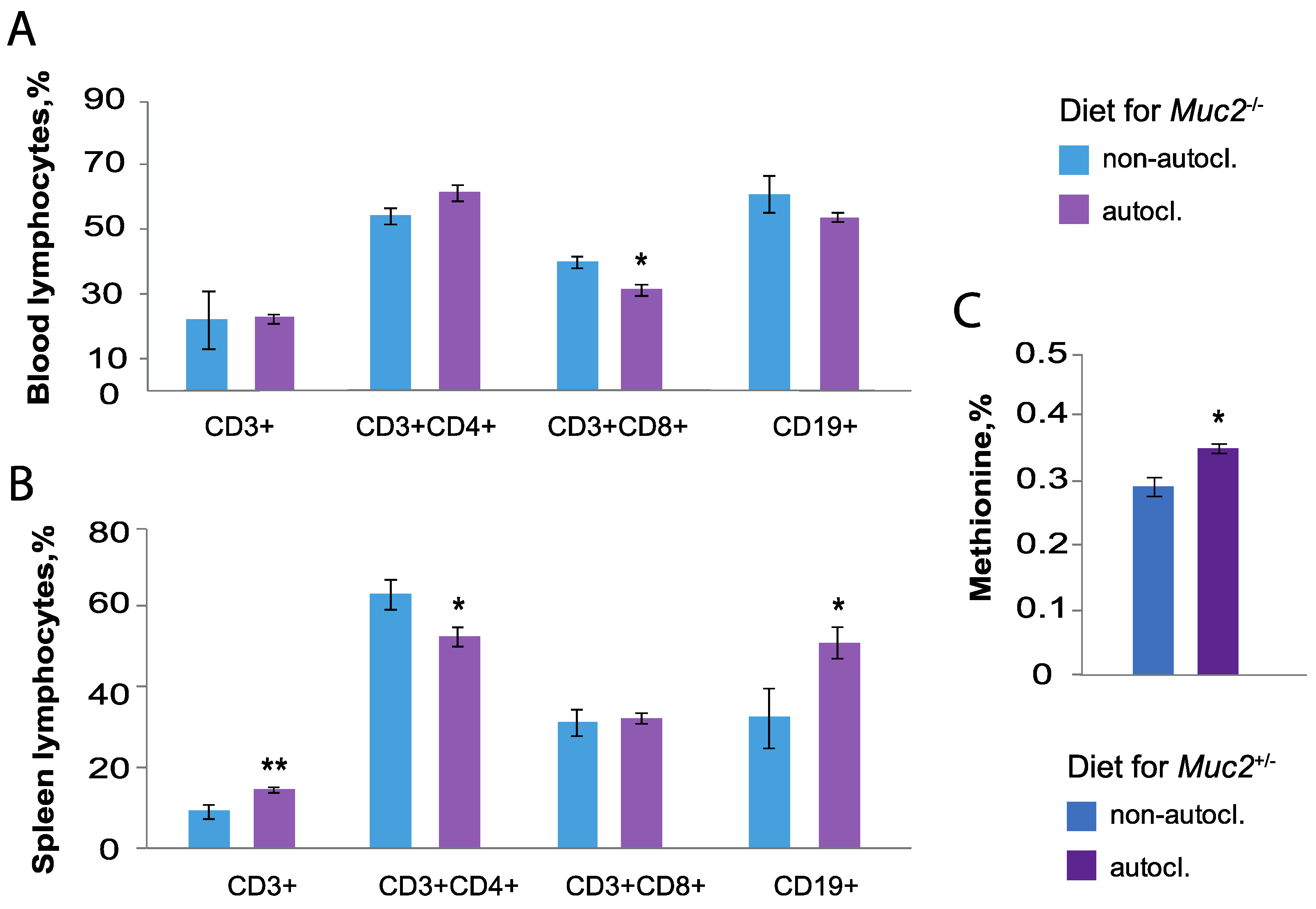
3.6. Immune Cell of Mice Fed on the Autoclaved and Non-Autoclaved Diet

To assess the effect of the autoclaved diet on the blood immune cells there were determined percent of B cells (CD19+), T helper (CD3+CD4+) and T killer (CD3+CD8+) cells (Figure 3A). Muc2−/− mice fed on the non-autoclaved diet increased the percentages of CD3+CD8+ cells (Z = 1.22, p < 0.05) (Table S2, Figure 3). The non-autoclaved diet enhanced in spleen the percentage of CD3+CD4+ (Z = 1.19, p < 0.05) and increased CD19+ cells (Z = −1.98, p < 0.05). (Figure 3B). There was no significant effect on the percentage of CD3+ and CD3+CD8+ cells in spleen of Muc2−/− mice fed on the non-autoclaved diet (p > 0.05) (Table S2).
Thus, the consumption of the non-autoclaved diet containing Bacillus spp. spores causes a decrease of Bacteroides spp. and Lactobacillus spp. in mice feces, as well as a decrease of the percent of T helper and B cells in the blood, which are involved in the humoral immune response. The bacteria Bacillus spp. are known for their ability to synthesize digestive enzymes and improve host digestion [35,36,37,38]. Therefore, bacterial strains from the diet and feces were tested for amylase, proteolytic, and lipolytic activities.

3.7. Enzymatic Activity of Bacteria from the Diet and Feces—Blood Biochemical Analysis of Muc2+/− Mice Fed on Autoclaved and Non-Autoclaved Diets

All bacterial strains from the feces and diet had enzymatic activity and could take part in the digestion (Table 1, Figure S1).
We assumed that such enzymatic activity has a positive effect on the absorption of nutrients and also affects the metabolism of mice. Therefore, we assayed the level of the following metabolites in the blood: glucose, creatinine, total protein, low-density lipoproteins. The data are presented in Table 2. We showed that in Muc2+/− females fed on the autoclaved diet stable levels of creatinine, glucose, proteins, and low-density lipoproteins were maintained. Thus, despite the detected enzymatic activity of Bacillus spp. from the diet and feces, there were no significant differences in the levels of the blood metabolites.

3.8. Amino Acid Composition of the Thigh Muscle of Muc2+/− Mice

Amino acid composition of the thigh muscle showed increased levels of methionine (%) in Muc2+/− mice fed on the autoclaved diet (Z = −2.40, p < 0.05) (Figure 3C). There were no significant differences in the levels of arginine, lysine, tyrosine, phenylalanine, histidine, leucine-isoleucine, valine, proline, threonine, serine, alanine, glycine, and tryptophan (Z = −0.73, Z = −0.73, Z = −1.15, Z = −0.94, Z = 0.31, Z = −0.73, Z = −2.40, Z = 0.00, Z = −0.31, Z = −1.76, Z = −1.37, Z = −0.94, Z = −0.73, and Z = 1.04, p > 0.05, respectively, Table S3). Amino acids in the thigh muscle of mice did not depend on presence or absence of Bacillus spp. The exception is methionine, which is elevated in mice on the autoclaved diet.

3.9. Analysis of the Mineral and Chemical Composition of the Diet

Analysis of the feed for amino acids did not show the influence of autoclaving on arginine, lysine, tyrosine, phenylalanine, histidine, leucine-isoleucine, valine, proline, threonine, serine, alanine, glycine, glutamine, asparagine, and cystine levels after autoclaving (Z = −1.53, Z = 1.53, Z = 0.65, Z = 1.53, Z = −1.53, Z = 0.65, Z = 1.52, Z = 0.65, Z = 1.52, Z = 1.53, Z = 1.53, Z = 0.6553, Z = 0.65, Z = −1.53, and Z = 0.65, p > 0.05, respectively). The amount of methionine and tryptophan was decreased after autoclaving at the trend level (Z = 1.96, Z = −1.96, p = 0.05, respectively, Table S4).
Feed testing for dry matter (%), moisture (%), crude protein (%), crude fat (%), crude fiber (%), sugar (%), starch (%), calcium (%), and phosphorus (%), also showed no effect from autoclaving (Z = −0.65, Z = −0.65, Z = 0.65, Z = −1.53, Z = 1.53, Z = −0.65, Z = 0.65, and Z = 1.53, p > 0.05, respectively). However, the raw ash content was reduced in the feed after autoclaving at trend level (Z = 1.96, p = 0.05, Table S4).
Thus, both in Muc2−/− and Muc2+/− mice autoclaved diet significantly reduced reproductive performance and increased pathological defect of the rectum, which was manifested by early prolapse. The presence of Bacillus spp. in the non-autoclaved diet was confirmed by microbiological assay. Being an allochthonous, Bacillus spp. appeared in the feces of mice that were fed on the non-autoclaved diet for two weeks. In mice fed on the autoclaved diet since birth, the bacterial composition of the intestinal microbiota changes and the amount of Bacteroides spp. and Lactobacillus spp. increased. The feeding on an autoclaved diet since birth affects the spleen immune cells of mice, increasing T and B cells percentage and declining T helper cells numbers. Sequencing of bacterial strains from the diet and feces confirmed that they all belonged to the Bacillus genus. The presence of amylase, protease, and lipase activity of isolated Bacillus strains can improve the absorption of nutrients from food, which can affect the metabolism of the body and increase the fertility of females. However, the presence of Bacillus in the diet and intestines did not change the following biochemical parameters in the blood and tissues: creatinine, glucose, protein, and low-density lipoproteins, and amino acids in muscles. Only methionine, which reduces autophagy and accelerates the aging process, decreased in the animals fed on the non-autoclaved diet. As autoclaving reduced crude ash, methionine, and tryptophan in the diet at trend level, it could be also critical for reproduction performance and manifested by early prolapse.

4. Discussion

The effects of housing conditions (autoclaved and non-autoclaved diets) on the reproductive performance of Muc2−/− females with defect of intestinal barrier function and physically healthy of Muc2+/− females were studied. The intestinal microbiota of mammals consists mainly of bacteria belonging to the Firmicutes and Bacteroidetes (90%), and a lesser to the Actinobacteria, Proteobacteria, Fusobacteria, and Verrucomicrobia [39]. These are autochthonous microorganisms, i.e., resident organisms [40]. Some microorganisms of intestine swallowed accidentally along with food, water, or air pass transiently and do not colonize the intestines. However, they play an important role in the structure of the gut microbiota [41,42].
The deviations of composition and diversity of the intestinal microbiota lead to disorders, primarily of the intestine (inflammatory bowel disease, irritable bowel syndrome, colorectal cancer) [43,44,45], type 2 diabetes, obesity [46], and cardiovascular diseases (atherosclerosis, hypertension, heart failure) [47]. Therefore, a limitation of the microbial load on mice in barrier facilities by sterilizing the diet, water, and bedding alters the microbial composition of the intestine and could have a negative effect on laboratory animals.
It had previously been shown that antibiotic treatment of Muc2−/− mice made to eliminate Helicobacter spp. Led to decline of reproductive performance [48]. This is consistent with our results, which showed the decrease of reproductive rates of Muc2−/− females fed on the autoclaved diet. This decrease is due to the early onset of intestinal prolapse of Muc2−/− mice. However, decrease of the reproductive index of Muc2+/− mice fed on the autoclaved diet indicates that there are other reasons. There are many facts that spores of Bacillus spp. In the diet have positive effects on reproductive functions [49,50,51]. This allows us to assume that the decrease in reproductive functions in animals of different genotypes on the autoclaved diet is associated with inactivation of Bacillus spp. Spores.
Increased intestinal inflammation, which was manifested by early prolapse of Muc2−/− animals fed on the autoclaved diet, can also be caused by the absence of viable spores of Bacillus spp. It was shown that the bacteria Bacillus spp. Are effective in the prevention and treatment of intestinal diseases such as diarrhea, colitis, irritable bowel syndrome, and colorectal cancer [52,53,54,55,56,57]. Bacillus spores from food can grow in the gastrointestinal tract and again form spores from vegetative cells [58,59]. This may explain why complete elimination from Bacillus spores took about 20–30 days. This period significantly exceeded the time of complete passage of food through the digestive tract. In another study, a complete gut clearance from Bacillus spores occurred in 3 weeks [60].
Bacillus can change the composition of the gut microbiota by releasing bacteriocins, peptide, and lipopeptide antibiotics [16,37,61,62], affecting the adhesion of other microorganisms [16] and changing the pH of the environment [63]. Changes in the gut microbiota fatally lead to abnormalities in the host’s metabolic and immune profile [64,65]. In addition, Bacillus can directly affect the host’s immunity. For example, spores of B. subtilis play an important role in the development of intestinal lymphoid tissue. Thus, spore of Bacillus in gut affected on the diversity of the primary antibody population in rabbits [66], inhibited phospholipase A2, resulting in suppression of pro-inflammatory cytokines and an increase in anti-inflammatory cytokines [56]. Therefore, we investigated the effect of the autoclaved diet and non-autoclaved diet on the commensal microorganisms and the lymphocytes in blood and spleen cells. It is known that microbiota inhabits the intestine in early ontogenesis [28]. To study the effect of the autoclaved diet on the gut microbiota, we used sibling mice fed on the non-autoclaved diet since birth and on the autoclaved diet for 2 weeks. The initial intestinal microbiota was the same in mice of different groups. Two weeks feeding on the non-autoclaved diet decreased only the quantity of E. coli. Feeding on the non-autoclaved diet since birth decreased two genera of bacteria: Bacteroides spp. and Lactobacillus spp. We assumed that continued breeding on the non-autoclaved diet would make changes more pronounced. It was shown [67], that the number of Bacteroides and Lactobacillus in the fecal bacterial community structure of broiler chickens decreased when B.amyloliquefaciens was used. Another study also showed that B. subtilis is able to inhibit enterotoxic E. coli infection [68]. For instance, the alterations in intestinal microbiota that we discovered are consistent with the findings of other investigators.
The autoclaved diet increased the percent of T helper and B cells in blood and spleen. This may indicate an increase in antibody formation, and may also lead to the development of allergic reactions [69,70]. Changes in the bacterial composition of the intestine and the number of immune cells indicate the response of mice to differences in the diets. These facts should be taken into account when planning experiments, for example, those involving the purchase of animals in a barrier facility and transferring them to a non-autoclaved diet.
It can be assumed that on the diets with or without Bacillus, the microbiota and the immune system of mice come to some equilibrium, adjusting to the existing conditions. However, Bacillus is also involved in the metabolism due to digestion of nutrients by releasing proteases, lipases, and amylases [35,36,37,38]. Maximum absorption of nutrients leads to sufficient energy balance in cells and its efficient functioning. It was shown that in Muc2−/− mice fed on the autoclaved diet, the functions of mitochondria in the intestinal epithelium were impaired [25]. Lack of additional digestive aid is difficult to compensate and can be a key negative factor on the health of mice fed on an autoclaved diet. We have not studied mitochondrial function in mice fed on a non-autoclaved diet. Such studies need to be performed in the future, using probiotic strains of Bacillus. However, housing on both autoclaved and non-autoclaved diets did not affect the level of major blood metabolites and amino acid composition of muscles, which confirms that the organism is able to normalize the blood and tissue composition. It is interesting to note that only methionine increased in mice fed on the autoclaved diets. It is known that high level of methionine can reduce autophagy and accelerate aging [71,72]. The increase of methionine in mice fed on the autoclaved diets did not depend on changes of the methionine level in the diet, since after autoclaving the level of methionine was even slightly lower in the diets.
Bacillus spp. is widely used as probiotic. We assume that the mice diet contained some of these species. Microscopical analysis and identification of bacteria by morphology of colony and is important but not always reliable. Therefore, sequence by Sanger of 16S rRNA gene of bacterial cultures isolated from the diet and feces were used. It was confirmed that all isolated bacterial cultures were Bacillus spp., with an identity of 99% or more [73]. Sequence by Sanger showed that the feces and diet contained the bacteria Bacillus spp. The microbiota composition of different aliquots of food and feces may differ. In addition, since colonies were manually selected for sequencing by morphotype and microscopy, visually similar colonies might not be included in the analysis. We have shown that the non-autoclaved bedding is free of Bacillus spp. bacteria. Our results confirm that Bacillus spp. are not residents of the digestive tract but enter it from the external environment. The results show the possible diversity of Bacillus spp. in the diet. Some of the bacterial species identified as probiotic are B. amyloliquefaciens, B. velezensis, B. subtilis, and Lysinibacillus macroides [16,37,62,74,75]. B. thuringiensis, B. paramycoides, and Bacillus paranthracis belong to the Bacillus cereus group. Among the representatives of this group there are both probiotic and toxic strains [76]. It is known that the toxic amount of B. cereus in a diet is 105–108 CFU/g [77]. In the diet, the total number of Bacillus spores was at the level of 103–104 CFU/g. Previously, it had been shown that only 3 of 13 strains (about 20%) from the diet exhibited lecithinase activity, a trait of B. cereus [32]. Therefore, presence of toxic strains in the diet is not enough to cause toxicity. Since B. cereus and B. thuringiensis are genetically close, for more accurate differentiation of bacterial strains from feces they were examined for crystal formation. All samples of bacteria from the diet formed para-sporal inclusions and can be classified as B. thuringiensis. These crystals contain proteins that are used as insecticides but are considered harmless to mammals [78]. This result is consistent with earlier results regarding 20% B. cereus content in the diet. Only one strain from the feces did not form crystals and could be assigned to B. cereus. It was found that all representatives of Bacillus spp. entered the intestines of these mice with the diet.

5. Conclusions

Thus, while a barrier facility that uses an autoclaved diet reduces the number of factors that could affect the reproduction of the results and limits the emergence and spread of infectious diseases among animals, these conditions negatively affect the reproductive performance of mice. This effect is associated with the presence of genus Bacillus in a non-autoclaved diet. It is known that autoclaving reduces the nutritional value of feed, in particular, destroys vitamins. However, our results did not reveal significant reductions in amino acids, crude protein, crude fat, etc., in the diet after autoclaving. Only methionine, tryptophan, and raw ash levels were reduced at trend level. It can be assumed that both factors may affect the reduced reproductive function of mice on autoclaved food: a relative decrease in the nutritional value of feed and inactivation of spores of beneficial Bacillus strains. The age of onset of prolapse in Muc2−/− mice was significantly affected by the presence or absence of Bacillus spores.
We believe that further research on the addition of Bacillus probiotic cultures or their metabolites to an autoclaved laboratory animal diet would be interesting and useful.

Supplementary Materials

The following supporting information can be downloaded at: https://www.mdpi.com/article/10.3390/ani12182399/s1, Table S1. Primers used for the qPCR analysis. Table S2. Data normality and statistical significance tests. Table S3. Amino acid composition of the thigh muscle of Muc2+/− mice, %. Table S4. Analysis of the mineral and chemical composition of the diet, %. Figure S1. Enzymatic activity of bacterial strains from diet and feces. Qualitative test. (A) Amylolytic activity, (B) Proteolytic properties. (C) Lipase activity.

Author Contributions

Formal analysis, M.V.M. and E.A.L.; Funding acquisition, M.V.M. and E.A.L.; Investigation, M.V.M., G.V.K., N.I.A., Y.V.I., V.I.K., E.A.L.; Methodology, M.V.M. and E.A.L.; Supervision, E.A.L.; Writing—original draft, review and editing, M.V.M. and E.A.L. All authors have read and agreed to the published version of the manuscript.

Funding

Laboratory animals were obtained with the support of the Budgetary funding for basic scientific research # 122042700001-9. Diet analysis and microbiological assay were supported by Russian Laboratory Animal Science Association research grant (# NI2 03/04/2021). Analysis of immune cells and bacteria was supported by the Russian Science Foundation # 20-64-47020.

Institutional Review Board Statement

Bioethical committee of Scientific-Research Institute of Neurosciences and Medicine, protocol # 3 (03/11/2021).

Informed Consent Statement

Not applicable.

Data Availability Statement

The data presented in this study are available on request from thecorresponding author.

Conflicts of Interest

The authors declare no conflict of interest.

References

  1. Davies, G.; Greenhough, B.; Hobson-West, P.; Kirk, R.G.W. Science, Culture, and Care in Laboratory Animal Research: Interdisciplinary Perspectives on the History and Future of the 3Rs. Sci. Technol. Hum. Values 2018, 43, 603–621. [Google Scholar] [CrossRef]
  2. Patel, K.B.; Galav, V.; Ramachandra, S.G.; Patel, K.B.; Galav, V. Planning and Designing of Laboratory Animal Facilities. In Essentials of Laboratory Animal Science: Principles and Practices; Nagarajan, P., Gudde, R., Srinivasan, R., Eds.; Springer: Singapore, 2021. [Google Scholar] [CrossRef]
  3. Schlapp, G.; Fernández-Graña, G.; Arévalo, A.P.; Crispo, M. Establishment of an environmental microbiological monitoring program in a mice barrier facility. An. Acad. Bras. Cienc. 2018, 90, 3155–3164. [Google Scholar] [CrossRef]
  4. Marx, J.O.; Gaertner, D.J.; Smith, A.L. Results of Survey Regarding Prevalence of Adventitial Infections in Mice and Rats at Biomedical Research Facilities. J. Am. Assoc. Lab. Anim. Sci. 2017, 56, 527–533. [Google Scholar] [PubMed]
  5. Mohan, G.H.; Shwetha Reddy, R.V.; Yogesh, C. Management of Specific Pathogen-Free (SPF) Mice and Rats. In Essentials of Laboratory Animal Science: Principles and Practices; Nagarajan, P., Gudde, R., Srinivasan, R., Eds.; Springer: Singapore, 2021. [Google Scholar] [CrossRef]
  6. Hallmann, J.; Quadt-Hallmann, A.; Mahaffee, W.F.; Kloepper, J.W. Bacterial endophytes in agricultural crops. Can. J. Microbiol. 2011, 43, 895–914. [Google Scholar] [CrossRef]
  7. Vinodkumar, S.; Nakkeeran, S.; Renukadevi, P.; Mohankumar, S. Diversity and antiviral potential of rhizospheric and endophytic Bacillus species and phyto-antiviral principles against tobacco streak virus in cotton. Agric. Ecosyst. Environ. 2018, 267, 42–51. [Google Scholar] [CrossRef]
  8. Hashem, A.; Tabassum, B.; Fathi Abd Allah, E. Bacillus subtilis: A plant-growth promoting rhizobacterium that also impacts biotic stress. Saudi J. Biol. Sci. 2019, 26, 1291–1297. [Google Scholar] [CrossRef]
  9. Grant, A.; Gay, C.G.; Lillehoj, H.S. Bacillus spp. as direct-fed microbial antibiotic alternatives to enhance growth, immunity, and gut health in poultry. Avian Pathol. 2018, 47, 339–351. [Google Scholar] [CrossRef]
  10. Mazanko, M.S.; Gorlov, I.F.; Prazdnova, E.V.; Makarenko, M.S.; Usatov, A.V.; Bren, A.B.; Chistyakov, V.A.; Tutelyan, A.V.; Komarova, Z.B.; Mosolova, N.I.; et al. Bacillus Probiotic Supplementations Improve Laying Performance, Egg Quality, Hatching of Laying Hens, and Sperm Quality of Roosters. Probiotics Antimicrob. Proteins 2018, 10, 367–373. [Google Scholar] [CrossRef]
  11. Hung, A.T.; Lin, S.-Y.; Yang, T.-Y.; Chou, C.-K.; Liu, H.-C.; Lu, J.-J.; Wang, B.; Chen, S.-Y.; Lien, T.-F.; Hung, A.T.; et al. Effects of Bacillus coagulans ATCC 7050 on growth performance, intestinal morphology, and microflora composition in broiler chickens. Anim. Prod. Sci. 2012, 52, 874–879. [Google Scholar] [CrossRef]
  12. Zhang, H.L.; Li, W.S.; Xu, D.N.; Zheng, W.W.; Liu, Y.; Chen, J.; Qiu, Z.B.; Dorfman, R.G.; Zhang, J.; Liu, J. Mucosa-reparing and microbiota-balancing therapeutic effect of Bacillus subtilis alleviates dextrate sulfate sodium-induced ulcerative colitis in mice. Exp. Ther. Med. 2016, 12, 2554–2562. [Google Scholar] [CrossRef]
  13. Sheng, K.; Xu, Y.; Kong, X.; Wang, J.; Zha, X.; Wang, Y. Probiotic Bacillus cereus Alleviates Dextran Sulfate Sodium-Induced Colitis in Mice through Improvement of the Intestinal Barrier Function, Anti-Inflammation, and Gut Microbiota Modulation. J. Agric. Food Chem. 2021, 69, 14810–14823. [Google Scholar] [CrossRef] [PubMed]
  14. Zhu, K.; Hölzel, C.S.; Cui, Y.; Mayer, R.; Wang, Y.; Dietrich, R.; Didier, A.; Bassitta, R.; Märtlbauer, E.; Ding, S. Probiotic Bacillus cereus strains, a potential risk for public health in China. Front. Microbiol. 2016, 7, 718. [Google Scholar] [CrossRef] [PubMed]
  15. Lee, N.K.; Kim, W.S.; Paik, H.D. Bacillus strains as human probiotics: Characterization, safety, microbiome, and probiotic carrier. Food Sci. Biotechnol. 2019, 28, 1297–1305. [Google Scholar] [CrossRef] [PubMed]
  16. Elshaghabee, F.M.F.; Rokana, N.; Gulhane, R.D.; Sharma, C.; Panwar, H. Bacillus as potential probiotics: Status, concerns, and future perspectives. Front. Microbiol. 2017, 8, 1490. [Google Scholar] [CrossRef] [PubMed]
  17. Suva, M.; Sureja, V.; Kheni, D. Novel insight on probiotic Bacillus subtilis: Mechanism of action and clinical applications. J. Curr. Res. Sci. Med. 2016, 2, 65. [Google Scholar] [CrossRef]
  18. Hong, H.A.; Le, H.D.; Cutting, S.M. The use of bacterial spore formers as probiotics. FEMS Microbiol. Rev. 2005, 29, 813–835. [Google Scholar] [CrossRef]
  19. Stein, T. Bacillus subtilis antibiotics: Structures, syntheses and specific functions. Mol. Microbiol. 2005, 56, 845–857. [Google Scholar] [CrossRef]
  20. Andrić, S.; Meyer, T.; Ongena, M. Bacillus Responses to Plant-Associated Fungal and Bacterial Communities. Front. Microbiol. 2020, 11, 1350. [Google Scholar] [CrossRef]
  21. Kimelman, H.; Shemesh, M. Probiotic Bifunctionality of Bacillus subtilis—Rescuing Lactic Acid Bacteria from Desiccation and Antagonizing Pathogenic Staphylococcus aureus. Microorganisms 2019, 7, 407. [Google Scholar] [CrossRef]
  22. Velcich, A.; Yang, W.C.; Heyer, J.; Fragale, A.; Nicholas, C.; Viani, S.; Kucherlapati, R.; Lipkin, M.; Yang, K.; Augenlicht, L. Colorectal cancer in mice genetically deficient in the mucin Muc2. Science 2002, 295, 1726–1729. [Google Scholar] [CrossRef]
  23. Wenzel, U.A.; Magnusson, M.K.; Rydström, A.; Jonstrand, C.; Hengst, J.; Johansson, M.E.; Velcich, A.; Öhman, L.; Strid, H.; Sjövall, H.; et al. Spontaneous Colitis in Muc2-Deficient Mice Reflects Clinical and Cellular Features of Active Ulcerative Colitis. PLoS ONE 2014, 9, e100217. [Google Scholar] [CrossRef]
  24. Van der Sluis, M.; De Koning, B.A.E.; De Bruijn, A.C.J.M.; Velcich, A.; Meijerink, J.P.P.; Van Goudoever, J.B.; Büller, H.A.; Dekker, J.; Van Seuningen, I.; Renes, I.B.; et al. Muc2-Deficient Mice Spontaneously Develop Colitis, Indicating That MUC2 Is Critical for Colonic Protection. Gastroenterology 2006, 131, 117–129. [Google Scholar] [CrossRef] [PubMed]
  25. Borisova, M.A.; Achasova, K.M.; Morozova, K.N.; Andreyeva, E.N.; Litvinova, E.A.; Ogienko, A.A.; Morozova, M.V.; Berkaeva, M.B.; Kiseleva, E.; Kozhevnikova, E.N. Mucin-2 knockout is a model of intercellular junction defects, mitochondrial damage and ATP depletion in the intestinal epithelium. Sci. Rep. 2020, 10, 21135. [Google Scholar] [CrossRef] [PubMed]
  26. Bergstrom, K.S.B.; Kissoon-Singh, V.; Gibson, D.L.; Ma, C.; Montero, M.; Sham, H.P.; Ryz, N.; Huang, T.; Velcich, A.; Finlay, B.B.; et al. Muc2 protects against lethal infectious colitis by disassociating pathogenic and commensal bacteria from the colonic mucosa. PLoS Pathog. 2010, 6, e1000902. [Google Scholar] [CrossRef] [PubMed]
  27. Wu, M.; Wu, Y.; Li, J.; Bao, Y.; Guo, Y.; Yang, W. The Dynamic Changes of Gut Microbiota in Muc2 Deficient Mice. Int. J. Mol. Sci. 2018, 19, 2809. [Google Scholar] [CrossRef] [PubMed]
  28. Senn, V.; Bassler, D.; Choudhury, R.; Scholkmann, F.; Righini-Grunder, F.; Vuille-dit-Bile, R.N.; Restin, T. Microbial Colonization From the Fetus to Early Childhood—A Comprehensive Review. Front. Cell. Infect. Microbiol. 2020, 10, 637. [Google Scholar] [CrossRef]
  29. Mähler, M.; Berar, M.; Feinstein, R.; Gallagher, A.; Illgen-Wilcke, B.; Pritchett-Corning, K.; Raspa, M. FELASA recommendations for the health monitoring of mouse, rat, hamster, guinea pig and rabbit colonies in breeding and experimental units. Lab. Anim. 2014, 48, 178–192. [Google Scholar] [CrossRef]
  30. Handelsman, D.J.; Walters, K.A.; Ly, L.P. Simplified Method to Measure Mouse Fertility. Endocrinology 2020, 161, bqaa114. [Google Scholar] [CrossRef]
  31. Kryukov, V.Y.; Rotskaya, U.; Yaroslavtseva, O.; Polenogova, O.; Kryukova, N.; Akhanaev, Y.; Krivopalov, A.; Alikina, T.; Vorontsova, Y.L.; Slepneva, I.; et al. Fungus Metarhizium robertsii and neurotoxic insecticide affect gut immunity and microbiota in Colorado potato beetles. Sci. Rep. 2021, 11, 1299. [Google Scholar] [CrossRef]
  32. Morozova, M.V.; Kalmykova, G.V.; Akulova, N.I.; Litvinova, E.A. Analysis of Bacillus spp. in the diet and feces of laboratory mice under barrier-housing and non-sterile conditions. Lab. Zhivotnye Dlya Nauchnych Issled (Laboratory Anim. Sci.) 2021, 3, 11–16. [Google Scholar] [CrossRef]
  33. Logan, N.A.; De Vos, P. Bacillus. In Bergey’s Manual of Systematics of Archaea and Bacteria; Whitman, W.B., Ed.; John Wiley & Sons: Hoboken, NJ, USA, 2015. [Google Scholar] [CrossRef]
  34. Popov, I.V.; Algburi, A.; Prazdnova, E.V.; Mazanko, M.S.; Elisashvili, V.; Bren, A.B.; Chistyakov, V.A.; Tkacheva, E.V.; Trukhachev, V.I.; Donnik, I.M.; et al. A Review of the Effects and Production of Spore-Forming Probiotics for Poultry. Animals 2021, 11, 1941. [Google Scholar] [CrossRef] [PubMed]
  35. Hu, S.; Cao, X.; Wu, Y.; Mei, X.; Xu, H.; Wang, Y.; Zhang, X.; Gong, L.; Li, W. Effects of probiotic Bacillus as an alternative of antibiotics on digestive enzymes activity and intestinal integrity of piglets. Front. Microbiol. 2018, 9, 2427. [Google Scholar] [CrossRef] [PubMed]
  36. Jujjavarapu, S.E.; Dhagat, S. Evolutionary Trends in Industrial Production of α-amylase. Recent Pat. Biotechnol. 2018, 13, 4–18. [Google Scholar] [CrossRef] [PubMed]
  37. Woldemariam Yohannes, K.; Wan, Z.; Yu, Q.; Li, H.; Wei, X.; Liu, Y.; Wang, J.; Sun, B. Prebiotic, Probiotic, Antimicrobial, and Functional Food Applications of Bacillus amyloliquefaciens. J. Agric. Food Chem. 2020, 68, 14709–14727. [Google Scholar] [CrossRef]
  38. Ziaei-Nejad, S.; Rezaei, M.H.; Takami, G.A.; Lovett, D.L.; Mirvaghefi, A.R.; Shakouri, M. The effect of Bacillus spp. bacteria used as probiotics on digestive enzyme activity, survival and growth in the Indian white shrimp Fenneropenaeus indicus. Aquaculture 2006, 252, 516–524. [Google Scholar] [CrossRef]
  39. Rinninella, E.; Raoul, P.; Cintoni, M.; Franceschi, F.; Miggiano, G.A.D.; Gasbarrini, A.; Mele, M.C. What is the Healthy Gut Microbiota Composition? A Changing Ecosystem across Age, Environment, Diet, and Diseases. Microorganisms 2019, 7, 14. [Google Scholar] [CrossRef]
  40. Nava, G.M.; Stappenbeck, T.S. Diversity of the autochthonous colonic microbiota. Gut Microbes 2011, 2, 99–104. [Google Scholar] [CrossRef]
  41. McNulty, N.P.; Yatsunenko, T.; Hsiao, A.; Faith, J.J.; Muegge, B.D.; Goodman, A.L.; Henrissat, B.; Oozeer, R.; Cools-Portier, S.; Gobert, G.; et al. The impact of a consortium of fermented milk strains on the gut microbiome of gnotobiotic mice and monozygotic twins. Sci. Transl. Med. 2011, 3, 106ra106. [Google Scholar] [CrossRef]
  42. Veiga, P.; Pons, N.; Agrawal, A.; Oozeer, R.; Guyonnet, D.; Brazeilles, R.; Faurie, J.M.; Van Hylckama Vlieg, J.E.T.; Houghton, L.A.; Whorwell, P.J.; et al. Changes of the human gut microbiome induced by a fermented milk product. Sci. Rep. 2014, 4, 6328. [Google Scholar] [CrossRef]
  43. Liverani, E.; Scaioli, E.; John Digby, R.; Bellanova, M.; Belluzzi, A. How to predict clinical relapse in inflammatory bowel disease patients. World J. Gastroenterol. 2016, 22, 1017–1033. [Google Scholar] [CrossRef]
  44. Ghoshal, U.C.; Shukla, R.; Ghoshal, U. Small Intestinal Bacterial Overgrowth and Irritable Bowel Syndrome: A Bridge between Functional Organic Dichotomy. Gut Liver 2017, 11, 196–208. [Google Scholar] [CrossRef] [PubMed]
  45. Rapozo, D.C.M.; Bernardazzi, C.; De Souza, H.S.P. Diet and microbiota in inflammatory bowel disease: The gut in disharmony. World J. Gastroenterol. 2017, 23, 2124–2140. [Google Scholar] [CrossRef] [PubMed]
  46. Everard, A.; Cani, P.D. Diabetes, obesity and gut microbiota. Best Pract. Res. Clin. Gastroenterol. 2013, 27, 73–83. [Google Scholar] [CrossRef] [PubMed]
  47. Tang, W.H.W.; Kitai, T.; Hazen, S.L. Gut Microbiota in Cardiovascular Health and Disease. Circ. Res. 2017, 120, 1183–1196. [Google Scholar] [CrossRef]
  48. Litvinova, E.A.; Kozhevnikova, E.N.; Achasova, K.M.; Kontsevaya, G.V.; Moshkin, M.P. Eradication of Helicobacter spp. In mucin2-deficient mice. Lab. Anim. 2017, 51, 311–314. [Google Scholar] [CrossRef]
  49. Zhang, Q.; Li, J.; Cao, M.; Li, Y.; Zhuo, Y.; Fang, Z.; Che, L.; Xu, S.; Feng, B.; Lin, Y.; et al. Dietary supplementation of Bacillus subtilis PB6 improves sow reproductive performance and reduces piglet birth intervals. Anim. Nutr. 2020, 6, 278–287. [Google Scholar] [CrossRef]
  50. Zhang, B.; Sui, F.; Wang, B.; Wang, Y.; Li, W. Dietary combined supplementation of iron and Bacillus subtilis enhances reproductive performance, eggshell quality, nutrient digestibility, antioxidant capacity, and hematopoietic function in breeder geese. Poult. Sci. 2020, 99, 6119–6127. [Google Scholar] [CrossRef]
  51. Prazdnova, E.V.; Mazanko, M.S.; Chistyakov, V.A.; Denisenko, Y.V.; Makarenko, M.S.; Usatov, A.V.; Bren, A.B.; Tutelyan, A.V.; Komarova, Z.B.; Gorlov, I.F.; et al. Effect of Bacillus subtilis KATMIRA1933 and Bacillus amyloliquefaciens B-1895 on the productivity, reproductive aging, and physiological characteristics of hens and roosters. Benef. Microbes 2019, 10, 395–412. [Google Scholar] [CrossRef]
  52. Camilleri, M. Probiotics and irritable bowel syndrome: Rationale, mechanisms, and efficacy. J. Clin. Gastroenterol. 2008, 42 Pt 1 (Suppl. S3), S123–S125. [Google Scholar] [CrossRef]
  53. Sazawal, S.; Hiremath, G.; Dhingra, U.; Malik, P.; Deb, S.; Black, R.E. Efficacy of probiotics in prevention of acute diarrhoea: A meta-analysis of masked, randomised, placebo-controlled trials. Lancet Infect. Dis. 2006, 6, 374–382. [Google Scholar] [CrossRef]
  54. Pillai, A.; Nelson, R. Probiotics for treatment of Clostridium difficile-associated colitis in adults. Cochrane Database Syst. Rev. 2008, 23, CD004611. [Google Scholar] [CrossRef] [PubMed]
  55. Bene, K.; Varga, Z.; Petrov, V.O.; Boyko, N.; Rajnavolgyi, E. Gut microbiota species can provoke both inflammatory and tolerogenic immune responses in human dendritic cells mediated by retinoic acid receptor alpha ligation. Front. Immunol. 2017, 8, 427. [Google Scholar] [CrossRef] [PubMed]
  56. Selvam, R.; Maheswari, P.; Kavitha, P.; Ravichandran, M.; Benedikt Sas, R.C.N. Effect of Bacillus subtilis PB6, a natural probiotic on colon mucosal inflammation and plasma cytokines levels in inflammatory bowel disease. Indian J. Biochem. Biophys. 2009, 46, 79–85. [Google Scholar] [PubMed]
  57. Catinean, A.; Neag, A.M.; Nita, A.; Buzea, M.; Buzoianu, A.D. Bacillus spp. Spores—A Promising Treatment Option for Patients with Irritable Bowel Syndrome. Nutrients 2019, 11, 1968. [Google Scholar] [CrossRef]
  58. Tam, N.K.M.; Uyen, N.Q.; Hong, H.A.; Duc, L.H.; Hoa, T.T.; Serra, C.R.; Henriques, A.O.; Cutting, S.M. The intestinal life cycle of Bacillus subtilis and close relatives. J. Bacteriol. 2006, 188, 2692–2700. [Google Scholar] [CrossRef]
  59. Ghelardi, E.; Celandroni, F.; Salvetti, S.; Gueye, S.A.; Lupetti, A.; Senesi, S. Survival and persistence of Bacillus clausii in the human gastrointestinal tract following oral administration as spore-based probiotic formulation. J. Appl. Microbiol. 2015, 119, 552–559. [Google Scholar] [CrossRef]
  60. Hong, H.A.; Huang, J.M.; Khaneja, R.; Hiep, L.V.; Urdaci, M.C.; Cutting, S.M. The safety of Bacillus subtilis and Bacillus indicus as food probiotics. J. Appl. Microbiol. 2008, 105, 510–520. [Google Scholar] [CrossRef]
  61. Ek-Ramos, M.J.; Gomez-Flores, R.; Orozco-Flores, A.A.; Rodríguez-Padilla, C.; González-Ochoa, G.; Tamez-Guerra, P. Bioactive products from plant-endophytic Gram-positive bacteria. Front. Microbiol. 2019, 10, 463. [Google Scholar] [CrossRef]
  62. Khalid, F.; Khalid, A.; Fu, Y.; Hu, Q.; Zheng, Y.; Khan, S.; Wang, Z. Potential of Bacillus velezensis as a probiotic in animal feed: A review. J. Microbiol. 2021, 59, 627–633. [Google Scholar] [CrossRef]
  63. Ilinskaya, O.N.; Ulyanova, V.V.; Yarullina, D.R.; Gataullin, I.G. Secretome of Intestinal bacilli: A natural guard against pathologies. Front. Microbiol. 2017, 8, 1666. [Google Scholar] [CrossRef]
  64. Nyangale, E.P.; Farmer, S.; Cash, H.A.; Keller, D.; Chernoff, D.; Gibson, G.R. Bacillus coagulans GBI-30, 6086 Modulates Faecalibacterium prausnitzii in Older Men and Women. J. Nutr. 2015, 145, 1446–1452. [Google Scholar] [CrossRef] [PubMed]
  65. Kishida, S.; Kato-Mori, Y.; Hagiwara, K. Influence of changes in the intestinal microflora on the immune function in mice. J. Vet. Med. Sci. 2018, 80, 440–446. [Google Scholar] [CrossRef] [PubMed]
  66. Rhee, K.-J.; Sethupathi, P.; Driks, A.; Lanning, D.K.; Knight, K.L. Role of Commensal Bacteria in Development of Gut-Associated Lymphoid Tissues and Preimmune Antibody Repertoire. J. Immunol. 2004, 172, 1118–1124. [Google Scholar] [CrossRef]
  67. Hong, Y.; Cheng, Y.; Li, Y.; Li, X.; Zhou, Z.; Shi, D.; Li, Z.; Xiao, Y. Preliminary Study on the Effect of Bacillus amyloliquefaciens TL on Cecal Bacterial Community Structure of Broiler Chickens. BioMed Res. Int. 2019, 2019, 5431354. [Google Scholar] [CrossRef]
  68. Ye, X.; Li, P.; Yu, Q.; Yang, Q. Bacillus subtilis inhibition of enterotoxic Escherichia coli-induced activation of MAPK signaling pathways in Caco-2 cells. Ann. Microbiol. 2013, 63, 577–581. [Google Scholar] [CrossRef]
  69. Vogel, K.; Blümer, N.; Korthals, M.; Mittelstädt, J.; Garn, H.; Ege, M.; von Mutius, E.; Gatermann, S.; Bufe, A.; Goldmann, T.; et al. Animal shed Bacillus licheniformis spores possess allergy-protective as well as inflammatory properties. J. Allergy Clin. Immunol. 2008, 122, 307–312. [Google Scholar] [CrossRef]
  70. Swartzendruber, J.A.; Incrocci, R.W.; Wolf, S.A.; Jung, A.; Knight, K.L. Bacillus subtilis exopolysaccharide prevents allergic eosinophilia. Allergy 2019, 74, 819. [Google Scholar] [CrossRef]
  71. Sutter, B.M.; Wu, X.; Laxman, S.; Tu, B.P. Methionine Inhibits Autophagy and Promotes Growth by Inducing the SAM-Responsive Methylation of PP2A. Cell 2013, 154, 403. [Google Scholar] [CrossRef]
  72. Kitada, M.; Ogura, Y.; Monno, I.; Xu, J.; Koya, D. Effect of Methionine Restriction on Aging: Its Relationship to Oxidative Stress. Biomedicines 2021, 9, 130. [Google Scholar] [CrossRef]
  73. Bosshard, P.P.; Abels, S.; Zbinden, R.; Böttger, E.C.; Altwegg, M. Ribosomal DNA sequencing for identification of aerobic gram-positive rods in the clinical laboratory (an 18-month evaluation). J. Clin. Microbiol. 2003, 41, 4134–4140. [Google Scholar] [CrossRef]
  74. Mani, S.R.; Vijayan, K.; Jacob, J.P.; Vijayakumar, S.; Kandhasamy, S. Evaluation of probiotic properties of Lysinibacillus macroides under in vitro conditions and culture of Cyprinus carpio on growth parameters. Arch. Microbiol. 2021, 203, 4705–4714. [Google Scholar] [CrossRef] [PubMed]
  75. Rhayat, L.; Maresca, M.; Nicoletti, C.; Perrier, J.; Brinch, K.S.; Christian, S.; Devillard, E.; Eckhardt, E. Effect of Bacillus subtilis Strains on Intestinal Barrier Function and Inflammatory Response. Front. Immunol. 2019, 10, 564. [Google Scholar] [CrossRef] [PubMed]
  76. Bianco, A.; Capozzi, L.; Monno, M.R.; Del Sambro, L.; Manzulli, V.; Pesole, G.; Loconsole, D.; Parisi, A. Characterization of Bacillus cereus Group Isolates From Human Bacteremia by Whole-Genome Sequencing. Front. Microbiol. 2021, 11, 599524. [Google Scholar] [CrossRef] [PubMed]
  77. Jessberger, N.; Dietrich, R.; Granum, P.E.; Märtlbauer, E. The Bacillus cereus Food Infection as Multifactorial Process. Toxins 2020, 12, 701. [Google Scholar] [CrossRef] [PubMed]
  78. Domanska, B.; Fortea, E.; West, M.J.; Schwartz, J.L.; Crickmore, N. The role of membrane-bound metal ions in toxicity of a human cancer cell-active pore-forming toxin Cry41Aa from Bacillus thuringiensis. Toxicon 2019, 167, 123–133. [Google Scholar] [CrossRef] [PubMed]
Figure 1. Effect of the autoclaved diet on the reproductive performance and age at onset of prolapse of Muc2+/− and Muc2−/− female mice. (A) Fertility index of female mice fed on the autoclaved (Muc2+/− n = 74, Muc2−/− n = 34) and non-autoclaved diets (Muc2+/− n = 21, Muc2−/− n = 10). (B) Age at onset of prolapse of Muc2−/− female mice fed on the autoclaved (n = 23) and non-autoclaved diets (n = 20). (C) Number of Muc2−/− female mice with prolapse at 4 months fed on the autoclaved and non-autoclaved diets. # p < 0.05, ### p < 0.001, Fisher’s exact test, *** p < 0.001 Mann–Whitney U-test.
Animals 12 02399 g001
Figure 2. The Bacillus spp. spores in the diet and feces of mice. Amount of 16s rRNA gene of intestinal bacteria of mice fed on the autoclaved and non-autoclaved diets. (A) The Bacillus spp. spore in the diet and feces of Muc2−/− and Muc2+/− mice (n = 3), log10. (B) The number of spore in the feces of Muc2−/− and Muc2+/− mice on not autoclaved, 10 days autoclaved, 20 days autoclaved, and 30 days autoclaved food (n = 3 for each group). (C) Amount of 16s rRNA gene of intestinal bacteria (Akkermansia muciniphila, Enterococcus faecalis, Lactobacillus murinus, Bacteroides spp., Staphylococcus spp., Escherichia coli) of Muc2−/− mice fed on the autoclaved or non-autoclaved diet since birth, and 2 weeks feeding of the autoclaved diet of mice after eating the non-autoclaved diet since birth. *** p < 0.001, Fisher’s exact test, * p < 0.05, Mann–Whitney U-test.
Animals 12 02399 g002
Figure 3. Percent of immune cells of Muc2−/− mice fed on the autoclaved and non-autoclaved diets. (A) The percent of immune cells in the blood of mice fed on the autoclaved (Muc2−/− n = 5) and non-autoclaved diets (Muc2−/− n = 4). (B) The percent of immune cells in the spleen of mice fed on the autoclaved (Muc2−/− n = 5) and non-autoclaved diets (Muc2−/− n = 5). (C) The methionine level in thigh muscle of mice (Muc2+/− on the autoclaved diet n = 5, Muc2+/− on the non-autoclaved diet n = 5). * p < 0.05, ** p < 0.01 Mann–Whitney U-test.
Animals 12 02399 g003
Table 1. Amylase, protease, and lipase activities of bacterial strains isolated from the diet and feces.
SourceBacterial StrainsEnzymatic Activity, Zone Diameter
NoDescriptionAmylase, mmProtease, mmLipase
Diet1 Bacillus subtilis strain qx-4, Endophytic bacterium MD337.7 ± 1.528.7 ± 0.3+
2Bacillus thuringiensis strain GZDF1, Bacterium strain MIS_YL_J5533.0 ± 1.020.3 ± 0.3
3Lysinibacillus spp. FWQSR5, Lysinibacillus macroides strain Z01026.7 ± 0.9
4Bacillus cereus strain S43, Bacillus thuringiensis strain BT62 chromosome, Bacillus paramycoides strain23.0 ± 1.528.0 ± 0.7
Feces1Bacillus amyloliquefaciens strain QT-162, Bacillus velezensis strain 264528.3 ± 0.928.0 ± 0.6
2Bacillus thuringiensis strain GZDF1, Bacillus sp. (in: Bacteria) strain 201705CJKOP-5927.3 ± 1.522.3 ± 2.1
3Bacillus subtilis subsp. stercoris strain EGI137, Bacillus subtilis strain a2224.0 ± 0.619.0 ± 0.6+++
4Bacillus cereus strain YB1806, Bacillus parathracis, Bacillus thuringiensis strain NO.830.0 ± 0.022.3 ± 0.3
5Bacillus cereus strain D21, Bacillus parathracis, Bacillus cereus strain YB180621.0 ± 1.2++++
Table 2. Biochemical analysis of blood serum.
MetaboliteGroup of MiceKolmogorov–
Smirnov Test, p
Mann–Whitney U Test
Non-Autoclaved DietAutoclaved DietZp-Value
Createnin, µmol/L36.92 ± 7.1842.99 ± 11.14p > 0.10−0.310.75
Total protein, g/L42.69 ± 1.0941.81 ± 0.47p > 0.100.730.46
LDL, µmol/L2.57 ± 0.523.28 ± 0.82p > 0.10−0.310.75
Glucose, µmol/L9.83 ± 0.298.84 ± 0.60p > 0.101.150.25
Publisher’s Note: MDPI stays neutral with regard to jurisdictional claims in published maps and institutional affiliations.

