Article

Vitamin D3 Metabolic Enzymes in Plateau Zokor (Myospalax baileyi) and Plateau Pika (Ochotona curzoniae): Expression and Response to Hypoxia

1
Research Center for High Altitude Medicine, Qinghai University, Xining 810016, China
2
State Key Laboratory of Plateau Ecology and Agriculture, Qinghai University, Xining 810016, China
*
Author to whom correspondence should be addressed.
Animals 2022, 12(18), 2371; https://doi.org/10.3390/ani12182371
Submission received: 3 August 2022 / Revised: 2 September 2022 / Accepted: 6 September 2022 / Published: 11 September 2022
(This article belongs to the Section Mammals)

Abstract

:

Simple Summary

Vitamin D3 is mainly synthesized in the skin after exposure to sunlight and plays a fundamental role in the absorption of calcium and phosphate. Lithocholic acid increases intestinal calcium absorption in vitamin D-deficient rats. The plateau zokor, which lives underground throughout its life, and plateau pika are unique animal species on the Qinghai-Tibet Plateau. To investigate how animals that are rarely exposed to sunlight deal with the expected deficit in vitamin D3 levels and the effect of hypoxia on vitamin D3 metabolism, the levels of key biomarkers related to vitamin D3 metabolism, and the mRNA and protein levels of enzymes involved in key pathways were measured. Plateau zokor was deficient in vitamin D3 and has a higher level of lithocholic acid, but its calcium levels were within the normal range. The levels of vitamin D3 and the expression levels of vitamin D3 metabolism-related genes CYP2R1 and CYP27B1 decreased with increasing altitude in plateau pika. Plateau zokor was deficient in vitamin D3, and lithocholic acid appears to substitute it. In addition, hypoxia suppresses vitamin D3 in plateau pika by down-regulating the expression of vitamin D3 metabolism-related genes.

Abstract

Vitamin D3 (D3) is produced endogenously from 7-dehydrocholesterol by irradiation and is an important secosteroid for the absorption of calcium and phosphate. Lithocholic acid (LCA) increases intestinal paracellular calcium absorption in a vitamin D receptor-dependent manner in vitamin D-deficient rats. The plateau zokor (Myospalax baileyi), a strictly subterranean species, and plateau pika are endemic to the Qinghai-Tibet Plateau. To verify whether the zokors were deficient in D3 and reveal the effects of hypoxia on D3 metabolism in the zokors and pikas, we measured the levels of 25(OH)D3, calcium, and LCA, and quantified the expression levels of D3 metabolism-related genes. The results showed an undetectable serum level of 25(OH)D3 and a significantly higher concentration of LCA in the serum of plateau zokor, but its calcium concentration was within the normal range compared with that of plateau pika and Sprague-Dawley rats. With increasing altitude, the serum 25(OH)D3 levels in plateau pika decreased significantly, and the mRNA and protein levels of CYP2R1 (in the liver) and CYP27B1 (in the kidney) in plateau pika decreased significantly. Our results indicate that plateau zokors were deficient in D3 and abundant in LCA, which might be a substitution of D3 in the zokor. Furthermore, hypoxia suppresses the metabolism of D3 by down-regulating the expression of CYP2R1 and CYP27B1 in plateau pika.

1. Introduction

Vitamin D3 is a fat-soluble secosteroid that has a fundamental role in the maintenance of calcium and phosphate homeostasis [1,2,3,4,5]. Vitamin D3 has a variety of pleiotropic functions in many extra-skeletal targets [2]. For instance, it regulates DNA stability, cell proliferation and differentiation, and innate and adaptive immunity [2,6,7,8]. Increased intestinal absorption of ingested calcium is vitamin D3′s key function in maintaining calcium homeostasis [4]. Regarding the absorption sites involved, the distal intestine, in addition to the duodenum, which only absorbs 8–10% calcium, plays an important role in intestinal calcium absorption [9,10,11,12]. Vitamin D3 promotes intestinal calcium absorption mainly via the transcellular pathway by a complex network of calcium-regulating substances, of which transient receptor potential vanilloid 6 (TRPV6) and calcium-binding protein D9k (calbindin-D9k) have been identified as the major intestinal targets and paracellular pathways, including cadherin-17, claudin-12, claudin-2, and claudin-1; however, the role of vitamin D3 in the regulation of passive calcium transport remains a matter of debate [2,4,13,14,15,16,17,18,19]. Thus, the mechanisms by which vitamin D3 regulates intestinal calcium absorption have remain unclear.
About 80% of vitamin D3 is endogenously produced in the skin, and the remaining 20% comes from food [20]. However, the majority of foods contain only small amounts of vitamin D3, and cutaneous synthesis is the main source of vitamin D3. Vitamin D3 is produced in the skin from its substrate 7-dehydrocholesterol (7-DHC) [21], an intermediate in cholesterol synthesis. Following an electrocyclic rearrangement of the ring at the C9 and C10 positions, 7-dehydrocholesterol is transformed into pre-vitamin D3 under the influence of ultraviolet B (UVB) radiation, which then undergoes thermal isomerization to vitamin D3 after rearranging the triene structure of the molecule [22,23]. This is a non-enzymatic process, but must be produced by irradiation with UV light and heat; vitamin D3 is not biologically active by itself [2]. Vitamin D3 undergoes two subsequent hydroxylation reactions to produce the active form 1, 25(OH)2D3. First, vitamin D3 is metabolized to 25-hydroxyvitamin D3 [25(OH)D3] mainly by CYP2R1 in the liver [24,25]. The only 1α-hydroxylase, CYP27B1, then transforms 25(OH)D3 into 1, 25-dihydroxy vitamin D3 [1, 25(OH)2D3] in the kidneys [26]. The total serum concentration of 25(OH)D3 is thought to be the best indicator of vitamin D3 status [27,28]. CYP24A1 is a catabolic enzyme that can convert 25(OH)D3 and 1, 25(OH)2D3 to 24, 25(OH)2D3 and 1, 24, 25(OH)3D3, respectively, via a negative feedback mechanism in vitamin D3 signaling, and is a helpful marker of vitamin D3 catabolism [29]. Vitamin D deficiency is also associated with low serum calcium levels, leading to musculoskeletal effects [30]. Vitamin D deficiency leads to rickets in children and osteoporosis in adults, resulting in poor bone mineralization [5]. In nature, a variety of rodents such as the naked mole-rat, Damara mole-rat, and plateau zokor live entirely underground, which effectively shields them from sunlight, and feed on an herbivorous diet [31,32,33]. Previous studies have shown that the subterranean rodents Damara mole-rat and naked mole-rat have a natural vitamin D-deficient status [34,35]. Therefore, sunlight is essential for the photolysis of 7-dehydrocholesterol to vitamin D3. Recently, a study showed that the biosynthesis of steroid hormones is activated in response to hypoxia in Sprague-Dawley (SD) rats [36]. Vitamin D plays a role in preventing and protecting lung injury in hypoxia tolerance. A previous study demonstrated that hypoxic sheep had higher circulating concentrations of 25(OH)D and 1, 25(OH)2D than normoxic sheep [37]. However, one study reported an alarmingly low level of 25(OH)D among nomads in Tibet [38]. Therefore, the effect of hypoxia on vitamin D3 metabolism has not been elucidated.
1, 25(OH)2D3 is a physiological ligand of the vitamin D receptor (VDR). Bile acids are steroid acids produced by hepatocytes from cholesterol and circulate between the liver and the intestine [39]. Primary bile acids, cholic acid (CA), chenodeoxycholic acid (CDCA), and their respective secondary bile acids, deoxycholic acid (DCA) and lithocholic acid (LCA), are the four major bile acids. LCA reportedly activates VDR in vitro [40,41,42]. A study found that LCA not only increases serum calcium levels, but also stimulates 24-hydroxylase expression in the kidney and acts on the bone to mobilize calcium, only in vitamin D-deficient rats [41]. Therefore, LCA may be a substitute for vitamin D3 for calcium homeostasis in vitamin D-deficient animals.
The Qinghai-Tibet Plateau is the highest and largest plateau on Earth. Hypoxia, strong ultraviolet radiation, and low temperatures are the major challenges at high altitudes, with hypoxia being the most obvious characteristic of the climate [43,44,45]. The plateau zokor (Myospalax baileyi) is a unique subterranean rodent in the Qinghai-Tibet Plateau that lives underground throughout its life [46]. Plateau pika (Ochotona curzoniae), an indigenous species of the Qinghai-Tibet Plateau, is a tiny, non-hibernating mammal that inhabits meadows at altitudes of 3000–5000 m [47,48,49]. Over long-periods of adaption and evolution, the plateau zokor and plateau pika have developed a variety of strategies to physiologically and genomically adjust to the hypoxic environment and become highly advanced hypoxia-tolerant animals [50,51,52]. Based on the above analysis, we speculated that the plateau zokor might have a naturally impoverished vitamin D3 state owing to the lack of sunlight, and LCA might be a substitute for vitamin D3. Moreover, whether hypoxia upregulates or inhibits vitamin D3 metabolism is currently unclear. Therefore, the contents of 25(OH)D3, calcium, and lithocholic acid of plateau zokor, plateau pika, and SD rats were quantified, and their mRNA and protein levels of vitamin D3 metabolism-related genes in the liver and kidney were evaluated. These indicators were compared in plateau zokor and plateau pika at different altitudes.

2. Materials and Methods

2.1. Animal Procedures

High-altitude group plateau zokors and high-altitude group plateau pikas were captured at an altitude of 3700 m (36°72′ N, 101°28′ E) at Laji Mountain in Guide County, Qinghai Province, China. The average oxygen level in the sampling area was 180.6 g/m3. Low-altitude group plateau zokors were captured at an altitude of 2700 m (36°26′ N, 101°69′ E) at Laji Mountain in Guide County, Qinghai Province, China. The average oxygen level in the sampling area was 212.6 g/m3. Low-altitude plateau pikas were captured in the Qinghai Lake area (37°16′ N, 99°52′ E; 3200 m) in Gangcha County, Qinghai Province, China, where the average oxygen level was 196.6 g/m3. Zokor and pika were live-trapped in the summer months (June and July). To avoid differences due to stimulation, the animals were placed in their cages for 4–5 h of silence and the zokor was placed in dark. The Sprague-Dawley rats were purchased from the Experimental Animal Center of Lanzhou University (36°02′ N, 103°51′ E; 1500 m), Lanzhou City, Gansu Province, China. The average oxygen level in Lanzhou City was 251.0 g/m3. The SD rats from Lanzhou (1500 m) were kept and moved to Xining (2200 m) one week in advance for adaptation to the lab environment. In order to avoid variation, the rats were fed adaptively for 1 week, subjected to a 12–12 h circadian rhythm, provided suitable feed, water, and bedding, and their hair, excrement, and activity were observed. All the animals were male, and the weights of the zokors, pikas, and SD rats were 200–250 g, 100–150 g, and 200–250 g, respectively. Six animals from each group were used to quantify 25(OH)D3 and LCA, and nine were used to quantify calcium and determine the expression levels of the vitamin D metabolism-related-genes.
Sample collection was conducted at same time (14:00–16:00) for the zokor, pika, and SD rats to minimize circadian rhythm effects. After the animals were anesthetized in situ with chloral hydrate (10%), 8 mL of blood was collected from the carotid artery in foil-wrapped tubes without anticoagulants. Care was taken disinfect the animal during blood collection to prevent microbial contamination. The blood samples were left for 40 min, then centrifuged and the supernatant collected. The samples were then stored in liquid nitrogen. Abdominal aortic blood samples were collected for calcium concentration analysis. The liver and kidneys were placed in liquid nitrogen. All the procedures were performed according to the China Practice for the Care and Use of Laboratory Animals and approved by the China Zoological Society (permit number: GB/T35892-2018).

2.2. Ultra Performance Liquid Chromatography-Tandem Mass Spectrometry (UPLC-MS/MS)

Analytical standard for 25-hydroxy D3 was purchased from Sigma-Aldrich (St. Louis, MO, USA). 4-phenyl-1, 2, 4-triazoline-3, and 5-dione and analytical standards of LCA were provided by Shanghai Yuanye Technology Development Co., Ltd. (Shanghai, China). The purities of the standards were >98%. The LC-MS-grade solvents acetonitrile, formic acid, and methanol were purchased from Merck (Darmstadt, Germany). Deionized water was obtained using a Millipore-Q system (Millipore, Billerica, MA, USA).

2.2.1. Quantitative Study of 25(OH)D3

Chromatographic separation was carried out on an Acquity UPLC (Waters, Milford, MA, USA) HSS T3 (100 mm × 2.1 mm, 1.8 μm) column operating in a programmed gradient mode at a flow rate of 0.3 mL min−1 using mobile phase A: formic acid (0.1%) in water and phase B: formic acid (0.1%) in methanol. The gradient elution program was optimized for the separation, and the program was as follows: 0–2 min, 50% B; 2–5 min, 100% B; and 5–6 min, 50% B. The temperature of the auto-sampler before analysis was 4 °C and the injection volume was 15 μL.
Mass spectrometry was performed on an AB SCIEX 5500 QQQ-MS (AB Sciex, CA, USA) equipped with an electrospray ionization (ESI) interface. The tandem mass spectrometer was operated in multiple reaction-monitoring modes. The optimal parameters were as follows: ion spray voltage, 4500 V; and turbo spray temperature, 450 °C. The flows of the curtain, collision, and ion source gases were set to 35, 9, and 55 psi, respectively.
The thawed samples were vortexed for 10 s, methanol/dichloromethane (1:2, v/v) was added and sonicated at 4 °C for 30 min. The samples were incubated at 4 °C for 60 min, then centrifuged and dried in a concentrator. PTAD (4-phenyl-1, 2, 4-triazoline-3, 5-dione) solution (0.25 mg/mL) was added to the dried samples and reacted for 1 h in the dark. After derivatization was completed, the sample was placed in a concentrator to dry, reconstituted with 50% acetonitrile-water, and the supernatant was collected for UPLC-MS/MS analysis. The prepared standard solution was added to the injection bottle, and the retention time of the substance peak was determined. Integration was performed using MultiQuant software, and content calculation was performed using a standard curve.

2.2.2. Quantitative Study of LCA

Chromatographic separation was carried out on an Acquity UPLC BEH C18 (100 × 2.1 mm, 1.7 μm) column. A programmed gradient mode of mobile phase A: formic acid (0.01%) in water and phase B: formic acid (0.01%) in acetonitrile was run at a flow rate of 0.3 mL min−1. The gradient elution program was optimized for the separation, and the program was as follows: 0–2 min, 10% B; 2–5 min, 40% B; 5–7.5 min, 45% B; 7.5–9.5 min, 60% B; 9.5–11.5 min, 80% B; 11.5–14 min, 80% B; and 14–16 min, 10% B. The temperature of the auto-sampler before analysis was 4 °C and the injection volume was 4 μL.
Mass spectrometry was as described for quantitative study of 25(OH)D3. The optimal parameters were as follows: ion spray voltage, −4200 V; and turbo spray temperature, 450 °C. The flows of the curtain, collision, and ion source gases were set to 35, 7, and 35 psi, respectively.
Thawed samples were vortexed for 10 s, and methanol was added to the sample (6:1, v/v), and sonicated at 4 °C for 30 min. The sample was centrifuged and dried in a concentrator. Then, it was reconstituted with 200 μL of methanol, and the supernatant was collected for UPLC-MS/MS analysis. The prepared standard solution was added to the injection bottle, and the retention time of the substance peak was determined. Integration was performed using MultiQuant software and content calculation was performed using the internal standard method.

2.3. Calcium Concentration Analysis

The arterial calcium levels of the plateau zokor, plateau pika, and SD rats were measured using an i-STAT analyzer (Abbott Point of Care Inc., Princeton, NJ, USA), and the measurement temperature was set at 37 °C. The reference range for calcium concentration was 0.25–2.50 mmol/L.

2.4. Total RNA Isolation and qRT-PCR

Total RNA was extracted from the liver and kidney of Myospalax baileyi using the TRIzol reagent (Invitrogen, Carlsbad, CA, USA). The RNA concentration was measured using an ultraviolet spectrophotometer. Next, 4 μg of total RNA was reverse-transcribed to first-strand cDNA using FastKing gDNA Dispelling RT SuperMix (Tiangen, Beijing, China). The mRNA expression levels of CYP2R1, CYP27B1, and CYP24A1 were evaluated by qRT-PCR, as described previously [53]. The nucleotide sequences of the primers used for the qPCR are listed in Table 1.

2.5. Western Blotting

Polyclonal antibodies against CYP2R1, CYP27B1, and CYP24A1 of plateau zokor and plateau pika were produced by Beijing Liuhe Huada Gene Technology Co., Ltd. (Beijing, China). SD rat antibodies against CYP2R1 (Invitrogen, PA5-87927), CYP27B1 (Invitrogen, PA5-79128), and CYP24A1 (Invitrogen, PA5-79127) were purchased from Invitrogen (Carlsbad, CA, USA). Polyclonal antibody against β-actin (Abcam, ab8227) was purchased from Abcam (Cambridge, UK).
The liver and kidney tissues (100 mg) were homogenized in RIPA lysis buffer containing protein inhibitors (Beyotime, Shanghai, China), and the supernatant was collected. Equivalent amounts of protein were separated by electrophoresis on 10% SDS-PAGE gels and transferred onto polyvinylidene fluoride (PVDF) membranes. The membranes were blocked with 5% fat-free milk and subsequently incubated with diluted primary antibodies. β-actin was used as the loading control. The membranes were then incubated with a goat anti-rabbit-HRP secondary antibody (1:5000, Servicebio, Wuhan, China). The protein-antibody complexes were detected using an enhanced chemiluminescence (ECL) system. The protein signals were analyzed and quantified using the ImageJ software.

2.6. Statistical Analysis

SPSS 23.0 was used for statistical analysis. All the values are expressed as the mean ± standard deviation (SD). The Kolmogorov–Smirnov and Levene tests were used to check the data for assumptions of normality and homogeneity of variance, respectively, before performing statistical analyses. Three species were analyzed using one-way ANOVA, followed by an LSD test. The student’s t-test was used to evaluate the differences between the two altitudes. p < 0.05 was considered statistically significant. All charts were visualized using GraphPad Prism 8.0 (GraphPad Software, San Diego, CA, USA).

3. Results

3.1. Serum Levels of 25(OH)D3, LCA, and Calcium and the Expression of Vitamin D3 Metabolism-Related Genes in Plateau Zokor, Plateau Pika, and SD Rats

As shown in Figure 1A, plateau zokors (3700 m) showed an undetectable serum level of 25(OH)D3, but the serum levels of 25(OH)D3 were significantly decreased in the plateau pikas (3700 m) compared with SD rats (1500 m) (Figure 1A). The serum levels of LCA in plateau zokors were significantly higher than those in the plateau pikas and SD rats (Figure 1B). Interestingly, the concentrations of calcium in the plateau zokors were within the normal range (1.10 ± 0.19 mmol/L), and there were no differences compared with the plateau pikas (1.10 ± 0.10 mmol/L) (Figure 1C). Compared with the SD rats (1.31 ± 0.02 mmol/L), the levels of calcium were significantly lower in plateau zokors and plateau pikas, but were still within the normal range (Figure 1C).
CYP2R1 is mainly expressed in the liver, whereas CYP27B1 and CYP24A1 are mainly expressed in the kidneys [2,13,54]. In the present study, the CYP2R1 mRNA and protein levels in the liver of plateau zokors (3700 m) were significantly higher than those in plateau pikas (3700 m) and SD rats (1500 m) (Figure 1D,G). CYP27B1 mRNA levels in the kidney of plateau zokors were significantly higher than those of plateau pikas, and those of plateau pikas were significantly higher than those of SD rats (Figure 1E). CYP27B1 protein levels in the kidney of plateau zokors (3700 m) and SD rats (1500 m) were significantly higher than those of plateau pikas (3700 m) (Figure 1H), and there was no significant difference between the plateau zokors and SD rats (Figure 1H). CYP24A1 mRNA levels in the kidneys of plateau pikas were significantly higher than those in plateau zokors and SD rats (Figure 1F). CYP24A1 protein levels in the kidneys of plateau pikas (3700 m) were significantly higher than those of plateau zokors (3700 m) and SD rats (1500 m) (Figure 1I), and there was no significant difference between the levels in plateau zokors and SD rats (Figure 1I).

3.2. Serum Levels of 25(OH)D3, LCA, and Calcium and the Expression of Vitamin D3 Metabolism-Related Genes in Plateau Zokors at Different Altitudes

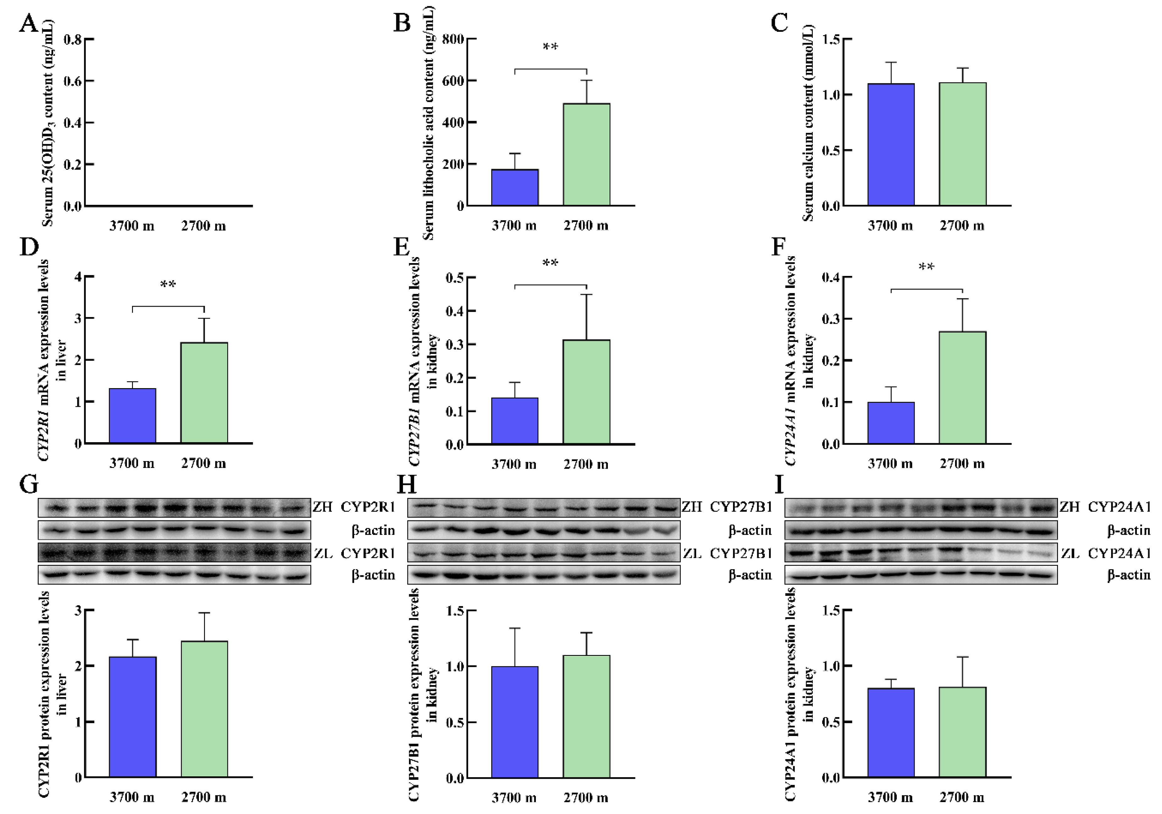
Exposure to high-altitude hypoxia has various effects on the body, including the cardiovascular system, nervous system, and digestive system, which cause physiological and pathological changes. The plateau zokors at elevations of 3700 m and 2700 m had undetectable serum levels of 25(OH)D3 (Figure 2A). The serum levels of LCA in the plateau zokors significantly decreased with increasing altitude (Figure 2B). Interestingly, the calcium concentrations in plateau zokors were within the normal range (Figure 2C), and there was no significant difference between the levels in plateau zokors at elevations of 3700 m and 2700 m (Figure 2C). With increasing altitude, there were significant decreases in the levels of CYP2R1, CYP27B1, and CYP24A1 mRNA in the liver and kidney, respectively, of plateau zokors (Figure 2D–F), and the levels of CYP2R1, CYP27B1, and CYP24A1 proteins were also decreased, but there were no significant differences (Figure 2G–I).

3.3. Serum Levels of 25(OH)D3, LCA, and Calcium and the Expression of Vitamin D3 Metabolism-Related Genes in Plateau Pikas at Different Altitudes

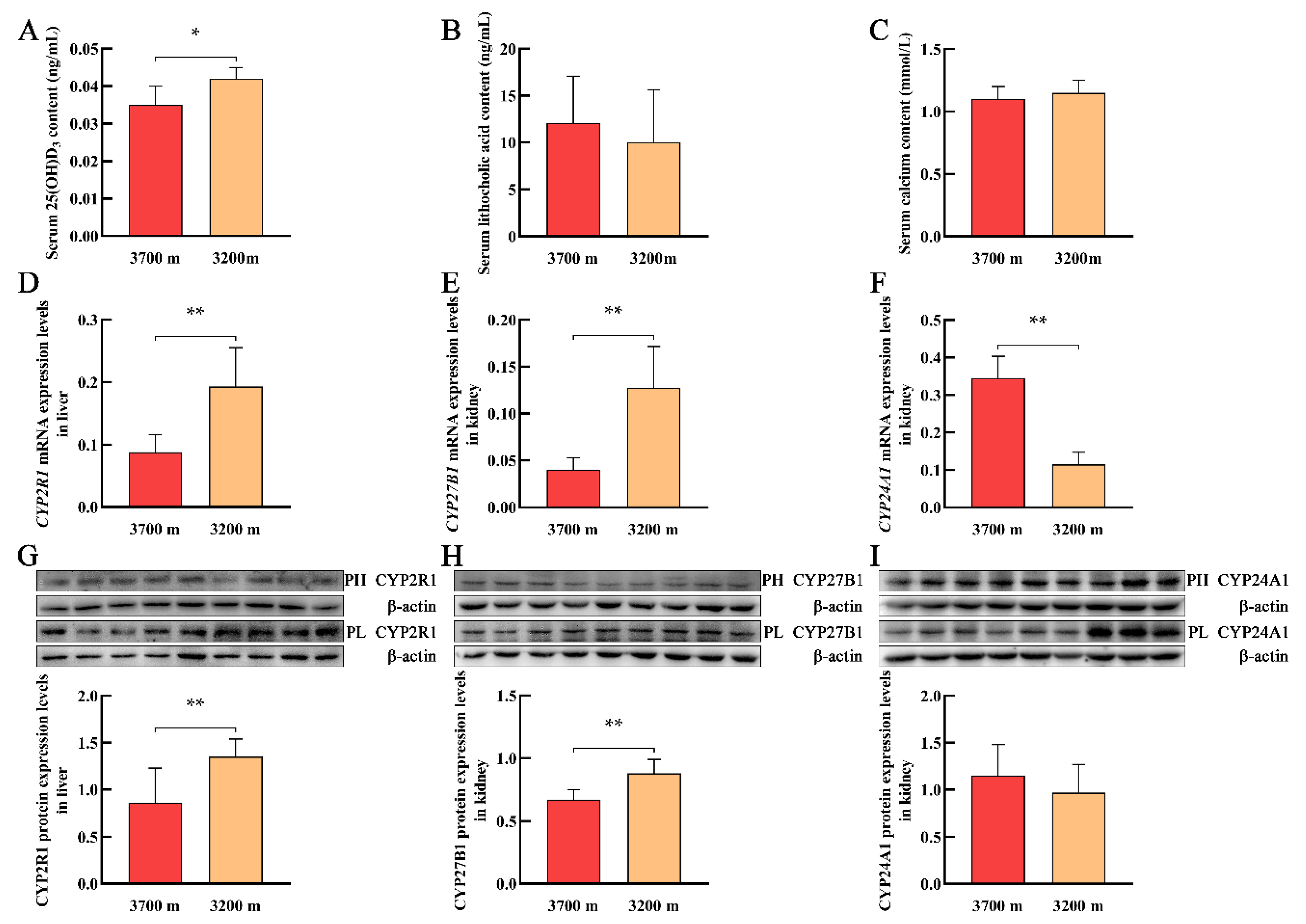
With increasing altitude, the serum levels of 25(OH)D3 in plateau pikas significantly decreased (Figure 3A). However, serum levels of LCA and calcium did not differ significantly with increasing altitude (Figure 3B,C). With increasing altitude, the mRNA and protein levels of CYP2R1 in the liver and CYP27B1 in the kidney of plateau pikas significantly decreased (Figure 3D,E,G,H). In contrast, CYP24A1 mRNA levels in the kidney of plateau pikas significantly increased (Figure 3F), whereas CYP24A1 protein levels were not significantly different (Figure 3I).

4. Discussion

The main source of vitamin D3 is cutaneous synthesis, for which sunlight is pivotal for photolysis of 7-dehydrocholesterol to vitamin D3. Vitamin D3 must undergo two subsequent hydroxylations, mainly by CYP2R1 and CYP27B1, to form the active form 1, 25(OH)2D3. Previous studies have shown that the subterranean rodents Damara mole-rats and naked mole-rats have a naturally impoverished vitamin D3 status [34,35]. The plateau zokor is a subterranean rodent that lives entirely underground in enclosed tunnels on the Qinghai-Tibet Plateau [55]. Similar to subterranean rodents, the plateau zokor lives in a hypoxic environment in the absence of sunlight. Our results showed that the plateau zokor was a vitamin D-deficient rodent with an undetectable serum level of 25(OH)D3. However, a previous study on Damara mole-rats and naked mole-rats showed that vitamin D3 supplementation or sun exposure resulted in an increase in serum level of 1, 25(OH)2D with a decline in 1-OHase activity and an increase in 24-OHase activity [34,35]. In the present research, we demonstrated that CYP2R1 mRNA and protein levels in the liver of plateau zokor were significantly higher than those of plateau pika and SD rats, and CYP27B1 mRNA and protein levels in the kidney of plateau zokor were significantly higher than those of plateau pika. CYP24A1 encodes vitamin D 24-hydroxylase and is largely silent under vitamin D deficiency [56]. CYP24A1 mRNA and protein levels in the kidney of plateau pika were significantly higher than those of the plateau zokor and SD rats, although the protein levels were not significantly different between the plateau zokor and SD rats. Moreover, the CYP2R1 and CYP27B1 mRNA and protein levels in the liver and kidney decreased with increasing altitude, although the protein levels were not significantly different in the liver and kidney. Taken together, our results suggest that the plateau zokor is a vitamin D-deficient rodent, although the vitamin D3 metabolic enzymes CYP2R1 and CYP27B1 are highly expressed in the liver and kidney, respectively. These two enzymes may be related to the metabolism of other steroid hormones, which needs to be studied in the future.
It is well established that vitamin D3 is produced endogenously from 7-dehydrocholesterol following irradiation [21]. The Qinghai-Tibet Plateau receives strong ultraviolet radiation. In theory, the level of 25(OH)D3 in plateau pikas should be higher than that in SD rats. However, our results indicated that serum levels of 25(OH)D3 were significantly lower in plateau pikas than in SD rats. Moreover, 25(OH)D3 levels in plateau pikas significantly decreased with increasing altitude. A previous study reported severe vitamin D deficiency among nomads in Tibet [38]. However, biosynthesis of steroid hormones in SD rats is increased under acute hypoxia [36]. Another study showed that hypoxia increased the levels of 25(OH)D3 in sheep [37]. Therefore, our findings prompted us to investigate whether hypoxia regulates the expression of vitamin D3 metabolic genes. The critical steps in vitamin D3 metabolism are catalyzed by two enzymes CYP2R1 and CYP27B1 [54]. The results of our present study showed that the mRNA levels and protein abundances of CYP2R1 and CYP27B1 in the liver and kidney, respectively, of plateau pikas were significantly decreased in the high-altitude group compared with those in the low-altitude group. However, CYP24A1 protein abundance in the kidney of plateau pikas was not significantly different among the different altitude groups. Therefore, hypoxia can suppress the conversion of D3 into 25(OH)D3 and 1, 25(OH)2D3 by down-regulating the expression of CYP2R1 and CYP27B1 in plateau pika. Future research is needed to determine if hypoxia treatment suppresses the expression of CYP2R1 and CYP27B1 in rats or mice, which would enhance the understanding of the relationship between hypoxia and D3 metabolism.
Calcium homeostasis is crucial for the functioning of numerous health maintenance and biological processes [4]. In fact, the duodenum absorbs only 8–10% of dietary calcium [12]. Ishizawa et al. reported that LCA is a VDR ligand that acts preferentially in the ileum, and LCA did not increase plasma calcium concentrations or intestinal TRPV6 expression in wild-type mice [57]. With regard to the absorption sites involved, the distal intestine might play a significant role in intestinal calcium absorption [9,10,11,12,13]. Studies have shown that calcium absorption is normal in vitamin D-deficient animals, including vitamin D-deficient rats and subterranean rodents such as Damara mole-rats and naked mole-rats [35,41,58]. Further, Nehring et al. reported that LCA restored serum calcium levels to the normal range by increasing VDR target gene expression and bone calcium mobilization in vitamin D-deficient rats [41]. Recently, intestinal calcium absorption was found to be increased by LCA in a vitamin D receptor-dependent manner [59]. The plateau zokor possesses morphological, behavioral, and physiological adaptations for digging, and they exhibit markedly powerful skeletal and muscle systems, lower pulse rates, and a stronger cardiovascular system compared with animals on the plain [46,60]. Despite the undetectable serum level of 25(OH)D3 in the plateau zokor, its calcium levels were within the normal range. Therefore, we speculated that there might have been a substance that replaced vitamin D3 in the plateau zokor. We also measured the LCA levels using UPLC-MS/MS, and the results showed that the LCA content of plateau zokors was significantly higher than that of plateau pikas and SD rats. These results suggest that LCA might be a substitute for vitamin D3 in the plateau zokor. As the most effective ligand of VDR, 1, 25(OH)2D3 promotes intestinal calcium absorption mainly through transcellular pathways mediated by TRPV6, calbindin-D9k, and plasma membrane Ca2+-ATPase 1b (PMCA1b), the expression of which is correlated with the calcium absorption efficiency [15,61,62]. However, Hashimoto et al. reported that LCA increased intestinal calcium absorption in a transcellular pathway-independent manner [59]. The physiological link connecting LCA and calcium metabolism in a naturally vitamin D-deficient rodent remains unclear. Therefore, the mechanisms underlying LCA-dependent calcium regulation in naturally vitamin D-deficient rodents requires further study.

5. Conclusions

In conclusion, the plateau zokor is a vitamin D-deficient subterranean rodent with an undetectable serum level of 25(OH)D3 as a result of the inhibition of the conversion of 7-dehydrocholesterol to vitamin D3, but its enzymes are highly expressed. Hypoxia significantly suppresses the conversion of D3 into 25(OH)D3 and 1, 25(OH)2D3 by down-regulating the expression of CYP2R1 and CYP27B1 in plateau pika. Further, LCA might be a substitution for D3 which could promote calcium absorption in the distal intestine of plateau zokors. However, the mechanisms underlying LCA-dependent calcium regulation in plateau zokors need to be identified.

Supplementary Materials

The following supporting information can be downloaded at: https://www.mdpi.com/article/10.3390/ani12182371/s1, Figure S1: The protein expression levels of CYP2R1, CYP27B1, and CYP24A1 in the liver and kidney of plateau zokor, plateau pika, and Sprague-Dawley (SD) rats; Figure S2: The protein expression levels of CYP2R1, CYP27B1, and CYP24A1 in the liver and kidney of plateau zokor at different altitudes; Figure S3: The protein expression levels of CYP2R1, CYP27B1, and CYP24A1 in the liver and kidney of plateau pika at different altitudes.

Author Contributions

X.C. performed the experiments and drafted the manuscript. Z.A., L.W. and J.Z. performed part of the experiments. X.C., Z.A., L.W., J.Z., Z.W., J.L., C.G. and D.W. collected the samples. X.C., J.Z. and J.L. analyzed the data. D.W. designed experiments and revised the manuscript. All authors have read and agreed to the published version of the manuscript.

Funding

This research was funded by the Natural Science Foundation of Qinghai Province of China (No. 2021-ZJ-904), The Second Tibetan Plateau Scientific Expedition and Research Program (STEP) (No. 2019 QZKK0501), and the National Natural Science Foundation of China (No. 31860606).

Institutional Review Board Statement

The animal care, use, and treatments in this study were reviewed and approved by the China Practice for the Care and Use of Laboratory Animals and the China Zoological Society (permit number: GB/T35892-2018).

Informed Consent Statement

Not applicable.

Data Availability Statement

Not applicable.

Conflicts of Interest

The authors declare no conflict of interest.

References

  1. Bikle, D.D. Vitamin D: An ancient hormone. Exp. Dermatol. 2011, 20, 7–13. [Google Scholar] [CrossRef] [PubMed]
  2. Christakos, S.; Dhawan, P.; Verstuyf, A.; Verlinden, L.; Carmeliet, G. Vitamin D: Metabolism, molecular mechanism of action, and pleiotropic effects. Physiol. Rev. 2016, 96, 365–408. [Google Scholar] [CrossRef] [PubMed]
  3. Bikle, D.; Christakos, S. New aspects of vitamin D metabolism and action-addressing the skin as source and target. Nat. Rev. Endocrinol. 2020, 16, 234–252. [Google Scholar] [CrossRef] [PubMed]
  4. Christakos, S. Vitamin D: A critical regulator of intestinal physiology. JBMR Plus 2021, 5, e10554. [Google Scholar] [CrossRef] [PubMed]
  5. Jones, G. 100 years of vitamin D: Historical aspects of vitamin D. Endocr. Connect. 2022, 11, e210594. [Google Scholar] [CrossRef] [PubMed]
  6. Baeke, F.; Takiishi, T.; Korf, H.; Gysemans, C.; Mathieu, C. Vitamin D: Modulator of the immune system. Curr. Opin. Pharmacol. 2010, 10, 482–496. [Google Scholar] [CrossRef]
  7. Muscogiuri, G.; Mitri, J.; Mathieu, C.; Badenhoop, K.; Tamer, G.; Orio, F.; Mezza, T.; Vieth, R.; Colao, A.; Pittas, A. Mechanisms in endocrinology: Vitamin D as a potential contributor in endocrine health and disease. Eur. J. Endocrinol. 2014, 171, R101–R110. [Google Scholar] [CrossRef]
  8. Mungai, L.N.W.; Mohammed, Z.; Maina, M.; Anjumanara, O. Vitamin D review: The low hanging fruit for human health. J. Nutr. Metab. 2021, 2021, 6335681. [Google Scholar] [CrossRef]
  9. Lee, D.B.; Walling, M.M.; Levine, B.S.; Gafter, U.; Silis, V.; Hodsman, A.; Coburn, J.W. Intestinal and metabolic effect of 1, 25-dihydroxyvitamin D3 in normal adult rat. Am. J. Physiol. 1981, 240, G90–G96. [Google Scholar] [CrossRef]
  10. Favus, M.J. Factors that influence absorption and secretion of calcium in the small intestine and colon. Am. J. Physiol. 1985, 248, G147–G157. [Google Scholar] [CrossRef]
  11. Favus, M.J.; Angeid-Backman, E. Effects of 1, 25(OH)2D3 and calcium channel blockers on cecal calcium transport in the rat. Am. J. Physiol. 1985, 248, G676–G681. [Google Scholar] [CrossRef] [PubMed]
  12. Wasserman, R.H. Vitamin D and the dual processes of intestinal calcium absorption. J. Nutr. 2004, 134, 3137–3139. [Google Scholar] [CrossRef] [PubMed]
  13. Christakos, S.; Seth, T.; Hirsch, J.; Porta, A.; Moulas, A.; Dhawan, P. Vitamin D biology revealed through the study of knockout and transgenic mouse models. Annu. Rev. Nutr. 2013, 33, 71–85. [Google Scholar] [CrossRef] [PubMed]
  14. Furuse, M.; Fujita, K.; Hiiragi, T.; Fujimoto, K.; Tsukita, S. Claudin-1 and -2: Novel integral membrane proteins localizing at tight junctions with no sequence similarity to occludin. J. Cell. Biol. 1998, 141, 1539–1550. [Google Scholar] [CrossRef] [PubMed]
  15. Bronner, F. Mechanisms of intestinal calcium absorption. J. Cell. Biochem. 2003, 88, 387–393. [Google Scholar] [CrossRef] [PubMed]
  16. Benn, B.S.; Ajibade, D.; Porta, A.; Dhawan, P.; Hediger, M.; Peng, J.B.; Jiang, Y.; Oh, G.T.; Jeung, E.B.; Lieben, L.; et al. Active intestinal calcium transport in the absence of transient receptor potential vanilloid type 6 and calbindin-D9k. Endocrinology 2008, 149, 3196–3205. [Google Scholar] [CrossRef] [PubMed]
  17. Fujita, H.; Sugimoto, K.; Inatomi, S.; Maeda, T.; Osanai, M.; Uchiyama, Y.; Yamamoto, Y.; Wada, T.; Kojima, T.; Yokozaki, H.; et al. Tight junction proteins claudin-2 and -12 are critical for vitamin D-dependent Ca2+ absorption between enterocytes. Mol. Biol. Cell. 2008, 19, 1912–1921. [Google Scholar] [CrossRef]
  18. Christakos, S. Recent advances in our understanding of 1, 25-dihydroxyvitamin D3 regulation of intestinal calcium absorption. Arch. Biochem. Biophys. 2012, 523, 73–76. [Google Scholar] [CrossRef]
  19. Lee, S.M.; Riley, E.M.; Meyer, M.B.; Benkusky, N.A.; Plum, L.A.; DeLuca, H.F.; Pike, J.W. 1, 25-dihydroxyvitamin D3 controls a cohort of vitamin D receptor target genes in the proximal intestine that is enriched for calcium-regulating components. J. Biol. Chem. 2015, 290, 18199–18215. [Google Scholar] [CrossRef]
  20. Adamczak, M.; Surma, S.; Więcek, A. Vitamin D and arterial hypertension: Facts and myths. Curr. Hypertens. Rep. 2020, 22. [Google Scholar] [CrossRef]
  21. Bikle, D.D. Vitamin D and the skin: Physiology and pathophysiology. Rev. Endocr. Metab. Disord. 2012, 13, 3–19. [Google Scholar] [CrossRef] [PubMed]
  22. Tian, X.Q.; Holick, M.F. Catalyzed thermal isomerization between previtamin D3 and vitamin D3 via β-cyclodextrin complexation. J. Biol. Chem. 1995, 270, 8706–8711. [Google Scholar] [CrossRef] [PubMed]
  23. DeLuca, H.F. Overview of general physiologic features and functions of vitamin D. Am. J. Clin. Nutr. 2004, 80, 1689S–1696S. [Google Scholar] [CrossRef]
  24. Bhattacharyya, M.H.; DeLuca, H.F. Subcellular location of rat liver calciferol-25-hydroxylase. Arch. Biochem. Biophys. 1974, 160, 58–62. [Google Scholar] [CrossRef]
  25. Zhu, J.G.; Ochalek, J.T.; Kaufmann, M.; Jones, G.; Deluca, H.F. CYP2R1 is a major, but not exclusive, contributor to 25-hydroxyvitamin D production in vivo. Proc. Natl. Acad. Sci. USA 2013, 110, 15650–15655. [Google Scholar] [CrossRef] [PubMed]
  26. Fu, G.K.; Lin, D.; Zhang, M.Y.H.; Bikle, D.D.; Shackleton, C.H.L.; Miller, W.L.; Portale, A.A. Cloning of human 25-hydroxyvitamin D-1α-hydroxylase and mutations causing vitamin D-dependent rickets type 1. Mol. Endocrinol. 1997, 11, 1961–1970. [Google Scholar] [CrossRef]
  27. Cashman, K.D.; van den Heuvel, E.G.; Schoemaker, R.J.; Prévéraud, D.P.; Macdonald, H.M.; Arcot, J. 25-hydroxyvitamin D as a biomarker of vitamin D status and its modeling to inform strategies for prevention of vitamin D deficiency within the population. Adv. Nutr. 2017, 8, 947–957. [Google Scholar] [CrossRef]
  28. Makris, K.; Sempos, C.; Cavalier, E. The measurement of vitamin D metabolites: Part I-metabolism of vitamin D and the measurement of 25-hydroxyvitamin D. Hormones 2020, 19, 81–96. [Google Scholar] [CrossRef]
  29. Bosworth, C.R.; Levin, G.; Robinson-Cohen, C.; Hoofnagle, A.N.; Ruzinski, J.; Young, B.; Schwartz, S.M.; Himmelfarb, J.; Kestenbaum, B.; de Boer, I.H. The serum 24, 25-dihydroxyvitamin D concentration, a marker of vitamin D catabolism, is reduced in chronic kidney disease. Kidney Int. 2012, 82, 693–700. [Google Scholar] [CrossRef]
  30. Bouillon, R.; Antonio, L.; Olarte, O.R. Calcifediol (25OH vitamin D3) deficiency: A risk factor from early to old age. Nutrients 2022, 14, 1168. [Google Scholar] [CrossRef] [PubMed]
  31. Lovegrove, B.G.; Knight-Eloff, A. Soil and burrow temperatures, and the resource characteristics of the social mole-rat Cryptomys damarensis (Bathyergidae) in the Kalahari desert. J. Zool. 1988, 216, 403–416. [Google Scholar] [CrossRef]
  32. Buffenstein, R.; Yahav, S. Cholecalciferol has no effect on calcium and inorganic phosphorus balance in a naturally cholecalciferol-deplete subterranean mammal, the naked mole rat (Heterocephalus glaber). J. Endocrinol. 1991, 129, 21–26. [Google Scholar] [CrossRef] [PubMed]
  33. Chu, B.; Tian, Y.L.; Zhou, J.W.; Tang, Z.S.; Dong, K.; Hua, L.M. Increased burrow oxygen levels trigger defensive burrow-sealing behavior by plateau zokors. Sci. Rep. 2021, 11. [Google Scholar] [CrossRef]
  34. Buffenstein, R.; Sergeev, I.N.; Pettifor, J.M. Vitamin D hydroxylases and their regulation in a naturally vitamin D-deficient subterranean mammal, the naked mole rat (Heterocephalus glaber). J. Endocrinol. 1993, 138, 59–64. [Google Scholar] [CrossRef]
  35. Pitcher, T.; Sergeev, I.N.; Buffenstein, R. Vitamin D metabolism in the Damara mole-rat is altered by exposure to sunlight yet mineral metabolism is unaffected. J. Endocrinol. 1994, 143, 367–374. [Google Scholar] [CrossRef]
  36. Lin, X.; Wang, H.Y.; Pu, X.Y. Protective mechanism of fdft1 in steroid hormone synthesis pathway in SD rats with acute hypoxic injury. Genes. Genom. 2020, 42, 1319–1326. [Google Scholar] [CrossRef]
  37. Goyal, R.; Billings, T.L.; Mansour, T.; Martin, C.; Baylink, D.J.; Longo, L.D.; Pearce, W.J.; Mata-Greenwood, E. Vitamin D status and metabolism in an ovine pregnancy model: Effect of long-term, high-altitude hypoxia. Am. J. Physiol. Endocrinol. Metab. 2016, 310, E1062–E1071. [Google Scholar] [CrossRef]
  38. Norsang, G.; Ma, L.W.; Dahlback, A.; Zhuoma, C.; Tsoja, W.; Porojnicu, A.; Lagunova, Z.; Moan, J. The vitamin D status among Tibetans. Photochem. Photobiol. 2009, 85, 1028–1031. [Google Scholar] [CrossRef]
  39. Ridlon, J.M.; Kang, D.J.; Hylemon, P.B.; Bajaj, J.S. Bile acids and the gut microbiome. Curr. Opin. Gastroenterol. 2014, 30, 332–338. [Google Scholar] [CrossRef]
  40. Makishima, M.; Lu, T.T.; Xie, W.; Whitfield, G.K.; Domoto, H.; Evans, R.M.; Haussler, M.R.; Mangelsdorf, D.J. Vitamin D receptor as an intestinal bile acid sensor. Science 2002, 296, 1313–1316. [Google Scholar] [CrossRef] [Green Version]
  41. Nehring, J.A.; Zierold, C.; DeLuca, H.F. Lithocholic acid can carry out in vivo functions of vitamin D. Proc. Natl. Acad. Sci. USA 2007, 104, 10006–10009. [Google Scholar] [CrossRef] [PubMed]
  42. Chaudhari, S.N.; Luo, J.N.; Harris, D.A.; Aliakbarian, H.; Yao, L.; Paik, D.; Subramaniam, R.; Adhikari, A.A.; Vernon, A.H.; Kiliç, A.; et al. A microbial metabolite remodels the gut-liver axis following bariatric surgery. Cell. Host Microbe. 2021, 29, 408–424. [Google Scholar] [CrossRef] [PubMed]
  43. Peacock, A.J. ABC of oxygen: Oxygen at high altitude. BMJ 1998, 317, 1063–1066. [Google Scholar] [CrossRef] [PubMed]
  44. Butaric, L.N.; Klocke, R.P. Nasal variation in relation to high-altitude adaptations among Tibetans and Andeans. Am. J. Hum. Biol. 2018, 30, e23104. [Google Scholar] [CrossRef]
  45. Song, D.S.; Navalsky, B.E.; Guan, W.; Ingersoll, C.; Wang, T.; Loro, E.; Eeles, L.; Matchett, K.B.; Percy, M.J.; Walsby-Tickle, J.; et al. Tibetan PHD2, an allele with loss-of-function properties. Proc. Natl. Acad. Sci. USA 2020, 117, 12230–12238. [Google Scholar] [CrossRef]
  46. Zhang, Y.M.; Zhang, Z.B.; Liu, J.K. Burrowing rodents as ecosystem engineers: The ecology and management of plateau zokors Myospalax fontanierii in alpine meadow ecosystems on the Tibetan Plateau. Mammal. Rev. 2003, 33, 284–294. [Google Scholar] [CrossRef]
  47. Smith, A.T.; Wang, X.G. Social relationships of adult black-lipped pikas (Ochotona curzoniae). J. Mammal. 1991, 72, 231–247. [Google Scholar] [CrossRef]
  48. Dobson, F.S.; Smith, A.T.; Wang, X.G. The mating system and gene dynamics of plateau pikas. Behav. Processes 2000, 51, 101–110. [Google Scholar] [CrossRef]
  49. Yin, B.F.; Yang, S.M.; Wei, W.H.; Zhang, Y.M. Male reproductive success in plateau pikas (Ochotona curzoniae): A microsatellite analysis. Mamm. Biol. 2009, 74, 344–350. [Google Scholar] [CrossRef]
  50. Sun, S.Z.; Wei, L.; Wei, D.B.; Wang, D.W.; Ma, B.Y. Differences of glycolysis in skeletal muscle and lactate metabolism in liver between plateau zokor (Myospalax baileyi) and plateau pika (Ochotona curzoniae). Acta Physiol. Sin. 2013, 65, 276–284. [Google Scholar] [CrossRef]
  51. Shao, Y.; Li, J.X.; Ge, R.L.; Zhong, L.; Irwin, D.M.; Murphy, R.W.; Zhang, Y.P. Genetic adaptations of the plateau zokor in high-elevation burrows. Sci. Rep. 2015, 5. [Google Scholar] [CrossRef] [PubMed]
  52. Zhang, T.; Chen, J.; Zhang, J.; Guo, Y.T.; Zhou, X.; Li, M.W.; Zheng, Z.Z.; Zhang, T.Z.; Murphy, R.W.; Nevo, E.; et al. Phenotypic and genomic adaptations to the extremely high elevation in plateau zokor (Myospalax baileyi). Mol. Ecol. 2021, 30, 5765–5779. [Google Scholar] [CrossRef] [PubMed]
  53. Li, S.H.; Xu, B.; An, Z.F.; Wang, Z.J.; Li, Y.X.; Wei, L.; Wei, D.B. Evolutionary analysis of TSP-1 gene in plateau zokor (Myospalax Baileyi) and its expression pattern under hypoxia. Cell. Mol. Biol. 2019, 65, 48–57. [Google Scholar]
  54. Bikle, D.D. Vitamin D metabolism, mechanism of action, and clinical applications. Chem. Biol. 2014, 21, 319–329. [Google Scholar] [CrossRef] [PubMed]
  55. Fan, N.C.; Shi, Y.Z. A revision of the zokors of subgenus. Acta Theriol. Sin. 1982, 2, 183–199. [Google Scholar] [CrossRef]
  56. Endres, B.; Kato, S.; DeLuca, H.F. Metabolism of 1α, 25-dihydroxyvitamin D3 in vitamin D receptor-ablated mice in vivo. Biochemistry 2000, 39, 2123–2129. [Google Scholar] [CrossRef]
  57. Ishizawa, M.; Akagi, D.; Makishima, M. Lithocholic acid is a vitamin D receptor ligand that acts preferentially in the ileum. Int. J. Mol. Sci. 2018, 19, 1975. [Google Scholar] [CrossRef]
  58. Skinner, D.C.; Moodley, G.; Buffenstein, R. Is vitamin D3 essential for mineral metabolism in the Damara mole-rat (Cryptomys damarensis)? Gen. Comp. Endocrinol. 1991, 81, 500–505. [Google Scholar] [CrossRef]
  59. Hashimoto, N.; Matsui, I.; Ishizuka, S.; Inoue, K.; Matsumoto, A.; Shimada, K.; Hori, S.; Lee, D.G.; Yasuda, S.; Katsuma, Y. Lithocholic acid increases intestinal phosphate and calcium absorption in a vitamin D receptor dependent but transcellular pathway independent manner. Kidney Int. 2020, 97, 1164–1180. [Google Scholar] [CrossRef]
  60. Zeng, J.X.; Wang, Z.W.; Shi, Z.X. Metabolic characteristics and some physiological parameters of mole rat (Myospalax baileyi) in alpine area. Acta Biol. Plat. Sin. 1984, 3, 163–171. [Google Scholar]
  61. Van Cromphaut, S.J.; Dewerchin, M.; Hoenderop, J.G.; Stockmans, I.; Van Herck, E.; Kato, S.; Bindels, R.J.; Collen, D.; Carmeliet, P.; Bouillon, R.; et al. Duodenal calcium absorption in vitamin D receptor-knockout mice: Functional and molecular aspects. Proc. Natl. Acad. Sci. USA 2001, 98, 13324–13329. [Google Scholar] [CrossRef] [PubMed]
  62. Replogle, R.A.; Li, Q.; Wang, L.B.; Zhang, M.; Fleet, J.C. Gene-by-diet interactions influence calcium absorption and bone density in mice. J. Bone Miner. Res. 2014, 29, 657–665. [Google Scholar] [CrossRef] [PubMed]
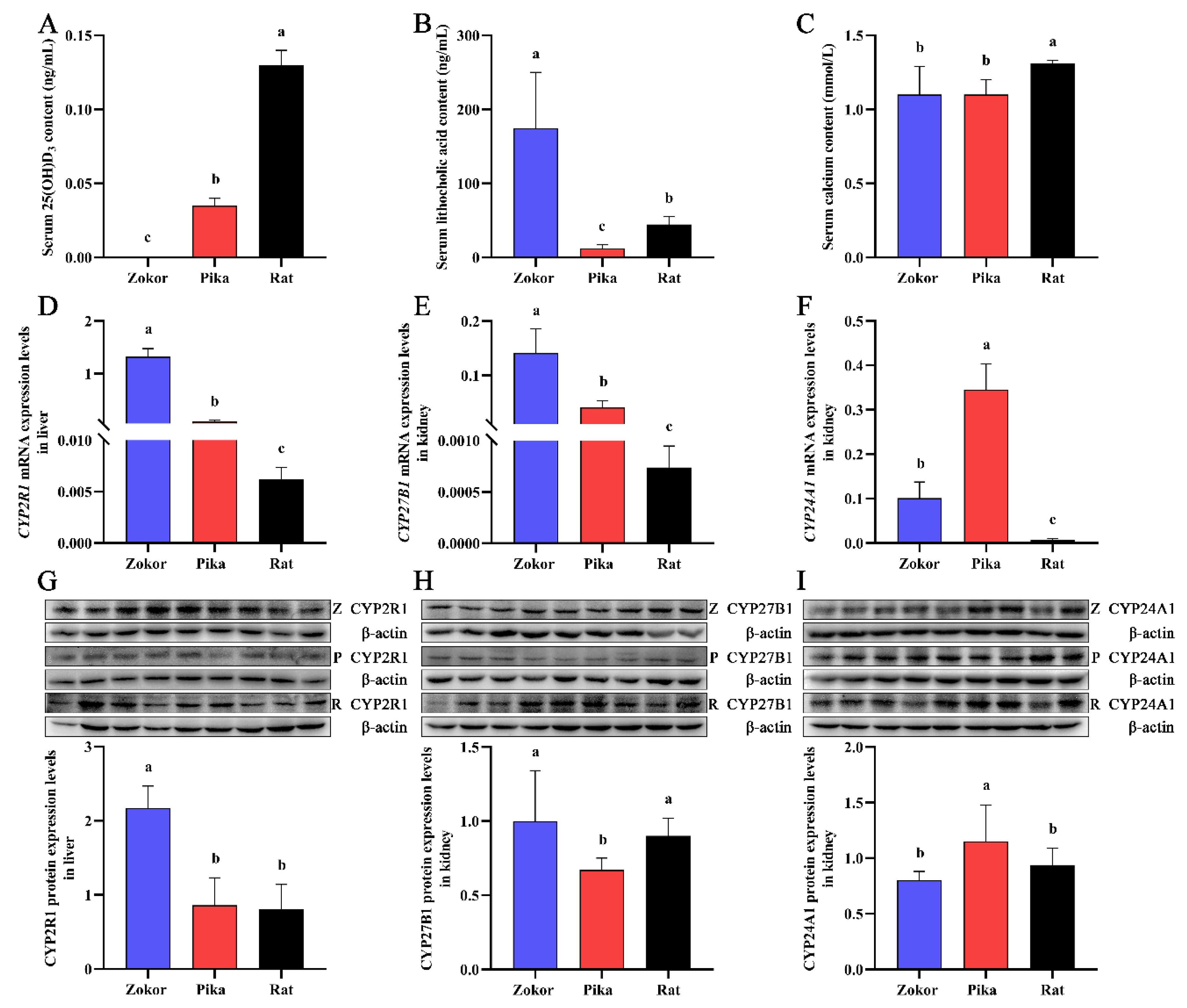
Figure 1. Serum levels of 25(OH)D3, lithocholic acid, and calcium and the expression levels of CYP2R1, CYP27B1, and CYP24A1 in the liver and kidney of plateau zokor, plateau pika, and Sprague-Dawley (SD) rats. (AC) represent the serum levels of 25(OH)D3, lithocholic acid, and calcium, respectively. The mRNA (D) and protein (G) expression levels of CYP2R1 in the liver. The mRNA (E) and protein (H) expression levels of CYP27B1 in the kidney. The mRNA (F) and protein (I) expression levels of CYP24A1 in the kidney. Z, P, and R represent the plateau zokor at 3700 m elevation, plateau pika at 3700 m elevation, and SD rat at 1500 m elevation, respectively. Significant differences among species are represented by different lowercase letters (p < 0.05). No significant differences among species are represented by the same lowercase letters (p > 0.05). (original Western blot figures are in Figure S1).
Animals 12 02371 g001
Figure 2. Serum levels of 25(OH)D3, lithocholic acid, and calcium and the expression levels of CYP2R1, CYP27B1, and CYP24A1 in the liver and kidney of plateau zokor at different altitudes. (AC) represent the serum levels of 25(OH)D3, lithocholic acid, and calcium, respectively. The mRNA (D) and protein (G) expression levels of CYP2R1 in the liver. The mRNA (E) and protein (H) expression levels of CYP27B1 in the kidney. The mRNA (F) and protein (I) expression levels of CYP24A1 in the kidney. ZH and ZL represent the plateau zokor at elevations of 3700 m and 2700 m, respectively. ** p < 0.01. (original Western blot figures are in Figure S2).
Animals 12 02371 g002
Figure 3. Serum levels of 25(OH)D3, lithocholic acid, and calcium and the expression levels of CYP2R1, CYP27B1, and CYP24A1 in the liver and kidney of plateau pika at different altitudes. (AC) represent the serum levels of 25(OH)D3, lithocholic acid, and calcium, respectively. The mRNA (D) and protein (G) expression levels of CYP2R1 in the liver. The mRNA (E) and protein (H) expression levels of CYP27B1 in the kidney. The mRNA (F) and protein (I) expression levels of CYP24A1 in the kidney. PH and PL represent the plateau pika at elevations of 3700 m and 3200 m, respectively. * p < 0.05, ** p < 0.01. (original Western blot figures are in Figure S3).
Animals 12 02371 g003
Table 1. Primers of vitamin D3 metabolism-related genes and β-actin for qPCR.
SpeciesGeneSense Primer (5′ to 3′)Anti-Sense Primer (5′ to 3′)
Plateau ZokorCYP2R1AGTTGTTCAGTGAGAATGTGGAACCGAGGTAAGTGAGGCTTTC
CYP27B1CCAAGCCACTGTTCTATCTAGTCACCTACACGGATG
CYP24A1GAGATTCGGACTCCTTCAGAGGTGTTGAGCCTCTTGTG
Plateau PikaCYP2R1TTCCTCGGCAACATCTACCTACATCATAGCCATTCAGAAC
CYP27B1CAATGCTCTGTCTCAACTGCGTGAAGTGGCATAGTGA
CYP24A1TCTCGCAATCCTCAAGTCACATATTCCTCAAGTCCTCTG
Sprague-Dawley ratCYP2R1GCATATCAACTGTGGTTCTCAATATCCATCCTCTGCCATATCTG
CYP27B1ATGGTGAAGAATGGCAGAGTGTCCAGAGTTCCAGCATA
CYP24A1TTCGCTCATCTCCCATTCCATCTCCACAGGTTCATTG
β-actinTCACCAACTGGGACGATATGGTTGGCCTTAGGGTTCAGAG
Publisher’s Note: MDPI stays neutral with regard to jurisdictional claims in published maps and institutional affiliations.