Article

DNA Multi-Marker Genotyping and CIAS Morphometric Phenotyping of Fasciola gigantica-Sized Flukes from Ecuador, with an Analysis of the Radix Absence in the New World and the Evolutionary Lymnaeid Snail Vector Filter

by
Maria Dolores Bargues
1,*,
Maria Adela Valero
1,
Gabriel A. Trueba
2,
Marco Fornasini
3,
Angel F. Villavicencio
4,
Rocío Guamán
4,
Alejandra De Elías-Escribano
1,
Ignacio Pérez-Crespo
1,
Patricio Artigas
1 and
Santiago Mas-Coma
1,*
1
Departamento de Parasitología, Facultad de Farmacia, Universidad de Valencia, Av. Vicent Andres Estelles s/n, 46100 Valencia, Spain
2
Instituto de Microbiología, Edificio Eugenio Espejo, EE-125, Colegio de Ciencias Biológicas y Ambientales, Campus Cumbayá, Universidad San Francisco de Quito, Av. Diego de Robles y Vía Interoceánica, Quito 170901, Ecuador
3
Escuela de Medicina, Universidad Internacional del Ecuador, Av. Jorge Fernández s/n y Av. Simón Bolivar, Quito 170411, Ecuador
4
Departamento de Ciencias de la Vida y Agricultura, Universidad de las Fuerzas Armadas-ESPE, Sede Santo Domingo de los Tsáchilas, Vía Santo Domingo-Quevedo km 24, Santo Domingo de los Tsáchilas, P.O. Box 171-5-231B, Luz de América 230118, Ecuador
*
Authors to whom correspondence should be addressed.
Animals 2021, 11(9), 2495; https://doi.org/10.3390/ani11092495
Submission received: 8 July 2021 / Revised: 13 August 2021 / Accepted: 22 August 2021 / Published: 25 August 2021

Abstract

:

Simple Summary

Fasciolid flukes collected from sheep and cattle in Ecuador showed a high diversity in DNA sequences whose analyses indicated introductions from South America, European and North American countries. These results agree with the numerous livestock importations performed by Ecuador. Abnormally big-sized liver flukes were found in Ecuadorian sheep. The morphometric phenotypic CIAS study showed that its size maximum and mean very pronouncedly and significantly surpassed those of the Fasciola hepatica populations from South America and Spain and proved to be intermediate between standard F. hepatica and F. gigantica populations. Such a feature is only known in intermediate fasciolid forms in Old World areas where the two species and their specific lymnaeid snail vectors overlap. This argues about a past hybridization after F. gigantica importation from Pakistan and/or introduction of intermediate hybrids previously generated in USA. The lack of heterozygotic rDNA ITS positions differentiating the two species, and of introgressed fragments and heteroplasmic positions in mtDNA genes, indicate a post-hybridization period sufficiently long as for rDNA concerted evolution to complete homogenization and mtDNA to return to homoplasmy. The vector specificity filter due to Radix absence should act as a driving force in accelerating such lineage evolution. Public health implications are finally emphasized.

Abstract

Fascioliasis is a disease caused by Fasciola hepatica worldwide transmitted by lymnaeid snails mainly of the Galba/Fossaria group and F. gigantica restricted to parts of Africa and Asia and transmitted by Radix lymnaeids. Concern has recently risen regarding the high pathogenicity and human infection capacity of F. gigantica. Abnormally big-sized fasciolids were found infecting sheep in Ecuador, the only South American country where F. gigantica has been reported. Their phenotypic comparison with F. hepatica infecting sheep from Peru, Bolivia and Spain, and F. gigantica from Egypt and Vietnam demonstrated the Ecuadorian fasciolids to have size-linked parameters of F. gigantica. Genotyping of these big-sized fasciolids by rDNA ITS-2 and ITS-1 and mtDNA cox1 and nad1 and their comparison with other countries proved the big-sized fasciolids to belong to F. hepatica. Neither heterozygotic ITS position differentiated the two species, and no introgressed fragments and heteroplasmic positions in mtDNA were found. The haplotype diversity indicates introductions mainly from other South American countries, Europe and North America. Big-sized fasciolids from Ecuador and USA are considered to be consequences of F. gigantica introductions by past livestock importations. The vector specificity filter due to Radix absence should act as driving force in the evolution in such lineages.

1. Introduction

Fascioliasis is a disease included within the group of foodborne trematodiases, among the Neglected Tropical Diseases (NTDs) given priority by the World Health Organization [1,2]. The two liver flukes Fasciola hepatica and F. gigantica are highly pathogenic [3,4,5] and may underlie the underdevelopment of rural communities of low-income countries. In these depauperated rural areas, they affect mainly children [6], sometimes even very early in life [7]. In these rural areas, infected subjects usually suffer from the chronic phase of the disease, which may last for many years due to lack of diagnosis and give rise to pathological effects such as anemia, litiasis, bacterobilia, and other clinical pictures [8,9,10], which in the long term may result in sequelae [4,11]. Moreover, the immunosuppression induced by these liver flukes in both the acute and chronic phases appear linked to very frequent coinfections by other pathogenic parasites within a scenario of great morbidity [12,13,14,15].
The fasciolid flukes causing this zoonotic parasitic disease infect mammals, mainly herbivorous ruminants, but also humans that become infected by a wide spectrum of sources by oral ingestion of the infective metacercarial stage together with different foods and drinks, mainly freshwater vegetables and natural water [16]. Fasciola hepatica is above all transmitted by small amphibious lymnaeid species belonging to the Galba/Fossaria group in mild/cold areas and highlands of warm regions of Europe, Asia, Africa, the Americas and Oceania. Fasciola gigantica is transmitted by aquatic, less amphibious usually bigger lymnaeid species of the Radix group in warmer lowland of parts of Africa and Asia [17]. These differences in geographical distribution appear mainly related to the minimum temperature threshold for their larval stage development: 10 °C for F. hepatica [18,19]; 16 °C for F. gigantica [20,21], although cercarial shedding of the latter at 13–18 °C has been reported [22].
This disease is markedly influenced by both climate change and anthropogenic modifications of the environment [21,23], including man-made movements and import/export of livestock [24,25]. The actual spread of F. gigantica related to the two aforementioned phenomena, mainly global warming, is worrying, both the parasite in northern Africa [26] and the northward spread of one of its main Radix species vectors in Asia [27]. Indeed, F. gigantica has been proven to show a slower development and higher pathogenicity than F. hepatica [28]. Although F. gigantica was considered to be involved in human infection only sporadically or rarely [29,30], the discovery of human endemic zones presenting F. gigantica in Africa and Asia suggests the convenience of assessing the capacity of this fasciolid species to also infect humans and originate problematic public health situations. Indeed, such situations have already been described in countries as Egypt [14,31], and Ethiopia [32], in Africa, and in Iran [33], Pakistan [21], Vietnam [34], and China [35], in Asia.
The palaeobiogeographical origin of F. gigantica is considered to have taken place in the warm lowlands linked to ancestors of Radix natalensis of eastern Africa during the early Miocene, whereas F. hepatica should originate more recently in the highlands linked to ancestors of Galba truncatula of the Near East [17]. Both fasciolid species should later spread throughout Africa, Europe and Asia together with livestock along the postdomestication period, and only F. hepatica was later introduced into the New World and Oceania during the last 500 years as a consequence of the European colonizations [17].
In the Americas, however, the presence of F. gigantica or F. gigantica-like worms have been reported in three countries. The report in the USA [36] has been analyzed later [17]. Regarding the reports in Mexico [37,38], more recent molecular studies by ISSR-PCR found small genetic distances between liver flukes from the northwest of Spain and Mexico [39] and genetic haplotyping with the ribosomal DNA intergenic region indicated an introduction into Mexico with livestock transported from Spain during the early colonization period [40], and, the most important, the size range reported for the Mexican F. gigantica specimens proved to phenotypically enter in the F. hepatica range [40]. In South America, F. gigantica has been reported only from Ecuador [41], as deduced from the concrete sentence saying that “In Vietnam and Ecuador where both F. hepatica and F. gigantica are endemic......”, although no additional detail on the presence of F. gigantica in Ecuador is included in this article, nor in any other subsequent publication by these or other authors.
The aim of the present study is to clarify whether the aforementioned report of F. gigantica in Ecuador is correct or a misunderstanding, by taking advantage of the finding of liver flukes morphologically resembling F. gigantica in sheep from an area westward from Quito city. For this purpose, a complete phenotypic assessment and a molecular study have been performed. This study is complemented by a similar DNA multimarker analysis of fasciolid flukes collected in cattle from southern Ecuador, where uncontrolled transborder exchange of livestock occurs [42], to assess a potential introduction route from the South. In this southernmost zone of Ecuador, Lymnaea neotropica has recently been found [42], a very efficient vector involved in the earlier spread of fascioliasis throughout South America [43] and linked to human fascioliasis endemic areas in both the neighboring northern Peru [44] and also Argentina [45].

2. Materials and Methods
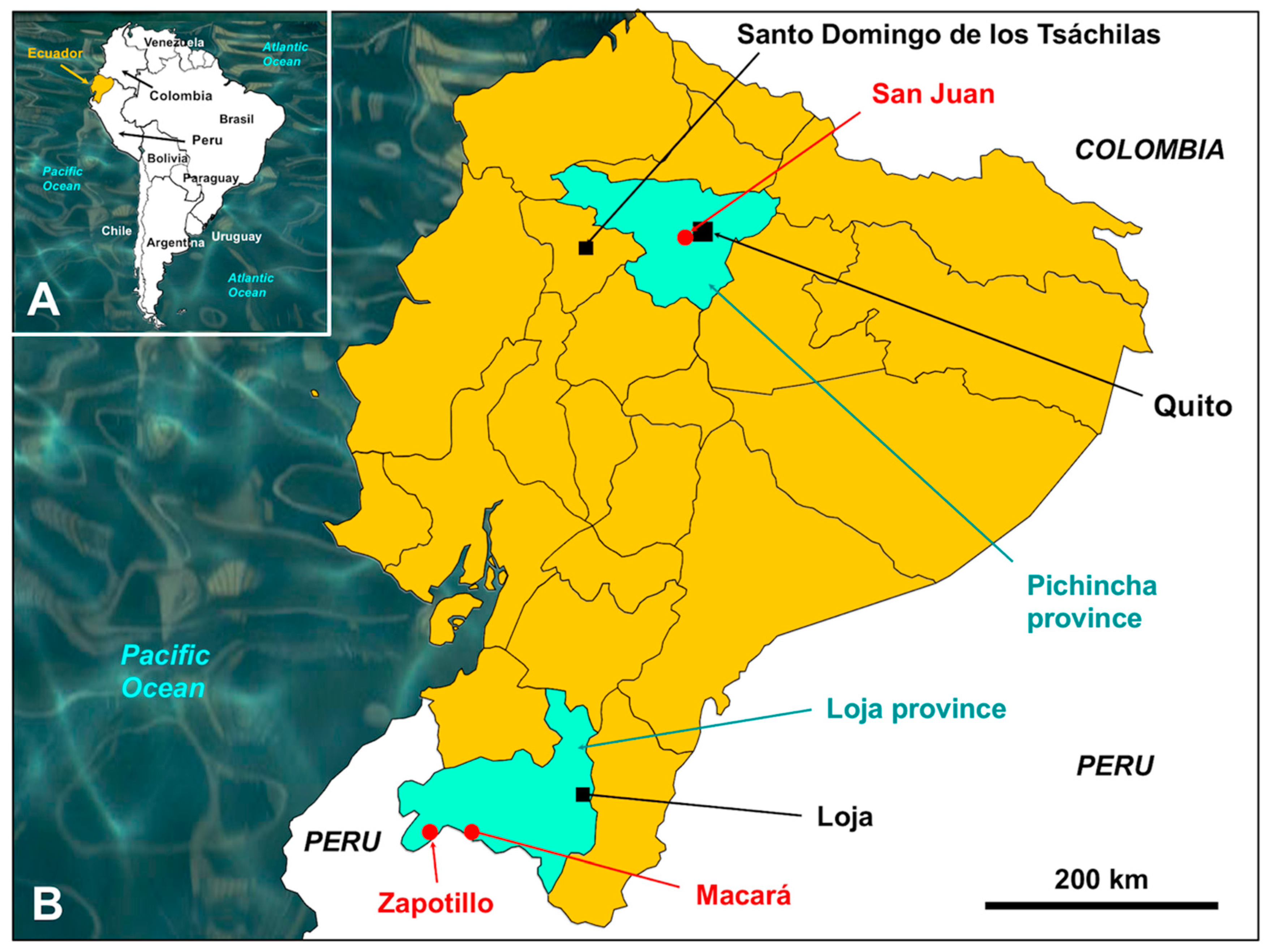
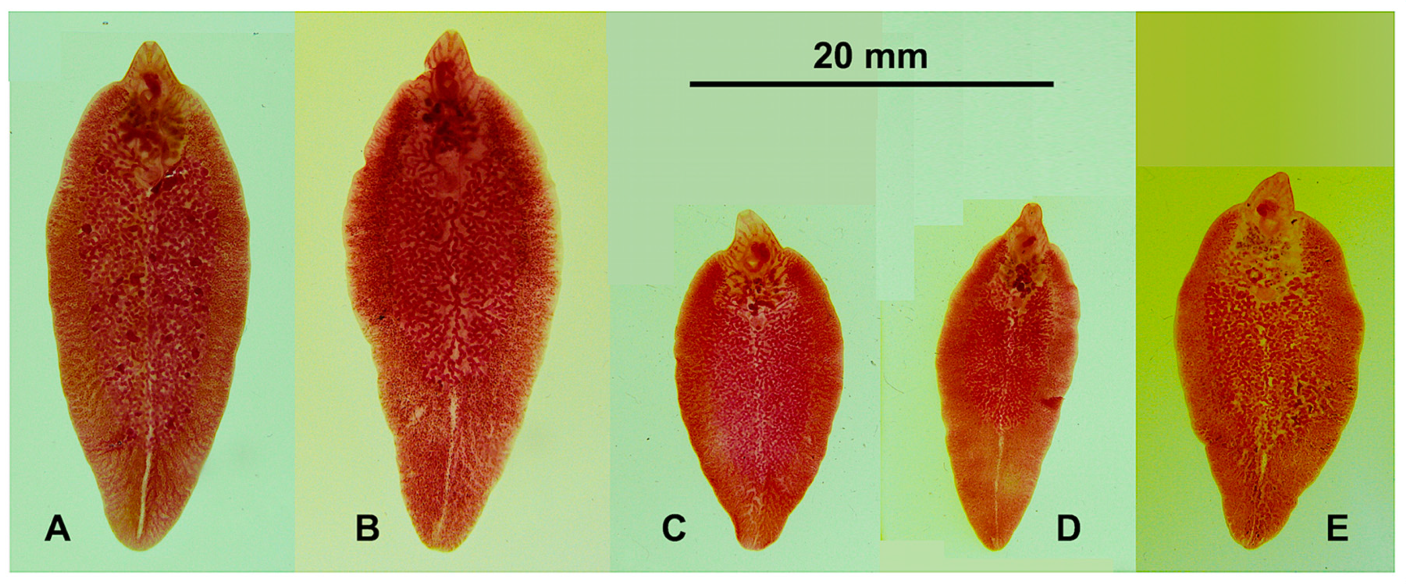
2.1. Parasites

In Ecuador, a total of 42 liver fluke specimens infecting sheep and 21 specimens infecting cattle from the zone of San Juan de Chillogallo, via Chiriboga, at an altitude of 2800–3000 m a.s.l., westward from Quito city, were obtained during slaughtering in the Camal Metropolitano de Quito. Additionally, other liver flukes infecting cattle were also obtained from the southern zone of Loja besides the border with Peru, namely 14 specimens from Macará and other 17 specimens from Zapotillo, at the lower altitudes of 480 and 153 m a.s.l., respectively [42] (Figure 1).

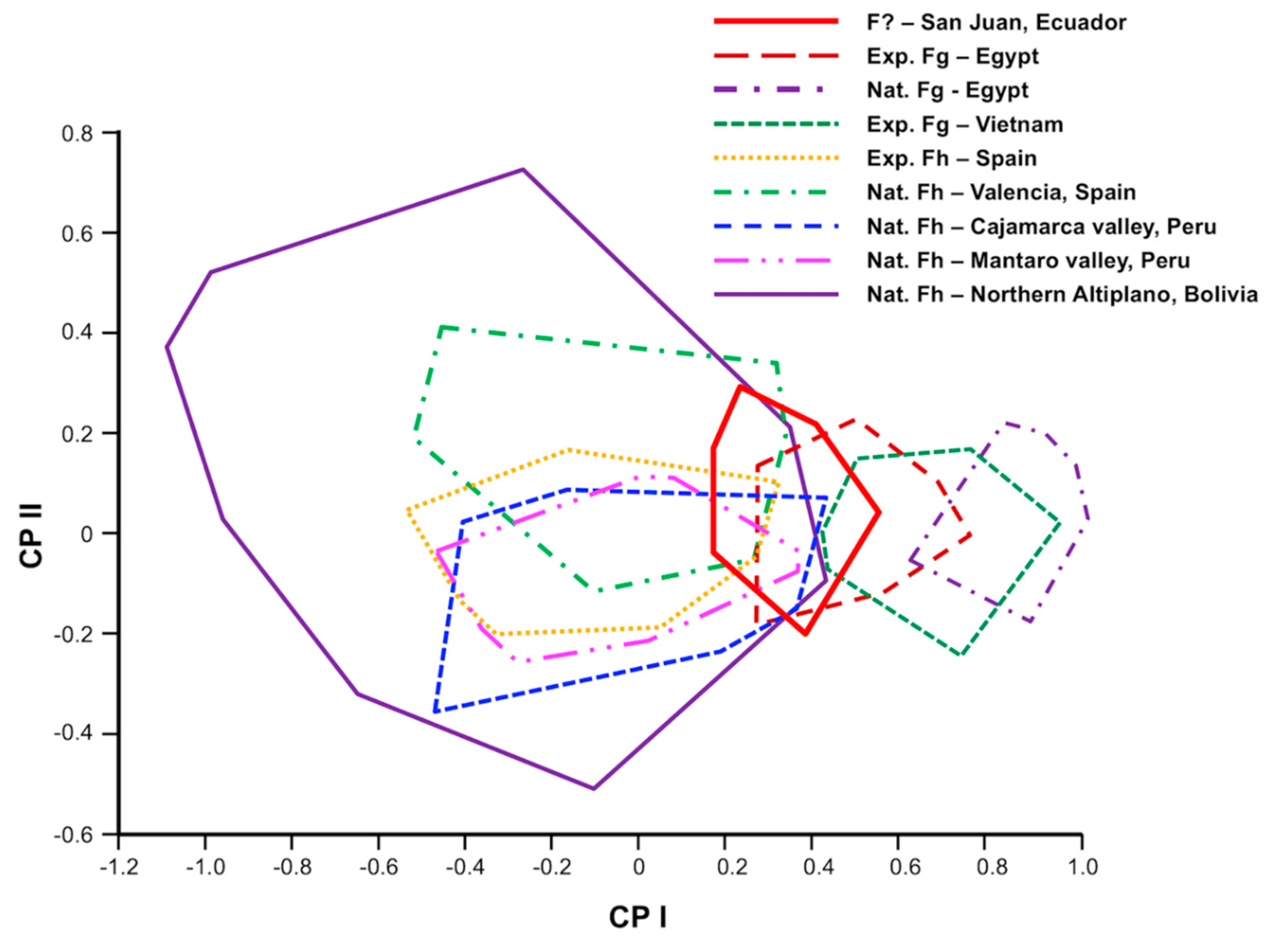
2.2. Phenotyping Analyses

2.2.1. Natural and Experimental Liver Fluke Materials Used for Phenotyping Comparison

Large-sized liver fluke specimens resembling F. gigantica were only found in the aforementioned sheep. Previous studies on liver fluke phenotype have revealed that the definitive host species decisively influences the size of fasciolid adults and eggs [46,47,48]. Therefore, all fasciolid specimens included in the needed comparative morphometric study were gravid adult flukes (i.e., containing from few to many eggs inside the uterus) from sheep.
For this phenotyping study, liver fluke adults obtained from naturally infected sheep were studied by means of appropriate morphometric analyses by Computer Image Analysis System (CIAS) according to previously established standards [49]. Post-mortem examinations were carried out on all animals as soon as possible. The liver parenchyma and bile ducts were examined and flukes inside were collected.
A phenomenon of crowding effect on liver flukes has been emphasized in sheep [50]. Worm size is dependent on the infection level, and that worm size decreases when the burden increases [51]. Therefore, the flukes studied were only from livers with low intensity infection (<70 adults per host), to avoid a possible crowding effect influence.
The studied flukes comprised the largest worm variability possible, i.e., including different stages of body size, maturity, and gravid uteri (i.e., from smallest specimens presenting only very few eggs in the uterus, up to the largest specimens presenting plenty of eggs in the uterus; nongravid specimens presenting no eggs were not included). Fluke specimens were fixed in Bouin’s solution between a slide and coverglass, considering that coverglass pressure could not produce distortion. The worms were colored with Grenacher´s borax, and subsequently differentiated, dehydrated and mounted in Canada balsam. The following materials from natural infections were used for comparisons with the aforementioned fluke materials from Ecuadorian sheep (code: nEc):
  • 47 adults of F. hepatica from 5 sheep (range: 3–21/sheep) from Rodicio, Cajamarca Valley, at an altitude of 2726 m a.s.l., northern Peru (nCaj);
  • 130 adults of F. hepatica from 8 sheep (range: 2–32/sheep) from Huayucachi, Pachacayo and Huancayo, in the Mantaro Valley, at an altitude of 3231–3989 m a.s.l., Peru (nMan);
  • 201 adults of F. hepatica collected in 12 sheep (range: 6–30 worms per sheep) from Batallas, Northern Bolivian Altiplano, at an altitude of 3858 m a.s.l., Bolivia (nAlt);
  • 37 adults of F. hepatica from 5 sheep (range: 2–10/sheep) from Massamagrell, Valencia, Comunidad Valenciana, at an altitude of 20 m a.s.l., Spain (nSp);
  • 31 adults of F. gigantica from 2 sheep (range: 6–32/sheep) from Giza, Giza Governorate, at an altitude of 40 m a.s.l., Egypt (nEg).
Additionally, for the same comparative phenotyping analysis, liver fluke adults of both species F. hepatica and F. gigantica experimentally obtained in sheep were also used. Fasciola hepatica and the lymnaeid Galba truncatula originated from Spain, whereas F. gigantica and its snail host Radix natalensis originated from Egypt [28]. The experimental group consisted of eight four-to-five-week-old sheep of the Guirra autochthonous breed (only sheep breed proved to be similarly susceptible to the two fasciolid species) divided into two groups of four animals each. The groups were infected per os with 200 F. hepatica metacercariae or 200 F. gigantica metacercariae, respectively. Food and water were provided ad libitum. Necropsy was carried out 40 weeks post infection. The following experimentally obtained materials were used for the same aforementioned comparative phenotyping study:
  • 127 adults of F. hepatica from 4 experimentally infected sheep (range: 33–66/sheep; 24-week-old) from Spain (expSp);
  • 100 adults of F. gigantica from 4 experimentally infected sheep (range: 17–69/sheep; 24-week-old) from Egypt (expEg);
  • 70 adults of F. gigantica from 7 experimentally infected sheep (range: 2–69/sheep; 52-week-old) from Vietnam (expVi).

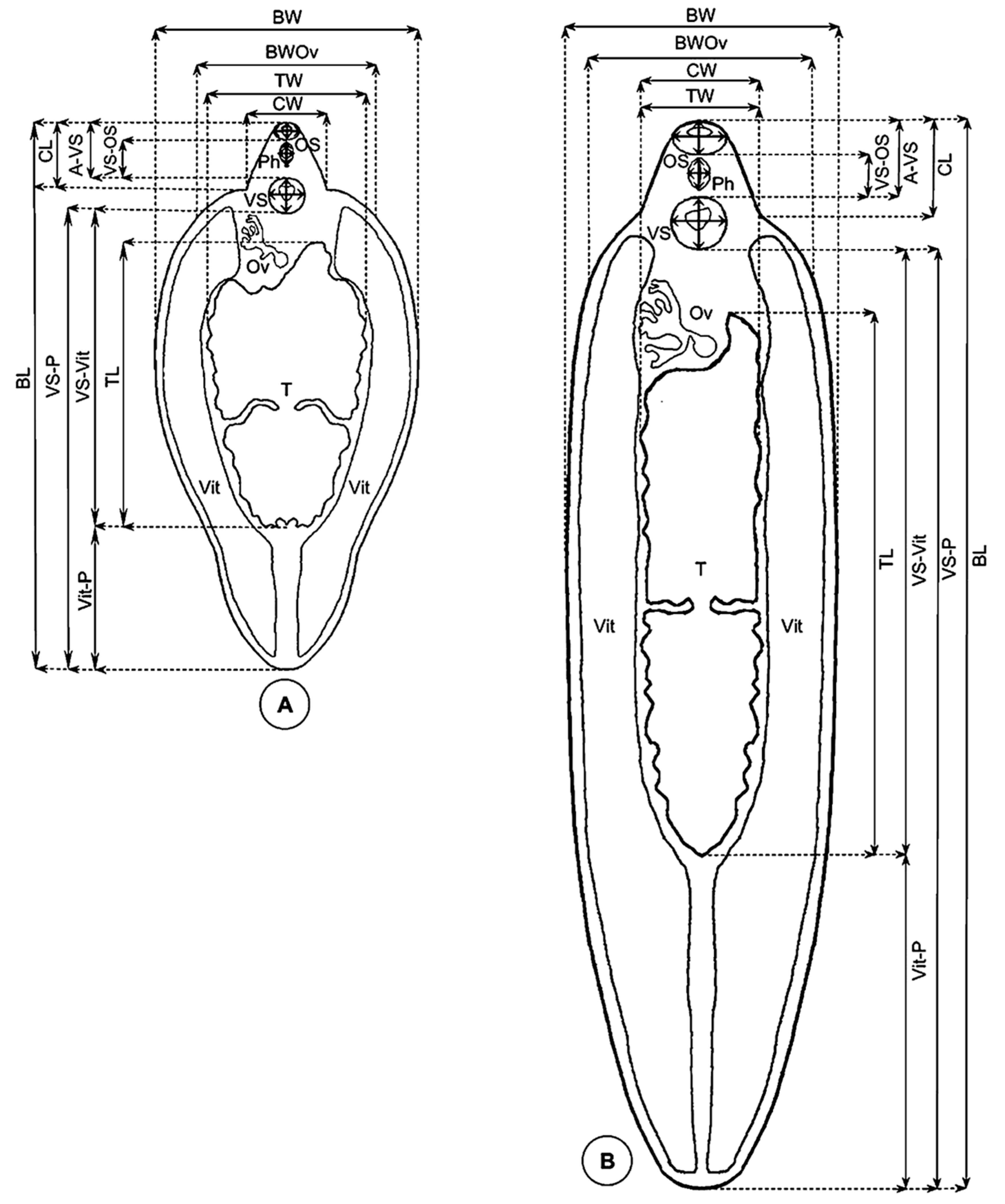
2.2.2. Measurement Techniques

A methodology whose accuracy has been previously verified for Fasciolidae (Figure 2) [49,52,53] was applied for the obtaining of all standardized measurements of adults needed:
  • Lineal biometric characters: body length (BL), maximum body width (BW), body width at ovary level (BWOv), body perimeter (BP), body roundness (BR), cone length (CL), cone width (CW), maximum diameter of oral sucker (OS max), minimum diameter of oral sucker (OS min), maximum diameter of ventral sucker (VS max), minimum diameter of ventral sucker (VS min), distance between the anterior end of the body and the ventral sucker (A-VS), distance between the oral sucker and the ventral sucker (OS-VS), distance between the ventral sucker and the union of the vitelline glands (VSVit), distance between the union of the vitelline glands and the posterior end of the body (Vit-P), distance between the ventral sucker and the posterior end of the body (VS-P), pharynx length (PhL), pharynx width (PhW), testicular length (TL), testicular width (TW), testicular perimeter (TP);
  • Areas: body area (BA), oral sucker area (OSA), ventral sucker area (VSA), pharynx area (PhA), and testicle area (taking both testes together, TA);
  • Ratios: body length over body width (BL/BW), body width at ovary level over cone width (BWOv/CW), oral sucker area over ventral sucker area (OSA/VSA), and body length over the distance between the ventral sucker and the posterior end of the body (BL/VS-P).
For the quantification of the body shape, the body roundness was measured (BR = BP2/4πBA). This measurement estimates how circular an object is, i.e., the expected perimeter of a circular object divided by the perimeter. Accordingly, a circular object has a roundness value of 1.0, whereas irregular objects show larger values [54,55].
A calibrated microscope was used for the measurements and images captured with a digital camera (Nikon Coolpix) were analyzed by CIAS (computer image analysis software) by means of the software Image-Pro Plus (version 5.0 for Windows, Media Cybernetics, Silver Spring, MD, USA).

2.2.3. Numerical Methods

Morphological variation can be quantified by means of geometrical morphometrics [56]. The technique offers a size estimation in a way that different growth axes are integrated into a unique variable, i.e., the “centroid size” [57]. This single size-estimating variable reflects the variation in many directions, depending on the landmarks under study. The shape is defined by the relative positions after the correction for size, position and orientation. In that way, a freely available software allows for a more accurate quantification by conducting complex analyses, including significant biological and epidemiological features [58]. In morphometrics, statistical techniques allow for the testing of the null hypothesis of conspecific populations, i.e., if they are simply the allometric extension of each other provided a common allometric trend is identifiable [56,59,60].
Multivariate analyses were used to explore the morphometric data. With the aim of assessing between-samples morphometric variation, a size-free canonical discriminant analysis was applied on the covariance matrix of log-transformed measurements. This technique consists of removing the effect of within-group ontogenetic variation, regressing each character separately on the within-group first principal component, which is a multivariate estimate of size [61]. The analyses were carried out using BAC v.2 software [60,62,63]. Values were considered statistically significant when p < 0.05. Nonredundant measurements were used (i.e., one is not included in another one): BL, BW, BP, OS max, OS min, vs. max, vs. min, A-VS, VS-Vit, Vit-P, PhL, PhW and TL, where at least one dimension of the most important morphological structures was included. These remaining variables were all significantly correlated with the first principal component (PC1), which contributed 69% to overall variation. Therefore, PC1 could be considered as a general indicator of size [57]. Thus, the factor maps obtained (Figure 2) clearly and appropriately illustrate the differences in size when comparing the populations in question. The influence of within-group allometries was afterwards removed by using variables equivalent to an orthogonal projection of the data onto the first common principal component (i.e., all the common principal components except the first one). The resulting “allometry-free”, or size-free, variables were subsequently submitted to a canonical variate analysis (CVA), and Mahalanobis distances were derived [60,64].
Given that the size variation is consequently based on one variable only—namely the first common principal component—the univariate equivalent of Mahalanobis distance, which is also known as the Pearson’s “Coefficient of Racial Likeliness” [60], was applied for the estimation of its variation. The relationships of altitudinal variation with these distances were further statistically analyzed by nonparametric tests.

2.3. Genotyping Analyses

The molecular methods and techniques were applied to the aforementioned three Ecuadorian groups of liver flukes obtained in sheep and cattle from the zone of San Juan de Chillogallo, as well as to those specimens infecting cattle from the southern zone of Loja.

2.3.1. DNA Markers

The sequence of the complete intergenic nuclear ribosomal DNA (rDNA) region, including the spacers ITS-2 and ITS-1 and the 5.8S gene, and the complete sequences of the two protein-coding genes cox1 and nad1 of the mitochondrial DNA (mtDNA) were selected to characterize the flukes. These molecular markers have already proved to be useful for the genetic characterization of Fasciola species and strains at local and regional levels [26,43] and in worldwide analyses [17], including the assessment of the spreading routes.

2.3.2. DNA Sequencing

For DNA extraction, a small part of the anterior body region of fasciolids was individually processed. The methods therefore used have been previously described [43,65]. Materials were suspended in 400 μL of lysis buffer (10 mM Tris-HCl, pH 8.0, 100 mM EDTA, 100 mM NaCl, 1% sodium dodecyl sulfate SDS) containing 500 μg/mL Proteinase K (Promega, Madison, WI, USA). The digestion was performed for 2 h at 55 °C, including alternate shaking every 15 min. Methods previously outlined were followed concerning the procedure steps [26,43]. The phenol-chloroform extraction and ethanol precipitation method were applied for total DNA isolation. Each pellet was dried and resuspended in 30 μL sterile TE buffer (pH 8.0), and subsequently, this suspension was stored at −20 °C until needed.
Each DNA marker was amplified by PCR in an independent way for each liver fluke individual. Each PCR product was sequenced for a bona-fide haplotype characterization. Forward and reverse primers were designed in the regions flanking the rRNA genes 18S and 28S for the subsequent amplification of the complete ITS-1, 5.8S, ITS-2 region [28,65]. Forward and reverse primers designed in flanking regions allowed for the sequencing of the cox1 and nad1 genes in their complete length [17,26,43].
For the PCR amplification, the Biotools DNA polymerase® (Biotools B&M Labs. S.A., Madrid, Spain) was used in a Verity-96 Well Thermal Cycler (Applied Biosystems, Thermo Fisher Scientific, Waltham, MA, USA). The program comprised one cycle of 2 min at 94 °C, 35 cycles of 1 min at 93 °C, 1 min at 55 °C and 1 min at 72 °C each, preceded by 2 min at 72 °C, and followed by a final cooling at 4 °C, for the ITS rDNA region, and one cycle of 1 min at 94 °C, 40–42 cycles of 1 min at 93 °C, 1 min at 52–55 °C and 2–3 min at 72 °C each, preceded by 5 min at 72 °C and followed by a final cooling at 4 °C, for the cox1 and nad1 mtDNA genes.
For the purification of the PCR product, the Ultra Clean™ PCR Clean-up DNA Purification System (MoBio, Solana Beach, CA, USA) was used following the manufacturer’s protocol and eluted in 50 μL of 10 mM TE buffer (pH 7.6). The final DNA concentration (in μg/mL) and the absorbance at 260/280 nm were determined in an Eppendorf BioPhotometer (Hamburg, Germany). Each molecular marker was sequenced on both strands by the dideoxy chain-termination method performed with the Taq dye-terminator chemistry kit on an Applied Biosystems 3730xl DNA Analyzer (Applied Biosystems, Foster City, CA, USA), by using the PCR primers
Sequence data from this article have been deposited in the GenBank Data Library under Accession Nos. MW867310–MW867323.

2.3.3. Sequence Analyses

The software Sequencher v. 5.4.6 (Gene Codes Co., Ann Arbor, MI, USA) was used to edit and assemble the sequences, and the software CLUSTAL Omega [66] was used to align them by means of default parameters. Corresponding penalties for gaps were included in pairwise and multiple alignments. Total character differences were used to measure divergence of the sequences within and among different ITS-1 and ITS-2, cox1 and nad1 markers. All changes, comprising transitions (ts), transversions (tv) and insertions/deletions (indels), were considered as character states in MEGA v7.0 [67]. By means of the ALTER web server [68], the sequences aligned were collapsed to haplotypes, counting gaps as differences. Closely related sequences were searched by utilizing the BLASTN programme from the National Center for Biotechnology Information website (http://www.ncbi.nlm.nih.gov/BLAST) (accessed 15 December 2020). Comparative sequences were analyzed by comparing with available rDNA and mtDNA sequences of F. gigantica, F. hepatica and Fasciola spp. downloaded from the GenBank and also with the Valencia centre fasciolid haplotype collection.

2.3.4. DNA Haplotype Nomenclature

The terminology to identify the haplotype (H) of the four aforementioned DNA markers follows the previously proposed combined haplotyping (CH) nomenclature [17,69]. According to this nomenclature, ITS-2 haplotypes are defined by numbers, and ITS-1 haplotypes by capital letters. Numbers are also utilized for the nucleotide and protein haplotypes of the mtDNA cox1 and nad1 genes. It is worth mentioning that haplotype codes are only definitive when the sequences are complete, i.e., full length sequences. When dealing with fragments or incomplete sequences, haplotype codes are considered only provisional.

3. Results

3.1. Morphometric Analyses of Sheep Liver Flukes

The morphometric values of South American populations of F. hepatica are noted in Table 1, with standard specimens shown in Figure 3. Values of F. hepatica from Spain and experimentally obtained F. gigantica from Egypt and Vietnam are shown in Table 2 and Table 3.
A general overlap is observed between populations of F. hepatica regardless of the geographical area of origin. The comparison of F. hepatica with F. gigantica also shows an overlap between the two species (including BR, BL/BW and VS-P).
However, it is worth mentioning the maximum and average values of the Ecuadorian fasciolid population, which are much higher than those of the other F. hepatica populations analyzed. The high values of the following parameters linked to size (BA, BL, BW, BWOv, BP, and BR) in the sheep specimens from Ecuador should be highlighted (Table 1) and compared with the same parameters in the F. gigantica populations from Egypt and Vietnam (Table 3).
A scatter plot of the first two principal components (PC) of the size variables shows that there are two differentiated zones along the horizontal axis (CP1) corresponding to the overlapping of the F. hepatica and F. gigantica populations. One zone is made up of geographical areas of Bolivia, Peru and Spain, where only F. hepatica is present. The other zone is made up of Egypt and Vietnam, corresponding to F. gigantica populations.
The resulting factor maps (Figure 4) illustrate global size differences in the populations analyzed. The F. hepatica populations from Peru (nCaj, nMan) and Spain (nSp) show a maximum and minimum size similar to that of the experimental F. hepatica standard population. The Bolivian population (nAlt) shares its maximum size with the other F. hepatica populations but presents a lower minimum size (Figure 4). The flukes from Ecuadorian sheep (nEc) have the biggest size recorded among F. hepatica populations. The experimental F. gigantica specimens from Egypt (expEg) included in the PCA (Figure 4) were 24 weeks old and show a clear separation from the experimental population of F. hepatica from Spain (expSp), which was also 24 weeks old. The factor maps show that, although in experimental populations the fluke size of F. hepatica and F. gigantica does not overlap (only a very little between expSp and expEg), the fluke size of experimental F. gigantica from Egypt (expEg) overlaps with the maximum size values of South-American F. hepatica populations (nCaj, nMan, nAlt). To avoid the effect of age, an experimental 52-week-old population from Vietnam (expVi) has been added to the comparison, showing a clear separation with all the F. hepatica populations excepting the population of Ecuador. To make the separation from F. hepatica even clearer, a natural population of F. gigantica found in slaughtered older sheep from Egypt (nEg) has additionally been included. The results show that the Ecuadorian samples (nEc) constitute the only population with a specimen size intermediate between that of F. hepatica populations (including both natural and experimental) and F. gigantica populations (also including both natural and experimental).
The size-free pattern of variation did not show any consistent relationship with altitudinal differences (Figure 5). Additionally, the presence of sperm in the seminal vesicle was microscopically confirmed in all the specimens found in sheep from Ecuador.

3.2. DNA Sequence Analyses

Two haplotypes of the complete ITS1-5.8S-ITS2 region were detected in the fasciolids infecting sheep and cattle in Ecuador. One haplotype was identical to the previously described F. hepatica haplotype Fh-1A (MG569980), including a 951 bp long intergenic region of 50.79% GC content. The second one includes a homozygous mutation in position 874 of the alignment and was described as Fh-2A (MG569978), with the same length and a 50.68% GC content. A variant of those two haplotypes was found and characterized by including a heterozygous mutation in the same 874 alignment position (Table 4). The most abundant in sheep and cattle in Ecuador proved to be the haplotype Fh-1A. The haplotype Fh-2A was detected only in cattle samples and the heterozygous variant (Fh-1/2A) only in some sheep samples.
ITS-1 proved to have the same sequence of 432 nucleotides and a 51.85% GC content in all specimens studied, corresponding to the haplotype Fh-HA of this spacer. The 5.8S gene was also very conserved in all specimens, with 154 base pairs and 53.25% GC. The 365-bp-long ITS-2 was the only one providing two haplotypes (Fh-H1, 48.49% GC; Fh-H2, 48.22% GC). There was only one single nucleotide polymorphism (SNP) in position 288 of the ITS-2 alignment, namely a “C” in Fh-H1, a “T” in Fh-H2, and “C/T” in the heterozygous variant (Fh1/2). This polymorphic position is not a position that differentiates between F. hepatica and F. gigantica (Table 4). The electropherograms of the rDNA intergenic sequence of all big-sized liver fluke specimens from sheep were thoroughly reviewed, especially in the positions that differentiate between F. hepatica and F. gigantica. No double peaks or ambiguous positions were detected, thus confirming its ascription to F. hepatica.
The mtDNA cox1 gene provided eight different sequences with the same length of 1533 bp and an average of AT content of 62.63%. Their alignment showed 15 variable positions (9 parsimony-informative and 6 singleton). Seven of these cox1 haplotypes are among the 69 reported previously in F. hepatica, corresponding to the haplotypes Fhcox1-16, Fhcox1-23, Fhcox1-53, Fhcox1-54, Fhcox1-55, Fhcox1-56, and Fhcox1-57. The eighth haplotype is a new one, found only in sheep, for which the code Fhcox1-70 has been assigned. Sheep and cattle samples only shared the haplotype Fhcox1-16. The COX1 protein was 511 aa long, with start/stop codons of ATG/TAG, and provided two haplotypes (Table 5). The difference between the two haplotypes was restricted to only one amino acid change in position 499 of the protein alignment (Table 5).
A comparative cox1 sequence analysis was performed with other complete length F. hepatica haplotypes, including Fhcox1-5, Fhcox1-16 and Fhcox1-42 from cattle and horses from Uruguay; haplotype M93388 from Salt Lake City, Utah, USA; and haplotype AF216697 corresponding to the Geelong strain from Australia. Their alignment was 1533 bp long and showed 30 nucleotide and 5 amino acid variable positions (Table 5).
The mtDNA nad1 gene provided six different sequences with the same length of 903 bp and an average AT content of 65.30%. Their alignment showed 6 variable positions (4 p-info and 2 singleton). All these six nad1 haplotypes are among the 51 nad1 haplotypes reported previously in F. hepatica and corresponding to the haplotypes Fhnad1-2, Fhnad1-6, Fhnad1-14, Fhnad1-23, Fhnad1-42 and Fhnad1-43. Sheep and cattle samples only shared the haplotype Fhnad1-14. The NAD1 protein showed only one 300-aa-long haplotype with start/stop codons of GTG/TAG in all specimens analyzed (Table 6).
Comparative nad1 alignment analyses were performed with other complete length F. hepatica haplotypes, including Fhnad1-2, and Fhnad1-12 and Fhnad1-14 reported from cattle and horses in Uruguay; haplotype M93388 from Salt Lake City, Utah, USA; and haplotype AF216697 corresponding to the Geelong strain from Australia. Their alignment was 903 bp long and contained 12 nucleotide and 1 amino acid variable positions that corresponded to the intraspecific variability reported for F. hepatica (Table 6).
Similarly, as done with the rDNA sequences, the electropherograms of the mtDNA cox1 and nad1 of all big-sized liver fluke specimens from sheep were carefully checked, especially in the positions that differentiate between F. hepatica and F. gigantica. Neither introgression fragments nor heteroplasmic positions were found, thus confirming its ascription to F. hepatica.
The distribution of the haplotypes of each of the aforementioned rDNA and mtDNA markers analyzed according to localities surveyed in Ecuador, and their previous findings in other countries are noted in Table 7.

4. Discussion

4.1. Phenotypic Analysis

4.1.1. Fasciola hepatica from Highland and Lowland Sheep

The geographical origins of the liver flukes from naturally infected sheep analyzed in this study include highlands and lowlands. Quantitative morphological variation informs about both genetic variation and external influences [70]. High-altitude environment factors exert an influence on mammals, so those born and living at high altitude show morphological and physiological characteristics different from those of mammals inhabiting low altitudes [71,72].
Although the F. hepatica population from the Bolivian Altiplano shows a wider size range, reaching uterus gravidity at a lower size (Figure 5) [73], this Altiplanic pattern differing from the valley pattern of the Peruvian Cajamarca and Mantaro [74], the results of the present study show that there is no apparent relationship between the shape of fasciolid adults with respect to the difference in altitude or geographical location.
Using an allometric model, the definitive host species proved to decisively influence the size of F. hepatica adults, but these influences do not persist in a subsequently infected rodent-definitive host model [46]. In natural populations, only slight differences were found in allometric models (BL, BW, P vs. BA or BL) between F. hepatica adults from highland and low-land populations of Bolivian and Spanish sheep [75]. Nevertheless, liver fluke populations from Altiplanic sheep and cattle, which have proved to be efficient reservoirs in the very high-altitude areas [76], proved to have a smaller uterus area than European lowland populations in the same host species [73]. Oxygen is needed for the production of eggs in F. hepatica [77,78,79]. Thus, hypoxia from which vertebrate hosts living in high altitude areas suffer may lead to a reduced egg production in trematodes. Moreover, although trematode uteri are traditionally not considered to be storage organs, the size of the uterus is primarily adapted for the egg to reach maturity along the uterine journey. In the Northern Bolivian Altiplano, climatic conditions, freshwater characteristics and ecological requirements of lymnaeids allow for fascioliasis transmission throughout the entire year [23,80]. In the Altiplanic pattern, egg storage is not essential, and this explains why smaller sizes are detected, in contrast to the valley pattern where fascioliasis presents a typical seasonal transmission just as in the northern Hemisphere.
Allometry-free shape appears as a more stable trait than size in fasciolid species [74]. This is in agreement with observations made on insect vectors of Trypanosoma cruzi (Triatominae), in which allometry-free shape may require important external changes to be significantly modified [58].

4.1.2. Liver Flukes from Ecuadorian Sheep Compared to F. hepatica and F. gigantica

The development of F. hepatica and F. gigantica in the definitive host [81,82] follows growth curves according to a logistic model, in which the morphometric development of the adult stage is “damped” and cannot exceed certain characteristic maximums of Ym. This logistic model of the body growth and development is characterized by two phases [49,82]: (i) the “exponential phase” of the logistic growth corresponds to the body development during the migration in the abdominal cavity and liver parenchyma and subsequent development and sexual maturation in the biliary duct system up to the onset of egg production; (ii) the “saturated phase“ of logistic growth starts from this moment and leads to a gradually stationary growth after oviposition.
In areas of countries of Africa and Asia where the two species of Fasciola overlap thanks to the coexistence of the respective specific lymnaeid species, the comparative multivariant analyses show flukes of a size intermediate between F. hepatica and F. gigantica, as demonstrated in Egypt [53], Iran [83], Pakistan [84] and Bangladesh [85].
The high maximum and mean values of all size-linked parameters in the Ecuadorian liver flukes from sheep are unexpected and should undoubtedly underlie, and justify, previous classifications of fasciolids in this country as F. gigantica, whether unpublished or published [41]. Indeed, size has traditionally been the main and even only characteristic considered in most of the past fasciolid species classifications in livestock, which led to several misclassifications later verified [26].
In cattle, a study showed that BL/BW, BR and VS-P are useful tools for differentiating between “genetically pure” F. hepatica from southern Europe and “genetically pure” F. gigantica from Burkina Faso [52]. In sheep, the results of the present study show an overlap in each of these three markers between F. hepatica and F. gigantica, although mean values are clearly different (compare Table 1, Table 2 and Table 3). Regarding the flukes from Ecuadorian sheep, VS-P appears to be a parameter showing evident intermediate characteristics which supports the conclusion of the phenotypic results on fluke size.

4.2. Molecular Analysis

4.2.1. DNA Sequence Characterization of Fasciolids from Ecuador

Big-sized fasciolid flukes found in sheep from Ecuador molecularly prove to belong to F. hepatica and all haplotypes of the four DNA markers used fall within the intraspecific variability reported for this species in European and American countries [17]. The same conclusion is reached in the haplotyping of the liver flukes collected in cattle from the three selected localities surveyed, one close to Quito and the other two in the southern zone neighboring the Peru border.
ITS-1 and ITS-2 are evolutionarily conserved markers very useful for species differentiation. Their consequent low variability at the local level does therefore provide little additional information about population genetics because it has been observed in invertebrates in general unless microsatellites and/or minisatellites varying in repeat numbers are included in their sequences [86]. In those regions of the world where only F. hepatica is present, and consequently there is no possibility for hybridization with F. gigantica, only FhITS1-HA is known [17], as observed in Ecuador in flukes from both sheep and cattle. Regarding ITS-2, the finding of the haplotype FhITS2-H1 in both sheep and cattle and in all localities surveyed agrees with the wide distribution of this haplotype. Similarly, it may be argued with the generally more geographically scarcely distributed FhITS2-H2. The detection of heterozygotic characteristics in the only variable position differentiating these two ITS-2 haplotypes (Table 4) speaks about a very recent mating or “hybridization” of parental flukes harboring one and the other haplotypes. This, in its turn, suggests usual mixing of populations coming from different parts of Ecuador in the area of San Juan for the daily supply needed for a highly populated city as Quito.
The faster evolutionary rate of the mtDNA genome underlies the wider haplotype variability of the cox1 and nad1 genes [86]. In Fasciola, the worldwide geographical spread of different haplotypes of these mtDNA genes is linked to man-made livestock movements occurred along the post-domestication period during the last 12,000–10,000 years [17], and in the Americas after European livestock introduction by the Spanish “conquistadores” and subsequent intracontinental spread during only the last 500 years [43]. The scattered distribution of the mutations throughout the whole length in these two long genes is worth mentioning (Table 5 and Table 6). This indicates that valuable information is lost when only using short fragments of these genes in analyses of fasciolid flukes. The assessment of past migrating routes may be misinterpreted or the correct overview may not be reached when only using short fragments of mtDNA genes, as it may happen with liver flukes in Ecuador [87]. Indeed, the difficulties in correctly assessing the spreading routes of fasciolid flukes become evident when considering the overall mixing of fluke populations as a consequence of livestock movements driven by humans, inside countries and between countries as in South America [43], but also between continents [17,88]. Additionally, this means that for a correct assessment of the usefulness of DNA markers for the differentiation between F. hepatica and F. gigantica, appropriate fasciolid populations should be selected; i.e., comparing fasciolid specimens from Ecuador with fasciolids from countries of Southeastern and Far East Asia [89], where the two fasciolid species overlap and hybrid fasciolids are widely distributed, does not furnish the adequate baseline for such a purpose.
The detection of eight haplotypes in cox1 and six haplotypes in nad1 indicates high genetic variability of F. hepatica in Ecuador. However, in cox1, only one nucleotide codon gives rise to an amino acid change (Table 5), whereas in nad1, all mutations prove to be silent (Table 6). This high haplotype mtDNA variability agrees with the heterogeneous origin of the livestock nowadays present in Ecuador. The present mix of F. hepatica haplotypes in Ecuador is the result of successive livestock introductions from abroad during the following two periods:
  • The colonial period: Three introduction routes were involved. (i) The maritime route through the Pacific coast was the initial entry for livestock from Central America, at the beginning of the colonization of South America. (ii) The southern terrestrial route from Peru started early thereafter, as soon as the European colonizers learned about the interest of Quito for the old Incas. (iii) The northern route through the border with present day Colombia was implemented later, with an exchange of silver transport from the Bolivian Potosi mine up to the Venezuelan haven of Cartagena. The latter silver transport route might have been the most important concerning the probability of introduction of F. hepatica haplotypes from both southern and northern countries into Ecuador.
  • The post-colonial period: The original European livestock evidenced problems of adaptation to the Ecuadorian environment both throughout the tropical lowlands along the coast as well as in the Andean highlands. During the XIXth and XXth centuries, Ecuador became a great livestock importer, mainly to improve the local cattle breeds. Importations of many thousands of animals were performed from the XIXth century, but mainly during the first two thirds of the XXth century. These importations were mainly from other countries of South America, but sporadically also from Europe and Asia, as well as North and Central America [90].
These livestock introductions from abroad underlie the wide diversity of haplotypes detected in Ecuadorian sheep and cattle, given the capacity of metacercariae to infect different livestock species [47,91]. Moreover, fasciolid infection does not induce premunition, so re-infections lead to fluke accumulation within the same host individual [92,93]. This means that livestock importation by terrestrial herd movements from one country to another and through other countries located in between may easily become the source of the introduction of more than one haplotype, and that introduced haplotypes may finally infect other livestock species in situ once in the new country. The detection of five cox1 haplotypes and three nad1 haplotypes suggests the mix of different lineages in the big-sized F. hepatica specimens in sheep (Table 7).
Haplotypes such as Fhcox1-16 and Fhcox1-23—but also Fhcox1-53 and Fhcox1-55, as well as Fhnad1-2, Fhnad1-14 and Fhnad1-23, all of them shared by Ecuador with many other countries of South America or even Mexico (Table 7)—suggest introductions by livestock exchange inside the Americas. The haplotypes Fhnad1-2 and Fhnad1-6 are shared by Ecuador with Spain and may be remains of introductions from Europe during the colonial period. The haplotypes Fhcox1-54, Fhcox1-56, Fhcox1-57, and the new Fhcox1-70, as well as Fhnad1-42 and Fhnad1-43, have so far never been found in other countries. Research today underway shall clarify whether they are also present in other countries.
The pronouncedly higher number of different haplotypes in San Juan when compared to the haplotype number in southern Peru (Table 7) also indicates a mixing of populations from different parts of Ecuador in the area of San Juan for the regular demand of the big city of Quito. Only Fhcox1-16 and Fhnad1-14 have been found in both San Juan and the southern Ecuador border, which suggests that livestock is nowadays not transported to Quito from so far inside the country.

4.2.2. American Big-Sized F. hepatica Introduction Routes

The F. hepatica worms here described from sheep in Ecuador share the characteristic of an abnormally big size with several liver flukes reported from another American country, namely USA.
In North America, zebu cattle and buffaloes were imported from India and probably also from Africa along the colonization period starting 500 years ago [94,95,96]. Importations from India into the Gulf coast occurred in 1875 and 1906 and may explain the presence of three different types of Fasciola in the USA [36]: in several states, they are identical to F. hepatica, those from Texas and Florida approach F. gigantica, and those from the Gulf Coast area show intermediate form characteristics. Fasciola halli described in Texas and Louisiana and F. californica in California [97] were probably intermediate forms resulting from introgressions of imported F. gigantica into USA-native F. hepatica [17]. Although F. gigantica was unable to adapt due to Radix species unavailability in USA, cross-breeding could have occurred within the livers of the initially imported animals (at that time, directly released into the field without prior quarantine, as in many developing countries today), enabling the foreign F. gigantica to encounter native F. hepatica, with subsequent DNA introgression. Hybrids unable to develop in USA-native snails subsequently disappeared, but others retained viability due to their capacity to use US-native lymnaeids [17].
In Ecuador, three questions are posed to understand such F. gigantica-sized flukes in this country: from where, when and how were F. gigantica or F. gigantica-like flukes introduced to subsequently give rise to such a local big sized strain.
The first sheep (criollo breeds) were introduced by the “Spanish conquistadores” already from the beginning of the colonization period. By the XVIth century, sheep were already widely spread in the Pichincha province [98]. After the long colonization period, sheep importations were also made but at a lesser level than with cattle because of the lower interest in sheep breed improving [99]. The origins of these sheep importations were initially Europe, and later Australia, New Zealand and USA in the 1964–1987 period [100]. Consequently, only those from southern States of the USA could have been risky if infected by the big-sized (F. gigantica-like) liver flukes recorded in the USA [36] and which could be adapted to be transmitted by North American non-Radix lymnaeids, given the absence of F. gigantica in Europe and Oceania
The first cattle arrived in Ecuador on boats from Nicaragua in 1537. Most of the numerous livestock importations during the XIX and XX centuries concerned cattle from different continents except Africa [90]. Most cattle importations were from other South American countries and have been implemented even very recently, as the importation of 12,000 bovines from Paraguay in the 2015–2017 period [101]. Of special interest was the importation of cattle of the Sahiwal breed from Pakistan through the Ecuadorian coast in 1974 [90]. This breed adapted very well to the Ecuadorian tropics. Fasciola gigantica is widely spread throughout most of Pakistan [21], where it has been seen to reach big size in buffaloes [84]. If these imported Pakistani animals were infected by F. gigantica—which is highly probable considering its very high prevalence in this Asian country [21]—their release in the environment of Ecuador might have offered their additional infection by local F. hepatica and subsequent hybridization giving rise to intermediate forms. Those hybrid flukes which succeeded to use original Ecuadorian lymnaeids for their transmission might have been the origin of the big sized liver fluke lineage having reached the present in Ecuador.
Worth mentioning also is the importation of Brown Swiss cattle from Ohio and Minnesota, USA, to Santo Domingo de los Tsáchilas in 1986. Although F. gigantica-like flukes or intermediate forms were not present in these northern states of the USA [36], the 132 cows imported were transported with trucks along 3000 km up to Miami along a three-day journey, allowing the animals to go out and freely feed locally in the different states every 12 h. Finally, the cows were transported by a direct flight from Miami to Ecuador [90]. Hence, the infection of these cows by F. gigantica-like flukes or intermediate forms could have occurred once in the southern states of the USA [36]. In that way, large-sized liver flukes could have been introduced in the area where reported big-sized F. hepatica infecting sheep have been found in Ecuador. Indeed, the old route from Quito to the Pacific Ocean covered the westward transect Quito–San Juan–Chiriboga–Santo Domingo de los Tsáchilas from 1888. All transport from the different western coastal localities to Quito was made through several routes which converged in Santo Domingo de los Tsáchilas [102].

4.3. Radix Absence and the Evolutionary Snail Vector Filter

Although the aforementioned date of 1974 as the most probable for a F. gigantica introduction with cattle from Pakistan [90] may be considered very recent, the time elapsed is more than sufficient to explain the absence of heterozygotic positions with F. gigantica in the two rDNA spacers ITS-2 and ITS-1 and the lack of detection of introgressed sequences or heteroplasmic positions in the complete mtDNA sequences of the cox1 and nad1 genes in the large-sized F. hepatica flukes from Ecuadorian sheep. In the case of the 1986 cow importation from USA [90], if liver fluke intermediate forms were introduced into Ecuador, these would have previously been originated in southern USA [36] by an adaptation to North American lymnaeid species of the Galba/Fossaria group such as L. humilis or L. bulimoides [103] and subsequently successfully established in Ecuador. Indeed, the concerted evolution mechanisms acting on the rDNA operon may reach sequence homogenization quite rapidly from the timely perspective [104]. Similarly in the case of mtDNA, heteroplasmy longevity until return to homoplasmy has also been observed to occur quite rapidly, from just a few generations in cattle up to 500 generations in the shorter life-span insects [105].
In studies on the epigenetic phenomenon of nucleolar dominance in hybrid organisms, it has been shown that the rRNA genes of one parental species may become transcriptionally dominant over the rRNA genes of the other parental species [104]. In fasciolid liver flukes, the specificity regarding different lymnaeid vector species should undoubtedly play a crucial role in such a phenomenon. The availability of given lymnaeid species and the asexual clonal intramolluscan larval development should act as a natural selection filter. In that way, the capacity for sexual crossbreeding of these hermaphroditic flukes and maintain a hybrid characteristic lineage becomes greatly reduced by the increasingly low probability of encountering other needed hybrids inside the liver of the same host individual, because of non-local-snail adapted hybrid elimination by the lymnaeid vector filter.
Such an evolutionary filter at vector level should additionally accelerate the elimination of hybrid genetic characteristics. In the Americas, F. gigantica lymnaeid vector species of the Old-World Radix group are absent. Only in the USA has a Radix species been found, namely introduced Radix auricularia, although it is mostly confined to artificial water collections in man-made environments and molecularly similar to the same European species [103]. In South America, a radicine lymnaeid has never been found. This absence of Radix lymnaeids explain the lack of capacity of F. gigantica to colonize the New World, even despite human activities having involuntarily offered occasions for such an introduction. Opposite to this, the anthropogenic introduction of F. hepatica was successful because of its subsequent adaptation to American native Galba/Fossaria lymnaeid species, additionally to the introduction of the European Galba truncatula [17].
In Ecuador, only five lymnaeid species have been reported, namely the more geographically restricted Pseudosuccinea columella [106,107] and four more widely spread species of the Galba/Fossaria lymnaeid group including Lymnaea cousini [108], L. schirazensis [109], L. cubensis [110] and L. neotropica [42]. All these species are F. hepatica vectors, except L. schirazensis which is a nonvector species [109,111]. Thus, F. gigantica from Pakistan could not found an appropriate scenario for a successful introduction.
The large size of the flukes found in the USA and Ecuador poses the question about why these fasciolids do not return to the normal F. hepatica size and keep the large phenotype at least in the mid-term of several decades. In fasciolids, adult stage phenotype has been observed to be linked to rDNA but not always to mtDNA [17]. Unequal crossing over, high-frequency gene conversion, and large deletion underlying recombination mechanisms driving the rapid concerted evolution of the rDNA tandem repeats are known to efficiently homogenize the 45S rDNA single precursor transcribing the rDNA genes (18S, 5.8S and 28S) and spacers (ITS-1, ITS-2 and IGS) sequences by, in essence, counteracting mutation effects [112]. However, concerted evolution does not act on two aspects with substantial ecological and evolutionary consequences such as the operon copy number and the length of the intergenic spacer IGS [104]. Indeed, unequal crossover plays an important role in the generation of such two heterogeneities, and rRNA transcription is directly related to the growth and development of the organisms. A higher number of rRNA units provides a higher transcription capacity enabling for increasing growth [104]. Hybridization of F. hepatica and F. gigantica may give rise to a number of rRNA unit copies higher than usual in genetically pure F. hepatica, thus allowing for bigger size development. Natural selection, including lymnaeid vector species specificity and local availability as the main effectors, may further contribute to maintain balanced rDNA dosage across unlinked rDNA arrays, as seen in other organisms [113]. This situation of a timely stable evolutionary bottleneck at the snail vector level always acting in the same sense is the opposite to what happens in areas of Africa and Asia, where the local coexistence of Galba/Fossaria vector species with Radix vector species in the same transmission focus offers daily alternating development filtering towards F. hepatica and F. gigantica to an evolving lineage, or zonal overlapping presence of Galba/Fossaria and Radix vector species offering similar alternating filtering possibilities but at seasonal scale (as e.g., in altitudinal transhumance) [17]. In the ten-chromosome-pair karyotype of F. hepatica, rDNA is located in the short arms of the fifth homologous pair [114].

5. Conclusions

Two important public health aspects may be concluded from the results obtained. First, the more pathogenic F. gigantica does not appear to be able to colonize the New World. Even if inadvertently introduced with imported livestock from Africa or Asia, the absence of Radix snail species did not allow for normal transmission, and the potential hybridization with local American F. hepatica would progressively lead the consequent lineage to a genetic uniformization towards F. hepatica in a relatively short period. This does not mean, however, that it should be taken into account that pathogenicity is also linked to fluke size and that the intermediate hybrid forms are pronouncedly bigger than normal F. hepatica.
Second, it is evident that transborder liver fluke introductions occur due to the livestock importations from other countries. The risk of transborder spread of the resistance to triclabendazole should be consequently considered. Livestock treatments should be therefore applied at the level of the exporting country and appropriate quarantine and specific diagnostics applied in the importation country immediately after livestock arrival.

Author Contributions

Conceptualization, S.M.-C.; methodology, M.D.B., M.A.V., A.D.E.-E., I.P.-C., P.A. and S.M.-C.; software, M.D.B. and M.A.V.; validation, M.D.B., M.A.V. and S.M.-C.; formal analysis, M.D.B., M.A.V. and S.M.-C.; investigation, M.D.B., M.A.V., G.A.T., M.F., A.F.V., R.G., A.D.E.-E., I.P.-C., P.A. and S.M.-C.; resources, G.A.T., M.F., A.F.V. and R.G.; writing—original draft preparation, S.M.-C.; writing—review and editing, M.D.B., M.A.V., G.A.T., A.F.V. and S.M.-C.; visualization, M.D.B., M.A.V. and S.M.-C.; supervision, S.M.-C.; project administration, M.D.B. and S.M.-C.; funding acquisition, M.D.B., M.A.V. and S.M.-C. All authors have read and agreed to the published version of the manuscript.

Funding

This research was funded by Project No. RD16/0027/0023, Red de Investigación de Centros de Enfermedades Tropicales—RICET, of the National Program I + D + I 2008–2011 and Fondos FEDER, ISCIII—Subdirección General de Redes y Centros de Investigación Cooperativa, Ministry of Health and Consumption, Madrid, Spain; by Project No. 2016/099 of the PROMETEO Program, Programa of Ayudas para Grupos de Investigación de Excelencia, Generalitat Valenciana, Valencia, Spain; and by Project No. 2017/01 of the V Convocatoria de Proyectos de Cooperación al Desarrollo de la Universidad de Valencia de 2016, Valencia, Spain. A.D.E. was supported by a Fellowship of the Programa de Ayudas de la Generalitat Valenciana y Fondo Social Europeo, 2019 (No. ACIF/2019/182).

Institutional Review Board Statement

All experimental research was performed with the approval of the Evaluation of Projects concerning Animal Research at University of Valencia (Organo Habilitado para la Evaluación de Proyectos de Experimentación Animal de la Universidad de Valencia) (A1263 915389140), strictly following the institution’s guidelines based on Directive 2010/63/EU. Permission for animal research was additionally obtained from the Servicio de Sanidad y Bienestar Animal, Dirección General de Producción Agraria y Ganadería, Consellería de Presidencia y Agricultura, Pesca, Alimentación y Agua, Generalitat Valenciana, Valencia, Spain (No. 2015/VSC/PEA/00001 tipo 2). Animal ethics guidelines regarding animal care were strictly adhered.

Data Availability Statement

Datasets generated for this study are available on request to the corresponding author.

Acknowledgments

Studies of this article have been performed within the framework of the Worldwide Initiative of WHO against Human Fascioliasis (WHO Headquarters, Geneva, Switzerland). The Universidad de las Fuerzas Armadas de Ecuador supported and funded the study in the Loja province and subsequent travel, stay and material transport to the WHO Collaborating Centre on Fascioliasis and Its Snail Vectors at the University of Valencia, Spain. The Agencia de Regulación y Control Fito y Zoosanitario (Agrocalidad), Loja, is acknowledged for the valuable information provided about fascioliasis in the province of Loja. Santiago Ulloa, Emil León, Darwin Pacha, Paul Carpio and Freddy Cueva furnished helpful data on condemned livers at slaughterhouses. Edison Medina is greatly acknowledged for his field work assistance. Technical support for DNA sequencing provided by the Servicio Central de Secuenciación para la Investigación Experimental (SCSIE) of the Universidad de Valencia (A. Martínez).

Conflicts of Interest

The authors declare no conflict of interest.

References

  1. World Health Organization. Sustaining the Drive to Overcome the Global Impact of Neglected Tropical Diseases; Department of Control of Neglected Tropical Diseases, World Health Organization: Geneva, Switzerland, 2013; pp. 1–138. [Google Scholar]
  2. World Health Organization. Ending the Neglect to Attain the Sustainable Development Goals. A Road Map for Neglected Tropical Diseases 2021–2030; World Health Organization: Geneva, Switzerland, 2020; pp. 1–47. Available online: https://www.who.int/neglected_diseases/Ending-the-neglect-to-attain-the-SDGs--NTD-Roadmap.pdf (accessed on 4 April 2021).
  3. Chen, M.G.; Mott, K.E. Progress in assessment of morbidity due to Fasciola hepatica infection: A review of recent literature. Trop. Dis. Bull. 1990, 87, R1–R38. [Google Scholar]
  4. Mas-Coma, S.; Agramunt, V.H.; Valero, M.A. Neurological and ocular fascioliasis in humans. Adv. Parasitol. 2014, 84, 27–149. [Google Scholar] [PubMed]
  5. Gonzalez-Miguel, J.; Valero, M.A.; Reguera-Gomez, M.; Mas-Bargues, C.; Bargues, M.D.; Simon-Martin, F.; Mas-Coma, S. Numerous Fasciola plasminogen-binding proteins may underlie blood-brain barrier leakage and explain neurological disorder complexity and heterogeneity in the acute and chronic phases of human fascioliasis. Parasitology 2019, 146, 284–298. [Google Scholar] [CrossRef] [Green Version]
  6. Mas-Coma, S.; Bargues, M.D.; Valero, M.A. Diagnosis of human fascioliasis by stool and blood techniques: Update for the present global scenario. Parasitology 2014, 141, 1918–1946. [Google Scholar] [CrossRef]
  7. De, N.V.; Le, T.H.; Agramunt, V.H.; Mas-Coma, S. Early postnatal and preschool age infection by Fasciola spp.: Report of five cases from Vietnam and worldwide review. Am. J. Trop. Med. Hyg. 2020, 103, 1578–1589. [Google Scholar] [CrossRef] [PubMed]
  8. Valero, M.A.; Santana, M.; Morales, M.; Hernandez, J.L.; Mas-Coma, S. Risk of gallstone disease in advanced chronic phase of fascioliasis: An experimental study in a rat model. J. Infect. Dis. 2003, 188, 787–793. [Google Scholar] [CrossRef] [Green Version]
  9. Valero, M.A.; Navarro, M.; Garcia-Bodelon, M.A.; Marcilla, A.; Morales, M.; Garcia, J.E.; Hernandez, J.L.; Mas-Coma, S. High risk of bacterobilia in advanced experimental chronic fasciolosis. Acta Trop. 2006, 100, 17–23. [Google Scholar] [CrossRef] [PubMed]
  10. Valero, M.A.; Girones, N.; Garcia-Bodelon, M.A.; Periago, M.V.; Chico-Calero, I.; Khoubbane, M.; Fresno, M.; Mas-Coma, S. Anaemia in advanced chronic fasciolosis. Acta Trop. 2008, 108, 35–43. [Google Scholar] [CrossRef]
  11. Rondelaud, D.; Dreyfuss, G.; Vignoles, P. Clinical and biological abnormalities in patients after fasciolosis treatment. Med. Mal. Infect. 2006, 36, 466–468. [Google Scholar] [CrossRef] [PubMed]
  12. Esteban, J.G.; Flores, A.; Angles, R.; Strauss, W.; Aguirre, C.; Mas-Coma, S. A population-based coprological study of human fascioliasis in a hyperendemic area of the Bolivian Altiplano. Trop. Med. Int. Health 1997, 2, 695–699. [Google Scholar] [CrossRef]
  13. Esteban, J.G.; Gonzalez, C.; Bargues, M.D.; Angles, R.; Sanchez, C.; Naquira, C.; Mas-Coma, S. High fascioliasis infection in children linked to a man-made irrigation zone in Peru. Trop. Med. Int. Health 2002, 7, 339–348. [Google Scholar] [CrossRef] [PubMed] [Green Version]
  14. Esteban, J.G.; Gonzalez, C.; Curtale, F.; Muñoz-Antoli, C.; Valero, M.A.; Bargues, M.D.; El Sayed, M.; El Wakeel, A.; Abdel-Wahab, Y.; Montresor, A.; et al. Hyperendemic fascioliasis associated with schistosomiasis in villages in the Nile Delta of Egypt. Am. J. Trop. Med. Hyg. 2003, 69, 429–437. [Google Scholar] [CrossRef] [PubMed]
  15. Gonzalez, L.C.; Esteban, J.G.; Bargues, M.D.; Valero, M.A.; Ortiz, P.; Naquira, C.; Mas-Coma, S. Hyperendemic human fascioliasis in Andean valleys: An altitudinal transect analysis in children of Cajamarca province, Peru. Acta Trop. 2011, 120, 119–129. [Google Scholar] [CrossRef] [PubMed]
  16. Mas-Coma, S.; Bargues, M.D.; Valero, M.A. Human fascioliasis infection sources, their diversity, incidence factors, analytical methods and prevention measures. Parasitology 2018, 145, 1665–1699. [Google Scholar] [CrossRef] [PubMed] [Green Version]
  17. Mas-Coma, S.; Valero, M.A.; Bargues, M.D. Fasciola, lymnaeids and human fascioliasis, with a global overview on disease transmission, epidemiology, evolutionary genetics, molecular epidemiology and control. Adv. Parasitol. 2009, 69, 41–146. [Google Scholar] [PubMed]
  18. Ollerenshaw, C.B.; Rowlands, W.T. A method of forecasting the incidence of fascioliasis in Anglesey. Vet. Rec. 1959, 71, 591–598. [Google Scholar]
  19. Armour, J. The epidemiology and control of bovine fascioliasis. Vet. Rec. 1975, 96, 198–201. [Google Scholar] [CrossRef]
  20. Dinnik, J.A.; Dinnik, N.N. Effects of seasonal variations of temperature on development of Fasciola gigantica in the snail host in the Kenyan highlands. Bull. Epizoot. Dis. Afr. 1963, 11, 197–207. [Google Scholar]
  21. Afshan, K.; Fortes-Lima, C.A.; Artigas, P.; Valero, M.A.; Qayyum, M.; Mas-Coma, S. Impact of climate change and man-made irrigation systems on the transmission risk, long-term trend and seasonality of human and animal fascioliasis in Pakistan. Geospat. Health 2014, 8, 317–334. [Google Scholar] [CrossRef]
  22. Schillhorn Van Veen, T.W. Dynamics of Lymnaea natalensis population in the Zaria area (Nigeria) and the relation to Fasciola gigantica infections. Acta Trop. 1980, 37, 183–194. [Google Scholar]
  23. Fuentes, M.V.; Valero, M.A.; Bargues, M.D.; Esteban, J.G.; Angles, R.; Mas-Coma, S. Analysis of climatic data and forecast indices for human fascioliasis at very high altitude. Ann. Trop. Med. Parasitol. 1999, 93, 835–850. [Google Scholar] [CrossRef] [PubMed]
  24. Mas-Coma, S.; Valero, M.A.; Bargues, M.D. Climate change effects on trematodiases, with emphasis on zoonotic fascioliasis and schistosomiasis. Vet. Parasitol. 2009, 163, 264–280. [Google Scholar] [CrossRef]
  25. Bargues, M.D.; Artigas, P.; Angles, R.; Osca, D.; Duran, P.; Buchon, P.; Gonzalez-Pomar, R.K.; Pinto-Mendieta, J.; Mas-Coma, S. Genetic uniformity, geographical spread and anthropogenic habitat modifications of lymnaeid vectors found in a One Health initiative in the highest human fascioliasis hyperendemic of the Bolivian Altiplano. Parasit Vectors 2020, 13, 171. [Google Scholar] [CrossRef] [PubMed]
  26. Chougar, L.; Mas-Coma, S.; Artigas, P.; Harhoura, K.; Aissi, M.; Agramunt, V.H.; Bargues, M.D. Genetically "pure" Fasciola gigantica discovered in Algeria: DNA multimarker characterization, trans-Saharan introduction from a Sahel origin and spreading risk into northwestern Maghreb countries. Transb. Emerg. Dis. 2020, 67, 2190–2205. [Google Scholar] [CrossRef]
  27. Stift, M.; Michel, E.; Sitnikova, T.Y.; Mamonova, E.Y.; Sherbakov, D.Y. Palaearctic gastropod gains a foothold in the dominion of endemics: Range expansion and morphological change of Lymnaea (Radix) auricularia in Lake Baikal. Hydrobiologia 2004, 513, 101–108. [Google Scholar] [CrossRef]
  28. Valero, M.A.; Bargues, M.D.; Khoubbane, M.; Artigas, P.; Quesada, C.; Berinde, L.; Ubeira, F.M.; Mezo, M.; Hernandez, J.L.; Agramunt, V.H.; et al. Higher physiopathogenicity by Fasciola gigantica than by the genetically close F. hepatica: Experimental long-term follow-up of biochemical markers. Trans. Roy. Soc. Trop. Med. Hyg. 2016, 110, 55–66. [Google Scholar] [PubMed]
  29. Stemmermann, G.N. Human infestation with Fasciola gigantica. Am. J. Pathol. 1953, 29, 731–759. [Google Scholar]
  30. Janssens, P.G.; Fain, A.; Limbos, P.; De Muynck, A.; Biemans, R.; Van Meirvenne, N.; De Mulder, P. Trois cas de distomatose hépatique à Fasciola gigantica contractés en Afrique centrale. Ann. Soc. Belg. Med. Trop. 1968, 48, 637–650. [Google Scholar]
  31. Curtale, F.; Hassanein, Y.A.W.; Barduagni, P.; Yousef, M.M.; El Wakeel, A.; Hallaj, Z.; Mas-Coma, S. Human fascioliasis infection: Gender difference within school-age children from endemic areas of the Nile Delta, Egypt. Trans. Roy. Soc. Trop. Med. Hyg. 2007, 101, 155–160. [Google Scholar] [CrossRef]
  32. Fentie, T.; Erqou, S.; Gedefaw, M.; Desta, A. Epidemiology of human fascioliasis and intestinal parasitosis among schoolchildren in Lake Tana Basin, northwest Ethiopia. Trans. R. Soc. Trop. Med. Hyg. 2013, 107, 480–486. [Google Scholar] [CrossRef]
  33. Ashrafi, K.; Valero, M.A.; Peixoto, R.V.; Artigas, P.; Panova, M.; Mas-Coma, S. Distribution of Fasciola hepatica and F. gigantica in the endemic area of Guilan, Iran: Relationships between zonal overlap and phenotypic traits. Infect. Genet. Evol. 2015, 31, 95–109. [Google Scholar] [CrossRef] [PubMed]
  34. De, N.V.; Murrell, K.D.; Cong, L.D.; Cam, P.D.; Chau, L.V.; Toan, N.D.; Dalsgaard, A. The food-borne trematode zoonoses of Vietnam. Southeast. Asian J. Trop. Med. Publ. Health 2003, 34, S12–S34. [Google Scholar]
  35. Chen, J.X.; Chen, M.X.; Ai, L.; Xu, X.N.; Jiao, J.M.; Zhu, T.J.; Su, H.Y.; Zang, W.; Luo, J.J.; Guo, Y.H.; et al. An outbreak of human Fascioliasis gigantica in Southwest China. PLoS ONE 2013, 8, e71520. [Google Scholar]
  36. Price, E.W. The fluke situation in North American ruminants. J. Parasitol. 1953, 39, 119–134. [Google Scholar] [CrossRef]
  37. Gomez-Agudelo, T.; Perez-Reyes, R.; Zeron-Bravo, F. Fasciolosis en Mexico. Estado actual y huespedes intermediarios. Rev. Latinoamer. Microbiol. 1978, 120, 121–127. [Google Scholar]
  38. Carrada-Bravo, T. Fascioliasis. Diagnóstico, epidemiología y tratamientos. Rev. Gastroenterol. México 2003, 68, 135–142. [Google Scholar]
  39. Robles-Pérez, D.; García-García, P.; Martínez-Pérez, J.M.; Rojo-Vázquez, F.A.; Martínez-Valladares, M. Analysis of genetic variability of Fasciola hepatica populations from different geographical locations by ISSR-PCR. Parasitology 2015, 142, 527–533. [Google Scholar] [CrossRef] [Green Version]
  40. Valero, M.A.; Bargues, M.D.; Calderon, L.; Artigas, P.; Mas-Coma, S. First phenotypic and genotypic description of Fasciola hepatica infecting highland cattle in the State of Mexico, Mexico. Infect. Genet. Evol. 2018, 64, 231–240. [Google Scholar] [CrossRef]
  41. Cornelissen, J.B.W.J.; Gaasenbeek, C.P.H.; Borgsteede, F.H.M.; Holland, W.G.; Harmsen, M.M.; Boersma, W.J.A. Early immunodiagnosis of fasciolosis in ruminants using recombinant Fasciola hepatica cathepsin L-like protease. Int. J. Parasitol. 2001, 31, 728–737. [Google Scholar] [CrossRef]
  42. Villavicencio, A.F.; Bargues, M.D.; Artigas, P.; Guaman, R.; Ulloa, S.M.; Romero, J.; Osca, D.; Mas-Coma, S. Lymnaeid snail vectors of fascioliasis, including the first finding of Lymnaea neotropica in Ecuador, assessed by ribosomal DNA sequencing in the southern zone close to the Peru border. Acta Parasitol. 2019, 64, 839–849. [Google Scholar] [CrossRef]
  43. Bargues, M.D.; Gayo, V.; Sanchis, J.; Artigas, P.; Khoubbane, M.; BirrieI, S.; Mas-Coma, S. DNA multigene characterization of Fasciola hepatica and Lymnaea neotropica and its fascioliasis transmission capacity in Uruguay, with historical correlation, human report review and infection risk analysis. PLoS Negl. Trop. Dis. 2017, 11, e0005352. [Google Scholar] [CrossRef] [PubMed]
  44. Bargues, M.D.; Artigas, P.; Khoubbane, M.; Ortiz, P.; Naquira, C.; Mas-Coma, S. Molecular characterisation of Galba truncatula, Lymnaea neotropica and L. schirazensis from Cajamarca, Peru and their potential role in transmission of human and animal fascioliasis. Parasit. Vectors 2012, 5, 174. [Google Scholar] [CrossRef] [PubMed] [Green Version]
  45. Bargues, M.D.; Malandrini, J.B.; Artigas, P.; Soria, C.C.; Velasquez, J.N.; Carnevale, S.; Mateo, L.; Khoubbane, M.; Mas-Coma, S. Human fascioliasis endemic areas in Argentina: Multigene characterisation of the lymnaeid vectors and climatic-environmental assessment of the transmission pattern. Parasit. Vectors 2016, 9, 306. [Google Scholar] [CrossRef] [Green Version]
  46. Valero, M.A.; Darce, N.A.; Panova, M.; Mas-Coma, S. Relationship between host species and morphometric patterns in Fasciola hepatica adults and eggs from the northern Bolivian Altiplano hyperendemic region. Vet. Parasitol. 2001, 102, 85–100. [Google Scholar] [CrossRef]
  47. Valero, M.A.; Panova, M.; Comes, A.M.; Fons, R.; Mas-Coma, S. Patterns in size and shedding of Fasciola hepatica eggs by naturally and experimentally infected murid rodents. J. Parasitol. 2002, 88, 308–313. [Google Scholar] [CrossRef]
  48. Valero, M.A.; Perez-Crespo, I.; Periago, M.V.; Khoubbane, M.; Mas-Coma, S. Fluke egg characteristics for the diagnosis of human and animal fascioliasis by Fasciola hepatica and F. gigantica. Acta Trop. 2009, 111, 150–159. [Google Scholar] [CrossRef] [PubMed]
  49. Valero, M.A.; Panova, M.; Mas-Coma, S. Phenotypic analysis of adults and eggs of Fasciola hepatica by computer image analysis system. J. Helminthol. 2005, 79, 217–225. [Google Scholar] [CrossRef]
  50. Boray, J.C. Experimental fascioliasis in Australia. Adv. Parasitol. 1969, 8, 95–210. [Google Scholar]
  51. Valero, M.A.; De Renzi, M.; Panova, M.; García-Bodelon, M.A.; Periago, M.V.; Ordoñez, D.; Mas-Coma, S. Crowding effect on adult growth, pre-patent period and egg shedding of Fasciola hepatica. Parasitology 2006, 133, 453–463. [Google Scholar] [CrossRef]
  52. Periago, M.V.; Valero, M.A.; Panova, M.; Mas-Coma, S. Phenotypic comparison of allopatric populations of Fasciola hepatica and Fasciola gigantica from European and African bovines using a computer image analysis system (CIAS). Parasitol. Res. 2006, 99, 368–378. [Google Scholar] [CrossRef]
  53. Periago, M.V.; Valero, M.A.; El Sayed, M.; Ashrafi, K.; El Wakeel, A.; Mohamed, M.Y.; Desquesnes, M.; Curtale, F.; Mas-Coma, S. First phenotypic description of Fasciola hepatica/Fasciola gigantica intermediate forms from the human endemic area of the Nile Delta, Egypt. Infect. Genet. Evol. 2008, 8, 51–58. [Google Scholar] [CrossRef]
  54. Anonymous. The Proven Solution for Image Analysis. Image-Pro1 Plus, Version 4.5 for WindowsTM, Start-Up Guide; Media Cybernetics, Inc.: Silver Spring, MD, USA, 2001; pp. 8/1–8/16. [Google Scholar]
  55. Ohnuma, K.; Yomo, T.; Asashima, M.; Kaneko, K. Sorting of cells of the same size, shape, and cell cycle stage for a single cell level assay without staining. BMC Cell Biol. 2006, 7, 25. [Google Scholar] [CrossRef] [Green Version]
  56. Rohlf, F.J.; Marcus, L.F. A revolution in morphometrics. TREE 1993, 8, 129–132. [Google Scholar]
  57. Bookstein, F.L. Size and shape: A comment on semantics. Syst. Zool. 1989, 38, 173–180. [Google Scholar] [CrossRef]
  58. Dujardin, J.-P. Morphometrics applied to medical entomology. Infect. Genet. Evol. 2008, 8, 875–890. [Google Scholar] [CrossRef] [PubMed]
  59. Klingenberg, C.P. Multivariate allometry. In Advances in Morphometrics, Proceedings of the 1993 NATO-ASI on Morphometrics, Ciocco, Tuscany, Italy, 18–30 July 1993; NATO ASI; Series A Life Sciences; Marcus, L.F., Corti, M., Loy, A., Naylor, G.J.P., Slice, D., Eds.; Plenum Publishers: New York, NY, USA, 1996; pp. 23–49. 587p. [Google Scholar]
  60. Dujardin, J.-P.; Le Pont, F. Geographical variation of metric properties within the neotropical sandflies. Infect. Genet. Evol. 2004, 4, 353–359. [Google Scholar] [CrossRef] [PubMed]
  61. Dos Reis, S.F.; Pessoa, L.M.; Strauss, R.E. Application of size-free canonical discriminant analysis to studies of geographic differentation. Braz. J. Gen. 1990, 13, 509–520. [Google Scholar]
  62. Dujardin, J.-P. BAC Software; Institute de Reserche pour le Développement—IRD: Montpellier, France, 2002; Available online: http://www.fsf.org/copyleft/gpl.html (accessed on 14 March 2012).
  63. Dujardin, J.-P.; Kaba, D.; Henry, A.B. The exchangeability of shape. BMC Res. Notes 2010, 3, 266. [Google Scholar] [CrossRef] [PubMed] [Green Version]
  64. Mahalanobis, P.C. On the generalized distance in statistics. Proc. Natl. Inst. Sci. India 1936, 2, 49–55. [Google Scholar]
  65. Mas-Coma, S.; Funatsu, R.; Bargues, M.D. Fasciola hepatica and lymnaeid snails occurring at very high altitude in South America. Parasitology 2001, 123, S115–S127. [Google Scholar] [CrossRef]
  66. Sievers, F.; Wilm, A.; Dineen, D.G.; Gibson, T.J.; Karplus, K.; Li, W.; Lopez, R.; McWilliam, H.; Remmert, M.; Söding, J.; et al. Fast, scalable generation of high-quality protein multiple sequence alignments using Clustal Omega. Mol. Sys. Biol. 2011, 7, 539. [Google Scholar] [CrossRef]
  67. Kumar, S.; Stecher, G.; Tamura, K. MEGA7: Molecular Evolutionary Genetics Analysis Version 7.0 for Bigger Datasets. Mol. Biol. Evol. 2016, 33, 1870–1874. [Google Scholar] [CrossRef] [Green Version]
  68. Glez-Peña, D.; Gómez-Blanco, D.; Reboiro-Jato, M.; Fdez-Riverola, F.; Posada, D. ALTER: Program-oriented conversion of DNA and protein alignments. Nucleic Acids Res. 2010, 38, W14–W18. [Google Scholar] [CrossRef] [PubMed]
  69. Bargues, M.D.; Mas-Coma, S. Reviewing lymnaeid vectors of fascioliasis by ribosomal DNA sequence analyses. J. Helminthol. 2005, 79, 257–267. [Google Scholar] [CrossRef] [PubMed]
  70. Dujardin, J.-P.; Costa, J.; Bustamante, D.; Jaramillo, N.; Catalã, S. Deciphering morphology in Triatominae: The evolutionary signals. Acta Trop. 2009, 110, 101–111. [Google Scholar] [CrossRef] [PubMed]
  71. Jackson, M.R.; Mayhew, T.M.; Haas, J.D. The volumetric composition of human term placentae: Altitudinal, ethnic and sex differences in Bolivia. J. Anat. 1987, 52, 173–187. [Google Scholar]
  72. Frisancho, D.; Frisancho, O. Fisiología y patología digestiva en gran altitud. Rev. Gastroenterol. Peru 1992, 12, 155–158. [Google Scholar] [PubMed]
  73. Valero, M.A.; Panova, M.; Mas-Coma, S. Development differences in the uterus of Fasciola hepatica between livestock liver fluke populations from Bolivian highland and European lowlands. Parasitol. Res. 2001, 87, 337–342. [Google Scholar] [CrossRef]
  74. Valero, M.A.; Perez-Crespo, I.; Khoubbane, M.; Artigas, P.; Panova, M.; Ortiz, P.; Maco, V.; Espinoza, J.R.; Mas-Coma, S. Fasciola hepatica phenotypic characterisation in Andean human endemic areas: Valley versus altiplanic patterns analysed in liver flukes from sheep from Cajamarca and Mantaro, Peru. Infect. Genet. Evol. 2012, 12, 403–410. [Google Scholar] [CrossRef]
  75. Valero, M.A.; Marcos, M.D.; Comes, A.M.; Sendra, M.; Mas-Coma, S. Comparison of adult liver flukes from highland and lowland populations of Bolivian and Spanish sheep. J. Helminthol. 1999, 73, 341–345. [Google Scholar] [CrossRef]
  76. Mas-Coma, S.; Buchon, P.; Funatsu, I.R.; Angles, R.; Artigas, P.; Valero, M.A.; Bargues, M.D. Sheep and cattle reservoirs in the highest human fascioliasis hyperendemic area: Experimental transmission capacity, field epidemiology, and control within a One Health initiative in Bolivia. Front. Vet. Sci. 2020, 7, 583204. [Google Scholar] [CrossRef] [PubMed]
  77. Mansour, T.E. The effect of serotonin on phenol oxidase from the liver fluke Fasciola hepatica, and from other sources. Biochim. Biophys. Acta 1958, 30, 492–500. [Google Scholar] [CrossRef]
  78. Bjorkman, N.; Thorsell, W. On the fine morphology of the egg-shell globules in the vitelline glands of the liver fluke (F. hepatica). Exp. Cell. Res. 1963, 32, 153–156. [Google Scholar] [CrossRef]
  79. McGonigle, S.; Dalton, J.P. Isolation of Fasciola hepatica haemoglobin. Parasitology 1995, 111, 209–215. [Google Scholar] [CrossRef]
  80. Bargues, M.D.; Angles, R.; Coello, J.; Artigas, P.; Funatsu, I.R.; Cuervo, P.F.; Buchon, P.; Mas-Coma, S. One Health initiative in the Bolivian Altiplano human fascioliasis hyperendemic area: Lymnaeid biology, population dynamics, microecology and climatic factor influences. Braz. J. Vet. Parasitol. 2021, 30, e025620. [Google Scholar] [CrossRef] [PubMed]
  81. Valero, M.A.; Marcos, M.D.; Mas-Coma, S. A mathematical model for the ontogeny of Fasciola hepatica in the definitive host. Res. Rev. Parasitol. 1996, 56, 13–20. [Google Scholar]
  82. Valero, M.A.; Marcos, M.D.; Fons, R.; Mas-Coma, S. Fasciola hepatica development in experimentally infected black rat, Rattus rattus. Parasitol. Res. 1998, 84, 188–194. [Google Scholar] [CrossRef] [PubMed]
  83. Ashrafi, K.; Valero, M.A.; Panova, M.; Periago, M.V.; Massoud, J.; Mas-Coma, S. Phenotypic analysis of adults of Fasciola hepatica, Fasciola gigantica and intermediate forms from the endemic region of Gilan, Iran. Parasitol. Int. 2006, 55, 249–260. [Google Scholar] [CrossRef] [PubMed]
  84. Afshan, K.; Valero, M.A.; Qayyum, M.; Peixoto, R.V.; Magraner, A.; Mas-Coma, S. Phenotypes of intermediate forms of Fasciola hepatica and Fasciola gigantica in buffaloes from Central Punjab, Pakistan. J. Helminthol. 2014, 88, 417–426. [Google Scholar] [CrossRef]
  85. Ahasan, S.A.; Valero, M.A.; Chowdhury, E.H.; Islam, M.T.; Islam, M.R.; Mondal, M.M.H.; Peixoto, R.V.; Berinde, L.; Panova, M.; Mas-Coma, S. CIAS detection of Fasciola hepatica/F. gigantica intermediate forms in bovines from Bangladesh. Acta Parasitol. 2016, 61, 267–277. [Google Scholar] [CrossRef] [Green Version]
  86. Mas-Coma, S.; Bargues, M.D. Populations, hybrids and the systematic concepts of species and subspecies in Chagas disease triatomine vectors inferred from nuclear ribosomal and mitochondrial DNA. Acta Trop. 2009, 110, 112–136. [Google Scholar] [CrossRef] [PubMed]
  87. Kasahara, S.; Ohari, Y.; Jin, S.; Calvopina, M.; Takagi, H.; Sugiyama, H.; Itagaki, T. Molecular characterization revealed Fasciola specimens in Ecuador are all Fasciola hepatica, none at all of Fasciola gigantica or parthenogenic Fasciola species. Parasitol. Int. 2021, 80, 102215. [Google Scholar] [CrossRef]
  88. Semyenova, S.K.; Morozova, E.V.; Chrisanfova, G.C.; Gorokhov, V.V.; Arkhipov, I.A.; Moskvin, A.S.; Movsessian, S.O.; Ryskov, A.P. Genetic differentiation in eastern European and western Asian populations of the liver fluke, Fasciola hepatica, as revealed by mitochondrial nad1 and cox1 genes. J. Parasitol. 2006, 92, 525–530. [Google Scholar] [CrossRef] [PubMed]
  89. Saijuntha, W.; Tantrawatpan, C.; Agatsuma, T.; Wang, C.; Intapane, P.M.; Maleewong, W.c; Petney, T.N. Revealing genetic hybridization and DNA recombination of Fasciola hepatica and Fasciola gigantica in nuclear introns of the hybrid Fasciola flukes. Mol. Biochem. Parasitol. 2018, 223, 31–36. [Google Scholar] [CrossRef]
  90. Vizcarra, R.; Tapia, D.; Lasso, R.; Jimenez, M. La Leche del Ecuador—Historia de la Lechería Ecuatoriana; Centro de la Industria Láctea del Ecuador: Quito, Ecuador, 2015; 183p. [Google Scholar]
  91. Valero, M.A.; Mas-Coma, S. Comparative infectivity of Fasciola hepatica metacercariae from isolates of the main and secondary reservoir animal host species in the Bolivian Altiplano high human endemic region. Folia Parasitol. 2000, 47, 17–22. [Google Scholar] [CrossRef] [PubMed] [Green Version]
  92. Valero, M.A.; Perez-Crespo, I.; Chillon-Marinas, C.; Khoubbane, M.; Quesada, C.; Reguera-Gomez, M.; Mas-Coma, S.; Fresno, M.; Girones, N. Fasciola hepatica reinfection potentiates a mixed Th1/Th2/Th17/Treg response and correlates with the clinical phenotypes of anemia. PLoS ONE 2017, 12, e0173456. [Google Scholar] [CrossRef]
  93. Valero, M.A.; Girones, N.; Reguera-Gomez, M.; Perez-Crespo, I.; Lopez-Garcia, M.P.; Quesada, C.; Bargues, M.D.; Fresno, M.; Mas-Coma, S. Impact of fascioliasis reinfection on Fasciola hepatica egg shedding: Relationship with the immune-regulatory response. Acta Trop. 2020, 209, 105518. [Google Scholar] [CrossRef] [PubMed]
  94. Sanders, J.O. History and development of zebu cattle in the United States. J. Anim. Sci. 1980, 50, 1188–1200. [Google Scholar] [CrossRef]
  95. Meirelles, F.V.; Rosa, A.J.M.; Lobo, R.B.; García, J.M.; Smith, L.C.; Duarte, F.A.M. Is the American zebu really Bos indicus? Genet. Mol. Biol. 1999, 22, 543–546. [Google Scholar] [CrossRef] [Green Version]
  96. Mirol, M.; Giovambattista, G.; Liron, J.P.; Dulout, F.N. African and European mitochondrial haplotypes in South American Creole cattle. Heredity 2003, 91, 248–254. [Google Scholar] [CrossRef] [Green Version]
  97. Sinitsin, D.F. Studien über die Phylogenie der Trematoden. VI. The life histories of some American liver flukes. Z. Parasitenk. 1933, 6, 170–191. [Google Scholar] [CrossRef]
  98. Borchart de Moreno, C. Llamas y ovejas: El desarrollo del ganado lanar en la Audiencia de Quito, S. XVI. In La Audiencia de Quito. Aspectos Económicos y Sociales (Siglos XVI–XVIII); Colección Pendoneros, Ediciones del Banco Central de Ecuador y Abya-Yala: Quito, Ecuador, 1998; Volume 23, pp. 15–46. [Google Scholar]
  99. Mendoza, B.; Peña, L. Situación actual y perspectivas de los ovinos en Ecuador. In Biodiversidad Ovina Iberoamericana. Caracterización y Uso Sostenible; Delgado Bermejo, J.V., Nogales Baena, S., Eds.; Córdoba, Argentina, 2009; pp. 433–446. [Google Scholar]
  100. Edifarm. Vademécum Veterinario. Vol. 1; Grupo Latino Editores, Edifarm, División de Publicaciones Técnicas: Quito, Ecuador, 2006; pp. 1–189. [Google Scholar]
  101. Anonymous. Ecuador importará 12000 cabezas de ganado este año. Rev. Líderes Lect. 2015, 2588, 1–2. Available online: https://www.revistalideres.ec/lideres/ecuador-importacion-ganado.html (accessed on 24 March 2021).
  102. Ortiz Bonilla, M.J. Consolidación del Turismo de Aventura en la Ruta Quito-Chiriboga. Trabajo de Titulación, Previo a la Obtención del Título de Ingeniero en Administración Turística y Hotelera; Departamento de Ciencias Económicas Administrativas y de Comercio, Universidad de las Fuerzas Armadas: Sangolqui, Ecuador, 2017; pp. 1–185. [Google Scholar]
  103. Bargues, M.D.; Vigo, M.; Horak, P.; Dvorak, J.; Patzner, R.A.; Pointier, J.P.; Jackiewicz, M.; Meier-Brook, C.; Mas-Coma, S. European Lymnaeidae (Mollusca: Gastropoda), intermediate hosts of trematodiases, based on nuclear ribosomal DNA ITS-2 sequences. Infect. Genet. Evol. 2001, 1, 85–107. [Google Scholar] [CrossRef]
  104. Weider, L.J.; Elser, J.J.; Crease, T.J.; Mateos, M.; Cotner, J.B.; Markow, T.A. The functional significance of ribosomal (r)DNA Variation: Impacts on the evolutionary ecology of organisms. Annu. Rev. Ecol. Evol. Syst. 2005, 36, 219–242. [Google Scholar] [CrossRef] [Green Version]
  105. White, D.J.; Wolff, J.N.; Pierson, M.; Gemmell, N.J. Revealing the hidden complexities of mtDNA inheritance. Mol. Ecol. 2008, 17, 4925–4942. [Google Scholar] [CrossRef] [PubMed]
  106. Paraense, W.L. Lymnaea viatrix and Lymnaea columella in the Neotropical region: A distributional outline. Mem. Inst. Osw. Cruz 1982, 77, 181–188. [Google Scholar] [CrossRef]
  107. Paraense, W.L. Planorbidae, Lymnaeidae and Physidae of Ecuador (Mollusca: Basommatophora). Mem. Inst. Osw. Cruz 2004, 99, 357–362. [Google Scholar] [CrossRef] [PubMed] [Green Version]
  108. Bargues, M.D.; Artigas, P.; Khoubbane, M.; Mas-Coma, S. DNA sequence characterisation and phylogeography of Lymnaea cousini and related species, vectors of fascioliasis in northern Andean countries, with description of L. meridensis n. sp. (Gastropoda: Lymnaeidae). Parasit. Vectors 2011, 4, 132. [Google Scholar] [CrossRef] [PubMed] [Green Version]
  109. Bargues, M.D.; Artigas, P.; Khoubbane, M.; Flores, R.; Glöer, P.; Rojas-Garcia, R.; Ashrafi, K.; Falkner, G.; Mas-Coma, S. Lymnaea schirazensis, an overlooked snail distorting fascioliasis data: Genotype, phenotype, ecology, worldwide spread, susceptibility, applicability. PLoS ONE 2011, 6, e24567. [Google Scholar] [CrossRef]
  110. Narvaez, A.O.; Aroca, J.M.; Alda, P.; Macías Castro, V.; Lounnas, M.; Hurtrez-Bousses, S.; Noya, O.; Martini Robles, L.; Pointier, J.P. Primer reporte de Galba cubensis (Gastropoda: Lymnaeidae) en el Ecuador, hospedador potencial de Fasclola hepatica en arrozales de la cosla ecuatoriana. Misionero Agro 2017, 13, 36–47. [Google Scholar]
  111. Dreyfuss, G.; Correa, A.C.; Djuikwo-Teukeng, F.F.; Novobilsky, A.; Höglund, J.; Pankrac, J.; Kasny, M.; Vignoles, P.; Hurtrez-Bousses, S.; Pointier, J.P.; et al. Differences in the compatibility of infection between the liver flukes Fascioloides magna and Fasciola hepatica in a Colombian population of the snail Galba sp. J. Helminthol. 2015, 89, 720–726. [Google Scholar] [CrossRef] [PubMed] [Green Version]
  112. Ganley, A.R.D.; Kobayashi, T. Monitoring the rate and dynamics of concerted evolution in the ribosomal DNA repeats of Saccharomyces cerevisiae using experimental evolution. Mol. Biol. Evol. 2011, 28, 2883–2891. [Google Scholar] [CrossRef] [PubMed] [Green Version]
  113. Gibbons, J.G.; Branco, A.T.; Godinho, S.A.; Yu, S.; Lemos, B. Concerted copy number variation balances ribosomal DNA dosage in human and mouse genomes. Proc. Natl. Acad. Sci. USA 2015, 112, 2485–2490. [Google Scholar] [CrossRef] [Green Version]
  114. Reblanova, M.; Spakulova, M.; Orosova, M.; Kralova-Hromadova, I.; Bazsalovicsova, E.; Rajsky, D. A comparative study of karyotypes and chromosomal location of rDNA genes in important liver flukes Fasciola hepatica and Fascioloides magna (Trematoda: Fasciolidae). Parasitol. Res. 2011, 109, 1021–1028. [Google Scholar] [CrossRef]
Figure 1. Maps showing the localities surveyed: (A) map of South America showing the location of Ecuador neighboring northern Peru and southern Colombia; (B) map showing the Pichincha province including the San Juan endemic area providing liver fluke infected sheep and cattle and Santo Domingo de los Tsachilas along the route from Quito to the Pacific coast, and the southern zone of Loja province showing the localities where cattle was surveyed in Zapotillo and Macará close to the country border with Peru.
Animals 11 02495 g001
Figure 2. Standardized measurements in gravid fasciolid adults: (A) Fasciola hepatica; (B) Fasciola gigantica. For abbreviations, see text Section 2.2.2 (schematic drawing by M.A. Valero).
Animals 11 02495 g002
Figure 3. Standard liver fluke specimens of the populations infecting sheep in South America: (A,B) San Juan de Chillogallo, Ecuador (nEc); (C) Cajamarca, Peru (nCaj); (D) Mantaro valley, Peru (nMan); (E) Northern Altiplano, Bolivia (nAlt). All specimens shown at the same scale.
Animals 11 02495 g003
Figure 4. Factor map for the comparison of Fasciola specimens found naturally infecting sheep in San Juan in Ecuador (nEc), with F. hepatica specimens from Cajamarca (nCaj) and Mantaro (nMan) valleys in Peru, Northern Altiplano (nAlt) in Bolivia, and Valencia (nSp) in Spain, as well as with F. hepatica (Spanish isolate - expSp) and F. gigantica (Egyptian isolate - expEg, and Vietnamese isolate - expVi) from experimentally infected Guirra sheep. Samples are projected onto the first (PC1, 69%) and second (PC2, 11%) principal components. Each group is represented by its perimeter.
Animals 11 02495 g004
Figure 5. Plots of the pairwise Mahalanobis distances derived from allometry-free variables (vertical axis) against the corresponding altitudinal differences. Straight lines represent the linear regression prediction.
Animals 11 02495 g005
Table 1. Comparative morphometric data (extreme values, mean and standard deviation) of natural liver flukes collected in highland sheep from the Northern Altiplano in Bolivia, Mantaro valley and Cajamarca valley in Peru, and San Juan in Ecuador (n = number of individuals).
Adult Measurements
(mm)
Altiplano
Bolivia
n = 201 nAlt
Mantaro
Peru
n = 47 nMan
Cajamarca
Peru
n = 130 nCaj
San Juan
Ecuador
n = 42 nEc
Body area, BA31.11–236.1479.89–250.5847.34–283.95185,21–352.28
106.39 ± 3.35149.30 ± 5.64135.87 ± 3.73266.88 ± 4.78
Body length, BL9.64–31.0413.41–27.7513.48–30.9721.49–32.78
18.08 ± 0.3119.70 ± 0.4018.86 ± 0.3128.38 ± 0.41
Body width, BW4.23–13.417.60–13.935.06–14.2311.74–15.25
8.26 ± 0.1410.88 ± 0.2310.25 ± 0.1413.31 ± 0.14
BL/BW ratio1.41–3.741.30–2.461.31–3.731.68–2.62
2.22 ± 0.031.83 ± 0.041.86 ± 0.032.14 ± 0.04
BW at ovary level, BWOv3.51–11.715.48–11.314.23–11.978.20–11.63
6.71 ± 0.128.50 ± 0.198.30 ± 0.119.54 ± 0.12
Body perimeter, BP26.89–68.3533.71–64.5130.20–71.1144.36–77.30
47.51 ± 0.7248.15 ± 0.9845.70 ± 0.6665.84 ± 0.92
Body roundness, BR1.11–2.121.13–1.481.09–1.941.16–1.54
1.53 ± 0.011.26 ± 0.011.25 ± 0.011.33 ± 0.01
Cone length, CL1.32–3.041.36–2.591.32–2.981.88–2.81
2.12 ± 0.021.89 ± 0.042.11 ± 0.022.26 ± 0.03
Cone width, CW1.78–3.922.30–4.212.12–4.412.77–4.44
2.65 ± 0.033.04 ± 0.063.17 ± 0.043.54 ± 0.06
BWOv/CW ratio1.53–3.831.86–3.941.79–3.722.19–4.19
2.53 ± 0.032.82 ± 0.072.64 ± 0.032.72 ± 0.06
Oral sucker area, OSA0.21–0.660.19–0.720.20–0.670.29–0.66
0.38 ± 0.010.42 ± 0.010.46 ± 0.010.49 ± 0.01
Maximum diameter of the oral sucker, OSmax0.53–1.060.61–1.020.63–1.140.79–0.98
0.76 ± 0.010.81 ± 0.010.84 ± 0.010.88 ± 0.01
Minimum diameter of the oral sucker, OSmin0.42–0.86
0.63 ± 0.01
0.29–0.90
0.62 ± 0.02
0.36–0.85
0.66 ± 0.01
0.35–0.88
0.67 ± 0.02
Ventral sucker area, VSA0.44–1.230.49–1.260.53–1.220.82–1.20
0.78 ± 0.010.82 ± 0.020.89 ± 0.011.01 ± 0.02
Maximum diameter of the ventral sucker, VSmax0.75–1.250.83–1.310.89–1.291.07–1.35
1.00 ± 0.011.08 ± 0.011.12 ± 0.011.18 ± 0.01
Minimum diameter of the ventral sucker, VSmin0.67–1.290.72–1.220.77–1.210.95–1.19
0.98 ± 0.010.95 ± 0.010.99 ± 0.011.07 ± 0.01
OSA/VSA ratio0.31–0.710.31–0.690.23–0.760.28–0.64
0.49 ± 0.010.51 ± 0.010.52 ± 0.010.49 ± 0.01
Distance between the anterior body end and ventral sucker, A-VS1.52–3.351.75–2.881.61–3.262.19–3.22
2.24 ± 0.022.29 ± 0.042.48 ± 0.022.82 ± 0.03
Distance between oral sucker and ventral sucker, OS-VS0.87–2.561.16–2.151.19–2.401.68–2.52
1.60 ± 0.021.66 ± 0.031.81 ± 0.022.13 ± 0.03
Distance between ventral sucker and vitelline gland union, VS-Vit4.49–19.828.11–17.366.78–20.6014.14–23.60
11.32 ± 0.2212.30 ± 0.3011.56 ± 0.2119.40 ± 0.42
Distance between vitelline gland union and posterior body end, Vit-P0.49–9.832.64–8.092.69–9.473.65–10.44
3.76 ± 0.145.20 ± 0.194.91 ± 0.126.56 ± 0.23
Distance between ventral sucker and posterior body end, VS-P7.11–27.3911.27–25.3611.39–28.3718.78–30.47
15.07 ± 0.3117.50 ± 0.4316.47 ± 0.3025.96 ± 0.43
BL/VS-P ratio0.88–1.421.09–1.191.08–1.221.06–1.19
1.22 ± 0.011.13 ± 0.0041.15 ± 0.0031.09 ± 0.004
Pharynx area, PhA0.05–0.340.13–0.310.12–0.350.11–0.30
0.17 ± 0.0030.20 ± 0.010.21 ± 0.0040.20 ± 0.01
Pharynx length, PhL0.37–0.930.58–0.910.55–0.960.60–1.06
0.68 ± 0.010.75 ± 0.010.78 ± 0.010.85 ± 0.02
Pharynx width, PhW0.18–0.500.25–0.480.24–0.550.52–0.19
0.34 ± 0.0040.36 ± 0.010.36 ± 0.0050.33 ± 0.01
Testes area, TA9.31–89.4822.54–71.7113.10–112.8557.36–121.39
37.43 ± 1.3548.33 ± 1.9246.25 ± 1.3296.11 ± 2.22
Testes length, TL3.12–14.625.56–12.704.54–23.537.85–18.77
8.12 ± 0.179.05 ± 0.268.86 ± 0.2014.06 ± 0.35
Testes width, TW2.84–8.974.84–9.873.12–9.077.69–10.33
5.69 ± 0.107.01 ± 0.166.84 ± 0.098.93 ± 0.10
Testes perimeter, TP13.06–48.5320.08–41.4416.76–48.7533.35–51.66
26.94 ± 0.5630.68 ± 0.7528.71 ± 0.4444.55 ± 0.60
Testes roundness, TR1.26–2.501.22–2.071.19–1.941.36–1.99
1.57 ± 0.011.58 ± 0.031.45 ± 0.011.66 ± 0.02
Table 2. Comparative morphometric data (extreme values, mean and standard deviation) of natural and experimental liver flukes of Fasciola hepatica from lowland sheep (n = number of individuals).
Adult Measurements
(mm)
Experimental Spanish Isolate n = 127 expSpNatural, Valencia, Spain
n = 37 nSp
Extreme ValuesMean ± SDExtreme ValuesMean ± SD
Body area, BA68.09–227.11129.78 ± 3.1775.09–239.13142.75 ± 6.22
Body length, BL12.45–26.6818.52 ± 0.2914.21–31.1720.82 ± 0.64
Body width, BW6.88–12.7410.19 ± 0.107.49–12.769.75 ± 0.16
BL/BW ratio1.30–2.621.82 ± 0.021.70–2.892.14 ± 0.05
BW at ovary level, BWOv5.69–10.177.98 ± 0.086.45–10.578.13 ± 0.17
Body perimeter, BP33.22–66.0647.91 ± 0.6439.85–71.4152.90 ± 1.33
Body roundness, BR1.23–1.731.43 ± 0.011.31–1.761.46 ± 0.02
Cone length, CL1.10–3.072.01 ± 0,031.55–2.982.10 ± 0.06
Cone width, CW2.08–4.193.27 ± 0.032.46–4.123.25 ± 0.06
BWOv/CW ratio1.86–3.812.46 ± 0.032.03–3.902.51 ± 0–05
Oral sucker area, OSA0.24–0.530.39 ± 0.010.33–0.660.44 ± 0.01
Maximum diameter of the oral sucker, OSmax0.70–1.010.86 ± 0.010.63–1.000.84 ± 0.01
Minimum diameter of the oral sucker, OSmin0.33–0.740.57 ± 0.10.52–0.860.67 ± 0.01
Ventral sucker area, VSA0.56–1.310.98 ± 0.010.95–1.351.13 ± 0.01
Maximum diameter of the ventral sucker, VSmax0.89–1.401.19 ± 0.010.92–1.401.19 ± 0.02
Minimum diameter of the ventral sucker, VSmin0.79–1.231.04 ± 0.010.68–1.481.06 ± 0.03
OSA/VSA ratio0.24–0.580.41 ± 0.010.27–0.510.39 ± 0.01
Distance between anterior body end and ventral sucker, A-VS1.16–3.092.13 ± 0.031.66–3.152.32 ± 0.05
Distance between oral sucker and ventral sucker, OS-VS0.64–2.501.56 ± 0.031.13–2.491.65 ± 0.05
Distance between ventral sucker and vitelline gland union, VS-Vit7.71–19.7512.33 ± 0.239.65–22.9713.84 ± 0.47
Distance between vitelline gland union and posterior body end, Vit-P1.99–7.024.35 ± 0.091.12–6.893.45 ± 0.20
Distance between ventral sucker and posterior body end, VS-P10.43–24.6616.68 ± 0.2911.21–26.8417.29 ± 0.58
BL/VS-P ratio1.04–1.201.11 ± 0.0021.15–1.271.21 ± 0.004
Pharynx area, PhA0.11–0.340.20 ± 0.0030.15–0.310.22 ± 0.01
Pharynx length, PhL0.51–1.070.77 ± 0.010.59–0.890.74 ± 0.01
Pharynx width, PhW0.26–0.560.36 ± 0.0040.30–0.510.40 ± 0.01
Testes area, TA24.12–89.1050.99 ± 1.2929.90–87.3746.27 ± 2.35
Testes length, TL5.33–14.508.92 ± 0.176.03–14.539.39 ± 0.32
Testes width, TW4.66–9.167.37 ± 0.075.08–9.386.42 ± 0.16
Testes perimeter, TP22.88–47.9333.13 ± 0.4920.81–44.3630.93 ± 0.82
Testes roundness, TR1.41–2.251.74 ± 0.021.30–2.221.69 ± 0.04
Table 3. Comparative morphometric data (extreme values, mean and standard deviation) of natural and experimental liver flukes of Fasciola gigantica from lowland sheep (n = number of individuals).
Adult Measurements
(mm)
Experimental
Egyptian Isolate
n = 100 expEg
Natural
Giza, Egypt
n = 31 nEg
Experimental
Vietnam Isolate
n = 70 expVi
Body area, BA156.61–305.35385.29–556.58438.46–474.11
219.64 ± 2.87470.0 ± 26.35460.97 ± 9.58
Body length, BL24.41–40.5140.10–54.9826.88–46.85
31.47 ± 0.3048.39 ± 3.3636.17 ± 4.05
Body width, BW8.28–12.048.73–12.459.82–16.30
9.97 ± 0.0910.63 ± 0.9112.89 ± 1.27
BL/BW ratio2.23–4.313.59–5.451.84–4.59
3.18 ± 0.044.58 ± 0.472.83 ± 0.41
BW at ovary level, BWOv6.33–9.989.05–11.536.33–9.98
8.06 ± 0.0710.07 1.028.06 ± 0.07
Body perimeter, BP57.98–105.2457.63–114.1796.70–104.11
74.41 ± 0.07101.88 ± 10.32100.10 ± 3.73
Body roundness, BR1.53–3.472.11–2.481.67–1.81
2.02 ± 0.022.28 ± 0.181.73 ± 0.07
Cone length, CL1.46–3.143.46–4.243.22–4.01
2.42 ± 0.033.09 ± 0.402.99 ± 0.40
Cone width, CW3.05–4.713.05–4.713.20–4.95
3.83 ± 0.033.83 ± 0.033.99 ± 0.03
BWOv/CW ratio1.62–2.661.45–2.221.30–2.25
2.11 ± 0.022.01 ± 0.022.00 ± 0.02
Oral sucker area, OSA0.26–0.780.28–0.890.25–0.86
0.49 ± 0.010.53 ± 0.010.55 ± 0.01
Maximum diameter of the oral sucker, OSmax0.79–1.200.55–1.120.88–1.23
1.01 ± 0.011.01 ± 0.101.05 ± 0.08
Minimum diameter of the oral sucker, OSmin0.39–0.830.45–0.980.55–0.99
0.61 ± 0.010.82 ± 0.090.82 ± 0.08
Ventral sucker area, VSA1.39–2.211.39–2.211.40–2.10
1.83 ± 0.021.83 ± 0.021.88 ± 0.02
Maximum diameter of the ventral sucker, VSmax1.36–1.921.12–1.821.18–1.75
1.61 ± 0.011.57 ± 0.141.54 012
Minimum diameter of the ventral sucker, VSmin1.01–1.590.95–1.640.97–1.60
1.44 ± 0.011.41 ± 0.151.38 ± 0.12
OSA/VSA ratio0.13–0.470.23–0.570.27–0.64
0.27 ± 0.010.38 ± 0.070.41 ± 0.06
Distance between anterior body end and ventral sucker, A-VS1.40–3.482.61–3.872.39–3.63
2.37 ± 0.033.33 ± 0.313.02 ± 0.27
Distance between oral sucker and ventral sucker, OS-VS0.75–2.670.85–2.980.84–2.99
1.76 ± 0.031.78 ± 0.031.79 ± 0.03
Distance between ventral sucker and vitelline gland union, VS-Vit14.24–26.6121.57–38.1816.27–29.09
20.96 ± 0.2430.45 ± 3.7622.53 ± 2.92
Distance between vitelline gland union and posterior body end, Vit-P5.17–12.379.82–21.376.69–18.30
8.52 ± 0.1214.83 ± 2.5011.84 ± 2.04
Distance between ventral sucker and posterior body end, VS-P22.88–38.0637.25–51.2325.6–45.8
29.48 ± 0.3045.28 ± 3.2534.3 ± 3.7
BL/VS-P ratio1.02–1.271.04–1.130.97–1.08
1.07 ± 0.0031.07 ± 0.021.05 ± 0.02
Pharynx area, PhA0.14–0.410.25–0.400.19–0.39
0.26 ± 0.010.32 ± 0.010.25 ± 0.01
Pharynx length, PhL0.50–1.010.48–1.040.71–1.21
0.77 ± 0.010.86 ± 0.110.99 ± 0.10
Pharynx width, PhW0.30–0.660.27–0.610.32–0.71
0.46 ± 0.010.44 ± 0.070.48 ± 0.09
Testes area, TA41.51–107.0642.98–125.0642.50–120.02
75.96 ± 1.2985.09 ± 1.3085.01 ± 1.20
Testes length, TL8.23–18.8515.45–27.609.78–22.57
14.32 ± 0.2122.10 ± 3.5115.73 ± 2.57
Testes width, TW5.40–8.725.02–7.325.01–7.00
6.90 ± 0.076.01 ± 0.065.99 ± 0.06
Testes perimeter, TP30.22–56.1331.33–57.5631.33–57.56
44.29 ± 0.5144.45 ± 0.5244.45 ± 0.52
Testes roundness, TR1.48–2.761.48–2.661.50–2.69
2.07 ± 0.032.10 ± 0.032.18 ± 0.03
Table 4. Polymorphic sites in sequence comparison of the nuclear rDNA whole intergenic region and in the ITS-1 and ITS-2 between haplotypes of Fasciola hepatica from Ecuador and haplotypes of genetically “pure” F. gigantica from African countries.
Fasciola spp. and Combined HaplotypesPolymorphic Sites
Intergenic Region (ITS-1, 5.8S, ITS-2)
Positions24114208286306 821834860866874917924
Polymorphic sites
ITS-1
Polymorphic sites
ITS-2
Positions24114208286306 234248273279288330337
F. hepatica 1ACACTC TACCCTG
F. hepatica 2ACACTC TACCTTG
F. hepatica 1/2A *CACTC TACCC/TTG
F. gigantica 1ATTTAT CATTC-A
F. gigantica 2ATTTAT CCTTC-A
F. gigantica 1/2A **TTTAT CC/ATTC-A
* = heterozygotic in position 874/288 not differentiating between F. hepatica and F. gigantica. ** = heterozygotic in position 834/248 not differentiating between F. hepatica and F. gigantica (also designed as H3A in Chougar et al. [26]. GenBank accession numbers for whole intergenic region: F. hepatica H1A = MG569980; F. hepatica H2A = MG569981; F. gigantica H1A = AJ853848.
Table 5. Nucleotide sequence (1533 bp-long) and protein sequence (511 aa-long) of the complete mtDNA cox1 gene of Fasciola hepatica populations studied from Ecuador compared to other haplotypes of the same species. Positions = numbers (to be read in vertical) refer to variable positions obtained in the alignment made with MEGA 7.0; . = identical.
Fasciola hepatica
Haplotypes
cox1
Nucleotide Sequence
COX1 Amino Acid SequenceNucleotide Composition (AT%)
Positions                                     111 1111111111
  222333555 6777999112 2333444445
7145027467 2246023381 4666488992
2935985673 7670670788 0567325561

11444
01159
30469
F. hepatica cox1-1 *
cox1-H2-H69 ^
GGCGGTGAGA TCTCTGCATA GTTCAGTAAC
tttttcagag cactcatgcg acctgacttw
LSVSN
fpilf
Fh-cox1-16
Fh-cox1-23
Fh-cox1-53
Fh-cox1-54
Fh-cox1-55
Fh-cox1-56
Fh-cox1-57
Fh-cox1-70 (new)
Fh-cox1-5 §
Fh-cox1-42 §
Fh-AF216697
Fh-M93388
.T........ ......T... ..........
T......G.. C..TC.TG.. ..........
.......... .A....T.C. ....G.C...
.......... .AC...T... ....G.C...
T......G.. C..TC.TG.. .........T
.......... .A....T... ....G.CTT.
T......G.. ...TC.TG.. ..........
T......... ......T... ..........
.......... .A....T... ....G.....
........AG ......T..G ..........
T......G.. C.....T... ..CT......
..TTTCA... ...T.ATG.. AC...A....
.....
.....
.....
.....
.....
....F
.....
.....
.....
.....
...L.
FPI..
62.75
62.56
62.56
62.56
62.62
62.62
62.62
62.75
62.60
62.60
62.60
63.00
* = F. hepatica cox1 model represented by haplotype H1 from Spain; ^ other nucleotides appearing in these positions in haplotypes H2 to H69 (in these positions only the nucleotides of H1 or H2-H69 appear; w = only in position 1521 more than two different nucleotides appear) [17]; § = from Uruguay [43]; = Geelong strain from Australia [43]; = from Utah, USA [43].
Table 6. Nucleotide sequence (903 bp-long) and protein sequence (300 aa-long) of the complete mtDNA nad1 gene of Fasciola hepatica populations studied from Ecuador compared to other haplotypes of the same species. Positions = numbers (to be read in vertical) refer to variable positions obtained in the alignment made with MEGA 7.0; . = identical.
Fasciola hepatica Haplotypesnad1
Nucleotide Sequence
NAD1 Amino Acid SequenceNucleotide Composition (AT%)
Positions 22335667 88
2345097176 07
0663394523 14


7
F. hepatica nad1-1 *
nad1-H2-H51 ^
CACTTTTTTT AT
tgtccacccc gc
A
v
Fh-nad1-2
Fh-nad1-6
Fh-nad1-14
Fh-nad1-23
Fh-nad1-42
Fh-nad1-43
Fh-nad1-12 §
Fh-AF216697
Fh-M93388
..TC...C.. ..
..T...C... ..
.GT....... ..
..T...C..C ..
..TC...C.. .C
..TC..CC.. .C
..T....... ..
TGT.C.C.C. G.
..T..AC..C ..
.
.
.
.
.
.
.
V
.
65.23
65.34
65.34
65.23
65.12
65.01
65.50
65.01
65.23
* = F. hepatica nad1 model represented by haplotype H1 from Spain; ^ = other nucleotides appearing in these positions in haplotypes H2 to H51 (in these positions only the nucleotides of H1 or H2-H51 appear) [17]; § = from Uruguay [43]; = Geelong strain from Australia [43]; = from Utah, USA [43].
Table 7. DNA marker haplotypes distributed according to geographical origin, including comparison of Ecuador with other countries. Geographical data from other countries concern only complete sequences of each rDNA spacer and mtDNA gene.
DNA
Marker
HaplotypesEcuadorOther Countries
San JuanZapotilloMacará
ITS-2FhITS2-1sheep/cattlecattlecattleSpain, France, Poland, Mexico, Venezuela,
Peru, Bolivia, Uruguay, Argentina
FhITS2-2cattle Spain, Andorra, Mexico, Bolivia, Uruguay
FhITS2-1/2sheep
ITS-1FhITS2-Asheep/cattlecattlecattleSpain, France, Poland, Mexico, Venezuela,
Peru, Bolivia, Uruguay, Argentina
cox1Fhcox1-16sheepcattlecattleVenezuela (cattle), Peru (sheep/cattle),
Bolivia (sheep/cattle), Uruguay (cattle)
Fhcox1-23sheep Mexico (cattle), Peru (cattle),
Argentina (cattle), Chile (cattle)
Fhcox1-53cattle Chile (cattle)
Fhcox1-54cattle
Fhcox1-55cattle Mexico (cattle)
Fhcox1-56sheep
Fhcox1-57sheep
Fhcox1-70 newsheep
nad1Fhnad1-2sheep Spain (sheep/cattle), Poland (bison), Venezuela (cattle), Peru
(cattle), Bolivia (cattle), Uruguay (cattle),
Argentina (sheep/cattle), Chile (cattle)
Fhnad1-6cattle Spain (cattle)
Fhnad1-14sheepcattlecattleMexico (cattle), Venezuela (cattle), Peru (sheep/cattle), Uruguay (cattle), Argentina (cattle)
Fhnad1-23sheep Mexico (cattle), Peru (cattle), Bolivia (sheep),
Argentina (cattle), Chile (cattle)
Fhnad1-42cattle
Fhnad1-43cattle
Publisher’s Note: MDPI stays neutral with regard to jurisdictional claims in published maps and institutional affiliations.

