Article

Effects of Bacillus subtilis on Production Performance, Bone Physiological Property, and Hematology Indexes in Laying Hens

1
Joint International Research Laboratory of Animal Health and Animal Food Safety, College of Veterinary Medicine, Southwest University, Chongqing 400715, China
2
Immunology Research Center, Medical Research Institute, Southwest University, Chongqing 402460, China
3
College of Animal Science and Technology, Southwest University, Chongqing 400715, China
4
China College of Veterinary Medicine, Shandong Agricultural University, Tai’an 271018, China
5
Livestock Behavior Research Unit, USDA-Agricultural Research Service, West Lafayette, IN 47907, USA
*
Author to whom correspondence should be addressed.
Animals 2021, 11(7), 2041; https://doi.org/10.3390/ani11072041
Submission received: 20 May 2021 / Revised: 29 June 2021 / Accepted: 6 July 2021 / Published: 8 July 2021
(This article belongs to the Section Poultry)

Abstract

:

Simple Summary

Due to breeding for high egg production, laying hens are at great risk for developing osteoporosis. To develop an effective feed additive for reducing the bone damage and associated pain and economic loss has become a critical issue affecting the poultry industry. The aim of this study was to investigate the effects of Bacillus subtills as a feed supplement on production performance and bone pathophysiological characteristics of laying hens. The results showed that Bacillus subtilis increases marketable eggs, protects bone health, changes the distribution of phosphorus between blood and bone, and increases estrogen but decreases interleukin-1 and tumor necrosis factor-α concentrations in blood. Results indicate that Bacillus subtilis can be used as a dietary supplement to increase marketable egg production and bone health of laying hens by inhibiting gut and systemic inflammation via the microbiota-gut-immune and the microbiota-gut-bone axes.

Abstract

This study was to investigate the effects of Bacillus subtilis on production performance and bone pathophysiological characteristics of layers. Twenty-four 48-week-old Lohmann Pink-shell laying hens were randomly divided into two groups: a basic diet (control) and the basic diet mixed with Bacillus subtilis (0.5 g/kg) for a 60-day trial. Statistically, independent-sample t-test was used to assess the treatment differences. The results showed that Bacillus subtilis supplementation improved the percent of marketable eggs (p < 0.05) with reduced numbers of broken and soft-shelled eggs but had no effects on egg weight, height of albumen, yolk color, and Haugh unit (p > 0.05). Bacillus subtilis supplement also elevated maximum load (p = 0.06), maximum stress (p = 0.01), stiffness (p < 0.01), and Young’s modulus (p < 0.01) but suppressed maximum strain (p = 0.06) in the femur. In addition, compared with control birds, phosphorous concentration (p < 0.01) was reduced in serum at day 61 but increased in the femur (p < 0.05) in Bacillus subtilis fed birds. Bacillus subtilis fed birds also had lower magnesium concentrations in both femur (p = 0.04) and feces (p = 0.09). Furthermore, Bacillus subtilis increased plasma estrogen concentration (p = 0.01) and femur TNF receptor superfamily member 11b (OPG) expression (p < 0.05) but reduced plasma IL-1 (p < 0.01) and TNF-α (p < 0.01) concentrations. These results indicate that Bacillus subtilis could be used as a health promotor to reduce overproduction-induced inflammation and associated bone damage and to increase marketable egg production. The data provide evidence for developing a management strategy to use Bacillus subtilis as a feed additive to improve marketable egg production and health and welfare status of laying hens.

1. Introduction

Osteoporosis is one of the major threats to the health and welfare of laying hens, which causes chronic pain, bone fractures, paralysis, and mortality, resulting in poor egg production and quality as well as significant economic loss [1,2,3]. Before sexual maturity, the level of calcium (Ca) in the feed of pullets is low, which is requested only for maintaining their normal life activities, while the nutrient levels such as amino acids are high for growth of their body weight to reserve sufficient energy for laying eggs later [4]. Therefore, during the pullet period, their femur and tibia are in a hollow state. After sexual maturity begins (2 weeks before laying) along with increased estrogen levels, the skeletal state starts to change, a large amount of Ca is deposited in the bones, leading to the formation of medulla bone which services as Ca sources for laying eggs [5,6]. Thus, in order to prepare pullets to start and continue laying eggs smoothly, the composition of the feed is changed, including increasing the Ca amount from 2.2% to approximately 3.5–3.8% [7]. In addition, producers also use large particles of bone meal, limestone, or oyster shells as Ca sources [8] with phytase [9] for improvement of Ca bioavailability in laying hens by prolonging retention time in small intestines and effective quantity of Ca [10]. However, when hens become aged (40-week-old and beyond), reaching peak egg production, along with the need for a great amount of Ca for eggshell formation, which causes more Ca to be mobilized from the skeleton to the eggshell gland to remedy limited and inadequate Ca absorbed in the gut [7]. Approximately 60–70% Ca of eggshells is from diet and the rest is from bones [3]. Generally, bone loss may possibly be corrected by restoring Ca balance [11]. However, the bone-remodeling process of laying hens is affected by multiple factors, such as genetic background, individual physical and physiological characteristics, rearing environment, and nutrient status. Any alterations of either the internal, external, or both, factors will disrupt the balance between bone formation and bone resorption, mostly increasing bone resorption for eggshell formation, eventually leading to osteoporosis [5]. Thus, improvement of absorption efficiency of Ca in the intestines plays an important role in preventing osteoporosis. Probiotics function to regulate intestinal microbiota [12,13], protect intestinal integrity [14,15], and strengthen immune response [16,17]. Several probiotics have been used as alternatives to antibiotics in farm animal production [18,19]. Especially, Bacillus subtilis has been widely used due to its heat resistant property during processing, high survivability in acidic condition, ability to form biofilm in the small intestines, as well as due to its high stability property during storage and administration procedure.
Several studies have reported that Bacillus subtilis alone or combined with other bacteria can be used as growth promotors or immunomodulatory factors to increase energy retention, weight gain, and feed conversion in broiler chickens [20,21]. One of the mechanisms of its effects is to enhance feed digestion and nutrient resorption in the gastrointestinal tract (GIT) [22,23], leading to positive effects on preventing bone mass loss [24] by improving bone density and mineral content such as Ca and phosphorus (P) [24,25,26]. Compared to broilers, laying hens have a longer lifespan with a phase for great egg production, approximately 300 eggs/year or more, therefore, laying hens demand more Ca resulting in depleting structural bone over the course of production, consequently increasing risk of osteoporosis [27]. Some studies have shown that Bacillus subtilis promotes the absorption ability of laying hens by protecting the intestinal barrier and positively regulating intestinal microbiota composition [28,29], and increases egg quality [30,31] and mineral retention [32]. However, few studies have focused on the effects of Bacillus subtilis on bone pathophysiological alterations in aged laying hens and the underlying mechanisms. It is not clear if Bacillus subtilis can protect bone quality in laying hens with the similar effects found in broilers.
The aims of this study were to detect the influence of Bacillus subtilis additive in production performance, bone traits, and bone remodeling related hematology indexes in laying hens. The outcomes will provide information for better understanding the mechanisms of Bacillus subtilis in bone protection and the strategy using Bacillus subtilis to improve health and welfare of laying hens during production.

2. Materials and Methods

2.1. Animals, Housing, and Diets

All procedures in this experiment were approved by the Animal Ethics Committee of the Southwest University, Chongqing, China (permission number: IACUC-2019022519). Twenty-four 48-week-old Lohmann Pink-shell laying hens (Zhongwan Poultry Farm, Chongqing, China) were fed a regular layer diet and reared in conventional single-bird cages (one bird one cage; cage size: 40 cm × 35 cm × 35 cm) prior to the study. During the study, the birds were randomly divided into two groups (n = 12 replicates per treatment): a basal diet (Control, Table 1) and the basic diet supplemented with a commercial Bacillus subtilis (Fubon Inc., Wuhan, China) at 0.5 g/kg of feed (company recommended dose) for a 60-day trial. During the experimental period, 16 h light was provided daily, and water and feed were offered ad libitum.

2.2. Sample Collection

Each bird was weighed on day 0 and day 61 of the experiment. For egg production, eggs were collected in the morning (9:00–9:15) daily. The number of eggs and broken and soft-shelled eggs were recorded. The weekly total egg production and the percent of marketable eggs (normal eggs without cracks, misshapen and soft shells) [33] were calculated according to the formula: weekly egg production = total number of eggs during the week /12 birds; and the percent of marketable eggs = normal eggs/total number of eggs [34].
For egg and eggshell quality, one qualified egg was randomly collected from each cage on day 0 (the day immediately before the experiment) for measuring the initial egg and eggshell quality; and one marketable egg was per cage during day 59–60 for measuring treatment effects on egg and eggshell quality (n = 12 replicates per treatment).
The fresh feces samples were collected from each bird by putting a plastic bag on the bottom of each cage (n = 12 per treatment), at 9:00 during day 29–30 and day 59–60, respectively, to test if Bacillus subtilis has successfully colonized in the intestines. Feed intake was record and calculated on days 59–60 by following the procedure published previously [35].
For blood samples, 10 mL blood per bird was collected via the brachia vein into a common blood collection tube for getting serum at day 30 (n = 12 per treatment). After standing overnight at 4 °C, the blood was centrifuged at 3000 rpm for 15 min to obtain the supernatant. The collected serum was stored at −20 °C until biochemical indicator analyses. At day 61 (on completion of the experiment), the birds were anesthetized using 30 mg/kg of pentobarbital sodium prior to the sampling, then blood samples were collected through cardiac puncture into plasma separator tubes with EDTA and common blood collection tubes (n = 12 per treatment) for plasma hormones, inflammatory factors, and intestinal barrier factors and serum biochemical indicator analyses. To get plasma, the blood samples were centrifuged at 3000 rpm for 15 min, then kept at −20 °C until analysis, while the procedure for serum collection was the same as described above. For bone parameter analyses, following blood collection, the birds were euthanized immediately by cervical dislocation, and femur and tibia were collected from each bird (n = 12 per treatment). The right femur and tibia from each bird were frozen at −20 °C for bone strength analysis; the cancellous bone from each left distal femur was stored at −80 °C for real-time PCR analysis; and the sampled right femur, feces, and eggshell were stored at −20 °C for mineral (Ca, P, and Mg) content analysis.

2.3. Colonization Efficiency of Bacillus subtilis

Furthermore, 0.2 g per feces sample (n = 12 per group) were suspended and homogenized with 20 mL saline solution (0.9% NaCl) and then incubated at 75 °C in a water bath for 20 min. After vibration (Vortex mixer, XH-C, WoXin, Wuxi, China), each sample was diluted 10 to 1000 fold using 0.9% NaCl. A 100 μL per dilution was plated on nutrient agar media, then incubated at 37 °C for 36 h. The counts of Bacillus subtilis in the sample (CFU) = dilution ratio × 100 × 10 × colony number [36].

2.4. Egg Quality

Egg and eggshell qualities were assessed on the day before starting the experiment and day 59–60 (the last two days of the experiment) (n = 12 per group per time point). Egg weight, height of albumen, yolk color, and Haugh unit were measured by using an Egg Analyzer (EA-01, ORKA Food Technology Ltd., Herzliya, Israel). Eggshell strength was measured by using an Egg Force Reader (EFR-01, ORKA Food Technology Ltd., Herzliya, Israel). Eggshell thickness was tested by using an Eggshell Thickness gauge (TI-PVX, ORKA Technology, Herzliya, Israel). Egg shape index was calculated by measuring the major axis and minor axis of each egg with an Egg shape index tester (NFN383, Fujihira Industry Co., Ltd., Tokyo, Japan).

2.5. Hematological Measurements

Serum alkaline phosphatase (ALP), Ca, and P were analyzed using an automatic biochemistry analyzer (Olympus AU400, Tokyo, Japan), and tartrate resistant acid phosphatase (TRAP) was analyzed via a Tartrate Resistant Acid Phosphatase Assay Kit (Shanghai Biyuntian Biotechnology Co., Ltd., Shanghai, China) using a microplate reader (Olympus AU400, Tokyo, Japan). Additionally, the concentrations of interleukin (IL)-1, IL-6, tumor necrosis factor-alpha (TNF-α), 1.25-dihydroxy vitamin D (1.25-(OH)2D3), thyroid hormones (PTH), calcitonin (CT), lipopolysaccharide (LPS), D-lactic acid (D-LA), and estrogen (E2) were detected by using relative ELISA kits (Xiamen Huijia Biotechnology Co., Ltd., Fujian, China).

2.6. Bone Traits Analysis

The muscle of both right femur and tibia were removed, and then the bones were weighed and scanned for bone mass and densitometry by using an electronic analytical balance (JA2003A, JingTian, Shanghai, China) and the InAlyzer (MEDIKORS, Seongnam, Korea) (n = 12 per group). Relative mass of each bone = bone weight/body weight. Breaking strength of femur and tibia was measured using the Universal Test Machines (LR10K Plus, Lloyd Instruments Ltd., Wokingham, UK) with adopted three points bending method and applied mean extension rate of 10 mm/min until failure, and the maximum load (the point on the load deformation curve, beyond which plastic damage of the bone will be occurred), maximum stress (the internal resistance of bone to external forces, beyond which bone fractures will be occurred), maximum strain (the ratio of change in the length to the original size of bone under maximum stress), stiffness (the result is calculated as being the gradient of the modulus line on a load vs. extension graph), and Young’s modulus (the ratio of the stress to strain in the linear range of the curve) were calculated [35,37,38].

2.7. Real-Time PCR

Total RNA was randomly extracted from 6 of 12 sampled cancellous bones of the left distal femur (n = 6 per group) using the Bone tissue RNA Extract Kit (Coolaber technology co., Ltd., Beijing, China). The quality and concentration were examined using NanoPhotometer (P330, Implen, Munich, Germany). cDNA was synthesized from 1 μg of total RNA using NovoScript Plus All-in-one 1st Strand cDNA Synthesis SuperMix (gDNA Purge) (Novoprotein Scientific Inc., Suzhou, China). The mRNA levels of TNF receptor superfamily member 11b (OPG), collagen type I alpha 2 chain (COL1A2), sclerostin (SOST), TNF superfamily member 11 (RANKL), TNF receptor superfamily member 11a (RANK), and glyceraldehyde-3-phosphate dehydrogenase (GAPDH) were measured by using real-time PCR. The GAPDH was used as the reference gene. The expression of each target gene was determined by using the 2−ΔCt method. The specific formula was: ΔCt = Cttarget gene − Cthousekeeping gene. The sequences of primers were synthesized by Invitrogen Biotechnology (Shanghai, China) and listed in Table 2.

2.8. Ca and P Content Analysis

The sampled eggshells, feces, and femur were dried in an air oven at 105 °C until constant weight was reached. The weighed 0.1 g sample was placed into a crucible and carbonized on electric heating plates at 200 °C. Each sample was then ashed in a muffle furnace (FO811C, Yamato scientific America, Inc., Tokyo, Japan) at 550 °C for 3 h, and the ash was weighed to gain the inorganic mass. The ash was dissolved with nitric acid and diluted up to 50 mL with ultra-pure water. The contents of Ca, P, and Mg were analyzed using an inductively coupled plasma-optical emission spectrometer (ICP-OES) (iCAP 7000 SERIES, Thermo, Waltham, MA, USA) [39,40,41].

2.9. Statistical Analysis

All data were analyzed by using the Statistical Package for the Social Science (SPSS). The overall differences between the two groups were analyzed through independent-sample t-test and multiple comparisons of means produced by LSD. The effects of Bacillus subtilis and age on the biochemical indexes of bone metabolism were analyzed by two-way ANOVA. When there was an interaction effect, the t-test was used to compare the differences between groups. The correlation analysis between bone traits and Ca or P in femur and serum was performed using the Spearman procedure via the two-tailed test. p ≤ 0.05 were considered statistically significant and 0.05 < p < 0.1 was considered a trend difference.

3. Results

3.1. Colonization Efficiency of Bacillus subtilis

The results showed that dietary Bacillus subtilis supplement significantly increased the count of colonized Bacillus subtilis in the feces of treated group compared to the control group during the experiment period (p < 0.01; Table 3).

3.2. Egg Production and Egg Quality Parameters

There were no treatment effects on egg production (p > 0.05). In addition, there was no difference in egg quality between the control and treated groups before the experiment (data not shown). However, Bacillus subtilis feeding significantly increased the number of marketable eggs during the entire experiment (p < 0.01, Table 4) by reducing broken (17.08% vs. 7.12%, control vs. treated group, p < 0.01) and soft-shelled eggs (0.69% vs. 0%, control vs. treated group, p < 0.05), while there were no treatment effects on the egg weight, height of albumen, yolk color, Haugh unit, eggshell thickness, and strength as well as eggs’ shape index (p > 0.05).

3.3. Hematological Analysis

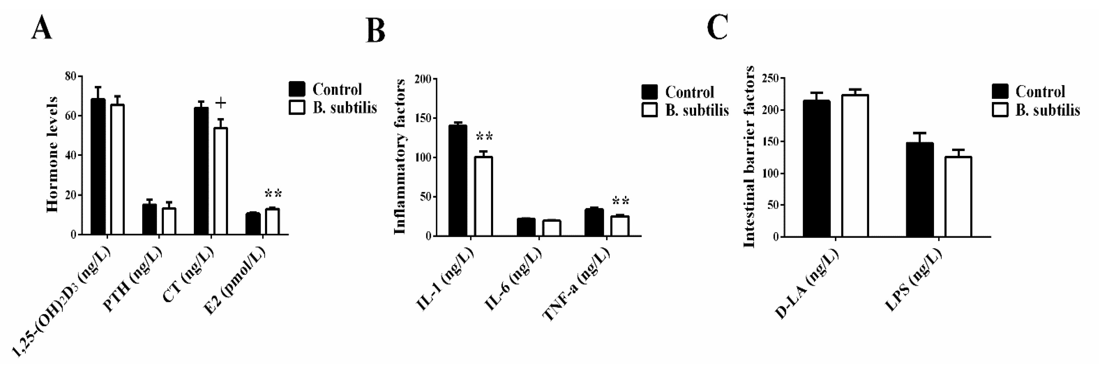
Bacillus subtilis did not affect serum Ca concentration, ALP, and TRAP activities (p > 0.05; Figure 1A,C,D) but reduced P concentration (p < 0.01, Figure 1B) in laying hens at day 61. The serum activity of ALP (p = 0.06) had a trend of decrease and TRAP activity (p < 0.01) significantly increased with age in laying hens (Figure 1C,D). Meanwhile, at day 61, there was a trend of lower plasma CT in Bacillus subtilis fed birds (p = 0.07; Figure 2A). In addition, Bacillus subtilis increased plasma concentrations of E2 (p = 0.01) without effects on plasma concentrations of IL-6, 1,25-(OH)2D3, PTH, D-LA, and LPS (p > 0.05; Figure 2A–C), but inhibited plasma IL-1 (p < 0.01; Figure 2B) and TNF-α (p < 0.01) concentrations.

3.4. Bone Pathophysiological Parameters

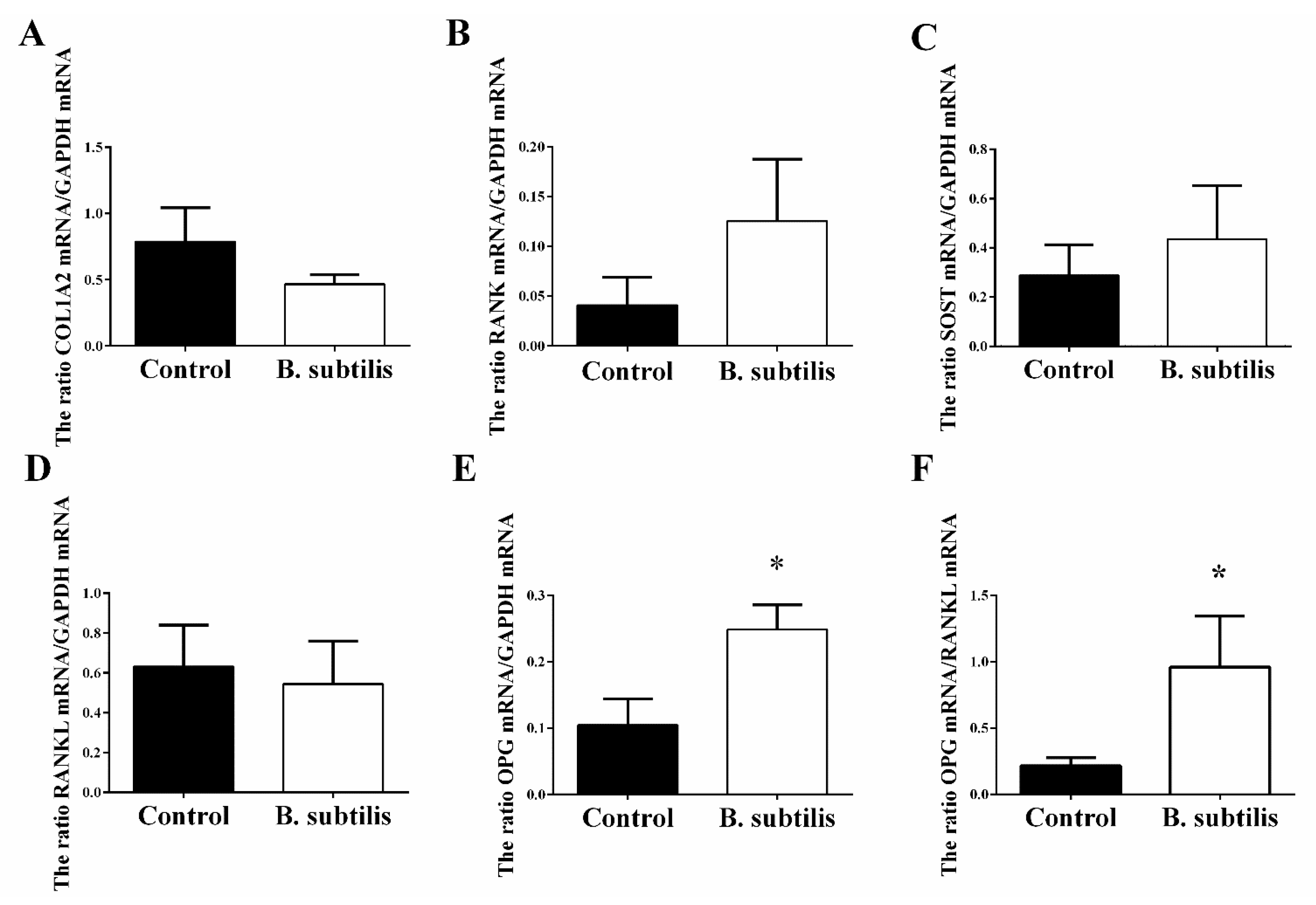
Compared with the control group, Bacillus subtilis administration did not change femur absolute and relative mass and density (p > 0.05; Table 5) but improved femur maximum stress (p = 0.01), stiffness (p < 0.01), and Young’s modulus (p < 0.01) in birds. Moreover, there was an upward trend of femur maximum load and a downward trend of femur maximum strain, respectively, in Bacillus subtilis fed birds (p = 0.06). However, Bacillus subtilis supplement did not affect other measured biomechanical properties of tibia, except for a slight increase in tibial density (p = 0.07). Furthermore, Bacillus subtilis supplement improved the expression of OPG mRNA (p = 0.02; Figure 3), leading to a greater OPG/RANKL ratio (p = 0.04), while there were no treatment effects on the expression of RANKL, RANK, SOST, and COL1A2 mRNA.

3.5. Ca and P Content Analysis

Compared with the control group, bone P content was significantly increased (p < 0.05; Table 6) in the Bacillus subtilis fed group. The Bacillus subtilis fed group also had a lower concentration of bone Mg (p = 0.04) and a trend of higher fecal Mg (p = 0.09). However, there were no treatment effects on the contents of fecal Ca and P, bone Ca and ash weight, and eggshell P (p > 0.05).

3.6. Correlation between Ca or P and Bone Quality

The femoral P but not Ca level was positively correlated with femur maximum stress (r = 0.55, p = 0.02; Table 7), stiffness (r = 0.45, p = 0.05), Young’s modulus (r = 0.56, p = 0.014), and femoral Ca (r = 0.80, p < 0.01) as well as being negatively correlated with serum P (r = −0.59, p < 0.01), serum Ca level was unrelated to any bone quality indexes. However, serum P level was negatively correlated with stiffness (r = −0.71, p = 0.001) and Young’s modulus (r = −0.58, p = 0.009).

4. Discussion

With age (beyond 40 weeks of age), especially into the later stage of egg production, their intestinal absorption and utilization of minerals including Ca are gradually declined [42,43], which could be due to the decrease of relative hormones and their receptors [39,44] or the change of gut epithelial architecture reducing absorption surface [45]. To maintain the need of Ca for egg production, layers may mobilize extra Ca stored in medullary bone. Approximately 25–40% of the eggshell Ca is from skeletal stores [46]. This process causes negative balance between bone formation (osteoblasts) and resorption (osteoclasts) with the enhanced osteoclast activity and releasing of large amounts of TRAP [47,48], resulting in significant expending of medullary bone at the expense of structural bone (cancellous and cortical bone), eventually leading to osteoporosis. To adapt to Ca shortage, laying hens adjust Ca usage for eggshell formation [49,50] by either decreasing eggshell thickness and strength [51], reducing egg production, or in combination [49].
As the egg cycle extended, the numbers of soft-shelled and cracked eggs increased, which may be attributed to reduction of Ca absorption, stress, or poor physical quality [52,53,54]. The current results indicate that hens fed Bacillus subtilis had a lower incidence of both soft-shelled and cracked eggs but there was no strong evidence of the improvement of eggshell strength and thickness [55]. Inconsistent findings about effects of Bacillus subtilis on egg quality have been reported. Some studies indicated that Bacillus subtilis could improve albumen height and Haugh unit [31,56]. On the contrary, our findings as well as some of others showed that egg quality was not affected by Bacillus subtilis supplement [57,58]. Previous study reported that probiotic efficiency as a growth promotor is affected by multiple factors, such as the type of probiotics, the length of feeding time and concentration [31], the isolation sources of probiotics, breed of laying hens, and their life-stage [59]. In addition, Alfonso-Carrillo et al. [54] indicated that the promotion of egg production, eggshell quality, and bone traits in laying hens can occur independently through dietary treatment. Therefore, we speculate that Bacillus subtilis is more likely to improve bone traits rather than egg or eggshell quality.
Stiffness and Young’s modulus have been used as indicators indicating the capability of bone to resist deformation, which is mainly determined by cortical bone density. Increased bone mass can improve bone strength and stiffness [60]. In the present study, dietary Bacillus subtilis supplement significantly improved bone strength, stiffness, and Young’s modulus of femurs, and while Bacillus subtilis changed both bone stress and strain in laying hens. Previous study has demonstrated that the fluctuations caused by changed bone strain and stress are mainly due to increased inhomogeneous mineral distribution in trabecular bone, namely, a higher bone stress but lower strain [61]. The outcomes of the present research suggest that Bacillus subtilis is able to prevent bone mass loss and reduce fracture risk. In addition, Ca and P are two necessary minerals for bone mineralization and bone homeostasis. Phosphorus is mainly deposited in bone in the form of hydroxyapatite with Ca and is involved in regulation of bone cells’ functions and ALP metabolism [62,63]. It has been revealed that besides being absorbed from diet, the skeletal system is a main source of minerals for eggshell formation [64]. It means that P is also mobilized from bone to blood for egg production. Shao [65] reported that the plasma concentration of P is an indicator of bone mineralization, and even a slight decline shows that bone mineralization is in a positive state with improved bone strength. This is consistent with the current results that the Bacillus subtilis reduced serum P concentration while it improved bone P content, and bone P content had a great correlation with serum P, and was positively associated with bone Ca content, stiffness, and other physical parameters. These results show that Bacillus subtilis provide the beneficial effects on bone health via regulation of P metabolism.
Estrogen is a kind of bone regulating hormone. Enterohepatic circulation is the main way to regulate E2 metabolism due to its bioconversion in the liver, conjugated E2 is mostly excreted with bile, but some is hydrolyzed by gut microbiota, and reabsorbed in the intestines for various biological processes including bone remodeling [66,67]. Similar to the current results, Zhou [68] reported that adding Bacillus amyloliquefaciens increased serum E2 level in laying hens. We speculate that the increase of E2 level is attributed to the colonization and functional activities of Bacillus subtilis in the intestines. Furthermore, E2 has been recognized as functionally preventing osteoporosis in postmenopausal women [69,70] and old laying hens [44]. Previous results have shown that E2 could decrease synthesis of inflammatory cytokines and regulate the OPG/RANKL/RANK system to prevent bone loss [71]. Estrogen deficiency leads to high productions of TNF and RANKL in bone marrow and small intestines [72,73]. However, probiotics supplements can protect ovariectomized mice from bone lose [74,75]. These evidences suggest the possible protective effects of probiotics and E2 on bone. Consistently, Bacillus subtilis supplement significantly reduced hens’ plasma pro-inflammatory factors, IL-1 and TNF-α, and increased the OPG gene expression in the present study.
The IL-1 and TNF-α are mainly secreted by peripheral monocytes, macrophagocytes, and T lymphocytes [76]. Reduced serum IL-1 and TNF-α in the hens suggests that Bacillus subtilis prevents the inflammatory process induced by aging-related immune response. Similarly, Lee et al. [77] found that probiotics prevent the progression of inflammation-associated intestine injury through intervening in the expression of inflammatory molecules, such as IL-1 and TNF-α, to maintain intestinal health. These pro-inflammatory factors play important roles in activation of osteoclasts and related bone resorption [78]. Moreover, the OPG/RANKL/RANK system has been identified as the dominant and final mediator of osteoclastogenesis [79]. OPG, as the only known decoy receptor of RANKL, is located on osteoblasts and can be upregulated by E2 [80]. OPG competitively binds with RANKL [78] to prevent osteoclasts from being activated by the signals released from the RANKL/RANK pathway, consequently, it inhibits bone resorption and bone loss [81]. The ratio of OPG/RANKL has been recognized as an anti-osteoclast indicator [82,83]. Taken together, these results indicate that the immune system is involved in regulation of bone remodeling (formation and resorption), especially influencing the activation of the OPG/RANKL/RANK pathway [71,84], for example, IL-1 indirectly inhibits OPG mRNA expression [85] without any impact on RANKL expression, and stimulates osteoclastogenesis resulting in bone loss [86]. Therefore, it is possible that Bacillus subtilis protects hen bone health through improving the E2 metabolism, inhibiting synthesis of pro-inflammatory factors, and increasing OPG synthesis.

5. Conclusions

Low-grade systemic inflammation is one of the major health and production issues in laying hens, resulting in osteoporosis with chronical pain, bone fractures, and poor egg production with significant economic loss. This study showed that Bacillus subtilis as an additive to laying hens’ diets can increase marketable egg production and bone traits by increasing P usage, improving estrogen metabolism, inhibiting pro-inflammatory factor synthesis, and increasing OPG expression. Thus, Bacillus subtilis can be a valuable feed supplement to improve the welfare and prevent osteoporosis in laying hens.

Author Contributions

Conceptualization, S.J.; methodology, S.J. and X.Z.; investigation, X.Z., M.Z., H.H., X.W., and M.J.; data analysis, X.Z.; writing, draft preparation, review and editing, X.Z., S.J., J.L., M.J., and H.C.; project administration, S.J. and X.Z.; funding acquisition, S.J. All authors have read and agreed to the published version of the manuscript.

Funding

This research was supported by Fundamental Research Funds for the Central Universities (XDJK2019B013) and the National Natural Science Foundation of China (No.31702307).

Institutional Review Board Statement

The study was conducted according to the guidelines of the Declaration of Helsinki, and approved by the Animal Ethics Committee of the Southwest University, Chongqing, China.The permission was approved on 25 February 2019, and the number is IACUC-20190225-19.

Data Availability Statement

Not applicable.

Acknowledgments

The authors thank Fubon Inc (Wuhan, China) who provided the Bacillus subtilis.

Conflicts of Interest

The authors declare no conflict of interest. The funders had no role in the design of the study; in the collection, analyses, or interpretation of data; in the writing of the manuscript, or in the decision to publish the results.

References

  1. Mehring, A.L.; Titus, H.W. The effects of low levels of calcium in the diet of laying chickens. Poult. Sci. 1964, 43, 1405–1414. [Google Scholar] [CrossRef]
  2. Webster, A.B. Welfare implications of avian osteoporosis. Poult. Sci. 2004, 83, 184–192. [Google Scholar] [CrossRef] [PubMed]
  3. Fleming, R.H. Nutritional factors affecting poultry bone health. Proc. Nutr. Soc. 2008, 67, 177–183. [Google Scholar] [CrossRef] [PubMed] [Green Version]
  4. Leeson, S.; Summers, A. Commercial Poultry Nutrition, 3rd ed.; Nottingham University Press: Nottingham, UK, 2008; pp. 123–160. [Google Scholar]
  5. Whitehead, C.C.; Fleming, R.H. Osteoporosis in cage layers. Poult. Sci. 2000, 79, 1033–1041. [Google Scholar] [CrossRef] [PubMed]
  6. Khanal, T.; Widowski, T.; Bédécarrats, G.; Kiarie, E. Effects of pre-lay dietary calcium (2.5 vs. 4.0%) and pullet strain (Lohmann Brown vs. Selected Leghorn LSL-Lite) on calcium utilization and femur quality at 1st through to the 50th egg. Poult. Sci. 2019, 98, 4919–4928. [Google Scholar] [CrossRef]
  7. Sumano-Lopez, H.; Carrillo-Gonzalez, L.; Monroy-Barreto, M.; Tapia-Perez, G.; Olvera, L.G. Bioavailability of four calcium sources in the second-cycle egg-producing hens. J. Appl. Poult. Res. 2021, 30, 8. [Google Scholar]
  8. Rathnayaka, R.; Mutucumarana, R.K.; Andrew, M.S. Free-choice feeding of three different dietary calcium sources and their influence on egg quality parameters of commercial layers. J. Agric. Sci. Sri Lanka 2020, 15, 50–62. [Google Scholar] [CrossRef] [Green Version]
  9. Habibollahi, M.; Abousadi, M.A.; Nakhaee, P. The effect of phytase on production performance, egg quality, calcium and phosphorus excretion, and fatty acids and cholesterol concentration in hy-line layers fed diets containing rice bran. J. Appl. Poult. Res. 2019, 28, 688–698. [Google Scholar] [CrossRef]
  10. Shi, H.; Lee, K.Y.; Kim, I.H. Dietary supplementation with protected calcium effects production and egg quality of Hy-line brown laying hens. Anim. Prod. Sci. 2020, 60, 1793–1799. [Google Scholar] [CrossRef]
  11. Bernard, B.D.; Stagni, N.; Camerotto, R.; Vittur, F.; Zanetti, M.; Zallone, A.Z.; Teti, A. Influence of calcium depletion on medullary bone of laying hens. Calcif. Tissue Int. 1980, 32, 221–228. [Google Scholar] [CrossRef]
  12. Mountzouris, K.C.; Tsitrsikos, P.; Palamidi, I.; Arvaniti, A.; Mohnl, M.; Schatzmayr, G.; Fegeros, K. Effects of probiotic inclusion levels in broiler nutrition on growth performance, nutrient digestibility, plasma immunoglobulins, and cecal microflora composition. Poult. Sci. 2010, 89, 58–67. [Google Scholar] [CrossRef] [PubMed]
  13. Forte, C.; Acuti, G.; Manuali, E.; Casagrande Proietti, P.; Pavone, S.; Trabalza-Marinucci, M.; Moscati, L.; Onofri, A. Effects of two different probiotics on microflora, morphology, and morphometry of gut in organic laying hens. Poult. Sci. 2016, 95, 2528–2535. [Google Scholar] [CrossRef] [PubMed]
  14. Zhang, P.; Yan, T.; Wang, X.; Kuang, S.; Xiao, Y.C.; Lu, W.W.; Bi, D.R. Probiotic mixture ameliorates heat stress of laying hens by enhancing intestinal barrier function and improving gut microbiota. Ital. J. Anim. Sci. 2017, 16, 292–300. [Google Scholar] [CrossRef] [Green Version]
  15. Song, J.; Xiao, K.; Ke, Y.L.; Jiao, L.F.; Hu, C.H.; Diao, Q.Y.; Shi, B.; Zou, X.T. Effect of a probiotic mixture on intestinal microflora, morphology, and barrier integrity of broilers subjected to heat stress. Poult. Sci. 2014, 93, 581–588. [Google Scholar] [CrossRef]
  16. Panda, A.K.; Reddy, M.R.; Rama Rao, S.V.; Praharaj, N.K. Production performance, serum/yolk cholesterol and immune competence of white leghorn layers as influenced by dietary supplementation with probiotic. Trop. Anim. Health Prod. 2003, 35, 85–94. [Google Scholar] [CrossRef] [PubMed]
  17. Bai, S.P.; Wu, A.M.; Ding, X.M.; Lei, Y.; Bai, J.; Zhang, K.Y.; Chio, J.S. Effects of probiotic-supplemented diets on growth performance and intestinal immune characteristics of broiler chickens. Poult. Sci. 2013, 92, 663–670. [Google Scholar] [CrossRef]
  18. Mikulski, D.; Jankowski, J.; Naczmanski, J.; Mikulska, M.; Demey, V. Effects of dietary probiotic (Pediococcus acidilactici) supplementation on performance, nutrient digestibility, egg traits, egg yolk cholesterol, and fatty acid profile in laying hens. Poult. Sci. 2012, 91, 2691–2700. [Google Scholar] [CrossRef]
  19. Park, J.W.; Jeong, J.S.; Lee, S.I.; Kim, I.H. Effect of dietary supplementation with a probiotic (Enterococcus faecium) on production performance, excreta microflora, ammonia emission, and nutrient utilization in ISA brown laying hens. Poult. Sci. 2016, 95, 2829–2835. [Google Scholar] [CrossRef]
  20. Molnar, A.K.; Podmaniczky, B.; Kurti, P.; Glavits, R.; Virag, G.; Szabo, Z.; Farkas, Z. Effect of different concentrations of Bacillus subtilis on immune response of broiler chickens. Probiotics Antimicrob. Proteins 2011, 3, 8–14. [Google Scholar] [CrossRef]
  21. Park, J.H.; Yun, H.M.; Kim, I.H. The effect of dietary Bacillus subtilis supplementation on the growth performance, blood profile, nutrient retention, and caecal microflora in broiler chickens. J. Appl. Anim. Res. 2018, 46, 868–872. [Google Scholar] [CrossRef] [Green Version]
  22. Knap, I.; Kehlet, A.B.; Bennedsen, M.; Mathis, G.F.; Hofacre, C.L.; Lumpkins, B.S.; Jensen, M.M.; Raun, M.; Lay, A. Bacillus subtilis (DSM17299) significantly reduces Salmonella in broilers. Poult. Sci. 2011, 90, 1690–1694. [Google Scholar] [CrossRef]
  23. Lei, X.; Piao, X.; Ru, Y.; Zhang, H.; Peron, A.; Zhang, H. Effect of Bacillus amyloliquefaciens-based direct-fed microbial on performance, nutrient utilization, intestinal morphology and cecal microflora in broiler chickens. Asian Aust. J. Anim. Sci. 2015, 28, 239–246. [Google Scholar] [CrossRef] [PubMed] [Green Version]
  24. Sadeghi, A.A. Bone mineralization of broiler chicks challenged with Salmonella enteritidis fed diet containing probiotic (Bacillus subtilis). Probiotics Antimicrob. Proteins 2014, 6, 136–140. [Google Scholar] [CrossRef]
  25. Mutuş, R.; Kocabagli, N.; Alp, M.; Acar, N.; Eren, M.; Gezen, S.S. The effect of dietary probiotic supplementation on tibial bone characteristics and strength in broilers. Poult. Sci. 2006, 85, 1621–1625. [Google Scholar] [CrossRef] [PubMed]
  26. Yan, F.F.; Wang, W.C.; Cheng, H.W. Bacillus subtilis based probiotic improved bone mass and altered brain serotoninergic and dopaminergic systems in broiler chickens. J. Funct. Foods. 2018, 49, 501–509. [Google Scholar] [CrossRef]
  27. Newberry, R.C.; Tarazona, A.M. Behavior and welfare of laying hens and broiler chickens. Rev. Colomb Cienc. Pecu. 2011, 24, 301–302. [Google Scholar]
  28. Guo, J.R.; Dong, X.F.; Liu, S.; Tong, J.M. High-throughput sequencing reveals the effect of Bacillus subtilis CGMCC 1.921 on the cecal microbiota and gene expression in ileum mucosa of laying hens. Poult. Sci. 2018, 97, 2543–2556. [Google Scholar] [CrossRef] [PubMed]
  29. Zhang, G.; Wang, H.; Zhang, J.; Tang, X.; Raheem, A.; Wang, M.; Lin, W.; Liang, L.; Qi, Y.; Zhu, Y. Modulatory effects of Bacillus subtilis on the performance, morphology, cecal microbiota and gut barrier function of laying hens. Animals 2021, 11, 1523. [Google Scholar] [CrossRef] [PubMed]
  30. Guo, J.R.; Dong, X.F.; Liu, S.; Tong, J.M. Effects of long-term Bacillus subtilis CGMCC 1.921 supplementation on performance, egg quality, and fecal and cecal microbiota of laying hens. Poult. Sci. 2017, 96, 1280–1289. [Google Scholar] [CrossRef] [PubMed]
  31. Neijat, M.; Shirley, R.B.; Barton, J.; Thiery, P.; Welsher, A.; Kiarie, E. Effect of dietary supplementation of Bacillus subtilis DSM29784 on hen performance, egg quality indices, and apparent retention of dietary components in laying hens from 19 to 48 weeks of age. Poult. Sci. 2019, 98, 5622–5635. [Google Scholar] [CrossRef]
  32. Wang, J.; Wang, W.W.; Qi, G.H.; Cui, C.F.; Wu, S.G.; Zhang, H.J.; Xu, L.; Wang, J. Effects of dietary Bacillus subtilis supplementation and calcium levels on performance and eggshell quality of laying hens in the late phase of production. Poult. Sci. 2021, 100, 100970. [Google Scholar] [CrossRef] [PubMed]
  33. Juzaitis-Boelter, C.P.; Benson, A.P.; Ahammad, M.U.; Jones, M.K.; Ferrel, J.; Davis, A.J. Dietary inclusion of AZOMITE improves feed efficiency in broilers and egg production in laying and broiler breeder hens. Poult. Sci. 2021, 100, 101144. [Google Scholar] [CrossRef] [PubMed]
  34. Ribeiro, C.L.N.; Barreto, S.L.T.; Reis, R.S.; Muniz, J.C.L.; Viana, G.S.; Ribeiro, V.; Mendonca, M.O.; Ferreira, R.C.; DeGroot, A.A. The effect of calcium and available phosphorus levels on performance, egg quality and bone characteristics of japanese quails at end of the egg-production phase. Braz. J. Poult. Sci. 2016, 18, 33–39. [Google Scholar] [CrossRef] [Green Version]
  35. Jiang, S.; Cheng, H.W.; Cui, L.Y.; Zhou, Z.L.; Hou, J.F. Changes of blood parameters associated with bone remodeling following experimentally induced fatty liver disorder in laying hens. Poult. Sci. 2013, 92, 1443–1453. [Google Scholar] [CrossRef]
  36. Guo, X.H.; Li, D.F.; Lu, W.Q.; Piao, X.S.; Chen, X.L. Screening of Bacillus strains as potential probiotics and subsequent confirmation of the in vivo effectiveness of Bacillus subtilis MA139 in pigs. Antonie Van Leeuwenhoek 2006, 90, 139–146. [Google Scholar] [CrossRef] [PubMed]
  37. Naghii, M.R.; Torkaman, G.; Mofid, M. Effects of boron and calcium supplementation on mechanical properties of bone in rats. Biofactors 2006, 28, 195–201. [Google Scholar] [CrossRef]
  38. Cole, J.H.; van der Meulen, M.C. Whole bone mechanics and bone quality. Clin. Orthop. Relat. Res. 2011, 469, 2139–2149. [Google Scholar] [CrossRef] [Green Version]
  39. Elaroussi, M.A.; Forte, L.R.; Eber, S.L.; Biellier, H.V. Calcium homeostasis in the laying hen. 1. Age and dietary calcium effects. Poult. Sci. 1994, 73, 1581–1589. [Google Scholar] [CrossRef] [PubMed]
  40. Abdelqader, A.; Al-Fataftah, A.-R.; Daş, G. Effects of dietary Bacillus subtilis and inulin supplementation on performance, eggshell quality, intestinal morphology and microflora composition of laying hens in the late phase of production. Anim. Feed Sci. Technol. 2013, 179, 103–111. [Google Scholar] [CrossRef]
  41. Fischer, A.; Malara, P.; Wiechula, D. The study of barium concentration in deciduous teeth, impacted teeth, and facial bones of polish residents. Biol. Trace Elem. Res. 2014, 161, 32–37. [Google Scholar] [CrossRef]
  42. Abe, E.; Hiroshi, H.; Masumura, T.; Sugahara, M.; Kubota, M.; Suda, T. Disorders of cholecaiciferol metabolism in old egg-laying hens. J. Nutr. 1982, 112, 436–446. [Google Scholar] [CrossRef]
  43. Al-Batshan, H.A.; Scheideler, S.E.; Black, B.L.; Garlich, J.D.; Anderson, K.E. Duodenal calcium uptake, femur ash, and eggshell quality decline with age and increase following molt. Poult. Sci. 1994, 73, 1590–1596. [Google Scholar] [CrossRef] [PubMed]
  44. Beck, M.M.; Hansen, K.K. Role of estrogen in avian osteoporosis. Poult. Sci. 2004, 83, 200–206. [Google Scholar] [CrossRef] [PubMed]
  45. Ren, W.Y.; Wu, K.F.; Li, X.; Luo, M.; Liu, H.C.; Zhang, S.C.; Hu, Y. Age-related changes in small intestinal mucosa epithelium architecture and epithelial tight junction in rat models. Aging Clin. Exp. Res. 2014, 26, 183–191. [Google Scholar] [CrossRef]
  46. Mueller, W.J.; Schraer, R.; Schraer, H. Calcium metabolism and skeletal dynamics of laying pullets. J. Nutr. 1964, 84, 20–26. [Google Scholar] [CrossRef] [PubMed]
  47. Irie, S.; Hayashida, N.; Shinkawa, T.; Taira, Y.; Sekitani, Y.; Teraoka, S.; Hashiguchi, K.; Yoshida, K.; Morishita, M.; Takamura, N. Suitability of tartrate-resistant acid phosphatase type 5b as a screening marker for bone mineral density in community-dwelling elderly individuals. Tohoku J. Exp. Med. 2011, 224, 105–110. [Google Scholar] [CrossRef] [PubMed] [Green Version]
  48. Jiang, S.; Wu, X.L.; Jin, M.L.; Wang, X.Z.; Tang, Q.; Sun, Y.X.; Cheng, H.W. Pathophysiological characteristics and gene transcriptional profiling of bone microstructure in a low calcium diet fed laying hens. Poult. Sci. 2019, 98, 4359–4368. [Google Scholar] [CrossRef]
  49. Morgan, C.L.; Mitchell, J.H. The calcium and phosphorus balance of laying hens. Poult. Sci. 1938, 17, 99–104. [Google Scholar] [CrossRef]
  50. Hurwitz, S.; Griminger, P. Observations on the calcium balance of laying hens. J. Agric. Sci. 1960, 54, 373–377. [Google Scholar] [CrossRef]
  51. An, S.H.; Kim, D.W.; An, B.K. Effects of dietary calcium levels on productive performance, eggshell quality and overall calcium status in aged laying hens. Asian Aust. J. Anim. Sci. 2016, 29, 1477–1482. [Google Scholar] [CrossRef] [Green Version]
  52. Reynard, M.; Savory, C.J. Stress-induced oviposition delays in laying hens: Duration and consequences for eggshell quality. Br. Poult. Sci. 1999, 40, 585–591. [Google Scholar] [CrossRef]
  53. Zhu, Y.Z.; Cheng, J.L.; Ren, M.; Yin, L.; Piao, X.S. Effect of gamma-aminobutyric acid-producing Lactobacillus strain on laying performance, egg quality and serum enzyme activity in hy-line brown hens under heat stress. Asian Aust. J. Anim. Sci. 2015, 28, 1006–1013. [Google Scholar] [CrossRef]
  54. Alfonso-Carrillo, C.; Benavides-Reyes, C.; de Los Mozos, J.; Dominguez-Gasca, N.; Sanchez-Rodríguez, E.; Garcia-Ruiz, A.I.; Rodriguez-Navarro, A.B. Relationship between bone quality, egg production and eggshell quality in laying hens at the end of an extended production cycle (105 weeks). Animals 2021, 11, 623. [Google Scholar] [CrossRef] [PubMed]
  55. Min, Y.N.; Liu, F.X.; Qi, X.; Ji, S.; Ma, S.X.; Liu, X.; Wang, Z.P.; Gao, Y.P. Effects of methionine hydroxyl analog chelated zinc on laying performance, eggshell quality, eggshell mineral deposition, and activities of Zn-containing enzymes in aged laying hens. Poult. Sci. 2018, 97, 3587–3593. [Google Scholar] [CrossRef]
  56. Chen, J.F.; Kuang, Y.H.; Qu, X.Y.; Guo, S.C.; Kang, K.L.; He, C.Q. The effects and combinational effects of Bacillus subtilis and montmorillonite supplementation on performance, egg quality, oxidation status, and immune response in laying hens. Livest. Sci. 2019, 227, 114–119. [Google Scholar] [CrossRef]
  57. Abd El-Hack, M.E.; Mahgoub, S.A.; Alagawany, M.; Ashour, E.A. Improving productive performance and mitigating harmful emissions from laying hen excreta via feeding on graded levels of corn DDGS with or without Bacillus subtilis probiotic. J. Anim. Physiol. Anim. Nutr. 2017, 101, 904–913. [Google Scholar] [CrossRef] [PubMed]
  58. Liu, X.; Peng, C.Y.; Qu, X.Y.; Guo, S.C.; Chen, J.F.; He, C.Q.; Zhou, X.B.; Zhu, S.W. Effects of Bacillus subtilis C-3102 on production, hatching performance, egg quality, serum antioxidant capacity and immune response of laying breeders. J. Anim. Physiol. Anim. Nutr. 2019, 103, 182–190. [Google Scholar] [CrossRef] [Green Version]
  59. Fathi, M.; Al-Homidan, I.; Al-Dokhail, A.; Ebeid, T.; Abou-Emera, O.; Alsagan, A. Effects of dietary probiotic (Bacillus subtilis) supplementation on productive performance, immune response and egg quality characteristics in laying hens under high ambient temperature. Ital. J. Anim. Sci. 2018, 17, 804–814. [Google Scholar] [CrossRef] [Green Version]
  60. Ferretti, J.L.; Vázquez, S.O.; Delgado, C.J.; Capozza, R.; Cointry, G. Biphasic dose-response curves of cortisol effects on rat diaphyseal bone biomechanics. Calcif. Tissue Int. 1992, 50, 49–54. [Google Scholar] [CrossRef] [PubMed]
  61. Van Ruijven, L.J.; Mulder, L.; van Eijden, T.M. Variations in mineralization affect the stress and strain distributions in cortical and trabecular bone. J. Biomech. 2007, 40, 1211–1218. [Google Scholar] [CrossRef]
  62. Heaney, R.P. Phosphorus nutrition and the treatment of osteoporosis. Mayo Clin. Proc. 2004, 79, 91–97. [Google Scholar] [CrossRef] [PubMed] [Green Version]
  63. Addison, W.N.; Azari, F.; Sørensen, E.S.; Kaartinen, M.T.; McKee, M.D. Pyrophosphate inhibits mineralization of osteoblast cultures by binding to mineral, up-regulating osteopontin, and inhibiting alkaline phosphatase activity. J. Biol. Chem. 2007, 282, 15872–15883. [Google Scholar] [CrossRef] [PubMed] [Green Version]
  64. Miles, R.D.; Junqueira, O.M.; Harms, R.H. Plasma phosphorus at 0, 6, and 21 hours postoviposition in hens laying in the morning or afternoon. Poult. Sci. 1984, 63, 354–359. [Google Scholar] [CrossRef]
  65. Shao, Y.; Sun, G.; Cao, S.; Lu, L.; Zhang, L.; Liao, X.; Luo, X. Bone phosphorus retention and bone development of broilers at different ages. Poult. Sci. 2019, 98, 2114–2121. [Google Scholar] [CrossRef]
  66. Taylor, W. The excretion of steroid hormone metabolites in bile and feces. Vitam. Horm. 1971, 29, 201–285. [Google Scholar]
  67. Adlercreutz, H.; Martin, F. Biliary excretion and intestinal metabolism of progesterone and estrogens in man. J. Steroid Biochem. 1980, 13, 231–244. [Google Scholar] [CrossRef]
  68. Zhou, Y.; Li, S.; Pang, Q.; Miao, Z. Bacillus amyloliquefaciens BLCC1-0238 can effectively improve laying performance and egg quality via enhancing immunity and regulating reproductive hormones of laying hens. Probiotics Antimicrob. Proteins 2020, 12, 246–252. [Google Scholar] [CrossRef]
  69. Tobias, J.H.; Compston, J.E. Does estrogen stimulate osteoblast function in postmenopausal women? Bone 1999, 24, 121–124. [Google Scholar] [CrossRef]
  70. Amonkar, M.M.; Mody, R. Developing profiles of postmenopausal women being prescribed estrogen therapy to prevent osteoporosis. J. Community Health 2002, 27, 335–350. [Google Scholar] [CrossRef] [PubMed]
  71. Clowes, J.A.; Riggs, B.L.; Khosla, S. The role of the immune system in the pathophysiology of osteoporosis. Immunol. Rev. 2005, 208, 207–227. [Google Scholar] [CrossRef]
  72. Li, J.Y.; Chassaing, B.; Tyagi, A.M.; Vaccaro, C.; Luo, T.; Adams, J.; Darby, T.M.; Weitzmann, M.N.; Mulle, J.G.; Gewirtz, A.T.; et al. Sex steroid deficiency-associated bone loss is microbiota dependent and prevented by probiotics. J. Clin. Investig. 2016, 126, 2049–2063. [Google Scholar] [CrossRef] [Green Version]
  73. Jones, R.M.; Mulle, J.G.; Pacifici, R. Osteomicrobiology: The influence of gut microbiota on bone in health and disease. Bone 2018, 115, 59–67. [Google Scholar] [CrossRef]
  74. Britton, R.A.; Irwin, R.; Quach, D.; Schaefer, L.; Zhang, J.; Lee, T.; Parameswaran, N.; McCabe, L.R. Probiotic L. reuteri treatment prevents bone loss in a menopausal ovariectomized mouse model. J. Cell. Physiol. 2014, 229, 1822–1830. [Google Scholar] [CrossRef] [Green Version]
  75. Ohlsson, C.; Engdahl, C.; Fak, F.; Andersson, A.; Windahl, S.H.; Farman, H.H.; Moverare-Skrtic, S.; Islander, U.; Sjogren, K. Probiotics protect mice from ovariectomy-induced cortical bone loss. PLoS ONE 2014, 9, 8. [Google Scholar] [CrossRef]
  76. Wigg, A.J.; Roberts-Thomson, I.C.; Dymock, R.B.; McCarthy, P.J.; Grose, R.H.; Cummins, A.G. The role of small intestinal bacterial overgrowth, intestinal permeability, endotoxaemia, and tumour necrosis factor alpha in the pathogenesis of non-alcoholic steatohepatitis. Gut 2001, 48, 206–211. [Google Scholar] [CrossRef] [Green Version]
  77. Lee, H.S.; Han, S.Y.; Bae, E.A.; Huh, C.S.; Ahn, Y.T.; Lee, J.H.; Kim, D.H. Lactic acid bacteria inhibit proinflammatory cytokine expression and bacterial glycosaminoglycan degradation activity in dextran sulfate sodium-induced colitic mice. Int. Immunopharmacol. 2008, 8, 574–580. [Google Scholar] [CrossRef] [PubMed]
  78. Jurado, S.; Garcia-Giralt, N.; Díez-Pérez, A.; Esbrit, P.; Yoskovitz, G.; Agueda, L.; Urreizti, R.; Pérez-Edo, L.; Saló, G.; Mellibovsky, L. Effect of IL-1beta, PGE(2), and TGF-beta1 on the expression of OPG and RANKL in normal and osteoporotic primary human osteoblasts. J. Cell. Biochem. 2010, 110, 304–310. [Google Scholar]
  79. Liu, X.H.; Kirschenbaum, A.; Yao, S.; Levine, A.C. Cross-talk between the interleukin-6 and prostaglandin E-2 signaling systems results in enhancement of osteoclastogenesis through effects on the osteoprotegerin/receptor activator of nuclear factor-kappa B (RANK) ligand/RANK system. Endocrinology 2005, 146, 1991–1998. [Google Scholar] [CrossRef] [PubMed] [Green Version]
  80. Liao, E.Y.; Luo, X.H.; Su, X. Comparison of the effects of 17 beta-E2 and progesterone on the expression of osteoprotegerin in normal human osteoblast-like cells. J. Endocrinol. Invest. 2002, 25, 785–790. [Google Scholar] [CrossRef] [PubMed]
  81. Trouvin, A.P.; Goëb, V. Receptor activator of nuclear factor-κB ligand and osteoprotegerin: Maintaining the balance to prevent bone loss. Clin. Interv. Aging 2010, 5, 345–354. [Google Scholar]
  82. Lindberg, M.K.; Erlandsson, M.; Alatalo, S.L.; Windahl, S.; Andersson, G.; Halleen, J.M.; Carlsten, H.; Gustafsson, J.A.; Ohlsson, C. Estrogen receptor alpha, but not estrogen receptor beta, is involved in the regulation of the OPG/RANKL (osteoprotegerin/receptor activator of NF-kappa B ligand) ratio and serum interleukin-6 in male mice. J. Endocrinol. 2001, 171, 425–433. [Google Scholar] [CrossRef] [PubMed] [Green Version]
  83. Kuehn, M.C.; Willenberg, H.S.; Schott, M.; Papewalis, C.; Stumpf, U.; Flohe, S.; Scherbaum, W.A.; Schinner, S. Adipocyte-secreted factors increase osteoblast proliferation and the OPG/RANKL ratio to influence osteoclast formation. Mol. Cell. Endocrinol. 2012, 349, 180–188. [Google Scholar] [CrossRef]
  84. Abrahamsen, B.; Shalhoub, V.; Larson, E.K.; Eriksen, E.F.; Beck-Nielsen, H.; Marks, S.C. Cytokine RNA levels in transiliac bone biopsies from healthy early postmenopausal women. Bone 2000, 26, 137–145. [Google Scholar] [CrossRef]
  85. Suda, K.; Udagawa, N.; Sato, N.; Takami, M.; Itoh, K.; Woo, J.T.; Takahashi, N.; Nagai, K. Suppression of osteoprotegerin expression by prostaglandin E-2 is crucially involved in lipopolysaccharide-induced osteoclast formation. J. Immunol. 2004, 172, 2504–2510. [Google Scholar] [CrossRef] [PubMed] [Green Version]
  86. Tanabe, N.; Maeno, M.; Suzuki, N.; Fujisaki, K.; Tanaka, H.; Ogiso, B.; Ito, K. IL-1 alpha stimulates the formation of osteoclast-like cells by increasing M-CSF and PGE2 production and decreasing OPG production by osteoblasts. Life Sci. 2005, 77, 615–626. [Google Scholar] [CrossRef] [PubMed]
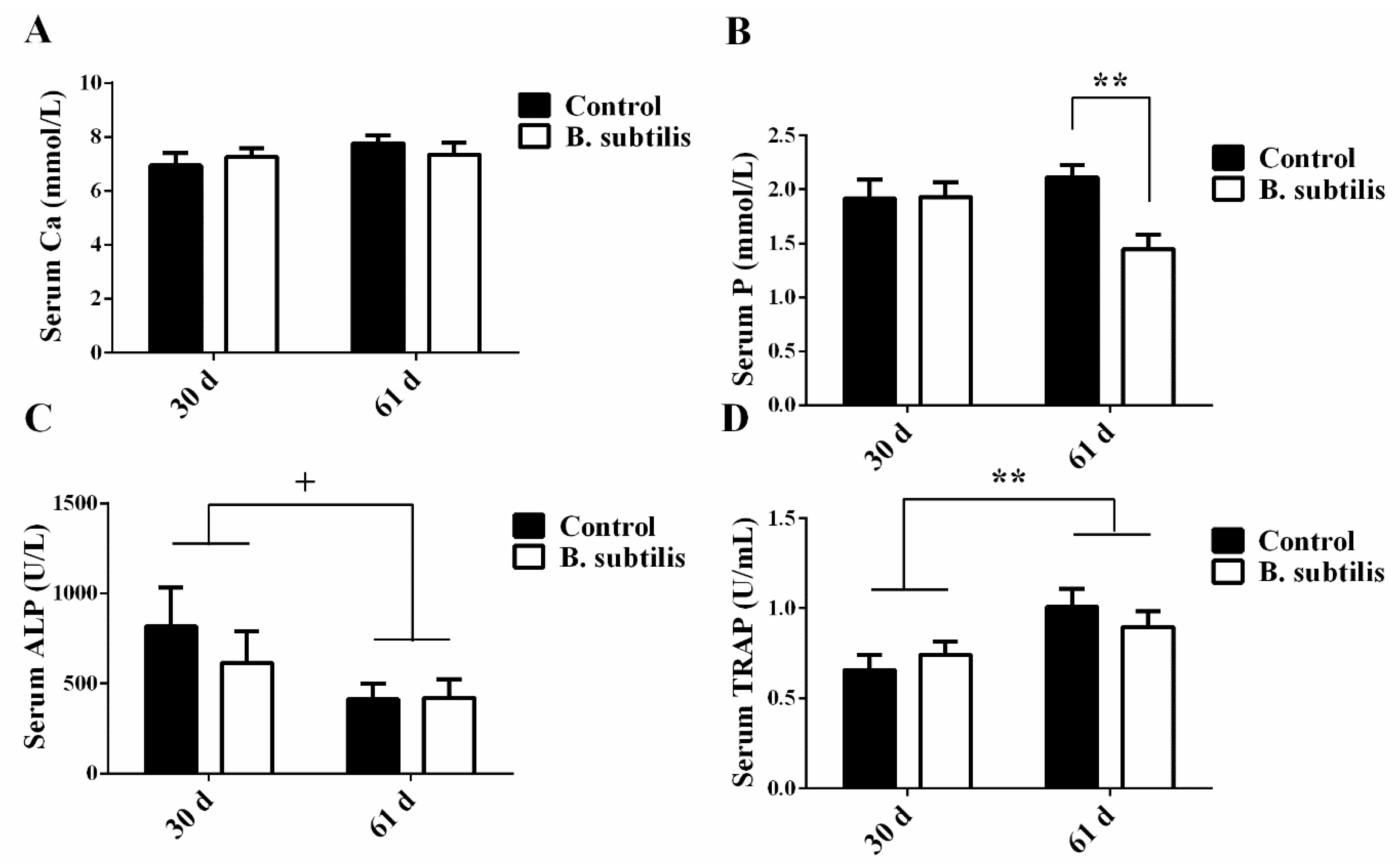
Figure 1. The effects of Bacillus subtilis and age on biochemical indicators of bone metabolism in laying hens. (A) Ca concentration in serum at day 30 and day 61. (B) P concentration in serum at day 30 and day 61. (C) ALP concentration in serum at day 30 and day 61. (D) TRAP concentration in serum at day 30 and day 61. Data are presented as mean ± SEM (n = 12 per group) ** means p ≤ 0.01; and + means 0.05 < p < 0.1. ALP: alkaline phosphatase; Ca: calcium; P: phosphorus; TRAP: tartrate-resistant acid phosphatase.
Animals 11 02041 g001
Figure 2. The effects of Bacillus subtilis on inflammatory factors, hormone levels, and intestinal barrier factors in laying hens at day 61. (A) Hormone levels in plasma. (B) Inflammatory factors concentrations in plasma. (C) Intestinal barrier factors concentrations in plasma. Data are presented as mean ± SEM (n = 12 per group). ** means p ≤ 0.01; and + means 0.05 < p < 0.1. PTH: thyroid hormones; CT: calcitonin; E2: estrogen; IL-1: interleukin-1; IL-6: interleukin-6; TNF-α: tumor necrosis factor-alpha; D-LA: D-lactic acid; LPS: lipopolysaccharide.
Animals 11 02041 g002
Figure 3. The effects of Bacillus subtilis on bone mRNA expressions in laying hens. (A) The change of COL1A2 mRNA expression. (B) The change of RANK mRNA expression. (C) The change of SOST mRNA expression. (D) The change of RANKL mRNA expression. (E) The change of OPG mRNA expression. (F) The ratio of OPG/RANKL mRNA expression. Data are presented as mean ± SEM (n = 12 per group) * means p ≤ 0.05. COL1A2: collagen type I alpha 2 chain; RANK: TNF receptor superfamily member 11a; SOST: sclerostin; RANKL: receptor activator for nuclear factor-κ B ligand; OPG: TNF receptor superfamily member 11b.
Animals 11 02041 g003
Table 1. Composition of the basal diet for layers.
ItemFactorControl DietB. subtilis Diet
IngredientCorn (%)6565
Soybean (%)2222
Shell powder (%)8.98.9
Zeolite powder (%)1.11.1
3% premix 1 (%)33
Bacillus subtilis (g/kg)00.5
Nutrient compositionCrude protein (%)15.0515.05
Calcium (%)3.73.7
Energy (MJ/kg)11.5711.57
1 Premix contains multivitamins, complex trace elements, DL-methionine, calcium hydrophosphate, light calcium carbonate, sodium chloride, phytase, choline chloride, antioxidant, and zeolite powder. Ingredients per kilogram of premix: vitamin A, 220,000–330,000 IU; vitamin D3, 55,000–85,000 IU; vitamin E: ≥320 mg; vitamin K3, 40–140 mg; vitamin B1, ≥75 mg; vitamin B2, ≥155 mg; vitamin B6, ≥75 mg; thiamine nitrate, ≥80 mg; calcium pantothenate, ≥155 mg; nicotinamide, ≥850 mg; iodine, 5–15; iron 2000–6000 mg; zinc, 2400–4830 mg; manganese, 2930–4820 mg; copper, 267–667 mg; selenium, 5–15 mg; calcium, ≥8%; total phosphorus, ≥3.3%; sodium chloride, 7–14%; methionine, ≥2.3%.
Table 2. Real-time PCR primers and amplified PCR product size.
GeneGenBank IDPCR Primers Sequence (5′ to 3′)PCR Products (bp)
OPGNM_001033641.1F: GTTCCTACTCGTTCCACACC
R: GCTCTTGTGAACTGTGCCTTTG
115
RANKLXM_015275777.2F: CTGGAACTCGCAAAGTGAACCT
R: TTTCCCATCACTGAACGTCATATTT
86
SOSTXM_025144077.1F: TTGTCTGTATTCGTCTCGCTAT
R: AACGTCCTTTCTGAGTCACCT
180
COL1A2NM_001079714.2F: GGCTTTGATGCAGAATACTACCG
R: GTTGTTCAATGTTTTCAGAGTGGC
90
RANKXM_004939689.3F: GCCATGTCCCAGAGGATACT87
R: GCCAATCCCAGAGCTGAACA
GAPDHNM_204305.1F: TTGACGTGCAGCAGGAACAC
R: ATGGCCACCACTTGGACTTT
124
OPG: TNF receptor superfamily member 11b; RANKL: receptor activator for nuclear factor-κ B ligand; SOST: sclerostin; COL1A2: collagen type I alpha 2 chain; RANK: TNF receptor superfamily member 11a; GAPDH: glyceraldehyde-3-phosphate dehydrogenase.
Table 3. The capability of colonization of Bacillus subtilis expressed in feces.
TimeLg (Control)Lg (B. subtilis)SEMp-Value
D 30 2.676.41 **0.980.007
D 60 3.316.08 **0.750.007
Values are represented by mean ± SEM (n = 12 per group), ** means p ≤ 0.01. Lg: the original count of colonized Bacillus subtilis data were presented by lg conversion.
Table 4. Effects of Bacillus subtilis on egg production and egg qualities of laying hens from 48 to 57 weeks of age.
ParametersControlB. subtilisSEMp-Value
48 week body weight (kg)1.691.700.080.32
57 week body weight (kg)1.721.730.060.76
Feed intake (g)120.33119.2810.950.93
Egg production, %80.6478.261.710.18
Marketable eggs, %76.1988.74 **2.29<0.01
Egg weight (g)62.0862.81.800.69
Egg shape index1.351.340.020.64
Height of albumen (mm)6.556.480.340.84
Yolk color5.084.580.350.16
Haugh unit79.7579.052.460.78
Eggshell thickness (mm)0.470.480.000.45
Eggshell strength (kg)3.543.470.410.87
Data are presented as Mean ± SEM, ** means p ≤ 0.01 (n = 12 per group).
Table 5. Effects of Bacillus subtilis on the parameters of bone strength.
Bone Strength ParametersControlB. subtilisSEMp-Value
-Femur-
Absolute mass (g)8.678.470.320.54
Relative mass%50.6349.311.850.48
Maximum load (N)118.85143.5 +12.10.06
Maximum stress (MPa)115.63211.90 **32.40.01
Maximum strain0.0140.012 +<0.00010.06
Stiffness (N/m)148,607205,039 **13,906<0.01
Young’s modulus (MPa)10,226.728,910.1 **3586.1<0.01
Density (g/cm2)0.490.430.110.57
-Tibia-
Absolute mass (g)9.939.520.350.25
Relative mass%58.1055.051.910.13
Maximum load (N)97.86101.348.980.70
Maximum stress (MPa)191.31202.3935.930.76
Maximum strain0.0400.0370.0010.31
Stiffness (N/m)44,657.2146,069.662705.60.61
Young’s modulus (MPa)6372.696679.991299.80.82
Density (g/cm2)0.280.38 +0.050.07
Data are presented as mean ± SEM. (n = 12 per group), + means 0.05 < p < 0.1; and ** means p ≤ 0.01. SEM: Standard Error of Mean.
Table 6. The effects of Bacillus subtilis on the Ca, P, and Mg content in laying hens.
SampleParametersControlB. subtilisSEMp-Value
EggshellCa (mg/g)281.77270.566.910.12
-P (mg/g)1.041.020.190.94
-Mg (mg/g)3.153.040.120.49
ExcretionCa (mg/g)53.7675.5412.460.10
-P (mg/g)13.2612.811.640.79
-Mg (mg/g)3.514.26 +0.420.09
BoneCa (mg/g)240.98243.128.430.80
-P (mg/g)94.60103.78 **3.17<0.01
-Mg (mg/g)3.393.09 *0.140.04
-bone ash (%)58.4159.873.200.65
Data are presented as mean ± SEM (n = 12 per group), + means 0.05 < p < 0.1; * means p ≤ 0.05; and ** means p ≤ 0.01.
Table 7. The correlation between bone P and Ca contents as well as serum P and Ca levels and bone physical parameters in laying hens.
FemurCorrelation AnalysisSerum CaSerum PFemoral CaFemoral P
Maximum loadr0.26−0.320.210.42
P0.280.180.400.07
Maximum stressr0.34−0.440.250.55 *
P0.160.060.300.02
Maximum strainr0.130.28−0.08−0.26
P0.590.240.750.28
Stiffnessr−0.07−0.71 **0.110.45 *
P0.77<0.010.670.05
Young’s modulusr0.16−0.58 **0.120.56 *
P0.51<0.010.630.01
Serum Car-0.35−0.13−0.11
P-0.150.590.67
Serum Pr--−0.31−0.59 **
P--0.20<0.01
Femoral Car---0.80 **
P---<0.01
Values are represented as the correlation coefficients between femur qualities and Ca or P in femur and serum, respectively (n = 12 per group), * means p ≤ 0.05; and ** means p ≤ 0.01.
Publisher’s Note: MDPI stays neutral with regard to jurisdictional claims in published maps and institutional affiliations.