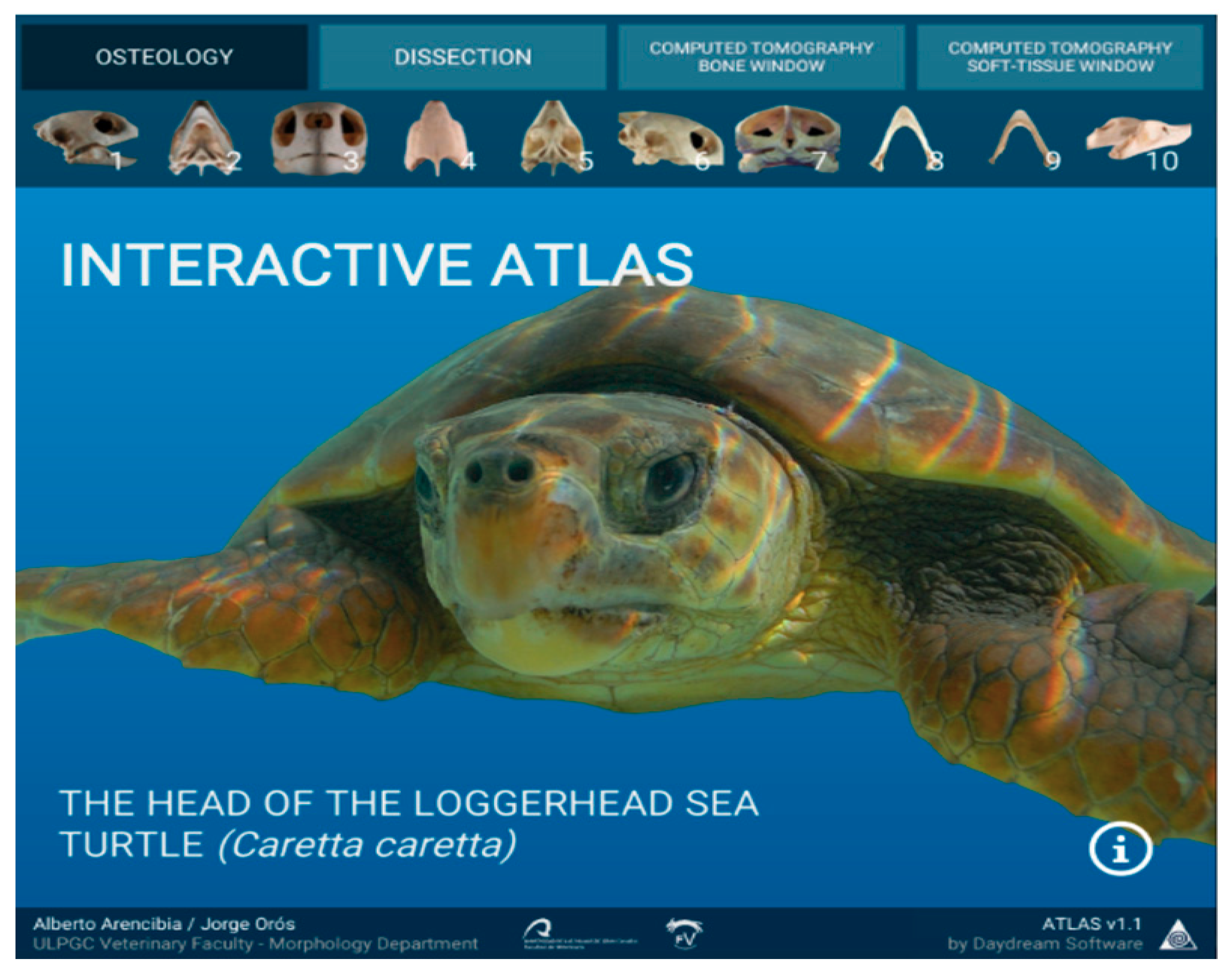
Anatomic Interactive Atlas of the Loggerhead Sea Turtle (Caretta caretta) Head


Abstract
Simple Summary
Abstract
1. Introduction
2. Materials and Methods
2.1. Animals
2.2. CT Technique
2.3. Anatomic Evaluation
2.4. Atlas Technical Methods
2.4.1. ITK-SNAP
- Full HD image.
- Segmentation file (with the defined anatomic area).
- Information file (containing the color and label of each anatomic structure).
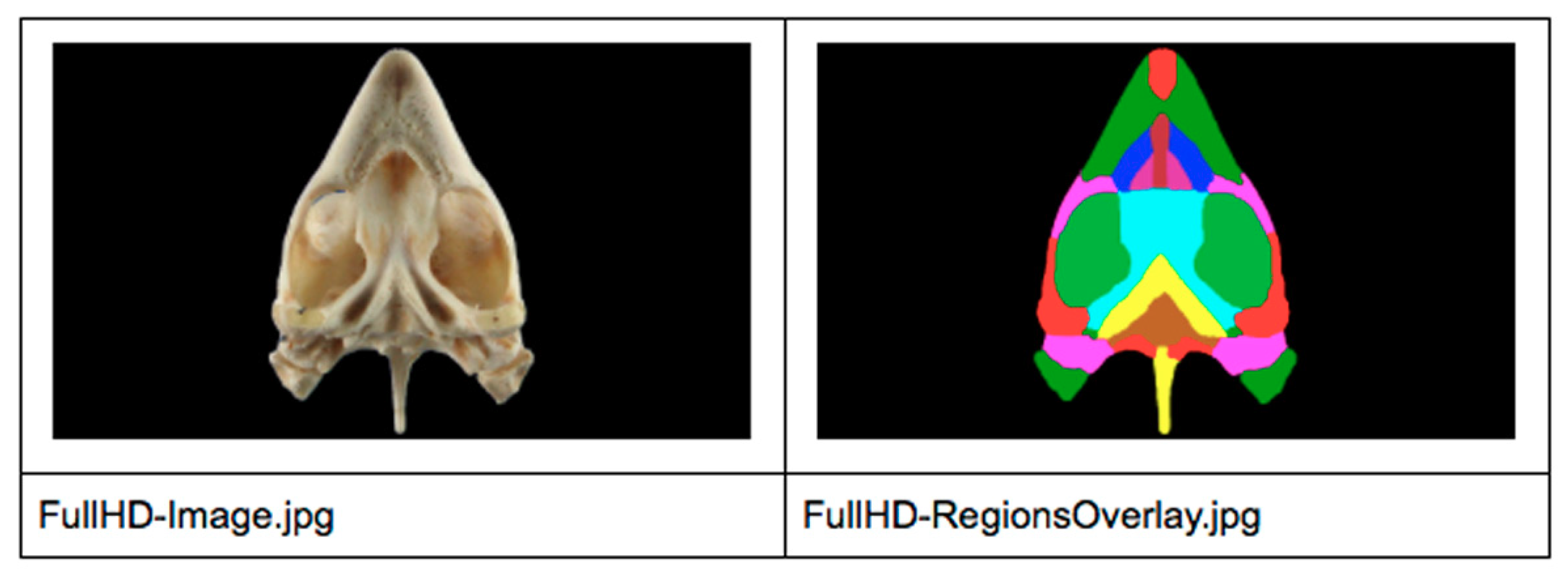
2.4.2. 2D Image Digital Processing
- Open the package into ITK-SNAP (Full HD image + Segmentation file + Information File).
- Check all region colors were unique so they could be identified later by their colors.
- Check all labels had no typographical errors.
- Define a common image resolution (we used Full HD image max resolution).
- Export Full HD Image as JPG.
- Enable all region colors (unique) and overlay them with full opacity.
- Export Full HD Regions Overlay (same resolution as Full HD image).
- Export Information File (for regions identification).
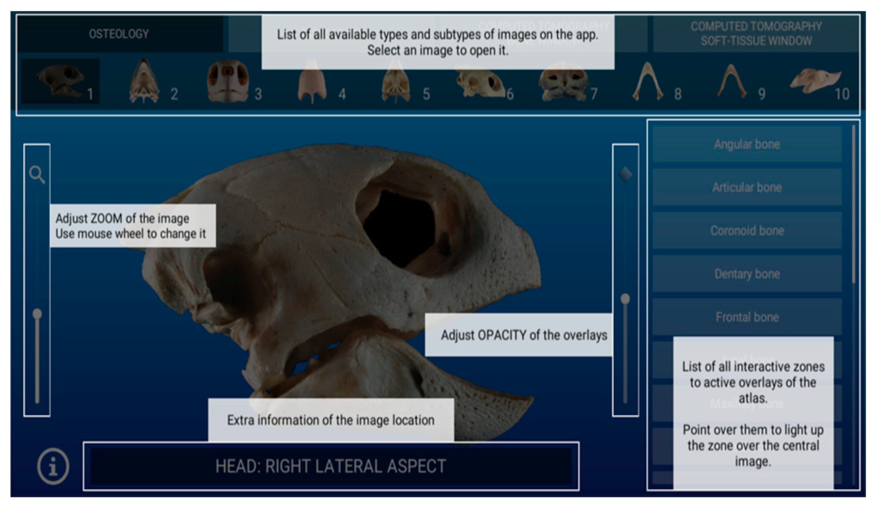
2.4.3. Unity 3D
2.4.4. Atlas Builder
- Select a parent folder with all image types for the atlas.
- Iterate over all image type folders and subfolders.
- Create needed data structures inside Unity 3D for each image found on iteration.
- Clean Full HD images by removing all black pixels from the main image and setting them to transparent.
- Generate Masks: new images for each region with the same resolution reading Information File to obtain the color code, select image pixels using Regions Overlay, and create a mask image with those pixels in white and all others transparent.
2.4.5. Atlas Renderer
2.4.6. Deployment
3. Results
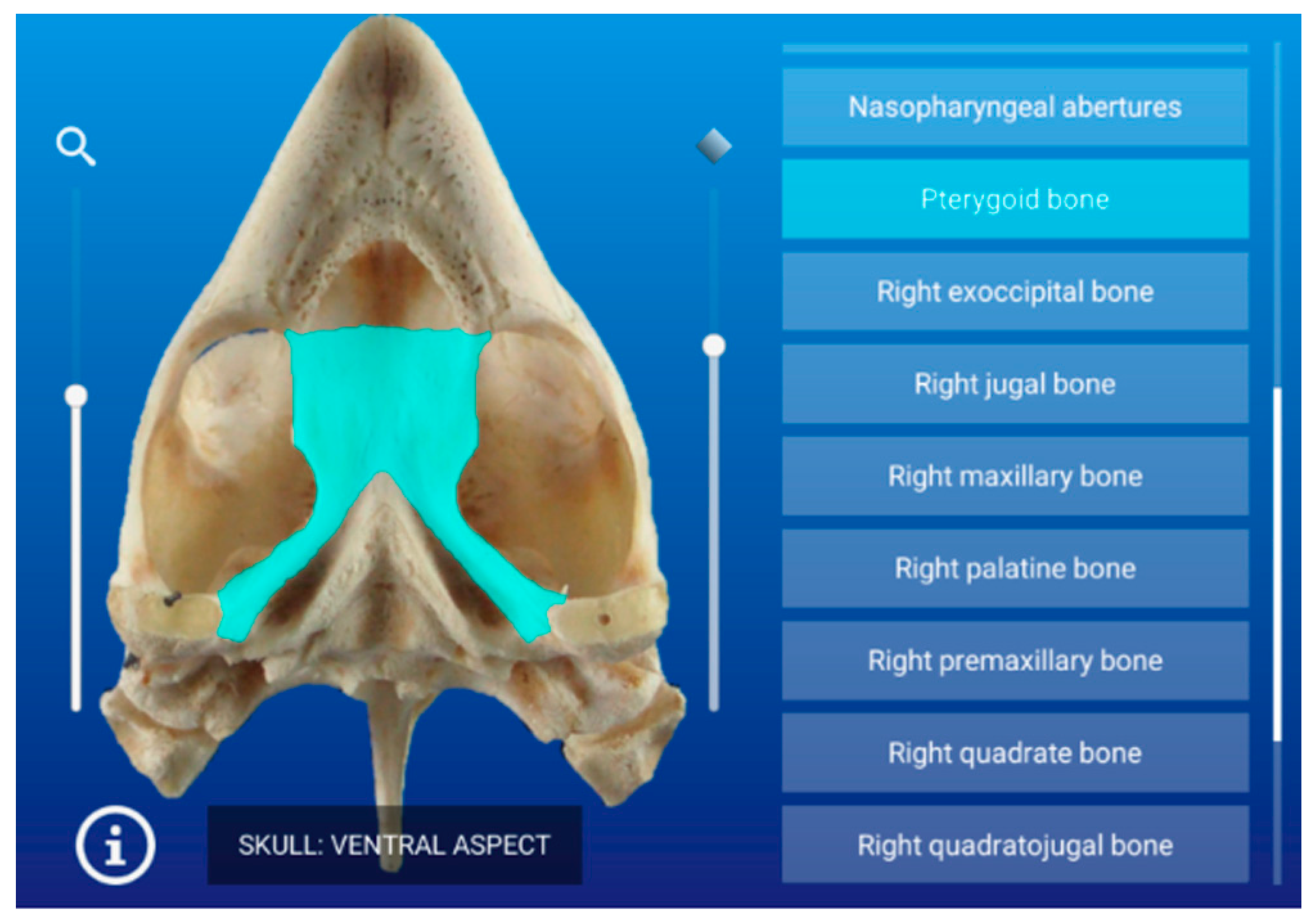
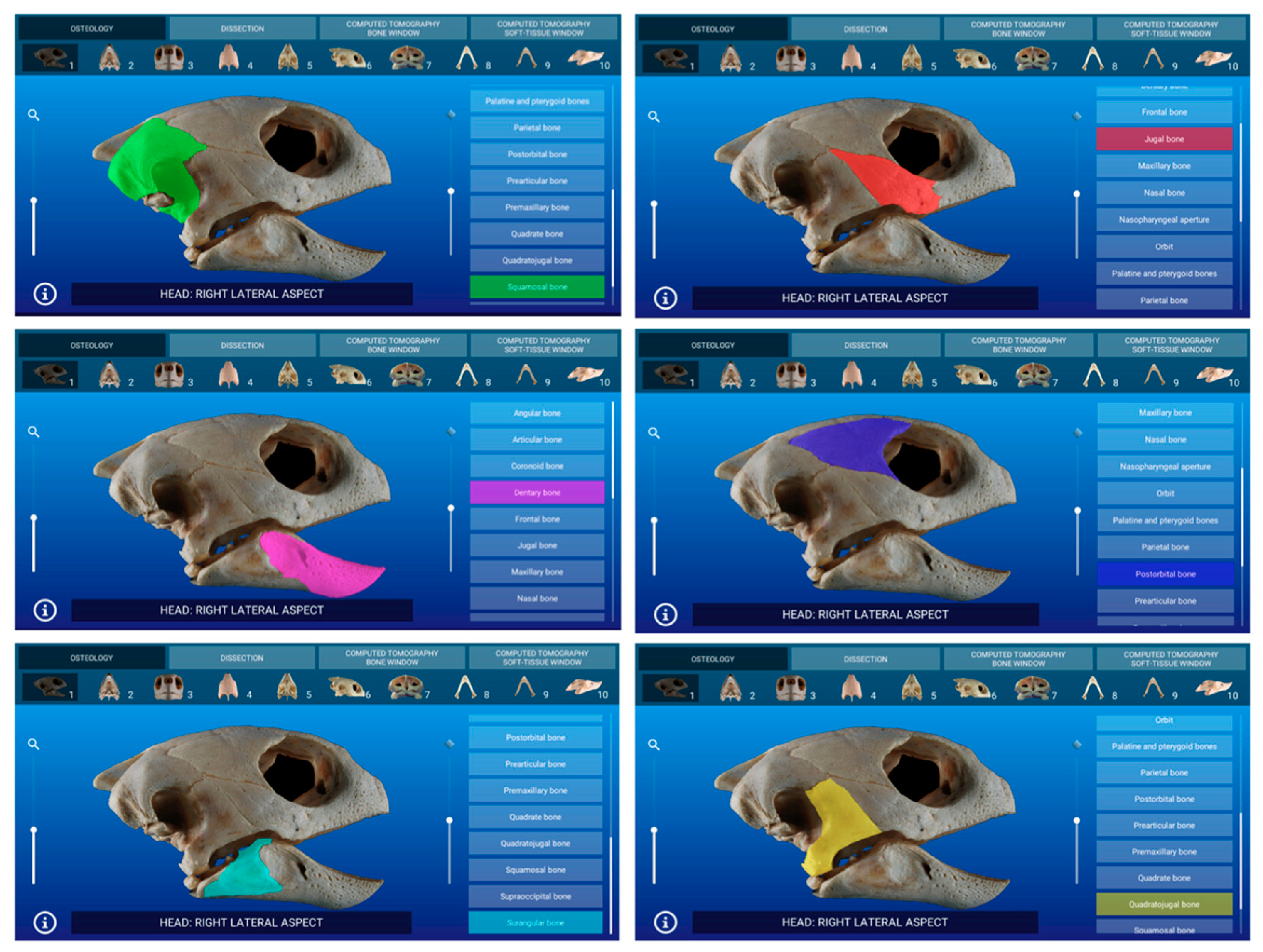
3.1. Osteology
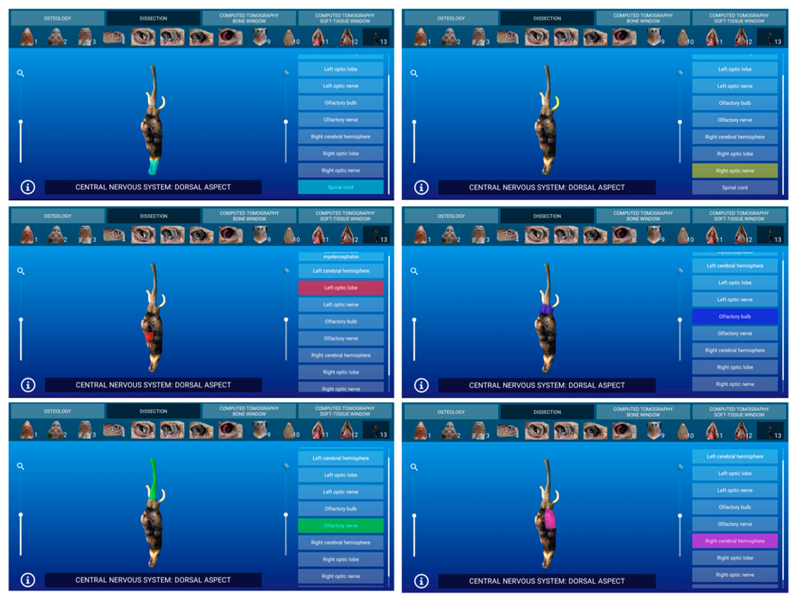
3.2. Dissections
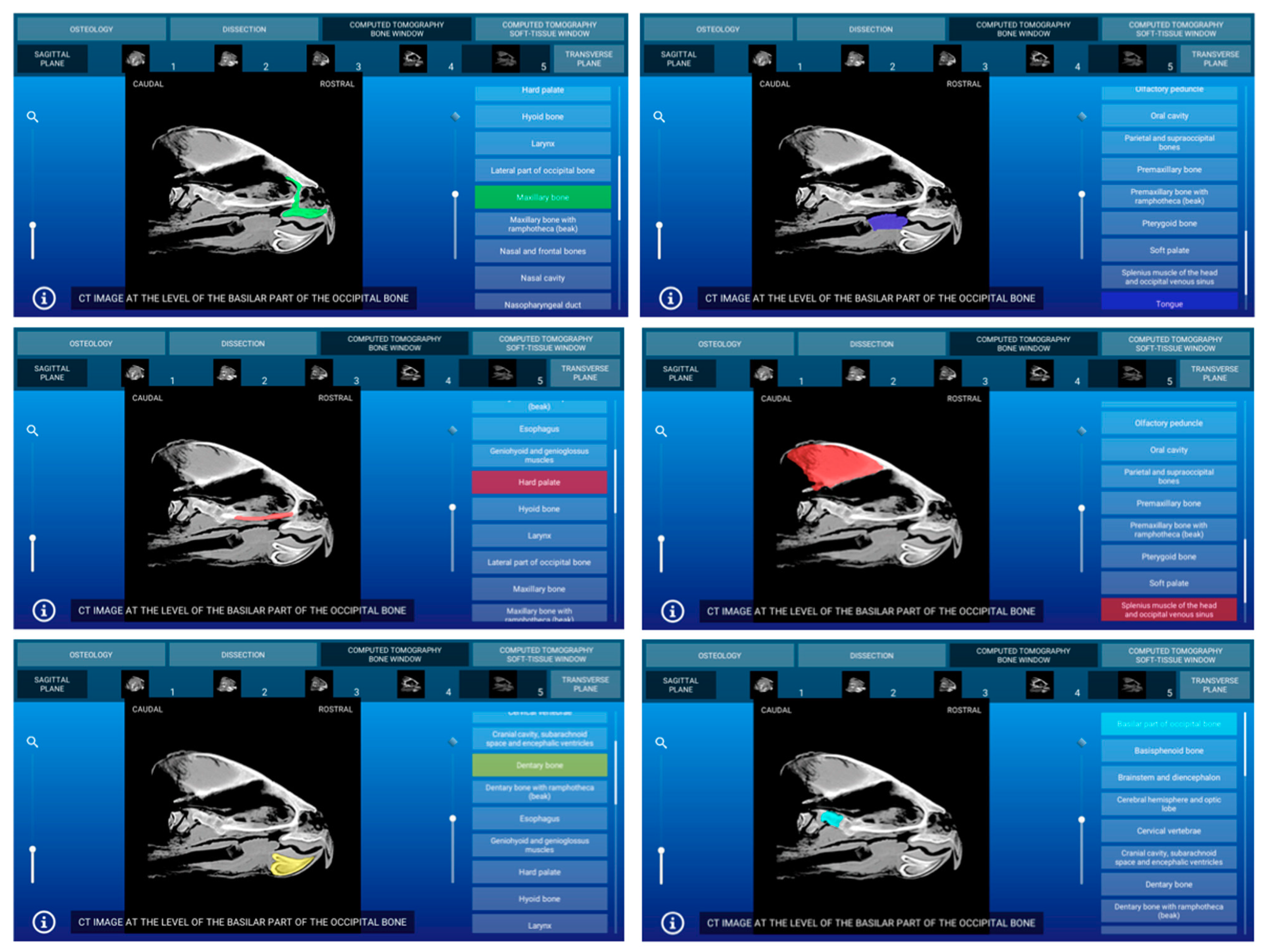
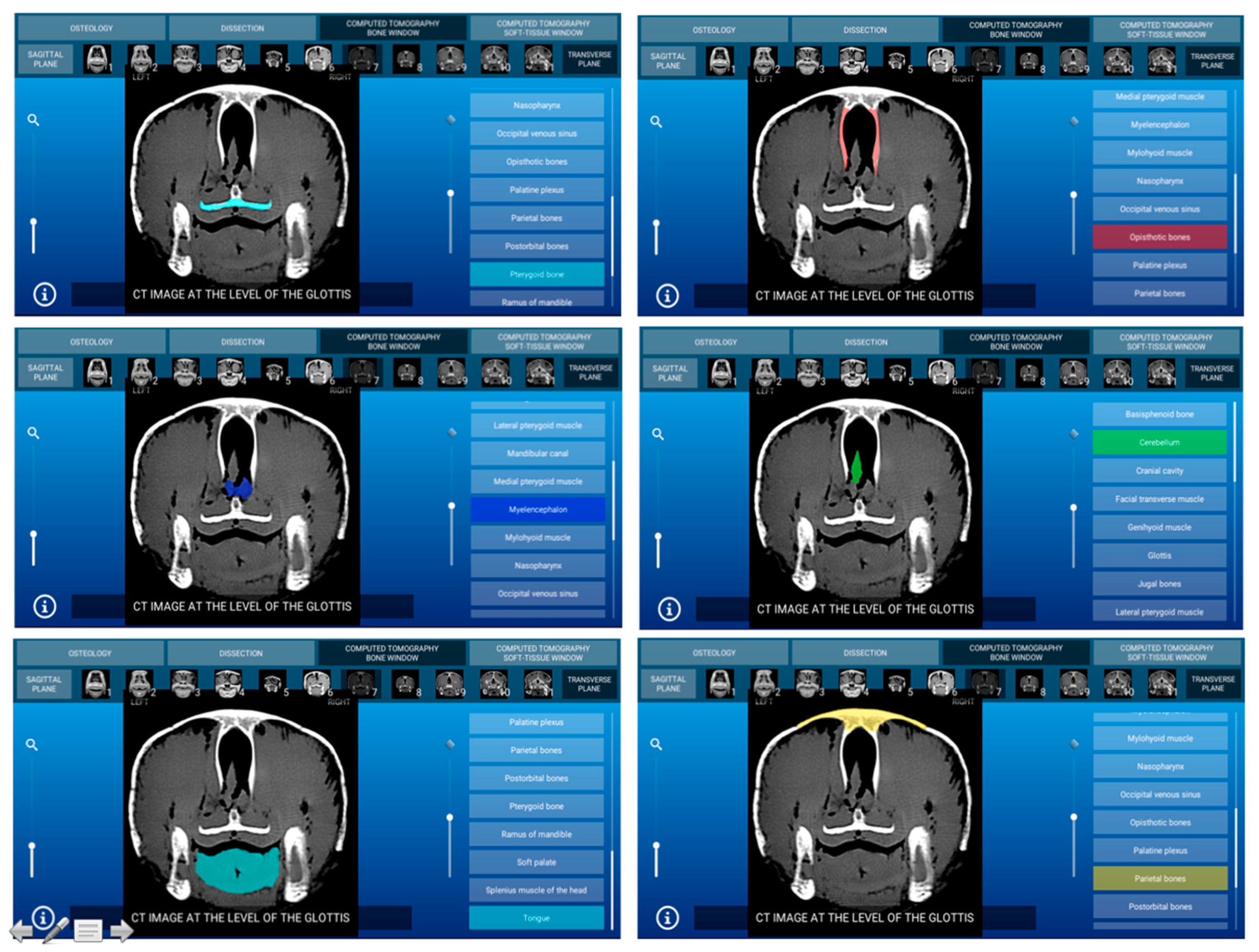
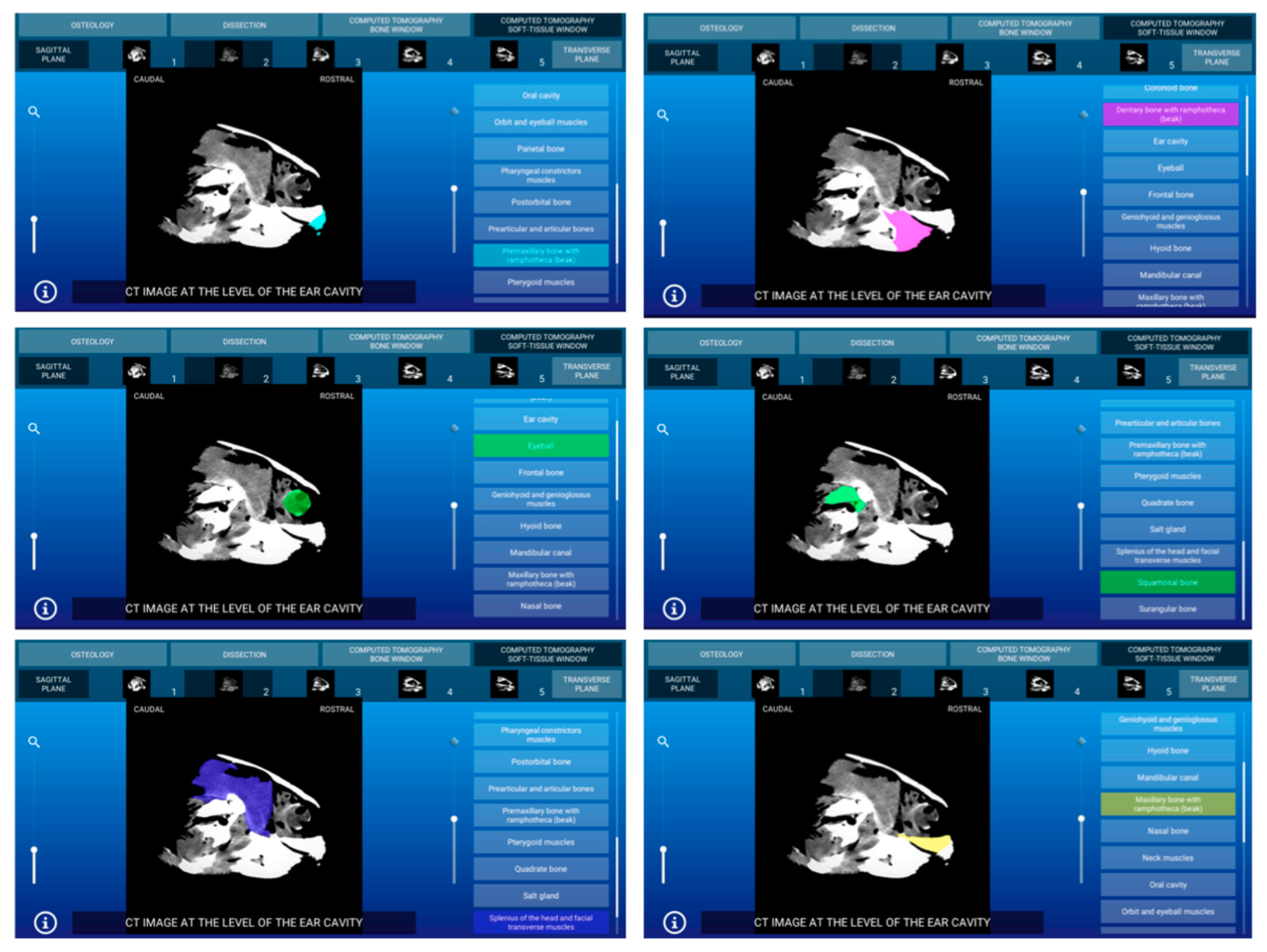
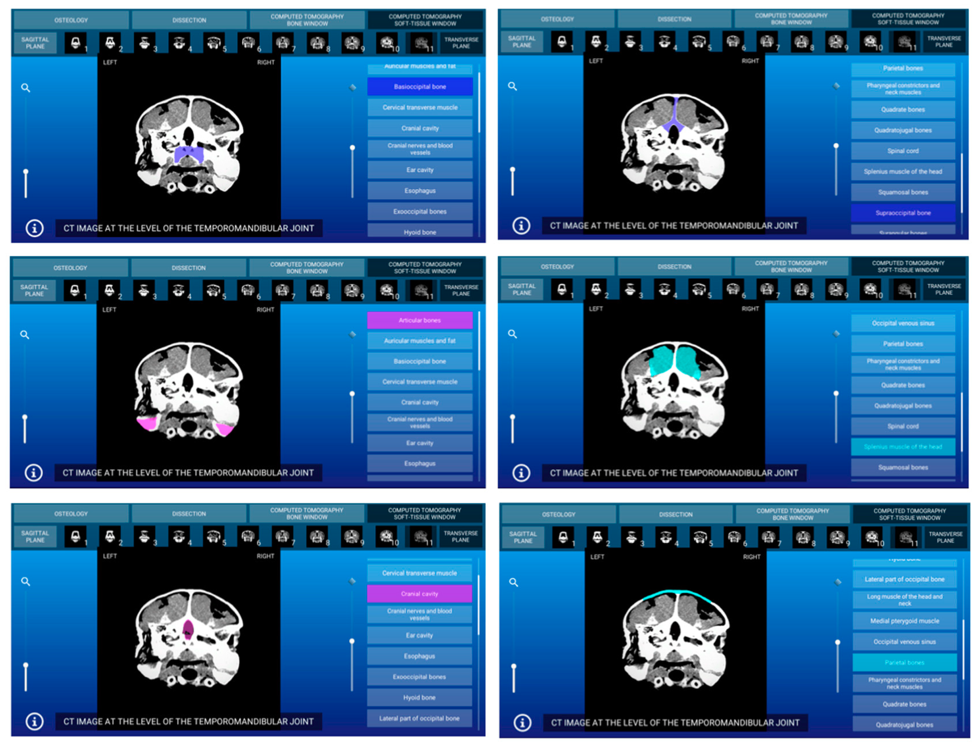
3.3. Computed Tomography Images
3.3.1. CT Bone Window
3.3.2. CT Soft Tissue Window
4. Discussion
5. Conclusions
Author Contributions
Funding
Institutional Review Board Statement
Data Availability Statement
Acknowledgments
Conflicts of Interest
References
- Monzón-Argüello, C.; Rico, C.; Carreras, C.; Calabuig, P.; Marco, A.; López-Jurado, L.F. Variation in spatial distribution of juvenile loggerhead turtles in the eastern Atlantic and western Mediterranean Sea. J. Exp. Mar. Biol. Ecol. 2009, 373, 79–86. [Google Scholar] [CrossRef]
- Casale, P.; Tucker, A.D. Caretta caretta, loggerhead turtle. IUCN Red List Threat. Species 2017, e.T3897A119333622. [Google Scholar] [CrossRef]
- Arencibia, A.; Rivero, M.A.; De Miguel, I.; Contreras, S.; Cabrero, A.; Orós, J. Computed tomographic anatomy of the head of the loggerhead sea turtle (Caretta caretta). Res. Vet. Sci. 2006, 81, 165–169. [Google Scholar] [CrossRef]
- Arencibia, A.; Hidalgo, M.R.; Vázquez, J.M.; Contreras, S.; Ramírez, G.; Orós, J. Sectional anatomic and magnetic resonance imaging features of the head of juvenile loggerhead sea turtles (Caretta caretta). Am. J. Vet. Res. 2012, 73, 1119–1127. [Google Scholar] [CrossRef] [PubMed]
- Camacho, M.; Quintana, M.P.; Luzardo, O.P.; Estévez, M.D.; Calabuig, P.; Orós, J. Metabolic and respiratory status of stranded juvenile loggerhead sea turtles (Caretta caretta): 66 cases (2008-2009). J. Am. Vet. Med. Assoc. 2013, 242, 396–401. [Google Scholar] [CrossRef]
- Orós, J.; Montesdeoca, N.; Camacho, M.; Arencibia, A.; Calabuig, P. Causes of stranding and mortality, and final disposition of loggerhead sea turtles (Caretta caretta) admitted to a wildlife rehabilitation center in Gran Canaria Island, Spain (1998-2014): A long-term retrospective study. PLoS ONE 2016, 11, e0149398. [Google Scholar] [CrossRef]
- Yamaguchi, Y.; Kitayama, C.; Tanaka, S.; Kondo, S.; Miyazaki, A.; Okamoto, K.; Yanagawa, M.; Kondoh, D. Computed tomographic analysis of internal structures within the nasal cavities of green, loggerhead and leatherback sea turtles. Anat. Rec. 2020, 1–7. [Google Scholar] [CrossRef]
- Manire, C.A.; Norton, T.M.; Stacy, B.A.; Innis, C.J.; Harms, C.A. Sea Turtle Health & Rehabilitation; Ross Publishing: Plantation, FL, USA, 2017; pp. 1–1010. [Google Scholar]
- Divers, S.J.; Stahl, S.J. Mader’s Reptile and Amphibian Medicine and Surgery; Elsevier: St. Louis, MO, USA, 2019; pp. 1–1511. [Google Scholar]
- Jacobson, E.R.; Garner, M.M. Diseases and Pathology of Reptiles Vol. I. Infectious Diseases and Pathology of Reptiles, Color Atlas and Text, 2nd ed.; CRC Press: Boca Raton, FL, USA, 2021; pp. 1–1013. [Google Scholar]
- Garner, M.M.; Jacobson, E.R. Diseases and Pathology of Reptiles Vol. II. Noninfectious Diseases and Pathology of Reptiles, Color Atlas and Text, 2nd ed.; CRC Press: Boca Raton, FL, USA, 2021; pp. 1–534. [Google Scholar]
- Aguirre, A.A.; Gómez, A. Essential veterinary education in conservation medicine and ecosystem health: A global perspective. Rev. Sci. Tech. Off. Int. Epiz. 2009, 28, 597–603. [Google Scholar] [CrossRef]
- Aguirre, A.A. Essential veterinary education in zoological and wildlife medicine: A global perspective. Rev. Sci. Tech. Off. Int. Epiz. 2009, 28, 605–610. [Google Scholar] [CrossRef]
- Wyneken, J. The Anatomy of Sea Turtles; US Department of Commerce NOAA Technical Memorandum; US Department of Commerce: Miami, FL, USA, 2001; pp. 1–27, 42–44, 68–70, 105, 109, 110, 115, 125–141. [Google Scholar]
- Valente, A.L.; Cuenca, R.; Zamora, M.A.; Parga, M.L.; Lavín, S.; Alegre, F.; Marco, I. Sectional anatomic and magnetic resonance imaging of coelomic structures of loggerhead sea turtles. Am. J. Vet. Res. 2006, 67, 1347–1353. [Google Scholar] [CrossRef]
- Latorre, R.; García-Sanz, M.P.; Moreno, M.; Hernández, F.; Gil, F.; López, O.; Ayala, M.D.; Ramirez, G.; Vázquez, J.M.; Arencibia, A.; et al. How useful is plastination in learning anatomy? J. Vet. Med. Educ. 2007, 34, 172–176. [Google Scholar] [CrossRef] [PubMed]
- Tiplady, C.; Lloyd, S.; Morton, J. Veterinary science student preferences for the source of dog cadavers used in anatomy teaching. Altern. Lab. Anim. 2011, 39, 461–469. [Google Scholar] [CrossRef] [PubMed]
- Rochelle, A.B.; Pasian, S.R.; Silva, R.H.; Rocha, M.J. Perceptions of undergraduate students on the use of animals in practical classes. Adv. Physiol. Educ. 2016, 40, 422–424. [Google Scholar] [CrossRef] [PubMed][Green Version]
- El Sharaby, A.A.; Alsafy, M.A.; El-Gendy, S.A. Equine anatomedia: Development, integration and evaluation of an e-learning resource in applied veterinary anatomy. Int. J. Morphol. 2015, 33, 1577–1584. [Google Scholar] [CrossRef]
- Little, W.B.; Artemiou, E.; Conan, A.; Sparks, C. Computer assisted learning: Assessment of the veterinary virtual anatomy education software IVALA™. Vet. Sci. 2018, 5, 58. [Google Scholar] [CrossRef]
- ITK-SNAP Home. Available online: http://www.itksnap.org/ (accessed on 5 November 2020).
- Yushkevich, P.A.; Piven, J.; Hazlett, H.C.; Smith, R.G.; Ho, S.; Gee, J.C.; Gerig, G. User-guided 3D active contour segmentation of anatomical structures: Significantly improved efficiency and reliability. Neuroimage 2006, 31, 1116–1128. [Google Scholar] [CrossRef]
- Cabezas, M.; Oliver, A.; Lladó, X.; Freixenet, J.; Cuadra, M.B. A review of atlas-based segmentation for magnetic resonance brain images. Comput. Methods Programs Biomed. 2011, 104, 158–177. [Google Scholar] [CrossRef]
- Yushkevich, P.A.; Gao, Y.; Gerig, G. ITK-SNAP: An interactive tool for semi-automatic segmentation of multi-modality biomedical images. Conf. Proc. IEEE Eng. Med. Biol. Soc. 2016, 3342–3345. [Google Scholar] [CrossRef]
- Sunico, S.K.; Hamel, C.; Styner, M.; Robertson, I.D.; Kornegay, J.N.; Bettini, C.; Parks, J.; Wilber, K.; Smallwood, J.E.; Thrall, D.E. Two anatomic resources of canine pelvic limb muscles based on CT and MRI. Vet. Radiol. Ultrasound 2012, 53, 266–272. [Google Scholar] [CrossRef]
- Liyanage, K.A.; Steward, C.; Moffat, B.A.; Opie, N.L.; Rind, G.S.; John, S.E.; Ronayne, S.; May, C.N.; O’Brien, T.J.; Milne, M.E.; et al. Development and implementation of a corriedale ovine brain atlas for use in atlas-based segmentation. PLoS ONE 2016, 11, e0155974. [Google Scholar] [CrossRef]
- Milne, M.E.; Steward, C.; Firestone, S.M.; Long, S.N.; O’Brien, T.J.; Moffat, B.A. Development of representative magnetic resonance imaging–based atlases of the canine brain and evaluation of three methods for atlas-based segmentation. Am. J. Vet. Res. 2016, 77, 395–403. [Google Scholar] [CrossRef] [PubMed]
- Orós, J.; Torrent, A.; Calabuig, P.; Déniz, S. Diseases and causes of mortality among sea turtles stranded in the Canary Islands, Spain (1998–2001). Dis. Aquat. Org. 2005, 63, 13–24. [Google Scholar] [CrossRef] [PubMed]
- Orós, J.; Camacho, M.; Calabuig, P.; Arencibia, A. Salt gland adenitis as only cause of stranding of loggerhead sea turtles Caretta caretta. Dis. Aquat. Org. 2011, 95, 163–166. [Google Scholar] [CrossRef] [PubMed]
- Bárcenas-Ibarra, A.; de la Cueva, H.; Rojas-Lleonart, I.; Abreu-Grobois, F.A.; Lozano-Guzmán, R.I.; Cuevas, E.; García-Gasca, A. First approximation to congenital malformation rates in embryos and hatchlings of sea turtles. Bird Defects Res. A Clin. Mol. Teratol. 2015, 103, 203–224. [Google Scholar] [CrossRef]
- Franchini, D.; Cavaliere, L.; Valastro, C.; Carnevali, F.; Van Der Esch, A.; Lai, O.; Di Bello, A. Management of severe head injury with brain exposure in three loggerhead sea turtles Caretta caretta. Dis. Aquat. Org. 2016, 119, 145–152. [Google Scholar] [CrossRef]
- Craven, K.S.; Sheppard, S.; Stallard, L.B.; Richardson, M.; Belcher, C.N. Investigating a link between head malformations and lack of pigmentation in loggerhead sea turtle embryos (Caretta caretta, Linnaeus, 1758) in the southeastern United States. Herpetol. Notes 2019, 12, 819–825. [Google Scholar]
- Oraze, J.S.; Beltran, E.; Thornton, S.M.; Gumpenberger, M.; Weller, R.; Biggi, M. Neurologic and computed tomography findings in sea turtles with history of traumatic injury. J. Zoo Wildl. Med. 2019, 50, 350–361. [Google Scholar] [CrossRef]
- Gaffney, E.S.; Parsons, T.S.; Williams, E.E. An Illustrated Glossary of Turtle Skull Nomenclature; American Museum Novitates: New York, NY, USA, 1972; pp. 1–33. [Google Scholar]
- Orós, J.; Torrent, A. Manual de Necropsia de Tortugas Marinas; Ediciones del Cabildo de Gran Canaria: Las Palmas de Gran Canaria, Spain, 2001; pp. 1–74. [Google Scholar]
- Unity3D User Manual: Copyright© 2020 Unity Technologies. Available online: https://docs.unity3d.com/Manual/index.html (accessed on 10 December 2020).
- Godbold, A. Mastering UI Development with Unity: An In-Depth Guide to Developing Engaging User Interfaces with Unity 5, Unity 2017, and Unity 2018; Packt Publishing: Birmingham, UK, 2018; Available online: https://www.amazon.es/Mastering-Development-Unity-depth-developing-ebook/dp/B079DX2N7R (accessed on 10 December 2020).
- Schiegl, J. Plugin to Identify Mouse over Irregular Shapes: 2014. Available online: https://github.com/senritsu/unitility/blob/master/Assets/Unitility/GUI/RaycastMask.cs (accessed on 10 December 2020).
- Naganobu, K.; Ogawa, H.; Oyadomari, N.; Sugimoto, M. Surgical repair of a depressed fracture in a green sea turtle, Chelonia mydas. J. Vet. Med. Sci. 2000, 62, 103–104. [Google Scholar] [CrossRef]
- Gordon, A.N.; Kelly, W.R.; Cribb, T.H. Lesions caused by cardiovascular flukes (Digenea: Spirorchidae) in stranded green turtles (Chelonia mydas). Vet. Pathol. 1998, 35, 21–30. [Google Scholar] [CrossRef]
- Jacobson, E.R.; Homer, B.L.; Stacy, B.A.; Greiner, E.C.; Szabo, N.J.; Chrisman, C.L.; Origgi, F.; Coberley, S.; Foley, A.M.; Landsberg, J.H.; et al. Neurological disease in wild loggerhead sea turtles Caretta caretta. Dis. Aquat. Org. 2006, 70, 139–154. [Google Scholar] [CrossRef]
- Brooks, D.E.; Ginn, P.E.; Miller, T.R.; Bramson, L.; Jacobson, E.R. Ocular fibropapillomas of green turtles (Chelonia mydas). Vet. Pathol. 1994, 31, 335–339. [Google Scholar] [CrossRef]
- Orós, J.; Lackovich, J.K.; Jacobson, E.R.; Brown, D.R.; Torrent, A.; Tucker, S.; Klein, P.A. Fibropapilomas cutáneos y fibromas viscerales en una tortuga verde (Chelonia mydas). Rev. Esp. Herpetol. 1999, 13, 17–26. [Google Scholar]
- Flint, M.; Limpus, C.J.; Patterson-Kane, J.C.; Murray, P.J.; Mills, P.C. Corneal fibropapillomatosis in green sea turtles (Chelonia mydas) in Australia. J. Comp. Pathol. 2010, 142, 341–346. [Google Scholar] [CrossRef] [PubMed]
- Camacho, M.; Calabuig, P.; Luzardo, O.P.; Boada, L.D.; Zumbado, M.; Orós, J. Crude oil as a stranding cause among loggerhead sea turtles (Caretta caretta) in the Canary Islands, Spain (1998-2011). J. Wildl. Dis. 2013, 49, 637–640. [Google Scholar] [CrossRef] [PubMed][Green Version]
- Wyneken, J. Computed tomography and magnetic resonance imaging anatomy of reptiles. In Reptile Medicine and Surgery, 2nd ed.; Mader, D.R., Ed.; Saunders Elsevier: St. Louis, MO, USA, 2006; pp. 1088–1096. [Google Scholar]
- Spadola, F.; Barillaro, G.; Morici, M.; Nocera, A.; Knotek, Z. The practical use of computed tomography in evaluation of shell lesions in six loggerhead turtles (Caretta caretta). Vet. Med. 2016, 61, 394–398. [Google Scholar] [CrossRef]


Publisher’s Note: MDPI stays neutral with regard to jurisdictional claims in published maps and institutional affiliations. |
© 2021 by the authors. Licensee MDPI, Basel, Switzerland. This article is an open access article distributed under the terms and conditions of the Creative Commons Attribution (CC BY) license (http://creativecommons.org/licenses/by/4.0/).
Share and Cite
Arencibia, A.; Melián, A.; Orós, J. Anatomic Interactive Atlas of the Loggerhead Sea Turtle (Caretta caretta) Head. Animals 2021, 11, 198. https://doi.org/10.3390/ani11010198
Arencibia A, Melián A, Orós J. Anatomic Interactive Atlas of the Loggerhead Sea Turtle (Caretta caretta) Head. Animals. 2021; 11(1):198. https://doi.org/10.3390/ani11010198
Chicago/Turabian StyleArencibia, Alberto, Aday Melián, and Jorge Orós. 2021. "Anatomic Interactive Atlas of the Loggerhead Sea Turtle (Caretta caretta) Head" Animals 11, no. 1: 198. https://doi.org/10.3390/ani11010198
APA StyleArencibia, A., Melián, A., & Orós, J. (2021). Anatomic Interactive Atlas of the Loggerhead Sea Turtle (Caretta caretta) Head. Animals, 11(1), 198. https://doi.org/10.3390/ani11010198