Histological Comparison of Testicular Needle Biopsy and En Bloc Samples in Abattoir Calves


 and
and
Simple Summary
Abstract
1. Introduction
2. Materials and Methods
2.1. Animals
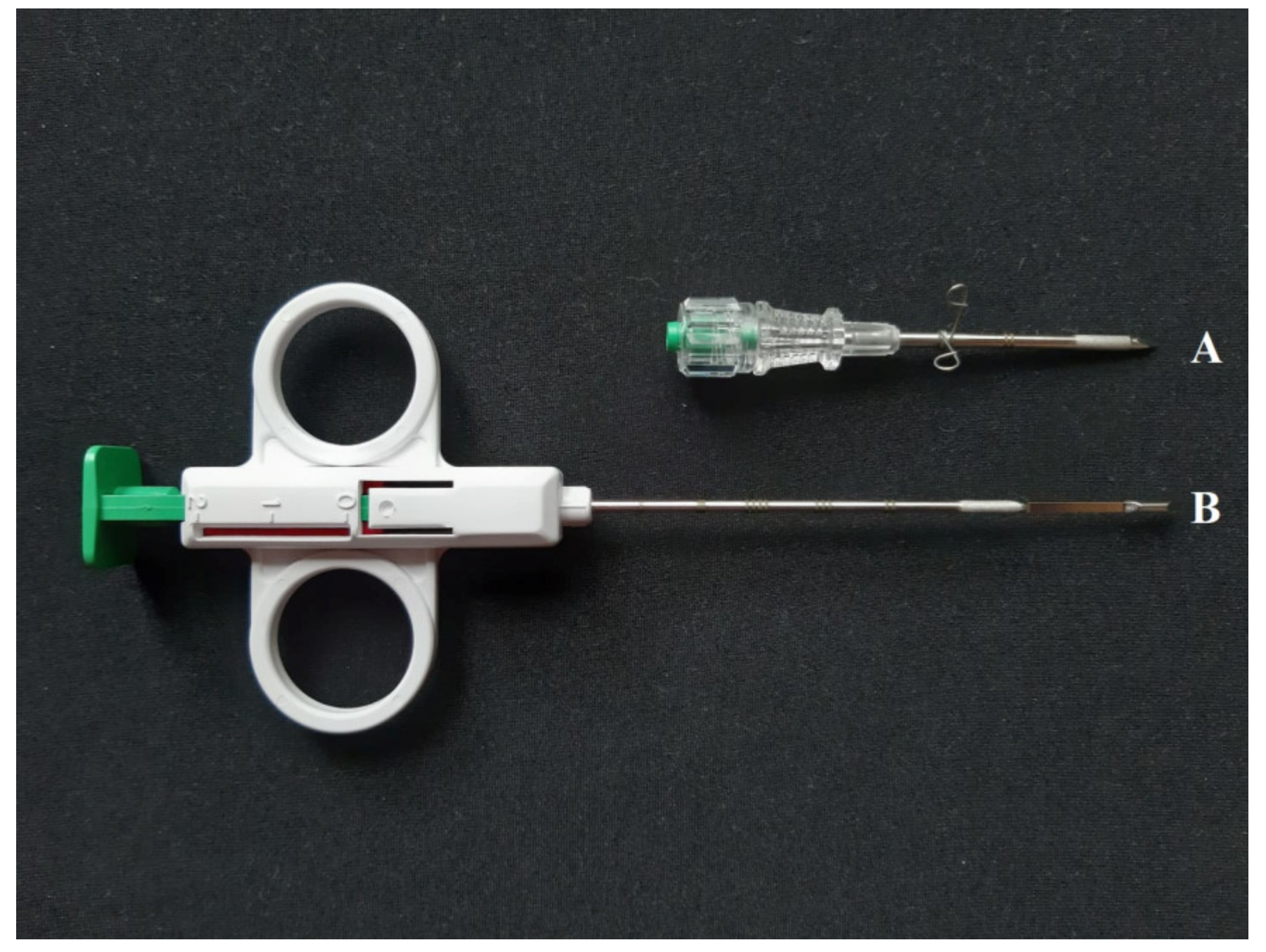
2.2. Collection of Testicular Samples
2.3. Histological Processing
2.4. Immunohistochemistry
2.5. Statistical Analysis
3. Results
3.1. General Data of the Animals
3.2. Data of the Histological Examination
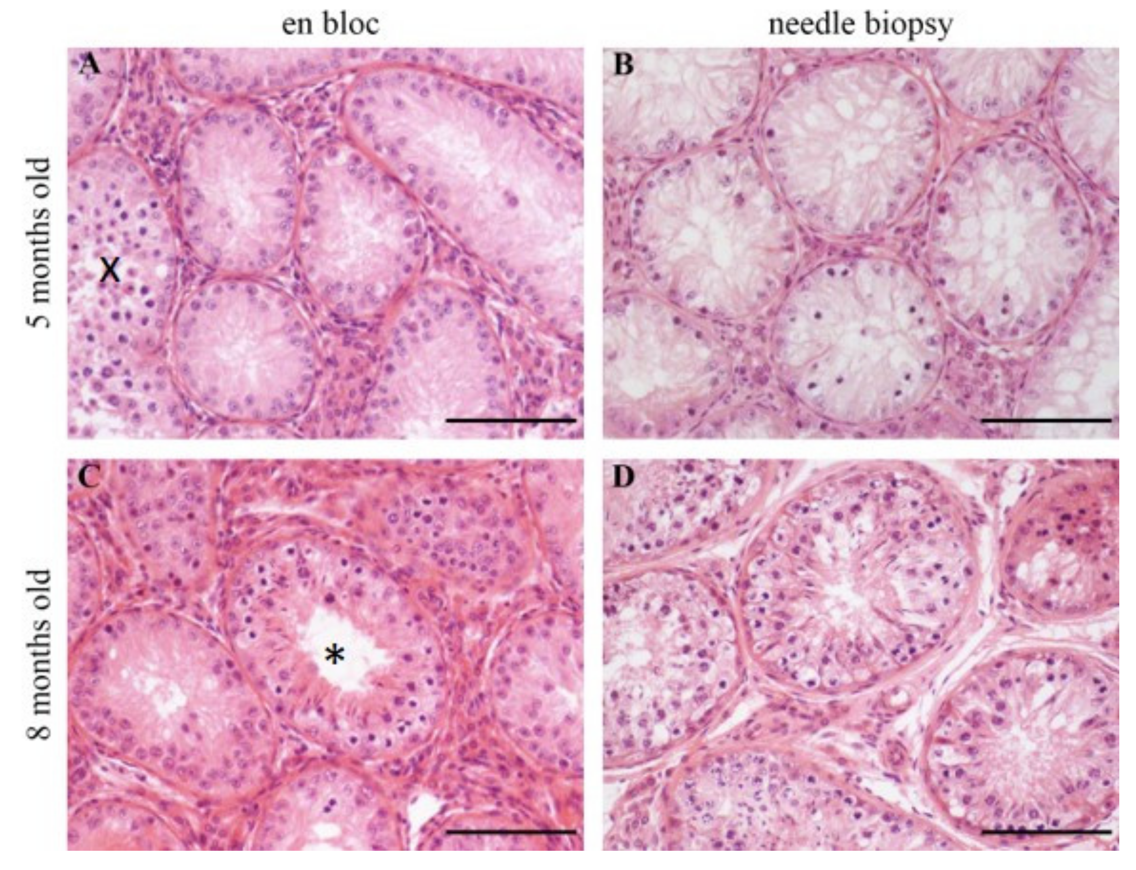
3.2.1. Investigations in HE Staining
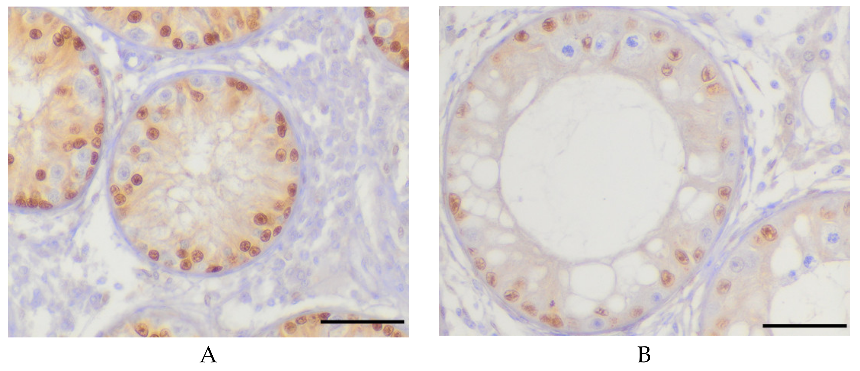
3.2.2. Investigations in Immunohistochemistry
4. Discussion
5. Conclusions
Author Contributions
Funding
Acknowledgments
Conflicts of Interest
References
- Rode, K.; Sieme, H.; Richterich, P.; Brehm, R. Characterization of the equine blood–testis barrier during tubular development in normal and cryptorchid stallions. Theriogenology 2015, 84, 763–772. [Google Scholar] [CrossRef]
- Heath, A.M.; Carson, R.L.; Purohit, R.C.; Sartin, E.M.; Wenzel, J.G.; Wolfe, D.F. Effects of testicular biopsy in clinically normal bulls. J. Am. Vet. Med. Assoc. 2002, 220, 507–512. [Google Scholar] [CrossRef]
- Odabaş, Ö.; Uĝraş, S.; Yilmaz, Y.; Aydin, S.; Atilla, M. Testicular needle biopsy: Is it a safe and adequate method? Int. Urol. Nephrol. 1997, 29, 591–595. [Google Scholar] [CrossRef] [PubMed]
- Rode, K.; Sieme, H.; Otzen, H.; Schwennen, C.; Lüpke, M.; Richterich, P.; Schrimpf, R.; Distl, O.; Brehm, R. Effects of repeated testicular biopsies in adult warmblood stallions and their diagnostic potential. J. Equine Vet. Sci. 2016, 38, 33–47. [Google Scholar] [CrossRef]
- Otzen, H. Intra- and peri-testicular damage resulting in alterations in sperm morphology after testicular biopsy in a Holstein bull—Poster Presentations. In Proceedings of the 21st Annual Conference of the European Society for Domestic Animal Reproduction (ESDAR), Bern, Switzerland; p. 120.
- Barth, A. The use of bull breeding soundness evaluation to identify subfertile and infertile bulls. Anim. Int. J. Anim. Biosci. 2018, 12, 158–164. [Google Scholar] [CrossRef] [PubMed]
- Kastelic, J.; Thundathil, J. Breeding soundness evaluation and semen analysis for predicting bull fertility. Reprod. Domest. Anim. 2008, 43, 368–373. [Google Scholar] [CrossRef] [PubMed]
- Leifland, K.; Lagerstedt, U.; Svane, G. Comparison of stereotactic fine needle aspiration cytology and core needle biopsy in 522 non-palpable breast lesions. Acta Radiol. 2003, 44, 387–391. [Google Scholar] [CrossRef]
- Wrobel, K.-H.; Dostal, S.; Schimmel, M. Postnatal development of the tubular lamina propria and the intertubular tissue in the bovine testis. Cell Tissue Res. 1988, 252, 639–653. [Google Scholar] [CrossRef]
- Abdel-Raouf, M. The postnatal development of the reproductive organs in bulls with special reference to puberty. Acta Endocrinol. (Copenh.) 1960, 34, 9–109. [Google Scholar] [CrossRef]
- Rawlings, N.; Evans, A.; Chandolia, R.; Bagu, E. Sexual maturation in the bull. Reprod. Domest. Anim. 2008, 43, 295–301. [Google Scholar] [CrossRef]
- Evans, A.; Pierson, R.; Garcia, A.; McDougall, L.; Hrudka, F.; Rawlings, N. Changes in circulating hormone concentrations, testes histology and testes ultrasonography during sexual maturation in beef bulls. Theriogenology 1996, 46, 345–357. [Google Scholar] [CrossRef]
- Curtis, S.K.; Amann, R. Testicular development and establishment of spermatogenesis in Holstein bulls. J. Anim. Sci. 1981, 53, 1645–1657. [Google Scholar] [CrossRef] [PubMed]
- Bagu, E.; Cook, S.; Gratton, C.; Rawlings, N. Postnatal changes in testicular gonadotropin receptors, serum gonadotropin, and testosterone concentrations and functional development of the testes in bulls. Reproduction 2006, 132, 403–411. [Google Scholar] [CrossRef] [PubMed]
- Byrne, C.; Fair, S.; English, A.; Urh, C.; Sauerwein, H.; Crowe, M.; Lonergan, P.; Kenny, D. Effect of breed, plane of nutrition and age on growth, scrotal development, metabolite concentrations and on systemic gonadotropin and testosterone concentrations following a GnRH challenge in young dairy bulls. Theriogenology 2017, 96, 58–68. [Google Scholar] [CrossRef]
- Lunstra, D.; Ford, J.; Echternkamp, S. Puberty in beef bulls: hormone concentrations, growth, testicular development, sperm production and sexual aggressiveness in bulls of different breeds. J. Anim. Sci. 1978, 46, 1054–1062. [Google Scholar] [CrossRef]
- Brito, L.; Barth, A.; Wilde, R.; Kastelic, J. Testicular ultrasonogram pixel intensity during sexual development and its relationship with semen quality, sperm production, and quantitative testicular histology in beef bulls. Theriogenology 2012, 78, 69–76. [Google Scholar] [CrossRef]
- Da Silva, K.M.; Zart, A.L.; Brum, K.B.; dos Santos Fernandes, C.E. Histopathological and histomorphometric testicular characteristics associated to reproductive condition in Bos indicus (Nellore) bulls. Semina Ciências Agrárias Londrina 2015, 36, 1935–1944. [Google Scholar] [CrossRef]
- Alturkistani, H.A.; Tashkandi, F.M.; Mohammedsaleh, Z.M. Histological stains: a literature review and case study. Glob. J. Health Sci. 2016, 8, 72. [Google Scholar] [CrossRef]
- Kobayashi, A.; Chang, H.; Chaboissier, M.C.; Schedl, A.; Behringer, R.R. Sox9 in testis determination. Ann. N. Y. Acad. Sci. 2005, 1061, 9–17. [Google Scholar] [CrossRef]
- Harikae, K.; Tsunekawa, N.; Hiramatsu, R.; Toda, S.; Kurohmaru, M.; Kanai, Y. Evidence for almost complete sex-reversal in bovine freemartin gonads: formation of seminiferous tubule-like structures and transdifferentiation into typical testicular cell types. J. Reprod. Dev. 2012. [Google Scholar] [CrossRef]
- Rajak, S.; Kumaresan, A.; Gaurav, M.; Aslam, M.M.; Mohanty, T.; Prasad, S.; Chakravarty, A.; Venkatasubramanian, V. Testicular biometry and semen quality is not altered by the process of fine needle aspiration in crossbred bulls. Indian J. Anim. Sci. 2013, 83, 732–735. [Google Scholar]
- Chapwanya, A.; Callanan, J.; Larkin, H.; Keenan, L.; Vaughan, L. Breeding soundness evaluation of bulls by semen analysis, testicular fine needle aspiration cytology and trans-scrotal ultrasonography. Ir. Vet. J. 2008, 61, 315–318. [Google Scholar] [CrossRef] [PubMed]
- Cohen, M.S.; Frye, S.; Warner, R.S.; Leiter, E. Testicular needle biopsy in diagnosis of infertility. Urology 1984, 24, 439–442. [Google Scholar] [CrossRef]
- Gassner, F.; Hill, H. Testicular biopsy in the bull: II. Effect on morphology of testes. Fertil. Steril. 1955, 6, 290–301. [Google Scholar] [CrossRef]
- Cohen, M.S.; Warner, R.S. Needle biopsy of testes: a safe outpatient procedure. Urology 1987, 29, 279–281. [Google Scholar] [CrossRef]
- Gruber, L.; Urdl, M.; Schauer, A.; Steinwender, R. Einfluss des Kraftfutterniveaus in der Stiermast auf die Mast-und Schlachtleistung bei Maissilage mit niedriger Energiekonzentration. 36. Viehwirtschaftliche Fachtagung 2009; Lehr- und Forschungszentrum für Landwirtschaft Raumberg-Gumpenstein, 2009; pp. 77–83. [Google Scholar]
- Amann, R.; Almquist, J. Reproductive Capacity of Dairy Bulls. VIII. Direct and Indirect Measurement of Testicular Sperm Production. J. Dairy Sci. 1962, 45, 774–781. [Google Scholar] [CrossRef]
- Swierstra, E. Structural composition of Shorthorn bull testes and daily spermatozoa production as determined by quantitative testicular histology. Can. J. Anim. Sci. 1966, 46, 107–119. [Google Scholar] [CrossRef]
- Swierstra, E. A comparison of spermatozoa production and spermatozoa output of Yorkshire and Lacombe boars. Reproduction 1968, 17, 459–469. [Google Scholar] [CrossRef]
- Barth, A.; Brito, L.; Kastelic, J. The effect of nutrition on sexual development of bulls. Theriogenology 2008, 70, 485–494. [Google Scholar] [CrossRef]
- Kastelic, J.P. Understanding and evaluating bovine testes. Theriogenology 2014, 81, 18–23. [Google Scholar] [CrossRef]
- Berndtson, W.; Igboeli, G.; Pickett, B. Relationship of absolute numbers of Sertoli cells to testicular size and spermatogenesis in young beef bulls. J. Anim. Sci. 1987, 64, 241–246. [Google Scholar] [CrossRef] [PubMed]
- Gebauer, M.; Pickett, B.; Swierstra, E. Reproductive physiology of the stallion. II. Daily production and output of sperm. J. Anim. Sci. 1974, 39, 732–736. [Google Scholar] [CrossRef] [PubMed]
- Bagu, E.; Madgwick, S.; Duggavathi, R.; Bartlewski, P.; Barrett, D.; Huchkowsky, S.; Cook, S.; Rawlings, N. Effects of treatment with LH or FSH from 4 to 8 weeks of age on the attainment of puberty in bull calves. Theriogenology 2004, 62, 861–873. [Google Scholar] [CrossRef] [PubMed]
- Brito, L.; Barth, A.; Rawlings, N.; Wilde, R.; Crews, D.; Mir, P.; Kastelic, J. Circulating metabolic hormones during the peripubertal period and their association with testicular development in bulls. Reprod. Domest. Anim. 2007, 42, 502–508. [Google Scholar] [CrossRef] [PubMed]


| Age (mo) | 5 | 6 | 7 | 8 |
| Number (n) | 5 | 10 | 10 | 7 |
| ANOVA – Mixed Model | Proximal – Middle | Proximal – Distal | Middle – Distal |
|---|---|---|---|
| Number of tubules with ES 1 | 0.11 | 0.01 | 0.67 |
| Number of tubular cross sections | 0.68 | 0.22 | 0.69 |
| Outer tubular diameter | 0.97 | 0.45 | 0.32 |
| Inner tubular diameter | 0.99 | 0.27 | 0.22 |
| Thickness of the tubular wall | 0.76 | 0.77 | 1.00 |
| Number of SC 2 per tubular cross section | 0.85 | 0.98 | 0.75 |
| Histological Parameter | En bloc Samples | Testicular NB 3 | p-Value (Wilcoxon Test) |
|---|---|---|---|
| Number of tubular cross sections | 241 ± 62 | 152 ± 63 | <0.05 |
| Number of tubules with ES 1 | 20 ± 28 | 11 ± 15 | 0.11 |
| Outer tubular diameter | 148 ± 27 | 134 ± 17 | 0.01 |
| Inner tubular diameter | 63 ± 31 | 29 ± 25 | <0.01 |
| Thickness of the tubular wall | 1.87 ± 0.19 | 2.06 ± 0.24 | <0.01 |
| Number of SC 2 per tubular cross section | 18 ± 5 | 14 ± 5 | <0.01 |
| ANOVA—Mixed Model | Proximal—NB3 | Middle—NB | Distal—NB |
|---|---|---|---|
| Number of tubules with ES 1 | 0.0170 | 0.0028 | 0.0028 |
| Number of tubular cross sections | <0.0001 | <0.0001 | <0.0001 |
| Outer tubular diameter | <0.0001 | <0.0001 | <0.0001 |
| Inner tubular diameter | <0.0001 | <0.0001 | <0.0001 |
| Thickness of the tubular wall | 0.0012 | 0.0003 | 0.0002 |
| Number of SC 2 per tubular cross section | 0.0002 | 0.0002 | <0.0001 |
| Number of Animals With Tubules With ES 1 in the Different Sample Types | ||
|---|---|---|
| Age (mo) | En bloc Samples | Needle Biopsy |
| 5 | 1/5 (20%) | 0/5 (0 %) |
| 6 | 8/10 (80%) | 7/10 (70%) |
| 7 | 8/10 (80%) | 6/10 (60%) |
| 8 | 7/7 (100%) | 7/7 (100%) |
| Staining | Mean Values of Number of SC 1 Per Tubular Cross Section | p-Value | |
|---|---|---|---|
| En Bloc Samples | Needle Biopsy Samples | ||
| HE ² | 17.9 ± 5.0 | 14.2 ± 4.9 | 0.0001 |
| SOX9 | 21.5 ± 0.5 | 21.1 ± 1.0 | 0.4097 |
© 2020 by the authors. Licensee MDPI, Basel, Switzerland. This article is an open access article distributed under the terms and conditions of the Creative Commons Attribution (CC BY) license (http://creativecommons.org/licenses/by/4.0/).
Share and Cite
Rohländer, M.; Otzen, H.; Rode, K.; Jung, K.; Schmicke, M.; Harborth, T.; Langeheine, M.; Brehm, R.; Bajcsy, Á.C. Histological Comparison of Testicular Needle Biopsy and En Bloc Samples in Abattoir Calves. Animals 2020, 10, 918. https://doi.org/10.3390/ani10050918
Rohländer M, Otzen H, Rode K, Jung K, Schmicke M, Harborth T, Langeheine M, Brehm R, Bajcsy ÁC. Histological Comparison of Testicular Needle Biopsy and En Bloc Samples in Abattoir Calves. Animals. 2020; 10(5):918. https://doi.org/10.3390/ani10050918
Chicago/Turabian StyleRohländer, Maike, Henning Otzen, Kristina Rode, Klaus Jung, Marion Schmicke, Teresa Harborth, Marion Langeheine, Ralph Brehm, and Árpád Csaba Bajcsy. 2020. "Histological Comparison of Testicular Needle Biopsy and En Bloc Samples in Abattoir Calves" Animals 10, no. 5: 918. https://doi.org/10.3390/ani10050918
APA StyleRohländer, M., Otzen, H., Rode, K., Jung, K., Schmicke, M., Harborth, T., Langeheine, M., Brehm, R., & Bajcsy, Á. C. (2020). Histological Comparison of Testicular Needle Biopsy and En Bloc Samples in Abattoir Calves. Animals, 10(5), 918. https://doi.org/10.3390/ani10050918