Article

Supplementation of Lactobacillus plantarum or Macleaya cordata Extract Alleviates Oxidative Damage Induced by Weaning in the Lower Gut of Young Goats

1
CAS Key Laboratory for Agro-Ecological Processes in Subtropical Region, National Engineering Laboratory for Pollution Control and Waste Utilization in Livestock and Poultry Production, Hunan Provincial Key Laboratory of Animal Nutritional Physiology and Metabolic Process, Institute of Subtropical Agriculture, The Chinese Academy of Sciences, Changsha 410125, Hunan, China
2
College of Animal Science and Technology, Hunan Agricultural University, Changsha 410128, Hunan, China
3
Hunan Co-Innovation Center of Animal Production Safety, CICAPS, Changsha 410128, Hunan, China
4
University of Chinese Academy of Sciences, Beijing 100049, China
*
Authors to whom correspondence should be addressed.
Animals 2020, 10(4), 548; https://doi.org/10.3390/ani10040548
Submission received: 18 February 2020 / Revised: 20 March 2020 / Accepted: 21 March 2020 / Published: 25 March 2020
(This article belongs to the Section Animal Physiology)

Abstract

:

Simple Summary

Weaning stress is a serious problem in the goat production industry. This article demonstrates that weaning can induce intestinal oxidative damage, and adding Lactobacillus plantarum or Macleaya cordata can alleviate the oxidative damage of the lower gut in weaned young goats in an intestinal region-specific way.

Abstract

Weaning usually leads to stress in livestock, which has a negative impact on their growth and development. Research on oxidative stress and inflammation induced by weaning has not been reported in goats. Here, we focused on oxidative stress profile and inflammation status of the lower gut (jejunum, ileum, and colon) of goats. First, we illustrated the status of antioxidant activity and inflammation in the intestine of young goats on pre-(2 weeks postnatal, 2 wkpn) or post-(11 wkpn, weaning at day 45 postnatal)-weaned period of young goats. Malondialdehyde (MDA) was higher (p < 0.0001) in jejunum and ileum of the young goats in 11 wkpn than that in 2 wkpn, whereas superoxide dismutase (SOD) activity was lower (p = 0.012) in the lower gut of the young goats with 11 wkpn than that in 2 wkpn. Furthermore, we intended to explore the protective influence of a probiotic additive (Lactobacillus plantarum (LAC) P-8, 10 g/d) and a prebiotic additive (Sangrovit®, Macleaya cordata (MAC) extract 3.75% w/w premix, 0.3 g/d) on intestinal oxidative stress and inflammation status of early-weaned young goats (average weights of 5.63 ± 0.30 kg, weaned on d 45 postnatal). We observed that LAC reduced MDA in jejunum and ileum (p < 0.0001), increased SOD activity in ileum (p < 0.01), and increased glutathione peroxidase (GSH-Px) activity in jejunum (p < 0.05). Similarly, MAC reduced MDA contents (p < 0.0001), increased SOD activities (p < 0.01) in both of ileum and jejunum, and increased GSH-Px activity (p < 0.05) in jejunum. However, there were no differences in feed intake, average daily gain, inflammation parameters (interleukin 2 and interleukin 6), and colon oxidative stress profile (MDA, SOD, or GSH-Px) among treatments. These results provide evidence that weaning induces oxidative damage in the lower gut of young goats, and the oxidative damage in the small intestine can be reduced by adding the addition of LAC or MAC in diets depending on the region of the lower gut.

1. Introduction

Weaning stress is still a long-standing challenge for the livestock productivity and health. The critical period in early life, around pre- and postweaning, results in digestive problem and diarrhea due to colonization of pathogenic bacteria in the gut [1]. Moreover, weaning switches the diet of young mammals from a liquid feed to a completely solid feed, and is commonly associated with the upregulation of proinflammatory cytokine and overproduction of reactive oxygen species (ROS) [2]. Increased oxidative stress and inflammation induced by weaning in mammals have been reported [3,4,5]. Strategies to mitigate the adverse effects of ROS and inflammation include nutrient regulation and feeding management.
Genus Lactobacillus (LAC) used as a probiotic additive to avoid the imbalance of redox homeostasis has been widely investigated in other animal species. For instance, Wu et al. [6] have reported that LAC delbrueckii extracellular secretions have the most robust antioxidant activity, which can remove superoxide free radicals. Glutathione peroxidase (GSH-Px) and superoxide dismutase (SOD) can decrease oxidative production by preventing lipid peroxidation [7]. The upregulation of GSH and SOD, and reduction of oxidative stress as a result of lactobacillus supplementation in weaning pigs has been reported [8]. Paszti-Gere et al. [9] have reported that LAC can inhibit intestinal interleukin 8 (IL-8), which confirms the anti-inflammatory properties of the active metabolites by LAC under acute oxidative stress. Furthermore, lactic acid bacteria reduce inflammation caused by bacteria and LPS, by reducing the levels of pro-inflammatory cytokines interleukin 1 (IL-1), interleukin 6 (IL-6), tumor necrosis factor-α (TNF-α), interleukin 17 (IL-17), and interleukin 22 (IL-22), as well as increasing the levels of anti-inflammatory cytokines interleukin 2, 4, and 10 (IL-2, -4, and -10) [10,11,12,13,14].
The probiotic additive of Macleaya cordata (MAC) is a plant extract, containing sanguinarine and chelerythrine. The two components are from benzylisoquinoline alkaloids and beneficial for animal intestinal health [15]. Li et al. [16] have found that MAC can upregulate antioxidant activity. The authors also reported that the extracts of MAC enhance the intestinal barrier function of piglets and have antibacterial and anti-inflammatory effects on the intestinal tract. We hypothesized that weaning induces oxidative damage and inflammation in young goats, and the inclusion of LAC or MAC in the diets of goats improves growth performance and alleviates oxidative damage and inflammation induced by weaning.
The importance of the lower gut in ruminants, especially in young ruminants, has received more attention in recent years. Therefore, the objectives of this study were: (1) to investigate the weaning effects on the oxidative stress and inflammation in the lower gut of young goats and (2) to explore the effects of supplementing probiotics or prebiotics additives on growth performance as well as oxidative stress and inflammation in the lower gut of young weaned goats.

2. Materials and Methods

2.1. Experimental Design and Animal Managements

For the sake of exploring the protective influence of pre-/probiotics on early-weaned young goats, we conducted two different experiments. The objective of experiment 1 (Exp. 1) was to investigate the weaning effects on the oxidative stress and inflammation in the lower gut of young goats, while the objective of experiment 2 (Exp. 2) was to evaluate the protective function of pre-/probiotics on the hindgut of early-weaned goats. The Animal Care Committee approved all the protocols for the animal procedure at the Institute of Subtropical Agriculture, Chinese Academy of Sciences (ISA-CAS), following the Animal Care and the Use Guidelines of ISA-CAS, China (KYNEAAM-2017-ZLT-0009).
Exp. 1: Early-weaned procedure was conducted on postnatal day 45; 17 young female goats (10 preweaned young goats and seven postweaned young goats (weaned at 45 days old)) were used to investigate the effects of weaning on intestinal oxidative stress and inflammation in the young goats. The young goats (Liuyang black goat, local breed) were all from pluriparous ewes and were provided by a local goat farm (Liuyang, Hunan province, China).
After birth, kids were ear-tagged, weighed, and reared together with their mothers until weaning at day 45. Before weaning, all goat mothers were grazed and received a 300 g/d concentrate diet. The concentrate contained (dry matter, DM, basis) of 740 g corn meal, 203 g soybean meal, 13 g calcium bicarbonate, 16 g calcium carbonate, 8 g sodium chloride, and 2 g mineral–vitamin premix per kg. It supplied 11.0 MJ/kg of metabolizable energy and 154 g/kg of crude protein. From birth to day 21, kids received no supplemental feed other than their mother’s milk. From days 22 to 45, the kids were provided goat milk and a starter feed (321 g extruded soybean, 115 g whey powder, 167 g maize flour, 182 g fat powder, 133 g soybean meal, 25 g calcium hydrophosphate, 11 g calcium carbonate, 13 g sodium chloride, and 33 g premix per kg; dry matter basis) for ad libitum intake. After weaning at day 45, kids received a starter diet and fresh grass in a ratio of 6:4 for ad libitum intake without milk. Ten preweaned kids (an average body weight of 2.28 ± 0.0230 kg; means ± SE) with ages between 7 and 14 d and seven postweaned young goats (an average body weight of 5.70 ± 0.957 kg) with an age of 77 days were slaughtered.
Exp. 2: Twenty-one weaned young female goats (weaned at day 45 with an averaged body weight of 5.63 ± 0.30 kg) were provided by the same local goat farm as for Exp. 1. The diets and management of the goats preweaning were also the same as described in Exp. 1. Goats were housed individually, and randomly assigned into one of the three diets: (1) a control diet without a probiotics additive or a prebiotics additive (CON), (2) a control diet supplementing with 10 g/day Lactobacillus plantarum P-8 (4.0 × 10⁹ CFU/g, LAC), the detail information of Lactobacillus plantarum P-8 strains was described by Bao et al. [17], and (3) a control diet supplementing with 0.3 g/day plant extract (Sangrovit®, MAC extract 3.75% w/w premix). The supplementing rates of additive used in this study were suggested by previous studies [15,17]. The goats were fed ad libitum in two equal amounts at 8:00 a.m. and 6:00 p.m., and had free access to water. The amount of a probiotics additive or a prebiotics additive was top-dressed every day to make sure that the goats could consume the entire additive. The feeding trial lasted for 31 days, and daily feed intake and initial and final body weights were recorded to monitor the growth performance of the young goats.

2.2. Collection Procedures and Sampling

After slaughtering the goats, the tissues of jejunum, ileum, and colon were separated and washed with 0.9% sodium chloride solution. The samples were then immediately frozen in liquid N2 and stored at −80 °C for the measurements of antioxidant activity and inflammation.

2.3. Measurements of Antioxidant Activities and Inflammation

The tissues were homogenized in 9 mg/mL precooling sodium chloride and centrifuged at 3000× g for 20 min at 4 °C, after which, the supernatant fraction was collected and stored at −80 °C for malondialdehyde (MDA) content and SOD and GSH-Px activities analyses. The MDA content and activities of SOD and GSH-Px were measured spectrophotometrically with a microplate reader (Tecan, Austria) as described by Girotti et al. [18], Flohé et al. [19], and Lawrence [20], respectively. The chemical kits were purchased from Nanjing Jiancheng Bioengineering Institute (Nanjing, Jiangsu, China).
The interleukin 2 and interleukin 6 concentrations were determined using ELISA kits (Beyotime, Shanghai, China) following the instructions of the manufacturer. Briefly, 96 well plates coated with antigoat IL-2 or IL-6 monoclonal antibodies and HRP-tagged antigoat IL-2 or IL-6 antibodies were incubated for 60 min at 37 °C after the samples were added. Wells were then washed five times using the washing buffers, and further, a chromogen solution was added. Plates were incubated for 15 min at 37 °C in the dark, and the reaction was stopped by adding the stop solution. The optical density was measured at 450 nm by a microplate reader. The intra- and interassay coefficient of variations were all below 10%.

2.4. Statistical Analysis

Data were analyzed using the MIXED procedure of SAS (SAS Inst. Inc., Cary, NC, USA). The model included treatment (day of age in Exp. 1 and additive in Exp. 2), gut region, and their interaction as fixed effects. If the interaction term was not significant, it was subsequently excluded from the final model. Differences between group means were tested using the Bonferroni adjustment, and statistical significance was declared at p ≤ 0.05.

3. Results

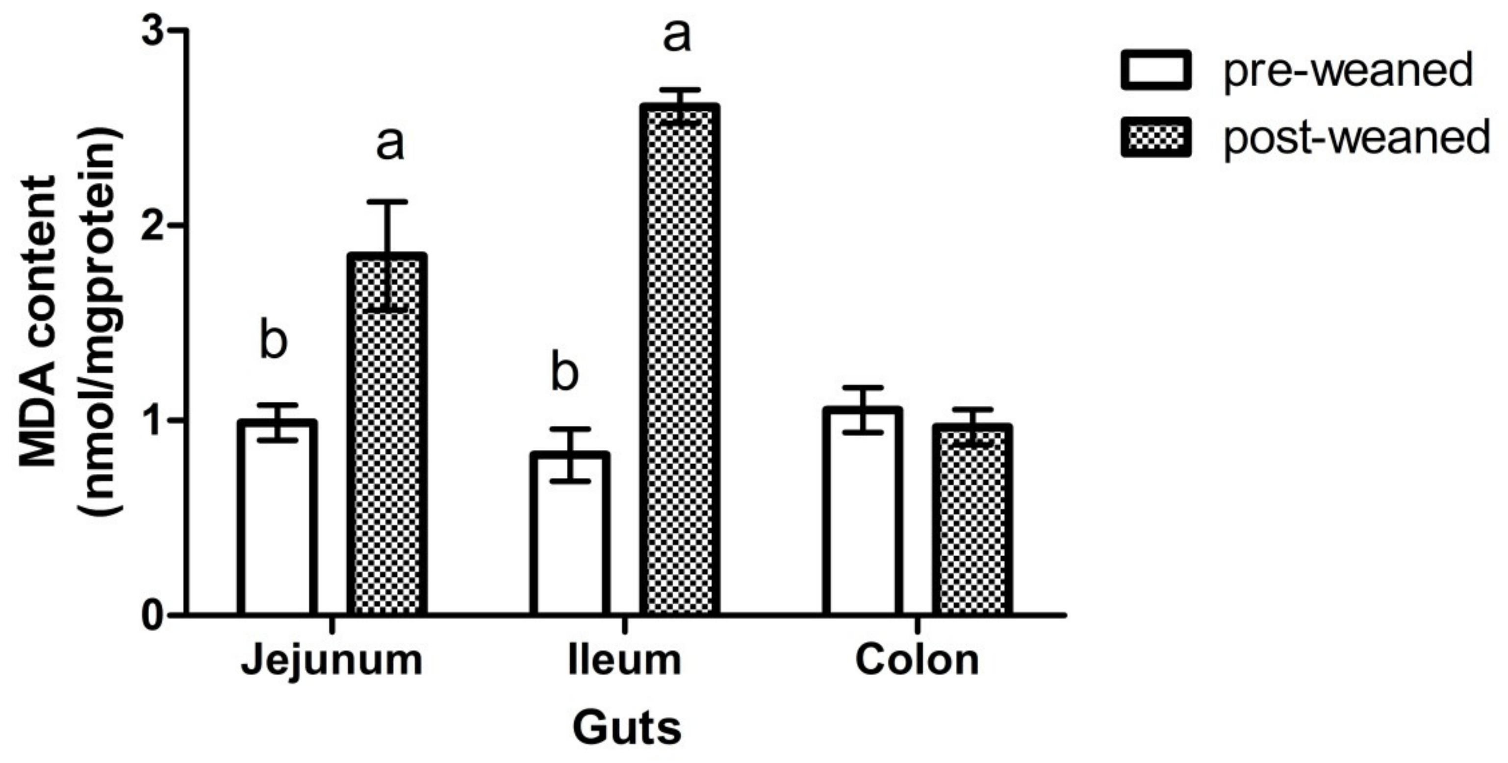
3.1. Weaning Effects on Activities of Antioxidant Enzyme and Inflammatory Factor

The MDA contents in the jejunum and ileum for the postweaned young goats were higher than the preweaned young goats (0.99 ± 0.089 vs. 1.84 ± 0.28 and 0.82 ± 0.13 vs. 2.61 ± 0.09 nmol/mg·protein for jejunum and ileum, respectively; Figure 1). There were no interactions (p = 0.35) between age and gut region for the activities of SOD and GSH-Px. The activity of SOD in postweaned young goats was lower (p = 0.012) than the preweaned young goats (98.8 vs. 56.3 U/mg·protein), and no difference (p = 0.829) was observed for the SOD activity among gut regions (Table 1). There was no difference (p = 0.846) in GSH-Px activity observed between pre- and postweaned young goats; however, the GSH-Px activity was higher in the jejunum (p = 0.037) than the ileum and colon. Moreover, neither age nor gut region effect (p > 0.092) was observed on IL-2 and IL-6 concentration in young goats (Table 1).

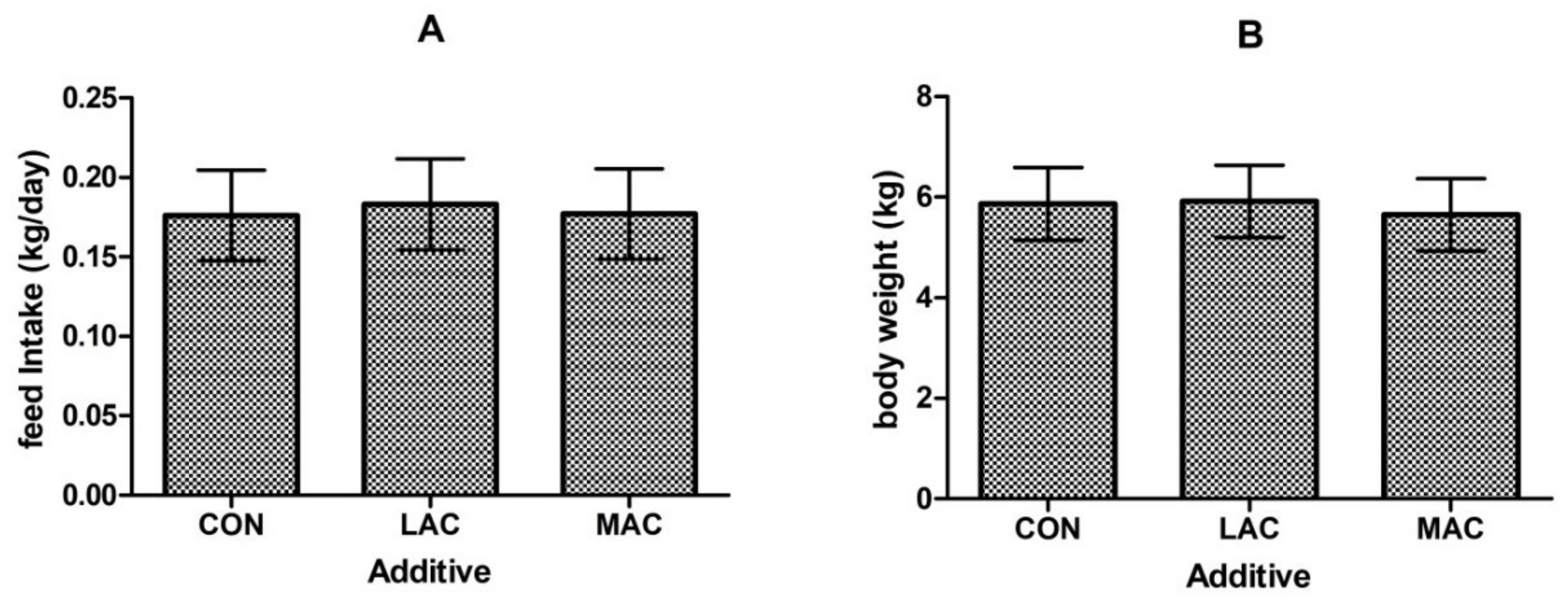
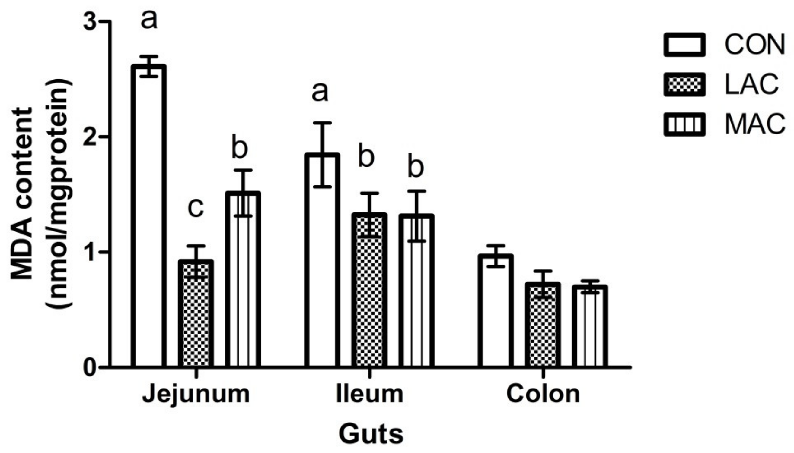
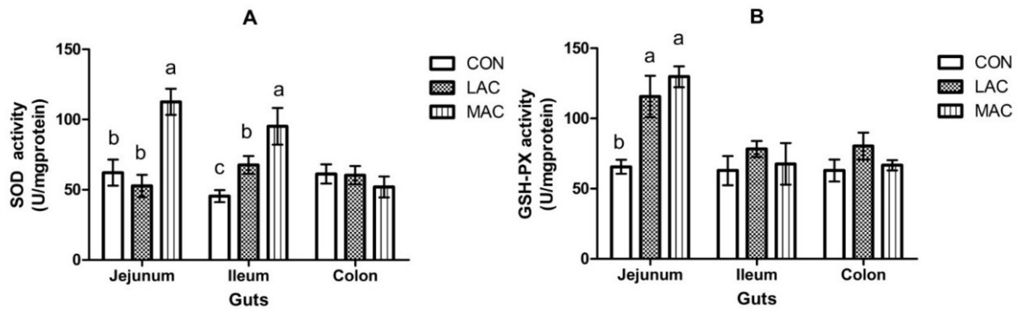
3.2. Effects of Supplementing Probiotics and Prebiotics on Antioxidant Enzyme, Inflammatory Factor, and Growth Performance

No difference (p > 0.05) in feed intake (Figure 2A) or final body weight (Figure 2B) was observed among groups. The activity of MDA in the ileum and jejunum of LAC goats was less (p < 0.001) than CON goats (Figure 3; 1.84 ± 0.28 vs. 1.32 ± 0.19 and 2.61 ± 0.09 vs. 0.92 ± 0.13 nmol/mg·protein). The SOD activity in the ileum (Figure 4A; 45.41 ± 4.29 vs. 67.68 ± 6.32 U/mg·protein) and GSH-Px activity (Figure 4B; 65.52 ± 5.00 vs. 115.69 ± 14.71 U/mg·protein) of LAC goats were higher (p < 0.03) than the corresponding activities in CON goats. Similarly, MDA contents in the ileum and jejunum from MAC goats were less (p < 0.001) than that from CON goats (Figure 3; 1.84 ± 0.28 vs. 1.31 ± 0.22 and 2.61 ± 0.09 vs. 1.51 ± 0.20 nmol/mg·protein). The SOD activity in ileum and jejunum (Figure 4A; 45.41 ± 4.29 vs. 95.15 ± 13.05 and 62.20 ± 9.37 vs. 112.60 ± 9.32 U/mg·protein) and GSH-Px activity (Figure 4B; 65.52 ± 5.00 vs. 129.76 ± 7.48 U/mg·protein) in the jejunum from MAC goats were higher (p < 0.03) than those in CON goats. However, there were no differences (p > 0.05) observed among treatments in MDA, SOD, and GSH-Px in the colon. For the inflammation variables, there were region effects (p < 0.05) both on IL-2 and IL-6, with higher activity in the jejunum (Table 2). Supplementation of LAC or MAC additive failed to alter (p > 0.19) the concentrations of IL-2 and IL-6 in the young goats (Table 2).

4. Discussion

It has been widely reported that weaning in mammals leads to intestinal dysfunction, intestinal microbial disorder, tight connection injury, inflammation, and oxidative stress [21]. Mammalian cells play a vital role in antioxidant defense systems when subjected to extreme stress. Previously, Wei et al. [4] have reported that the activity of ROS is significantly increased with the decrease of the activity of SOD in weaned groups as compared to the preweaning group in the jejunum of piglets. Superoxide dismutase is an antioxidase enzyme that changes the superoxide anion into hydrogen peroxides, and GSH-Px can inactivate peroxides [22]. The imbalance of the intestinal redox status usually restrains the capacity of eliminating ROS, resulting in oxidative damage to the intestinal epithelial cells [23,24]. In addition, MDA is one of the lipid peroxide metabolites and an essential symbol of oxidative damage, which can be enhanced with weaning stress [25,26]. In this study, the higher MDA content with less antioxidant enzymes activity in weaned young goats was expected, which suggests that early weaning can induce oxidative damage in the young goats. Moreover, the previous study in piglets has shown that a significant increase in messenger RNA (mRNA) levels of TNF-α, IL-1β, and IL-6 in the jejunal mucosa was induced by weaning [4]. However, the absence of a significant impact of weaning on IL-2 or IL-6 in the present study may indicate the distinct responses between ruminants and monogastric animals. It has been reported that the change of gastrointestinal inflammatory cytokines is related to the diversity of gastrointestinal microorganisms [27]. Ruminating goats have a unique digestive organ called the rumen, where the microorganisms can convert feed components into readily available sources of energy and protein. Our results suggest that the ruminating goats may be more tolerant of pathogens than monogastric animals.
Lactobacillus is an active microorganism that is used as a probiotic additive in livestock [28,29]. The less MDA content and increased SOD and GSH-Px activities in the jejunum and ileum of young goats due to LAC were expected, which suggest a protective effect of LAC in improving animal health. In agreement with our results, it has been reported that LAC increased activities of GSH and reduced oxidative stress in growing lambs. [29]. In addition, Maekawa et al. (2016) have reported a protective effect of LAC pentosus strain S-PT84 against Candida infection of the gastrointestinal tract in mice [11]. The mechanism that LAC can regulate the oxidative stress of animals may be related to modulation of nuclear factor erythroid-2-related factor 2 (Nrf2) signals and suppression of nuclear factor kappa-B (NF-kB) inflammatory pathway [30]. Moreover, it has been reported that Lactobacillus plays a significant role in regulating gastrointestinal mucosal immune cells and maintaining a healthy gastrointestinal tract [31]. Lactobacillus can produce bactericidal substances and compete for the attachment plaque with pathogens and toxins [31].
It has been reported that MAC can reduce the reproduction of pathogenic bacteria which lead to severe intestinal stress, inflammation, and even death after weaning in piglets [32]. Li et al. [16] have observed an antioxidant ability of MAC using 1,1-diphenyl-2-picrylhydrazyl (DPPH) radical scavenging and β-carotene–linoleic acid assays. The main active components in MAC are sanguinarine and chelerythrine [16], which can induce antioxidant effects [33]. Thus, the improved activities of SOD and GSH-Px in the jejunum and reduced MDA production in jejunum and ileum were expected in our study.
Although decreased MDA with an increased SOD and GSH-Px in jejunum and ileum in young goats for the LAC and MAC groups were observed in this study, the inclusion of LAC and MAC in the diets failed to change the oxidative status in the colons of the young goats. Plausibly, the majority of additives played a role in the small intestine, and colonization of the additives in the large intestine was inefficient [34]. Moreover, since the variety and quantity of microorganisms in the colon are more abundant and numerous compared with that in the small intestine, the added additives are possibly hard to be attached to the intestinal walls, and can be only kept for a very short time [34,35].
Interestingly, we did not observe a difference between the control and LAC-supplemented groups in feed intake and body weight gain. This result was consistent with Whitley et al. [36] that they have found the average daily gain, feed intake, and feed conversion are not influenced by a commercial probiotic supplementation containing LAC acidophilus and Enterococcus faecium in meat goats. Conversely, in the study of Sharma et al. [37] using Murrah buffalo calves showed that supplementation of LAC acidophilus improved the growth performance of the calves, which suggests that the effect of probiotic and prebiotic may relate with animal species.

5. Conclusions

In this study, weaning increased MDA content and decreased SOD activity in the lower gut of young goats. Supplementation of LAC or MAC reduced the MDA concentration and increased the SOD and GSH-Px activity in the jejunum or ileum of the young goats. However, there were no differences in MDA, SOD, or GSH-Px in the colon of the young goats supplementing with or without a probiotic and prebiotic additive. There was no alternation in inflammation among the goats in this study. The changes in oxidation status indicate that weaning induces oxidative damage in the lower gut in the young goats, and the oxidative damage in the small intestine can be alleviated by adding LAC or MAC to the diets.

Author Contributions

Z.H., Y.L., and Z.T. designed this study; K.C. and Y.L. performed the data analysis; Y.C. and K.C. performed the experiment parts; K.C. wrote the primary manuscript; Z.H. and J.H. were the joint corresponding authors; All authors have read and agreed to the published version of the manuscript.

Funding

This work was jointly supported by the National Key Research and Development Program of China (Grant No. 2018YFD0501903), National Natural Science Foundation of China (Grant Nos. 31772631, 31702141), Key Research Program of the Chinese Academy of Sciences (Grant No. KFZD-SW-219), CAS Science and Technology Service Network Initiative (Grant No. KFJ-STS-ZDTP-075), and Pioneer Hundred Talents Program of the Chinese Academy of Sciences (There was no grant number for this funding).

Acknowledgments

Authors acknowledge the staff of Liuyang Black Goat Reproduction Centre for their care and coordinate management of experimental animals. Helpful comments and suggestions from Emilio M. Ungerfeld (Instituto de Investigaciones Agropecuarias INIA Carillanca, Temuco, Chile) are also gratefully acknowledged. Language was revised by a native speaker—Kefyalew Gebeyew Mekonnen (Institute of Subtropical Agriculture, The Chinese Academy of Sciences, Changsha 410125, Hunan, China).

Conflicts of Interest

The authors declare no conflicts of interest.

References

  1. Alugongo, G.M.; Xiao, J.X.; Chung, Y.H.; Dong, S.Z.; Li, S.L.; Yoon, I.; Wu, Z.H.; Cao, Z.J. Effects of Saccharomyces cerevisiae fermentation products on dairy calves: Performance and health. J. Dairy Sci. 2017, 100, 1–11. [Google Scholar] [CrossRef] [Green Version]
  2. Zhou, X.; Zhang, Y.; Wu, X.; Wan, D.; Yin, Y. Effects of Dietary Serine Supplementation on Intestinal Integrity, Inflammation and Oxidative Status in Early-Weaned Piglets. Cell Physiol. Biochem. 2018, 48, 993–1002. [Google Scholar] [CrossRef]
  3. Luo, Z.; Zhu, W.; Guo, Q.; Luo, W.; Zhang, J.; Xu, W.; Xu, J. Weaning Induced Hepatic Oxidative Stress, Apoptosis, and Aminotransferases through MAPK Signaling Pathways in Piglets. Oxid. Med. Cell Longev. 2016, 2016, 10. [Google Scholar] [CrossRef] [PubMed]
  4. Wei, H.K.; Xue, H.X.; Zhou, Z.X.; Peng, J. A carvacrol-thymol blend decreased intestinal oxidative stress and influenced selected microbes without changing the messenger RNA levels of tight junction proteins in jejunal mucosa of weaning piglets. Animal 2017, 11, 193–201. [Google Scholar] [CrossRef] [Green Version]
  5. Li, Y.; Guo, Y.; Wen, Z.; Jiang, X.; Ma, X.; Han, X. Weaning Stress Perturbs Gut Microbiome and Its Metabolic Profile in Piglets. Sci. Rep. 2018, 8, 1–12. [Google Scholar] [CrossRef] [Green Version]
  6. Wu, D.; Sun, M.Z.; Zhang, C.; Xin, Y. Antioxidant properties of lactobacillus and its protecting effects to oxidative stress caco-2 cells. J. Anim. Plant Sci. 2014, 24, 1766–1771. [Google Scholar]
  7. Kullisaar, T.; Zilmer, M.; Mikelsaar, M.; Vihalemm, T.; Annuk, H.; Kairane, C.; Kilk, A. Two antioxidative lactobacilli strains as promising probiotics. Int. J. Food Microbiol. 2002, 72, 215–224. [Google Scholar] [CrossRef]
  8. Wang, J.; Ji, H.F.; Wang, S.X.; Zhang, D.Y.; Liu, H.; Shan, D.C.; Wang, Y.M. Lactobacillus plantarum ZLP001: In vitro Assessment of Antioxidant Capacity and Effect on Growth Performance and Antioxidant Status in Weaning Piglets. Asian Austral. J. Anim. 2012, 25, 1153–1158. [Google Scholar] [CrossRef] [PubMed] [Green Version]
  9. Paszti-Gere, E.; Csibrik-Nemeth, E.; Szeker, K.; Csizinszky, R.; Palocz, O.; Farkas, O.; Galfi, P. Lactobacillus plantarum 2142 prevents intestinal oxidative stress in optimized in vitro systems. Acta Physiol. Hung. 2013, 100, 89–98. [Google Scholar] [CrossRef]
  10. Pena, J.A.; Rogers, A.B.; Ge, Z.M.; Ng, V.; Li, S.Y.; Fox, J.G.; Versalovic, J. Probiotic Lactobacillus spp. diminish Helicobacter hepaticus-induced inflammatory bowel disease in interleukin-10-deficient mice. Infect. Immun. 2005, 73, 912–920. [Google Scholar] [CrossRef] [Green Version]
  11. Maekawa, T.; Ishijima, A.S.; Ida, M.; Izumo, T.; Ono, Y.; Shibata, H.; Abe, S. Prophylactic Effect of Lactobacillus pentosus strain S-PT84 on Candida Infection and Gastric Inflammation in a Murine Gastrointestinal Candidiasis Model Errata. Med. Mycol. 2016, 57, E81–E92. [Google Scholar]
  12. Tobita, K.; Watanabe, I.; Saito, M. Specific vaginal lactobacilli suppress the inflammation induced by lipopolysaccharide stimulation through downregulation of toll-like receptor 4 expression in human embryonic intestinal epithelial cells. Biosci. Microb. Food Health 2017, 36, 39–44. [Google Scholar] [CrossRef] [PubMed] [Green Version]
  13. Holowacz, S.; Blondeau, C.; Guinobert, I.; Guilbot, A.; Hidalgo, S.; Bisson, J.F. Lactobacillus salivarius LA307 and Lactobacillus rhamnosus LA305 attenuate skin inflammation in mice. Benef. Microbes 2018, 9, 299–309. [Google Scholar] [CrossRef]
  14. Pan, F.; Zhang, L.; Li, M.; Hu, Y.; Zeng, B.; Yuan, H.; Zhao, L.; Zhang, C. Predominant gut Lactobacillus murinus strain mediates anti-inflammaging effects in calorie-restricted mice. Microbiome 2018, 6, 54. [Google Scholar] [CrossRef] [PubMed]
  15. Huang, P.; Zhang, Y.; Xiao, K.; Jiang, F.; Wang, H.; Tang, D.; Liu, D.; Liu, B.; Liu, Y.; He, X.; et al. The chicken gut metagenome and the modulatory effects of plant-derived benzylisoquinoline alkaloids. Microbiome 2018, 6, 1–17. [Google Scholar] [CrossRef] [PubMed] [Green Version]
  16. Li, C.M.; Yang, X.Y.; Zhong, Y.R.; Yu, J.P. Chemical composition, antioxidant and antimicrobial activity of the essential oil from the leaves of Macleaya cordata (Willd) R. Br. Nat. Prod. Res. 2016, 30, 438–442. [Google Scholar] [CrossRef]
  17. Bao, Y.; Zhang, Y.; Li, H.; Liu, Y.; Wang, S.; Dong, X.; Su, F.; Yao, G.; Sun, T.; Zhang, H. In vitro screen of Lactobacillus plantarum as probiotic bacteria and their fermented characteristics in soymilk. Ann. Microbiol. 2012, 62, 1311–1320. [Google Scholar] [CrossRef]
  18. Girotti, M.J.; Khan, N.; McLellan, B.A. Early measurement of systemic lipid-peroxidation products in the plasma of major blunt trauma patients. J. Trauma 1991, 31, 32–35. [Google Scholar] [CrossRef]
  19. Flohe, L.; Otting, F. superoxide-dismutase assays. Methods Enzymol. 1984, 105, 93–104. [Google Scholar]
  20. Lawrence, R.A.; Burk, R.F. Glutathione peroxidase activity in selenium-deficient rat liver. Biochem. Biophys. Res. Commun. 1976, 71, 952–958. [Google Scholar] [CrossRef]
  21. Mach, N.; Foury, A.; Kittelmann, S.; Reigner, F.; Moroldo, M.; Ballester, M.; Esquerre, D.; Riviere, J.; Salle, G.; Gerard, P.; et al. The Effects of Weaning Methods on Gut Microbiota Composition and Horse Physiology. Front. Physiol. 2017, 8, 535. [Google Scholar] [CrossRef] [Green Version]
  22. He, Z.X.; Sun, Z.H.; Tan, Z.L.; Tang, S.X.; Zhou, C.S.; Han, X.F.; Wang, M.; Wu, D.Q.; Kang, J.H.; Beauchemin, K.A. Effects of maternal protein or energy restriction during late gestation on antioxidant status of plasma and immune tissues in postnatal goats. J. Anim. Sci. 2012, 90, 4319–4326. [Google Scholar] [CrossRef]
  23. Mates, J.M.; Segura, J.A.; Alonso, F.J.; Marquez, J. Intracellular redox status and oxidative stress: Implications for cell proliferation, apoptosis, and carcinogenesis. Arch. Toxicol. 2008, 82, 273–299. [Google Scholar] [CrossRef]
  24. Wang, L.; Wang, H.; Hu, M.; Cao, J.; Chen, D.; Liu, Z. Oxidative stress and apoptotic changes in primary cultures of rat proximal tubular cells exposed to lead. Arch. Toxicol. 2009, 83, 417–427. [Google Scholar] [CrossRef] [PubMed]
  25. Dmitriev, L.F.; Titov, V.N. Lipid peroxidation in relation to ageing and the role of endogenous aldehydes in diabetes and other age-related diseases. Ageing Res. Rev. 2010, 9, 200–210. [Google Scholar] [CrossRef] [PubMed]
  26. Verona, C.; Hackenhaar, F.S.; Teixeira, C.; Medeiros, T.M.; Alabarse, P.V.; Salomon, T.B.; Shüller, Á.K.; Maccari, J.A.G.; Condessa, R.L.; Oliveira, R.P.; et al. Blood markers of oxidative stress predict weaning failure from mechanical ventilation. J. Cell Mol. Med. 2015, 19, 1253–1261. [Google Scholar] [CrossRef] [PubMed]
  27. Jiao, J.; Zhou, C.; Guan, L.L.; McSweeney, C.S.; Tang, S.; Wang, M.; Tan, Z. Shifts in Host Mucosal Innate Immune Function Are Associated with Ruminal Microbial Succession in Supplemental Feeding and Grazing Goats at Different Ages. Front. Microbiol. 2017, 8, 1655. [Google Scholar] [CrossRef]
  28. Wang, T.W.; Teng, K.L.; Liu, Y.Y.; Shi, W.X.; Zhang, J.; Dong, E.Q.; Zhang, X.; Tao, Y.; Zhong, J. Lactobacillus plantarum PFM 105 promotes intestinal development through modulation of gut microbiota in weaning piglets. Front. Microbiol. 2019, 10, 90. [Google Scholar] [CrossRef]
  29. Wang, J.; Lin, L.; Jiang, Q.; Huang, W.; Liu, N. Effect of supplemental lactic acid bacteria on growth performance, glutathione turnover and aflatoxin B1 removal in lambs. Czech J. Anim. Sci. 2019, 64, 272–278. [Google Scholar] [CrossRef] [Green Version]
  30. Finamore, A.; Ambra, R.; Nobili, F.; Garaguso, I.; Raguzzini, A.; Serafini, M. Redox Role of Lactobacillus casei Shirota against the cellular damage induced by 2,2’-Azobis (2-Amidinopropane) dihydrochloride-induced oxidative and inflammatory stress in enterocytes-like epithelial cells. Front. Immunol. 2018, 9, 1131. [Google Scholar] [CrossRef] [Green Version]
  31. Raabis, S.; Li, W.; Cersosimo, L. Effects and immune responses of probiotic treatment in ruminants. Vet. Immunol. Immunopathol. 2018, 12, 006. [Google Scholar] [CrossRef] [PubMed]
  32. Kong, X.F.; Wu, G.Y.; Liao, Y.P.; Hou, Z.P.; Liu, H.J.; Yin, F.G.; Li, T.J.; Huang, R.L.; Zhang, Y.M.; Deng, D.; et al. Effects of Chinese herbal ultra-fine powder as a dietary additive on growth performance, serum metabolites and intestinal health in early-weaned piglets. Livest. Sci. 2007, 108, 272–275. [Google Scholar] [CrossRef]
  33. Bavarsadi, M.; Mahdavi, A.H.; Ansari-Mahyari, S.; Jahanian, E. Effects of different levels of sanguinarine on antioxidant indices, immunological responses, ileal microbial counts and jejunal morphology of laying hens fed diets with different levels of crude protein. J. Anim. Physiol. Anim. Nutr. 2017, 101, 936–948. [Google Scholar] [CrossRef] [PubMed]
  34. Fernandez, M.F.; Boris, S.; Barbes, C. Probiotic properties of human lactobacilli strains to be used in the gastrointestinal tract. J. Appl. Microbiol. 2003, 94, 449–455. [Google Scholar] [CrossRef]
  35. Zuo, Z.H.; Shang, B.J.; Shao, Y.C.; Li, W.Y.; Sun, J.S. Screening of intestinal probiotics and the effects of feeding probiotics on the growth, immune, digestive enzyme activity and intestinal flora of Litopenaeus vannamei. Fish Shellfish Immun. 2019, 86, 160–168. [Google Scholar] [CrossRef]
  36. Whitley, N.C.; Cazac, D.; Rude, B.J.; Jackson-O’Brien, D.; Parveen, S. Use of a commercial probiotic supplement in meat goats. J. Anim. Sci. 2009, 87, 723–728. [Google Scholar] [CrossRef] [Green Version]
  37. Sharma, A.N.; Kumar, S.; Tyagi, A.K. Effects of mannan-oligosaccharides and Lactobacillus acidophilus supplementation on growth performance, nutrient utilization and faecal characteristics in Murrah buffalo calves. J. Anim. Physiol. Anim. Nutr. 2018, 102, 679–689. [Google Scholar] [CrossRef]
Figure 1. Weaning effects on the content (nmol/mg·protein) of malondialdehyde (MDA) in the colon, ileum, and jejunum of young goats (Exp. 1). a, b Row means that do not have a common superscript differ (p < 0.05).
Animals 10 00548 g001
Figure 2. Effects of supplementing probiotics and prebiotics additives on feed intake (A) and body weight (B) of young goats (Exp. 2). CON = control group; LAC = a control diet supplementing with 10 g/day Lactobacillus plantarum P-8; MAC = a control diet supplementing with 0.3 g/day plant extract (Sangrovit®, MAC extract 3.75% w/w premix).
Animals 10 00548 g002
Figure 3. Effects of supplementing probiotics and prebiotics additives on the content (nmol/mg·protein) of MDA in the colon, ileum, and jejunum of young goats (Exp. 2). CON = control group; LAC = a control diet supplementing with 10 g/day Lactobacillus plantarum P-8; MAC = a control diet supplementing with 0.3 g/day plant extract (Sangrovit®, MAC extract 3.75% w/w premix). a, b Row means that do not have a common superscript differ (p < 0.05).
Animals 10 00548 g003
Figure 4. Effects of supplementing probiotics and prebiotics additives on the activities (U/mg·protein) of SOD (Panel A) and GSH-Px (Panel B) in the colon, ileum, and jejunum of young goats (Exp. 2). CON = control group; LAC = a control diet supplementing with 10 g/day Lactobacillus plantarum P-8; MAC = a control diet supplementing with 0.3 g/day plant extract (Sangrovit®, MAC extract 3.75% w/w premix). a, b Row means that do not have a common superscript differ (p < 0.05).
Animals 10 00548 g004
Table 1. Weaning effects on the activities (U/mg·protein) of SOD and GSH-Px, and the concentration (pg/mg·protein) of IL-2 and IL-6 in the colon, ileum, and jejunum of young goats.
Item 1AgeSEMGutSEMp Value
PreweanedPostweanedJejunumIleumColonGutAgeInteraction
SOD
(U/mg·protein)
98.8 a56.3 b8.085.173.174.3511.90.8290.0120.701
GSH-Px
(U/mg·protein)
62.064.410.788.8 a53.0 b47.87 b12.50.0370.8460.349
IL-2
(pg/mg·protein)
319.5327.323.9329.8267.3373.234.20.1470.8550.427
IL-6
(pg/mg·protein)
28.933.22.228.326.638.23.20.0920.3070.683
a, b Row means that do not have a common superscript differ (p < 0.05). 1 SOD = superoxide dismutase; GSH-Px = glutathione peroxidase; IL-2 = interleukin 2; IL-6 = interleukin 6.
Table 2. Effects of probiotics and prebiotics additives on concentrations (pg/mg·protein) of IL2 and IL6 in the colon, ileum and jejunum of young goats.
Item 1Additive 2SEMGutSEMp Value
CONLACMACJejunumIleumColonGutAdditiveInteraction
IL2327.3294.9278.024.0381.46 a254.5 b266.0 b26.80.0200.5360.304
IL633.226.927.12.238.9 a23.4 b25.0 b2.3<0.0010.1850.397
a, b Row means that do not have a common superscript differ (p < 0.05). 1 IL2 = interleukin 2; IL6 = interleukin 6; 2 CON = control group; LAC = a control diet supplementing with 10 g/day Lactobacillus plantarum P-8; MAC = a control diet supplementing with 0.3 g/day plant extract (Sangrovit®, MAC extract 3.75% w/w premix).

