Article

A High Dietary Incorporation Level of Chlorella vulgaris Improves the Nutritional Value of Pork Fat without Impairing the Performance of Finishing Pigs

1
CIISA-Centro de Investigação Interdisciplinar em Sanidade Animal, Faculdade de Medicina Veterinária, Universidade de Lisboa, 1300-477 Lisboa, Portugal
2
INIAV-Instituto Nacional de Investigação Agrária e Veterinária, Fonte Boa, 2005-048 Vale de Santarém, Portugal
*
Author to whom correspondence should be addressed.
Animals 2020, 10(12), 2384; https://doi.org/10.3390/ani10122384
Submission received: 5 November 2020 / Revised: 25 November 2020 / Accepted: 10 December 2020 / Published: 12 December 2020
(This article belongs to the Special Issue Effects of Dietary Supplements on Livestock and Poultry Products)

Abstract

:

Simple Summary

Pork is one of the most consumed meats worldwide but its production and quality are facing significant challenges, including feeding sustainability and the unhealthy image of fat. In fact, corn, and soybean, the two main conventional feedstuffs for pig production, are in unsustainable competition with the human food supply and biofuel industry. Moreover, the nutritional value of pork lipids is small due to their low contents of the beneficial n-3 polyunsaturated fatty acids and lipid-soluble antioxidants. The inclusion of microalgae in pig diets represents a promising approach for the development of sustainable pork production and the improvement of its quality. The current study aimed to investigate the impact of Chlorella vulgaris as ingredient (5% in the diet), alone and in combination with carbohydrases, on growth performance, carcass characteristics and pork quality traits in finishing pigs. Our data indicate that the use of 5% C. vulgaris in finishing pig diets does not impair animal growth and ameliorates the nutritional value of pork. Therefore, C. vulgaris could be used advantageously as an alternative sustainable ingredient in swine feeding.

Abstract

The influence of a high inclusion level of Chlorella vulgaris, individually and supplemented with two carbohydrase mixtures, in finishing pig diets was assessed on zootechnical performance, carcass characteristics, pork quality traits and nutritional value of pork fat. Forty crossbred entire male pigs, sons of Large White × Landrace sows crossed with Pietrain boars, with an initial live weight of 59.1 ± 5.69 kg were used in this trial. Swines were randomly assigned to one of four dietary treatments (n = 10): cereal and soybean meal-based diet (control), control diet with 5% C. vulgaris (CV), CV diet supplemented with 0.005% Rovabio® Excel AP (CV + R) and CV diet supplemented with 0.01% of a four-CAZyme mixture (CV + M). Animals were slaughtered, after the finishing period, with a BW of 101 ± 1.9 kg. Growth performance, carcass characteristics and meat quality traits were not influenced (p > 0.05) by the incorporation of C. vulgaris in the diets. However, the inclusion of the microalga in finishing pig diets increased some lipid-soluble antioxidant pigments and n-3 PUFA, and decreased the n-6:n-3 ratio of fatty acids, thus ameliorating the nutritional value of pork fat. Moreover, the supplementation of diets with the carbohydrase mixtures did not change (p > 0.05) neither animal performance nor meat quality traits, indicating their inefficacy in the increase of digestive utilization of C. vulgaris by pigs under these experimental conditions. It is concluded that the use of C. vulgaris in finishing pig diets, at this high incorporation level, improves the nutritional value of pork fat without compromising pig performance.

1. Introduction

Pork industry is currently facing the big challenges of feeding sustainability and the unhealthy image of fat. In fact, pork production is about 38% of the total amount of meat produced in the world, being is the most commonly consumed meat in different European, American, and Asian countries [1]. Moreover, the combination between rise of global population and the increase in income, will double the overall demand for animal-derived products by 2050, including pork [2]. The increased demand for these products will necessarily bring dramatic consequences in terms of sustainability, as cereal grains and soybean food crops are the two main conventional feedstuffs for animal feeding [3]. Therefore, alternative feed ingredients are needed to sustain animal agriculture and human food security [4,5].
In addition, pork is frequently considered unhealthy due to the lower proportions of polyunsaturated fatty acids (PUFA) and lipid-soluble antioxidant vitamins, and higher percentages of saturated fatty acids (SFA) [6]. However, it is well established that pig diet provides an effective approach for altering the fat composition of pork, thereby modifying the impact of human dietary fat intake from pork [7]. Functionally, the most important n-3 fatty acids are eicosapentaenoic acid (EPA, 20:5n-3) and docosahexaenoic acid (DHA, 22:6n-3), although the roles for docosapentaenoic acid (DPA, 22:5n-3) are now also emerging [8]. Lipid-soluble antioxidant vitamins comprise vitamin E homologues (tocopherols and tocotrienols) and vitamin A and its precursors (some carotenoids, including β-carotene). In general, the intakes of EPA and DHA are typically small and much lower than the recommended values [9]. This fact raised substantial interest in food enrichment with EPA and DHA, by using feed ingredients from marine origin in animal nutrition.
Microalgae, an important aquatic resource, could be a good sustainable alternative to conventional feedstuffs, since they have similar nutritional compositions [10]. Chlorella vulgaris is a freshwater eukaryotic green microalga. This microalga, one of the most cultivated microalgae worldwide, is known for its high biomass productivity, relative ease of cultivation and a balanced nutritional composition, making it an attractive alternative for monogastric diets [11]. In particular, regarding fatty acid profile, C. vulgaris displays a high percentage of SFA, mainly myristic acid (14:0), palmitic acid (16:0) and stearic acid (18:0). In addition, C. vulgaris presents an interesting content in some n-6 PUFA (18:2n-6 and 18:3n-6) and α-linolenic acid (18:3n-3), but much less quantity of EPA and DHA [12]. However, C. vulgaris cell wall is composed by a diverse and complex matrix of cross-linked insoluble carbohydrates [13]. Thus, the incorporation of C. vulgaris in monogastric diets could be a problem since the recalcitrant cell wall is largely indigestible, impairing the bioavailability of its valuable nutrients [14].
Exogenous carbohydrate-active enzymes (CAZymes) are now completely accepted as feed supplements for monogastric livestock species to improve feed nutritive value and enhance animal performance and health [15]. In addition to cereal cell walls, several in vitro studies demonstrated the ability of CAZymes to degrade microalgae cell walls [16,17,18,19]. Recently, Coelho et al. [20] described a four-CAZyme mixture, composed by an exo-β-glucosaminidase, an alginate lyase, a peptidoglycan N-acetylmuramic acid deacetylase and a lysozyme, that was shown to disrupt microalgae C. vulgaris cell walls to a significant extent, in in vitro assays, enabling the release of trapped nutrients with important nutritional value.
Therefore, the supplementation with the four-CAZyme mixture mentioned above could enable the incorporation of C. vulgaris in monogastric diets, at high incorporation levels (>2% in diet), without impairing animal performance and health. In line with this, the aim of this study was to assess how the dietary incorporation of C. vulgaris at a 5% high level, supplemented or not with two exogenous CAZyme mixtures (the commercially available Rovabio® Excel (ADISSEO, Antony, France) AP and the four-CAZyme mixture described by Coelho et al. [20]), influences finishing pigs’ performance, carcass characteristics, and pork quality traits.

2. Materials and Methods

2.1. Production of Recombinant Four-CAZyme Mixture

The genes encoding the four recombinant CAZymes that compose the mixture (exo-β-glucosaminidase, alginate lyase, peptidoglycan N-acetylmuramic acid deacetylase and lysozyme) were cloned according to Coelho et al. [20]. Briefly, BL21 Escherichia coli cells were transformed with the generated recombinant plasmids and were grown on Luria–Bertani media, at 37 °C under agitation (190 rpm) to mid exponential phase (absorbance was measured at λ = 595 nm as being 0.4–0.6). Isopropyl β-d-thiogalactoside was added to a final concentration of 1 mM in order to induce recombinant gene expression. Cells were incubated overnight at 19 °C with agitation (140 rpm). After induction, the culture media was centrifuged and the protein extracts were prepared by ultrasonication, followed by centrifugation and freeze dried. The four-CAZyme protein extracts were mixed in equal weight proportions at a final level of 0.01%.

2.2. Animal Care, Experimental Design and Experimental Diets

The trial was conducted at the facilities of Unidade de Investigação em Produção Animal (Instituto Nacional de Investigação Agrária e Veterinária (UEISPA-INIAV, Santarém). The experimental procedures were reviewed by the Ethics Commission of the Centro de Investigação Interdisciplinar em Sanidade Animal/Faculdade de Medicina Veterinária (CIISA/FMV) and approved by the Animal Care Committee of the National Veterinary Authority (Direção-Geral de Alimentação e Veterinária), following the appropriated European Union guidelines (2010/63/EU Directive). The staff members involved in animal trial hold license for conducting experiments on live animals from the Portuguese Veterinary Services.
Forty crossbred entire male pigs, sons of Large White × Landrace sows crossed with Pietrain boars, were obtained from a commercial farm. Before the beginning of the trial, pigs were submitted to an adaptation period of one week. Then, pigs with an initial weight of 59.1 ± 5.69 kg were randomly distributed into 10 pens with 4 animals in each pen (7.8 m2). Pens were equipped with one stainless steel nipple and four creep feeders separated by a system of gates, thus allowing individual feed intake control. The 4 experimental diets were randomly assigned to the four animals within each pen, with each animal in each pen receiving a different diet, thus being the pig the experimental unit. Pigs had access to feed and water ad libitum. The experimental diets were: cereal and soybean meal-based diet (Control), control diet with 5% of C. vulgaris supplied by Allmicroalgae (Natural Products, Portugal) (CV), control diet with 5% of C. vulgaris supplemented with 0.005% of Rovabio® Excel AP (Adisseo, Antony, France) (CV + R), and control diet with 5% of C. vulgaris supplemented with 0.01% of four-CAZyme mixture (CV + M).
The ingredient composition of the experimental diets is described in Table 1, and their chemical composition is presented in detail in Table 2. For further information on the feed analysis see details below.

2.3. Animal Performance, Slaughter, and Sampling

During the experiment, supplied feed and refusals were recorded daily, whereas pig were weighed weekly just before feeding, with the purpose of calculate average daily feed intake (ADFI), average daily weight gain (ADG), feed conversion ratio (FCR) and gain:feed ratio (G:F). Food was withdrawn from animals 17–19 h before slaughter. Animals were slaughtered at a BW of 101 ± 1.9 kg, after a trial period of 41 ± 7.8 days, at the Unidade de Investigação em Produção Animal experimental slaughterhouse (Santarém, Portugal), with electrical stunning followed by exsanguination. The hot carcass weight (HCW) was measured in order to calculate carcass yield. Perirenal and mesenteric fat depot was removed and weighed. Longissimus lumborum muscle was collected from the right carcass side between the third and fifth lumbar vertebras, minced, immediately vacuum packed and stored at −20 °C, to assess meat quality, and at −80 °C, for meat oxidative stability determinations.
At 24 h post mortem, backfat thickness was measured in the left side of carcass at the last rib position (P2) (the most representative location), last lumbar vertebra (L6) and second sacral vertebra (S2), using a calibrated engineering calliper (150 mm Electronic Digital Vernier Calliper CE ROHS) as described by Frederick [21]. The loin was excised from the left side of carcass, between the last cervical and L6 lumbar vertebras, weighted and sliced into 2.5-cm-thick chops for sensory evaluation, shear force measurements, color and drip loss determinations. Chops were vacuum packed, frozen and stored at −20 °C until sensory analysis and shear force measurements.

2.4. Microalga and Experimental Diets Analyses

Experimental diets were collected three times during the entire trial. The Association of Official Analytical Chemists (AOAC) [22] methods were used to determine the proximal composition of C. vulgaris microalga and experimental diets. Samples were dried at 103 °C until reach constant weight to determine dry matter (DM). Crude protein of samples was calculated through the determination of the nitrogen content (N) by the Kjeldahl method using the factor 6.25 × N following the method 954.01 [22]. Ash and starch contents of samples were determined according to the method 942.05 [22] and Clegg [23] procedure, respectively. Crude fat of samples was determined after automatic Soxhlet extraction with petroleum ether (Gerhardt Analytical Systems, Königswinter, Germany). Crude fiber, acid detergent fiber (ADF) and neutral detergent fiber (NDF) were determined by the method 989.03 [22]. Metabolizable energy (ME) was calculated according to Noblet et al. [24].
The amino acid composition of C. vulgaris and experimental diets was determined according to the method 994.12 [25] and quantified by High-Performance Liquid Chromatography (HPLC) (Agilent 1100, Agilent Technologies, Avondale, PA, USA), as described by Henderson et al. [26]. The fatty acid methyl esters (FAME) profile of C. vulgaris and experimental diets were analyzed by one-step extraction and acid transesterification, followed by gas chromatography using heneicosaenoic acid (21:0) methyl ester as the internal standard [27].
The diterpene profile of C. vulgaris and experimental diets was analyzed by direct saponification, using a single n-hexane extraction followed by HPLC analysis [28]. The determination of pigments in C. vulgaris and experimental diets was performed according to Teimouri et al. [29], with slight modifications as described in Pestana et al. [30]. The quantification of pigments in C. vulgaris and experimental diet samples were performed according to Hynstova et al. [31].

2.5. Meat Quality Traits

The pH and temperature of longissimus lumborum muscle were measured in the right carcass side at 45 min and 24 h post mortem using a pH meter equipped with a penetrating electrode (HI8424, Hanna Instruments, Woonsocket, RI, USA). Meat color was measured on the cut surface of longissimus lumborum section, 24 h post mortem, using a colorimeter (Minolta CR-400, Konica Minolta, Tokyo, Japan) with the illuminant D65, at an observer angle of 2° and 1 cm diameter of measurement area. Three measurements on different locations per sample were recorded following the CIE color convention L* (lightness), a* (redness) and b* (yellowness) system after 1 h of blooming at 4 °C [32].
Drip loss of fresh longissimus lumborum muscle was performed according to Hope-Jones et al. [33]. The amount of drip measured between 24 h and 144 h post mortem was expressed as a percentage of the initial mass of the sample, and calculated through the difference between the sample mass at the beginning and end of the storage period.

2.6. Cooking Loss and Shear Force Measurements

Meat cooking loss and shear force were determined according to the procedure adapted from Honikel [34] and Oillic et al. [35]. Frozen meat samples were thawed at 2 ± 1 °C overnight, weighed and cooked in a water bath at 80 ± 0.5 °C until reaching the temperature of 75 ± 0.5 °C in the geometric center, using an internal thermocouple (Thermometer Omega RDXL4SD, Manchester, NH, USA). The samples were cooled for 20 h (2 ± 1 °C), weighed in order to calculate the cooking loss, and longitudinally cut in the fiber axis parallel to muscle fiber direction into 8 to 12 cores, with a 1-cm2 cross-section area for shear force determinations. Cooking loss, expressed as percentage, was calculated by the difference of the weights before and after cooking divided by the initial weight of the sample [36].
The Warner–Bratzler shear force (WBSF) was measured in a texture analyzer (TA-XT Plus texture analyzer; Stable Micro Systems, Surrey, UK) with a Warner–Bratzler shear device with a 30-kg compression load cell, trigger force was 25 g and crosshead speed during pre-test, test and post-test set were 5.0, 2.0, and 10.0 mm/s, respectively. Force and distance were recorded at 200 points/s and analyzed with the Version 6.1.16 of Exponent software (Stable Micro Systems, Surrey, UK). The value of the peak shear force of cores from each sample was determined and averaged to obtain a single WBSF value per sample.

2.7. Trained Sensory Panel Analysis

A trained sensory panel with five sessions was used to evaluate meat sensory characteristics. The eleven panelists were selected and trained according to Cross et al. [37]. For each session, meat samples were thawed at 2 ± 1 °C overnight and cooked at 170 ± 5 °C in a Ceramic Contact Grill 1.6 kW (UNOX Catering Equipment, Padova, Italy) with an internal thermocouple in each sample (Thermometer Omega RDXL4SD, Manchester, NH, USA) until reached 71 °C in the geometric center. After 10 min of stabilization at 40 °C, the sample was trimmed of the six external surfaces, included connective tissue, cut into 1 × 1 × 1 cm subsamples and maintained, on individual covered plates, in an incubator at 40 °C until tasting (no longer than 30 min) [38]. Samples were randomly distributed across sections and the attributes evaluated were juiciness, tenderness, flavor intensity, off-flavor, flavor acceptability, and overall acceptability. These attributes were classified on a grading scale from 1 (extremely dry, tough, soft, weak, or unacceptable) to 8 (extremely juicy, tender, strong, positive and positive), with the exception of off-flavor quantified from 0 (absence) or 1 (presence) [39].

2.8. Determination of Total Cholesterol and Diterpene Profile in Meat

The simultaneous quantification of total cholesterol, β-carotene and vitamin E homologues (tocopherols and tocotrienols) in longissimus lumborum samples was performed, in duplicate, as previously described by Prates et al. [28]. Muscle samples were submitted to a saponification reaction in a water bath at 80 °C for 15 min under agitation. Afterwards, the diterpenes were extracted with n-hexane and analyzed by HPLC system (Agilent 1100 Series, Agilent Technologies Inc., Palo Alto, CA, USA), using a normal-phase silica column (Zorbax RX-Sil, 250 mm × 4.6 mm i.d., 5 μm particle size, Agilent Technologies Inc, Santa Clara, CA, USA). The HPLC analysis was performed using UV-visible photodiode array detector for cholesterol (λ = 202 nm) and β-carotene (λ = 450 nm) coupled to fluorescence detector for tocopherols and tocotrienols (excitation λ = 295 nm and emission λ = 325 nm). Standard curves of peak area versus concentration was used to quantify total cholesterol, β-carotene and vitamin E homologues contents in meat samples.

2.9. Determination of Pigments in Meat

The contents of chlorophyll a, chlorophyll b and total carotenoids were measured according to the procedure of Teimouri et al. [29] modified by Pestana et al. [30]. One g of each sample was incubated overnight with 10 mL of acetone (Merck KGaA, 249 Darmstadt, Germany) under agitation at room temperature in absence of light. Then, samples were centrifuged at 3345× g for 5 min and the absorbance was measured in the supernatants using a UV–VIS spectrophotometer (Ultrospec 3100 pro, Amersham Biosciences, Little Chalfont, UK). The results were calculated according to Hynstova et al. [31], as described above for C. vulgaris microalga and experimental diets.

2.10. Determination of Total Lipid Content and Fatty Acid Composition

Longissimus lumborum muscle samples were lyophilized (−60 °C and 2.0 hPa) using a lyophilizator Edwards Modulyo (Edwards High Vacuum International, Crawley, UK) for total lipids determination according to Folch et al. [40]. Total lipid content was determined gravimetrically, in duplicate, by weighing the fat residue obtained after solvent evaporation. Then, the fat residue was re-suspended in dry toluene and submitted to sequential alkaline and acid transesterification procedure at 50 °C for 30 and 10 min, respectively, to convert fatty acids into FAME [41]. FAME were separated through gas chromatography (HP7890A Hewlett-Packard, Avondale, PA, USA) comprising a Supelcowax® 10 capillary column (30 m × 0.20 mm internal diameter, 0.20 μm film thickness; Supelco, Bellefonte, PA, USA) and a flame ionization detector as described by Madeira et al. [42]. For FAME identification, a reference standard (FAME mix 37 components, Supelco Inc, Bellefonte, PA, USA) was used and confirmed by gas chromatography with a mass spectrometry detector using a GC-MS QP2010-Plus (Shimadzu, Kyoto, Japan). FAME were quantified by the internal standard method using heneicosanoic acid (21:0) methyl ester as internal standard. The fatty acids identified were expressed as percentage of total fatty acids.

2.11. Determination of Meat Lipid Oxidation

The extent of meat lipid oxidation was evaluated at day 0, 4 and 8 post mortem (storage at 4 °C), by quantifying thiobarbituric acid reactive substances (TBARS), following the spectrophotometric method described by Grau et al. [43]. TBARS values were calculated, in duplicate, from a standard curve constructed with 1,1,3,3-tetraethoxypropane (Fluka, Neu Ulm, Germany), as a precursor of malonaldehyde, and the results were presented as mg of malonaldehyde per kg of meat [42]. In addition, lipid peroxidation levels in meat were also measured by the concentration of TBARS, after chemical oxidation through a ferrous-hydrogen peroxide system, as described by Mercier et al. [44]. The TBARS were quantified after 0, 30, 120, and 300 min of oxidation induction following the method described above.

2.12. Statistical Analysis

All data were checked for normal distribution and variance homogeneity. Data were analyzed by analysis of variance using the PROC GLM of SAS software package (version 9.4; SAS Institute Inc., Cary, NC, USA) and measurements over time analyzed with PROC MIXED of SAS. The statistical model considered the dietary treatment the fixed effect and the pig the experimental unit. Least square means for multiple comparisons were generated using the PDIFF option adjusted with the Tukey–Kramer method. The significance level was set at p < 0.05.

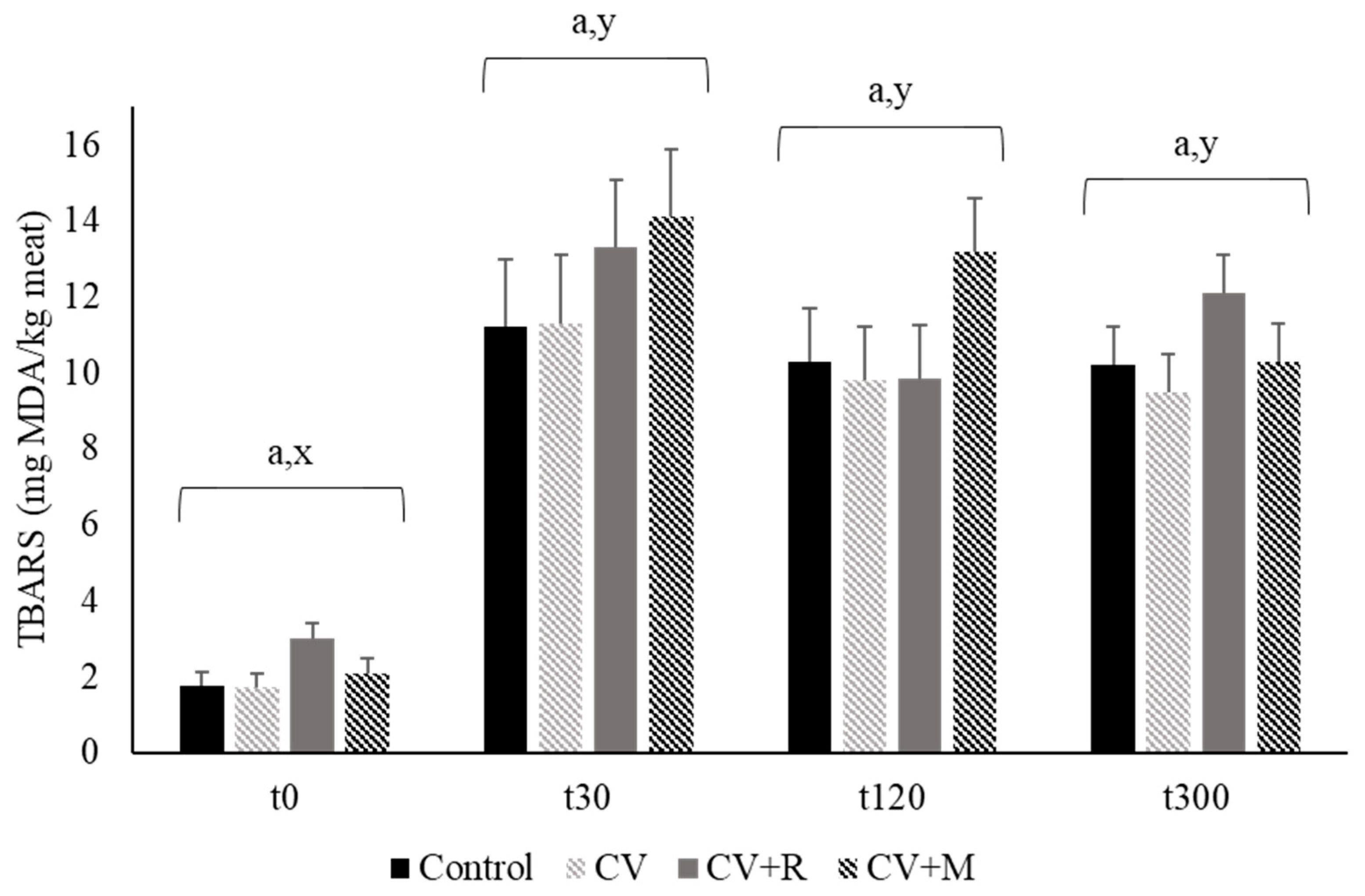
3. Results and Discussion

3.1. Feed Intake, Growth Performance and Carcass Characteristics of Pigs

Data on feed intake, growth performance and carcass traits of finishing pigs are shown in Table 3. Growth performance variables had no significant differences among animals fed with different experimental diets (p > 0.05). The average values of ADG, ADFI, and FCR were 1.02 kg, 2.62 kg, and 2.59 kg, respectively. No significant differences in carcass characteristics were obtained among the experimental groups (p > 0.05), with the exception of perirenal fat (p = 0.026). The control group displayed a higher value of perirenal fat than the group fed with the C. vulgaris diet (+34%).
We assessed, for the first time, the impact of a high dietary level (>2% in diet) of C. vulgaris, individually and combined with two exogenous CAZymes, on pig performance. In fact, some studies reported the use of C. vulgaris in pig diets but at much lower levels (1% in the diet or lower), compared with the 5% incorporated in the current trial [45,46,47]. Baňoch et al. [46] investigated the effect of a very low level (0.0002%) of incorporation of C. vulgaris in female pigs, with an initial weight of 30 kg, and found no significant differences in ADG, HCW, lean muscle thickness and backfat thickness. Later, Furbeyre and colleagues [47] showed no significant effects on ADG, ADFI, and FCR, by using 1% of C. vulgaris in weaned piglet diets, with an initial weight of 9.1 kg, during 14 days. In another study, the same authors assessed the effect of oral supplementation with C. vulgaris (385 mg/kg BW) on growth and digestive health of weaning piglets and also found no significant changes in ADG, ADFI and G:F [48]. In addition, a study conducted in growing pigs, with an initial weight of 26.6 kg and C. vulgaris incorporation of 0.1% and 0.2% in the diet, described an increase of ADG with the lower dietary level without significant variations in ADFI and G:F [45]. In the present study, no significant effects on zootechnical parameters and carcass characteristics were obtained, which indicates that dietary incorporation of 5% C. vulgaris does not compromise the productive parameters of finishing pigs. Moreover, the dietary supplementation with exogenous carbohydrases, aiming at improving C. vulgaris digestibility by finishing pigs, does not seem to be necessary at this high incorporation level.

3.2. Pork Quality Traits and Sensory Evaluation

Data concerning the effect of experimental diets on quality traits of longissimus lumborum muscle from finishing pigs are presented on Table 4. Experimental treatments had no significant effect on temperature 45 min post mortem, pH 45 min and 24 h post mortem, color parameters, WBSF and cooking loss (p > 0.05). Table 5 summarizes the trained panel scores obtained for pork. No significant differences were obtained among experimental diets for the several items evaluated by the trained sensory panel (p > 0.05).
Similar results for meat quality traits were reported by Baňoch et al. [46], who found that a 0.0002% level of incorporation of C. vulgaris in pig diets had no significant effect on color, pH, cooking loss and drip loss of pork. Here, the dietary incorporation of 5% C. vulgaris did not change pork quality traits and sensory parameters, which is very important for the consumer acceptance of this meat. By contrast, Oh et al. [49] observed an increase of b*, pH and shear force in breast meat, and an increase of L* and b* in leg meat, of male Pekin ducks fed with 0.1–0.2% C. vulgaris during 42 days. Therefore, pork quality traits seem to be less sensitive to the dietary inclusion of C. vulgaris than poultry meat characteristics, although both are meats-derived from monogastric animals. Finally, it was also indicated here that the dietary use of CAZyme mixtures does not affect pork quality characteristics.

3.3. Vitamin E Profile and Pigments of Pork

The effect of experimental diets on vitamin E profile and pigments of longissimus lumborum muscle from finishing pigs is shown in Table 6. Experimental diets did not contribute to significant differences on the diterpene profile (p > 0.05). Regarding pigments analysis, β-carotene was not detected in any group fed with experimental diets, and there were no significant differences among experimental groups for chlorophyll a, chlorophyll b and total chlorophylls (p > 0.05). However, for total carotenoids, there were significant differences between animals fed Control diet and pigs fed C. vulgaris diets (p = 0.042), with approximately the double content of meat carotenoids in animals fed with the microalga. This could be explained by the much higher level of carotenoids in C. vulgaris diets that in the control diet (about nine times). In addition, there was also significant differences among groups fed with experimental diets for total chlorophylls and carotenoids (p = 0.038), being the sum two-fold higher in the group fed with CV + R diet compared with the control group; pork from animals fed with CV and CV + M diets had intermediate values of total pigments.
α-Tocopherol was the major diterpene in all groups fed with the experimental diets, while the other vitamin E homologues were present at lower concentrations. Concerning pigments, β-carotene (a pro-vitamin A) was not detected in pork, which could indicate that β-carotene in the diet is quickly metabolized into vitamin A [50], as animals cannot synthesize carotenoids by themselves [51]. C. vulgaris, due to the photosynthetic pathway, is also rich in pigments, such as chlorophylls and carotenoids. Despite the fact that β-carotene was not detected, the inclusion of 5% C. vulgaris in pig diets, combined or not with the two exogenous CAZyme mixtures, improved the carotenoid content of pork, thus providing further nutritional benefits for consumers. Total carotenoids were strongly in conformity with diet composition. Similar results were reported by Lemahieu et al. [52], who studied the effect of dietary supplementation of laying hens with different n-3 rich autotrophic microalgae, including Chlorella, on meat carotenoids. These authors reported that the transference of carotenoids from the microalgae to the meat provides an additional value for microalgae supplementation.

3.4. Total Lipids, Cholesterol and Fatty Acid Composition of Pork

Table 7 shows the effect of dietary inclusion of C. vulgaris, alone or combined with exogenous CAZymes, on total lipids, cholesterol, and fatty acid composition of longissimus lumborum muscle from pigs. Pork contents of total lipids and cholesterol were not affected by experimental diets (p > 0.05). In addition, experimental diets promoted only significant differences in the percentage of some minor fatty acids (<1% of total fatty acids). Control group had a higher percentage of the saturated fatty acid 10:0 relative to CV and CV + M groups (p = 0.013). In contrast, the percentages of the monounsaturated fatty acid 14:1c9 and n-3 fatty acids 18:3n-3, 18:4n-3, 20:3n-3, 20:5n-3, 22:5n-3, and 22:6n-3 were generally lower in the Control group relative to the C. vulgaris groups. Among microalga experimental groups, the group fed with CV + M usually had the highest percentage of these unsaturated fatty acids. In fact, both percentages of DPA and DHA increased 1.6-fold for CV + M diet comparing with the control diet. Contrarily, α-linolenic acid had higher percentages in all microalga-fed animals relative to the control group (+48%).
Regarding partial sums of fatty acids, total n-3 PUFA in pork had a significant increase of approximately 50% in microalga-fed groups comparing with the control group (p = 0.001). This increase reflects the individual effects of the predominant n-3 PUFA (α-linolenic acid, DPA, and DHA). The other partial sums of fatty acids, as well as the PUFA:SFA ratio, were not affected by the dietary treatment. However, the n-6:n-3 ratio decreased in all microalga-fed groups, in an extension of 24%, comparing with the Control group (p < 0.001). Feeding pigs with 5% of C. vulgaris increased the n-3 PUFA content in pork, which showed a correspondence between dietary and deposited n-3 PUFA in muscle. This finding reveals the ability of muscle to capture the precursor α-linolenic acid from C. vulgaris diets and its ability to convert it into n-3 PUFA derivatives. The n-3 long-chain PUFA (n-3 LC-PUFA), such as EPA and DHA, are of great interest for human diets due to their recognized positive effects, which includes anti-atherogenic, anti-thrombotic, and anti-inflammatory properties [53]. In fact, a well-balanced fatty acids intake is crucial to reduce the risk of cardiovascular and related diseases [54]. However, the intake of n-3 PUFA remains relatively low in human populations. In Europe, n-3 PUFA consumption is inferior to the recommendations of several international health organizations, which advise consuming one n-3 PUFA for five n-6 PUFA [55]. Although the intake of 250 mg per day already affords protection against cardiovascular diseases [56], the recommended daily intake of n-3 LC-PUFA ranges from 140 to 667 mg/day [57]. Herein, the dietary inclusion of 5% C. vulgaris in pig diets, supplemented or not with the two CAZyme mixtures, could be an interesting source to supply these beneficial fatty acids to animals and humans, since the usual n-3 PUFA content in pig muscle is very low (about 0.41–0.68 g/100 g of total fatty acids) [58]. In opposition to our findings, El-Bahr et al. [59] found higher levels of n-3 fatty acids, particularly of EPA and DHA, in breast muscle of broiler chickens fed Spirulina platensis and Amphora coffeaformis compared to those fed C. vulgaris and control birds. Interestingly, fatty acid profile in the microalgae supplemented contrasted with that of poultry meat, since C. vulgaris had higher n-3 fatty acids than S. platensis and A. coffeaformis [59].
Concerning the ratio of n-6:n-3 PUFA, pork from microalga-fed groups had lower values than that from Control group (−21%). Although these lower values are more health-protecting to the consumers, they are considerable higher (approximately 12) than the recommended ratio of 4 to prevent cardiovascular diseases [60].

3.5. Oxidative Stability of Pork

Table 8 displays the effect of experimental diets on oxidative stability of pig longissimus lumborum muscle after 0, 4, and 8 days of storage at 4 °C. Data showed that for 0 days of storage TBARS are not detected in any group fed with the different experimental diets, as well as for the group fed with CV + R diet with 4 days of storage. Although TBARS are detected for the other groups at day 4, and for all groups after 8 days of storage, experimental diets did not cause significant effects among them with regard to meat oxidative stability (p > 0.05). To complement these results, TBARS were also quantified after 0, 30, 120, and 300 min of chemical induction of lipid oxidation, through a ferrous/hydrogen peroxide system. No significant differences were observed among experimental diets for each time of lipid oxidation induction (p > 0.05), in spite of a significant increase of TBARS concentration between 0 and 30 min of lipid oxidation induction (p = 0.0001). Figure 1 presents the values of TBARS after 0, 30, 120, and 300 min of chemical induction of pork lipid oxidation for each experimental diet. The chemical lipid oxidation induced by the Fenton reaction corroborates the values of TBARS found for pork with the conventional TBARS method. In the current study, all the TBARS values during storage were below to the 0.9 mg malondialdehyde/kg of meat reported by Jayasingh and Cornforth [61] for ground and cooked pork meat. TBARS values above 0.5 mg malondialdehyde/kg of fresh meat are considered critical because at this level of lipid oxidation rancid odor and taste can be already detected by consumers [62]. Our values were all below this critical point, with exception of pork from CV + R group with eight days of storage, which was slightly higher (0.517).
The inclusion of microalgae rich in antioxidants as natural feed ingredients in animal diet can be a promising and sustainable alternative to enhance not only the nutritional value and health aspects of pork lipids, decreasing the ratio n-6:n-3 PUFA, but also delaying meat susceptibility to lipid oxidation [63]. However, PUFA represent the best candidates for the propagation of oxidative reactions that could depreciate the sensory and nutritional properties of foods [64]. Herein, the incorporation of 5% C. vulgaris in pig diets did not protect pork lipids from peroxidation, which is probably related to similar contents of PUFA, in spite of an important increase of carotenoids in microalga-fed groups in comparison to the control group. Baňoch et al. [46] and Vossen et al. [65] also documented no changes on pork and dry cured ham oxidative stability with the incorporation of 0.0002% and 0.3–1.2% levels of Chlorella sp., respectively. Notwithstanding C. vulgaris is an excellent source of antioxidant compounds, such as α-tocopherol and carotenoids, as previously documented by Safi et al. [66], the oxidative stability of pork did not reflect the antioxidant activity of C. vulgaris. In addition, Müller et al. [67] showed a large variation on the reactivity of the different types of carotenoids toward antioxidant activity. Therefore, changes in antioxidant activity are not only associated to the quantity of carotenoids but also with the specific characteristics of carotenoids identity [68]. This aspect deserves further investigation.

4. Conclusions

Dietary incorporation of 5% C. vulgaris did not negatively affect neither growth variables of finishing pigs nor carcass and meat quality traits (physicochemical and sensory analyses). In contrast, the inclusion of this microalga at this level in finishing pig diets improved the nutritional value of pork fat, through the increase of the beneficial lipid-soluble antioxidant pigments and n-3 PUFA, as well as the decrease of the n-6:n-3 ratio. In addition, the use of carbohydrases in the feed did not improve the digestive utilization of this microalga by pigs, at this incorporation level.
Overall, data indicate that C. vulgaris can be included in finishing pig diets up to 5%, with no need of feed enzymes supplementation, to increase pork fat nutritional value without impairing pig performance. As far as we know, this is the first study depicting the feasibility of the use of C. vulgaris as an alternative sustainable ingredient (incorporation at high levels) in swine feeding. In order to maximize both, the sustainability of swine diets and the pork nutritional quality, further research should be conducted with higher incorporation levels of C. vulgaris, combined or not with exogenous carbohydrases.

Author Contributions

Conceptualization: O.M. and J.A.M.P.; methodology: D.C., J.P., J.M.A., C.M.A., and C.M.G.A.F.; writing—original draft preparation: D.C.; writing—review and editing: J.A.M.P.; project administration: J.A.M.P.; funding acquisition: J.A.M.P. All authors have read and agreed to the published version of the manuscript.

Funding

This work was supported by Fundação para a Ciência e a Tecnologia (FCT, Lisbon, Portugal) grant (PTDC/CVT-NUT/5931/2014), Portugal2020 grant (08/SI/3399/2015), and CIISA (Project UIDB/00276/2020). Author D.C. acknowledge a PhD fellowship (SFRH/BD/126198/2016) and author J.P. (SFRH/BPD/116816/2016) acknowledge a Postdoctoral fellowship, all from FCT.

Acknowledgments

The authors thank Eng. Teresa Costa (Indukern, Lda., Sintra, Portugal) for offering the Rovabio® Excel AP used in this study. The authors are also grateful to Eng. António Sequeira, for zootechnical assistance, and to the Experimental Slaughterhouse staff, for help with carcass data and meat samples collection, from UEISPA (INIAV, Santarém, Portugal).

Conflicts of Interest

The authors declare no conflict of interest.

References

  1. Zhang, M.; Zou, X.; Zhao, D.; Zhao, F.; Li, C. Pork Meat Proteins Alter Gut Microbiota and Lipid Metabolism Genes in the Colon of Adaptive Immune-Deficient Mice. Mol. Nutr. Food Res. 2020, 64. [Google Scholar] [CrossRef]
  2. FAO. The State of Food and Agriculture. 2007. Available online: http://www.fao.org (accessed on 9 September 2020).
  3. FAO. The State of Food Insecurity in the World. 2011. Available online: http://www.fao.org (accessed on 9 September 2020).
  4. Ekmay, R.; Gatrell, S.; Lum, K.; Kim, J.; Lei, X.G. Nutritional and Metabolic Impacts of a Defatted Green Marine Microalgal (Desmodesmussp.) Biomass in Diets for Weanling Pigs and Broiler Chickens. J. Agric. Food Chem. 2014, 62, 9783–9791. [Google Scholar] [CrossRef] [PubMed]
  5. Taelman, S.E.; De Meester, S.; Van Dijk, W.; Da Silva, V.; Dewulf, J. Environmental sustainability analysis of a protein-rich livestock feed ingredient in The Netherlands: Microalgae production versus soybean import. Resour. Conserv. Recycl. 2015, 101, 61–72. [Google Scholar] [CrossRef]
  6. Morgan, C.A.; Noble, R.C.; Cocchi, M.; McCartney, R. Manipulation of the fatty acid composition of pig meat lipids by dietary means. J. Sci. Food Agric. 1992, 58, 357–368. [Google Scholar] [CrossRef]
  7. Wood, J.D.; Enser, M. Factors influencing fatty acids in meat and the role of antioxidants in improving meat quality. Br. J. Nutr. 1997, 78, S49–S60. [Google Scholar] [CrossRef] [PubMed] [Green Version]
  8. Kaur, G.; Cameron-Smith, D.; Garg, M.; Sinclair, A.J. Docosapentaenoic acid (22:5n-3): A review of its biological effects. Prog. Lipid Res. 2011, 50, 28–34. [Google Scholar] [CrossRef] [Green Version]
  9. Calder, P.C. Omega-3: The good oil. Nutr. Bull. 2017, 42, 132–140. [Google Scholar] [CrossRef] [Green Version]
  10. Liu, J.; Chen, F. Biology and Industrial Applications of Chlorella: Advances and Prospects. Adv. Biochem. Eng. Biotechnol. 2014, 153, 1–35. [Google Scholar] [CrossRef]
  11. Kotrbáček, V.; Doubek, J.; Doucha, J. The chlorococcalean alga Chlorella in animal nutrition: A review. Environ. Boil. Fishes 2015, 27, 2173–2180. [Google Scholar] [CrossRef]
  12. Batista, A.P.; Gouveia, L.; Bandarra, N.M.; Franco, J.M.; Raymundo, A. Comparison of microalgal biomass profiles as novel functional ingredient for food products. Algal Res. 2013, 2, 164–173. [Google Scholar] [CrossRef] [Green Version]
  13. Baudelet, P.-H.; Ricochon, G.; Linder, M.; Muniglia, L. A new insight into cell walls of Chlorophyta. Algal Res. 2017, 25, 333–371. [Google Scholar] [CrossRef]
  14. Teuling, E.; Wierenga, P.A.; Agboola, J.O.; Gruppen, H.; Schrama, J.W. Cell wall disruption increases bioavailability of Nannochloropsis gaditana nutrients for juvenile Nile tilapia (Oreochromis niloticus). Aquaculture 2019, 499, 269–282. [Google Scholar] [CrossRef]
  15. Son, J.-H.; Ravindran, V. Feed enzyme technology: Present status and future developments. Recent Patents Food Nutr. Agric. 2011, 3, 102–109. [Google Scholar] [CrossRef] [PubMed]
  16. Sander, K.B.; Murthy, G.S. Enzymatic Degradation of Microalgal Cell Walls; American Society of Agricultural and Biological Engineers (ASABE): San Jose, MI, USA, 2009; p. 1. [Google Scholar]
  17. Zheng, H.; Yin, J.; Gao, Z.; Huang, H.; Ji, X.; Dou, C. Disruption of Chlorella vulgaris Cells for the Release of Biodiesel-Producing Lipids: A Comparison of Grinding, Ultrasonication, Bead Milling, Enzymatic Lysis, and Microwaves. Appl. Biochem. Biotechnol. 2011, 164, 1215–1224. [Google Scholar] [CrossRef]
  18. Cho, H.-S.; Oh, Y.-K.; Park, S.-C.; Lee, J.-W.; Park, J.-Y. Effects of enzymatic hydrolysis on lipid extraction from Chlorella vulgaris. Renew. Energy 2013, 54, 156–160. [Google Scholar] [CrossRef]
  19. Gerken, H.G.; Donohoe, B.; Knoshaug, E.P. Enzymatic cell wall degradation of Chlorella vulgaris and other microalgae for biofuels production. Planta 2013, 237, 239–253. [Google Scholar] [CrossRef]
  20. Coelho, D.; Lopes, P.A.; Cardoso, V.; Ponte, P.; Brás, J.; Madeira, M.S.; Alfaia, C.M.; Bandarra, N.M.; Gerken, H.G.; Fontes, C.M.G.A.; et al. Novel combination of feed enzymes to improve the degradation of Chlorella vulgaris recalcitrant cell wall. Sci. Rep. 2019, 9, 5382. [Google Scholar] [CrossRef] [Green Version]
  21. Frederick, K.R. Pork Carcass Evaluation and Procedures; Oklahoma Coop. Ext. Service: Stillwater, OK, USA, 1972. [Google Scholar]
  22. Association of Official Analytical Chemists International. Official Methods of Analysis, 17th ed.; AOAC International: Arlington, VA, USA, 2000. [Google Scholar]
  23. Clegg, K.M. The application of the anthrone reagent to the estimation of starch in cereals. J. Sci. Food Agric. 1956, 7, 40–44. [Google Scholar] [CrossRef]
  24. Noblet, J.; Fortune, H.; Dubois, S.; Henry, Y. Nouvelles Bases D’estimation des Teneurs en Énergie Digestible Métabolisable et Nette des Aliments Pour le Porc; INRA: Paris, France, 1989. [Google Scholar]
  25. Association of Official Analytical Chemists International. Official Methods of Analysis, 18th ed.; AOAC International: Gaithersburg, MD, USA, 2005. [Google Scholar]
  26. Henderson, J.W.; Ricker, R.D.; Bidlingmeyer, B.A.; Woodward, C. Rapid, Accurate, Sensitive and Reproducible Analysis of Amino Acids; Agilent Publication Number 5980-1193EN; Agilent Technologies: Palo Alto, CA, USA, 2000. [Google Scholar]
  27. Sukhija, P.S.; Palmquist, D.L. Rapid method for determination of total fatty acid content and composition of feedstuffs and feces. J. Agric. Food Chem. 1988, 36, 1202–1206. [Google Scholar] [CrossRef]
  28. Prates, J.; Quaresma, M.A.G.; Bessa, R.J.B.; Fontes, C.M.A.; Alfaia, C.M.M. Simultaneous HPLC quantification of total cholesterol, tocopherols and β-carotene in Barrosã-PDO veal. Food Chem. 2006, 94, 469–477. [Google Scholar] [CrossRef]
  29. Teimouri, M.; Amirkolaie, A.K.; Yeganeh, S. The effects of Spirulina platensis meal as a feed supplement on growth performance and pigmentation of rainbow trout (Oncorhynchus mykiss). Aquaculture 2013, 396, 14–19. [Google Scholar] [CrossRef]
  30. Pestana, J.; Puerta, B.; Santos, H.; Madeira, M.; Alfaia, C.; Lopes, P.; Pinto, R.; Lemos, J.; Fontes, C.; Lordelo, M.; et al. Impact of dietary incorporation of Spirulina (Arthrospira platensis) and exogenous enzymes on broiler performance, carcass traits, and meat quality. Poult. Sci. 2020, 99, 2519–2532. [Google Scholar] [CrossRef] [PubMed]
  31. Hynstova, V.; Sterbova, D.; Klejdus, B.; Hedbavny, J.; Huska, D.; Adamab, V. Separation, identification and quantification of carotenoids and chlorophylls in dietary supplements containing Chlorella vulgaris and Spirulina platensis using High Performance Thin Layer Chromatography. J. Pharm. Biomed. Anal. 2018, 148, 108–118. [Google Scholar] [CrossRef] [PubMed]
  32. Madeira, M.S.M.D.S.; Costa, P.; Alfaia, C.M.; Lopes, P.A.A.B.; Bessa, R.J.B.; Lemos, J.P.; Prates, J. The increased intramuscular fat promoted by dietary lysine restriction in lean but not in fatty pig genotypes improves pork sensory attributes1. J. Anim. Sci. 2013, 91, 3177–3187. [Google Scholar] [CrossRef] [PubMed] [Green Version]
  33. Hope-Jones, M.; Strydom, P.E.; Frylinck, L.; Webb, E. Effect of dietary beta-agonist treatment, vitamin D3 supplementation and electrical stimulation of carcasses on colour and drip loss of steaks from feedlot steers. Meat Sci. 2012, 90, 607–612. [Google Scholar] [CrossRef] [Green Version]
  34. Honikel, K.O. Reference methods for the assessment of physical characteristics of meat. Meat Sci. 1998, 49, 447–457. [Google Scholar] [CrossRef]
  35. Oillic, S.; Lemoine, E.; Gros, J.-B.; Kondjoyan, A. Kinetic analysis of cooking losses from beef and other animal muscles heated in a water bath—Effect of sample dimensions and prior freezing and ageing. Meat Sci. 2011, 88, 338–346. [Google Scholar] [CrossRef]
  36. American Meat Science Association. Research Guidelines for Cookery, Sensory Evaluation, and Instrumental Tenderness Measurements of Meat, 2nd ed.; AMSA: Champaign, IL, USA, 2016. [Google Scholar]
  37. Cross, H.R.; Moen, R.; Stanfield, M.S. Training and testing of judges for sensory analysis of meat quality. Food Technol. 1979, 32, 48–54. [Google Scholar]
  38. Almeida, J.M.; Bressan, M.C.; Santos-Silva, J.; Moreira, O.; Bettencourt, C.; Da Gama, L.T. Physicochemical characteristics and sensory attributes of meat from heavy-weight Iberian and F1 Large White × Landrace pigs finished intensively or in free-range conditions. J. Anim. Sci. 2018, 96, 2734–2746. [Google Scholar] [CrossRef]
  39. Belk, K.E.; Dikeman, M.E.; Calkins, C.R.; Andy King, D.; Shackelford, S.D.; Hale, D. Research Guidelines for Cookery, Sensory Evaluation, and Instrumental Tenderness Measurements of Meat; American Meat Science Association: Champaign, IL, USA, 2015; pp. 6–104. [Google Scholar]
  40. Folch, J.; Lees, M.; Stanley, G.S. A simple method for the isolation and purification of total lipides from animal tissues. J. Biol. Chem. 1957, 226, 497–509. [Google Scholar]
  41. Raes, K.; De Smet, S.; Demeyer, D. Effect of double-muscling in Belgian Blue young bulls on the intramuscular fatty acid composition with emphasis on conjugated linoleic acid and polyunsaturated fatty acids. Anim. Sci. 2001, 73, 253–260. [Google Scholar] [CrossRef]
  42. Madeira, M.S.M.D.S.; Pires, V.M.R.; Alfaia, C.M.; Luxton, R.; Doran, O.; Bessa, R.J.B.; Prates, J. Combined effects of dietary arginine, leucine and protein levels on fatty acid composition and gene expression in the muscle and subcutaneous adipose tissue of crossbred pigs. Br. J. Nutr. 2014, 111, 1521–1535. [Google Scholar] [CrossRef] [PubMed] [Green Version]
  43. Grau, A.; Guardiola, F.; Boatella, J.; Barroeta, A.C.; Codony, R. Measurement of 2-thiobarbituric acid values in dark chicken meat through derivative spectrophotometry: Influence of various parameters. J. Agric. Food Chem. 2000, 48, 1155–1159. [Google Scholar] [CrossRef] [PubMed]
  44. Mercier, Y.; Gatellier, P.; Renerre, M. Lipid and protein oxidation in vitro, and antioxidant potential in meat from Charolais cows finished on pasture or mixed diet. Meat Sci. 2004, 66, 467–473. [Google Scholar] [CrossRef]
  45. Yan, L.; Lim, S.U.; Kim, I. Effect of Fermented Chlorella Supplementation on Growth Performance, Nutrient Digestibility, Blood Characteristics, Fecal Microbial and Fecal Noxious Gas Content in Growing Pigs. Asian Australas. J. Anim. Sci. 2012, 25, 1742–1747. [Google Scholar] [CrossRef] [Green Version]
  46. Baňoch, T.; Svoboda, M.; Kuta, J.; Saláková, A.; Fajt, Z. The effect of iodine from iodine-enriched alga Chlorella spp. on the pork iodine content and meat quality in finisher pigs. Acta Veter Brno 2012, 81, 339–346. [Google Scholar] [CrossRef]
  47. Furbeyre, H.; Van Milgen, J.; Mener, T.; Gloaguen, M.; Labussière, E. Effects of dietary supplementation with freshwater microalgae on growth performance, nutrient digestibility and gut health in weaned piglets. Animal 2017, 11, 183–192. [Google Scholar] [CrossRef]
  48. Furbeyre, H.; Van Milgen, J.; Mener, T.; Gloaguen, M.; Labussière, E. Effects of oral supplementation with Spirulina and Chlorella on growth and digestive health in piglets around weaning. Animal 2018, 12, 2264–2273. [Google Scholar] [CrossRef]
  49. Oh, S.T.; Zheng, L.; Kwon, H.J.; Choo, Y.K.; Lee, K.W.; Kang, C.W.; An, B.K. Effects of Dietary Fermented Chlorella vulgaris (CBT®) on Growth Performance, Relative Organ Weights, Cecal Microflora, Tibia Bone Characteristics, and Meat Qualities in Pekin Ducks. Asian Australas. J. Anim. Sci. 2014, 28, 95–101. [Google Scholar] [CrossRef]
  50. Nogareda, C.; Moreno, J.A.; Angulo, E.; Sandmann, G.; Portero, M.; Capell, T.; Zhu, C.; Christou, P. Carotenoid-enriched transgenic corn delivers bioavailable carotenoids to poultry and protects them against coccidiosis. Plant. Biotechnol. J. 2016, 14, 160–168. [Google Scholar] [CrossRef]
  51. Gouveia, L.; Veloso, V.; Reis, A.; Fernandas, H.; Novais, J.; Empis, J. Chlorella vulgaris used to colour egg yolk. J. Sci. Food Agric. 1996, 70, 167–172. [Google Scholar] [CrossRef]
  52. Lemahieu, C.; Bruneel, C.; Termote-Verhalle, R.; Muylaert, K.; Buyse, J.; Foubert, I. Impact of feed supplementation with different omega-3 rich microalgae species on enrichment of eggs of laying hens. Food Chem. 2013, 141, 4051–4059. [Google Scholar] [CrossRef] [PubMed]
  53. Mason, R.P. New Insights into Mechanisms of Action for Omega-3 Fatty Acids in Atherothrombotic Cardiovascular Disease. Curr. Atheroscler. Rep. 2019, 21, 1–11. [Google Scholar] [CrossRef] [Green Version]
  54. Shahidi, F.; Ambigaipalan, P. Omega-3 Polyunsaturated Fatty Acids and Their Health Benefits. Annu. Rev. Food Sci. Technol. 2018, 9, 345–381. [Google Scholar] [CrossRef] [PubMed]
  55. Agence Nationale de Sécurité Sanitaire de L’alimentation, de L’environnement et du Travail. Actualisation des Apports Nutritionnels Conseillés Pour les Acides Gras; ANSES: Maisons-Alfort, France, 2011.
  56. Kris-Etherton, P.M.; Grieger, J.A.; Etherton, T.D. Dietary reference intakes for DHA and EPA. Prostaglandins Leukot. Essent. Fat. Acids 2009, 81, 99–104. [Google Scholar] [CrossRef]
  57. Molendi-Coste, O.; Legry, V.; Leclercq, I.A. Why and How Meet n-3 PUFA Dietary Recommendations? Gastroenterol. Res. Pr. 2010, 2011, 1–11. [Google Scholar] [CrossRef] [Green Version]
  58. Domínguez, R.; Pateiro, M.; Gagaoua, M.; Barba, F.J.; Zhang, W.; Lorenzo, J.M. A Comprehensive Review on Lipid Oxidation in Meat and Meat Products. Antioxidants 2019, 8, 429. [Google Scholar] [CrossRef] [Green Version]
  59. El-Bahr, S.M.; Shousha, S.; Shehab, A.; Khattab, W.; Ahmed-Farid, O.; Sabike, I.; El-Garhy, O.; Albokhadaim, I.; Al-Busada, K.A. Effect of Dietary Microalgae on Growth Performance, Profiles of Amino and Fatty Acids, Antioxidant Status, and Meat Quality of Broiler Chickens. Animal 2020, 10, 761. [Google Scholar] [CrossRef]
  60. HMSO, UK. Department of Health. Nutritional aspects of cardio-vascular disease. Rep. Health Soc. Subj. 1994, 46, 37–46. [Google Scholar]
  61. Jayasingh, P.; Cornforth, D.P. Comparison of antioxidant effects of milk mineral, butylated hydroxytoluene and sodium tripolyphosphate in raw and cooked ground pork. Meat Sci. 2004, 66, 83–89. [Google Scholar] [CrossRef]
  62. Wood, J.D.; Enser, M.; Fisher, A.V.; Nute, G.R.; Sheard, P.R.; Richardson, R.I.; Hughes, S.I.; Whittington, F.M. Fat deposition, fatty acid composition and meat quality: A review. Meat Sci. 2008, 78, 343–358. [Google Scholar] [CrossRef]
  63. Wojtasik-Kalinowska, I.; Guzek, D.; Górska-Horczyczak, E.; Brodowska, M.; Sunabcd, D.-W.; Wierzbicka, A. Diet with linseed oil and organic selenium yields low n-6/n-3 ratio pork Semimembranosus meat with unchanged volatile compound profiles. Int. J. Food Sci. Technol. 2018, 53, 1838–1846. [Google Scholar] [CrossRef]
  64. Martini, S.; Tagliazucchi, D.; Minelli, G.; Fiego, D.P.L. Influence of linseed and antioxidant-rich diets in pig nutrition on lipid oxidation during cooking and in vitro digestion of pork. Food Res. Int. 2020, 137, 109528. [Google Scholar] [CrossRef]
  65. Vossen, E.; Raes, K.; Van Mullem, D.; De Smet, S. Production of docosahexaenoic acid (DHA) enriched loin and dry cured ham from pigs fed algae: Nutritional and sensory quality. Eur. J. Lipid Sci. Technol. 2017, 119, 1600144. [Google Scholar] [CrossRef] [Green Version]
  66. Safi, C.; Zebib, B.; Merah, O.; Pontalier, P.-Y.; Vaca-Garcia, C. Morphology, composition, production, processing and applications of Chlorella vulgaris: A review. Renew. Sustain. Energy Rev. 2014, 35, 265–278. [Google Scholar] [CrossRef] [Green Version]
  67. Müller, L.; Fröhlich, K.; Böhm, V. Comparative antioxidant activities of carotenoids measured by ferric reducing antioxidant power (FRAP), ABTS bleaching assay (αTEAC), DPPH assay and peroxyl radical scavenging assay. Food Chem. 2011, 129, 139–148. [Google Scholar] [CrossRef]
  68. Goiris, K.; Muylaert, K.; Fraeye, I.; Foubert, I.; De Brabanter, J.; De Cooman, L. Antioxidant potential of microalgae in relation to their phenolic and carotenoid content. Environ. Boil. Fishes 2012, 24, 1477–1486. [Google Scholar] [CrossRef]
Figure 1. Determination of thiobarbituric acid reactive substances (TBARS) of pig longissimus lumborum muscle after time (t0, t30, t120 and t300 min) of chemical induction of lipid oxidation. Experimental diets: Control-corn-soybean basal diet; CV-basal diet plus 5% C. vulgaris; CV + R -basal diet plus 5% C. vulgaris + 0.005% Rovabio® Excel AP; CV + M-basal diet plus 5% C. vulgaris + 0.01% mix of 4 CAZymes. Values with different letters within diet (a) and time (x,y) are significantly different (p < 0.05).
Animals 10 02384 g001
Table 1. Ingredients and additives of the experimental diets (%, as fed basis).
Ingredients (%)Experimental Diets
ControlCVCV + RCV + M
Corn56565656
Soybean meal19.311.711.611.7
Barley10101010
Sunflower meal5.46.86.86.8
Wheat5555
Calcium carbonate1.31.21.21.2
Soybean oil0.50.20.20.2
Wheat bran0.41.71.661.65
Salt0.40.40.40.4
Vitamin–trace mineral premix 10.40.40.40.4
Dicalcium phosphate0.260.360.360.36
Sodium bicarbonate0.10.10.10.1
Betaine-HCl0.150.150.150.15
Mold inhibitor mixture 20.0750.0750.0750.075
Fatty acid mixture 3 0.0750.0750.0750.075
L-Lysine0.410.570.570.57
L-Threonine0.11800.20000.20000.2000
DL-Methionine0.07120.10800.10800.1080
L-Tryptophan0.0064---
Chlorella vulgaris-555
Mix of 4 CAZymes---0.01
Rovabio® Excel AP --0.005-
Experimental diets: Control-corn-soybean basal diet; CV-basal diet plus 5% C. vulgaris; CV + R -basal diet plus 5% C. vulgaris + 0.005% Rovabio® Excel AP; CV + M-basal diet plus 5% C. vulgaris + 0.01% mix of 4 CAZymes. 1 VitaTec (Tecadi, Santarém, Portugal). Provided the following nutrients per kg of diet: Premix provided per kg of complete diet: 6000 IU vitamin A; 1500 IU vitamin D3; 15 mg vitamin E (acetate DL-alpha-tocopherol); 0.3 mg vitamin B2; 3.75 mg vitamin B12; 0.1 mg biotin; 12 mg calcium pantothenate, 15 mg nicotinic acid; 0.75 mg folic acid; 200 mg choline chloride; 15 mg Cu (cupric sulphate pentahydrate); 100 mg Zn (zinc oxide); 35 mg Mn (manganese oxide); 0.7 mg I (potassium iodide); 0.05 mg Co (basic cobaltous carbonate mono hydrous); 0.2 mg Se (sodium selenite); 80 mg Fe(ferrous carbonate); and 0.2 mg butylated hydroxyl-toluene. 2 Yeast extracts, high absorbent clay mineral, plant derivatives, calcium propionate and antioxidant premix (Escent® S, Innovad, Berchem, Belgium). 3 Esterified butyric acid, medium chain fatty acid, plant extract and essential oil (Lumance®, Innovad, Berchem, Belgium).
Table 2. Chemical composition of Chlorella vulgaris and experimental diets.
ItemMicroalga C. vulgarisExperimental Diets
ControlCVCV + RCV + M
ME, kcal/kg DM 135573576354036443547
Proximate composition, %
Dry matter93.190.089.789.590.0
Crude protein42.814.015.915.215.2
Starch1.8645.545.344.747.4
Crude fat8.732.603.003.103.10
Crude fiber1.524.605.005.305.20
NDF1.0513.713.912.713.7
ADF0.2864.905.505.505.90
Ash11.84.034.704.604.60
Amino acid composition, %
Alanine2.770.6820.8480.8060.776
Arginine3.890.8901.111.030.969
Asparagine0.0620.0230.0220.0150.018
Aspartate3.041.001.081.111.01
Cysteine0.6650.2920.2680.2370.248
Glutamate4.072.332.222.212.10
Glutamine0.016ndndndnd
Glycine1.720.5440.6870.6140.584
Histidine0.6540.5120.5930.5280.489
Hydroxyproline 0.7410.8801.331.191.16
Isoleucine1.260.4780.5360.5210.482
Leucine2.450.9421.051.030.984
Lysine2.631.041.431.421.32
Methionine 0.4510.1160.1240.1440.088
Phenylalanine1.490.5780.6340.6210.587
Proline1.871.061.041.041.01
Serine1.560.6890.7710.7270.679
Threonine 2.320.7610.9891.000.943
Tryptophan0.4710.1560.1720.1470.133
Tyrosine1.180.4290.4950.4700.437
Valine3.521.201.431.321.26
Fatty acid profile, % total fatty acids
14:01.130.1500.2180.1900.190
16:017.216.316.616.316.5
16:1c93.900.2281.140.9890.972
17:00.2340.1890.1820.1530.154
17:1c90.6100.0380.7040.7390.732
18:03.002.893.293.113.08
18:1c911.727.427.427.627.5
18:1c11nd0.8851.701.381.42
18:2n-611.248.144.145.144.9
18:3n-310.12.573.473.283.28
20:00.1740.5280.5130.5170.500
20:1c110.1270.2920.2880.3200.320
20:5n-3ndndndndnd
22:00.0600.3040.2940.2620.266
22:1n-9nd0.1550.1550.1310.149
22:6n-3ndndndndnd
Diterpene profile, μg/g
α-Tocopherol19.216.518.719.416.5
α-Tocotrienolnd4.843.703.884.36
β-Tocopherol0.3400.3800.2680.2440.258
γ -Tocopherol0.5203.532.742.352.65
γ-Tocotrienol0.5607.235.937.306.02
δ-Tocopherol0.3600.3400.3310.3120.314
δ-Tocotrienolnd0.2870.2300.2460.247
Pigments, μg/g
β-Carotene1981.197.107.406.49
Chlorophyll a 29064.31127139126
Chlorophyll b 31717.4633.936.634.2
Total Chlorophylls 4 107711.8161176160
Total Carotenoids 52283.9736.539.534.9
Total Chlorophylls+ carotenoids 6130515.7198215195
Experimental diets: Control-corn-soybean basal diet; CV-basal diet plus 5% C. vulgaris; CV + R -basal diet plus 5% C. vulgaris + 0.005% Rovabio® Excel AP; CV + M-basal diet plus 5% C. vulgaris + 0.01% mix of 4 CAZymes. ME–metabolized energy; DM–dry matter; NDF–neutral detergent fiber; ADF–acid detergent fiber; nd–not detected. 1 Metabolizable energy (kcal/kg DM) = 4412−11.06 × Ash (g/kg DM) + 3.37 × Crude Fat (g/kg DM) − 5.18 × ADF (g/kg DM). 2 Chlorophyll a = 11.24 × A662 nm − 2.04 × A645 nm. 3 Chlorophyll b = 20.13 × A645 nm − 4.19 × A662 nm. 4 Total chlorophylls (Ca + b) = 7.05 × A662 nm + 18.09 × A645 nm. 5 Total carotenoids (Cx + c) = (1000 × A470 nm − 1.90 ×Ca − 63.14 × Cb)/214. 6 Total chlorophylls and carotenoids = (Ca + b) + (Cx + c).
Table 3. Effect of experimental diets on growth performance and carcass characteristics of pigs.
ItemControlCVCV + RCV + MSEMp-Value
Growth performance
Initial weight, kg62.856.158.459.41.790.075
Final weight, kg1011011011010.6430.927
ADFI, kg2.562.672.652.600.0520.409
ADG, kg0.9591.081.011.040.0370.141
FCR2.692.492.632.550.0790.286
G:F, kg/kg0.3740.4040.3820.3980.0110.244
Carcass characteristics
HCW, kg80.179.579.378.90.7350.703
Carcass yield, %77.477.176.976.80.4300.749
Perirenal fat, kg0.897 b0.666 a0.806 ab0.711 ab0.0550.026
Mesenteric fat, kg0.5250.5300.5720.5830.0240.231
P2 backfat thickness, mm6.385.547.176.400.6330.359
L6 backfat thickness, mm9.3310.110.89.640.7580.535
S2 backfat thickness, mm4.985.225.425.770.7370.891
Loin weight, kg2.142.112.102.180.0660.850
Drip loss % 15.825.637.276.510.4600.065
Experimental diets: Control-corn-soybean basal diet; CV-basal diet plus 5% C. vulgaris; CV + R -basal diet plus 5% C. vulgaris + 0.005% Rovabio® Excel AP; CV + M-basal diet plus 5% C. vulgaris + 0.01% mix of 4 CAZymes. SEM–standard error of the mean; ADFI–average daily feed intake; ADG – average daily weight gain; FCR–feed conversion ratio; G:F–gain-feed ratio; HCW–hot carcass weight; P2–at the last rib position; L6–at the last lumbar vertebra; S2–at the second sacral vertebra. 1 Measured as the amount of drip between 24 h and 144 h post mortem. a, b Values within a row with different superscripts differ significantly at p < 0.05.
Table 4. Effect of experimental diets on meat quality traits of longissimus lumborum muscle from pigs.
ItemControlCVCV + RCV + MSEMp-Value
Temperature, °C
45 min39.939.839.740.00.2460.911
pH
45 min6.116.346.126.280.1090.351
24 h5.495.545.505.510.0160.260
Color measurements
Lightness (L*)57.056.557.956.90.9760.791
Redness (a*)6.505.686.286.390.6000.770
Yellowness (b*)7.266.467.247.070.5260.679
Other traits
WBSF, kg6.927.176.446.950.3730.574
Cooking loss, %30.830.731.030.10.6050.740
Experimental diets: Control-corn-soybean basal diet; CV-basal diet plus 5% C. vulgaris; CV + R -basal diet plus 5% C. vulgaris + 0.005% Rovabio® Excel AP; CV + M-basal diet plus 5% C. vulgaris + 0.01% mix of 4 CAZymes. SEM–standard error of the mean. * Color parameters CIE L* a* b* system. WBSF–Warner-Bratzler shear force.
Table 5. Effect of experimental diets on sensory panel scores of longissimus lumborum muscle from pigs.
ItemControlCVCV + RCV + MSEMp-Value
Tenderness4.454.614.574.540.1170.788
Juiciness3.723.853.743.840.1110.760
Flavor4.094.204.294.200.1090.649
Off-flavor0.0610.1110.1710.1310.0290.064
Flavor acceptability5.555.295.365.320.1040.260
Overall acceptability5.235.225.135.100.1010.756
Experimental diets: Control-corn-soybean basal diet; CV-basal diet plus 5% C. vulgaris; CV + R -basal diet plus 5% C. vulgaris + 0.005% Rovabio® Excel AP; CV + M-basal diet plus 5% C. vulgaris + 0.01% mix of 4 CAZymes. SEM–standard error of the mean.
Table 6. Effect of experimental diets on vitamin E profile and pigments of longissimus lumborum muscle from pigs.
ItemControlCVCV + RCV + MSEMp-Value
Diterpene profile, µg/100 g
α-Tocopherol95.473.674.979.46.20.062
γ-Tocopherol3.53.73.53.20.20.441
γ-Tocotrienol10.29.010.48.21.90.821
Pigments, µg/100 g
β-Carotenendndndnd--
Chlorophyll a14.723.931.328.04.750.094
Chlorophyll b27.747.256.954.79.000.109
Total chlorophylls 42.471.288.182.813.70.103
Total carotenoids7.18 a16.4 b16.4 b15.1 b2.550.042
Total chlorophylls and carotenoids49.6 a87.6 ab104 b97.9 ab13.90.038
Experimental diets: Control-corn-soybean basal diet; CV-basal diet plus 5% C. vulgaris; CV + R -basal diet plus 5% C. vulgaris + 0.005% Rovabio® Excel AP; CV + M-basal diet plus 5% C. vulgaris + 0.01% mix of 4 CAZymes. SEM–standard error of the mean. nd–not detected. a, b Values within a row with different superscripts differ significantly at p < 0.05.
Table 7. Effect of experimental diets on total lipid content, total cholesterol and fatty acid (FA) composition of longissimus lumborum muscle from pigs.
ItemControlCVCV + RCV + MSEMp-Value
Total lipids, g/100 g1.181.031.050.9330.0730.141
Cholesterol, mg/g0.3630.3630.3610.3670.0150.993
FA composition, g/100 g FA
10:00.053 b0.023 a0.042 ab0.023 a0.0070.013
12:00.0560.0450.0530.0510.0060.536
14:01.050.9520.9940.9040.0450.126
14:1c90.034 a0.062 ab0.064 ab0.068 b0.0080.021
15:00.0810.0720.0670.0690.0070.519
DMA 16:00.0890.0470.0540.1400.0290.107
16:023.422.823.222.50.2790.119
16:1c70.3350.3520.3380.3880.0150.065
16:1c92.942.672.792.420.1310.054
17:00.4320.4350.4170.4600.0380.882
17:1c90.3400.3690.3630.3340.0230.647
DMA 18:00.0450.0190.0670.0760.0320.597
DMA 18:10.0230.0060.0340.0390.0200.637
18:011.911.611.912.20.3730.698
18:1c937.336.136.734.80.9330.270
18:1c113.993.943.913.790.0720.260
18:2n-611.813.412.413.90.8460.291
18:2t9t120.0390.0340.0260.0320.0060.494
18:3n-60.1210.1290.1230.1330.0140.934
18:3n-30.279 a0.408 b0.377 b0.381 b0.020>0.001
18:4n-30.027 a0.050 b0.041 ab0.058 b0.0060.004
20:00.1670.1540.1610.1710.0070.422
20:1c110.6040.5930.5940.5950.0330.996
20:2n-60.3410.3580.3260.3360.0180.675
20:3n-60.3620.4150.3830.4570.0350.270
20:4n-62.302.722.402.930.2800.368
20:3n-30.056 a0.080 ab0.089 b0.092 b0.0080.008
20:5n-30.064 a0.119 b0.114 b0.112 b0.0150.042
22:00.0680.0700.0690.0880.0080.240
22:1n-90.0470.0490.0550.0430.0080.740
22:5n-30.266 a0.385 ab0.356 ab0.428 b0.0400.036
22:6n-30.241 a0.328 ab0.342 ab0.393 b0.0380.035
23:00.1620.1890.1700.2110.0210.366
Others0.9461.031.021.350.2270.615
Partial sums of FA, g/100 g FA
SFA37.436.437.136.70.5340.564
MUFA45.644.244.842.41.090.213
PUFA15.918.417.019.31.250.243
n-6 PUFA14.917.015.617.81.170.306
n-3 PUFA0.932 a1.37 b1.32 b1.46 b0.0930.001
Ratios of FA
PUFA:SFA0.4270.5080.4610.5300.0380.232
n-6:n-316.1 b12.3 a11.9 a12.3 a0.395<0.001
Experimental diets: Control-corn-soybean basal diet; CV-basal diet plus 5% C. vulgaris; CV + R -basal diet plus 5% C. vulgaris + 0.005% Rovabio® Excel AP; CV + M - basal diet plus 5% C. vulgaris + 0.01% mix of 4 CAZymes. SEM–standard error of the mean; FA–fatty acids; DMA–dimethylacetal; SFA–saturated fatty acids; MUFA–monounsaturated fatty acids; PUFA-polyunsaturated fatty acids. SFA = Sum of (10:0, 12:0, 14:0, 15:0, 16:0, 17:0, 18:0, 20:0, 22:0 and 23:0). MUFA = Sum of (14:1c9, 16:1c7, 16:1c9, 17:1c9, 18:1c9, 18:1c11, 20:1c11 and 22:1n-9). PUFA = Sum of (18:2n-6, 18:3n-6, 18:2t9t12, 18:3n-3, 18:4n-3, 20:2n-6, 20:3n-6, 20:4n-6, 20:3n-3, 20:5n-3, 22:5n-3 and 22:6n-3). n-3 PUFA = Sum of (18:3n-3, 18:4n-3, 20:3n-3, 20:5n-3, 22:5n-3 and 22:6n-3). n-6 PUFA = Sum of (18:2n-6, 18:3n-6, 20:2n-6, 20:3n-6, and 20:4n-6). a, b Values within a row with different superscripts differ significantly at p < 0.05.
Table 8. Effect of experimental diets on oxidative stability of pig longissimus lumborum muscle after 0, 4 and 8 days of storage at 4 °C.
TBARS, mg MDA/kg meatControlCVCV + RCV + MSEMp-Value
Day 0ndndndnd--
Day 40.0270.047nd0.0310.0170.604
Day 80.1860.1740.5170.1600.1420.234
Experimental diets: Control-corn-soybean basal diet; CV-basal diet plus 5% C. vulgaris; CV + R -basal diet plus 5% C. vulgaris + 0.005% Rovabio® Excel AP; CV + M-basal diet plus 5% C. vulgaris + 0.01% mix of 4 CAZymes. TBARS–Thiobarbituric acid reactive substances; MDA–malonaldehyde; SEM–standard error of the mean; nd–not detected.
Publisher’s Note: MDPI stays neutral with regard to jurisdictional claims in published maps and institutional affiliations.

