Article

Expression of Phosphatonin-Related Genes in Sheep, Dog and Horse Kidneys Using Quantitative Reverse Transcriptase PCR

by
Keren E. Dittmer
1,*,
Rosemary W. Heathcott
1,
Jonathan C. Marshall
2 and
Sara Azarpeykan
1
1
School of Veterinary Science, Massey University, Palmerston North 4410, New Zealand
2
School of Fundamental Sciences, Massey University, Palmerston North 4410, New Zealand
*
Author to whom correspondence should be addressed.
Animals 2020, 10(10), 1806; https://doi.org/10.3390/ani10101806
Submission received: 10 September 2020 / Revised: 25 September 2020 / Accepted: 2 October 2020 / Published: 5 October 2020
(This article belongs to the Section Animal Physiology)

Abstract

:

Simple Summary

Traditionally, it has been thought that control of body phosphorus was secondary to the tighter control of calcium. However, over the last 20 years, an extensive system for control of body phosphorus by proteins called phosphatonins has been shown to exist. Most research on phosphatonins has been done in rat or mouse models. This paper looks at whether important proteins and phosphorus channels in the phosphatonin pathways are present in the kidneys of dogs, horses and sheep. The results showed that all of the components of the phosphatonin system are present in these species, but that there are species differences in which protein or channel is most common, and in the relationships between the proteins and channels. This research is important because the phosphatonin system is involved in the progression of chronic kidney disease in humans and animals, and differences in the systems between animal species may affect treatment of chronic kidney disease.

Abstract

The aim of this preliminary study was to determine the relative expression of phosphatonin pathway-related genes in normal dog, sheep and horse kidneys and to explore the relationships between the different genes. Kidneys were collected post-mortem from 10 sheep, 10 horses and 8 dogs. RNA was extracted, followed by reverse transcriptase quantitative polymerase chain reaction for fibroblast growth factor receptor 1 IIIc (FGFR1IIIC), sodium-phosphate co-transporter (NPT) 1 (SLC17A1), NPT2a (SLC34A1), NPT2c (SLC34A3), parathyroid hormone 1 receptor (PTH1R), klotho (KL), vitamin D receptor (VDR), 1a-hydroxylase (CYP27B1) and 24-hydroxylase (CYP24A1). NPT2a was highly expressed in the dog kidneys, compared with those of the horses and sheep. NPT1 had greatest expression in horses and sheep, although the three different NPTs all had relatively similar expression in sheep. There was little variability in FGFR1IIIc expression, particularly in the dogs and horses. FGFR1IIIc expression was negatively correlated with NPT genes (except NPT2a in sheep), while NPT genes were all positively correlated with each other. Unexpectedly, klotho was positively correlated with NPT genes in all three species. These results provide the basis for further research into this important regulatory system. In particular, species differences in phosphatonin gene expression should be considered when considering the pathogenesis of chronic kidney disease.

1. Introduction

The traditional paradigm of body phosphorus control has been that regulation of phosphorus was secondary to the tighter control of calcium. However, research over the last 20 years has shown that there is an extensive regulatory system to control body phosphorus by proteins known as phosphatonins [1]. This system is centered on fibroblast growth factor 23 (FGF23), a hormone that is produced by osteocytes in response to high plasma phosphorus concentrations [2]. In the kidneys, FGF23 forms a complex with α-klotho (klotho) and fibroblast growth factor receptor 1c (FGFR1c); limited binding to FGFR3 and -4 may also occur [3,4]. This leads to downregulation of sodium-phosphate co-transporters (NPT2a and 2c) and, subsequently, increased renal phosphorus excretion [5].
The other major effect of FGF23 on plasma phosphorus concentration is via inhibition of 25-hydroxyvitamin D 1α-hydroxylase and, possibly, activation of 25-hydroxyvitamin D 24-hydroxylase [6,7]. Additionally, binding of FGF23 to the klotho-FGFR1c complex in the parathyroid gland results in decreased parathyroid hormone (PTH) secretion [8].
FGF23 is a key factor in the pathogenesis of chronic kidney disease–metabolic bone disease, where significant increases in plasma FGF23 concentrations occur in humans, cats and dogs in parallel with the stages of kidney disease [9,10,11]. Plasma FGF23 concentrations in humans and animals with chronic kidney disease are also correlated with serum phosphorus concentrations, and, in some studies, with plasma PTH concentrations [9,12,13].
Species differences in phosphorus metabolism have been described. In mice, NPT2a (SLC34A1) is considered to be the major sodium phosphate co-transporter, with NPT2c (SLC34A3) playing a minor role [14]. The opposite appears to be the case in humans [15,16]. However, while the expression of phosphatonin-related proteins is relatively well described in the kidneys of goats [17,18,19] and in the intestines of horses, goats and cattle [20,21,22,23], knowledge on the expression of phosphatonin-related proteins in the kidneys of most domestic species is lacking.
The aim of this preliminary study was to determine the relative expression of phosphatonin pathway-related genes in normal dog, sheep and horse kidneys and to explore the relationships between the different genes. In addition, we aimed to compare the patterns of expression and gene relationships between species. These species were chosen to cover a wide range of evolutionary adaptations, for example, carnivores, monogastric herbivores and ruminants.

2. Materials and Methods
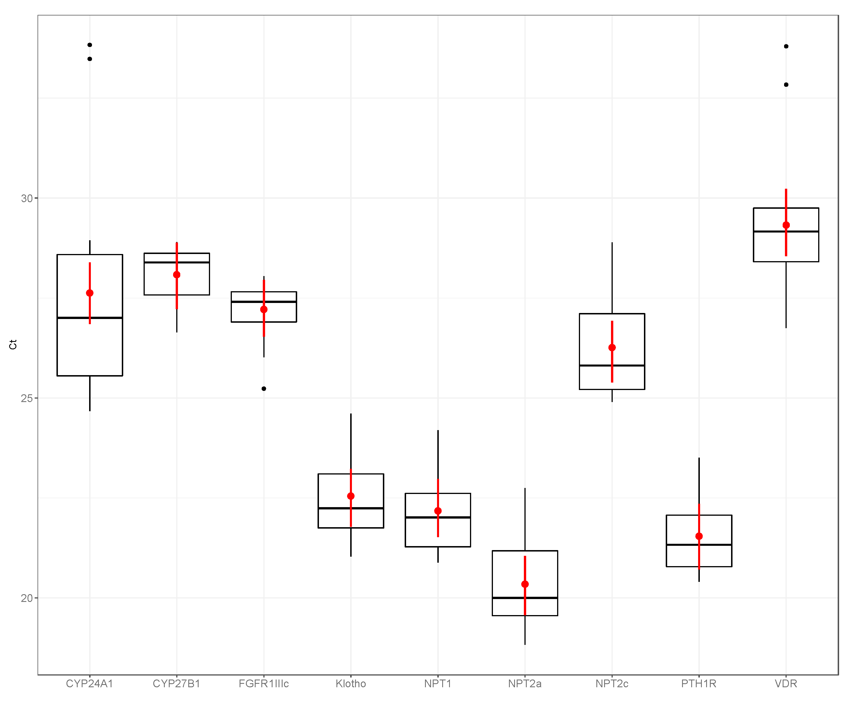
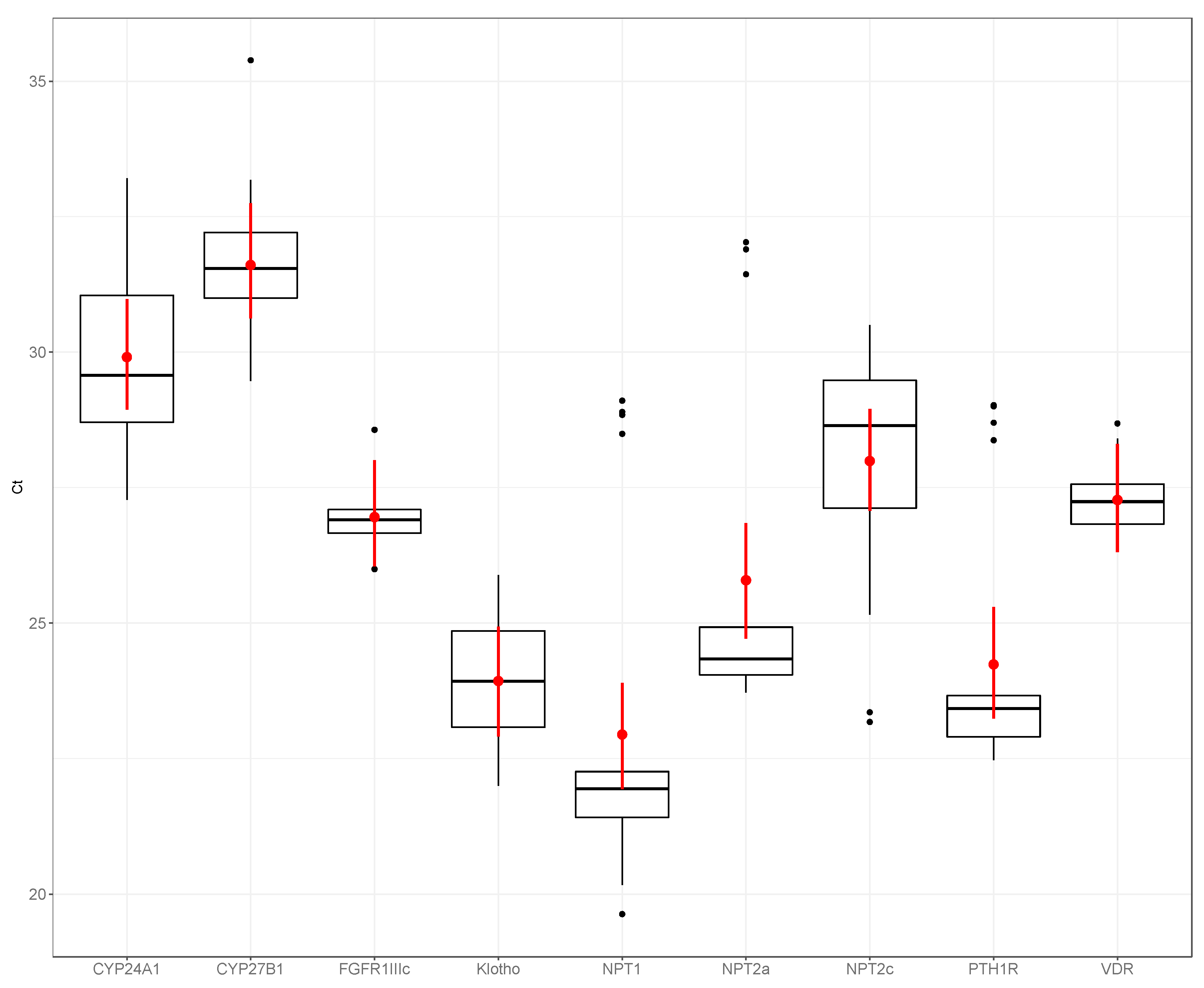
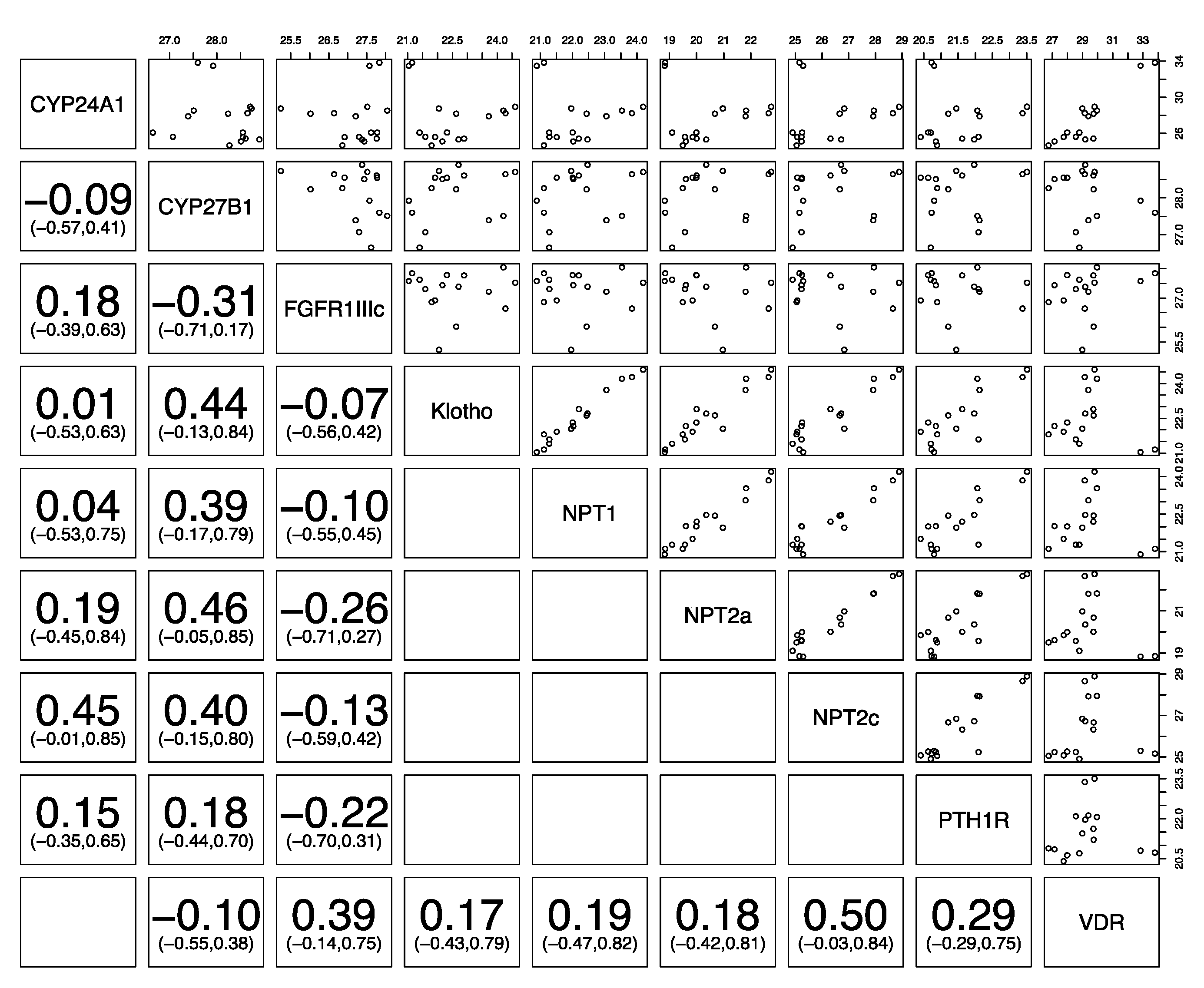
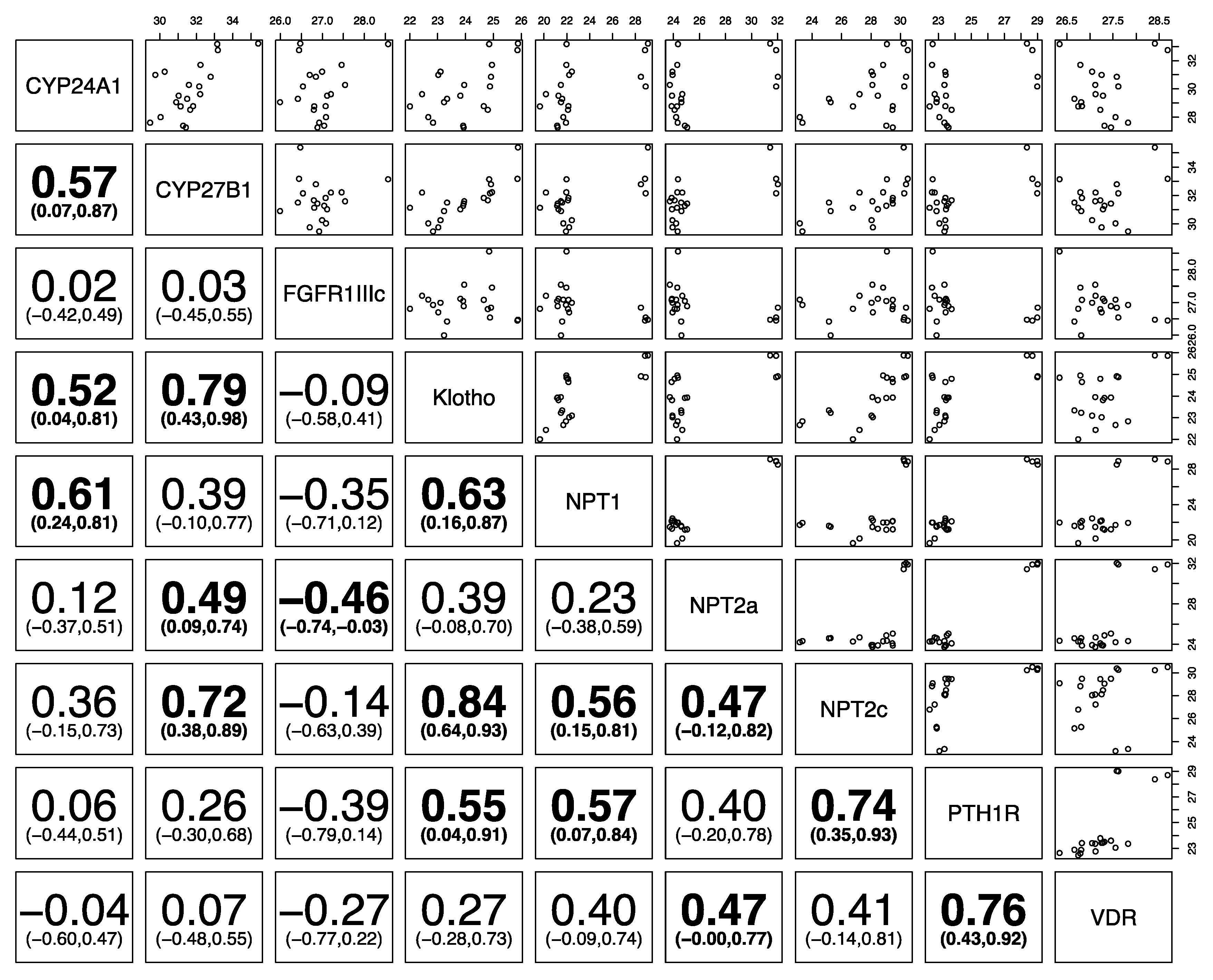
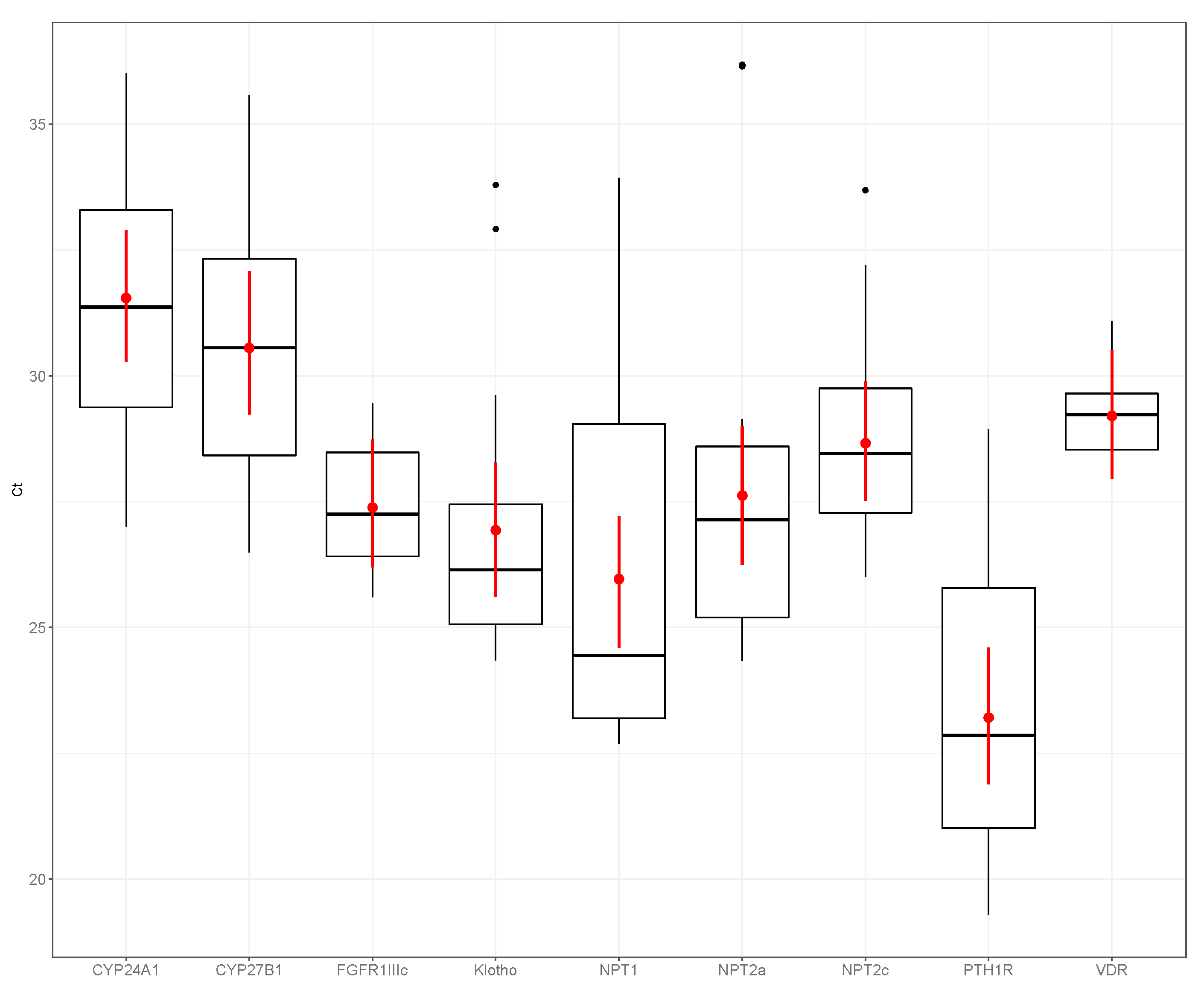
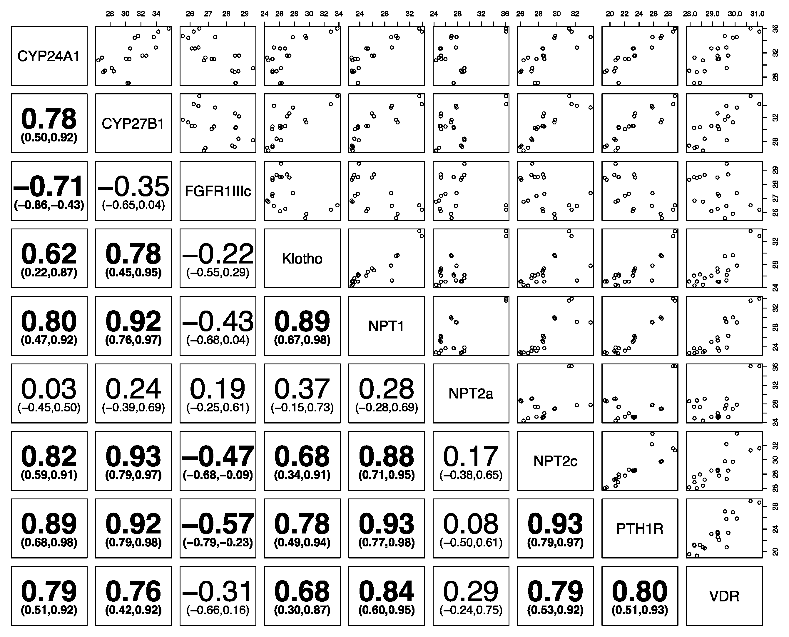
Kidney samples were collected post-mortem from 10 healthy adult horses, 1–14 years of age (6 male neutered, 2 male, 2 female; 5 Thoroughbreds, 5 Standardbreds), 10 healthy adult sheep, 4–6 years of age (all female, all Romney), and eight healthy dogs, 1–2 years of age (5 female, 3 male; 1 Neapolitan Mastiff, 2 Staffordshire bull terrier cross, 5 Pit bull terrier cross) that were euthanized for reasons unrelated to this study. The horses had been kept at pasture and supplemented with hay prior to euthanasia; the sheep were also kept at pasture prior to euthanasia; the dogs were fed the same commercial dog kibble for at least 2 weeks prior to euthanasia. As the kidney collection was post-mortem and unrelated to the reasons for euthanasia, animal ethics approval was not required as per national guidelines.
Kidney samples were collected within half an hour of euthanasia, divided into two, with one sample either snap-frozen in liquid nitrogen or placed into Ambion RNAlater (Thermo Fisher Scientific Inc., Waltham, MA, USA), and the other placed into 10% neutral buffered formalin and processed for histology. Hematoxylin and eosin-stained sections were examined by the primary author to confirm the absence of significant lesions. Samples for qPCR were stored at −80 °C until processing.
RNA was extracted using Tri Reagent (Sigma-Aldrich Inc., Merck KGaA, Darmstadt, Germany) as per the manufacturer’s instructions. RNA was treated with the Ambion Turbo DNA-free kit (Thermo Fisher Scientific Inc., Waltham, MA, USA) to remove any contaminating genomic RNA as per the manufacturer’s instructions. RNA and DNA concentrations (to check for absence of DNA) were measured using the Qubit 2.0 Fluorometer with Qubit RNA HS (high sensitivity), and DNA HS assays (Thermo Fisher Scientific Inc., Waltham, MA, USA). In addition, RNA integrity was assessed by running 5 μL of the RNA sample on a 1.5% agarose gel containing SYBR Safe (Thermo Fisher Scientific Inc., Waltham, MA, USA) with visualization of sharp 28S and 18S bands using an ultraviolet transilluminator.
The Transcriptor First Strand cDNA Synthesis Kit (Roche, Mannheim, Germany) was used to synthesize cDNA. Each 20 μL reaction mix contained 600 ng of RNA, 2.5 μM oligo (dT), 8 mM RT (reverse transcriptase) reaction buffer, 1 mM dNTP, 10 U reverse transcriptase, 20 U RNase inhibitor and RNase/DNase-free water. The reaction was performed at 55 °C for 30 min, 85 °C for 5 min, and then chilled at 4 °C using an Applied Biosystems Veriti Thermal Cycler (Thermo Fisher Scientific Inc., Waltham, MA, USA).
Primers were designed using Geneious 8.1 (Biomatters Ltd., Auckland, New Zealand) and the Primer 3 algorithm, as described previously [24]. The primers were designed with the following features: PCR product less than 150 bp, span an exon–exon junction, and show no complementarity to unrelated targets. Each primer set was tested for secondary structures at 60 °C using the mFold program (http://mfold.rit.albany.edu).
Real-time qPCR was performed using the StepOne Plus real-time PCR machine (Applied Biosystems, Thermo Fisher Scientific Inc., Waltham, MA, USA). Each real-time PCR reaction mix contained 5 μL Power SYBR Green PCR master mix (Thermo Fisher Scientific Inc., Waltham, MA, USA), the primer pair at concentrations determined to be optimal, as in Tables S1–S3, and 10 ng of cDNA, made up to 10 μL with RNase/DNase-free water. The PCR protocol consisted of a denaturation step at 95 °C for 20 s, followed by 40 cycles at 95 °C for 3 s and 60 °C for 30 s, and a melt curve ranging from 60 °C to 95 °C with a heating rate of 0.3 °C/15 s. Negative controls of water and reaction mix without reverse transcriptase were included in every PCR run, and all samples were run in duplicate. Standard curves were produced for each gene target in order to determine the efficiency and R2 (regression coefficient).
All samples were normalized relative to the expression of the same housekeeping gene found to be most stable in the kidneys of all three species: zeta polypeptide (YWHAZ) [24,25,26].
Statistical analysis was performed as previously described [24] using R-studio 1.1.456 with R version 3.5.1 (R Core Team, 2016) [27] and the lme4 package [28].

3. Results

Figure 1, Figure 2 and Figure 3 show the expression of the different phosphatonin pathway genes examined in the dogs, sheep and horses. Expression of FGFR1c showed little variability, particularly in the dogs (range count threshold (Ct) 25.23–28.04) and horses (range Ct 25.99–28.57). NPT2a expression in the dogs was high (range Ct 18.82–22.74) compared with the sheep (range Ct 24.34–36.18) and horse (range Ct 23.72–32.02). Of the sodium-phosphate cotransporters, NPT1 had the greatest expression in the horses and sheep, and NPT2a in the dogs. Although expression of all the sodium-phosphate cotransporters in the sheep was similar. NPT2c expression was the lowest of all the cotransporters in all three species.
Figure 4, Figure 5 and Figure 6 show the correlation of the expression of the different phosphatonin pathway genes with each other. Klotho was positively correlated in all species with the sodium-phosphate co-transporters NPT1, NPT2a and NPT2c and PTH1R. FGFR1IIIc was negatively correlated with klotho, the sodium-phosphate co-transporters NPT1, NPT2a, and NPT2c and PTH1R in all species, except NPT2a in sheep. Similarly, PTH1R was positively correlated with all genes in the three species (except FGFR1IIIc in all species). The sodium-phosphate co-transporters NPT1, NPT2 and NPT2c were positively correlated with each other in all species.

4. Discussion

The results show that phosphatonin system genes are expressed in the kidney of dogs, sheep and horses. Each species showed a different pattern of sodium-phosphate co-transporter gene expression. The sodium-phosphate co-transporters were all positively correlated with each other in the 3 species. NPT2a had profoundly greater expression in dogs than in sheep and horses, as shown by the mean Ct value of 20.4 in the dog, compared with 27.6 and 25.8 in the sheep and horse respectively. The dog, as a carnivore, has a higher dietary phosphorus consumption in comparison to herbivores such as the horse and sheep [29]. Therefore, higher expression of NPT2a mRNA could be an evolutionary adaptation to allow for greater renal phosphorus excretion.
NPT2a is considered to be the predominant sodium-phosphate co-transporter in the kidneys of mice, although NPT2c is considered the predominant human co-transporter. NPT2c expression was the lowest of all the co-transporters in the three species examined. In mice, NPT2 was found to account for 84% of total Na-P co-transporter expression, with NPT1 expression accounting for 15% [30]. In horses and sheep, NPT1 had the greatest expression of the sodium-phosphate co-transporters (although all three co-transporters had relatively similar expression in the sheep).
NPT1, when first discovered, was thought to be primarily a phosphate transporter; however, further study has revealed that, in addition to phosphate, it transports a variety of different anions, in particular urates [31]. PTH was found to have no effect on renal phosphate transport in Npt2-/- mice [32], suggesting there is a role for NPT1 in renal phosphate excretion. However, expression of both NPT1 and NPT2 was significantly decreased in the Hyp mouse (a mouse model of hypophosphatemic rickets with increased serum FGF23 concentrations) [30], and NPT1 expression is upregulated in Npt2-/- mouse models [33]. In this present study, expression of NPT1 was correlated with that of NPT2a, NPT2c and PTH1R in the three species examined. This could suggest species differences in the function and control of NPT1. High NPT1 expression in horses could be a physiological adaptation. Plasma uric acid concentrations in horses increase with intense exercise [34] and endurance exercise [35], and perhaps high renal NPT1 expression may assist the horse with removal of uric acid produced during exercise. Similarly, a study examining the kinetic parameters of sodium-phosphate co-transporter IIa in sheep and goats found that dietary phosphorus or calcium deficiency did not alter NPT2a transport capacity or affinity [36]. The authors suggested that PTH-independent pathways for phosphorus reabsorption may be important in the kidneys of ruminants, perhaps suggesting a role for NPT1 in renal tubular phosphate transport in sheep and goats.
Binding of membrane-bound klotho to the FGFR1c receptor activates the ERK1/2 pathway, resulting in decreased numbers of sodium-phosphate co-transporters in the membrane of proximal tubular epithelial cells [3]. Therefore, it could be expected that klotho and FGFR1c would be negatively correlated with renal sodium-phosphate co-transporters. This expectation holds true for FGFR1c in all species, where there is a negative association with the sodium-phosphate co-transporters, with the exception of NPT2a in the sheep.
However, klotho is significantly positively associated with sodium-phosphate co-transporters in all three species examined, a result that is inconsistent with knowledge on the regulation of sodium-phosphate co-transporters. Klotho does, however, have many other functions, including an association with aging [37]. Klotho is also involved in renal calcium and potassium transport and control of insulin-like growth factor-1 receptors and transforming growth factor-β1 [38,39,40,41]. Therefore, klotho gene expression in this study could be related to its other functions rather than its action on sodium-phosphate co-transporters. Expression of klotho in the body is highest in the kidneys and two forms may be produced—membrane bound klotho and secreted klotho—depending on RNA splicing. The design of the PCR primers would have detected both forms of klotho, perhaps also explaining the discrepancy in the relationship between klotho and the sodium-phosphate co-transporters.
Expression of FGFR1c showed little variability, particularly in the dogs and horses, despite the age and breed diversity of these two groups. FGFR1c is one of a group of four FGF receptors (FGFR1–4), and FGFR1 has three isoforms (a–c) with different binding affinities for different FGFs [42]. FGF23 can bind to FGFR1, 3 and 4, but signaling is maximum when bound to FGFR1c complexed to klotho [4]. There is limited literature on the regulation of FGFR1 receptors in the kidneys and on the relationship between FGFR1 and other phosphatonin pathway genes.
FGFR1c was significantly negatively correlated with CYP27B1 gene expression in sheep and dogs (but not horses), again likely reflecting the action of FGF23 in suppression of CYP27B1 [43]. Conversely, the relationship between FGFR1c and CYP24A1 was the opposite in sheep as to what might have been expected. There is some suggestion that FGF23 may increase CYP24A1 expression [44] as part of its role in decreasing plasma 1,25(OH)2D concentrations. However, whether it has a direct role in this is controversial, as 1,25(OH)2D itself is a potent inhibitor of CYP24A1 and separating the actions of FGF23 on 1,25(OH)2D and CYP24A1 is difficult. Other studies have found no effect of FGF23 on CYP24A1 gene expression [45]. Renal expression of VDR and CYP24A1 was significantly correlated in the sheep and dogs in this study, and this has also been reported in other studies [24]. These results suggest that control of CYP24A1 expression in the sheep may be indirect, due to decreased production of 1,25(OH)2D rather than a direct action of FGF23 itself.
PTH1R was highly expressed in all three species and had a positive correlation with all three sodium-phosphate co-transporters in all three species. Activation of PTH1R by PTH leads to inhibition of NPT2a, counter to the findings in our study. However, as with klotho, the regulation of PTH1R and the sodium-phosphate co-transporters is more complex than a simple two-way relationship. For example, PTH1R may couple with a number of different stimulatory G proteins, which may activate different secondary messenger pathways, and PTH1R on the apical and basolateral membranes of the renal tubular epithelial cell may have different roles [46]. In addition, the function of PTH1R on sodium-phosphate co-transporter expression in renal tubular epithelial cells is modulated by the binding of PTH1R to NHERF1 (sodium hydrogen exchanger regulatory factor 1) and activation of the phospholipase C pathway, which ultimately leads to NPT2a internalization from the cell membrane [47,48]. As such, there are a number of physiological intermediates between PTH1R and the sodium-phosphate co-transporters, suggesting that direct correlation may not be appropriate.
The major limitation of this study is the lack of concurrent data on plasma phosphorus and calcium concentrations. In addition, the control of calcium-phosphorus homeostasis by FGF23, 1,25OH2D3 and PTH is so inter-linked that determining relationships in isolation should be treated with caution. Therefore, future studies into the phosphatonin system should also assess other calciotropic genes, such as calcium channels and binding proteins, and assess intermediary cell messengers, such as NHERF, phosphokinase A and phospholipase C pathways. An additional limitation of the study is that the kidney samples were collected from animals of different age, sex and breed, and it is possible these differences may have resulted in the observed changes in gene expression. In particular, the wide age range in the horse samples could have contributed to the greater variation seen in gene expression in this species.
In summary, phosphatonin pathway genes are expressed in the kidneys of dogs, sheep and horses. NPT2a expression was greater in dogs compared with sheep and horses, and NPT2c expression was the lowest of all the sodium-phosphate transporters in all species. There was little variation in FGFR1c expression, suggesting that it may be tightly regulated, and FGFR1c was negatively correlated with the sodium-phosphate co-transporters. Due to the variability in expression and relationships between the different phosphatonin genes in the different species in this study, species differences in phosphatonin pathway regulation should be considered when investigating the pathogenesis of and possible treatments for chronic kidney disease. Additionally, further studies investigating the effect of sex and age would also be warranted.

Supplementary Materials

The following are available online at https://www.mdpi.com/2076-2615/10/10/1806/s1, Table S1: Canine phosphatonin and selected vitamin D-related kidney genes primer sequences and concentrations, Table S2: Ovine phosphatonin and selected vitamin D-related kidney genes primer sequences and concentrations, Table S3: Equine phosphatonin and selected vitamin D-related kidney genes primer sequences and concentrations.

Author Contributions

Conceptualization, K.E.D. and R.W.H.; methodology, K.E.D., R.W.H. and S.A.; formal analysis, K.E.D. and J.C.M.; resources, K.E.D. and S.A.; writing—original draft preparation, K.E.D.; writing—review and editing, K.E.D., R.W.H., J.C.M. and S.A.; visualization, K.E.D. and J.C.M.; funding acquisition, K.E.D. All authors have read and agreed to the published version of the manuscript.

Funding

This research was funded by the Massey University School of Veterinary Science McGeorge Fund.

Acknowledgments

The authors would like to acknowledge the assistance of Mike Hogan and Allan Nutman in collecting the kidney samples.

Conflicts of Interest

The authors declare no conflict of interest. The funders had no role in the design of the study; in the collection, analyses, or interpretation of data; in the writing of the manuscript, or in the decision to publish the results.

References

  1. Hardcastle, M.R.; Dittmer, K.E. Fibroblast Growth Factor 23: A New Dimension to Diseases of Calcium-Phosphorus Metabolism. Vet. Pathol. 2015, 52, 770–784. [Google Scholar] [CrossRef]
  2. Bonewald, L.F.; Wacker, M.J. FGF23 production by osteocytes. Pediatr. Nephrol. 2013, 28, 563–568. [Google Scholar] [CrossRef]
  3. Erben, R.G.; Andrukhova, O. FGF23-Klotho signaling axis in the kidney. Bone 2017, 100, 62–68. [Google Scholar] [CrossRef] [Green Version]
  4. Urakawa, I.; Yamazaki, Y.; Shimada, T.; Iijima, K.; Hasegawa, H.; Okawa, K.; Fujita, T.; Fukumoto, S.; Yamashita, T. Klotho converts canonical FGF receptor into a specific receptor for FGF23. Nature 2006, 444, 770. [Google Scholar] [CrossRef] [PubMed]
  5. Tomoe, Y.; Segawa, H.; Shiozawa, K.; Kaneko, I.; Tominaga, R.; Hanabusa, E.; Aranami, F.; Furutani, J.; Kuwahara, S.; Tatsumi, S.; et al. Phosphaturic action of fibroblast growth factor 23 in Npt2 null mice. Am. J. Physiol.-Ren. Physiol. 2010, 298, F1341–F1350. [Google Scholar] [CrossRef] [PubMed]
  6. Shimada, T.; Kakitani, M.; Yamazaki, Y.; Hasegawa, H.; Takeuchi, Y.; Fujita, T.; Fukumoto, S.; Tomizuka, K.; Yamashita, T. Targeted ablation of Fgf23 demonstrates an essential physiological role of FGF23 in phosphate and vitamin D metabolism. J. Clin. Investig. 2004, 113, 561–568. [Google Scholar] [CrossRef] [PubMed]
  7. Wöhrle, S.; Bonny, O.; Beluch, N.; Gaulis, S.; Stamm, C.; Scheibler, M.; Müller, M.; Kinzel, B.; Thuery, A.; Brueggen, J.; et al. FGF receptors control vitamin D and phosphate homeostasis by mediating renal FGF-23 signaling and regulating FGF-23 expression in bone. J. Bone Miner. Res. 2011, 26, 2486–2497. [Google Scholar] [CrossRef]
  8. Silver, J.; Naveh-Many, T. FGF23 and the parathyroid. Adv. Exp. Med. Biol. 2012, 728, 92–99. [Google Scholar]
  9. Dittmer, K.E.; Perera, K.C.; Elder, P.A. Serum fibroblast growth factor 23 concentrations in dogs with chronic kidney disease. Res. Vet. Sci. 2017, 114, 348–350. [Google Scholar] [CrossRef]
  10. Geddes, R.F.; Finch, N.C.; Elliott, J.; Syme, H.M. Fibroblast growth factor 23 in feline chronic kidney disease. J. Vet. Intern. Med. 2013, 27, 234–241. [Google Scholar] [CrossRef]
  11. Fliser, D.; Kollerits, B.; Neyer, U.; Ankerst, D.P.; Lhotta, K.; Lingenhel, A.; Ritz, E.; Kronenberg, F.; Kuen, E.; Konig, P.; et al. Fibroblast growth factor 23 (FGF23) predicts progression of chronic kidney disease: The Mild to Moderate Kidney Disease (MMKD) Study. J. Am. Soc. Nephrol. 2007, 18, 2600–2608. [Google Scholar] [CrossRef]
  12. Finch, N.C.; Geddes, R.F.; Syme, H.M.; Elliott, J. Fibroblast growth factor 23 (FGF-23) concentrations in cats with early nonazotemic chronic kidney disease (CKD) and in healthy geriatric cats. J. Vet. Intern. Med. 2013, 27, 227–233. [Google Scholar] [CrossRef]
  13. Isakova, T.; Wahl, P.; Vargas, G.S.; Gutierrez, O.M.; Scialla, J.; Xie, H.; Appleby, D.; Nessel, L.; Bellovich, K.; Chen, J.; et al. Fibroblast growth factor 23 is elevated before parathyroid hormone and phosphate in chronic kidney disease. Kidney Int. 2011, 79, 1370–1378. [Google Scholar] [CrossRef] [PubMed] [Green Version]
  14. Myakala, K.; Motta, S.; Murer, H.; Wagner, C.A.; Koesters, R.; Biber, J.; Hernando, N. Renal-specific and inducible depletion of NaPi-IIc/Slc34a3, the cotransporter mutated in HHRH, does not affect phosphate or calcium homeostasis in mice. Am. J. Physiol.-Ren. Physiol. 2014, 306, F833–F843. [Google Scholar] [CrossRef] [Green Version]
  15. Bergwitz, C.; Roslin, N.M.; Tieder, M.; Loredo-Osti, J.C.; Bastepe, M.; Abu-Zahra, H.; Frappier, D.; Burkett, K.; Carpenter, T.O.; Anderson, D.; et al. SLC34A3 mutations in patients with hereditary hypophosphatemic rickets with hypercalciuria predict a key role for the sodium-phosphate cotransporter NaPi-IIc in maintaining phosphate homeostasis. Am. J. Hum. Genet. 2006, 78, 179–192. [Google Scholar] [CrossRef] [PubMed] [Green Version]
  16. Dinour, D.; Davidovits, M.; Ganon, L.; Ruminska, J.; Forster, I.C.; Hernando, N.; Eyal, E.; Holtzman, E.J.; Wagner, C.A. Loss of function of NaPiIIa causes nephrocalcinosis and possibly kidney insufficiency. Pediatr. Nephrol. 2016, 31, 2289–2297. [Google Scholar] [CrossRef] [PubMed]
  17. Muscher-Banse, A.S.; Breves, G. Mechanisms and regulation of epithelial phosphate transport in ruminants: Approaches in comparative physiology. Pflügers Arch.-Eur. J. Physiol. 2019, 471, 185–191. [Google Scholar] [CrossRef]
  18. Firmenich, C.S.; Elfers, K.; Wilkens, M.R.; Breves, G.; Muscher-Banse, A.S. Modulation of renal calcium and phosphate transporting proteins by dietary nitrogen and/or calcium in young goats1. J. Anim. Sci. 2018, 96, 3208–3220. [Google Scholar] [CrossRef]
  19. Shirazi-Beechey, S.P.; Penny, J.I.; Dyer, J.; Wood, I.S.; Tarpey, P.S.; Scott, D.; Buchan, W. Epithelial phosphate transport in ruminants, mechanisms and regulation. Kidney Int. 1996, 49, 992–996. [Google Scholar] [CrossRef] [Green Version]
  20. Muscher, A.S.; Wilkens, M.R.; Mrochen, N.; Schröder, B.; Breves, G.; Huber, K. Ex vivo intestinal studies on calcium and phosphate transport in growing goats fed a reduced nitrogen diet. Br. J. Nutr. 2012, 108, 628–637. [Google Scholar] [CrossRef]
  21. Muscher-Banse, A.S.; Marholt, L.; Eigendorf, N.; Wilkens, M.R.; Schröder, B.; Breves, G.; Cehak, A. Segmental diversity of phosphate transport along the intestinal axis in horses1. J. Anim. Sci. 2017, 95, 165–172. [Google Scholar] [CrossRef] [PubMed]
  22. Foote, A.P.; Lambert, B.D.; Brady, J.A.; Muir, J.P. Short communication: Phosphate transporter expression in Holstein cows. J. Dairy Sci. 2011, 94, 1913–1916. [Google Scholar] [CrossRef] [PubMed]
  23. Huber, K.; Walter, C.; Schröder, B.; Breves, G. Phosphate transport in the duodenum and jejunum of goats and its adaptation by dietary phosphate and calcium. Am. J. Physiol. Integr. Comp. Physiol. 2002, 283, R296–R302. [Google Scholar] [CrossRef] [PubMed] [Green Version]
  24. Azarpeykan, S.; Dittmer, K.E.; Marshall, J.C.; Perera, K.C.; Gee, E.K.; Acke, E.; Thompson, K.G. Evaluation and Comparison of Vitamin D responsive gene expression in ovine, canine and equine kidney. PLoS ONE 2016, 11, e0162598. [Google Scholar] [CrossRef]
  25. Azarpeykan, S.; Dittmer, K.E. Evaluation of housekeeping genes for quantitative gene expression analysis in the equine kidney. J. Equine Sci. 2016, 27, 165–168. [Google Scholar] [CrossRef] [PubMed] [Green Version]
  26. Peters, I.R.; Peeters, D.; Helps, C.R.; Day, M.J. Development and application of multiple internal reference (housekeeper) gene assays for accurate normalisation of canine gene expression studies. Vet. Immunol. Immunopathol. 2007, 117, 55–66. [Google Scholar] [CrossRef] [PubMed]
  27. R Core Team. R: A Language and Environment for Statistical Computing; R Foundation for Statistical Computing: Vienna, Austria, 2016. [Google Scholar]
  28. Bates, D.; Machler, M.; Bolker, B.; Walker, S. Fitting linear mixed-effects models using lme4. J. Stat. Softw. 2015, 67, 1–48. [Google Scholar] [CrossRef]
  29. Simesen, M.G. Calcium, inorganic phosphorus, and magnesium metabolism in health and disease. In Clinical Biochemistry of Domestic Animals; Kaneko, J.J., Cornelius, C.E., Eds.; Academic Press, Inc.: New York, NY, USA; London, UK, 1970; pp. 313–375. [Google Scholar]
  30. Tenenhouse, H.S.; Roy, S.; Martel, J.; Gauthier, C. Differential expression, abundance, and regulation of Na+-phosphate cotransporter genes in murine kidney. Am. J. Physiol. 1998, 275, F527–F534. [Google Scholar] [CrossRef]
  31. Lederer, E.; Miyamoto, K. Clinical Consequences of Mutations in Sodium Phosphate Cotransporters. Clin. J. Am. Soc. Nephrol. 2012, 7, 1179–1187. [Google Scholar] [CrossRef] [Green Version]
  32. Zhao, N.; Tenenhouse, H.S. Npt2 gene disruption confers resistance to the inhibitory action of parathyroid hormone on renal sodium-phosphate cotransport. Endocrinology 2000, 141, 2159–2165. [Google Scholar] [CrossRef]
  33. Hoag, H.M.; Martel, J.; Gauthier, C.; Tenenhouse, H.S. Effects of Npt2 gene ablation and low-phosphate diet on renal Na(+)/phosphate cotransport and cotransporter gene expression. J. Clin. Investig. 1999, 104, 679–686. [Google Scholar] [CrossRef] [PubMed] [Green Version]
  34. Rasanen, L.A.; Wiitanen, P.A.; Lilius, E.M.; Hyyppa, S.; Poso, A.R. Accumulation of uric acid in plasma after repeated bouts of exercise in the horse. Comp. Biochem. Physiol. B Biochem. Mol. Biol. 1996, 114, 139–144. [Google Scholar] [CrossRef]
  35. Castejon, F.; Trigo, P.; Munoz, A.; Riber, C. Uric acid responses to endurance racing and relationships with performance, plasma biochemistry and metabolic alterations. Equine Vet. J. 2006, 38, 70–73. [Google Scholar] [CrossRef] [PubMed]
  36. Schröder, B.; Walter, C.; Breves, G.; Huber, K. Comparative studies on Na-dependent Pi transport in ovine, caprine and porcine renal cortex. J. Comp. Physiol. B Biochem. Syst. Environ. Physiol. 2000, 170, 387–393. [Google Scholar] [CrossRef]
  37. Kuro-o, M. Klotho and the Aging Process. Korean J. Intern. Med. 2011, 26, 113–122. [Google Scholar] [CrossRef]
  38. Cha, S.-K.; Ortega, B.; Kurosu, H.; Rosenblatt, K.P.; Kuro-O, M.; Huang, C.-L. Removal of sialic acid involving Klotho causes cell-surface retention of TRPV5 channel via binding to galectin-1. Proc. Natl. Acad. Sci. USA 2008, 105, 9805–9810. [Google Scholar] [CrossRef] [Green Version]
  39. Cha, S.-K.; Hu, M.-C.; Kurosu, H.; Kuro-o, M.; Moe, O.; Huang, C.-L. Regulation of renal outer medullary potassium channel and renal K(+) excretion by Klotho. Mol. Pharmacol. 2009, 76, 38–46. [Google Scholar] [CrossRef] [Green Version]
  40. Kurosu, H.; Yamamoto, M.; Clark, J.D.; Pastor, J.V.; Nandi, A.; Gurnani, P.; McGuinness, O.P.; Chikuda, H.; Yamaguchi, M.; Kawaguchi, H.; et al. Suppression of aging in mice by the hormone Klotho. Science (80-.) 2005, 309, 1829–1833. [Google Scholar] [CrossRef] [Green Version]
  41. Doi, S.; Zou, Y.; Togao, O.; Pastor, J.V.; John, G.B.; Wang, L.; Shiizaki, K.; Gotschall, R.; Schiavi, S.; Yorioka, N.; et al. Klotho inhibits transforming growth factor-β1 (TGF-β1) signaling and suppresses renal fibrosis and cancer metastasis in mice. J. Biol. Chem. 2011, 286, 8655–8665. [Google Scholar] [CrossRef] [Green Version]
  42. Martin, A.; David, V.; Quarles, L.D. Regulation and function of the FGF23/klotho endocrine pathways. Physiol. Rev. 2012, 92, 131–155. [Google Scholar] [CrossRef]
  43. Chanakul, A.; Zhang, M.Y.H.; Louw, A.; Armbrecht, H.J.; Miller, W.L.; Portale, A.A.; Perwad, F. FGF-23 Regulates CYP27B1 Transcription in the Kidney and in Extra-Renal Tissues. PLoS ONE 2013, 8, e72816. [Google Scholar] [CrossRef] [PubMed] [Green Version]
  44. Dai, B.; David, V.; Martin, A.; Huang, J.; Li, H.; Jiao, Y.; Gu, W.; Quarles, L.D. A Comparative Transcriptome Analysis Identifying FGF23 Regulated Genes in the Kidney of a Mouse CKD Model. PLoS ONE 2012, 7, e44161. [Google Scholar] [CrossRef] [PubMed] [Green Version]
  45. Kägi, L.; Bettoni, C.; Pastor-Arroyo, E.M.; Schnitzbauer, U.; Hernando, N.; Wagner, C.A. Regulation of vitamin D metabolizing enzymes in murine renal and extrarenal tissues by dietary phosphate, FGF23, and 1,25(OH)2D3. PLoS ONE 2018, 13, e0195427. [Google Scholar] [CrossRef] [PubMed]
  46. Mahon, M.J. The Parathyroid Hormone 1 Receptor Directly Binds to the FERM Domain of Ezrin, an Interaction that Supports Apical Receptor Localization and Signaling in LLC-PK1 Cells. Mol. Endocrinol. 2009, 23, 1691–1701. [Google Scholar] [CrossRef] [PubMed] [Green Version]
  47. Mahon, M.J.; Donowitz, M.; Yun, C.C.; Segre, G. V Na(+)/H(+ ) exchanger regulatory factor 2 directs parathyroid hormone 1 receptor signalling. Nature 2002, 417, 858–861. [Google Scholar] [CrossRef]
  48. Bastepe, M.; Turan, S.; He, Q. Heterotrimeric G proteins in the control of parathyroid hormone actions. J. Mol. Endocrinol. 2017, 58, R203–R224. [Google Scholar] [CrossRef] [Green Version]
Figure 1. Expression of phosphatonin and selected vitamin D-related transcripts in canine kidney—cytochrome P450 family 24 subfamily A polypeptide 1 (CYP24A1), cytochrome P450 family 27 subfamily B polypeptide 1 (CYP27B1), fibroblast growth factor receptor 1 IIIc (FGFR1IIIc), α-klotho (klotho, KL), sodium-phosphate co-transporter 1 (NPT1), NPT2a, NPT2c, parathyroid hormone 1 receptor (PTH1R) and vitamin D receptor (VDR). Boxplots are normalized cycle threshold numbers (Ct values), where the median expression levels of genes are shown as black bars, the red dots show the mean expression of genes and the red line bars represent 95% confidence interval of the genes, accounting for replicates.
Animals 10 01806 g001
Figure 2. Expression of phosphatonin and selected vitamin D-related transcripts in ovine kidneys—cytochrome P450 family 24 subfamily A polypeptide 1 (CYP24A1), cytochrome P450 family 27 subfamily B polypeptide 1 (CYP27B1), fibroblast growth factor receptor 1 IIIc (FGFR1IIIc), α-klotho (klotho, KL), sodium-phosphate co-transporter 1 (NPT1), NPT2a, NPT2c, parathyroid hormone 1 receptor (PTH1R) and vitamin D receptor (VDR). Boxplots are normalized cycle threshold numbers (Ct values), where the median expression levels of genes are shown as black bars, the red dots show the mean expression of genes and the red line bars represent 95% confidence interval of the genes, accounting for replicates.
Animals 10 01806 g002
Figure 3. Expression of phosphatonin and selected vitamin D-related transcripts in equine kidneys—cytochrome P450 family 24 subfamily A polypeptide 1 (CYP24A1), cytochrome P450 family 27 subfamily B polypeptide 1 (CYP27B1), fibroblast growth factor receptor 1 IIIc (FGFR1IIIc), α-klotho (klotho, KL), sodium-phosphate co-transporter 1 (NPT1), NPT2a, NPT2c, parathyroid hormone 1 receptor (PTH1R) and vitamin D receptor (VDR). Boxplots are normalized cycle threshold numbers (Ct values), where the median expression levels of genes are shown as black bars, the red dots show the mean expression of genes and the red line bars represent 95% confidence interval of the genes, accounting for replicates.
Animals 10 01806 g003
Figure 4. Correlation of phosphatonin and selected vitamin D-related transcripts in canine kidneys. Spearman’s correlation (95% confidence intervals) between cytochrome P450 family 24 subfamily A polypeptide 1 (CYP24A1), cytochrome P450 family 27 subfamily B polypeptide 1 (CYP27B1), fibroblast growth factor receptor 1 IIIc (FGFR1IIIc), α-klotho (klotho, KL), sodium-phosphate co-transporter 1 (NPT1), NPT2a, NPT2c, parathyroid hormone 1 receptor (PTH1R) and vitamin D receptor (VDR). Statistically significant positive and/or negative correlations of different genes with each other (p < 0.05) are bolded, and the numbers in parentheses show the 95% confidence interval. On the top and right, the numbers indicate normalized gene expression.
Animals 10 01806 g004
Figure 5. Correlation of phosphatonin and selected vitamin D-related transcripts in ovine kidneys. Spearman’s correlation (95% confidence intervals) between cytochrome P450 family 24 subfamily A polypeptide 1 (CYP24A1), cytochrome P450 family 27 subfamily B polypeptide 1 (CYP27B1), fibroblast growth factor receptor 1 IIIc (FGFR1IIIc), α-klotho (klotho, KL), sodium-phosphate co-transporter 1 (NPT1), NPT2a, NPT2c, parathyroid hormone 1 receptor (PTH1R) and vitamin D receptor (VDR). Statistically significant positive and/or negative correlations of different genes with each other (p < 0.05) are bolded, and the numbers in parentheses show the 95% confidence interval. On the top and right, the numbers indicate normalized gene expression.
Animals 10 01806 g005
Figure 6. Correlation of phosphatonin and selected vitamin D-related transcripts in equine kidneys. Spearman’s correlation (95% confidence intervals) between cytochrome P450 family 24 subfamily A polypeptide 1 (CYP24A1), cytochrome P450 family 27 subfamily B polypeptide 1 (CYP27B1), fibroblast growth factor receptor 1 IIIc (FGFR1IIIc), α-klotho (klotho, KL), sodium-phosphate co-transporter 1 (NPT1), NPT2a, NPT2c, parathyroid hormone 1 receptor (PTH1R) and vitamin D receptor (VDR). Statistically significant positive and/or negative correlations of different genes with each other (p < 0.05) are bolded, and the numbers in parentheses show the 95% confidence interval. On the top and right, the numbers indicate normalized gene expression.
Animals 10 01806 g006

