Article

Characterization of Martelella soudanensis sp. nov., Isolated from a Mine Sediment

1
Groundwater Research Center, Geologic Environment Division, Korea Institute of Geoscience and Mineral Resources, Daejeon 34132, Korea
2
Department of Microbiology, Chungbuk National University, Cheongju 28644, Korea
*
Author to whom correspondence should be addressed.
Microorganisms 2021, 9(8), 1736; https://doi.org/10.3390/microorganisms9081736
Submission received: 23 July 2021 / Accepted: 10 August 2021 / Published: 14 August 2021
(This article belongs to the Section Environmental Microbiology)

Abstract

:
Gram-stain-negative, strictly aerobic, non-spore-forming, non-motile, and rod-shaped bacterial strains, designated NC18T and NC20, were isolated from the sediment near-vertical borehole effluent originating 714 m below the subsurface located in the Soudan Iron Mine in Minnesota, USA. The 16S rRNA gene sequence showed that strains NC18T and NC20 grouped with members of the genus Martelella, including M. mediterranea DSM 17316T and M. limonii YC7034T. The genome sizes and G + C content of both NC18T and NC20 were 6.1 Mb and 61.8 mol%, respectively. Average nucleotide identity (ANI), the average amino acid identity (AAI), and digital DNA–DNA hybridization (dDDH) values were below the species delineation threshold. Pan-genomic analysis showed that NC18T, NC20, M. mediterranea DSM 17316T, M. endophytica YC6887T, and M. lutilitoris GH2-6T had 8470 pan-genome orthologous groups (POGs) in total. Five Martelella strains shared 2258 POG core, which were mainly associated with amino acid transport and metabolism, general function prediction only, carbohydrate transport and metabolism, translation, ribosomal structure and biogenesis, and transcription. The two novel strains had major fatty acids (>5%) including summed feature 8 (C18:1 ω7c and/or C18:1 ω6c), C19:0 cyclo ω8c, C16:0, C18:1 ω7c 11-methyl, C18:0, and summed feature 2 (C12:0 aldehyde and/or iso-C16:1 I and/or C14:0 3-OH). The sole respiratory quinone was uniquinone-10 (Q-10). On the basis of polyphasic taxonomic analyses, strains NC18T and NC20 represent novel species of the genus Martelella, for which the name Martelella soudanensis sp. nov. is proposed. The type strain is NC18T (=KTCT 82174T = NBRC 114661T).

1. Introduction

Although deep subsurface environments are characterized by relatively low available organic carbon and elevated pressure, they are the largest habitats for prokaryotes, estimated to contain from 12% to 20% of the total biomass of microorganisms on Earth [1,2]. The deep biosphere contains a variety of functionally active microbial communities, but microbes face challenges such as limited available electron donors or acceptors, pore space, and fracture networks, and competition with other microorganisms for growth [3]. The Soudan Iron Mine is located in northern Minnesota, USA, along the southern edge of the Canadian Shield Transects and the Archaean Animikie ocean basin. The mine reaches a depth of 714 m below the surface, providing access to deep subsurface brines entrained in a massive hematite formation [3,4]. Previous cultivation-based and metagenomics studies reported the presence of diverse microbial communities, including the iron-oxidizing Marinobacter and iron-reducing Desulfuromonas in a calcium- and sodium-rich brine that reaches salinities as high as 4.2% (w/v) from these boreholes [3,5,6].
Halophiles and halotolerant bacteria are able to survive even in highly saline environments that are unfavorable for the existence of most life forms [7]. In recent decades, halophilic and halotolerant bacteria have been studied in biotechnological applications [8]. Halophiles have stable enzymes that function in high salinity and are used in industrial processes where high salt concentrations would inhibit enzymatic transformations [9]. Many compounds synthesized by halophiles (e.g., enzymes, polysaccharides, polyhydroxyalkanoates, and biosurfactants) are available for commercial uses [10,11,12,13]. Several halophilic enzymes, including proteases, amylases, lipases, and xylanases, have been characterized in recent years and are used in biofuel production [14], food processing [15], and the biodegradation of organic contaminants [16,17]. These halophilic and halotolerant bacteria are spread over a large number of phylogenetic branches, with most species being grouped in Proteobacteria [18].
The genus Martelella was originally described in 2005 by Rivas et al., isolated from a subterranean saline lake [19]. This genus belongs to the phylum Proteobacteria, the class Alphaproteobacteria, the order Hyphomicrobiales, and the family Aurantimonadaceae (Available online: https://lpsn.dsmz.de/genus/martelella (accessed on 16 July 2021)). Thus far, a total of 8 species were reported in this genus: Martelella mediterranea [19], Martelella endophytica [20], Martelella radicis [21], Martelella mangrovi [21], Martelella suaedae [22], Martelella limonii [22], Martelella caricis [23], and Martelella lutilitoris [24]. The members of Martelella are Gram-stain-negative, aerobic, rod-shaped, non-motile, non-spore-forming, and chemoheterotrophic bacteria. Martelella strains were isolated from diverse environments such as a saline lake, the roots or mud plat of halophytes, and the soils of mangrove roots [19,20,21,22,23,24]. Additionally, a halophilic polycyclic aromatic hydrocarbon (PAH)-degrading strain was reported [25]. Therefore, this genus could be of great biotechnological interest, as halotolerant and halophilic microorganisms are well-known for their potential applications in biotechnology.
Here, we report the taxonomic characterization of bacterial strains, NC18T and NC20, isolated from mine sediment by using polyphasic approaches.

2. Materials and Methods

2.1. Enrichment Culture and Isolation

Strains NC18T and NC20 were isolated from sediment near a descending exploratory borehole (47°49.2′ N, 92°14.5′ W), Soudan Mine Diamond Drill Hole 942, located at the bottom (level 27) of the Soudan Underground Mine State Park in Soudan, Minnesota, USA. The sediment sample was serially diluted with 0.85% NaCl, and suspensions were plated on R2A agar (BD, Franklin Lakes, NJ, USA) supplemented with 2% NaCl (w/v) and incubated at room temperature for 7 days. Circular, smooth, and white-to-cream-colored colonies of designated strains NC18T and NC20 were isolated and subsequently purified three times. Cultures were maintained on R2A plates supplemented with 2% NaCl at 30 °C, and stocks were preserved in R2A broth with glycerol (20%, v/v) at −80 °C.

2.2. Phenotypic Analysis

Cell morphology and flagellation of strains NC18T and NC20 were observed by transmission electron microscope (CM20, Philips, Amsterdam, The Netherlands) operated at 80 kV with cells grown in a marine agar (MA; BD, Franklin Lakes, NJ, USA) plate for 2 days at 30 °C. Cells were negatively stained using 2% (w/v) uranyl acetate, air-dried, and had their grids examined. The presence of spores was analyzed by phase-contrast microscopy (ECLIPSE 80i, Nikon, Tokyo, Japan) at a magnification of 1500×, using cells that had been grown for 1 week at 30 °C on MA. Gram staining was determined by using the bioMérieux Gram-staining kit according to the manufacturer’s instructions. The colony color and morphology of strains NC18T and NC20 were investigated on MA plate incubated for 2 days at 30 °C.
Growth at different temperature (4, 10, 12, 15, 20, 25, 30, 35, and 40 °C) and pH (pH 4.0–10.0 at intervals of 1.0 pH units) levels were tested on marine broth 2216 (MB; BD, Franklin Lakes, NJ, USA). pH was adjusted with 0.1 N NaOH or 0.1 N HCl solutions and checked after autoclaving. Salt tolerance was investigated in marine broth supplemented with 0–15% (w/v, at 1% intervals) NaCl. Growth of strains NC18T and NC20 at different temperatures, pH, and NaCl concentrations were determined by OD600 using a spectrophotometric method (Optizen POP, Mechasus, Daejeon, Korea). Growth under anaerobic conditions was determined on MA plates at 30 °C for 10 days in an anaerobic Gaspak jar (OXOID) with Anaero-PACK (Mitsubishi Gas Chemical Co., Tokyo, Japan).
Catalase activity was determined with 3% (v/v) hydrogen peroxide. Casein hydrolysis was examined on MA supplemented with 1% (w/v) skim milk. Enzyme activities, acid production from different carbohydrates, and the assimilation of various substrates were determined using API ZYM, API 20E, and API 20NE, respectively, according to the manufacturer’s instructions (bioMérieux, Marcy l’Etoile, France).

2.3. Chemotaxonomic Analysis

For the analysis of cellular fatty acid and respiratory quinone, cells of NC18T and NC20 were prepared from cells grown on MA for 2 days at 30 °C. Fatty acid methyl esters were prepared and analyzed according to the standard MIDI protocol (Sherlock Microbial Identification System, version 6.2) and identified by the RTSBA 6.0 database of the Microbial Identification System [26].
Respiratory quinone was extracted by the chloroform-methanol extraction method and analyzed using high-performance liquid chromatography (HPLC) as previously described [27].

2.4. Phylogenetic Analysis

Genomic DNA was prepared using an AccuPrep Genomic DNA Extraction Kit (Bioneer, Daejeon, Korea) according to the manufacturer’s instructions. DNA was precipitated using 1 volume of chilled isopropanol and 0.1 volume of 3 M sodium acetate, followed by overnight incubation at −20 °C. DNA pellet was collected by centrifugation at 13,800× g for 30 min at 4 °C. The DNA pellet was washed with 70% ethanol, air-dried, and resuspended in nuclease-free water (Qiagen, Germantown, MD, USA).
The 16S rRNA gene was amplified using universal primers 27F and 1492R [28]. The 16S rRNA gene sequences (1482 nt) of strains NC18T and NC20 were identical with the corresponding region of its genomic sequence and were compared with the related sequences from the EzBioCloud server (Available online: www.ezbiocloud.net (accessed on 16 July 2021)) [29]. The 16S rRNA gene sequences were aligned with those of closely related species using the CLUSTAL X software program [30]. Gaps were edited in the BioEdit program [31] using the neighbor-joining, maximum-parsimony, and maximum-likelihood algorithms in MEGA 6.0 software [32]. Bootstrap analysis was performed to determine confidence values of individual branches in the phylogenetic tree with 1000 replications. The 16s rRNA gene sequences of strains NC18T and NC20 were deposited in GenBank/EMBL/DDBJ under accession numbers MT367774 and MT367775, respectively.
Multilocus sequence analysis (MLSA) was also conducted to support phylogenetic analysis using 31 housekeeping gene products: dnaG, frr, infC, nusA, pgk, pyrG, rplA, rplB, rplC, rplD, rplE, rplF, rplK, rplL, rplM, rplN, rplP, rplS, rplT, rpmA, rpoB, rpsB, rpsC, rpsE, rpsI, rpsJ, rpsK, rpsM, rpsS, smpB, and tsf. The available genomic data of the genus Martelella were obtained from the GeneBank database (Supplementary Table S1). Then, 31 universally conserved proteins were extracted using the AmphoraNet server [33]. A multiple alignment of 31core proteins was built using MUSCLE [34]. The resulting alignment was concatenated using MEGA 6.0 [32], resulting in 9333 amino acid positions. Gaps were then removed using Gblocks version 0.91b [35], resulting in 4988 amino acid positions, covering about 53% of the original alignment. The phylogenetic tree was reconstructed by using the neighbor-joining, maximum-likelihood, and maximum-parsimony algorithms with the JTT matrix-based model [36] and 1000 bootstrap replicates in MEGA 6.0 [32].

2.5. Whole-Genome Sequencing, Assembly, and Annotation

Whole-genome sequencing and assembly were carried out using Illumina MiSeq and PacBio RS II platforms at the ChunLab Inc. (Seoul, Korea). Illumina sequencing data were assembled with SPAdes 3.9.0 (Available online: http://cab.spbu.ru/software/spades/ (accessed on 5 December 2020)). MiSeq sequencing data were quality-controlled with Trimmomatic 0.36, and PhiX sequences were removed with BBMap 38.32. PacBio RS II sequencing data were assembled with PacBio SMRT Link 7.0.1 using the HGAP4 protocol (Pacific Biosciences, Menlo Park, CA, USA). Hybrid assembly was performed by Pilon version 1.22 and reassembled using quality-controlled MiSeq data and assembled contigs from Pacbio data. The resulting contigs from the hybrid assembly were circularized using Circlator 1.4.0 (Sanger Institute). Contamination of the whole-genome sequence was checked using ContEst16S [37]. Gene prediction and functional annotation pipeline of whole-genome assemblies used in EzBioCloud database. Protein coding sequences (CDSs) were predicted by Prodigal v.2.6.2 [38]. Genes coding for tRNA were identified using tRNAscan SE 1.3.1 [39]. rRNA operons were predicted by a covariance model search with Rfam 12.0 database [40]. CRISPRs were detected by PilerCR 1.06 [41] and CRT 1.2 [42]. Identified protein coding sequences (CDSs) were classified into clusters of orthologous groups (COGs) on the basis of their roles, with reference to orthologous groups (EggNOG 4.5; Available online: http://eggnogdb.embl.de (accessed on 20 December 2020)) [43]. For more functional annotation, the predicted CDSs were compared with Swissprot [44], KEGG [45], and SEED [46] databases using UBLAST [47]. The genomic sequences of strains NC18T and NC20 were deposited in DDBJ/ENA/GenBank under accession numbers CP054858-CP054860 and CP054861-CP054863, respectively.

2.6. Comparative Genomic Analysis

DNA G + C contents of strains NC18T and NC20 were calculated from the whole-genome sequence. Overall genome relatedness index (OGRI) of strains NC18T, NC20, and reference strains with available genomic sequences, M. mediterranea DSM 17316T (GenBank assembly accession GCA_002043005.1), M. endophytica YC6887T (GCA_000960975.1), and M. lutilitoris GH2-6T (GCA_005924265.1) were estimated on the basis of average nucleotide identity (ANI) using the ANI calculator employing the OrthoANIu algorithm [48] and digital DNA–DNA hybridizations (dDDH) values using the genome-to-genome distance-calculation (GGDC) method [49]. An online calculator GGDC2.1; available online: http://ggdc.dsmz.de/ggdc.php# (accessed on20 May 2021) was used for calculating the dDDH value with recommended Formula 2 [50]. Identified CDSs were classified into groups on the basis of their roles according to the reference to orthologous groups (EggNOG available online: http://eggnogdb.embl.de (accessed on 20 May 2021) [43]. To calculate the similarity at the orthologous protein level between genomes, given that average amino acid sequences change more slowly than nucleotide sequences do, two-way average amino acid identity (AAI), which is more sensitive over greater evolutionary distances, based on reciprocal best hits, was calculated using the AAI calculator available online: http://enve-omics.ce.gatech.edu/aai (accessed on 20 May 2021) [51]. The Bacterial Pan-Genome Analysis Tool (BPGA) pipeline [52] was used to define core (shared with all five strains), accessory (shared with more than two but not all strains), and unique (strain-specific) pan-genome orthologous groups (POGs) of the five Martelella strains. POG clustering was carried out using the USEARCH algorithm with an identity value of 0.5.

3. Results and Discussion

3.1. Phenotypic Characterization

Cells of strains NC18T and NC20 were short rod-shaped (0.8–0.9 × 1.2–1.3 μm and 0.8–1.1 × 1.2–2.5 μm, respectively; Supplementary Figure S1), Gram-stain-negative, strictly aerobic, non-motile, non-spore-forming, and oxidase-, and catalase-positive. The colonies of the two strains were cream-colored, smooth, convex, and circular.
Phenotypic examination revealed several common traits between the novel strains and closely related type strains. However, strains NC18T and NC20 could be clearly differentiated from type strains by their ability to maximally grow in a higher NaCl concentration (13%); the ability for L-rhamnose fermentation-oxidation; the presence of urease; the absence of aesculin hydrolysis and acetoin production; and the inability for esterase lipase activity. Strain NC18T could also be differentiated from strain NC20 by its ability to optimally grow at pH 7; ability to maximally grow at a temperature of 40 °C and the presence of β-glucosidase; and N-acetyl-β-glucosaminidase activity. The detailed morphological, physiological, and biochemical characteristics of strains NC18T and NC20 are given in Table 1; the species description is in Section 4.1. Thus, the distinguished phenotypic properties suggest that strains NC18T and NC20 are separated from other species of the genus Martelella.

3.2. Chemotaxonomic Characterization

Predominant cellular fatty acids of strain NC18T were summed feature 8 (C18:1 ω7c and/or C18:1 ω6c, 37.5%), C19:0 cyclo ω8c (25.8%), C16:0 (13.0%), C18:1 ω7c 11-methyl (6.4%), C18:0 (6.2%), and summed feature 2 (C12:0 ALDE, an unidentified fatty acid with an equivalent chain-length of 10.9525, iso-C16:1 I and/or C14:0 3-OH; 5.9%) (Table 2). The predominant cellular fatty acids of strain NC20 were summed feature 8 (C18:1 ω7c and/or C18:1 ω6c, 35.0%), C19:0 cyclo ω8c (28.2%), C16:0 (13.4%), C18:1 ω7c 11-methyl (6.6%), C18:0 (6.3%), and summed feature 2 (C12:0 ALDE, an unidentified fatty acid with an equivalent chain-length of 10.9525, iso-C16:1 I and/or C14:0 3-OH; 5.6%) (Table 2). The fatty acid profiles of strains NC18T and NC20 had similar patterns to those of members of the genus Martelella. However, there were some differences in the respective compositions of some long-chain fatty acids, such as C18:1 ω5c, C19:0 iso, and C20:1 ω7c. The respiratory quinones of strains NC18T and NC20 were Q-10, typical for the genus Martelella [1,2,3,4,5,6].

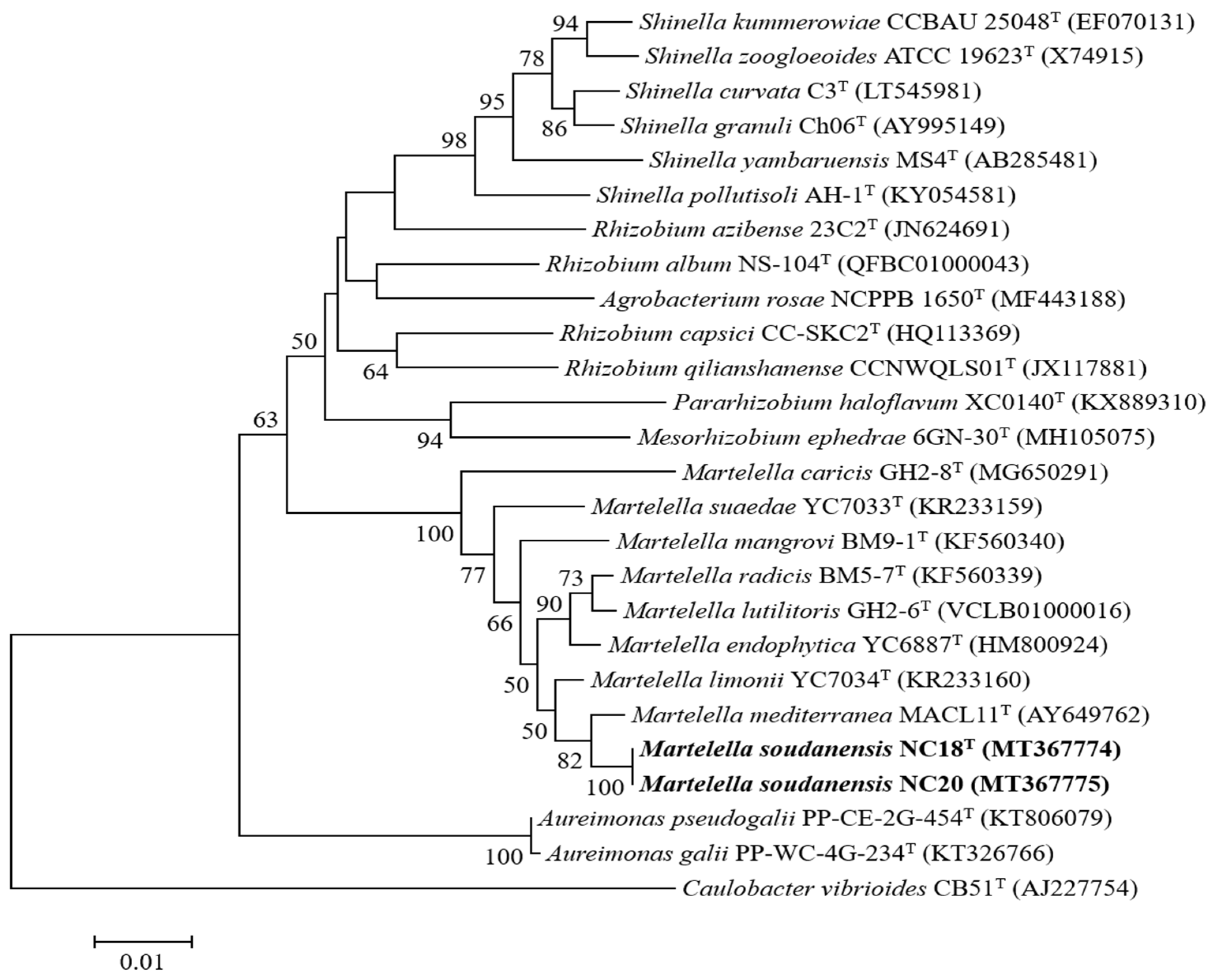
3.3. Phylogenetic Characterization

The 16S rRNA gene sequences of strains NC18T and NC20 were 1482 bp, showing 100% similarity to each other. NC18T and NC20 showed the highest sequence similarity values with those of M. mediterranea DSM 17316T (99.0%), M. limonii YC7034T (98.6%), M. endophytica YC6887T (98.1%), M. mangrovi BM9-1T (97.9%), M. lutilitoris GH2-6T (97.9%), M. suaedae YC7033T (97.6%), M. radicis BM5-7T (97.6%), and M. caricis GH2-8T (97.2%). The 16S rRNA gene sequence similarities indicated that strain M. mediterranea DSM 17316T and M. limonii YC7034T were the nearest phylogenetic neighbors to the novel isolate. The 16S rRNA gene sequence phylogenetic analysis based on the neighbor-joining (Figure 1), maximum-likelihood (Supplementary Figure S2), and maximum-parsimony (Supplementary Figure S3) algorithms indicated that strains NC18T and NC20 formed a lineage within the clade of the genus Martelella and separated it from the clade composed of the species M. mediterranea and M. limonii.
The MLSA tree also agreed with the taxonomic positions of the two strains as shown by the phylogenetic tree based on the 16S rRNA gene (Figure 2, Supplementary Figures S4 and S5).

3.4. General Genomic Features

The genomic features of strains NC18T and NC20 are shown in Table 3. The genome sizes of NC18T and NC20 were 6,109,459 and 6,109,677 bp, respectively. The NC18T genome was predicted to have 5849 genes, 5502 protein-encoding genes, 6 rRNAs, and 48 tRNAs. The NC20 genome was predicted to have 5830 genes, 5585 protein-encoding genes, 6 rRNAs, and 48 tRNAs.
The DNA G + C contents of both NC18T and NC20 were 61.8 mol% (Table 1), which are within the known DNA G + C contents of genus Martelella (52.8–62.6 mol%). The predicted functional genes based on the COG database of both strains mainly belong to amino acid transport and metabolism (E; 537 and 526 orthologs for NC18T and NC20, respectively), carbohydrate transport and metabolism (G; 534 and 524 orthologs), transcription (K; 500 and 496 orthologs), and inorganic ion transport and metabolism (P; 458 and 451 orthologs), except only a general function prediction (R) (Supplementary Table S2).

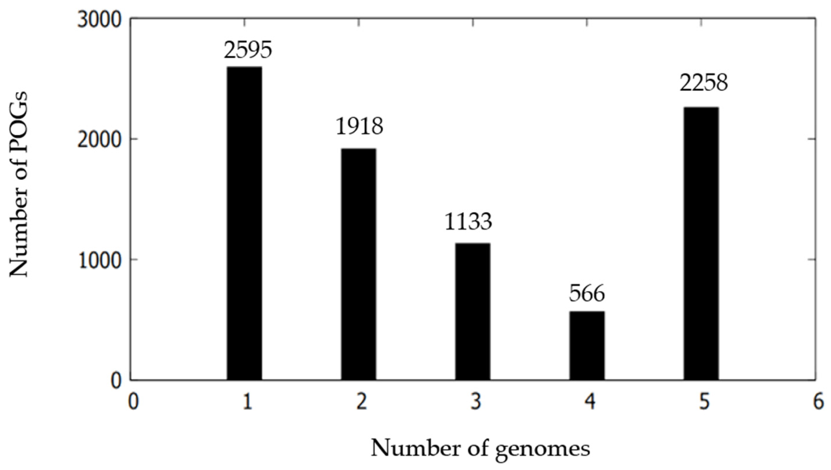
3.5. Comparative Genomic Characterization

ANI, AAI, and dDDH values between NC18T and NC20 were 99.9%, 100%, and 100%, respectively (Table 4 and Table 5). These indicate that the two isolates belonged to a single species. In contrast, ANI values between NC18T and the reference strains of M. mediterranea DSM 17316T, M. endophytica YC6887T, and M. lutilitoris GH2-6T were 88.1%, 80.2%, and 80.4%, respectively (Table 4). AAI values between NC18T and the reference strains of M. mediterranea DSM 17316T, M. endophytica YC6887T, and M. lutilitoris GH2-6T were 87.7%, 76.3%, and 77.8%, respectively (Table 5). These ANI and AAI values were lower than the 95% threshold used to identify isolates as belonging to the same bacterial species [53,54]. The dDDH values of strain NC18T and the reference strains of M. mediterranea DSM 17316T, M. endophytica YC6887T, and M. lutilitoris GH2-6T were 34.9%, 23.9%, and 23.7%, respectively (Table 4), which were below the threshold of 70% for species delineation [53]. Altogether, these results indicate that strain NC18T represents a novel species of the genus Martelella.
To gain an in-depth understanding of the intra-species genomic diversity of the Martelella species, pan-genome analysis was performed. The pan-genome curve showed that the size of the pan-genome increased with the addition of new genomes (Supplementary Figure S6). The core genome slowly decreased as the genomes were added one by one. Pan-genomic analysis shows that the two strains and three related species had 8470 POGs: 2258 POG core, 3617 POG accessory, and 2595 POG unique (Figure 3). The five strains contained a certain number of strain-specific genes (POG unique), and the number varied considerably (9–971) depending on each strain, showing that the changing genetic flow led to the generation of strain specificity (Supplementary Table S3). Most of the POG core was classified into basic functions: amino acid transport and metabolism (E), general function prediction only (R), carbohydrate transport and metabolism (G), translation, ribosomal structure and biogenesis (J), and transcription (K) (Supplementary Figure S7), which were related to necessary nutrients obtaining from various environments and maintaining a lifestyle. Function unknown (S) occupied a large proportion, which showed the current lacing in understanding Martelella genomes. Most of the POG accessory was also classified as a similar pattern to that of the POG core.
In addition, Kyoto Encyclopedia of Genes and Genomes (KEGG) analysis indicated that the POG unique were mainly involved in carbohydrate metabolism, membrane transport, amino acid metabolism, and lipid metabolism, especially carbohydrate metabolism (Supplementary Figure S8), which corresponded with the variable ability of carbon source use tested by API (Table 1). These genomic characteristics indicated the diversity of metabolic pathways in different Martelella strains. In addition, the concatenated POG core-based phylogenetic tree (Supplementary Figure S9) showed that the two novel strains were distinct from the three other Martelella strains, as clustering with M. mediterranea DSM 17316T.
This result was consistent with the phylogenetic tree based on 16S rRNA gene sequences of strains NC18T and NC20 with other related taxa. Thus, the distinguished genetic distinctiveness by ANI, AAI, and dDDH values suggest that strain NC18T is separated from other recognized species of the genus Martelella. Pan-genomic analysis also showed the distinct patterns of gene content and metabolism pathway within this new Martelella species.

4. Conclusions

We isolated two Martelella strains and performed phenotypic, chemotaxonomic, phylogenetic, and genomic analyses to identify these strains as novel species. Phylogenetic analysis showed that strains NC18T and NC20 grouped with members of the genus Martelella, including M. mediterranea DSM 17316T and M. limonii YC7034T. Genetic distinctiveness by ANI, AAI, and dDDH values suggested that strains NC18T and NC20 are separated from other recognized species of the genus Martelella. Pan-genomic analysis showed that most POG cores of NC18T and NC20 were relevant to amino acid transport and metabolism in the COG category and carbohydrate metabolism in the KEGG pathway. On the basis of polyphasic taxonomic data, strain NC18T represents the type strain of a novel species of the genus Martelella, for which the name Martelella soudanensis sp. nov. is proposed.

4.1. Description of Martelella Soudanensis sp. nov.

Martelella soudanensis (sou.da.nen′sis. N.L. masc./fem. adj. soudanensis, named after Soudan Iron Mine).
Cells are Gram-stain-negative, strictly aerobic, oxidase-positive, catalase-positive, non-spore-forming, non-flagellated rods (0.8–0.9 × 1.2–1.3 μm). Colonies on MA are cream-colored, smooth, convex, circular, and reach 0.5 mm in diameter after incubation for 2 days at 30 °C. Growth occurs at 20–40 °C with an optimum of 30 °C. Growth does not occur at 10 °C or above 45 °C. The pH range for growth is pH 5.0–9.0 (optimum, pH 7.0). Growth occurs in the range of 0%–13% (w/v) NaCl with an optimum of 1% (w/v). Positive for alkaline phosphatase, esterase (C4), leucine arylamidase, acid phosphatase, naphthol-AS-BI-phosphohydrolase, α-glucosidase, β-glucosidase, and N-acetyl-β-glucosaminidase. Negative for esterase lipase (C8), lipase (C14), valine arylamidase, cystine arylamidase, trypsin, α-chymotrypsin, α-galactosidase, β-glucuronidase, α-mannosidase, α-fucosidase, lysine decarboxylase, ornithine decarboxylase, acetoin production, H2S production, and tryptophane deaminase. Nitrate reduction and urease were observed. Aesculin hydrolysis, glucose fermentation, gelatin hydrolysis, indole production, arginine dihydrolase, caseinase are not observed. Potassium gluconate, malate, D-maltose, adipate, caprate, D-mannose, N-acetyl-D-glucosamine, phenylacetate, D-glucose, D-mannitol, L-arabinose, and trisodium citrate are not used as carbon and energy sources. Positive for acid production from L-rhamnose and L-arabinose, but negative for acid production from amygdalin, inositol, D-melibiose, D-sorbitol, D-sucrose, D-glucose, and D-mannitol. The major respiratory quinone is Q-10. Predominant fatty acids are summed feature 8 (C18:1 ω7c and/or C18:1 ω6c), C19:0 cyclo ω8c, C16:0, C18:1 ω7c 11-methyl, C18:0, and summed feature 2 (C12:0 aldehyde and/or iso-C16:1 I and/or C14:0 3-OH). The type strain, NC18T (=KCTC 82174T = NBRC 114661T), was isolated from mine sediment in Soudan, Minnesota, USA. The genome size of the type strain is 6.10 Mb and has a DNA G + C content of 61.8 mol %. The GenBank/EMBL/DDBJ accession numbers for sequences of strain NC18T generated in this study are as follows: MT367774 (16S rRNA gene), CP054858 (chromosome), CP054859 (plasmid), and CP054860 (plasmid).

Supplementary Materials

The following are available online at https://www.mdpi.com/article/10.3390/microorganisms9081736/s1, Figure S1: Morphology of strains NC18T (a) and NC20 (b) examined transmission electron microscope, Figure S2: Phylogenetic tree based on 16S rRNA gene sequences of strains NC18T and NC20 with other related taxa using 1393 bp sequence. Evolutionary distances calculated using the Jukes–Cantor model. Evolutionary history inferred using the maximum-likelihood method, Figure S3: Phylogenetic tree based on 16S rRNA gene sequences of strains NC18T and NC20 with other related taxa using 1393 bp sequence. Evolutionary history inferred using the maximum-parsimony method, Figure S4: Multilocus sequence analysis (MLSA) tree based on universally conserved protein sequences of strains NC18T and NC20 with other related taxa. Evolutionary distances computed using JTT matrix-based method. Evolutionary history inferred using the maximum-likelihood method, Figure S5: Multilocus sequence analysis (MLSA) tree based on universally conserved protein sequences of strains NC18T and NC20 with other related taxa. Evolutionary history inferred using the maximum-parsimony method, Figure S6: Pan-genome curve of five Martelella strains. Analysis performed using the Bacterial Pan-Genome Analysis Tool (BPGA) pipeline with default parameters, Figure S7: Functional POGs annotation of five Martelella strains using COG database, Figure S8: Functional POGs annotation of five Martelella strains using KEGG database, Figure S9: Phylogenetic tree using concatenated POG core based on pan-matrix of five Martelella strains, Table S1: Accession numbers of 31 universally conserved gene sequences used in multilocus sequence analysis (MLSA) tree, Table S2: COG functional classification of the genome belonging to strains NC18T and NC20, Table S3: Pan-genomes of five Martelella strains.

Author Contributions

Conceptualization, D.-H.K. and J.-Y.L.; methodology, J.-Y.L.; investigation, J.-Y.L.; data curation, D.-H.K.; writing—original draft preparation, J.-Y.L.; writing—review and editing, D.-H.L.; visualization, D.-H.K. and J.-Y.L.; supervision, D.-H.K.; funding acquisition, D.-H.K. All authors have read and agreed to the published version of the manuscript.

Funding

This work was supported by the Basic Research Project (GP2020-012) of the Korea Institute of Geoscience and Mineral Resources (KIGAM), funded by the Ministry of Science and ICT (MSIT).

Institutional Review Board Statement

Not applicable.

Informed Consent Statement

Not applicable.

Data Availability Statement

Generated sequences can be found as stated under the species description.

Acknowledgments

We are grateful to Michael J. Sadowsky at BioTechnology Institute, University of Minnesota, MN, USA.

Conflicts of Interest

The authors declare no conflict of interest.

References

  1. Kadnikov, V.V.; Mardanov, A.V.; Beletsky, A.V.; Karnachuk, O.V.; Ravin, N.V. Microbial Life in the Deep Subsurface Aquifer Illuminated by Metagenomics. Front. Microbiol. 2020, 11, 2146. [Google Scholar] [CrossRef]
  2. Breuker, A.; Köweker, G.; Blazejak, A.; Schippers, A. The deep biosphere in terrestrial sediments in the Chesapeake Bay area, Virginia, USA. Front. Microbiol. 2011, 2, 156. [Google Scholar] [CrossRef] [Green Version]
  3. Sheik, C.; Badalamenti, J.; Telling, J.; Hsu, D.; Alexander, S.C.; Bond, D.R.; Gralnick, J.A.; Sherwood Lollar, B.; Toner, B.M. Novel microbial groups drive productivity in an Archean Iron formation. Front. Microbiol. 2021, 12, 616. [Google Scholar] [CrossRef]
  4. Badalamenti, J.P.; Summers, Z.M.; Chan, C.H.; Gralnick, J.A.; Bond, D.R. Isolation and genomic characterization of ‘Desulfuromonas soudanensis WTL’, a metal-and electrode-respiring bacterium from anoxic deep subsurface brine. Front. Microbiol. 2016, 7, 913. [Google Scholar] [CrossRef] [PubMed] [Green Version]
  5. Bonis, B.M.; Gralnick, J.A. Marinobacter subterrani, a genetically tractable neutrophilic Fe (II)-oxidizing strain isolated from the Soudan Iron Mine. Front. Microbiol. 2015, 6, 719. [Google Scholar] [CrossRef]
  6. Held, B.W.; Salomon, C.E.; Blanchette, R.A. Diverse subterranean fungi of an underground iron ore mine. PLoS ONE 2020, 15, e0234208. [Google Scholar] [CrossRef]
  7. Ventosa, A.; Márquez, M.C.; Sánchez-Porro, C.; Rafael, R. Taxonomy of halophilic archaea and bacteria. In Advances in Understanding the Biology of Halophilic Microorganisms; Springer: Berlin/Heidelberg, Germany, 2012; pp. 59–80. [Google Scholar]
  8. Fathepure, B.Z. Recent studies in microbial degradation of petroleum hydrocarbons in hypersaline environments. Front. Microbiol. 2014, 5, 173. [Google Scholar] [CrossRef] [Green Version]
  9. Corral, P.; Amoozegar, M.A.; Ventosa, A. Halophiles and their biomolecules: Recent advances and future applications in biomedicine. Mar. Drugs 2020, 18, 33. [Google Scholar] [CrossRef] [Green Version]
  10. Le Borgne, S.; Paniagua, D.; Vazquez-Duhalt, R. Biodegradation of organic pollutants by halophilic bacteria and archaea. J. Mol. Microbiol. Biotechnol. 2008, 15, 74–92. [Google Scholar] [CrossRef]
  11. Cai, Z.; Ge, H.; Yi, Z.; Zeng, R.; Zhang, G. Characterization of a novel psychrophilic and halophilic β-1, 3-xylanase from deep-sea bacterium, Flammeovirga pacifica strain WPAGA1. Int. J. Biol. Macromol. 2018, 118, 2176–2184. [Google Scholar] [CrossRef] [PubMed]
  12. Amoozegar, M.A.; Safarpour, A.; Noghabi, K.A.; Bakhtiary, T.; Ventosa, A. Halophiles and their vast potential in biofuel production. Front. Microbiol. 2019, 10, 1895. [Google Scholar] [CrossRef] [PubMed]
  13. Ruginescu, R.; Gomoiu, I.; Popescu, O.; Cojoc, R.; Neagu, S.; Lucaci, I.; Batrinescu-Moteau, C.; Enache, M. Bioprospecting for novel halophilic and halotolerant sources of hydrolytic enzymes in brackish, saline and hypersaline lakes of Romania. Microorganisms 2020, 8, 1903. [Google Scholar] [CrossRef] [PubMed]
  14. Giani, M.; Garbayo, I.; Vílchez, C.; Martínez-Espinosa, R.M. Haloarchaeal carotenoids: Healthy novel compounds from extreme environments. Mar. Drugs 2019, 17, 524. [Google Scholar] [CrossRef] [Green Version]
  15. Yin, J.; Chen, J.; Wu, Q.; Chen, G. Halophiles, coming stars for industrial biotechnology. Biotechnol Adv. 2015, 33, 1433–1442. [Google Scholar] [CrossRef]
  16. Liu, C.; Baffoe, D.K.; Zhan, Y.; Zhang, M.; Li, Y.; Zhang, G. Halophile, an essential platform for bioproduction. J. Microbiol. Methods 2019, 166, 105704. [Google Scholar] [CrossRef]
  17. Oren, A. Industrial and environmental applications of halophilic microorganisms. Environ. Technol 2010, 31, 825–834. [Google Scholar] [CrossRef] [Green Version]
  18. Oren, A. Diversity of halophilic microorganisms: Environments, phylogeny, physiology, and applications. J. Ind. Microbiol. Biotechnol. 2002, 28, 56–63. [Google Scholar] [CrossRef] [PubMed]
  19. Rivas, R.; Sanchez-Marquez, S.; Mateos, P.F.; Martínez-Molina, E.; Velazquez, E. Martelella mediterranea gen. nov., sp. nov., a novel α-proteobacterium isolated from a subterranean saline lake. Int. J. Syst. Evol. Microbiol. 2005, 55, 955–959. [Google Scholar] [CrossRef]
  20. Bibi, F.; Chung, E.J.; Khan, A.; Jeon, C.O.; Chung, Y.R. Martelella endophytica sp. nov., an antifungal bacterium associated with a halophyte. Int. J. Syst. Evol. Microbiol. 2013, 63, 2914–2919. [Google Scholar] [CrossRef] [Green Version]
  21. Zhang, D.; Margesin, R. Martelella radicis sp. nov. and Martelella mangrovi sp. nov., isolated from mangrove sediment. Int. J. Syst. Evol. Microbiol. 2014, 64, 3104–3108. [Google Scholar] [CrossRef]
  22. Chung, E.J.; Hwang, J.M.; Kim, K.H.; Jeon, C.O.; Chung, Y.R. Martelella suaedae sp. nov. and Martelella limonii sp. nov., isolated from the root of halophytes. Int. J. Syst. Evol. Microbiol. 2016, 66, 3917–3922. [Google Scholar] [CrossRef] [PubMed] [Green Version]
  23. Lee, S.D. Martelella caricis sp. nov., isolated from a rhizosphere mudflat. Int. J. Syst. Evol. Microbiol. 2019, 69, 266–270. [Google Scholar] [CrossRef] [PubMed]
  24. Kim, Y.; Lee, S.D. Martelella lutilitoris sp. nov., isolated from a tidal mudflat. J. Microbiol. 2019, 57, 976–981. [Google Scholar] [CrossRef] [PubMed]
  25. Cui, C.; Ma, L.; Shi, J.; Lin, K.; Luo, Q.; Liu, Y. Metabolic pathway for degradation of anthracene by halophilic Martelella sp. AD-3. Int. Biodeterior. Biodegrad. 2014, 89, 67–73. [Google Scholar] [CrossRef]
  26. Sasser, M. Identification of Bacteria by Gas Chromatography of Cellular Fatty Acids; MIDI, Inc.: Newark, Delaware, 1990. [Google Scholar]
  27. Shin, Y.K.; Lee, J.; Lee, K.C.; Chun, C.O.; Kim, H.; Joo, W.; Lee, J.; Park, Y. Isoprenoid quinone profiles in microbial taxonomy. J. Life Sci. 1995, 5, 211–217. [Google Scholar]
  28. Weisburg, W.G.; Barns, S.M.; Pelletier, D.A.; Lane, D.J. 16S ribosomal DNA amplification for phylogenetic study. J. Bacteriol. 1991, 173, 697–703. [Google Scholar] [CrossRef] [Green Version]
  29. Yoon, S.; Ha, S.; Kwon, S.; Lim, J.; Kim, Y.; Seo, H.; Chun, J. Introducing EzBioCloud: A taxonomically united database of 16S rRNA gene sequences and whole-genome assemblies. Int. J. Syst. Evol. Microbiol. 2017, 67, 1613. [Google Scholar] [CrossRef]
  30. Larkin, M.A.; Blackshields, G.; Brown, N.P.; Chenna, R.; McGettigan, P.A.; McWilliam, H.; Valentin, F.; Wallace, I.M.; Wilm, A.; Lopez, R. Clustal W and Clustal X version 2.0. Bioinformatics 2007, 23, 2947–2948. [Google Scholar] [CrossRef] [Green Version]
  31. Hall, T.; Biosciences, I.; Carlsbad, C. BioEdit: An important software for molecular biology. Gerf. Bull. Biosci. 2011, 2, 60–61. [Google Scholar]
  32. Tamura, K.; Stecher, G.; Peterson, D.; Filipski, A.; Kumar, S. MEGA6: Version 6.0. Molecular evolutionary genetics analysis. Mol. Biol. Evol. 2013, 30, 2725–2729. [Google Scholar] [CrossRef] [Green Version]
  33. Kerepesi, C.; Bánky, D.; Grolmusz, V. AmphoraNet: The webserver implementation of the AMPHORA2 metagenomic workflow suite. Gene 2014, 533, 538–540. [Google Scholar] [CrossRef] [PubMed]
  34. Edgar, R.C. MUSCLE: Multiple sequence alignment with high accuracy and high throughput. Nucleic Acids Res. 2004, 32, 1792–1797. [Google Scholar] [CrossRef] [Green Version]
  35. Castresana, J. Selection of conserved blocks from multiple alignments for their use in phylogenetic analysis. Mol. Biol. Evol. 2000, 17, 540–552. [Google Scholar] [CrossRef] [Green Version]
  36. Jones, D.T.; Taylor, W.R.; Thornton, J.M. The rapid generation of mutation data matrices from protein sequences. Bioinformatics 1992, 8, 275–282. [Google Scholar] [CrossRef]
  37. Lee, I.; Chalita, M.; Ha, S.; Na, S.; Yoon, S.; Chun, J. ContEst16S: An algorithm that identifies contaminated prokaryotic genomes using 16S RNA gene sequences. Int. J. Syst. Evol. Microbiol. 2017, 67, 2053–2057. [Google Scholar] [CrossRef] [PubMed]
  38. Hyatt, D.; Chen, G.; LoCascio, P.F.; Land, M.L.; Larimer, F.W.; Hauser, L.J. Prodigal: Prokaryotic gene recognition and translation initiation site identification. BMC Bioinform. 2010, 11, 119. [Google Scholar] [CrossRef] [Green Version]
  39. Schattner, P.; Brooks, A.N.; Lowe, T.M. The tRNAscan-SE, snoscan and snoGPS web servers for the detection of tRNAs and snoRNAs. Nucleic Acids Res. 2005, 33, W686–W689. [Google Scholar] [CrossRef] [PubMed]
  40. Nawrocki, E.P.; Burge, S.W.; Bateman, A.; Daub, J.; Eberhardt, R.Y.; Eddy, S.R.; Floden, E.W.; Gardner, P.P.; Jones, T.A.; Tate, J. Rfam 12.0: Updates to the RNA families database. Nucleic Acids Res. 2015, 43, D130–D137. [Google Scholar] [CrossRef]
  41. Edgar, R.C. PILER-CR: Fast and accurate identification of CRISPR repeats. BMC Bioinform. 2007, 8, 18. [Google Scholar] [CrossRef] [Green Version]
  42. Bland, C.; Ramsey, T.L.; Sabree, F.; Lowe, M.; Brown, K.; Kyrpides, N.C.; Hugenholtz, P. CRISPR recognition tool (CRT): A tool for automatic detection of clustered regularly interspaced palindromic repeats. BMC Bioinform. 2007, 8, 209. [Google Scholar] [CrossRef] [Green Version]
  43. Powell, S.; Forslund, K.; Szklarczyk, D.; Trachana, K.; Roth, A.; Huerta-Cepas, J.; Gabaldon, T.; Rattei, T.; Creevey, C.; Kuhn, M. eggNOG v4. 0: Nested orthology inference across 3686 organisms. Nucleic Acids Res. 2014, 42, D231–D239. [Google Scholar] [CrossRef] [PubMed]
  44. UniProt Consortium UniProt: A hub for protein information. Nucleic Acids Res. 2015, 43, D204–D212. [CrossRef]
  45. Kanehisa, M.; Goto, S.; Sato, Y.; Kawashima, M.; Furumichi, M.; Tanabe, M. Data, information, knowledge and principle: Back to metabolism in KEGG. Nucleic Acids Res. 2014, 42, D199–D205. [Google Scholar] [CrossRef] [PubMed] [Green Version]
  46. Overbeek, R.; Begley, T.; Butler, R.M.; Choudhuri, J.V.; Chuang, H.; Cohoon, M.; de Crécy-Lagard, V.; Diaz, N.; Disz, T.; Edwards, R. The subsystems approach to genome annotation and its use in the project to annotate 1000 genomes. Nucleic Acids Res. 2005, 33, 5691–5702. [Google Scholar] [CrossRef] [PubMed] [Green Version]
  47. Edgar, R.C. Search and clustering orders of magnitude faster than BLAST. Bioinformatics 2010, 26, 2460–2461. [Google Scholar] [CrossRef] [Green Version]
  48. Yoon, S.; Ha, S.; Lim, J.; Kwon, S.; Chun, J. A large-scale evaluation of algorithms to calculate average nucleotide identity. Antonie Van Leeuwenhoek 2017, 110, 1281–1286. [Google Scholar] [CrossRef]
  49. Meier-Kolthoff, J.P.; Auch, A.F.; Klenk, H.; Göker, M. Genome sequence-based species delimitation with confidence intervals and improved distance functions. BMC Bioinform. 2013, 14, 60. [Google Scholar] [CrossRef] [PubMed] [Green Version]
  50. Goris, J.; Konstantinidis, K.T.; Klappenbach, J.A.; Coenye, T.; Vandamme, P.; Tiedje, J.M. DNA–DNA hybridization values and their relationship to whole-genome sequence similarities. Int. J. Syst. Evol. Microbiol. 2007, 57, 81–91. [Google Scholar] [CrossRef] [Green Version]
  51. Rodriguez, L.M.; Konstantinidis, K.T. Bypassing cultivation to identify bacterial species. Microbe 2014, 9, 111–118. [Google Scholar] [CrossRef]
  52. Chaudhari, N.M.; Gupta, V.K.; Dutta, C. BPGA-an ultra-fast pan-genome analysis pipeline. Sci. Rep. 2016, 6, 1–10. [Google Scholar] [CrossRef] [Green Version]
  53. Chun, J.; Oren, A.; Ventosa, A.; Christensen, H.; Arahal, D.R.; da Costa, M.S.; Rooney, A.P.; Yi, H.; Xu, X.; De Meyer, S. Proposed minimal standards for the use of genome data for the taxonomy of prokaryotes. Int. J. Syst. Evol. Microbiol. 2018, 68, 461–466. [Google Scholar] [CrossRef] [PubMed]
  54. Richter, M.; Rosselló-Móra, R. Shifting the genomic gold standard for the prokaryotic species definition. Proc. Natl. Acad. Sci. USA 2009, 106, 19126–19131. [Google Scholar] [CrossRef] [PubMed] [Green Version]
Figure 1. Phylogenetic tree based on 16S rRNA gene sequences of strains NC18T and NC20 with other related taxa using 1393 bp sequence. Evolutionary distances calculated using the Jukes–Cantor model. Evolutionary history was inferred using the neighbor-joining method. Bootstrap values (tested as 1000 replications) above 50% are shown next to branches. Sequence of Caulobacter vibrioides CB51T used as outgroup. Bar, 0.01 nucleotide substitution per position.
Microorganisms 09 01736 g001
Figure 2. Multilocus sequence analysis (MLSA) tree based on 31 universally conserved protein sequences of strains NC18T and NC20 with other related taxa. Evolutionary distances computed using JTT matrix-based method. Evolutionary history inferred using the neighbor-joining method. Bootstrap values (tested as 1000 replications) above 50% are shown next to branches. Sequence of Caulobacter vibrioides CB51T used as outgroup. Bar, 0.05 nucleotide substitution per position. Accession numbers for each sequence are shown in Supplementary Table S1.
Microorganisms 09 01736 g002
Figure 3. Comparison of POG distribution of strains NC18T and NC20 with related taxa within the genus Martelella.
Microorganisms 09 01736 g003
Table 1. Differential characteristics of strains NC18T and NC20 with related taxa within the genus Martelella.
Characteristic12345678910
Cell size (μm)0.8–0.9 × 1.2–1.30.6–0.8 × 1.1–1.31.0–1.1 × 1.3–1.50.8–1.1 × 1.2–2.50.5–0.8 × 1.2–2.31.0 × 1.20.8–0.9 × 1.9–2.80.4–0.7 × 1.0–1.51.0–1.2 × 1.2–1.50.8–0.9 × 1.3–1.4
Conditions for growth:
Temperature (°C)10–40
(30)
4–35
(30)
4–37
(28)
4–40
(25–30)
4–40
(28–30)
10–4020–45
(30)
10–40
(25–30)
10–4010–30
(30)
pH5–9
(7)
5–9
(8)
5–8
(7)
5–9
(6–8.5)
5–9
(7–8.5)
5–9.55–10
(6–8)
5–10
(7–8)
5–84–10
(7–8)
NaCl (%)0–13
(1)
0–13
(1)
0–5
(2)
1–6
(2–5)
0–9
(4–5)
2–120.5–9
(0.5–3)
1–9
(2–6)
2–100–11
(0–2)
Nitrate reduction+++NR+--NR+-
Aesculin hydrolysis--++++w+++
acetoin production--+++NRNR+NRNR
Fermentation-oxidation:
L-rhamnose++---NRNR-NRNR
D-glucose--NR--++-+NR
D-mannitol--NR--NR+-NRNR
L-arabinose++NR-+NR++NRNR
Assimilation:
potassium gluconate--+----+--
malate--+--+-+--
D-maltose--+-+--++-
D-mannose----+NR-+NR-
phenylacetate---NR-+-NR+-
D-glucose--+-++-++-
D-mannitol----++-++-
L-arabinose----+NR-+w-
Enzyme activity:
Urease+++------+
esterase lipase (C8)--++-+++++
lipase (C14)--+-------
trypsin--++---+--
naphthol-AS-BI-phosphohydrolase++-w-w+-++
α-galactosidase--+--w-w--
β-glucuronidase--+----+--
β-glucosidase+-+-++++++
N-acetyl-β-glucosaminidase+-+-+----+
α-fucosidase----+-----
G + C content (%)61.861.857.462.262.159.761.952.86153.4
Strain: 1, NC18T; 2, NC20; 3, M. mediterranea DSM 17316T; 4, M. limonii YC7034T; 5, M. endophytica YC6887T; 6, M. mangrovi BM9-1T; 7, M. lutilitoris GH2-6T; 8, M. suaedae YC7033T; 9, M. radicis BM5-7T; 10, M. caricis GH2-8T. Data for strain 1 from this study. Data for strain 3 from Rivas et al. [1], strains 4 and 8 from Chung et al. [4], strain 5 from Bibi et al. [2], strains 6 and 9 from Zhang et al. [3], strain 7 from Kim et al. [6], and strain 10 from Lee [5]. +, positive; -, negative; w, weak; NR, not reported.
Table 2. Cellular fatty acid compositions (%) of strains NC18T and NC20 with related taxa within the genus Martelella.
Fatty Acid12345678910
Saturated:
C16:013.013.412.08.44.85.910.07.82.517.2
C17:00.80.7-1.1-1.2<11.2--
C18:06.26.34.36.98.88.910.46.67.62.9
C19:0 cyclo ω8c25.828.241.425.328.023.79.820.824.912.5
Branched:
C18:1 ω7c --21.7-17.935.7--41.7-
C18:1 ω5c0.40.4--------
C18:1 ω7c 11-methyl6.46.68.817.87.95.48.416.26.88.9
C19:0 iso0.3---------
Unsaturated:
C20:1 ω7c0.30.3--------
C20:2 ω6,9c0.60.70.7------1.4
Hydroxy:
C16:0 3-OH0.70.70.50.3--<10.3-0.9
C18:1 2-OH0.2--0.5-1.5<10.51.90.9
C18:0 3-OH1.01.00.70.5<10.7<10.40.70.5
Summed feature*
25.95.67.714.69.614.87.514.512.48.8
30.50.50.60.5--1.30.5-1.9
70.50.6-----0.5-2.5
837.535.0-23.4--35.930.2-39.7
Strain: 1, NC18T; 2, NC20; 3, M. mediterranea DSM 17316T; 4, M. limonii YC7034T; 5, M. endophytica YC6887T; 6, M. mangrovi BM9-1T; 7, M. lutilitoris GH2-6T; 8, M. suaedae YC7033T; 9, M. radicis BM5-7T; 10, M. caricis GH2-8T. Data for strain 1 from this study. Data for strain 3 from Rivas et al. [1], strains 4 and 8 from Chung et al. [4], strain 5 from Bibi et al. [2], strains 6 and 9 from Zhang et al. [3], strain 7 from Kim et al. [6], and strain 10 from Lee [5]. Strain 1 was grown at MA for 2 days at 30 °C. -, not detected. *Summed features represent groups of two or three fatty acids that could not be separated by GLC with the MIDI system. Summed feature 2 contained C12:0 aldehyde and/or iso-C16:1 I and/or C14:0 3-OH. Summed feature 3 contained C16:1 ω7c and/or C16:1 ω6c. Summed feature 7 contained unidentified 18.846 and/or C19:1 ω6c and/or C19:0 cyclo ω10c and/or C19 ω6. Summed feature 8 contained C18:1 ω7c and/or C18:1 ω6c.
Table 3. Genomic features of strains NC18T and NC20 with related taxa within the genus Martelella.
Feature12345
Genome size (bp)6,109,4596,109,6775,693,0674,817,3354,447,501
N50 (bp)5,586,6235,586,8234,671,4774,817,335402,005
Contig number334118
Assembly levelcompletecompletecompletecompletecontig
Total genes 58495830530345394093
Protein coding genes55025585511544093981
rRNA 66693
tRNA4848485347
GenBank assembly accession numberGCA_013459415.1GCA_013459645.1GCA_002043005.1GCA_000960975.1GCA_005924265.1
Strain: 1, NC18T; 2, NC20; 3, M. mediterranea DSM 17316T; 4, M. endophytica YC6887T; 5, M. lutilitoris GH2-6T.
Table 4. ANI values (%) and dDDH values (%) of strains NC18T and NC20 with related taxa within the genus Martelella.
StrainANI Values (%)dDDH Values (%)
NC18TNC20NC18TNC20
Martelella soudanensis NC18T-99.9-100
Martelella soudanensis NC2099.9-100-
Martelella mediterranea DSM 17316T88.188.034.934.9
Martelella endophytica YC6887T80.280.423.923.9
Martelella lutilitoris GH2-6T80.480.423.723.7
Table 5. AAI results of strains NC18T and NC20 with related taxa within the genus Martelella.
StrainsM. soudanensis
NC18T
M. soudanensis
NC20
M. mediterranea
DSM 17316T
M. endophytica
YC6887T
M. lutilitoris
GH2-6T
M. soudanensis NC18T 10087.776.377.8
M. soudanensis NC20100 87.976.578.0
M. mediterranea DSM 17316T87.787.9 77.378.2
M. endophytica YC6887T76.376.577.3 78.5
M. lutilitoris GH2-6T77.878.078.278.5
Publisher’s Note: MDPI stays neutral with regard to jurisdictional claims in published maps and institutional affiliations.

