Article

Phylogenomic Reconstruction and Metabolic Potential of the Genus Aminobacter

1
Department of Science, Roma Tre University, Viale G. Marconi 446, 00146 Rome, Italy
2
Department of Veterinary and Animal Sciences, University of Copenhagen, Stigbøjlen 4, 1870 Frederiksberg C, Denmark
3
Laboratory of Probiogenomics, Department of Chemistry, Life Sciences, and Environmental Sustainability, University of Parma, Parco Area delle Scienze 11a, 43124 Parma, Italy
4
Interdepartmental Research Centre “Microbiome Research Hub”, University of Parma, 43124 Parma, Italy
*
Author to whom correspondence should be addressed.
These authors contributed equally.
Microorganisms 2021, 9(6), 1332; https://doi.org/10.3390/microorganisms9061332
Submission received: 27 May 2021 / Revised: 15 June 2021 / Accepted: 16 June 2021 / Published: 19 June 2021
(This article belongs to the Special Issue Microbial Genetics and Evolution)

Abstract

:
Bacteria belonging to the genus Aminobacter are metabolically versatile organisms thriving in both natural and anthropized terrestrial environments. To date, the taxonomy of this genus is poorly defined due to the unavailability of the genomic sequence of A. anthyllidis LMG 26462T and the presence of unclassified Aminobacter strains. Here, we determined the genome sequence of A. anthyllidis LMG 26462T and performed phylogenomic, average nucleotide identity and digital DNA-DNA hybridization analyses of 17 members of genus Aminobacter. Our results indicate that 16S rRNA-based phylogeny does not provide sufficient species-level discrimination, since most of the unclassified Aminobacter strains belong to valid Aminobacter species or are putative new species. Since some members of the genus Aminobacter can utilize certain C1 compounds, such as methylamines and methyl halides, a comparative genomic analysis was performed to characterize the genetic basis of some degradative/assimilative pathways in the whole genus. Our findings suggest that all Aminobacter species are heterotrophic methylotrophs able to generate the methylene tetrahydrofolate intermediate through multiple oxidative pathways of C1 compounds and convey it in the serine cycle. Moreover, all Aminobacter species carry genes implicated in the degradation of phosphonates via the C-P lyase pathway, whereas only A. anthyllidis LMG 26462T contains a symbiosis island implicated in nodulation and nitrogen fixation.

1. Introduction

Members of the genus Aminobacter (family Alphaproteobacteria) are soil bacteria described as motile aerobic rods with a strictly respiratory metabolism, capable of growing heterotrophically in the presence of a wide variety of organic substrates and colonizing natural and anthropized terrestrial ecosystems [1].
The taxonomy of the genus Aminobacter has a recent history of debate and change [2,3,4]. The first known member of the genus was a methylamine-utilizing bacterium isolated from soils enriched with various amines [5]. This bacterium was originally assigned to the genus Pseudomonas and named Pseudomonas aminovorans for its ability to utilize various amines as sole carbon and energy sources [5]. In 1990, tetramethylammonium hydroxide- and N,N-dimethylformamide-utilizing bacteria (strains TH-3T and DM-81T, respectively) were isolated from soils contaminated with industrial solvents [6,7] and found to resemble P. aminovorans [6,7]. Following chemotaxonomic investigations, Urakami et al. [2] proposed the transfer of these bacteria to a new genus within the Phyllobacteriaceae family and coined the name Aminobacter to denote the genus ability to catabolize methylated amines [1,5]. The genus Aminobacter was initially composed of Aminobacter aminovorans (basonym P. aminovorans [2]) as type species, together with Aminobacter aganoensis (strain TH-3T = DSM 7051T) and Aminobacter niigataensis (strain DM-81T = DSM 7050T) as novel species [2]. Subsequently, two strains, that is, IMB-1T and CC495T, isolated from a fumigated agricultural soil and a beech forest soil, respectively [8,9], were included in genus Aminobacter with the species name Aminobacter ciceronei (strain IMB-1T = DSM 15910T = DSM 17455T) and Aminobacter lissarensis (strain CC495T = DSM 17454T) [10], both able to utilize methyl halides as sole carbon and energy sources. In 2012, the genus Aminobacter was further expanded by the addition of the new monotypic species, Aminobacter anthyllidis, whose type strain LMG 26462T originated from the root nodule of the plant Anthyllis vulneraria [11]. Recently, the genus Carbophilus was incorporated into the genus Aminobacter [3], and Aminobacter carboxidus DSM 1086T (basonym Carbophilus carboxidus [12]) was subsequently classified as a distinct strain of the species A. lissarensis [4].
From a metabolic perspective, Aminobacter species are a new group of facultatively methylotrophic bacteria due to their ability to utilize reduced carbon substrates containing no carbon-carbon bonds (C1 compounds) as sole carbon and energy source [13,14]. However, genetic information about the degradative and/or assimilatory pathways of C1 compounds in the genus is so far limited.
A. aminovorans, A. aganoensis and A. niigataensis are known to degrade C1 methylated amines [1]. These reduced nitrogen compounds ([CH3]nNHx) are produced in nature as by-products of protein decomposition [15], and their oxidation takes place via two major routes: (i) direct oxidation to formaldehyde and ammonia by methylamine dehydrogenase (MADH), (ii) conversion of methylamine to N-methylglutamate (NMG), followed by the oxidation of NMG to methylene tetrahydrofolate (CH2=THF) [16,17]. The assimilation of C1 compounds can take place at the level of: (i) formaldehyde, via the ribulose monophosphate (RuMP) cycle; (ii) CO2, via the Calvin cycle or (iii) through the combination of CH2=THF and CO2, via the serine cycle [14].
A. ciceronei and A. lissarensis are both endowed with the ability to degrade methyl halides [8,9], a group of atmospheric pollutants that contribute to the destruction of the stratospheric ozone layer [18]. Methyl halide degradation occurs by the transfer of the methyl group to tetrahydrofolate, followed by oxidation to methylene tetrahydrofolate, thus generating reducing equivalents for biosynthesis [19]. Of note, methyltransferase gene clusters have been described in both A. ciceronei and A. lissarensis [20,21].
Genes encoding the form I carbon monoxide (CO) dehydrogenase (CODH), a heterotrimeric enzyme responsible for the oxidation of CO to carbon dioxide (CO2), were detected in A. lissarensis [4,22]. Indeed, A. lissarensis DSM 1086 is described as a “carboxydobacterium” for its ability to grow aerobically on CO as the sole carbon and energy source [12,23].
Recently, glyphosate degradation has been documented for A. aminovorans strain KCTC 2477 [24]. Glyphosate is a phosphonate herbicide containing a direct covalent bond between adjacent carbon and phosphorus atoms (the C-P bond) [25]. Many soil bacteria have developed sophisticated enzymatic machinery, known as carbon-phosphorus (C-P) lyase, which allows them to extract phosphate from a wide range of phosphonate compounds [26].
The genus Aminobacter also includes some unclassified strains with interesting bioremediation properties [27], namely Aminobacter sp. SR38 and Amininobacter sp. MSH1, capable of degrading the atrazine herbicide [28,29] and the 2,6-dichlorobenzamide (BAM) pesticide [30], respectively.
A. anthyllidis LMG 26462T is somehow unique in the genus since it is the only species capable of fixing nitrogen from the atmosphere [11]. Preliminary nodulation and nitrogen fixation tests suggest that this monotypic species could represent the first, maybe the only, legume symbiont in the genus Aminobacter [11].
At present, the phylogeny of the genus Aminobacter is still uncertain due to the unavailability of the genomic sequence of A. anthyllidis LMG 26462T and the presence of several strains presumptively assigned to the genus Aminobacter. In addition, while a broad metabolic potential for several members of Aminobacter genus has been suggested, the genetic basis of the catabolic/assimilatory pathways used by Aminobacter species to degrade recalcitrant compounds remains largely unknown. Two main questions can therefore be raised: (i) which are the evolutionary relationships among members of the genus Aminobacter and (ii) to which extent are the catabolic/assimilatory pathways conserved within the genus? Here, we report the draft genome sequence of A. anthyllidis LMG 26462T together with a complete phylogenomic reconstruction of the whole genus Aminobacter with the aim to better delineate the taxonomic relationships between all Aminobacter species, including so far unclassified strains. Moreover, the gene clusters implicated in the catabolism of methylated amines, methyl halides and CO were investigated in all Aminobacter species, along with the degradation pathways of xenobiotic compounds, including atrazine, BAM and glyphosate. Assimilatory processes of monocarbon units and nitrogen fixation were also considered, to get a complete picture of the metabolic potential of the genus Aminobacter.

2. Materials and Methods

2.1. DNA Extraction and Genome Sequencing

A. anthyllidis LMG 26462T was obtained from BCCM/LMG Bacteria Collection (Laboratorium voor Microbiologie, Universiteit Gent, Ghent, Belgium) and aerobically grown at 27 (±1) °C in Trypticase soy broth. DNA extraction was performed using a QIAamp DNA minikit (Qiagen). The quantity and quality of the extracted DNA were tested using a Thermo Scientific™ NanoDrop 2000 spectrophotometer (NanoDrop Technologies, Thermo Scientific) and by agarose gel electrophoresis, respectively. Additionally, the genomic DNA quality was evaluated by using Agilent TapeStation Systems 2200 (Agilent Technologies, Santa Clara, CA, USA). A genomic library of A. anthyllidis LMG 26462T was obtained with the TruSeq DNA PCR-free sample preparation kit (Illumina, Inc., San Diego, CA, USA). Genome sequencing was performed with a NextSeq 500 sequencing system, according to the supplier’s protocol (Illumina, UK), and library samples were loaded into a midoutput kit v2.5 (300 cycles) (Illumina, UK), producing 1,642,495 paired-end reads. The raw sequence reads were filtered and trimmed using the command-line fastq-mcf software (https://expressionanalysis.github.io/ea-utils/ accessed on 1 March 2021). Fastq files of Illumina paired-end reads (150 bp) were used as input in the MEGAnnotator pipeline for microbial genome assembly and annotation [31]. This pipeline employed the program SPAdes v3.14.0 for de novo assembly of the genome sequence with the option “–careful” and a list of k-mer sizes of 21, 33, 55, 77, 99, 127 [32]. The genome quality was evaluated with the program CheckM v1.0.18 [33], estimating a genome completeness of 99.3% and 2.4% contamination. The contigs were submitted to the National Center for Biotechnology Information (NCBI) for the prediction of protein-encoding open reading frames (ORFs) and tRNA and rRNA genes using the NCBI Prokaryotic Genome Annotation Pipeline (PGAP) v5.2 [34]. The presence of genomic islands (GIs) was predicted by IslandViewer 4 [35], which uses SIGI-HMM, IslandPath-DIMOB and IslandPick prediction algorithms to generate a dataset of GIs.

2.2. Dataset Collection

Available genome sequences of Aminobacter strains were searched in the NCBI database, and 22 genome assemblies were retrieved, including type strains and unclassified Aminobacter spp. Redundant genomes were trimmed from the dataset, and only the last released genome for each strain was selected. The resulting dataset contained the genomes of 17 Aminobacter strains, including A. anthyllidis LMG 26462T (Table 1). Plasmids were predicted from short-read data analysis with PlasmidSPAdes v3.14.0 program [36].

2.3. Phylogenetic Analysis of the 16S rRNA Gene

The 16S rRNA gene sequences of type strains for all validly published species belonging to the Phyllobacteriaceae family were retrieved from the NCBI database, and 16S rRNA gene sequences of unclassified Aminobacter strains were extracted from their genomes, when available. Alignment and phylogenetic analysis of the 16S rRNA gene sequences (Supplementary Dataset S1) were performed using MAFFT v7.48 [43], and positions containing gaps (585 positions) were removed using Gblocks v.0.91b [44], resulting in partial 16S rRNA gene sequences of 1098 nt in the final dataset. Due to the incomplete 16S rRNA gene sequence (919 bp), Aminobacter sp. J15 was excluded from the analysis. Brucella melitensis 16MT was included as an outgroup in the analysis (Supplementary Dataset S1). Genetic distances were corrected using the Kimura two-parameter model [45], and a phylogenetic tree was constructed using the Neighbor-Joining (NJ) method [46], which was visualized using iTOL v6.1.2 [47]. The robustness of the phylogenetic tree was statically tested with a bootstrap of 1000 replicates [48].

2.4. Whole Genome-Based Phylogeny

A whole genome-based phylogeny was inferred for all available genome sequences of Aminobacter strains (n = 17) (Table 1), using the Type (Strain) Genome Server (TYGS) web-based pipeline (https://tygs.dsmz.de, accessed on 27 May 2021) [49]. Mesorhizobium loti DSM 2626T (GenBank accession no. GCF_003148495.1) was included as an outgroup in the analysis. All pairwise comparisons among the set of genomes were conducted using Genome BLAST Distance Phylogeny (GBDP), and accurate intergenomic distances inferred under the algorithm “trimming” and distance formula d5 [50]; 100 distance replicates were calculated each. The resulting intergenomic distances were used to infer a balanced minimum evolution tree with branch support via FASTME v2.1.4 including SPR post-processing [51]. Branch support was inferred from 100 pseudo-bootstrap replicates each [48]. The tree was rooted in the selected outgroup and visualized using iTOL v6.1.2 [47].

2.5. Digital DNA–DNA Hybridization (dDDH) and Average Nucleotide Identity (ANI)

Species boundaries between Aminobacter members were investigated by the digital dDDH tool, as implemented in the Genome-To-Genome Distance Calculator (GGDC) v2.1 [50] and by whole genome ANI, as determined by FastANI v1.33 [52]. Average nucleotide identity and digital DNA–DNA hybridization were expressed as ANI and GGDC values, respectively. GGDC values > 70% in combination with ANI values > 96% were used as the boundary for species demarcation [53,54,55].

2.6. Detection and Structural Analysis of Nodulation and Nitrogen-Fixation Genes

The translated nucleotide sequences of nif-fix and nod genes from Mesorhizobium japonicum MAFF 303099T (GCF_000009625.1) were individually used as queries in BLASTp v2.11.0 searches against the translated genome sequences of A. anthyllidis LMG 26462T and M. Japonicum R7A (GCF_012913625.1). The presence of nif-fix and nod gene clusters was also investigated in all Aminobacter strains using the translated nucleotide sequences from A. anthyllidis LMG 26462T. Protein homologs were selected showing E-value < 10−4 and >60% identity across at least 80% of the protein sequence.

2.7. Detection and Structural Analysis of Methylotrophy Genes

The translated nucleotide sequences of the methylamine oxidizing genes and serine cycle genes from Paracoccus aminovorans JCM 7685T (GCF_900005615.1) were individually used as queries in BLASTp v2.11.0 searches against the translated genome sequences of A. aminovorans DSM 7048T. The presence of the methylotrophy genes was investigated in all Aminobacter strains using the translated nucleotide sequences of the methylamine oxidizing genes and serine cycle genes from A. aminovorans DSM 7048T, the methyl halides oxidizing genes from A. lissarensis DSM 17454T and A. ciceronei DSM 15910T, and the CODH and RuBisCO genes from A. lissarensis DSM 1086. Protein homologs were selected showing E-value < 10−4 and >60% identity across at least 80% of the protein sequence. When complete homologs were not detectable in draft genome sequences, partial coding sequences located at the end of the contigs were also considered.

2.8. Detection and Structural Analysis of Genes Implicated in Xenobiotic Degradation

The translated nucleotide sequences of genes implicated in glyphosate oxidation from A. aminovorans KCTC 2477, the atrazine degradation from Aminobacter sp. SR38, and the BAM degradation from Aminobacter sp. MSH1 were individually used as queries in BLASTp v2.11.0 searches against the translated genome sequences of all Aminobacter strains. Protein homologs were selected showing E-value < 10−4 and >60% identity across at least 80% of the protein sequence.

3. Results

3.1. Aminobacter Anthyllidis LMG 26462T Genome Sequence

The draft genome sequence of A. anthyllidis LMG 26462T consists of 6,717,907 bp, which results fragmented into 30 contigs with an N50 value of 670,596 bp, an average coverage of 113× and a mean GC content of 62.58%. Genome annotation identified 6486 ORFs, 51 tRNA genes and three rRNA genes.

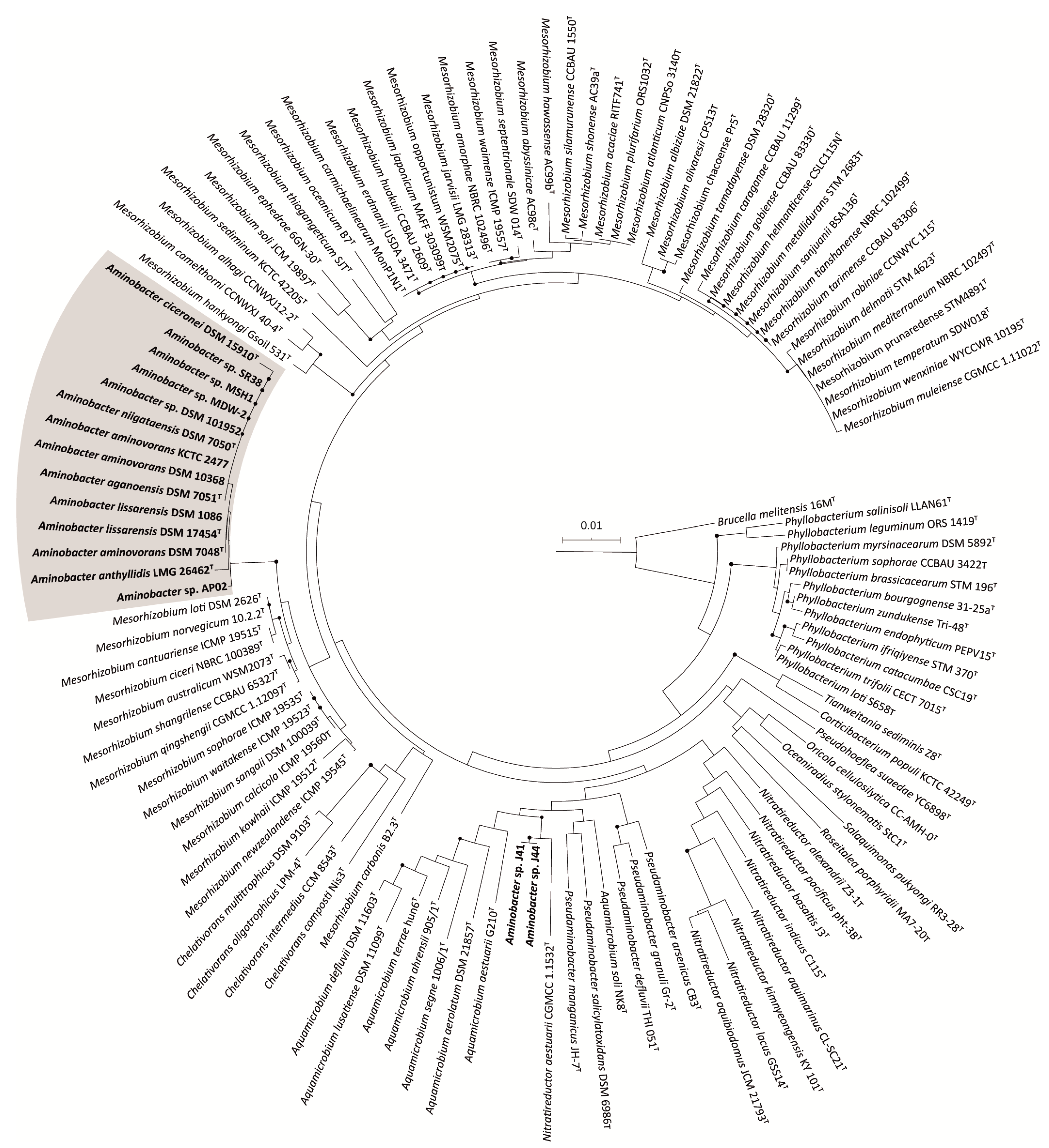
3.2. 16S rRNA Gene-Based Phylogeny of Phyllobacteriaceae

A phylogenetic tree of 16S rRNA gene sequences (1098 positions) of 118 strains including all type strains from the Phyllobacteriaceae family and the selected Aminobacter strains, was obtained using NJ. Aminobacter sp. J15 was excluded from the analysis due to its incomplete 16S rRNA gene sequence (919 bp), which would lower the alignment length (aligned positions would decrease from 1698 to 666 bp). The phylogenetic analysis showed that most of the members of the Aminobacter genus form a discrete, statistically supported clade, clearly distinct from all other members of Phyllobacteriaceae (Figure 1). Notably, Aminobacter sp. J41 and Aminobacter sp. J44 formed a separate cluster from other Aminobacter species.

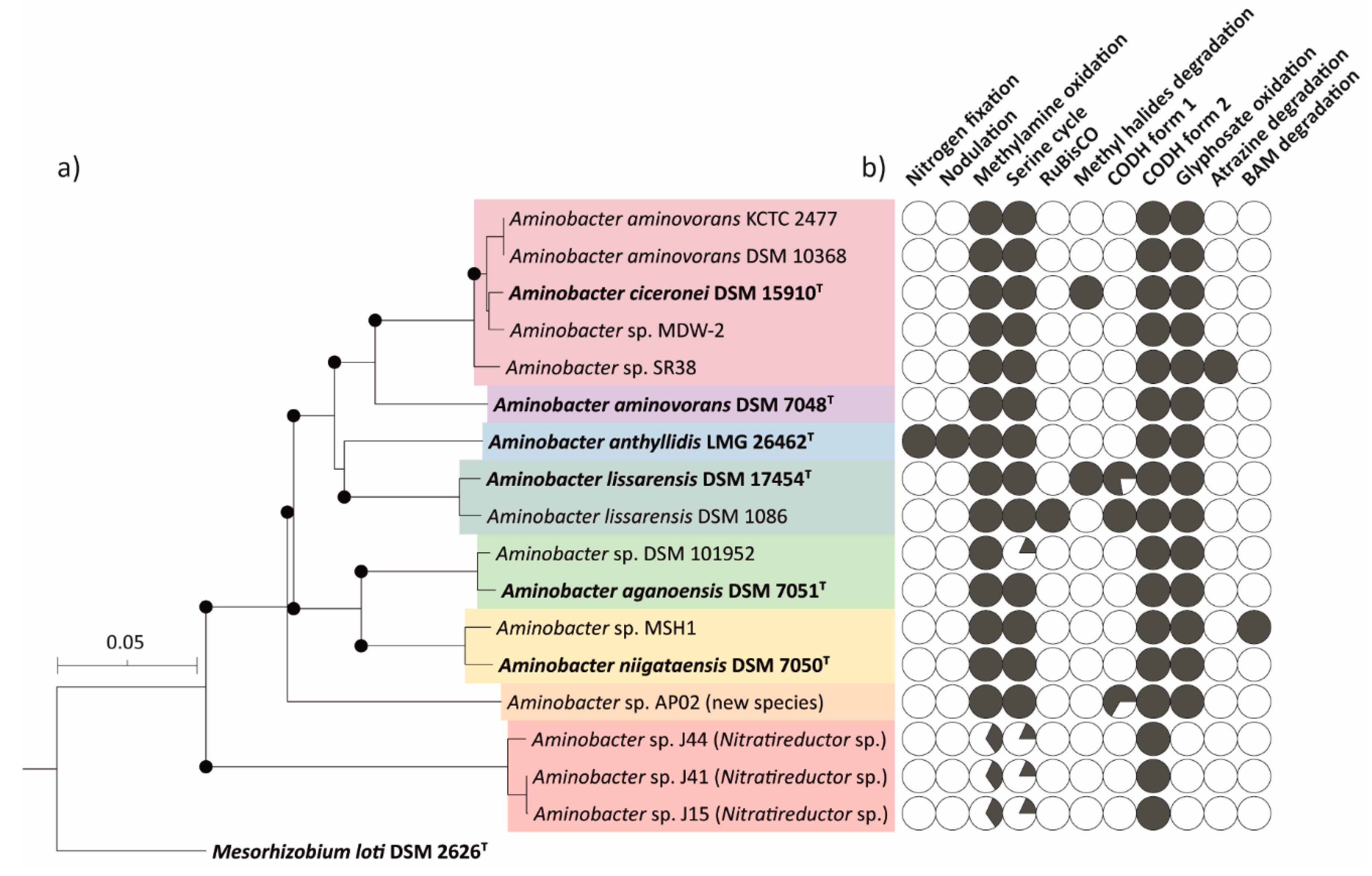
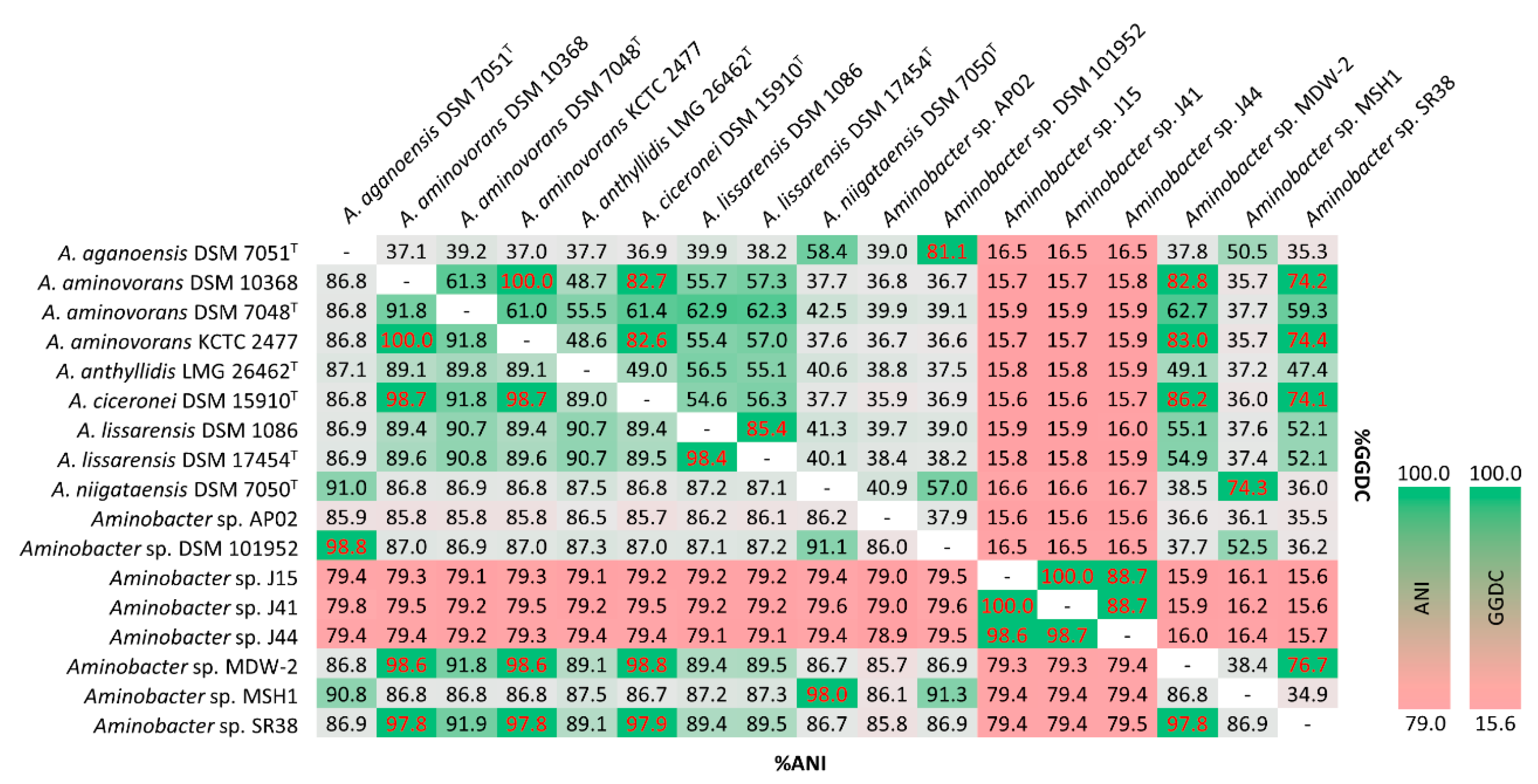
3.3. Whole Genome Phylogeny of Aminobacter Strains

A total of 17 non-redundant genome sequences representative of all members of the Aminobacter genus available in the NCBI database were combined with the A. anthyllidis LMG 26462T genome sequence and used for the whole genome-based phylogenetic analysis (Table 1). Genome-wide phylogenomic relationships between Aminobacter members showed the presence of two distinct clades, supported by >70% bootstrap values (Figure 2a). The first clade was formed by the majority (14/17) of Aminobacter species. Within this clade, A. lissarensis strains were the closest neighbors of the newly sequenced A. anthyllidis LMG 26462T strain (Figure 2a). A. ciceronei DSM 15910T, A. aminovorans KCTC 2477, A. aminovorans DSM 10368, Aminobacter sp. MDW-2 and Aminobacter sp. SR38 formed a separate subclade (A. ciceronei subclade hereafter) (Figure 2a). Similarly, a small inter-species distance was also noticed between Aminobacter sp. DSM 101952 and A. aganoensis DSM 7051T and between Aminobacter sp. MSH1 and A. niigataensis DSM 7050T (Figure 2a). Aminobacter sp. AP02 did not cluster with any validly published Aminobacter species, likely representing a distinct species. Consistent with 16S rRNA gene analysis, Aminobacter sp. J15, Aminobacter sp. J41 and Aminobacter sp. J44 formed a separate clade, unrelated to any Aminobacter spp.

3.4. ANI and dDDH of Aminobacter Species

ANI and GGDC values of the pairwise comparison between Aminobacter strains are shown in Figure 3. Pairwise ANI and GGDC values between A. ciceronei DSM 15910T and all members of the A. ciceronei subclade, namely A. aminovorans KCTC 2477, A. aminovorans DSM 10368, Aminobacter sp. MDW-2 and Aminobacter sp. SR38, was above the species demarcating threshold (>96% for ANI and >70% for GGDC; [53,54,55]), suggesting that all these strains belong to the A. ciceronei species (Figure 3). ANI and GGDC values between Aminobacter sp. DSM 101952 and A. aganoensis DSM 7051T, and between Aminobacter sp. MSH1 and A. niigataensis DSM 7050T, were also above the species demarcating threshold (Figure 3). ANI and GGDC values between Aminobacter sp. AP02 and all other Aminobacter strains are below the species threshold (Figure 3), further supporting the hypothesis that this strain could represent a new Aminobacter species.
Notably, Aminobacter sp. J15, Aminobacter sp. J41 and Aminobacter sp. J44 showed the lowest ANI and GGDC values in any reciprocal comparison with any other Aminobacter strains, although the pairwise comparison between them suggested that these three strains belong to the same species (Figure 3). According to 16S phylogeny, Nitratireductor aestuarii CGMCC 1.1532T was the closest relative to the three strains (Figure 1). Pairwise ANI and GGDC comparisons between N. aestuarii CGMCC 1.1532T and Aminobacter sp. J15 (78.5% and 14.7%, respectively), Aminobacter sp. J41 (78.5% and 14.7%, respectively) and Aminobacter sp. J44 (78.5% and 14.7%, respectively) were below the species identity threshold, suggesting that these strains also represent a new species of genus Nitratireductor.
Based on the above observations, reclassification of some members of the genus is proposed in Supplementary Table S1.

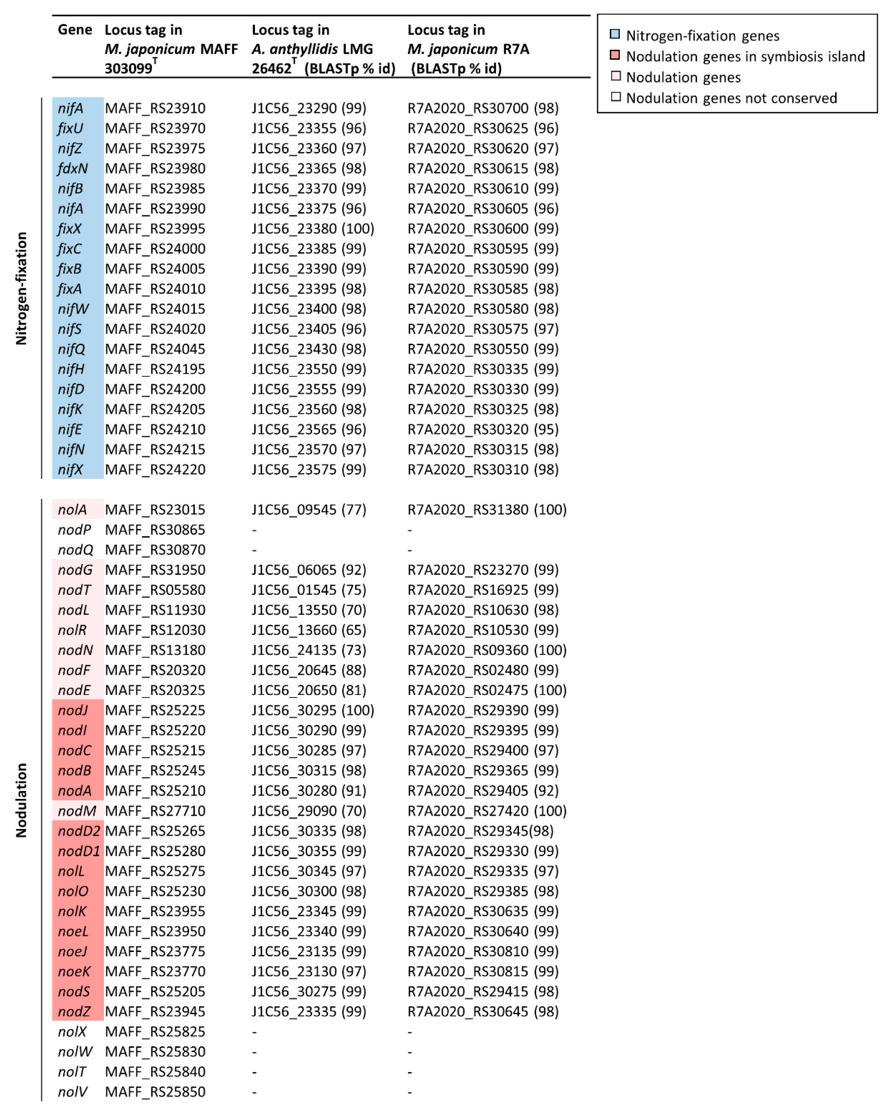
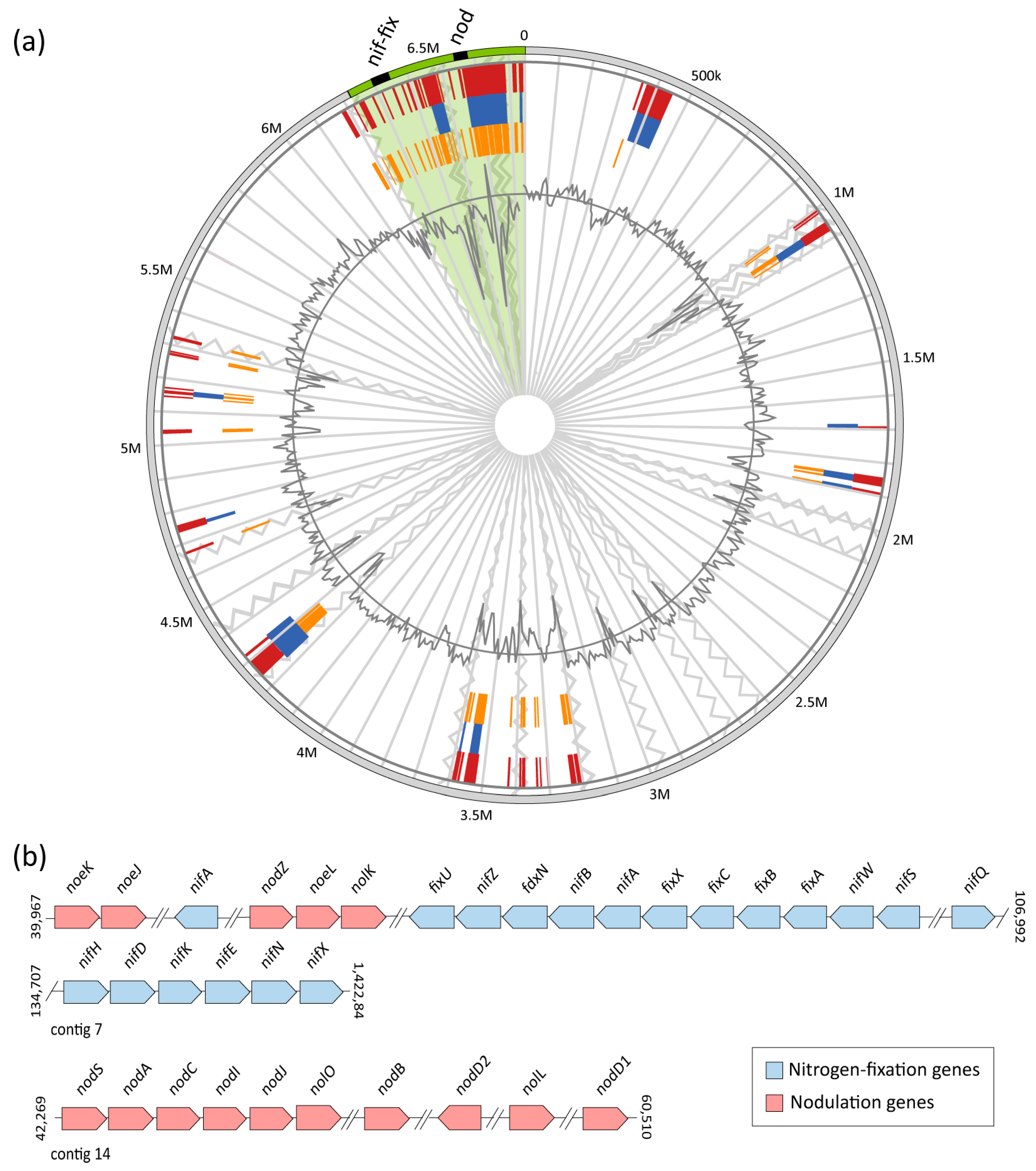
3.5. Nodulation and Nitrogen-Fixation Genes in Members of the Aminobacter Genus

The N-acyltransferase nodA gene, implicated in nodulation, has previously been detected in A. anthyllidis LMG 26462T by PCR with nodA-specific primers and showed extensive similarity with the Mesorizobium loti nodA homolog [11]. By inspecting the nodA (J1C56_30280) flanking ORFs in A. anthyllidis LMG 26462T, additional genes of the nod cluster were also identified (see locus tags in Figure 4).
To further characterize the composition and physical arrangement of the nod gene cluster and detect nif-fix genes in the A. anthyllidis LMG 26462T genome, a comparative analysis with Mesorhizobium japonicum MAFF 303099T and M. japonicum R7A (formerly Mesorhizobium loti; [56]) genomes, was performed. The well-characterized nod and nif-fix gene products from M. japonicum MAFF 303099T [57,58] were used as query sequences. All 19 nif-fix genes and 24 out of 30 nod genes formerly described in M. japonicum MAFF 303099T [57], were identified in both M. japonicum R7A and A. anthyllidis LMG 26462T (Figure 4).
Although several species of rhizobia carry nodulation and nitrogen fixation genes on large plasmids [59], M. japonicum R7A and M. japonicum MAFF 303099T harbor most of nod and nif-fix genes in a 501-kb chromosomal symbiosis island [57,58]. IslandViewer prediction revealed the presence of a 500-kb symbiosis island also in A. anthyllidis LMG 26462T, containing the same nif-fix and nod genes as those detected in M. Japonicum R7A (Figure 4 and Figure 5a). Moreover, a nearly identical arrangement of nif-fix and nod genes in A. anthyllidis LMG 26462T and M. Japonicum R7A was observed (Figure 5b; [58]).
The A. anthyllidis LMG 26462T nodABC genes encode key enzymes for the biosynthesis of the Nod factor backbone. Nod factors are oligosaccharide signal molecules playing a key role in the early stages of nodulation [60]. While the chemical structure of the A. anthyllidis LMG 26462T Nod factor is unknown, a number of nodulation genes were detected (nod, nol and noe genes) likely implicated in modification of the Nod factor backbone to generate species-specific signal molecule(s) (Figure 5b). The nodD1 and nodD2 genes are predicted to encode regulators of the expression of the nod structural genes upon interaction with a flavonoid inducer of Nod factor synthesis, secreted by the symbiotic plant [58,61], whereas nodIJ genes encode for a Nod factor secretion system [62]. The symbiosis island of A. anthyllidis LMG 26462T also contains nif-fix genes, including nifA which encodes for the nitrogen fixation transcriptional regulator, and nifKDH genes involved in the synthesis of the MoFe-nitrogenase complex [58].
Remarkably, nodulation and nitrogen-fixation genes were absent from the genomes of Aminobacter species other than A. anthyllidis LMG 26462T (Figure 2b).

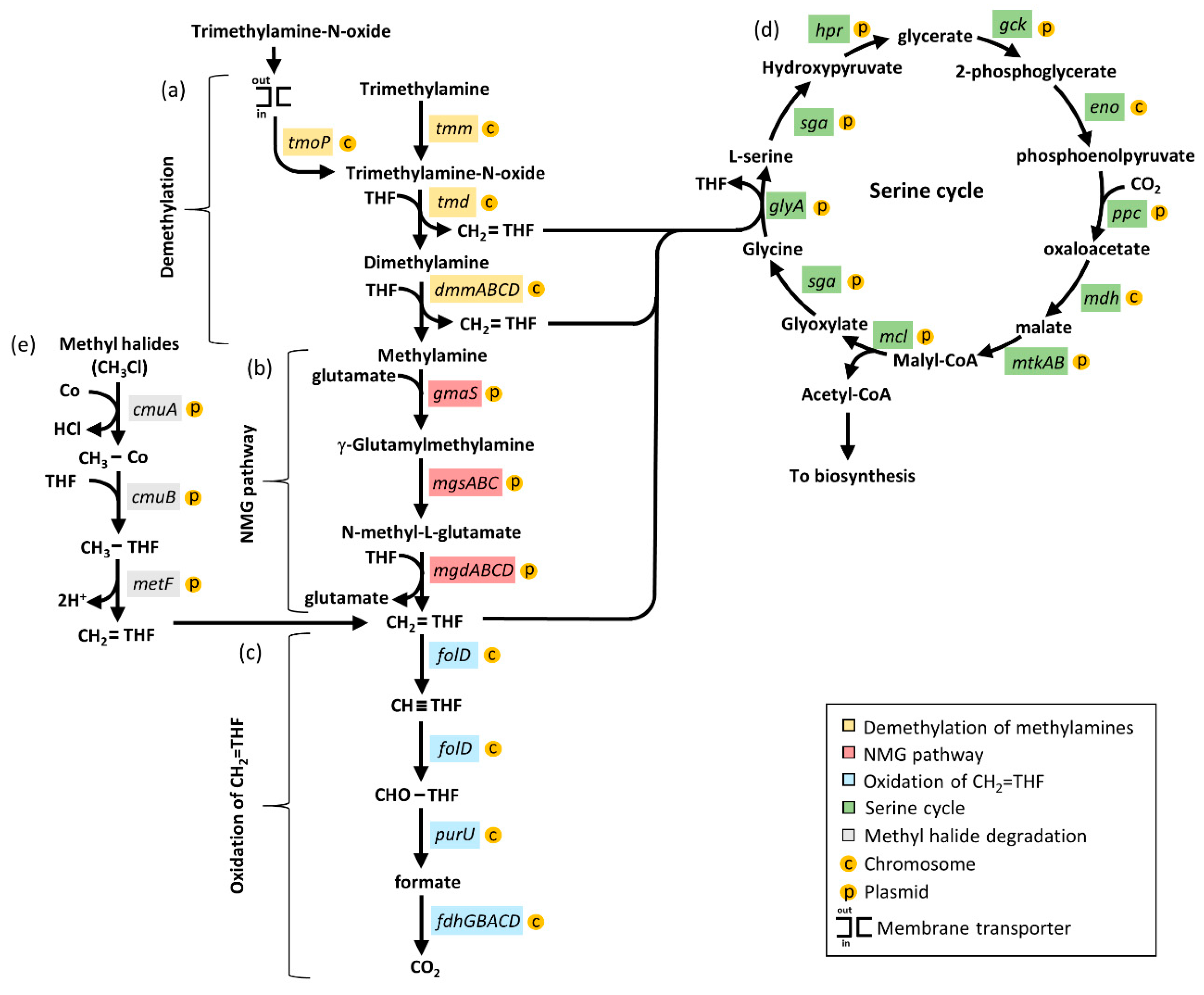
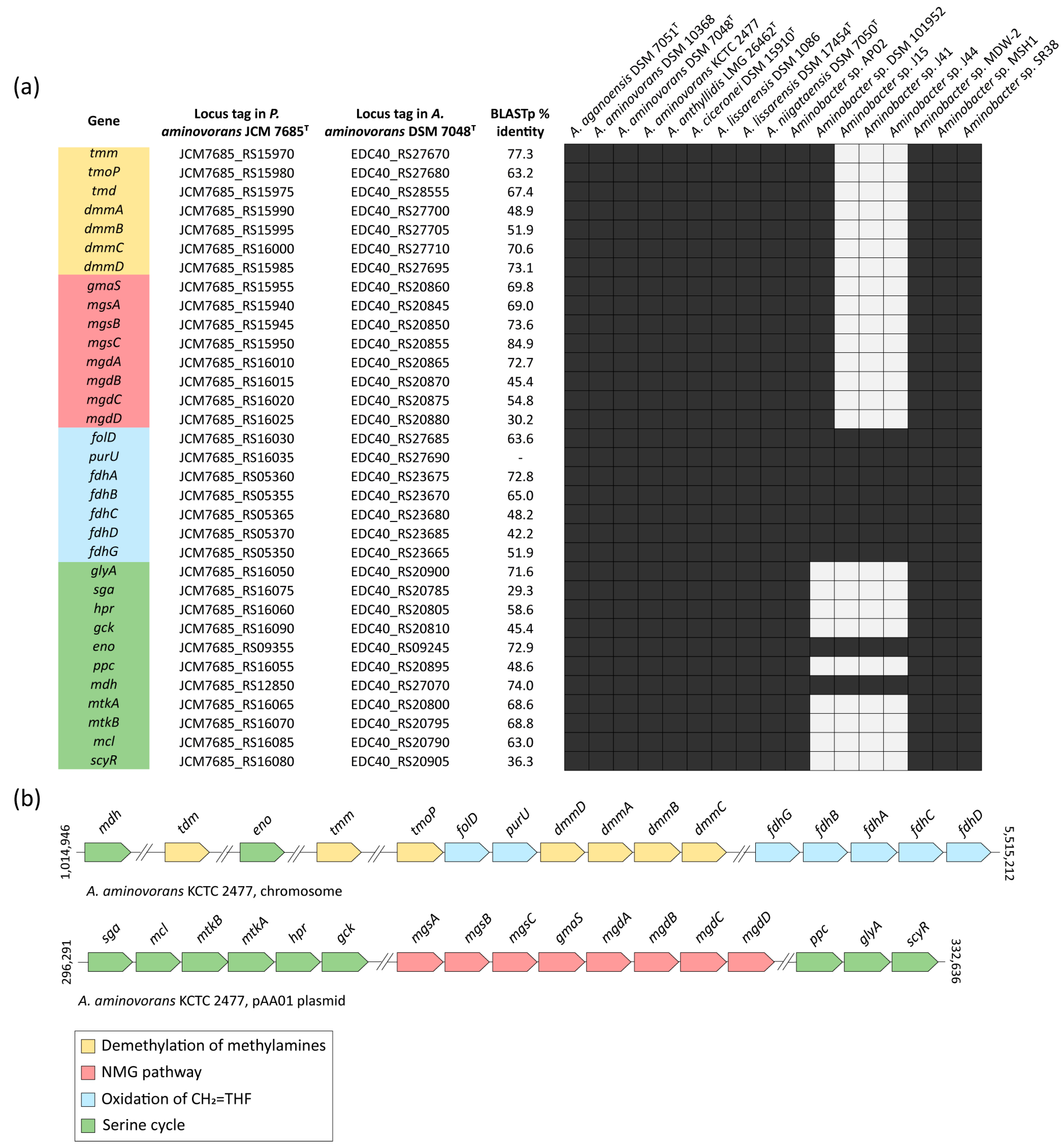
3.6. Methylotrophy Genes in Members of the Aminobacter Genus

Degradation of C1 methylated amines takes place via the alternative methylamine dehydrogenase (MADH) or N-methylglutamate (NMG) pathways [14]. The methylamine dehydrogenase madH gene, considered a hallmark of the MADH pathway, was not detected in any Aminobacter species (Table 1). Hence, oxidation of methylamines was assumed to be carried out by the NMG pathway. A comprehensive description of genes involved in the NMG pathway, in association with the serine cycle, is available for Paracoccus aminovorans JCM 7685T, in which all these genes map in a 40-kb methylotrophy island (MEI) located on plasmid pAMV1 [63]. The first step of the methylamine-oxidation pathway relies on seven genes involved in the demethylation of the methylamine precursors (Figure 6a). The second step involves eight genes of the NMG pathway for methylamine oxidation to CH2=THF (Figure 6b). The last step includes seven genes involved in the oxidation of CH2=THF to CO2 (Figure 6c). The C1 intermediates generated by the methylamine-oxidation and the NGM pathways are funneled into the serine cycle to generate acetyl-CoA (Figure 6d).
Starting from the P. aminovorans JCM 7685T genome annotation, the presence of genes involved in the methylamine-oxidizing pathway genes was investigated in the A. aminovorans DSM 7048T type species. A total of 33 genes implicated in all steps of both the methylamine-oxidation and the serine pathways were detected in the A. aminovorans DSM 7048T type species (Figure 7a). Notably, the whole set of both NMG and serine cycle genes was observed for 13 strains referable to all validly published Aminobacter species (Figure 7a; Supplementary Table S2), whereas they were absent in Aminobacter sp. J15, J41 and J44, consistent with their belonging to a different genus (Figure 7a and Supplementary Table S1). Moreover, the whole serine cycle was not detected in Aminobacter sp. DSM 101952, except mdh and eno genes, along with the citric acid cycle and glycolysis, respectively.
To investigate the physical localization and the organization of genes involved in the methylamine-oxidation and the serine pathways, A. aminovorans KCTC 2477 was selected as a reference strain, since it carries all the genes involved in both pathways (33 genes; Figure 7a) and is characterized by a complete well-refined genome, including the circular chromosome and 4 plasmids (Table 1). Genome inspection revealed that 16 out of 33 genes (48.5%) mapped in the A. aminovorans KCTC 2477 chromosome. These included seven genes involved in the demethylation of the methylamine precursors, seven genes involved in the CH2=THF oxidation pathway, and the eno and mdh genes of the serine cycle, which are shared with other fundamental pathways (Figure 7b). The remaining 17 genes, namely nine genes from the serine cycle and eight genes involved in the NMG pathway, were mapped in plasmid pAA01 (Figure 7b).
To determine if Aminobacter strains can also fix CO2 through the Calvin cycle, the presence of the ribulose-1,5-bisphosphatecarboxylase/oxygenase (RuBisCO) genes were investigated. The RuBisCO genes were only identified in A. lissarensis DSM 1086, corresponding to locus tags IHE39_RS24920 and IHE39_RS24925, annotated as ribulose bisphosphate carboxylase small and large subunits, respectively (Figure 2b).

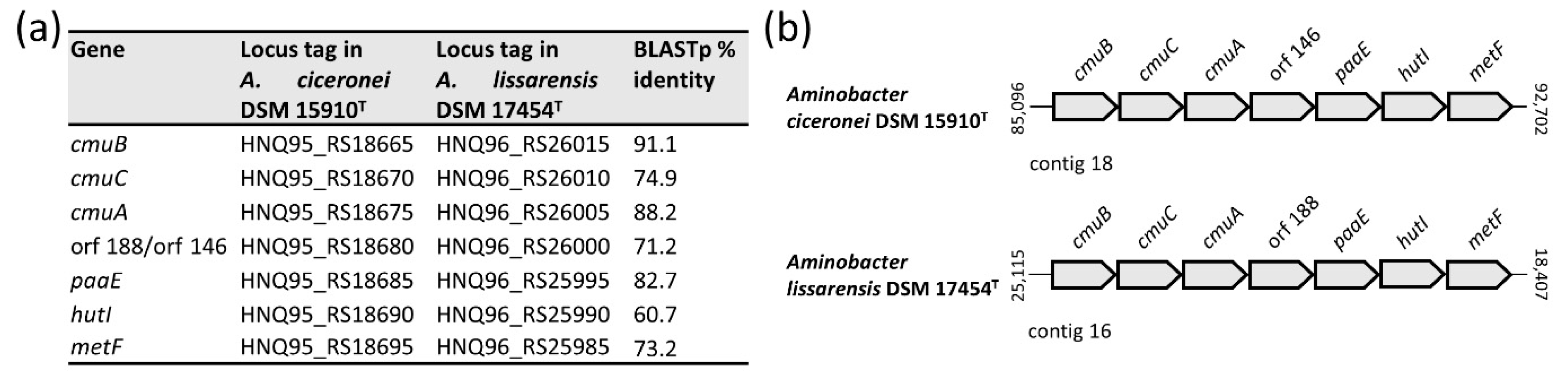
3.7. Methyl Halide Utilisation Gene Cluster in Members of the Aminobacter Genus

Methyl halide degradation genes were previously described in A. lissarensis CC495T (=DSM 17454T) [21] and A. ciceronei IMB-1T (=DSM 15910T) [20]. Genome inspection showed that both strains shared the same organization of the gene cluster and high similarity at the protein level. In addition to previous work [21], cmuB and metF were identified in A. ciceronei DSM 15910T and A. lissarensis DSM 17454T, respectively (Figure 8).
CmuA is a corrinoid-binding methyltransferase implicated in the transfer of the methyl group from the methyl halides to the corrinoid Co atom, CmuB transfers the methyl group onto tetrahydrofolate (THF), forming methyl THF, which is ultimately oxidized to the key intermediate methylene THF by MetF (Figure 6e). The paaE gene product is a putative ferredoxin—NADP reductase likely implicated in the reduction of the inactive Co(I) to the active Co(II) state of the corrinoid cofactor. CmuC and HutI are putative methyltransferase and imidazolonepropionase enzymes, respectively, whose role in the methyl halide degradation pathway is still uncertain.
To investigate the genetic location of the methyl halide degradation gene cluster, putative plasmids were assembled from short-read data with plasmidSPAdes [36]. Three and four large plasmids were predicted in A. lissarensis DSM 17454T and A. ciceronei DSM 15910T, respectively. One of the three plasmids predicted in A. lissarensis DSM 17454T (138,736 bp) showed 100% identity with contig 16 (138,704 bp), containing the methyl halide degradation gene cluster and form I CODH genes, together with previously characterized genes involved in plasmid replication and transfer [4]. Likewise, one of the four A. ciceronei DSM 15910T plasmids (151,924 bp) showed 100% identity with contig 18 (151,942 bp) encompassing the methyl halide degradation gene cluster. Additional plasmid features were detected in A. ciceronei DSM 15910T contig 18, particularly genes encoding for putative replication functions, namely RepA DNA helicase (HNQ95_RS18770), the RepB DNA primase (HNQ95_RS18775), the RepC DNA binding protein (HNQ95_RS18780), together with traGDCAFBH (HNQ95_RS18495-HNQ95_RS18525) and trbIHGFLKJ (HNQ95_RS18710-HNQ95_RS18740) genes, presumably implicated in plasmid transfer. The physical association between methyl halide degradation and plasmid replication and conjugal transfer genes, together with data from in silico plasmid assemblies, argues for a plasmid location of the methyl halide degradation cluster. Of note, methyl halide degradation genes were not detected in Aminobacter strains other than A. lissarensis DSM 17454T and A. ciceronei DSM 15910T (Figure 2b).

3.8. Carbon Monoxide Dehydrogenase Genes in Members of the Aminobacter Genus

Genome inspection revealed the presence of the form II CODH gene cluster in all Aminobacter genomes, as opposed to form I CODH, which was detected in only three genomes, namely in A. lissarensis DSM 17454T, A. lissarensis DSM 1086 and Aminobacter sp. AP02 genomes (Figure 2b; Supplementary Table S3). The form II CODH structural genes invariably showed the typical coxSLM organization, and CoxL contained the distinctive AYRGAGR signature [64], whereas form I CODH structural genes were all organized in the coxMSL order, followed by coxDEF accessory genes, with CoxL containing the distinctive AYRCSFR signature [4,64]. Additional genes of the form I CODH cluster were coxG in A. lissarensis DSM 17454T and coxH and coxI in A. lissarensis DSM 1086 [4]. In both A. lissarensis DSM 17454T and A. lissarensis DSM 1086, the structural genes encoding form I CODH were mapped in putative plasmids [4]. To investigate the genomic location of form I CODH genes (i.e., coxMSLDEF) in Aminobacter sp. AP02, putative plasmids were assembled from short-read data with plasmidSPAdes [36]. None of the two predicted plasmids of Aminobacter sp. AP02 were associated with contig 4 (343,490 bp), containing the coxMSLDEF genes, suggesting a chromosomal location of form I CODH genes.

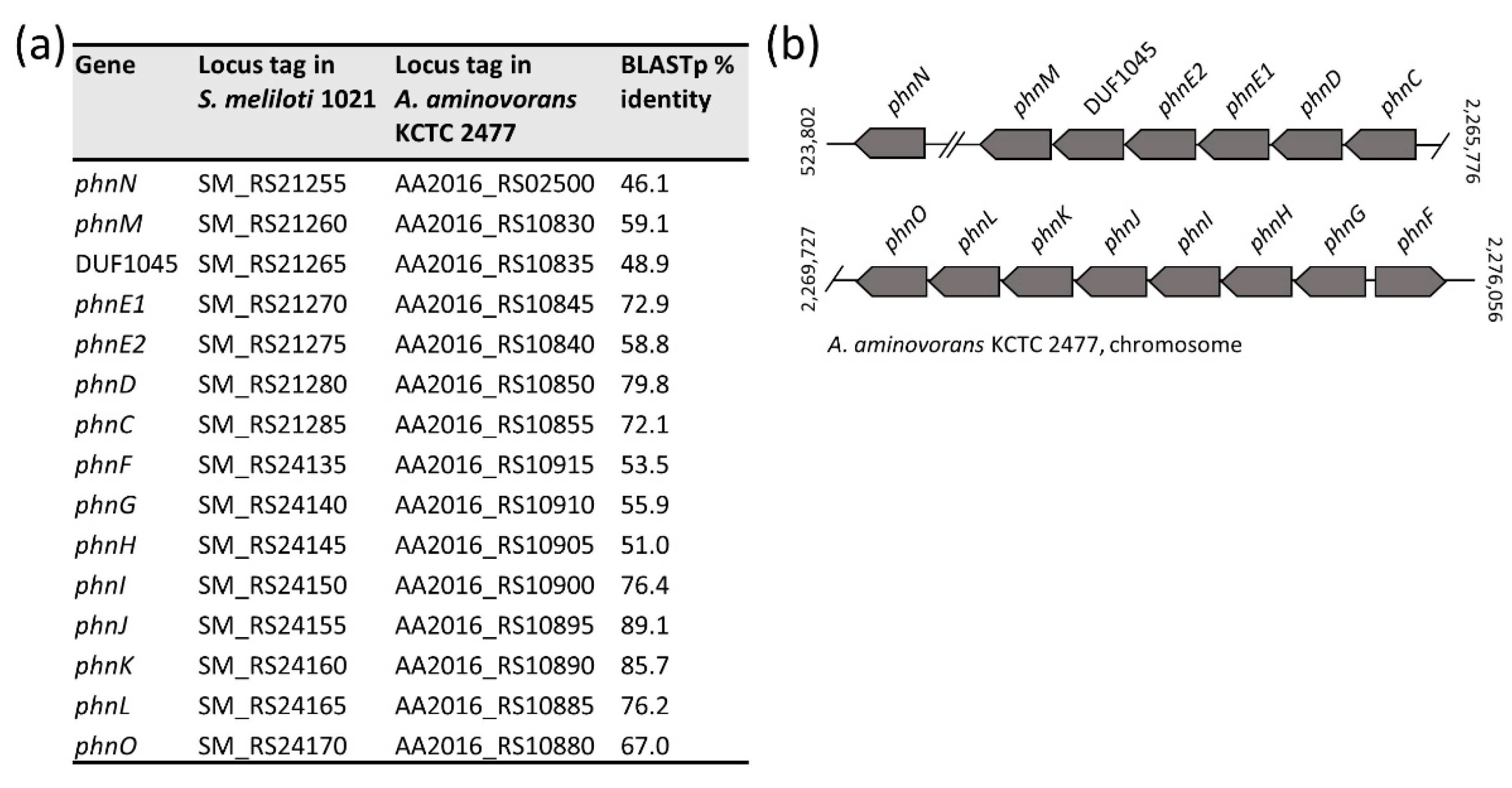
3.9. Glyphosate Oxidation Genes in Members of the Aminobacter Genus

Genome inspection revealed that A. aminovorans KCTC 2477 carries 15 phn genes encoding for enzymes of the glyphosate oxidation pathway, all showing significant similarity at the protein level with homologs from Sinorhizobium meliloti 1021 (GCF_000006965.1) (Figure 9a; [26]). The phn genes were mapped on the A. aminovorans KCTC 2477 chromosome, though their physical organization resembled that observed in the pSymB plasmid of S. meliloti 1021 (Figure 9b; [26]). The glyphosate oxidation pathway involves phnGHIJKLM gene cluster which embodies the core components of the C-P lyase pathway (Figure 9b). The DUF1045 gene, encoding a member of the two-histidine phosphodiesterase superfamily, has frequently been found next to phnM in several bacteria [26]. While the function of DUF1045 is still uncertain, it has been proposed that it could act as a phosphoribosyl cyclic phosphodiesterase implicated in the hydrolysis of cyclic ribose-phosphate, the product of the C-P lyase reaction [26]. Similarly arranged phn genes, including DUF1045, were detected in all Aminobacter genomes, with exception of Aminobacter sp. J15, Aminobacter sp. J41 and Aminobacter sp. J44 (Figure 2b; Supplementary Table S4).

3.10. 2,6-dichlorobenzamide and Atrazine Degradation Genes in Members of the Aminobacter Genus

Previous studies on Aminobacter sp. MSH1 reported that 2,6-dichlorobenzamide (BAM) degradation genes map on distinct plasmids: pBAM1 carries the BbdA amidase, responsible for the initial transformation of BAM into 2,6-DCBA [65], whereas pBAM2 harbors a large region for the complete degradation of 2,6-DCBA, encompassing two major gene clusters: bbdR1B1B2B3CDE and bbdR2FGHIJ [30,66] (Supplementary Table S5). Genome inspection of all Aminobacter members detected BAM degradation genes only in Aminobacter sp. MSH1 (Figure 2b).
Aminobacter sp. SR38 was described as an atrazine-degrading strain with the atzA gene located on plasmid pSR4 and atzB, atzC and trzD genes on plasmid pSR8 (Supplementary Table S6; [42]). These genes were unique to Aminobacter sp. SR38, since they were undetectable in other Aminobacter genomes (Figure 2b).

4. Discussion

Currently, the genus Aminobacter comprises six validly published species, namely A. aganoensis [2], A. aminovorans [2,5], A. anthyllidis [11], A. ciceronei [10], A. lissarensis [10] and A. niigataensis [2]. Consistent with previous reports [3,10], the phylogenetic analysis based on 16S rRNA gene sequences clearly delineates the genus Aminobacter as a clade distinct from all other members of Phyllobacteriaceae. However, overall poor resolution and short inter-species distances were observed in the 16S-based phylogeny, prompting us to infer phylogenetic relationships from genome-scale analysis of all members of the genus Aminobacter. For this purpose, the whole genome of A. anthyllidis LMG 26462T was de novo sequenced, and comprehensive phylogenomic reconstruction of 17 members of the Aminobacter genus, including both type strains and the so far unclassified isolates, was inferred. By combining whole genome-based phylogeny with ANI and dDDH analyses, novel taxonomic relationships were reliably established.
Whole genome-based phylogeny revealed that the closest neighbor of A. anthyllidis LMG 26462T is A. lissarensis. This phylogenetic relationship does not strictly correlate with previous chemotaxonomic data obtained from the Biolog GEN III MicroPlate assay [4], which provided a quite similar metabolic profile for A. anthyllidis LMG 26462T and A. aminovorans DSM 7048T. However, A. aminovorans DSM 7048T was the second nearest neighbor of A. anthyllidis LMG 26462T, and both shared identical morphological traits, such as cell size and the presence of lophotrichous flagella [4], consistent with established phylogenetic relationships.
Whole genome phylogeny, combined with ANI and dDDH analyses, provided compelling evidence that formerly unclassified Aminobacter strains can be assigned to definite Aminobacter species, with the only exception of Aminobacter sp. AP02, which plausibly represents a new, still uncharacterized species. Moreover, the nearly identical Aminobacter sp. J15, Aminobacter sp. J41 and Aminobacter sp. J44 strains were phylogenetically distant from all members of the genus Aminobacter and were taxonomically allocated within the genus Nitratireductor by 16S rRNA gene analysis. Based on both ANI e dDDH values, A. aminovorans DSM 10368, A. aminovorans KCTC 2477, Aminobacter sp. MDW-2 and Aminobacter sp. SR38 could be assigned to the species A. ciceronei. In addition, ANI and dDDH results classified Aminobacter sp. DSM 101952 and Aminobacter sp. MSH1 as A. aganoensis and A. niigataensis, respectively (Supplementary Table S1).
The genus Aminobacter comprises a group of environmental bacteria that thrive in polluted soil, with a single species capable of establishing symbiotic interactions with plants. Indeed, A. anthyllidis LMG 26462T was isolated from root nodules of Anthyllis vulneraria (Fabaceae) and showed nitrogen fixation properties [11]. In this study, accurate inspection of the A. anthyllidis LMG 26462T genome unraveled the presence of a symbiosis island containing both nodulation and nitrogen-fixation genes, identical to those identified in M. japonicum R7A [58]. The advantage of acquiring such a genomic island by A. anthyllidis LMG 26462T is huge, as it would allow the exploitation of a new ecological niche, ultimately resulting in “evolution in quantum leaps” [67]. This is because the acquisition of a complete set of nod-nif-fix genes, probably by horizontal gene transfer, converted A. anthyllis LMG 26462T from a soil saprophyte to a symbiotic nitrogen-fixing bacterium. Profiting from the published A. anthyllis LMG 26462T genome sequence, future studies will help to better define the structural and functional organization of the symbiosis island in A. anthyllidis LMG 26462T.
A metabolic feature common to all Aminobacter species is their ability to use certain C1 compounds as the only carbon and energy sources. However, the genetic basis underlying the oxidation and the assimilation of C1 units into biomass have never been investigated in the genus Aminobacter. Methylotrophy is the metabolic ability of microorganisms to build biomass and obtain energy from C1 compounds [13,14]. The substrates supporting methylotrophic growth include methane and methanol, as well as methylamines, methyl halides and methylated sulfur species [13]. Members of the Aminobacter genus are able to degrade a variety of methylamines and methyl halides, but cannot utilize methane and methanol [1]. The genetic analysis performed in this study provides a comprehensive characterization of the genes involved in methylamine oxidation, starting from its precursors (trimethylamine, trimethylamine-N-oxide, dimethylamine) until complete oxidation to CO2. All Aminobacter strains, with the exception of Aminobacter sp. 15, Aminobacter sp. 41 and Aminobacter sp. 44, carried genes involved in the methylamine oxidation via the NMG pathway, as well as the serine cycle genes for the assimilation of C1 units. A plasmid location of all genes of the NMG pathway, together with essential genes of the serine cycle, was predicted in A. aminovorans KTCT 2477. The co-localization of NMG pathway and serine cycle genes in the same plasmid suggests their assembly as a methylotrophic module that could favor the metabolic efficiency, and thus undergo positive selection [14]. Besides the methylamine pathway, A. lissarensis DSM 17454T and A. ciceronei DSM 15910T carried genes for methyl halide oxidation to CH2=THF, which is then assimilated in the serine cycle or further oxidized to CO2.
Since all Aminobacter species are equipped with a complete set of serine cycle genes and all but one lack genes encoding for key enzymes of the autotrophic Calvin cycle (i.e., RuBisCO), they can be classified as heterotrophic methylotrophs [68]. The only exception is Aminobacter sp. DSM 101952 also lacked essential genes of the serine cycle, and should therefore be considered a non-methylotrophic bacterium, given that the NMG pathway is also present in non-methylotrophs, which employ it for utilization of methylamine as a nitrogen source [69]. Moreover, since A. lissarensis DSM 1086 carries both serine and Calvin cycle genes, it can be classified as a facultative autotrophic methylotroph [68]. This bacterium, together with A. lissarensis DSM 17454T and Aminobacter sp. AP02, also showed carboxydotrophic properties, due to the presence of genes encoding form I CODH. Two CODH forms are known, designated I and II, sharing the same nomenclature (CoxL, CoxM and CoxS subunits) but differing in sequence [22]; form I specifically oxidizes CO with high affinity, whereas form II has a lower affinity for CO and still uncertain function [64], and it was found in all members of the genus Aminobacter.
Methylamines are organic nitrogen compounds widespread in the atmosphere. Their deposition may constitute a substantive input of atmospheric nitrogen to terrestrial and aquatic ecosystems [15,70]. A prominent source of methylamine compounds is provided by agricultural systems, where a variety of aliphatic amines, including methylamines, can be emitted in the atmosphere from animal husbandry [71] and biomass burning [72]. Therefore, understanding the sources and sinks of these gases in the environment will contribute to better assess their impact on public health and ecosystem function. Data from this study expand previous knowledge on the methylotrophic metabolism in genus Aminobacter and shed more light on the role of methylamine oxidation via the NMG pathway in soil bacteria.
Soil bacteria are highly adaptable organisms, able to survive in extremely difficult conditions. They have evolved smart strategies for surviving during nutritional stress, including the expression of specialized enzyme systems that allow them to grow on rare nutrient sources. Inorganic phosphate (Pi) is a limiting factor in many ecosystems, and phosphonates represent organic compounds containing Pi [25]. Many soil bacteria can degrade phosphonates via the C-P lyase pathway, an enzymatic process responsible for the cleavage of the phosphonate C-P bond, resulting in the formation of N-methylglycine (sarcosine) and a phosphorus-containing molecule [26]. Through this enzymatic pathway, bacteria are also able to degrade the herbicide glyphosate [26,73]. The phnJ gene, formerly detected in A. aminovorans KCTC 2477 [24], was used by us as a probe to search for the C-P lyase pathway in Aminobacter genomes. Our investigation revealed that all Aminobacter species, except Aminobacter sp. J15, Aminobacter sp. J41 and Aminobacter sp. J44, carried a complete set of genes for the C-P lyase pathway (phn genes) in their chromosome, showing a similar organization as that observed in S. meliloti 1021 pSymB plasmid [26]. It can be speculated that the C-P lyase pathway is expressed under conditions of limiting Pi, to enable its acquisition from phosphonates.
Two unclassified Aminobacter spp. were previously reported to have a potential role in the bioremediation of additional xenobiotic compounds, namely the atrazine herbicide [28] and the 2,6-dichlorobenzamide (BAM) water pollutant [74]. Aminobacter sp. SR38 (A. ciceronei, according to this study) degrades atrazine [29] via plasmid-borne atz genes [75], whereas Aminobacter sp. MSH1 (A. niigataensis, according to this study) is the only known strain capable of mineralizing BAM to CO2 [41,76], through catabolic enzymes encoded by bbd genes carried on a plasmid [30]. The ability to degrade these xenobiotic compounds is unique of these two Aminobacter strains, consistent with the location of degradative genes onto extrachromosomal DNA elements.

5. Conclusions

This study provides a comprehensive taxonomic reassessment of all species and strains formerly referred to as the genus Aminobacter. The de novo sequencing of A. anthyllidis LMG 26462T genome combined with an in-depth phylogenomic investigation of Aminobacter genus made it possible to (i) refine the evolutionary relationships between six validly published Aminobacter species, (ii) assign formerly uncharacterized Aminobacter strains to a definite species and (iii) presumptively identify new Aminobacter species. Overall, members of Aminobacter appear to be metabolically versatile organisms characterized by broad assimilatory and catabolic potentials. These bacteria are endowed with degradative and assimilatory properties which may contribute to the environmental C, N and P cycles. Here, evidence is provided that some metabolic pathways may have been acquired by horizontal gene transfer, involving either genomic island or plasmids, as in the case of genes implicated in nodulation and nitrogen fixation, methylamine and CO oxidation, the serine pathway, methyl halide, 2,6-dichlorobenzamide and atrazine detoxification. Horizontal gene transfer is the main driver of bacterial evolution [77], and it has probably modeled the genome of Aminobacter species to broaden their metabolic potential. The adaptive evolution of Aminobacter species to face challenging environmental conditions, including fluctuations in nutrients availability or exposure to toxic compounds, holds promise for the employment of these bacteria in bioaugmentation and bioremediation processes.

Supplementary Materials

The following are available online at https://www.mdpi.com/article/10.3390/microorganisms9061332/s1, Dataset S1: Species, strain ID, accession number and 16S rRNA gene sequences of the Phyllobacteriaceae family. Table S1: Proposed reclassification of Aminobacter strains. Table S2: Summary results of BLASTp searches of translated nucleotide sequences of the methylotrophy genes from A. aminovorans DSM 7048T in Aminobacter strains, Table S3: Summary results of BLASTp searches of translated nucleotide sequences of form II coxSLM (IHE39_RS20400-IHE39_RS20410) and of form I coxMSLDEFGHI (IHE39_RS24825-IHE39_RS24785) from A. lissarensis DSM 1086 in Aminobacter strains, Table S4: Summary results of BLASTp searches of translated nucleotide sequences of the glyphosate oxidation genes from A. aminovorans KCTC 2477 in Aminobacter strains, Table S5: Overview of the BAM degradation genes described in plasmids pBAM1 and pBAM2 of Aminobacter sp. MSH1, Table S6: Overview of the atrazine metabolic degradation genes described in Aminobacter sp. SR38.

Author Contributions

Conceptualization, P.V.; methodology, I.A., P.T. and P.V.; formal analysis, I.A.; investigation, I.A., P.T., M.P., G.A.L. and M.V.; resources, G.A.L. and M.V.; writing—original draft preparation, I.A., and P.T.; writing—review and editing, M.P., G.A.L., M.V. and P.V.; visualization, I.A., M.P. and P.T.; supervision, P.V.; project administration, P.V.; funding acquisition, P.V. All authors have read and agreed to the published version of the manuscript.

Funding

This work was supported by the Excellence Departments grant (art. 1, commi 314–337 Legge 232/2016) to the Department of Science, Roma Tre University, and grant PRIN 2017 (Prot. 20177J5Y3P) to P.V., both from the Italian Ministry of Education, University and Research (MIUR).

Data Availability Statement

The whole-genome shotgun project of A. anthyllidis LMG 26462T has been deposited at DDBJ/ENA/GenBank under accession number JAFLWW000000000. The version described in this paper is JAFLWW010000000. The raw sequencing reads are available at the Sequence Read Archive under accession number SRR13863412 and are associated with BioProject accession number PRJNA706925. BioSample SAMN18148366.

Conflicts of Interest

The authors declare no conflict of interest.

References

  1. Urakami, T. Aminobacter. In Bergey’s Manual of Systematics of Archaea and Bacteria, 1st ed.; Whitman, W.B., Rainey, F., Kämpfer, P., Trujillo, M., Chun, J., DeVos, P., Hedlund, B., Dedysh, S., Eds.; Wiley: Hoboken, NJ, USA, 2015; pp. 1–5. ISBN 978-1-118-96060-8. [Google Scholar]
  2. Urakami, T.; Araki, H.; Oyanagi, H.; Suzuki, K.-I.; Komagata, K. Transfer of Pseudomonas aminovorans (den Dooren de Jong 1926) to Aminobacter gen. nov. as Aminobacter aminovorans comb. nov. and Description of Aminobacter aganoensis sp. nov. and Aminobacter niigataensis sp. nov. Int. J. Syst. Bacteriol. 1992, 42, 84–92. [Google Scholar] [CrossRef]
  3. Hördt, A.; López, M.G.; Meier-Kolthoff, J.P.; Schleuning, M.; Weinhold, L.-M.; Tindall, B.J.; Gronow, S.; Kyrpides, N.; Woyke, T.; Göker, M. Analysis of 1,000+ Type-Strain Genomes Substantially Improves Taxonomic Classification of Alphaproteobacteria. Front. Microbiol. 2020, 11, 468. [Google Scholar] [CrossRef]
  4. Artuso, I.; Turrini, P.; Pirolo, M.; Lucidi, M.; Tescari, M.; Visaggio, D.; Mansi, A.; Lugli, G.A.; Ventura, M.; Visca, P. Phylogenomic analysis and characterization of carbon monoxide utilization genes in the family Phyllobacteriaceae with reclassification of Aminobacter carboxidus (Meyer et al. 1993, Hördt et al. 2020) as Aminobacter lissarensis comb. nov. (McDonald et al. 2005). Syst. Appl. Microbiol. 2021, 44, 126199. [Google Scholar] [CrossRef] [PubMed]
  5. Den Dooren de Jong, L.E. Bijdrage Tot de Ken Nisvan Het Mineralisatieproces; Nijgh and van Ditmar Uitgevers-Mij: Rotterdam, The Netherlands, 1926; pp. 1–200. [Google Scholar]
  6. Urakami, T.; Araki, H.; Kobayashi, H. Isolation and identification of tetramethylammonium-biodegrading bacteria. J. Ferment. Bioeng. 1990, 70, 41–44. [Google Scholar] [CrossRef]
  7. Urakami, T.; Kobayashi, H.; Araki, H. Isolation and identification of N,N-Dimethylformamide-biodegrading bacteria. J. Ferment. Bioeng. 1990, 70, 45–47. [Google Scholar] [CrossRef]
  8. Hancock, T.L.C.; Costello, A.M.; Lidstrom, M.E.; Oremland, R.S. Strain IMB-1, a Novel Bacterium for the Removal of Methyl Bromide in Fumigated Agricultural Soils. Appl. Environ. Microbiol. 1998, 64, 2899–2905. [Google Scholar] [CrossRef] [Green Version]
  9. Coulter, C.; Hamilton, J.T.G.; McRoberts, W.C.; Kulakov, L.; Larkin, M.J.; Harper, D.B. Halomethane:Bisulfide/Halide Ion Methyltransferase, an Unusual Corrinoid Enzyme of Environmental Significance Isolated from an Aerobic Methylotroph Using Chloromethane as the Sole Carbon Source. Appl. Environ. Microbiol. 1999, 65, 4301–4312. [Google Scholar] [CrossRef] [PubMed] [Green Version]
  10. McDonald, I.; Kampfer, P.; Topp, E.; Warner, K.L.; Cox, M.J.; Hancock, T.L.C.; Miller, L.; Larkin, M.J.; Ducrocq, V.; Coulter, C.; et al. Aminobacter ciceronei sp. nov. and Aminobacter lissarensis sp. nov., isolated from various terrestrial environments. Int. J. Syst. Evol. Microbiol. 2005, 55, 1827–1832. [Google Scholar] [CrossRef]
  11. Maynaud, G.; Willems, A.; Soussou, S.; Vidal, C.; Mauré, L.; Moulin, L.; Cleyet-Marel, J.-C.; Brunel, B. Molecular and phenotypic characterization of strains nodulating Anthyllis vulneraria in mine tailings, and proposal of Aminobacter anthyllidis sp. nov., the first definition of Aminobacter as legume-nodulating bacteria. Syst. Appl. Microbiol. 2012, 35, 65–72. [Google Scholar] [CrossRef]
  12. Meyer, O.; Schlegel, H.G. Biology of Aerobic Carbon Monoxide-Oxidizing Bacteria. Annu. Rev. Microbiol. 1983, 37, 277–310. [Google Scholar] [CrossRef]
  13. Anthony, C. The Biochemistry of Methylotrophs; Academic Press: London, UK; New York, NY, USA, 1982; ISBN 978-0-12-058820-6. [Google Scholar]
  14. Chistoserdova, L. Modularity of methylotrophy, revisited. Environ. Microbiol. 2011, 13, 2603–2622. [Google Scholar] [CrossRef]
  15. Neff, J.; Holland, E.A.; Dentener, F.J.; McDowell, W.; Russell, K.M. The origin, composition and rates of organic nitrogen deposition: A missing piece of the nitrogen cycle? Biogeochemistry 2002, 57, 99–136. [Google Scholar] [CrossRef]
  16. Chistoserdova, L.; Kalyuzhnaya, M.G.; Lidstrom, M.E. The Expanding World of Methylotrophic Metabolism. Annu. Rev. Microbiol. 2009, 63, 477–499. [Google Scholar] [CrossRef] [Green Version]
  17. Latypova, E.; Yang, S.; Wang, Y.-S.; Wang, T.; Chavkin, T.A.; Hackett, M.; Schäfer, H.; Kalyuzhnaya, M.G. Genetics of the glutamate-mediated methylamine utilization pathway in the facultative methylotrophic beta-proteobacteriumMethyloversatilis universalisFAM5. Mol. Microbiol. 2010, 75, 426–439. [Google Scholar] [CrossRef]
  18. Butler, J.H. Better budgets for methyl halides? Nat. Cell Biol. 2000, 403, 260–261. [Google Scholar] [CrossRef] [PubMed]
  19. McDonald, I.R.; Warner, K.L.; McAnulla, C.; Woodall, C.; Oremland, R.S.; Murrell, J.C. A review of bacterial methyl halide degradation: Biochemistry, genetics and molecular ecology. Environ. Microbiol. 2002, 4, 193–203. [Google Scholar] [CrossRef] [PubMed]
  20. Woodall, C.; Warner, K.L.; Oremland, R.S.; Murrell, J.C.; McDonald, I.R. Identification of Methyl Halide-Utilizing Genes in the Methyl Bromide-Utilizing Bacterial Strain IMB-1 Suggests a High Degree of Conservation of Methyl Halide-Specific Genes in Gram-Negative Bacteria. Appl. Environ. Microbiol. 2001, 67, 1959–1963. [Google Scholar] [CrossRef] [PubMed] [Green Version]
  21. Warner, K.L.; Larkin, M.J.; Harper, D.B.; Murrell, J.C.; McDonald, I.R. Analysis of genes involved in methyl halide degradation inAminobacter lissarensisCC495. FEMS Microbiol. Lett. 2005, 251, 45–51. [Google Scholar] [CrossRef] [Green Version]
  22. King, G.M.; Weber, C.F. Distribution, diversity and ecology of aerobic CO-oxidizing bacteria. Nat. Rev. Genet. 2007, 5, 107–118. [Google Scholar] [CrossRef] [PubMed]
  23. Turrini, P.; Artuso, I.; Tescari, M.; Lugli, G.A.; Frangipani, E.; Ventura, M.; Visca, P. Draft Genome Sequence of the Carboxydotrophic Alphaproteobacterium Aminobacter carboxidus Type Strain DSM 1086. Microbiol. Resour. Announc. 2020, 9, e01170-20. [Google Scholar] [CrossRef]
  24. Gorodylova, N.; Michel, C.; Seron, A.; Joulian, C.; Delorme, F.; Bresch, S.; Garreau, C.; Giovannelli, F.; Michel, K. Modified zeolite-supported biofilm in service of pesticide biodegradation. Environ. Sci. Pollut. Res. 2021, 1–21. [Google Scholar] [CrossRef]
  25. Manav, M.C.; Sofos, N.; Hove-Jensen, B.; Brodersen, D.E. The Abc of Phosphonate Breakdown: A Mechanism for Bacterial Survival. BioEssays 2018, 40, e1800091. [Google Scholar] [CrossRef]
  26. Hove-Jensen, B.; Zechel, D.L.; Jochimsen, B.; Genois, M.-M.; Paquet, E.R.; Laffitte, M.-C.N.; Maity, R.; Rodrigue, A.; Ouellette, M.; Masson, J.-Y. Utilization of Glyphosate as Phosphate Source: Biochemistry and Genetics of Bacterial Carbon-Phosphorus Lyase. Microbiol. Mol. Biol. Rev. 2014, 78, 176–197. [Google Scholar] [CrossRef] [PubMed] [Green Version]
  27. Schultz-Jensen, N.; Aamand, J.; Sørensen, S.R. Bioaugmentation potential of free and formulated 2,6-dichlorobenzamide (BAM) degrading Aminobacter sp. MSH1 in soil, sand and water. AMB Express 2016, 6, 33. [Google Scholar] [CrossRef] [Green Version]
  28. Mudhoo, A.; Garg, V. Sorption, Transport and Transformation of Atrazine in Soils, Minerals and Composts: A Review. Pedosphere 2011, 21, 11–25. [Google Scholar] [CrossRef]
  29. Billet, L.; Devers, M.; Rouard, N.; Martin-Laurent, F.; Spor, A. Labour sharing promotes coexistence in atrazine degrading bacterial communities. Sci. Rep. 2019, 9, 1–8. [Google Scholar] [CrossRef] [Green Version]
  30. T’Syen, J.; Raes, B.; Horemans, B.; Tassoni, R.; Leroy, B.; Lood, C.; Van Noort, V.; Lavigne, R.; Wattiez, R.; Kohler, H.-P.E.; et al. Catabolism of the groundwater micropollutant 2,6-dichlorobenzamide beyond 2,6-dichlorobenzoate is plasmid encoded in Aminobacter sp. MSH1. Appl. Microbiol. Biotechnol. 2018, 102, 7963–7979. [Google Scholar] [CrossRef] [PubMed]
  31. Lugli, G.A.; Milani, C.; Mancabelli, L.; Van Sinderen, D.; Ventura, M. MEGAnnotator: A user-friendly pipeline for microbial genomes assembly and annotation. FEMS Microbiol. Lett. 2016, 363, fnw049. [Google Scholar] [CrossRef] [PubMed] [Green Version]
  32. Bankevich, A.; Nurk, S.; Antipov, D.; Gurevich, A.A.; Dvorkin, M.; Kulikov, A.; Lesin, V.M.; Nikolenko, S.; Pham, S.; Prjibelski, A.D.; et al. SPAdes: A New Genome Assembly Algorithm and Its Applications to Single-Cell Sequencing. J. Comput. Biol. 2012, 19, 455–477. [Google Scholar] [CrossRef] [PubMed] [Green Version]
  33. Parks, D.H.; Imelfort, M.; Skennerton, C.T.; Hugenholtz, P.; Tyson, G.W. CheckM: Assessing the quality of microbial genomes recovered from isolates, single cells, and metagenomes. Genome Res. 2015, 25, 1043–1055. [Google Scholar] [CrossRef] [Green Version]
  34. Tatusova, T.; DiCuccio, M.; Badretdin, A.; Chetvernin, V.; Nawrocki, P.; Zaslavsky, L.; Lomsadze, A.; Pruitt, K.D.; Borodovsky, M.; Ostell, J. NCBI prokaryotic genome annotation pipeline. Nucleic Acids Res. 2016, 44, 6614–6624. [Google Scholar] [CrossRef]
  35. Bertelli, C.; Laird, M.; Williams, K.P.; Lau, B.Y.; Hoad, G.; Winsor, G.L.; Brinkman, F.S.L. Simon Fraser University Research Computing Group IslandViewer 4: Expanded prediction of genomic islands for larger-scale datasets. Nucleic Acids Res. 2017, 45, W30–W35. [Google Scholar] [CrossRef] [PubMed]
  36. Antipov, D.; Hartwick, N.; Shen, M.; Raiko, M.; Lapidus, A.; Pevzner, P.A. plasmidSPAdes: Assembling plasmids from whole genome sequencing data. Bioinformatics 2016, 32, btw493. [Google Scholar] [CrossRef] [PubMed] [Green Version]
  37. Kampfer, P.; Neef, A.; Salkinoja-Salonen, M.S.; Buss, H.-J. Chelatobacter heintzii (Auling et al. 1993) is a later subjective synonym of Aminobacter aminovorans (Urakami et al. 1992). Int. J. Syst. Evol. Microbiol. 2002, 52, 835–839. [Google Scholar] [CrossRef] [PubMed]
  38. Lee, S.-H.; Choe, H.; Nasir, A.; Park, D.-S.; Kim, K.M. Complete Genome Sequence of Nitrilotriacetate-Degrading Aminobacter aminovorans KCTC 2477 T. Genome Announc. 2016, 4, e01363-16. [Google Scholar] [CrossRef] [Green Version]
  39. Meyer, O.; Stackebrandt, E.; Auling, G. Reclassification of Ubiquinone Q-10 Containing Carboxidotrophic Bacteria: Transfer of “[Pseudomonas] carboxydovorans” OM5T to Oligotropha, gen. nov., as Oligotropha carboxidovorans, comb. nov., Transfer of “[Alcaligenes] carboxydus” DSM 1086T to Carbophilus, gen. nov., as Carbophilus carboxidus, comb. nov., Transfer of “[Pseudomonas] compransoris” DSM 1231T to Zavarzinia, gen. nov., as Zavarzinia compransoris, comb. nov., and Amended Descriptions of the New Genera. Syst. Appl. Microbiol. 1993, 16, 390–395. [Google Scholar] [CrossRef]
  40. Zhang, C.; Yang, Z.; Jin, W.; Wang, X.; Zhang, Y.; Zhu, S.; Yu, X.; Hu, G.; Hong, Q. Degradation of methomyl by the combination of Aminobacter sp. MDW-2 and Afipia sp. MDW-3. Lett. Appl. Microbiol. 2017, 64, 289–296. [Google Scholar] [CrossRef]
  41. Sørensen, S.R.; Holtze, M.S.; Simonsen, A.; Aamand, J. Degradation and Mineralization of Nanomolar Concentrations of the Herbicide Dichlobenil and Its Persistent Metabolite 2,6-Dichlorobenzamide by Aminobacter spp. Isolated from Dichlobenil-Treated Soils. Appl. Environ. Microbiol. 2007, 73, 399–406. [Google Scholar] [CrossRef] [Green Version]
  42. Billet, L.; Devers-Lamrani, M.; Serre, R.-F.; Julia, E.; Vandecasteele, C.; Rouard, N.; Martin-Laurent, F.; Spor, A. Complete Genome Sequences of Four Atrazine-Degrading Bacterial Strains, Pseudomonas sp. Strain ADPe, Arthrobacter sp. Strain TES, Variovorax sp. Strain 38R, and Chelatobacter sp. Strain SR38. Microbiol. Resour. Announc. 2021, 10, e01080-20. [Google Scholar] [CrossRef]
  43. Katoh, K.; Misawa, K.I.; Kuma, K.; Miyata, T. MAFFT: A novel method for rapid multiple sequence alignment based on fast Fourier transform. Nucleic Acids Res. 2002, 30, 3059–3066. [Google Scholar] [CrossRef] [Green Version]
  44. Castresana, J. Selection of Conserved Blocks from Multiple Alignments for Their Use in Phylogenetic Analysis. Mol. Biol. Evol. 2000, 17, 540–552. [Google Scholar] [CrossRef] [PubMed] [Green Version]
  45. Kimura, M. A simple method for estimating evolutionary rates of base substitutions through comparative studies of nucleotide sequences. J. Mol. Evol. 1980, 16, 111–120. [Google Scholar] [CrossRef] [PubMed]
  46. Saitou, N.; Nei, M. The neighbor-joining method: A new method for reconstructing phylogenetic trees. Mol. Biol. Evol. 1987, 4, 406–425. [Google Scholar] [CrossRef] [PubMed]
  47. Letunic, I.; Bork, P. Interactive Tree Of Life (iTOL) v4: Recent updates and new developments. Nucleic. Acids Res. 2019, 47, W256–W259. [Google Scholar] [CrossRef] [Green Version]
  48. Felsenstein, J. Confidence limits on phylogenies: An approach using the bootstrap. Evolution 1985, 39, 783–791. [Google Scholar] [CrossRef] [PubMed]
  49. Meier-Kolthoff, J.P.; Göker, M. TYGS is an automated high-throughput platform for state-of-the-art genome-based taxonomy. Nat. Commun. 2019, 10, 1–10. [Google Scholar] [CrossRef] [PubMed]
  50. Meier-Kolthoff, J.P.; Auch, A.F.; Klenk, H.-P.; Göker, M. Genome sequence-based species delimitation with confidence intervals and improved distance functions. BMC Bioinform. 2013, 14, 60. [Google Scholar] [CrossRef] [Green Version]
  51. Lefort, V.; Desper, R.; Gascuel, O. FastME 2.0: A Comprehensive, Accurate, and Fast Distance-Based Phylogeny Inference Program: Table 1. Mol. Biol. Evol. 2015, 32, 2798–2800. [Google Scholar] [CrossRef] [Green Version]
  52. Jain, C.; Rodriguez-R, L.M.; Phillippy, A.M.; Konstantinidis, K.T.; Aluru, S. High throughput ANI analysis of 90K prokaryotic genomes reveals clear species boundaries. Nat. Commun. 2018, 9, 1–8. [Google Scholar] [CrossRef] [Green Version]
  53. Goris, J.; Konstantinidis, K.T.; Klappenbach, J.A.; Coenye, T.; Vandamme, P.; Tiedje, J.M. DNA–DNA hybridization values and their relationship to whole-genome sequence similarities. Int. J. Syst. Evol. Microbiol. 2007, 57, 81–91. [Google Scholar] [CrossRef] [Green Version]
  54. Kim, M.; Oh, H.-S.; Park, S.-C.; Chun, J. Towards a taxonomic coherence between average nucleotide identity and 16S rRNA gene sequence similarity for species demarcation of prokaryotes. Int. J. Syst. Evol. Microbiol. 2014, 64, 346–351. [Google Scholar] [CrossRef] [PubMed]
  55. Richter, M.; Rosselló-Móra, R. Shifting the genomic gold standard for the prokaryotic species definition. Proc. Natl. Acad. Sci. USA 2009, 106, 19126–19131. [Google Scholar] [CrossRef] [PubMed] [Green Version]
  56. Martínez-Hidalgo, P.; Ramírez-Bahena, M.H.; Felix, J.D.F.; Igual, J.M.; Sanjuán, J.; León-Barrios, M.; Peix, A.; Velázquez, E. Reclassification of strains MAFF 303099T and R7A into Mesorhizobium japonicum sp. nov. Int. J. Syst. Evol. Microbiol. 2016, 66, 4936–4941. [Google Scholar] [CrossRef] [PubMed]
  57. Wang, S.; Hao, B.; Li, J.; Gu, H.; Peng, J.; Xie, F.; Zhao, X.; Frech, C.; Chen, N.; Ma, B.; et al. Whole-genome sequencing of Mesorhizobium huakuii 7653R provides molecular insights into host specificity and symbiosis island dynamics. BMC Genom. 2014, 15, 1–17. [Google Scholar] [CrossRef] [PubMed] [Green Version]
  58. Sullivan, J.; Trzebiatowski, J.R.; Cruickshank, R.W.; Gouzy, J.; Brown, S.D.; Elliot, R.M.; Fleetwood, D.J.; McCallum, N.G.; Rossbach, U.; Stuart, G.S.; et al. Comparative Sequence Analysis of the Symbiosis Island of Mesorhizobium loti Strain R7A. J. Bacteriol. 2002, 184, 3086–3095. [Google Scholar] [CrossRef] [Green Version]
  59. Martínez-Romero, E.; Caballero-Mellado, J. RhizobiumPhylogenies and Bacterial Genetic Diversity. Crit. Rev. Plant Sci. 1996, 15, 113–140. [Google Scholar] [CrossRef]
  60. Pueppke, S.G. The Genetic and Biochemical Basis for Nodulation of Legumes by Rhizobia. Crit. Rev. Biotechnol. 1996, 16, 1–51. [Google Scholar] [CrossRef]
  61. Perret, X.; Staehelin, C.; Broughton, W.J. Molecular Basis of Symbiotic Promiscuity. Microbiol. Mol. Biol. Rev. 2000, 64, 180–201. [Google Scholar] [CrossRef] [Green Version]
  62. Vázquez, M.; Santana, O.; Quinto, C. The NodI and NodJ proteins from Rhizobium and Bradyrhizobium strains are similar to capsular polysaccharide secretion proteins from Gram-negative bacteria. Mol. Microbiol. 1993, 8, 369–377. [Google Scholar] [CrossRef] [PubMed]
  63. Czarnecki, J.; Dziewit, L.; Puzyna, M.; Prochwicz, E.; Tudek, A.; Wibberg, D.; Schlüter, A.; Pühler, A.; Bartosik, D. Lifestyle-determining extrachromosomal replicon pAMV1 and its contribution to the carbon metabolism of the methylotrophic bacteriumParacoccus aminovoransJCM 7685. Environ. Microbiol. 2017, 19, 4536–4550. [Google Scholar] [CrossRef]
  64. King, G.M. Molecular andCulture-Based Analyses of Aerobic Carbon Monoxide Oxidizer Diversity. Appl. Environ. Microbiol. 2003, 69, 7257–7265. [Google Scholar] [CrossRef] [PubMed] [Green Version]
  65. T’Syen, J.; Tassoni, R.; Hansen, L.H.; Sorensen, S.J.; Leroy, B.; Sekhar, A.; Wattiez, R.; De Mot, R.; Springael, D. Identification of the Amidase BbdA That Initiates Biodegradation of the Groundwater Micropollutant 2,6-dichlorobenzamide (BAM) inAminobactersp. MSH1. Environ. Sci. Technol. 2015, 49, 11703–11713. [Google Scholar] [CrossRef] [PubMed]
  66. Nielsen, T.K.; Hylling, O.; Ellegaard-Jensen, L.; Aamand, J.; Hansen, L.H. The genome of BAM-degrading Aminobacter Sp. MSH1 with several low copy plasmids. BioRxiv 2018, 307967. [Google Scholar] [CrossRef] [Green Version]
  67. Groisman, E.A.; Ochman, H. Pathogenicity Islands: Bacterial Evolution in Quantum Leaps. Cell 1996, 87, 791–794. [Google Scholar] [CrossRef] [Green Version]
  68. Edziewit, L.; Eczarnecki, J.; Eprochwicz, E.; Ewibberg, D.; Eschlüter, A.; Epühler, A.; Ebartosik, D. Genome-guided insight into the methylotrophy of Paracoccus aminophilus JCM 7686. Front. Microbiol. 2015, 6, 852. [Google Scholar] [CrossRef] [Green Version]
  69. Chen, Y.; McAleer, K.L.; Murrell, J.C. Monomethylamine as a Nitrogen Source for a Nonmethylotrophic Bacterium, Agrobacterium tumefaciens. Appl. Environ. Microbiol. 2010, 76, 4102–4104. [Google Scholar] [CrossRef] [PubMed] [Green Version]
  70. Ge, X.; Wexler, A.S.; Clegg, S.L. Atmospheric amines—Part I. A review. Atmos. Environ. 2011, 45, 524–546. [Google Scholar] [CrossRef]
  71. Schade, G.W.; Crutzen, P.J. Emission of aliphatic amines from animal husbandry and their reactions: Potential source of N2O and HCN. J. Atmos. Chem. 1995, 22, 319–346. [Google Scholar] [CrossRef]
  72. Lobert, J.M.; Scharffe, D.H.; Hao, W.-M.; Kuhlbusch, T.A.; Warneck, P.; Crutzen, P.J. Experimental evaluation of biomass burning emissions: Nitrogen and carbon containing compounds. In Global Biomass Burning: Atmospheric, Climatic, and Biospheric Implications; Levine, J.S., Ed.; MIT Press: Cambridge, MA, USA, 1991; ISBN 978-0-262-12159-0. [Google Scholar]
  73. Ternan, N.G.; Mc Grath, J.W.; Mc Mullan, G.; Quinn, J.P. Review: Organophosphonates: Occurrence, synthesis and biodegradation by microorganisms. World J. Microbiol. Biotechnol. 1998, 14, 635–647. [Google Scholar] [CrossRef]
  74. Ellegaard-Jensen, L.; Horemans, B.; Raes, B.; Aamand, J.; Hansen, L.H. Groundwater contamination with 2,6-dichlorobenzamide (BAM) and perspectives for its microbial removal. Appl. Microbiol. Biotechnol. 2017, 101, 5235–5245. [Google Scholar] [CrossRef]
  75. Li, Q.; Li, Y.; Zhu, X.; Cai, B. Isolation and characterization of atrazine-degrading Arthrobacter sp. AD26 and use of this strain in bioremediation of contaminated soil. J. Environ. Sci. 2008, 20, 1226–1230. [Google Scholar] [CrossRef]
  76. Simonsen, A.; Holtze, M.S.; Sørensen, S.R.; Sørensen, S.J.; Aamand, J. Mineralisation of 2,6-dichlorobenzamide (BAM) in dichlobenil-exposed soils and isolation of a BAM-mineralising Aminobacter sp. Environ. Pollut. 2006, 144, 289–295. [Google Scholar] [CrossRef] [PubMed]
  77. Ochman, H.; Lawrence, J.G.; Groisman, E.A. Lateral gene transfer and the nature of bacterial innovation. Nat. Cell Biol. 2000, 405, 299–304. [Google Scholar] [CrossRef] [PubMed]
Figure 1. Rooted NJ tree based on the alignment of 1098 bp of 16S rRNA gene sequences, showing the phylogenetic position of the Aminobacter genus (highlighted in grey) within the Phyllobacteriaceae family. All Aminobacter strains are in bold. Bootstrap values ≥ 70% (1000 replicates) are illustrated by black, filled circles at the nodes. B. melitensis 16MT was used as an outgroup. The scale bar indicates the expected number of substitutions per site.
Microorganisms 09 01332 g001
Figure 2. (a) Whole-genome phylogeny of the 17 Aminobacter strains, including all type strains of validly published species. M. loti DSM 2626T was used as an outgroup. The branch lengths are scaled in terms of GBDP distance formula d5. Filled circles at the nodes are GBDP pseudo-bootstrap support values > 70% from 100 replications. The scale bar indicates the number of substitutions per variable site. Each colored box contains Aminobacter members belonging to the same species, according to ANI and GGDC values (Figure 3); type strains are in bold. (b) Black sectored circles denote the presence and degree of completeness of genes involved in the metabolic pathways shown on top of the figure (each gene accounts for 1/22 sector for methylamine oxidation; 1/11 sector for serine cycle; 1/9 sector for CODH form I). White circles denote the complete absence of genes involved in the pathway.
Microorganisms 09 01332 g002
Figure 3. Results of ANI calculation using FastANI and dDDH for the available genomes of Aminobacter strains. Text in red indicates ANI and GGDC values exceeding the threshold for species delineation (>96% for ANI and >70% for GGDC).
Microorganisms 09 01332 g003
Figure 4. Summary results of BLASTp searches of the translated nucleotide sequences of nitrogen-fixation and nodulation genes from M. japonicum MAFF 303099T against A. anthyllidis LMG 26462T and M. japonicum R7A homologs. Putative genes are colored according to nod/nif-fix pathways (see key), where lighter color denotes genes that are not predicted in the symbiosis island, and white denotes the genes not conserved in A. anthyllidis LMG 26462T and M. japonicum R7A. Hyphen indicates not detected genes.
Microorganisms 09 01332 g004
Figure 5. (a) Genomic island prediction in the A. anthyllidis LMG 26462T genome. The draft genome of A. anthyllidis LMG 26462T was aligned on the reference genome of Aminobacter sp. SR38. Circular visualization of the predicted genomic island is shown, with blocks colored according to the prediction method; IslandPath-DIMOB (blue) and SIGI-HMM (orange), as well as the integrated results (dark red). The outermost circle (grey) represents aligned contigs, while a region of unaligned contigs, which corresponds to a ca. 500 kb genomic island, is highlighted in green. Black zig-zag line demarcates each of the 30 contigs identified in A. anthyllidis LMG 26462T. The innermost black plot represents the GC content (%). The nod and nif-fix gene clusters are shown in black within the symbiosis island region in the outermost circle; (b) Organization of nitrogen-fixation and nodulation genes predicted in the A. anthyllidis LMG 26462T symbiosis island, colored according to nod/nif-fix pathways (see key). Coordinates are numbered according to the first base of each contig. Double slash marks denote genome regions that are not shown. The gene map is not to scale.
Microorganisms 09 01332 g005
Figure 6. Methylotrophic metabolism in the Aminobacter genus. Genes are colored according to the metabolic pathway (see key). (a) Demethylation of methylamines: tmm, trimethylamine monooxygenase; tmd, trimethylamine-N-oxide demethylase; tmoP, trimethylamine-N-oxide permease; dmmDABC, subunits of the putative dimethylamine monooxygenase. (b) NMG pathway: gmaS glutamate-methylamine ligase; mgsABC, N-methylglutamate synthase subunits; mgdABCD, N-methylglutamate dehydrogenase subunits. (c) CH2=THF oxidation: folD, 5,10 methylene tetrahydrofolate dehydrogenase/methenyl tetrahydrofolate cyclohydrolase, purU formyl tetrahydrofolate deformylase; fdhGBACD, NAD-dependent formate dehydrogenase subunits. (d) Serine cycle: glyA, serine hydroxymethyltransferase; sga, serine glyoxylate aminotransferase; hpr, hydroxypyruvate reductase, gck, glycerate 2-kinase; eno, enolase; ppc phosphoenolpyruvate carboxylase; mdh, malate dehydrogenase; mtkAB, malate-CoA ligase; mcl, malyl-CoA lyase. The scyR (serine cycle regulator) gene encoding a putative transcriptional regulator is not shown in figure. (e) Methyl halide degradation: cmuA, methyltransferase I; cmuB, methyltransferase II; metF, 5,10-methylenetetrahydrofolate reductase. Orange circles indicate the location in the A. aminovorans KCTC 2477 genome (C for chromosome and P for plasmid).
Microorganisms 09 01332 g006
Figure 7. (a) Summary results of BLASTp searches of translated nucleotide sequences from the methylotrophy gene clusters from P. aminovorans JCM 7685T in A. aminovorans DSM 7048T. Presence/absence of each gene in all Aminobacter genomes is represented by black/white blocks. Putative genes are colored according to the metabolic pathways (see key); (b) Methylamine degradation genes organization from A. aminovorans KCTC 2477. Genes are colored according to the metabolic pathways (see key). Double slash marks denote genome regions that are not shown. Coordinates are numbered according to the first base of each contig. The map is not to scale.
Microorganisms 09 01332 g007
Figure 8. (a) Summary results of BLASTp comparison of translated nucleotide sequences from methyl halide degradation genes from A. lissarensis DSM 17454T and A. ciceronei DSM 15910T; (b) Physical organization of methyl halide degradation genes in A. lissarensis DSM 17454T and A. ciceronei DSM 15910T; cmuA, methyltransferase I; cmuB, methyltransferase II; metF, 5,10-methylenetetrahydrofolate reductase; cmuC, paaE and hutI are genes of uncertain function. Coordinates are numbered according to the first base of each contig. The map is not to scale.
Microorganisms 09 01332 g008
Figure 9. (a) Summary results of BLASTp comparison of translated nucleotide sequences from glyphosate oxidation genes from S. meliloti 1021 and A. aminovorans KCTC 2477; (b) Glyphosate oxidation genes organization of A. aminovorans KCTC 2477; phnGHIJKLM core genes of the C-P lyase enzyme system; phnCDE1E2, phosphonate transporter subunits; phnN, ribosyl bisphosphate phosphokinase; phnF, transcription regulator; phnO, aminoalkylphosphonate N-acetyltransferase; DUF1045, putative phosphoribosyl cyclic phosphodiesterase ([26]; see also MetaCyc Pathway: glyphosate degradation III, https://biocyc.org/META/NEW-IMAGE?type=PATHWAY&object=PWY-7807 last accessed on 14 May 2021). Double slash marks denote regions that are not shown. Coordinates are numbered according to the first base of the genome. The gene map is not to scale.
Microorganisms 09 01332 g009
Table 1. Genome sequence features of selected Aminobacter strains.
Organism NameGenome Size (bp)Genome Coverage (n×)No. of ContigsN50 (bp)G + C (%)ORFs (no.)tRNA Genes (no.)rRNA Genes (no.)Source/CountryAccession No.Ref.
A. aganoensis DSM 7051 T5,765,88926052248,42363.895586463Soil/JPGCF_014206975.1[2]
A. aminovorans DSM 103686,811,07622067190,66963.146605483Soil/USGCF_014195595.1[37]
A. aminovorans DSM 7048 T5,848,36325629467,48363.195658473Soil/-GCF_004341645.1[2,5]
A. aminovorans KCTC 24776,890,7261441 + 4 a 63.146613543Soil/USGCF_001605015.1[38]
A. anthyllidis LMG 26462 T6,717,90711330670,59662.586486513Root nodule/FRGCF_018555685.1[11]
A. ciceronei DSM 15910 T6,774,75821496156,33563.076611513Soil/USGCF_014138625.1[10]
A. lissarensis DSM 10866,291,2756531458,93162.966023493Soil/RUGCF_014863355.1[3,4,39]
A. lissarensis DSM 17454 T6,541,12722945317,63462.696190463Soil/IEGCF_014207495.1[10]
A. niigataensis DSM 7050 T5,287,61328426468,74663.45083483Soil/JPGCF_014200015.1[2]
Aminobacter sp. AP025,603,68321072253,16162.055360485Populus root/USGCF_003148805.1-
Aminobacter sp. DSM 1019525,233,61728636512,35363.815047483-GCF_014201895.1-
Aminobacter sp. J154,216,55729312294,54863.323992493-GCF_007829635.1-
Aminobacter sp. J414,234,633-113102,05563.344011523-GCF_000526635.1-
Aminobacter sp. J444,186,0642509092,80663.43962515-GCF_007829415.1-
Aminobacter sp. MDW-26,607,8282001 + 3 a 63.196400526Soil/CNGCF_014250155.1[40]
Aminobacter sp. MSH16,321,6069001 + 7 a 62.896180526Soil/DKGCF_003063555.1[41]
Aminobacter sp. SR387,367,3532901 + 8 a 62.897146538Soil/FRGCF_014843375.1[42]
T Type strain. a Chromosome + plasmids number. -, unknown. Abbreviation: CN, China; DK, Denmark; FR, France; JP, Japan; IE, Ireland; RU, Russia; US, United States.
Publisher’s Note: MDPI stays neutral with regard to jurisdictional claims in published maps and institutional affiliations.

