Article

Modified Monosaccharides Content of Xanthan Gum Impairs Citrus Canker Disease by Affecting the Epiphytic Lifestyle of Xanthomonas citri subsp. citri

by
Simone Cristina Picchi
1,†,
Laís Moreira Granato
1,*,†,
Maria Júlia Festa Franzini
1,
Maxuel Oliveira Andrade
2,
Marco Aurélio Takita
1,
Marcos Antonio Machado
1 and
Alessandra Alves de Souza
1,*
1
Biotechnology Lab, Centro de Citricultura Sylvio Moreira, Instituto Agronômico de Campinas, Cordeirópolis, São Paulo 1349070, Brazil
2
Bioscience National Lab, Centro Nacional de Pesquisa em Energia e Materiais, Campinas, São Paulo 13083100, Brazil
*
Authors to whom correspondence should be addressed.
These authors contributed equally to this work.
Microorganisms 2021, 9(6), 1176; https://doi.org/10.3390/microorganisms9061176
Submission received: 31 March 2021 / Revised: 16 April 2021 / Accepted: 22 April 2021 / Published: 29 May 2021

Abstract

:
Xanthomonas citri subsp. citri (X. citri) is a plant pathogenic bacterium causing citrus canker disease. The xanA gene encodes a phosphoglucomutase/phosphomannomutase protein that is a key enzyme required for the synthesis of lipopolysaccharides and exopolysaccharides in Xanthomonads. In this work, firstly we isolated a xanA transposon mutant (xanA::Tn5) and analyzed its phenotypes as biofilm formation, xanthan gum production, and pathogenesis on the sweet orange host. Moreover, to confirm the xanA role in the impaired phenotypes we further produced a non-polar deletion mutant (ΔxanA) and performed the complementation of both xanA mutants. In addition, we analyzed the percentages of the xanthan gum monosaccharides produced by X. citri wild-type and xanA mutant. The mutant strain had higher ratios of mannose, galactose, and xylose and lower ratios of rhamnose, glucuronic acid, and glucose than the wild-type strain. Such changes in the saccharide composition led to the reduction of xanthan yield in the xanA deficient strain, affecting also other important features in X. citri, such as biofilm formation and sliding motility. Moreover, we showed that xanA::Tn5 caused no symptoms on host leaves after spraying, a method that mimetics the natural infection condition. These results suggest that xanA plays an important role in the epiphytical stage on the leaves that is essential for the successful interaction with the host, including adaptive advantage for bacterial X. citri survival and host invasion, which culminates in pathogenicity.

1. Introduction

Xanthomonas citri subsp. citri (X. citri) is a Gram-negative plant pathogenic bacterium causing citrus canker, one of the most economically damaging diseases that affects almost all commercial citrus varieties worldwide [1,2]. X. citri invades the host through natural openings, such as stomata and wounds [2], and many studies have shown that biofilms formed on the leaf surface and bacterial motility are important features in the early stages of infection [3,4]. Notably, mutants of X. citri impaired in biofilm formation, and/ or motility results in the decrease of citrus canker symptoms [3,4,5,6,7,8,9]. Additionally, either biofilm or sliding motility is affected by X. citri ability to produce exopolysaccharides (EPS) [4]. Therefore, EPS production in X. citri interferes directly with citrus canker disease development.
The main EPS produced by Xanthomonads is the xanthan, which is a polymer of repeating pentasaccharide units with the mannose-(β-1,4)- glucuronic acid-(β-1,2)-mannose-(α-1,3)-cellobiose structure [10,11]. Production of xanthan involves many genes, including the gum gene cluster, composed of twelve genes (gumBCDEFGHIJKLM), and the xanA gene (XAC3579), that encodes a phosphoglucomutase/phosphomannomutase (PGM/PMM) homologous [11,12]. XanA is a conserved enzyme in the Xanthomonadaceae family that provides essential 1-phosphosugars required for the biosynthesis of lipopolysaccharide and exopolysaccharide [11,13]. Genetic and biochemical analysis showed that X. campestris XanA is involved in the biosynthesis of both glucose 1-phosphate and mannose 1-phosphate and that a single mutation in xanA led to a drastic decrease of enzyme activity, though low levels of the activity remained detectable [11]. Furthermore, colonies of the X. citri xanA mutant were less viscous/sticky than the wild type and did not show PGM activity [12], besides demonstrated late canker symptoms on infiltrated and detached Citrus aurantifolia leaves [12].
Although a previous study in X. citri indicated that the PGM activity of XanA may be involved with xanthan synthesis, xanthan gum content and yield was not analyzed. Furthermore, the method used to verify bacterial pathogenicity did not evaluate the leaf epiphytic colonization, since X. citri survive on leaf surface forming biofilm before invading the parenchyma tissue. Indeed, the epiphytical stage has an important role in the X. citri pathogenesis [3] and is positively affected by xanthan gum production, which is crucial to pathogen survival and interaction with the host [4].
In this work, we expanded the investigation of xanA function, studying two different xanA mutants, to verify the xanthan gum composition and yield, besides to analyze important lifestyle traits of X. citri, as biofilm formation and motility. Further, we used a method that mimics the natural process of infection by X. citri to test the bacterial pathogenicity on Citrus sinensis leaves. Further, we showed changes in the xanthan gum monosaccharide composition that may be involved with the impairment of pathogenicity, which gives insight into the development of a targeted strategy to reduce citrus canker disease.

2. Materials and Methods

2.1. Screening Of Mutants and Identification of Transposon Insertion Sites

The screening for the biofilm formation-affected mutants in a mutant library of X. citri strain 306 (National Center for Biotechnology Information, NCBI, accession No: AE008923), constructed previously [14] using the EZ::TN < KAN-2 > transposome complex (Epicentre Technologies, Madison, WI) was performed using a polystyrene 96-well plate (Nunclon surface, Nuncbrand, Roskilde, Denmark) assay system [8]. After that, was selected a clone, identified as A7 that showed a decrease in biofilm formation.
For mapping the location of transposon insertion, thermal asymmetric interlaced PCR (TAIL-PCR) was performed to amplify unknown DNA sequences contiguous to known kanamycin gene sequences, according to [15]. Three successive high- and low-stringency PCR amplifications were performed with nested sequence-specific primers with a melting temperature (Tm) > 65 °C in consecutive reactions together with a short (15–16 nucleotides) arbitrary degenerate (AD1) primer with a Tm of about 45° C, and genomic templates from the mutant strain generated by random insertion. TAIL-PCR reactions were analyzed on a 1.0% agarose TBE gel and the brightest band was purified with a PCR Purification Kit (Wizard® SV Gel and PCR Clean-Up System, Promega). The insertion point of transposon mutant was confirmed by DNA sequencing with the forward and reverse primers of EZ::TN (Epicentre Technologies, Madison, WI, USA) (Table 1). Sequences were compared and aligned with sequences from the GenBank database, by using the BLAST program of the National Center for Biotechnology Information (NCBI) website (http://www.ncbi.nlm.nih.gov/, access date: 27 January 2015). To verify the accuracy of transposon insertions in the sequence, PCR amplification was performed using primers (XAC3579F and XAC3579R) cited in Table 2, designed from the sequences flanking the gene XAC3579 (xanA). The PCR amplification was carried out in 1 cycle at 94 °C for 2 min, 35 cycles at 94 °C for 30 s, 56 °C for 30 s and 72 °C for 4 min, and 1 cycle at 72 °C for 10 min.

2.2. Bacterial Strains and Growth Conditions

Xanthomonas citri subsp. citri strain 306 (ampicillin-resistant) [16] and mutant strains were grown at 28 °C in NBY nutrient medium (0.5% (w/v) peptone, 0.3% (w/v) meat extract, 0.2% (w/v) yeast extract, 0.2% (w/v) K2HPO4, 0.05% (w/v) KH2PO4, pH 7.2) shaking at 180 rpm overnight or on 1.2% (w/v) agar solid media by 48 h. When required, antibiotics were added at the following concentrations: ampicillin (Ap) 100 μg/mL, kanamycin (Km) 50 μg/mL, gentamycin (Gm) 5 μg/mL. Escherichia coli DH5α cells were grown at 37 °C in Luria-Bertani (LB) medium (1% (w/v) tryptone, 0.5% (w/v) yeast extract and 1% (w/v) sodium chloride, pH 7.5) shaking at 200 rpm or on plates. The bacterial strains and plasmids used in this study are listed in Table 2.
Bacterial growth in 96-well microtiter plates was measured in the Varioskan Flash Multimode Reader (Thermo Fisher Scientific) at 600 nm (OD600).

2.3. Construction of the XanA Deletion Mutant and Complementation of EZ-Tn5 Insertion and Deletion Mutants of the X. Citri

Bacterial genomic DNA and plasmid DNA were extracted using a Wizard genomic DNA purification kit and a Wizard miniprep DNA purification system according to the manufacturer’s instructions (Promega, Madison, WI, USA). The concentration and purity of the DNA were determined using a Nanodrop ND-1000 spectrophotometer (NanoDrop Technologies, Wilmington, DE, USA). DNA was stored in Tris-EDTA buffer (10 mM Tris, 1 mM EDTA, pH 8.0) at −20 °C. PCR was performed using standard procedures [20] with Pfu DNA polymerase (Promega Corporation, Madison, WI, USA). The restriction digestions and DNA ligations were performed according to the manufacturer’s instructions.
To construct the xanA deletion mutant, approximately 1 kbp of the upstream and downstream regions of the xanA gene (XAC3579) was amplified using PCR from genomic DNA obtained from X. citri strain 306 using the following primer pairs: xanAF1 (5′-GGCTGGCGCCAAGCTTCCGACTGCAGCCACACATCGA-3′) and xanAR1 (5′-CAATCAGGCGGGTAGCGTCATGGGCAAATCCTG-3′); and xanAF2 (5′-TACCCGCCTGATTGACCCCTCTCCCACCCATAGAC-3′) and xanAR2 (5′-TCCTGCAGAGAAGCTTGGTGTTCTGGCAATCGAGCTGGATCAC-3′), respectively. Fragments were amplified using a high-fidelity polymerase (Phusion, Thermo Scientific) and PCR reaction conditions were: 98 °C for 4 min, 35 amplification cycles were performed at 95 °C for 1 min, 60 °C for 30 s, 72 °C for 1 min, and final incubation at 72 °C for 5 min. The PCR products were digested with BamHI, and both fragments were ligated to produce a deletion construct fragment containing 2 kbp. Thus, the resulting fragment was cloned into the pNPTS138 suicide vector to generate pNPTS_xanA (Table 2) using the HindIII restriction site, as previously described in Andrade et al. (2014) [21]. The pNPTS_xanA construction was introduced into E. coli by heat-shock at 42 °C [20] according to standard procedures and into X. citri by electroporation [22]. X. citri transformants were grown in LB medium plus kanamycin for 3 days at 28 °C and after the second recombination event X. citri cells were plated on LB medium containing ampicillin and 5% sucrose [23]. X. citri recombinant cells were grown for 2 days at 28 °C and after confirmation of the loss of both the kanamycin resistance cassette and sacB gene, a PCR was performed using the primers xanAF1 and xanAR2 mentioned above. X. citri strains carrying the xanA mutant allele were selected for further studies.
To complement the xanA EZ-Tn5 insertion and deletion mutants, a 1300 bp DNA fragment containing the entire xanA gene was amplified by PCR using total DNA obtained from the X. citri wild-type strain 306 as the template and the specific primer pair xanA_p53_F (5′ATTATTGGTACCATGACGCTACCCGCCTTCAAG3′) and xanA_p53_R (5′ATTATTAAGCTTTCAGCCGCGCAGCAGGTTAGA3′). The amplified DNA fragment was cloned into pUFR053 [19] at the BamHI and HindIII restriction sites to obtain the recombinant plasmid pUFR053_xanA (Table 2), which was used for genetic complementation. The construction was confirmed using sequencing. The recombinant plasmid pUFR053_xanA was transferred into xanA::Tn5 and ΔxanA mutant strains using electroporation, and cells were selected on NBY solid media using gentamicin, resulting in the strain xanA::Tn5_c and ΔxanA_c (xanA+).

2.4. Biofilm Formation on Abiotic Surfaces

Biofilms that formed on glasses and polystyrene plates were examined as previously described [3] with some modifications. Wild-type, mutants (xanA::Tn5 and ΔxanA), and the complemented (xanA::Tn5_c and ΔxanA_c) strains were grown into NBY overnight and the bacteria were centrifuged for six minutes at 4456 g. The optical density (OD) at 600 nm was adjusted to 0.1 (108 CFU/mL) using fresh NBY plus glucose (1% w/v). The bacterial suspension was transferred into each glass tube or polystyrene plate and incubated at 28 °C without shaking for 48 h. Culture media were removed and bacterial cells attached to the abiotic surfaces were gently washed three times with sterilized distilled water and stained with 2 mL 0.1% Crystal Violet (CV) for 30 min. The unbound CV was poured off, and the surfaces were washed with water. The CV-stained cells were solubilized in 2 mL of ethanol. The samples were measured at 590 nm using a spectrometer (UV/Vis Spectrometer Lambda Bio; Perkin Elmer). Biofilm values were normalized to bacterial growth (OD at 600 nm). Assays were repeated two times independently with six replicates each.

2.5. Xanthan Gum Quantification

Bacteria were grown overnight at 28 °C with shaking at 180 rpm in NBY medium. Bacteria were adjusted to 0.1 (108 CFU/mL) and 5 mL was transferred to 45 mL of NBY plus glucose (2% w/v). After 48 h of growth at 28 °C and shaking at 180 rpm, the cells were pelleted by centrifugation (4456 g for 6 min), and 3 volumes of 100% ethanol were added to the supernatant. The crude xanthan was collected using a glass rod and placed on a Petri dish to dry at 60 °C for 24 h. The assay was repeated three times independently with three replicates for each one.

2.6. Bacterial Motility Assay

Assays were performed as previously described [3,5]. Briefly, bacteria were grown overnight in NBY medium, and 3 µL of bacterial culture (106 CFU/mL) was then plated onto SB medium plus 0.5% (w/v) agar for sliding analysis [24]. Plates were incubated at 28 °C for 48 h. Motility analysis was assessed qualitatively by examining the circular halo formed by the bacterial growth. The assay was performed in triplicate and repeated two times independently. The diameters of the circular halos that were occupied by each strain were measured, and the resulting values were taken to indicate the motility of X. citri strains. The assay was repeated three times independently with three replicates for each one.

2.7. Pathogenicity Assay

Pathogenicity assays were performed by three different methods. X. citri strains (X. citri wild-type and xanA EZ-Tn5 insertion mutant) that were grown in selective antibiotic NBY medium overnight at 28 °C, centrifuged at 4456× g, and then suspended in a 10 mM potassium phosphate buffer (pH 7.0).
Leaves from sweet orange (Citrus sinensis cv. ‘Bahia’) plants were inoculated by spraying and infiltration. In the first method, the abaxial surfaces of fully expanded immature leaves of each plant were sprayed with 108 CFU/mL from each X. citri strain. In the second method, leaves were inoculated by pressure infiltration of 100 uL of bacterial suspension (104 CFU/mL) from each X. citri strain. Phosphate buffer was used as the control in non-infected plants. All plant inoculations involved a minimum of three immature leaves from each plant, and three plants were inoculated for each bacterial strain. The plants were kept in the greenhouse at Instituto Agronômico de Campinas/IAC (Campinas, SP), at a temperature of 28 ± 4 °C in high humidity for 30 days. Disease symptoms were evaluated until 30 days post-inoculation, and assays were independently repeated twice.
In the third method, sweet orange leaves were pierced with a microneedle device (0.5 mm). Care was taken to apply gentle pressure onto the leaf abaxial surface with one move horizontally over the area. Bacteria strains were inoculated using a cotton pad soaked with the different X. citri strains (108 CFU/mL). Three plants were inoculated for each X. citri strain and five leaves of each plant were used. The plants were kept at a temperature of 28 ± 4 °C and disease symptoms were evaluated after 15 days post-inoculation (dpi). The assay was repeated two times independently.

2.8. Xanthan Gum Composition Assessment in XanA Mutant and Wild-Type

Xanthan gum produced by wt and xanA::Tn5 mutant were evaluated by high-performance liquid chromatography (HPLC) according to the pre-established method [25] at “Laboratório de Bioquímica de Plantas” of UNESP in Jaboticabal (SP/Brazil). First of all, bacteria were grown for 72 h in NBY medium plus glucose (1% w/v) at 28°C and 200 rpm. The cells were pelleted by centrifugation (4456 g for 6 min) and 3 volumes of 100% ethanol were added to the supernatant. The crude xanthan was collected using a glass rod. A blend of three samples, totalizing 1 mg xanthan gum, was hydrolyzed with trifluoracetic acid (4 mol/L) and the monosaccharides were marked with the addition of 1-fenil-3-metil-5-pirazolone (PMP) solution (0.5 mol/L and methanol) and sodium hydroxide solution (0.3 mol/L). The microtubes were vortexed and incubated at 70 °C for 2 h. After cooling, the mixture was neutralized by a hydrochloric acid (0.3 mol/L) addition. The monosaccharides extraction was performed by the addition of butyl ether. The organic phase (supernatant) was removed by centrifugation at 5000 g for 5 min and one mL of milli-Q water was added in the remaining phase. The monosaccharides detection was performed at 245 nm by HPLC equipped with UV/VIS detector (Shimadzu, model SPD-M10A) using column GHRC ODS-C-18 (4.6 mm i.d. × 15 cm), flow rate of 0.5 mL/min, buffers A (ammonium acetate 100 mmol/L, pH 5.5 + 10% (w/v) acetonitrile) and B (ammonium acetate 100 mmol/L, pH 5.5 + 25% (w/v) acetonitrile) and separation gradient of 0% (30 min) and 0–100% up 100 min. Pattern monosaccharides were used for standard curve construction in the following concentrations: 0.0125, 0.0250, 0.0500, 0.1000 mg/mL.

2.9. Statistical Analysis

Data from biofilm formation, sliding motility, gum production, and pathogenicity assays were statistically analyzed using a t-test (p < 0.05). The values were expressed as the means ± standard deviations of independent replicates.

3. Results

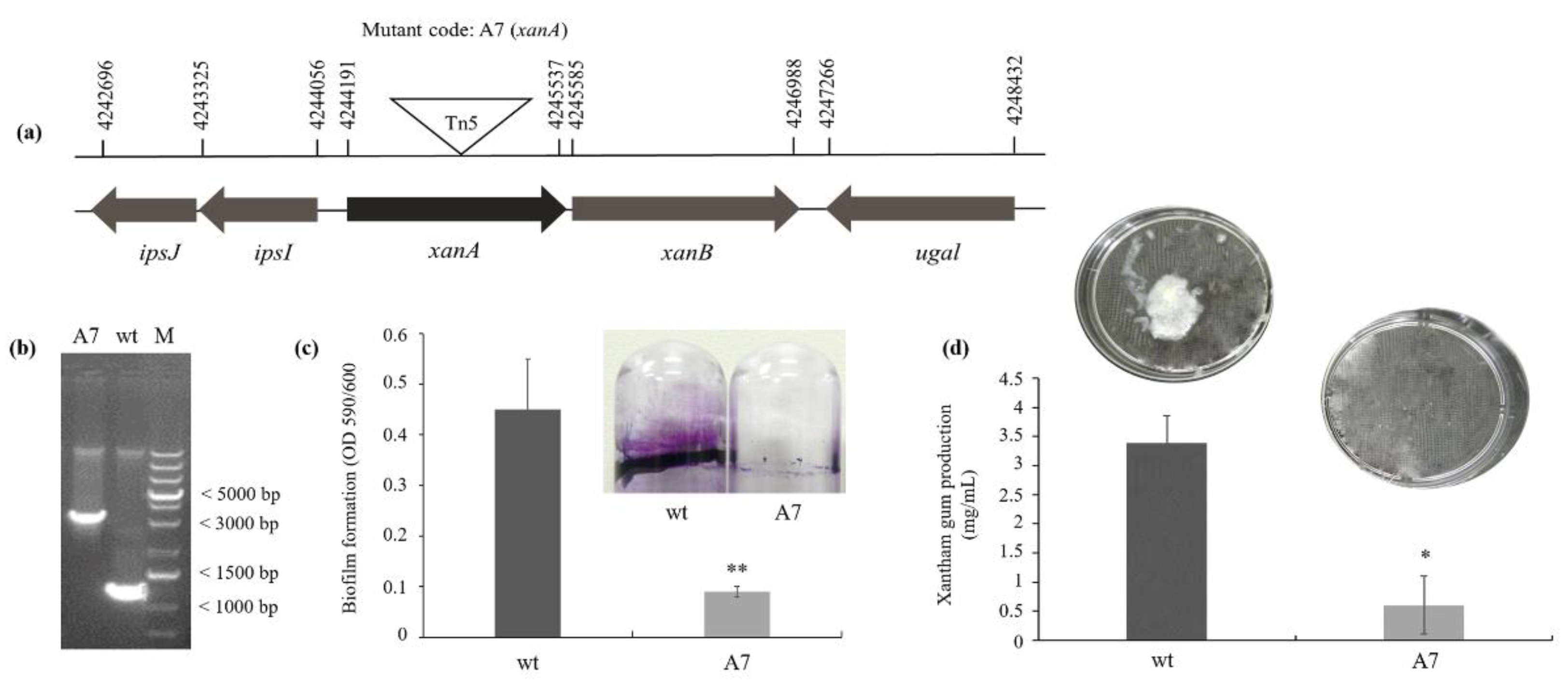
3.1. XanA Encodes a Phosphoglucomutase Protein, Important to the Biofilm Formation and Xanthan Gum Production

The A7 mutant was isolated from an EZ-Tn5 transposon mutagenesis library of X. citri strain 306 [5,14] since it exhibited a reduced ability to form biofilm. Sequencing indicated that EZ-Tn5 was inserted in the position 651 downstream of the translation start site of the locus XAC_RS18095 (Figure 1a). PCR using the primers XAC3579F and XAC3579R demonstrated the increase in the length of the obtained PCR amplicon by insertion of EZ-Tn5 transposon (1.22 kb) generating a PCR product with 2.22 kb, which confirmed the mutation of the xanA gene (locus XAC_RS18095) (Figure 1b). Thus, we will refer to the A7 mutant as xanA::Tn5. The xanA gene encodes a predicted phosphoglucomutase protein, required to synthesize the xanthan gum, a pathogenesis-related exopolysaccharide in Xanthomonads [13].
The xanA::Tn5 produced approximately 80% less biofilm compared with the X. citri wild-type (wt) strain (Figure 1c). We further verified the importance of the mutated gene in xanthan gum production. As summarized in Figure 1d, the xanthan gum yield of xanA::Tn5 was significantly different from that of the wild-type strain, showing 83% less xanthan gum production.

3.2. Mutant Strain XanA::Tn5 Developed No Symptoms on Sweet Orange Host

Previous studies have verified that xanthan plays a role in X. citri pathogenesis in detached leaves [12], however, this is not a natural condition to understand the host-pathogen interaction. We then inoculated the wild-type strain and xanA::Tn5 on host leaves by two different methods: spray and infiltration. The symptoms evaluation was monitored for 30 days after inoculation. No symptoms were observed in xanA::Tn5 when inoculated by spray, in opposition to severe citrus canker symptoms caused by wt cells (Figure 2a). However, when the strains were inoculated by infiltration, xanA::Tn5 mutant was able to cause canker symptoms, even though less severe than those observed by wt strain (Figure 2b). These results indicate that xanA plays an important role in the epiphytic stage of the X. citri lifestyle, since xanA mutated allele affected this ability, thus the bacterium was not able to cause disease when inoculated by spray, a method that mimics a natural condition of X. citri infection.

3.3. xanA Plays a Critical Role in Controlling Important Features in X. citri

To verified that the Tn5 mutation did not have a polar effect, we generated a deletion mutant of the xanA gene in X. citri that was also complemented with the construct pUFR053-xanA. Both xanA mutants (xanA::Tn5 and ΔxanA) and wt strains were tested to determine their ability to form biofilm on the abiotic surface, sliding motility, gum production, and pathogenicity. The growth curve of the mutant strains was not different from that in the wild-type (Figure S1).
Two independent mutant strains of xanA showed a decreased in biofilm formation, as shown in the first test (Figure 3a), and in sliding on medium plates (Figure 3b). On the plate surface, the diameters of the bacteria sliding resulted from migration away from the inoculation points in the agar surface were about 3-fold higher for wt than both xanA mutant strains after 48 h at 28 °C (Figure 3b). The complemented strain showed results like those of the wild-type strain, indicating that the sliding motility phenotype of the mutant was restored. Swimming motility analyses were also performed, but no difference was observed between the mutant and the wt strains (data not shown).
Xanthan gum amount also decreased about 4.5-fold in xanA::Tn5 and 3.4-fold in ΔxanA strain compared to the wt strain. This phenotype was restored to the wt level by complementation of the mutants, with no significant difference (p < 0.05) between xanA::tn5_c, ΔxanA_c, and wt, respectively. These differences in the amount of xanthan gum extracted from supernatants of these strains can also be visually observed on the plates (Figure 3c).
Impairment of pathogenesis after bacterial inoculation on leaf surfaces that has been verified for xanA::Tn5 mutant strain was also confirmed by ΔxanA and restored in xanA complementation strains (Figure 3d). A large number of pustules was observed on the leaves surfaces at 15 days after inoculation of wt and both complemented strains (xanA::Tn5_c and ΔxanA_c). However, very few pustules were observed on the leaves surfaces inoculated with each of the mutants (xanA::Tn5 and ΔxanA) strains.

3.4. Xanthan Gum from xanA Mutant Have Altered Monosaccharides Content

Xanthan gum produced by wt and xanA::Tn5 showed different percentages of some specific monosaccharides by HPLC analysis (Table 3). Xanthan gum produced by xanA::Tn5 exhibited a higher proportion of mannose, galactose, and xylose and reduced rates of rhamnose, glucuronic acid, and glucose. Thus, monosaccharide composition analysis indicated that xanA deletion altered the xanthan composition.

3.5. XanA Is Highly Conserved in the Xanthomonads Group

BLASTP search revealed that XAC3579 is highly conserved in other plant pathogenic species, including, X. oryzae pv. oryzae (99% identity), X. campestris pv. campestris (96% identity), and Xylella fastidiosa Temecula (84% identity) and Xylella fastidiosa 9a5c (84% identity). The phosphoglucomutase and phosphomannomutase, as well as XanA, share a conserved domain I that contains the catalytic phosphoserine (Figure 4), suggesting that this enzyme may be critical for cellular functions and give adaptative advantage for bacterial cells.

4. Discussion

In this work, we showed changes in the xanthan gum monosaccharide content that were associated with the xanA function in Xanthomonas. Disruption of xanA in X. citri genome, which codes for enzyme XanA that is necessary for isomerization of glucose-6-phosphate to glucose-1-phosphate and conversion of mannose-6- phosphate to mannose-1-phosphate [12,26,27], led to impairment of xanthan gum production. PGM/PMM possibly works as a valve, rerouting the metabolic flux originating from hexose phosphates either toward the biosynthesis of lipopolysaccharides (LPS) or xanthan, or the generation of energy or building blocks, such as amino acids, for cellular growth [27,28]. Thus, with the lack of XanA synthesis, the PGM/PMM activity levels decreased and consequently, the conversion of the substrates to UDP-D-glucose and GDP-mannose was compromised, leading to a reduction of glucose and mannose activated monosaccharides. This explains the lower percentages of glucose and mannose in the xanthan composition in xanA::Tn5 mutant compared with that observed in the wild-type strain. Indeed, the xanA deletion resulted in a lack of both activated monosaccharides UDP-glucose and GDP-mannose, which led to the formation of a xanthan gum structurally different [29].
For completion of xanthan gum synthesis, different steps are involved, and they require specific substrates and specific enzymes during this biosynthetic process. If either the substrate or the enzyme is absent, the step is compromised [30]. Some authors have shown that when mutation of specific genes involved in the xanthan gum synthesis are made to simplify the repeating unit structure, the xanthan gum yield was much lower as than that produced from the wild-type strain [29]. Betlach et al. [31] constructed a mutant lacking the glucuronic acid residues and the pyruvate and as a result, the xanthan gum solution produced by this strain was highly viscous. As we showed here, mutation of xanA reflected in changes of the repeating unit in the xanthan structure, and consequently, the xanthan gum yield was much lower than that produced from the wild-type and complemented strains, although low levels of xanthan gum remained detectable. The remaining xanthan may be explained by the presence of the xanB gene in the X. citri genome which codes for the enzyme phosphomannose-isomerase-GDP-mannose pyrophosphorylase that has involvement in GDP-mannose synthesis, one of those xanthan precursors [11].
The drastic reduction of xanthan gum in xanA mutants (xanA::Tn5 and ΔxanA) was responsible for the decrease of biofilm formation, sliding motility, and pathogenicity observed in this work. As xanthan gum contributes to the bacterial attachment to surfaces [8] and biofilm development with an organized structure on both abiotic and biotic surfaces [4], we suggest that such features are associates with the xanA function in determining EPS content. These findings, together with the demonstration that biofilm formation in xanA mutants can be complemented by the pUFR53-xanA construct, strongly suggest that a structurally intact xanthan gum harboring a particular composition of monosaccharides is critical for the X. citri lifestyle. Therefore, xanthan gum can affect bacterial cell adherence on host tissue and survival under stress conditions, as had already been demonstrated by other authors [32].
Further, sliding motility was previously shown to be promoted by xanthan gum and inhibited by type IV pili in X. citri [3,23]. These previous results corroborate our data which shows that xanA mutants also had a significant reduction in sliding motility compared with the wild-type strain. Consistently, it has been proposed that xanthan acts as a surfactant or surface-wetting agent to facilitate this type of movement [33,34]. Indeed, xanA::Tn5 and ΔxanA displayed less spread on the medium surface that may be due to low levels of the xanthan produced by these mutant strains, since anterior works have shown that X. citri mutants impaired in xanthan gum production also have sliding motility altered [3,4,5,35,36].
Moreover, reduced xanthan gum has also been related to the impairment of X. citri pathogenesis [3,12,13]. In our study, likewise, we also observed fewer symptoms when the xanA::Tn5 mutant cells are infiltrated into the leaves. Since inoculation by infiltration breaks the first physical defense layer, X. citri can use subsequent pathogenicity mechanisms, such as the type III secretion system to inject specific effectors into host cells, culminating in the development of citrus canker symptoms. Accordingly, previous results showed that the xanA mutant retained its infectivity when infiltrated into leaves and the authors speculated that PGM itself is not critical to canker progression, but it is related to the inoculation method used, which does not simulate the natural process of infection by X. citri [12]. Even though infiltration does not reflect a natural infection condition, xanA has a role when the bacteria are in plant mesophyll. On the other hand, we show that xanA::Tn5 caused no symptoms in sweet orange leaves after spraying, a method that simulates the natural infection condition. These results suggest that xanA has an important role in the epiphytical stage on the leaves that is essential for the interaction with the host, including adaptive advantage for bacteria cell survival under stress conditions and invasion of parenchyma tissue, which is required for the pathogenesis of X. citri.
Our data expanded the investigation on the xanA function, showing its role in affecting the monosaccharide composition of xanthan gum. The modified xanthan produced by xanA mutant compromised important features in X. citri and strongly impaired its ability to cause disease when in the epiphytic stage. Thus, chemical compounds that target the metabolic pathways involved in the activation of monosaccharides such as UDP-D-glucose and GDP-mannose, which was shown in this work that interferes in pathogenicity, could be a strategy to impair citrus canker disease. Further, as xanA is highly conserved in many bacteria, this strategy could be applied to other plant pathogenic species.

Supplementary Materials

The following are available online at https://www.mdpi.com/article/10.3390/microorganisms9061176/s1, Figure S1. Growth curve of X. citri strains in NBY medium.

Author Contributions

S.C.P., M.A.T., M.A.M. and A.A.d.S. participated in the conception of the work; S.C.P., L.M.G. and A.A.d.S. designed the experiments; S.C.P., L.M.G., M.J.F.F. and M.O.A. performed the experiments; S.C.P., L.M.G. and M.A.T. analyzed the resulting data; L.M.G. and A.A.d.S. wrote the paper. All authors have read and agreed to the published version of the manuscript.

Funding

This work was supported by research grant from Fundação de Amaparo à Pesquisa do Estado de São Paulo (2013/10957-0) and INCT Citrus (Proc. CNPQ465440/2014–2 and FAPESP 2014/50880–0). S.C.P. was a post-doctoral fellow from FAPESP (2013/01395-9) and L.M.G and M.O.A. are post-doctoral fellows from FAPESP (2019/01901-8 and 2017/18570-9, respectively). M.A.M. and A.A.S. are recipients of research fellowships from CNPq.

Institutional Review Board Statement

Not applicable.

Informed Consent Statement

Not applicable.

Conflicts of Interest

The authors declare no conflict of interest.

References

  1. Graham, J.H.; Gottwald, T.R.; Cubero, J.; Achor, D.S. Xanthomonas axonopodis pv. citri: Factors affecting successful eradication of citrus canker. Mol. Plant Pathol. 2004, 5, 1–15. [Google Scholar] [CrossRef]
  2. Gottwald, T.R.; Pierce, F.; Graham, J.H. Citrus Canker: The Pathogen and Its Impact Plant Health Progress Plant Health Progress. Plant Health Prog. 2002, 3, 15. [Google Scholar] [CrossRef] [Green Version]
  3. Granato, L.M.; Picchi, S.C.; Andrade, M.O.; Takita, M.A.; de Souza, A.A.; Wang, N.; Machado, M.A. The ATP-dependent RNA helicase HrpB plays an important role in motility and biofilm formation in Xanthomonas citri subsp. citri. BMC Microbiol. 2016, 16, 55. [Google Scholar] [CrossRef] [Green Version]
  4. Rigano, L.A.; Siciliano, F.; Enrique, R.; Sendín, L.; Filippone, P.; Torres, P.S.; Qüesta, J.; Dow, J.M.; Castagnaro, A.P.; Vojnov, A.A.; et al. Biofilm formation, epiphytic fitness, and canker development in Xanthomonas axonopodis pv. citri. Mol. Plant Microbe Interact. 2007, 20, 1222–1230. [Google Scholar] [CrossRef] [Green Version]
  5. Granato, L.M.; Picchi, S.C.; De Oliveira Andrade, M.; Martins, P.M.M.; Takita, M.A.; Machado, M.A.; De Souza, A.A. The EcnA antitoxin is important not only for human pathogens: Evidence of its role in the plant pathogen Xanthomonas citri subsp. citri. J. Bacteriol. 2019, 201, e00796-18. [Google Scholar] [CrossRef] [Green Version]
  6. Guo, Y.; Zhang, Y.; Li, J.-L.; Wang, N. DSF-mediated quorum sensing plays a central role in coordinating gene expression of Xanthomonas citri subsp. citri. Mol. Plant Microbe Interact. 2012, 25, 165–179. [Google Scholar] [CrossRef] [Green Version]
  7. Guo, Y.; Figueiredo, F.; Jones, J.; Wang, N. HrpG and HrpX play global roles in coordinating different virulence traits of Xanthomonas axonopodis pv. citri. Mol. Plant Microbe Interact. 2011, 24, 649–661. [Google Scholar] [CrossRef] [Green Version]
  8. Li, J.; Wang, N. The wxacO gene of Xanthomonas citri ssp. citri encodes a protein with a role in lipopolysaccharide biosynthesis, biofilm formation, stress tolerance and virulence. Mol. Plant Pathol. 2011, 12, 381–396. [Google Scholar] [CrossRef] [PubMed]
  9. Malamud, F.; Conforte, V.P.; Rigano, L.A.; Castagnaro, A.P.; Marano, M.R.; Morais do Amaral, A.; Vojnov, A.A. HrpM is involved in glucan biosynthesis, biofilm formation and pathogenicity in Xanthomonas citri ssp. citri. Mol. Plant Pathol. 2012, 13, 1010–1018. [Google Scholar] [CrossRef] [PubMed]
  10. Jansson, P.-E.; Kenne, L.; Bengt, L. Structure of the extracellular polysacchride from Xanthomonas campestris. Carbohydr. Res. 1975, 45, 275–282. [Google Scholar] [CrossRef]
  11. Koplin, R.; Arnold, W.; Hotte, B.; Simon, R.; Wang, G.; Puhler, A. Genetics of xanthan production in Xanthomonas campestris: The xanA and xanB genes are involved in UDP-glucose and GDP-mannose biosynthesis. J. Bacteriol. 1992, 174, 191–199. [Google Scholar] [CrossRef] [PubMed] [Green Version]
  12. Goto, L.S.; Vessoni Alexandrino, A.; Malvessi Pereira, C.; Silva Martins, C.; D’Muniz Pereira, H.; Brandão-Neto, J.; Marques Novo-Mansur, M.T. Structural and functional characterization of the phosphoglucomutase from Xanthomonas citri subsp. citri. Biochim. Biophys. Acta Proteins Proteom. 2016, 1864, 1658–1666. [Google Scholar] [CrossRef]
  13. Hung, C.-H.; Wu, H.-C.; Tseng, Y.-H. Mutation in the Xanthomonas campestris xanA gene required for synthesis of xanthan and lipopolysaccharide drastically reduces the efficiency of bacteriophage (phi)L7 adsorption. Biochem. Biophys. Res. Commun. 2002, 291, 338–343. [Google Scholar] [CrossRef]
  14. Baptista, J.C.; Machado, M.A.; Homem, R.A.; Torres, P.S.; Vojnov, A.A.; do Amaral, A.M. Mutation in the xpsD gene of Xanthomonas axonopodis pv. citri affects cellulose degradation and virulence. Genet. Mol. Biol. 2010, 33, 146–153. [Google Scholar] [CrossRef]
  15. Liu, Y.G.; Whittier, R.F. Thermal asymmetric interlaced PCR: Automatable amplification and sequencing of insert end fragments from P1 and YAC clones for chromosome walking. Genomics 1995, 25, 674–681. [Google Scholar] [CrossRef]
  16. da Silva, A.C.R.; Ferro, J.A.; Reinach, F.C.; Farah, C.S.; Furlan, L.R.; Quaggio, R.B.; Monteiro-Vitorello, C.B.; Van Sluys, M.A.; Almeida, N.F.; Alves, L.M.C.; et al. Comparison of the genomes of two Xanthomonas pathogens with differing host specificities. Nature 2002, 417, 459–463. [Google Scholar] [CrossRef]
  17. Hanahan, D. Studies on transformation of Escherichia coli with plasmids. J. Mol. Biol. 1983, 166, 557–580. [Google Scholar] [CrossRef]
  18. Schaad, N.W.; Postnikova, E.; Lacy, G.H.; Sechler, A.; Agarkova, I.; Stromberg, P.E.; Stromberg, V.K.; Vidaver, A.K. Reclassification of Xanthomonas campestris pv. citri (ex Hasse 1915) Dye 1978 forms A, B/C/D, and E as X. smithii subsp. citri (ex Hasse) sp. nov. nom. rev. comb. nov., X. fuscans subsp. aurantifolii (ex Gabriel 1989) sp. nov. nom. rev. comb. nov., and X. Syst. Appl. Microbiol. 2005, 28, 494–518. [Google Scholar] [CrossRef]
  19. El Yacoubi, B.; Brunings, A.M.; Yuan, Q.; Shankar, S.; Gabriel, D.W. In planta horizontal transfer of a major pathogenicity effector gene. Appl. Environ. Microbiol. 2007, 73, 1612–1621. [Google Scholar] [CrossRef] [PubMed] [Green Version]
  20. Sambrook, J.; Fritsch, E.; Maniatis, T. Molecular Cloning, 4th ed.; Cold Spring Harbor Laboratory Press: New York, NY, USA, 2012; Volume 1, ISBN 9781936113415. [Google Scholar]
  21. Andrade, M.O.; Farah, C.S.; Wang, N. The Post-transcriptional Regulator rsmA/csrA Activates T3SS by Stabilizing the 5′ UTR of hrpG, the Master Regulator of hrp/hrc Genes, in Xanthomonas. PLoS Pathog. 2014, 10, 1–19. [Google Scholar] [CrossRef]
  22. Amaral, A.M.; Toledo, C.P.; Baptista, J.C.; Machado, M.A. Transformation of Xanthomonas axonopodis pv. citri by Electroporation. Fitopatol. Bras. 2005, 30, 292–294. [Google Scholar] [CrossRef]
  23. Souza, D.P.; Andrade, M.O.; Alvarez-Martinez, C.E.; Arantes, G.M.; Farah, C.S.; Salinas, R.K. A component of the Xanthomonadaceae type IV secretion system combines a VirB7 motif with a N0 domain found in outer membrane transport proteins. PLoS Pathog. 2011, 7, e1002031. [Google Scholar] [CrossRef] [PubMed] [Green Version]
  24. Guzzo, C.R.; Salinas, R.K.; Andrade, M.O.; Farah, C.S. PILZ Protein Structure and Interactions with PILB and the FIMX EAL Domain: Implications for Control of Type IV Pilus Biogenesis. J. Mol. Biol. 2009, 393, 848–866. [Google Scholar] [CrossRef] [PubMed]
  25. Fu, D.; O’Neill, R. Monosaccharide composition analysis of oligosaccharides and glycoproteins by high-performance liquid chromatography. Anal. Biochem. 1995, 227, 377–384. [Google Scholar] [CrossRef] [PubMed]
  26. Mitra, S.; Cui, J.; Robbins, P.W.; Samuelson, J. A deeply divergent phosphoglucomutase (PGM) of Giardia lamblia has both PGM and phosphomannomutase activities. Glycobiology 2010, 20, 1233–1240. [Google Scholar] [CrossRef] [Green Version]
  27. Zandonadi, F.S.; Ferreira, S.P.; Alexandrino, A.V.; Carnielli, C.M.; Artier, J.; Barcelos, M.P.; Nicolela, N.C.S.; Prieto, E.L.; Goto, L.S.; Belasque, J.; et al. Periplasm-enriched fractions from Xanthomonas citri subsp. citri type A and X. fuscans subsp. aurantifolii type B present distinct proteomic profiles under in vitro pathogenicity induction. PLoS ONE 2020, 15, 1–24. [Google Scholar] [CrossRef]
  28. Schatschneider, S.; Huber, C.; Neuweger, H.; Watt, T.F.; Pühler, A.; Eisenreich, W.; Wittmann, C.; Niehaus, K.; Vorhölter, F.J. Metabolic flux pattern of glucose utilization by Xanthomonas campestris pv. campestris: Prevalent role of the Entner-Doudoroff pathway and minor fluxes through the pentose phosphate pathway and glycolysis. Mol. Biosyst. 2014, 10, 2663–2676. [Google Scholar] [CrossRef]
  29. Rosalam, S.; England, R. Review of xanthan gum production from unmodified starches by Xanthomonas campestris sp. Enzyme Microb. Technol. 2006, 39, 197–207. [Google Scholar] [CrossRef]
  30. Becker, A.; Katzen, F.; Pühler, A.; Ielpi, L. Xanthan gum biosynthesis and application: A biochemical /genetic perspective. Appl. Microbiol. Biotechnol. 1998, 50, 145–152. [Google Scholar] [CrossRef]
  31. Betlach, M.; Capage, D.; Doherty, D.; Hassler, R.; Henderson, N.; Vanderslice, R.; Marrelli, J.; Ward, M. Genetically engineered polymers: Manipulation of xanthan gum biosynthesis. In Industrial Polysaccharides Genetic Engineering, Structure/Property Relations and Applications; Yalpani, M., Ed.; Elsevier Science Publisher: Amsterdam, The Netherlands, 1987; pp. 35–50. [Google Scholar]
  32. Li, J.; Wang, N. Foliar application of biofilm formation-inhibiting compounds enhances control of citrus canker caused by Xanthomonas citri subsp. citri. Phytopathology 2014, 104, 134–142. [Google Scholar] [CrossRef] [Green Version]
  33. Murray, T.S.; Kazmierczak, B.I. Pseudomonas aeruginosa exhibits sliding motility in the absence of type IV pili and flagella. J. Bacteriol. 2008, 190, 2700–2708. [Google Scholar] [CrossRef] [PubMed] [Green Version]
  34. Malamud, F.; Torres, P.S.; Roeschlin, R.; Rigano, L.A.; Enrique, R.; Bonomi, H.R.; Castagnaro, A.P.; Marano, M.R.; Vojnov, A.A. The Xanthomonas axonopodis pv. citri flagellum is required for mature biofilm and canker development. Microbiology 2011, 157, 819–829. [Google Scholar] [CrossRef] [PubMed] [Green Version]
  35. Guo, Y.; Sagaram, U.S.; Kim, J.; Wang, N. Requirement of the galU gene for polysaccharide production by and pathogenicity and growth in planta of Xanthomonas citri subsp. citri. Appl. Environ. Microbiol. 2010, 76, 2234–2242. [Google Scholar] [CrossRef] [PubMed] [Green Version]
  36. Malamud, F.; Homem, R.A.; Conforte, V.P.; Yaryura, P.M.; Castagnaro, A.P.; Marano, M.R.; Morais do Amaral, A.; Vojnov, A.A. Identification and characterization of biofilm formation-defective mutants of Xanthomonas citri subsp. citri. Microbiology 2013. [Google Scholar] [CrossRef] [Green Version]
Figure 1. Identification of the A7 mutant from an EZ-Tn5 library of X. citri by biofilm and xanthan gum assays. (a) Genetic organization of the xanA gene in the X. citri subsp. citri strain 306 genome and the lengths of the open reading and transposon insertion site in the xanA mutant are indicated. The length of each arrow represents the relative open reading frame (ORF) size and indicates the direction of transcription. The triangle indicates the Tn5 insertion site. The annotation information and sizes of the genes were obtained from the genome sequence database of X. citri strain 306 (National Center for Biotechnology Information, NCBI, accession No: AE008923). (b) PCR analysis confirmed the insertion of EZ-Tn5 in the xanA gene (XAC_RS18095 or XAC3579). DNA was amplified using the primers XAC3579F and XAC3579R targeting a 500-bp region surrounding xanA from the wild-type (wt) and A7 (xanA::Tn5) strains, demonstrating the increase in the length of the obtained PCR amplicon by insertion of EZ-Tn5 transposon (1.22 kb) generating a PCR product with 2.22 kb in A7 mutant. M: Thermo Scientific O’GeneRuler 1 kb Plus DNA Ladder. (c) Biofilm formation on the abiotic surface by the wt and A7 mutant strains. The values were normalized to bacterial growth (OD at 600 nm). Values are expressed as the means ± standard deviations of six biological replicates. (d) Xanthan gum production. Values are expressed as the means ± standard deviations of three biological replicates. * indicates significant difference by t-test (p < 0.05) compared with wild-type.
Microorganisms 09 01176 g001
Figure 2. Pathogenicity assays. (a) Symptoms of sweet orange leave inoculated by spraying with wt and xanA::Tn5 mutant strains. (b) Symptoms of sweet orange leave infiltrated with wt and xanA::Tn5 mutant strains. Symptoms were analyzed for 30 days post-inoculation and pictures of leaves are representative of six independent replicates. wt, wild-type; xanA::Tn5, EZ-Tn5 insertion mutant.
Microorganisms 09 01176 g002
Figure 3. Biofilm, sliding motility, xanthan gum production, and pathogenicity of X. citri strains. (a) Biofilm formation on polystyrene surface. Values are expressed as the means ± standard deviations of six independent replicates. (b) Sliding motility phenotypes on SB agar medium plates after 48 h of incubation at 28 °C. Values are expressed as the means ± standard deviations of three independent replicates. (c) Xanthan gum production by X. citri strains. (d) Sweet orange leaves microneedle and inoculated with the different X. citri strains (108 CFU/mL). Pictures are representative of five leaves for each strain. wt, wild-type; xanA::Tn5, transposon mutant; xanA::Tn5_c, complemented transposon mutant; ΔxanA, deletion mutant; ΔxanA_c, complemented deletion mutant. * indicates significant difference by t-test (p < 0.05) compared with wt strain.
Microorganisms 09 01176 g003
Figure 4. Sequence alignments of XanA homologs. The * indicates the presence of a conserved motif. The highlight region (GGVMVTASHP) characterizes the conserved domain Phosphoglucumutase and phosphomannomutase phosphoserine (PS00710). Abbreviations are as follows: Xylella 9a5c—Xylella fastidiosa 9a5c (XF_0260), Xylella Temecula—Xylella fastidiosa Temecula (PDO213), X. citri—Xanthomonas citri subsp. citri (XAC3579), X. oryzae—Xanthomonas oryzae pv. oryzae (PXO_03174), XCC00626, X. campestris—Xanthomonas campestris pv. campestris str. ATCC 33913 (XCC0626). The protein motif was identified in Pfam (http://pfam.xfam.org/). Alignments were performed from residue 8 to residue 157 of X. citri XanA.
Microorganisms 09 01176 g004
Table 1. Primers used to transposon insertion point confirmation.
PrimerNucleotide Sequence (5′–3′)
KanACATGCAAGCTTCAGGGTTGA
KanBGCGGGGATCCTCTAGAGTCG
KanCACCTACAACAAAGCTCTCATCAACC
AD1NTCGA(G/C)T(A/T)T(G/C)G(A/T)GTT
XAC3579FTATTTCCAGACCGATTACCTCA
XAC3579RGAAACTCCAAAGTGCGTCTATG
Table 2. Strains and plasmids used in this study.
Strains/PlasmidsCharacteristicsReference or Source
Escherichia coliF- ɸ80dlacZhM15 h (lacZYA-argF) U169 endA1 deoR recA1 hsdR17(rK- mK+) phoA supE44 λ- thi-1 gyrA96 relA1[17]
Xanthomonas citri subsp. citri (X. citri)306 Syn. X. axonopodis pv. citri strain 306; wild-type, Rfr, Apr[16,18]
A7/xanA::Tn5 (xanA-)xanA (XAC3579), Kmr. Transposon (Tn5) mutant.[14]
xanA::Tn5_c (xanA+)xanA (contained in puFR053, Gmr)This study
ΔxanA (xanA-)Deletion (Tn5) mutant; xanA (XAC3579), Apr.This study
ΔxanA_c (xanA+)xanA (contained in puFR053, Gmr)This study
pNPTS138pNPTS138, Kmr, sacB, lacZα+M. R. Alley, unpublished
pNPTS_xanAxanA gene cloning on pNPTS138This study
pUFR053IncW Mob+mob (P) lacZα+ Par+, Cmr, Gmr, Kmr, shuttle vector[19]
pUFR053_xanAxanA (XAC3579) gene cloning on pUFR053This study
Apr, Kmr, Gmr, and Rfr indicate resistance to ampicillin, kanamycin, gentamicin, and rifamycin, respectively.
Table 3. Percentages (%) of monosaccharides present in the xanthan gum produced by Xanthomonas citri subsp. citri strains.
X. citri Strains%
ManRhaGlcAGalAGlcGalXyl
wt51.866.203.450.3927.324.601.92
xanA::Tn558.951.580.480.3516.687.982.64
Man (mannose), Rha (ramnose), GlcA (glucuronic acid), GalA (galacturonic acyd), Glc (glucose), Gal (galactose) and Xyl (xylose).
Publisher’s Note: MDPI stays neutral with regard to jurisdictional claims in published maps and institutional affiliations.

