Article

Survey of Pathogen-Lowering and Immuno-Modulatory Effects Upon Treatment of Campylobacter coli-Infected Secondary Abiotic IL-10−/− Mice with the Probiotic Formulation Aviguard®

Gastrointestinal Microbiology Research Group, Institute of Microbiology, Infectious Diseases and Immunology, Charité—Universitätsmedizin Berlin, Corporate Member of Freie Universität Berlin, Humboldt-Universität zu Berlin, and Berlin Institute of Health, 12203 Berlin, Germany
*
Authors to whom correspondence should be addressed.
These authors contributed equally to this study.
Microorganisms 2021, 9(6), 1127; https://doi.org/10.3390/microorganisms9061127
Submission received: 7 April 2021 / Revised: 17 May 2021 / Accepted: 21 May 2021 / Published: 23 May 2021
(This article belongs to the Special Issue Foodborne Bacteria–Host Interactions)

Abstract

:
The prevalence of infections with the zoonotic enteritis pathogen Campylobacter coli is increasing. Probiotic formulations constitute promising antibiotic-independent approaches to reduce intestinal pathogen loads and modulate pathogen-induced immune responses in the infected human host, resulting in acute campylobacteriosis and post-infectious sequelae. Here, we address potential antipathogenic and immuno-modulatory effects of the commercial product Aviguard® during experimental campylobacteriosis. Secondary abiotic IL-10−/− mice were infected with a C. coli patient isolate on days 0 and 1, followed by oral Aviguard® treatment on days 2, 3 and 4. Until day 6 post-infection, Aviguard® treatment could lower the pathogen burdens within the proximal but not the distal intestinal tract. In contrast, the probiotic bacteria had sufficiently established in the intestines with lower fecal loads of obligate anaerobic species in C. coli-infected as compared to uninfected mice following Aviguard® treatment. Aviguard® application did not result in alleviated clinical signs, histopathological or apoptotic changes in the colon of infected IL-10−/− mice, whereas, however, Aviguard® treatment could dampen pathogen-induced innate and adaptive immune responses in the colon, accompanied by less distinct intestinal proinflammatory cytokine secretion. In conclusion, Aviguard® constitutes a promising probiotic compound to alleviate enteropathogen-induced proinflammatory immune responses during human campylobacteriosis.

1. Introduction

Campylobacter infections are the leading causes of bacterial gastroenteritis in humans, with worldwide rising prevalences [1]. In the European Union, for instance, campylobacteriosis was the most frequent zoonotic disease in 2018, responsible for more than 240,000 reported cases. Campylobacter jejuni and Campylobacter coli constituted the most prevalent causative agents with relative abundances of 83.9% and 10.3%, respectively [2]. The curved or rod-shaped and highly motile Gram-negative bacteria are part of the commensal gut microbiota of many avian species, including chicken and turkey and of mammals, such as pigs, cattle and sheep and exhibit a growth optimum at 37–42 °C [3,4]. In addition, Campylobacter species might be isolated from natural environments, including surface waters and can survive extended periods outside a warm-blooded host [4,5]. The bacteria are mainly transmitted to humans via the food chain. Ingestion of undercooked or raw meat products from livestock, contaminated water, eggs or unpasteurized milk is a potential infection source. However, most Campylobacter outbreaks result from ingestion of poultry products and/or inappropriate handling and kitchen hygiene measures [6,7]. Of note, a European survey from 2018 revealed a Campylobacter prevalence of 26% in broiler chicken and of 72% in turkeys [8]. In slaughterhouses, Campylobacter has been shown to spread from the ceca of infected animals, subsequently contaminating the entire carcass [9,10]. Importantly, Campylobacter was not only isolated from the meat of Campylobacter colonized chicken but also from secondarily cross-contaminated chickens whose feces had initially been Campylobacter-negative [11]. Physical, chemical and radiation-based methods are generally applied to eliminate Campylobacter species from livestock carcasses and slaughterhouse equipment. However, those procedures may significantly reduce the Campylobacter loads but not eliminate the contaminating agents [11,12,13]. Notably, Campylobacter species can be highly infectious, given that relatively low doses of 500 bacteria may already lead to campylobacteriosis in infected humans depending on both the arsenal of virulence factors expressed by the pathogen and on the host immune status [5,14]. After an incubation period of up to five days, Campylobacter infected patients may present with symptoms of varying severities ranging from mild malaise to acute campylobacteriosis characterized by fever, chills, abdominal cramps, headache, and watery or even bloody diarrhea with mucous discharge and inflammatory cells due to underlying enterocolitis [15,16]. Notably, from the severity of the induced campylobacteriosis syndrome, one cannot predict the respective causative Campylobacter species [6]. The disease usually only requires symptomatic treatment, such as the substitution of fluids and minerals and resolves within two weeks post-infection (p.i.) without residues [14,15,16]. Post-infectious autoimmune morbidities affecting the nervous system (e.g., Guillain–Barré or Miller–Fisher syndrome), the joints (i.e., reactive arthritis) and the gastrointestinal tract (i.e., inflammatory bowel disease and irritable bowel syndrome), however, may develop in rare occasions weeks to months p.i. [16,17,18]. Particularly sialylated lipooligosaccharide (LOS) derived from the cell wall of Gram-negative bacterial species, including Campylobacter, is associated with severe forms of campylobacteriosis and increased risk of post-infectious manifestations [19].
Almost 40 years ago, distinct probiotic formulations were hypothesized as promising tools to exclude potentially pathogenic bacteria from their ecological niches within the vertebrate gastrointestinal tract [20,21]. Commercial competitive exclusion products have gained access to the agricultural industry in the meantime and have been successfully applied as antibiotic-independent animal feed additives to reduce the prevalence of enteropathogens, such as Salmonella and Clostridium perfringens in livestock and subsequently, to decrease the prevalence of foodborne infections in humans [22,23,24,25]. Among these commercial exclusion products, Aviguard® constitutes a mixture of viable probiotic bacteria representative for the main commensal species within the cecum of adult chicken [24]. This freeze-dried fermentation product exhibits a longer shelf life when compared to other compounds. It is applied as a drinking water additive or spray treatment to poultry [24].
To date, however, studies elucidating the triangle interplay between Campylobacter, probiotic bacteria derived from a commercial exclusion product including Aviguard® and the mammalian host are scarce. This prompted us in our present study for the first time to investigate the potential pathogen-lowering effects and immune-modulatory properties of peroral Aviguard® treatment during experimental campylobacteriosis. We, therefore, applied a murine Campylobacter-induced infection and inflammation model that had initially been generated to dissect C. jejuni-host interactions. Following peroral infection of secondary abiotic interleukin 10 gene-deficient (IL-10−/−) mice that had been generated following broad-spectrum antibiotic treatment, C. jejuni could not only stably infect the murine gastrointestinal tract but also induced non-self-limiting acute enterocolitis within a week p.i. [26]. The underlying mechanisms for this inflammatory scenario are pronounced Toll-like receptor-4 (TLR-4)-dependent, LOS host immune responses affecting not only the intestinal tract but also extra-intestinal, including systemic body parts [26,27]. Very recently, the secondary abiotic IL-10−/− mouse infection and inflammation model has been proven suitable also to unravel C. coli-host interactions [28,29]. In our actual study, we infected secondary abiotic IL-10−/− mice with a C. coli isolate derived from a diseased human patient by gavage, followed by oral Aviguard® treatment on three consecutive days, and surveyed (i.) the intestinal pathogen loads over time, (ii.) the clinical and microscopic inflammatory changes, and (iii.) the proinflammatory immune responses in intestinal and systemic compartments upon sacrifice at day 6 p.i.

2. Materials and Methods

2.1. Ethical Statement

All murine experiments had initially been approved by the commission for animal experiments headed by the “Landesamt für Gesundheit und Soziales” (LaGeSo, Berlin; registration number G0172/16) and were performed according to the European Guidelines for animal welfare (2010/63/EU) and the ARRIVE guidelines. Clinical conditions of mice were monitored daily.

2.2. Secondary Abiotic IL-10−/− Mice

IL-10−/− mice (C57BL/6j background) were bred in the Forschungsinstitute für Experimentelle Medizin, Charité—University Medicine Berlin (Berlin, Germany) and maintained in sterile cages covered by filter tops within an experimental semi-barrier facility. By the age of 3 weeks, mice were subjected to an 8-week-course of broad-spectrum antibiotic treatment for commensal gut microbiota depletion as described in detail elsewhere [30,31]. The secondary abiotic status of mice was confirmed by both culture and culture-independent (i.e., molecular, 16S rRNA-based) methods, as reported earlier [30,32]. Microbiota-depleted mice received autoclaved drinking water and chow and were handled under aseptic conditions to avoid contaminations. For antibiotic washout, the antibiotic cocktail was replaced by autoclaved tap water as early as three days before infection.

2.3. Campylobacter Coli Infection

The C. coli isolates (from the stool of a diseased patient with bloody diarrhea) were kindly provided by Dr. Torsten Semmler (Robert-Koch-Institute Berlin, Germany). Two days before infection, the C. coli isolate was thawed from stock and cultivated on Columbia agar (supplemented with 5% sheep blood) and Karmali agar plates (both from Oxoid, Wesel, Germany) that were incubated in a jar containing CampyGen gas packs (Oxoid, Wesel, Germany) under microaerophilic conditions (37 °C, 48 h). On days 0 and 1, sex- and age-matched secondary abiotic IL-10−/− mice (11 week-old litter mates, balanced gender ratio) were challenged with 109 colony-forming units (CFU) C. coli by gavage.

2.4. Treatment of Mice With the Commercial Exclusion Product Aviguard®

For probiotic treatment, 1 g Aviguard® (Lallemand Animal Nutrition, Worcestershire, UK) was dissolved in 10 mL phosphate-buffered saline (PBS; Thermo Fisher Scientific, Waltham, MA, USA) and perorally applied to mice on days 2, 3 and 4 p.i. (0.3 mL by gavage). The competitive exclusion product consists of the following bacterial species (approximately 109 CFU per mL): Escherichia coli, Citrobacter species, Enterococcus genus (E. faecalis, E. faecium), Lactobacillus species (L. casei, L. plantarum), Bacteroides species, Clostridium species (C. sporogenes), Eubacterium species, Propionibacterium species, Fusobacterium species, Ruminococcus species [33]. Placebo-treated C. coli-infected mice received vehicle only (0.3 mL by gavage). Naive control animals were neither infected with C. coli nor challenged with Aviguard® nor placebo.

2.5. Gastrointestinal C. coli Loads

For quantification of the gastrointestinal C. coli burdens, fecal and luminal stomach, duodenum, ileum, and colon samples were homogenized and serial dilutions streaked onto Columbia agar plates supplemented with 5% sheep blood and onto selective Karmali plates (both from Oxoid, Wesel, Germany). The inoculated plates were incubated in a jar containing CampyGen gas packs (Oxoid, Wesel, Germany) under microaerophilic conditions (37 °C, 48 h). C. coli were identified by the distinct macroscopic morphotypes, microscopic appearance following Gram-staining, and oxidase-positive reaction.

2.6. Cultural Intestinal Microbiota Analysis

To provide a comprehensive quantitative cultural survey of the bacterial compositions in both the Aviguard® suspensions and murine feces, respective samples were homogenized in sterile phosphate-buffered saline (PBS; Gibco, Life Technologies, Loughborough, UK) and cultivable bacterial species quantitated by plating serial dilutions on solid media and incubated under aerobic, microaerobic and anaerobic conditions (37 °C, 48 h) as stated elsewhere [30]. The numbers of respective bacteria were determined by enumeration of distinct colony morphotypes, followed by subcultivation, Gram-staining and biochemical analyses [30].

2.7. Culture-Independent Intestinal Microbiota Analysis

For quantitative assessment of fastidious and even uncultivable bacteria within bacterial suspensions and fecal samples, we applied culture-independent, 16S rRNA-based methods. The total genomic DNA was extracted from respective samples and adjusted to 1 ng per µL (Quant-iT PicoGreen reagent, Invitrogen, Carlsbad, CA, USA) as reported earlier [30]. The total eubacterial loads and the main bacterial groups abundant in the murine intestinal microbiota, including gamma-Proteobacteria/Enterobacteriaceae, Enterococcus genus, Lactobacillus group, Bifidobacterium genus, Bacteroides group, including Prevotella and Porphyromonas, Clostridium coccoides group, and Clostridium leptum group, were then assessed by quantitative RT–PCR (qRT–PCR) with species-, genera- or group-specific 16S rRNA gene primers (Tib MolBiol, Berlin, Germany), as shown in Table 1 and expressed as 16S rRNA gene copies per ng DNA [34].

2.8. Clinical Conditions

Before and once a day after the C. coli challenges, we quantitatively determined the clinical conditions of mice as stated previously [35]. In brief, the following parameters were assessed: the clinical aspect/wasting (0: normal; 1: ruffled fur; 2: less locomotion; 3: isolation; 4: severely compromised locomotion, pre-final aspect), the abundance of blood in feces (0: no blood; 2: microscopic detection of blood by the Guajac method using Hemoccult, Beckman Coulter/PCD, Krefeld, Germany; 4: macroscopic blood visible), and stool consistency (0: formed feces; 2: pasty feces; 4: liquid feces). The overall clinical score (maximum of 12) was calculated as the sum of the three scores assessing respective individual parameters [35].

2.9. Sampling Procedures

Mice were sacrificed by CO2 asphyxiation on day 6 p.i. Under sterile conditions, cardiac blood, large intestinal tissue samples (collected from each mouse in parallel for microbiological and immunohistopathological analyses) and luminal stomach, duodenum, ileum and colon samples were taken.

2.10. Histopathology

Histopathological changes were quantitated in 5 μm thin sections of colonic explants that had been immediately fixed in 5% formalin, embedded in paraffin and stained with hematoxylin and eosin using standardized histopathological (100× magnification, light microscopy, blinded investigator) as described earlier [36]. In brief, score 1: minimal inflammatory cell infiltrates in the mucosa with intact epithelium; score 2: mild inflammatory cell infiltrates in the mucosa and submucosa with mild hyperplasia and mild goblet cell loss; score 3: moderate inflammatory cell infiltrates in the mucosa with moderate goblet cell loss; score 4: marked inflammatory cell infiltration into in the mucosa and submucosa with marked goblet cell loss, multiple crypt abscesses and crypt loss.

2.11. In Situ Immunohistochemistry

In situ immunohistochemical analyses were performed in 5 μm thin large intestinal paraffin sections for quantification of apoptotic epithelial cells, macrophages and monocytes, T lymphocytes, regulatory T cells and B lymphocytes by using primary antibodies directed against cleaved caspase-3 (Asp175, Cell Signaling, Beverly, MA, USA, 1:200), F4/80 (no. 14-4801, clone BM8, eBioscience, San Diego, CA, USA, 1:50), CD3 (no. N1580, Dako, Glostrup, Denmark; 1:10), FOXP3 (clone FJK-165, no. 14-5773, eBioscience, 1:100) and B220 (no. 14-0452-81, eBioscience; 1:200), respectively [37]. The average number of positively stained cells in each section was determined within at least six high-power fields (HPF, 0.287 mm2, 400× magnification, blinded investigator).

2.12. Proinflammatory Cytokines

Ex vivo biopsies obtained from the colon and ileum (longitudinally cut strips of approximately 1 cm2) as well as from mesenteric lymph nodes (MLN; 3 lymph nodes) were washed in PBS (Gibco, Life Technologies, Loughborough, UK) and placed in 24-flat-bottom well culture plates (Nunc, Darmstadt, Germany) containing 500 μL serum-free RPMI 1640 medium (Gibco, Life Technologies, Loughborough, UK) supplemented with penicillin (100 µg/mL) and streptomycin (100 µg/mL; Biochrom, Berlin, Germany). Respective culture supernatants and serum samples were tested for interferon-γ (IFN-γ) and tumor necrosis factor-α (TNF-α) by the mouse inflammation cytometric bead assay (CBA; BD Biosciences, Heidelberg, Germany) in a BD FACSCanto II flow cytometer (BD Biosciences) after incubation at 37 °C for 18 h.

2.13. Statistical Analyses

Medians and significance levels were calculated by using GraphPad Prism (version 8, San Diego, CA, USA). The Mann–Whitney test was applied for pairwise comparisons of not normally distributed data. For multiple comparisons, the one-sided ANOVA with Tukey’s post-correction was used for normally distributed data and the Kruskal–Wallis test with Dunn’s post-correction for not normally distributed data. Two-sided probability (p) values ≤ 0.05 were considered significant. Definite outliers were removed after being identified by the Grubb’s test (α = 0.001). Data were pooled from three independent experiments.

3. Results

3.1. Gastrointestinal Pathogen Loads Following Oral Aviguard® Treatment of C. coli Infected Secondary Abiotic IL-10−/− Mice

Secondary abiotic IL-10−/− mice were infected with 109 viable C. coli cells on days 0 and 1 by gavage and perorally treated with the commercial competitive exclusion product Aviguard® on days 2, 3 and 4 p.i. Our cultural analyses of the bacterial suspensions revealed total numbers of 109 viable bacteria per mL, including 106 CFU Enterobacteriaceae per mL and between 108 and 5 ×10⁸ CFU Lactobacillus, Enterococcus, Bacteroides/Prevotella and Clostridium/Eubacterium species per mL (Figure S1A). We additionally performed culture-independent, 16S rRNA-based analyses for quantifying fastidious and non-cultivable bacteria. In support of the cultural data, our molecular analyses revealed consistently high gene numbers of Enterobacteriaceae, Enterococcus genus, Lactobacillus group, Bifidobacterium genus, Clostridium coccoides and Clostridium leptum groups among individual gavages (Figure S1B). Hence, the vast majority of cultivable, fastidious and uncultivable probiotic bacterial species abundant in the commercial competitive exclusion product Aviguard® (except for Fusobacterium and Propionibacterium species) could be assessed by cultural and molecular approaches.
Our cultural analyses revealed that C. coli could stably establish within the intestinal tract of both Aviguard® and placebo-treated mice with comparably high median loads of 109 CFU per g feces as early as day 2 p.i. (Figure 1, Figure S2). Upon necropsy on day 6 p.i., the pathogen numbers were slightly lower than day 2 p.i. in Aviguard® treated mice (less than 0.5 logs; p < 0.05; Figure S2B), which also held for the placebo control mice, however (p < 0.01; Figure S2A). Hence, Aviguard® could not sufficiently reduce fecal pathogen loads in C. coli-infected mice.
We further assessed the pathogen burdens in defined luminal parts of the gastrointestinal tract. On day 6 p.i., approximately two log orders of magnitude lower C. coli numbers could be determined in the duodenum and the ileum of Aviguard® as compared to placebo-treated mice (p < 0.001 and p < 0.01, respectively; Figure 2), which was, however, not the case in the large intestines (Figure 2). Notably, 47.1% of mice from the Aviguard® cohort, but only 7.1% of the placebo controls had expelled the pathogen from their stomach lumen. Furthermore, the former exhibited a trend towards approximately 2.5 log orders of magnitude lower median gastric C. coli numbers than the latter (not significant (n.s.) given high standard deviations; Figure 2). Hence, Aviguard® treatment lowered the pathogen burdens within the proximal but not the distal intestinal tract of C. coli-infected IL-10−/− mice.

3.2. Fecal Microbiota Composition Following Oral Aviguard® Treatment of C. coli-Infected Secondary Abiotic IL-10−/− Mice

We then addressed whether the applied Aviguard® bacteria could stably establish within the intestinal tract of C. coli-infected and uninfected secondary abiotic IL-10−/− mice and whether C. coli infection resulted in shifts within the gut microbiota composition. Our cultural analyses revealed that respective aerobic and anaerobic bacterial groups, genera and species could be detected in fecal samples obtained on day 6 p.i. with median loads ranging from approximately 106 up to 1011 CFU per g. Interestingly, C. coli-infected mice harbored lower obligate anaerobic bacterial species, such as Bacteroides/Prevotella and Clostridium/Eubacterium species in their feces (p < 0.01, Figure 3A). In support, when applying culture-independent, 16S rRNA-based approaches, lower fecal gene numbers for Bacteroides/Prevotella group/genera (p < 0.001), for Clostridium coccoides (p < 0.05) and for Clostridium leptum groups (p < 0.05) as well as for Bifidobacterium genus (p < 0.01) were detected in C. coli-infected than uninfected mice following Aviguard® challenge (Figure 3B). Hence, the probiotic bacteria could sufficiently establish in the intestinal tract of mice until day 6 p.i. Thus, 48 h after the latest of three consecutive peroral Aviguard® applications with lower fecal loads of obligate anaerobic species in the C. coli cohort than the uninfected counterparts.

3.3. Clinical and Histopathological Sequelae Upon Oral Aviguard® Treatment of C. coli-Infected Secondary Abiotic IL-10−/− Mice

We further assessed the clinical outcomes of C. coli-infected IL-10−/− mice following Aviguard® treatment. Therefore, we quantified the key clinical signs of campylobacteriosis, such as wasting, the abundance of fecal blood and diarrhea in mice before and after C. coli infection by using standardized clinical scores [35]. Overall, mice from either cohort displayed rather mild clinical signs of C. coli infection as indicated by median scores not exceeding 2. Immediately before the first (i.e., day 2 p.i.) until the last (i.e., day 4 p.i.) of three consecutive Aviguard® versus placebo applications, mice from the treatment cohort displayed slightly lower clinical scores than the placebo control group (p < 0.05–0.001; Figure S3), whereas at the end of the observation period (i.e., day 6 p.i.), the clinical conditions of C. coli-infected mice from either group were comparable (n.s.; Figure S3; Figure 4A).
We further quantitatively determined the large intestinal histopathological changes upon Aviguard® treatment of C. coli-infected mice in colonic paraffin sections applying standardized histopathological scores [36]. On day 6 p.i., mild to moderate histopathological changes within the colonic mucosa could be observed (p < 0.01–0.001 versus uninfected controls), but with no differences between the Aviguard® and the placebo cohorts (n.s.; Figure 4B).
Since apoptosis is well-known as a reliable parameter for the grading of intestinal inflammatory responses [31], we further enumerated colonic epithelial cells positive for cleaved caspase-3 following immunohistochemical staining of large intestinal paraffin ex vivo biopsies. In line with the obtained histopathological results, C. coli infection mounted in increased numbers of apoptotic colonic epithelial cells (p < 0.01–0.001), whereas comparable counts could be assessed in the Aviguard® and placebo groups at day 6 p.i. (n.s.; Figure 4C). Hence, Aviguard® did neither affect clinical signs of C. coli infection nor histopathological nor apoptotic changes in the large intestines of infected IL-10−/− mice.

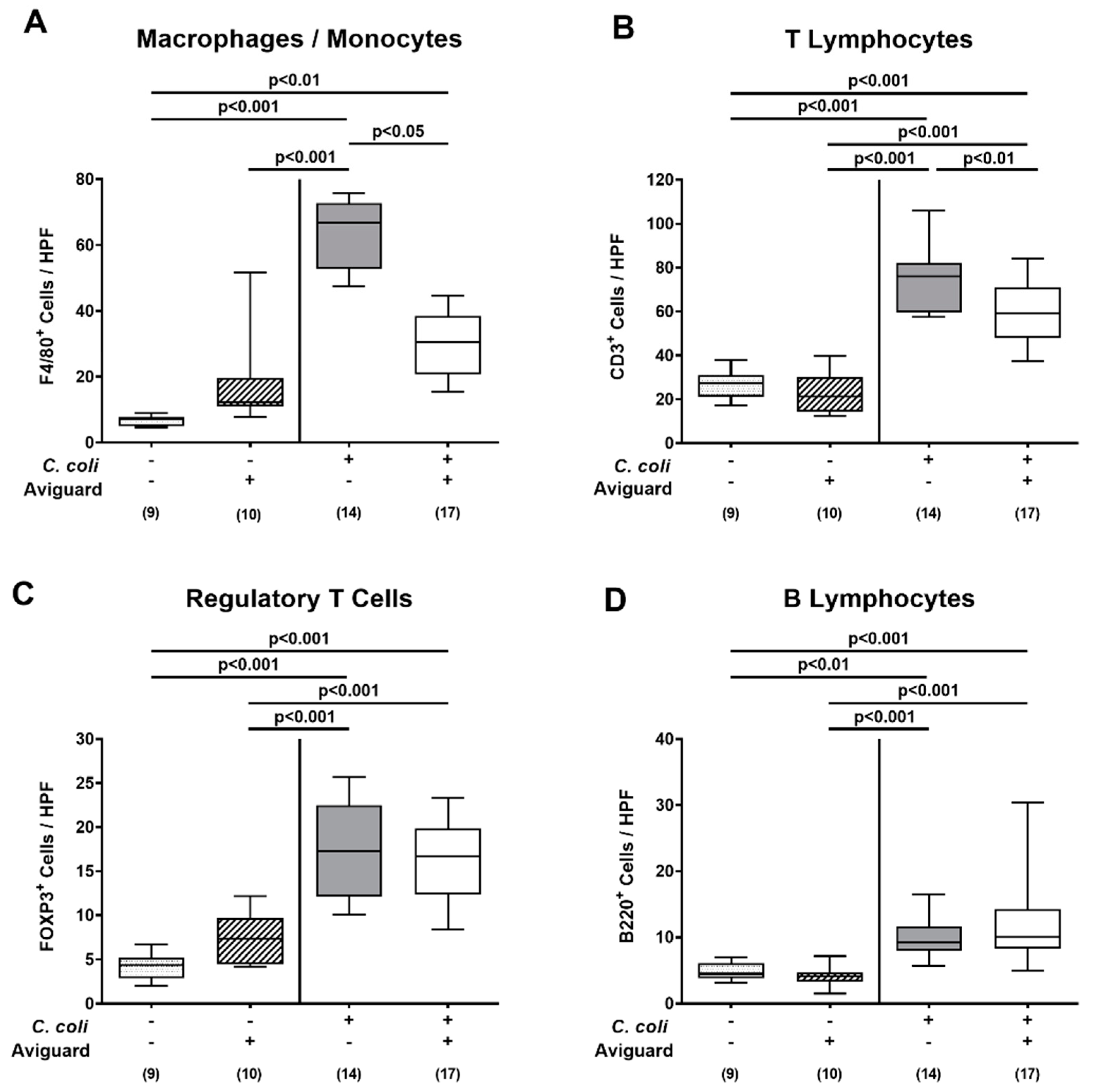
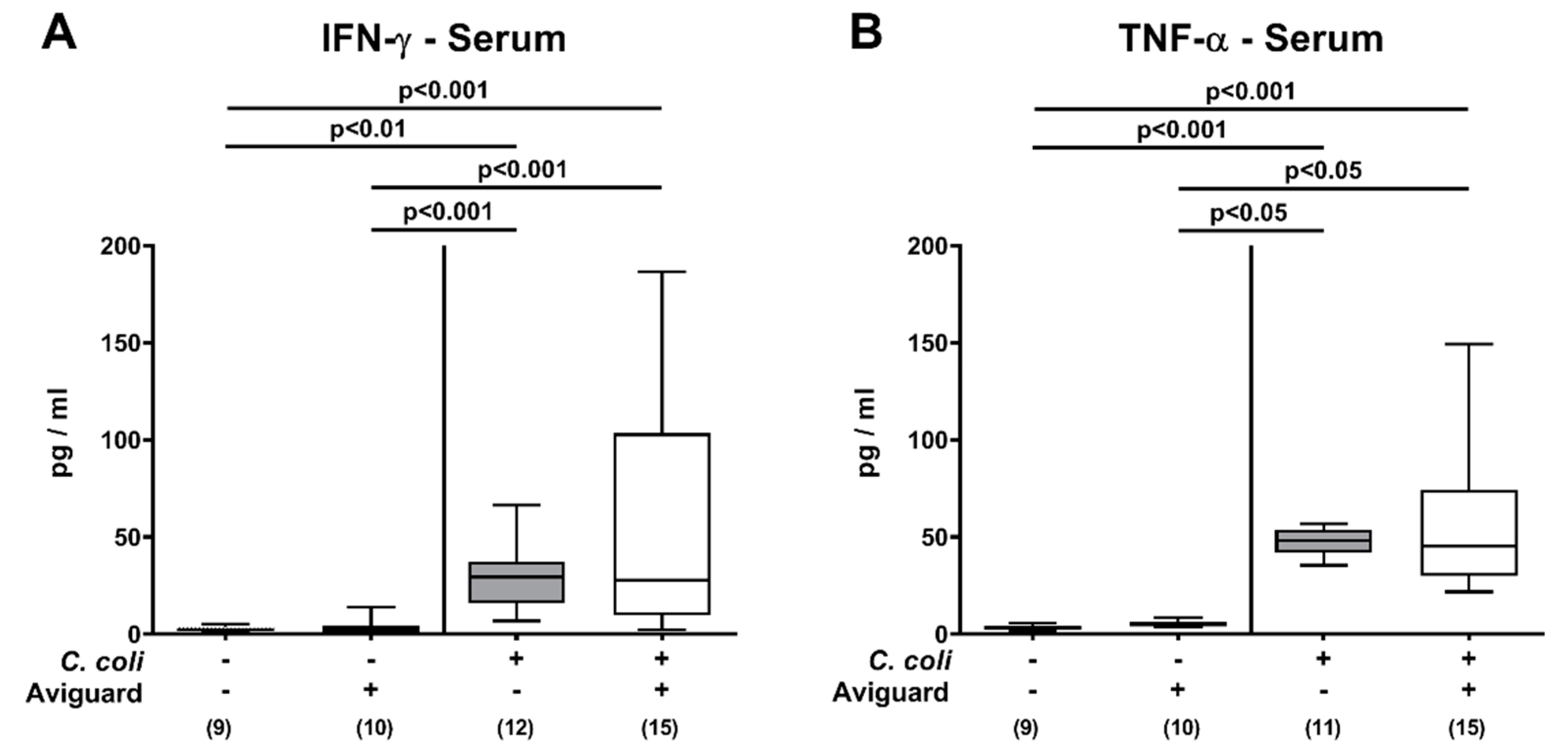
3.4. Intestinal and Systemic Proinflammatory Immune Responses Following Oral Aviguard® Treatment of C. coli-Infected Secondary Abiotic IL-10−/− Mice

We further addressed whether peroral Aviguard® application to C. coli-infected secondary abiotic IL-10−/− mice modulated pathogen-induced immune responses in the intestinal tract. Therefore, we quantified distinct innate and adaptive immune cell populations in colonic paraffin sections that had been subjected to defined immunohistochemical stainings. On day 6 p.i., increased numbers of innate immune cells, such as macrophages and monocytes as well as of adaptive immune cell subsets, including T lymphocytes, regulatory T cells and B lymphocytes, could be determined in the colonic mucosa and lamina propria of mice from either cohort (p < 0.01–0.001; Figure 5). The C. coli induced increases in macrophages and monocytes as well as in T lymphocytes were, however, less pronounced following Aviguard® than placebo treatment (p < 0.05 and p < 0.01 versus placebo, respectively; Figure 5A,B), whereas colonic numbers of regulatory T cells and B lymphocytes were comparable in both cohorts on day 6 p.i. (n.s.; Figure 5C,D).
We further assessed proinflammatory cytokine secretion in distinct intestinal compartments. Elevated IFN-γ concentrations were measured in ex vivo biopsies taken from the colon of C. coli-infected mice (p < 0.05–0.001), but with lower levels in Aviguard® than placebo-treated counterparts (p < 0.05; Figure 6A), whereas ileal IFN-γ secretion was less pronounced in the former versus the latter on day 6 p.i. (p < 0.05; Figure 6B). In MLN, increases in IFN-γ concentrations were comparable in both treatment groups on day 6 p.i. (p < 0.05–0.001; Figure 6C), while C. coli-induced TNF-α secretion was enhanced in placebo control mice only (p < 0.01; Figure 6D).
We finally addressed whether the immuno-modulatory effects of Aviguard® were restricted to the intestinal tract or could also be observed systemically. Therefore, we measured proinflammatory cytokines in serum samples taken in C. coli-infected and corresponding uninfected control groups. Upon C. coli infection, increased IFN-γ and TNF-α serum concentrations could be measured (p < 0.01–0.001) but did not differ between Aviguard® or placebo-treated mice on day 6 p.i. (n.s.; Figure 7). Hence, Aviguard® treatment of C. coli-infected IL-10−/− mice dampened pathogen-induced proinflammatory immune responses in different compartments of the intestinal tract.

4. Discussion

In our actual study, we addressed to the best of our knowledge for the first time the impact of the oral treatment with the competitive exclusion product Aviguard® in C. coli-induced murine campylobacteriosis. Following a 3-day oral treatment period starting two days after C. coli infection, Aviguard® when compared to placebo challenged secondary abiotic IL-10−/− mice i.) exhibited lower pathogen burdens within the proximal (i.e., duodenum, ileum), but not the distal intestinal tract (i.e., colon), ii.) presented with comparable pathogen-induced clinical signs, histopathological and apoptotic epithelial cell changes in the colon, whereas iii.) intestinal, but not systemic proinflammatory immune responses were dampened on day 6 p.i.
The decrease in pathogen loads by two log orders of magnitude within the small intestinal tract needs to be regarded as modest. One needs to take into consideration that the ecological niches taken by Campylobacter are predominantly the crypts within the large intestines [47,48]. In the colon, however, Aviguard® could not exhibit pathogen-lowering effects until day 6 p.i. One may argue that (i) the oral infection dose of 108 C. coli cells resulting in fecal and colonic pathogen loads of more than 108 CFU per gram was too high (and beyond usual infection doses); and/or, (ii) the Aviguard® treatment period was too short to achieve a biologically relevant pathogen-lowering effect and might hence have been more pronounced following longer application to mice or upon a prophylactic treatment starting prior C. coli infection, which we currently address in independent studies; (iii) furthermore, distinct bacterial components within the Aviguard® formulation may either not have fully established within the gastrointestinal tract of the infected secondary abiotic murine host following peroral application. In earlier studies, applying a defined mucosal competitive exclusion flora before infection resulted in approximately two log orders of magnitude lower C. jejuni loads in the ceca of chickens following challenge with 105 viable C. jejuni cells [49]. In line with a preceding trial by the same group applying standard competitive exclusion products [50], the authors questioned the relevant effectiveness of competitive exclusion products in reducing C. jejuni burdens in the avian host, which may result in the sustainable reduction of food safety risks as opposed to other enteropathogens, such as Salmonella, for instance [49,51].
Except for Fusobacterium and Propionibacterium genera that had not been included in our analytical panel, all other cultivable, fastidious and uncultivable probiotic bacterial species could be quantitatively assessed in the applied Aviguard® suspension as well as in the intestinal luminal samples upon sacrifice by our comprehensive cultural and molecular analyses. Interestingly, colonic loads of obligate anaerobic bacterial taxa, including Bacteroides/Prevotella, Clostridium and Bifidobacterium species, were lower following Aviguard® treatment of C. coli-infected than uninfected counterparts. In support, intestinal inflammatory conditions in mice and men are accompanied by pronounced shifts in the commensal gut microbiota composition as indicated by decreased diversity and particularly lower numbers of potentially probiotic species, such as Bifidobacterium species [52,53,54]. This further supports the pivotal triangle interactions between (entero)pathogens, commensal probiotic bacteria and host immunity during health and disease.
Apart from minor pathogen-lowering properties in the small intestines, oral Aviguard® application exhibited potent immuno-modulatory, such as inflammation-dampening effects during acute murine campylobacteriosis. C. coli-infected mice that had been treated with the probiotic formulation displayed lower numbers of innate as well as adaptive immune cell subsets, such as macrophages, monocytes and T lymphocytes, respectively, infiltrating the infected large intestines which resulted in dampened secretion of proinflammatory cytokines in distinct parts of the intestinal tract, including the colon, ileum and MLN. However, the inflammation-alleviating effects were restricted to the intestines and could not be detected systemically given comparable proinflammatory cytokine concentrations in serum samples obtained from the Aviguard® and placebo cohorts. Interestingly, lowered colonic T cell counts could be detected even in uninfected secondary abiotic wild-type mice until four weeks following three oral Aviguard® applications [55].
In our previous C. jejuni infection studies, we showed that oral application of a single Lactobacillus johnsonii strain that had been isolated from the gut of a naive wild-type mouse [56] or the probiotic compound VSL#3 consisting of eight bacterial strains, including three Bifidobacterium species, four Lactobacillus species and Streptococcus thermophilus [57] could not effectively exclude C. jejuni from the already taken ecological niches within the large intestines of infected secondary abiotic wild-type mice. Instead, it effectively dampened the recruitment of macrophages, monocytes and T cells to the colonic mucosa and lamina propria [56,57], thus, further supporting the here obtained results following Aviguard® treatment of C. coli-infected mice.

5. Conclusions

In summary, application of the probiotic formulation Aviguard® to C. coli-infected mice does not result in a biologically relevant decrease in intestinal pathogen loads. However, it does modulate the host immune responses upon the infecting C. coli strain resulting in less distinct proinflammatory sequelae in the intestinal tract. Our study provides evidence that oral application of probiotic formulations, including Aviguard®, may be promising antibiotic-independent approaches to dampen C. coli-induced immune responses during human campylobacteriosis. Since the competitive exclusion products are usually applied in a prophylactic manner and not as treatment measures for already established enteropathogenic infection like in our survey, we currently evaluate the effects of Aviguard® before Campylobacter infection.

Supplementary Materials

The following are available online at https://www.mdpi.com/article/10.3390/microorganisms9061127/s1, Figure S1: Microbiota composition of the perorally applied solutions containing the probiotic formulation Aviguard®, Figure S2: Fecal pathogen loads over time following peroral treatment of C. coli-infected secondary abiotic IL-10−/− mice with the probiotic formulation Aviguard®, Figure S3: Clinical conditions over time following peroral treatment of C. coli-infected secondary abiotic IL-10−/− mice with the probiotic formulation Aviguard®.

Author Contributions

D.W. Performed experiments, analyzed data, co-wrote paper; S.M. Performed experiments, analyzed data, co-edited paper; N.B. Performed experiments, analyzed data; S.B. Provided advice in experimental design, critically discussed results, co-edited the paper; M.M.H. Designed and performed experiments, analyzed data, wrote the paper. All authors have read and agreed to the published version of the manuscript.

Funding

This work was supported by the German Federal Ministries of Education and Research (BMBF) in the frame of the zoonoses research consortium PAC—Campylobacter to MMH and SB (IP7 / 01KI1725D) and from the Federal Ministry for Economic Affairs and Energy following a resolution of the German National Parliament, Deutscher Bundestag to MMH and SB (ZIM, ZF4117908 AJ8). The funders had no role in study design, data collection and analysis, decision to publish or preparation of the manuscript.

Informed Consent Statement

Not applicable.

Data Availability Statement

The data presented in this study are available on request from the corresponding authors.

Acknowledgments

Excellent technical assistance provided by Sumaya Abdul-Rahman, Alexandra Bittroff-Leben, Ulrike Fiebiger, Gernot Reifenberger, Ines Puschendorf, and the staff of the animal research facility at Charité—University Medicine Berlin is deeply acknowledged. Furthermore, we thank Uwe Rössler (Institute for Animal Hygiene and Environmental Health, Free University of Berlin, Germany) for providing Aviguard®. We acknowledge support from the German Research Foundation (DFG) and the Open Access Publication Fund of Charité—Universitätsmedizin Berlin.

Conflicts of Interest

The authors declare no conflict of interest.

References

  1. World Health Organisation. Campylobacter. Available online: https://www.who.int/news-room/fact-sheets/detail/campylobacter (accessed on 4 June 2020).
  2. European Food Safety Authority; European Centre for Disease. Prevention Control. The European Union One Health 2018 Zoonoses Report. EFSA J. 2019, 17, e05926. [Google Scholar] [CrossRef] [Green Version]
  3. Vandamme, P.; De Ley, J. Proposal for a new family, Campylobacteraceae. Int. J. Syst. Evol. Microbiol. 1991, 41, 451–455. [Google Scholar] [CrossRef]
  4. Silva, J.; Leite, D.; Fernandes, M.; Mena, C.; Gibbs, P.A.; Teixeira, P. Campylobacter spp. as a foodborne pathogen: A review. Front. Microbiol. 2011, 2, 200. [Google Scholar] [CrossRef] [PubMed] [Green Version]
  5. Walker, R.I.; Caldwell, M.B.; Lee, E.C.; Guerry, P.; Trust, T.J.; Ruiz-Palacios, G.M. Pathophysiology of Campylobacter enteritis. Microbiol. Rev. 1986, 50, 81–94. [Google Scholar] [CrossRef] [PubMed]
  6. Kaakoush, N.O.; Castaño-Rodríguez, N.; Mitchell, H.M.; Man, S.M. Global Epidemiology of Campylobacter Infection. Clin. Microbiol. Rev. 2015, 28, 687–720. [Google Scholar] [CrossRef] [PubMed] [Green Version]
  7. Cody, A.J.; Maiden, M.C.; Strachan, N.J.; McCarthy, N.D. A systematic review of source attribution of human campylobacteriosis using multilocus sequence typing. Eurosurveillance 2019, 24, 1800696. [Google Scholar] [CrossRef]
  8. European Food Safety Authority; European Centre for Disease Prevention Control. European Centre for Disease Prevention Control. The European Union summary report on trends and sources of zoonoses, zoonotic agents and food-borne outbreaks in 2017. EFSA J. 2018, 16, e05500. [Google Scholar] [CrossRef]
  9. Bull, S.; Allen, V.; Domingue, G.; Jørgensen, F.; Frost, J.; Ure, R.; Whyte, R.; Tinker, D.; Corry, J.; Gillard-King, J. Sources of Campylobacter spp. colonizing housed broiler flocks during rearing. Appl. Environ. Microbiol. 2006, 72, 645–652. [Google Scholar] [CrossRef] [Green Version]
  10. Powell, L.; Lawes, J.; Clifton-Hadley, F.; Rodgers, J.; Harris, K.; Evans, S.; Vidal, A. The prevalence of Campylobacter spp. in broiler flocks and on broiler carcases, and the risks associated with highly contaminated carcases. Epidemiol. Infect. 2012, 140, 2233–2246. [Google Scholar] [CrossRef] [Green Version]
  11. Newell, D.G.; Mughini-Gras, L.; Kalupahana, R.S.; Wagenaar, J.A. Campylobacter epidemiology—Sources and routes of transmission for human infection. In Campylobacter; Elsevier: Amsterdam, The Netherlands, 2017; pp. 85–110. [Google Scholar]
  12. Wagenaar, J.A.; French, N.P.; Havelaar, A.H. Preventing Campylobacter at the source: Why is it so difficult? Clin. Infect. Dis. 2013, 57, 1600–1606. [Google Scholar] [CrossRef] [Green Version]
  13. Peyrat, M.; Soumet, C.; Maris, P.; Sanders, P. Recovery of Campylobacter jejuni from surfaces of poultry slaughterhouses after cleaning and disinfection procedures: Analysis of a potential source of carcass contamination. Int. J. Food Microbiol. 2008, 124, 188–194. [Google Scholar] [CrossRef] [PubMed]
  14. Kist, M.; Bereswill, S. Campylobacter jejuni. Contrib. Microbiol. 2001, 8, 150–165. [Google Scholar] [CrossRef]
  15. Young, K.T.; Davis, L.M.; Dirita, V.J. Campylobacter jejuni: Molecular biology and pathogenesis. Nat. Rev. Microbiol. 2007, 5, 665–679. [Google Scholar] [CrossRef] [PubMed]
  16. Backert, S.; Tegtmeyer, N.; Cróinín, T.Ó.; Boehm, M.; Heimesaat, M.M. Human campylobacteriosis. In Campylobacter; Klein, G., Ed.; Academic Press: Cambridge, MA, USA, 2017; pp. 1–25. [Google Scholar] [CrossRef]
  17. Allos, B.M. Association between Campylobacter infection and Guillain-Barre syndrome. J. Infect. Dis 1997, 176 (Suppl. 2), S125–S128. [Google Scholar] [CrossRef] [Green Version]
  18. Hsieh, Y.-H.; Sulaiman, I.M. Campylobacteriosis: An Emerging Infectious Foodborne Disease. In Foodborne Diseases; Elsevier: Amsterdam, The Netherlands, 2018; pp. 119–155. [Google Scholar]
  19. Mortensen, N.P.; Kuijf, M.L.; Ang, C.W.; Schiellerup, P.; Krogfelt, K.A.; Jacobs, B.C.; van Belkum, A.; Endtz, H.P.; Bergman, M.P. Sialylation of Campylobacter jejuni lipo-oligosaccharides is associated with severe gastro-enteritis and reactive arthritis. Microbes Infect. 2009, 11, 988–994. [Google Scholar] [CrossRef]
  20. Nurmi, E.; Rantala, M. New aspects of Salmonella infection in broiler production. Nature 1973, 241, 210–211. [Google Scholar] [CrossRef]
  21. Rantala, M.; Nurmi, E. Prevention of the growth of Salmonella infantis in chicks by the flora of the alimentary tract of chickens. Br. Poult. Sci. 1973, 14, 627–630. [Google Scholar] [CrossRef] [PubMed]
  22. Mountzouris, K.C.; Balaskas, C.; Xanthakos, I.; Tzivinikou, A.; Fegeros, K. Effects of a multi-species probiotic on biomarkers of competitive exclusion efficacy in broilers challenged with Salmonella enteritidis. Br. Poult. Sci. 2009, 50, 467–478. [Google Scholar] [CrossRef]
  23. Ducatelle, R.; Eeckhaut, V.; Haesebrouck, F.; Van Immerseel, F. A review on prebiotics and probiotics for the control of dysbiosis: Present status and future perspectives. Animal 2015, 9, 43–48. [Google Scholar] [CrossRef]
  24. Nakamura, A.; Ota, Y.; Mizukami, A.; Ito, T.; Ngwai, Y.B.; Adachi, Y. Evaluation of aviguard, a commercial competitive exclusion product for efficacy and after-effect on the antibody response of chicks to Salmonella. Poult. Sci. 2002, 81, 1653–1660. [Google Scholar] [CrossRef] [PubMed]
  25. Abudabos, A.M. Use of a Competitive Exclusion Product (Aviguard®) to Prevent Clostridium perfringens Colonization in Broiler Chicken under Induced Challenge. Pak. J. Zool. 2013, 45, 371–376. [Google Scholar]
  26. Haag, L.M.; Fischer, A.; Otto, B.; Plickert, R.; Kuhl, A.A.; Gobel, U.B.; Bereswill, S.; Heimesaat, M.M. Campylobacter jejuni induces acute enterocolitis in gnotobiotic IL-10-/- mice via Toll-like-receptor-2 and -4 signaling. PLoS ONE 2012, 7, e40761. [Google Scholar] [CrossRef] [Green Version]
  27. Mousavi, S.; Bereswill, S.; Heimesaat, M.M. Novel Clinical Campylobacter jejuni Infection Models Based on Sensitization of Mice to Lipooligosaccharide, a Major Bacterial Factor Triggering Innate Immune Responses in Human Campylobacteriosis. Microorganisms 2020, 8, 482. [Google Scholar] [CrossRef] [PubMed] [Green Version]
  28. Kløve, S.; Genger, C.; Mousavi, S.; Weschka, D.; Bereswill, S.; Heimesaat, M.M. Toll-Like Receptor-4 Dependent Intestinal and Systemic Sequelae Following Peroral Campylobacter coli Infection of IL10 Deficient Mice Harboring a Human Gut Microbiota. Pathogens 2020, 9, 386. [Google Scholar] [CrossRef] [PubMed]
  29. Kløve, S.; Genger, C.; Weschka, D.; Mousavi, S.; Bereswill, S.; Heimesaat, M.M. Toll-Like Receptor-4 Is Involved in Mediating Intestinal and Extra-Intestinal Inflammation in Campylobacter coli-Infected Secondary Abiotic IL-10-/-Mice. Microorganisms 2020, 8, 1882. [Google Scholar] [CrossRef]
  30. Heimesaat, M.M.; Bereswill, S.; Fischer, A.; Fuchs, D.; Struck, D.; Niebergall, J.; Jahn, H.K.; Dunay, I.R.; Moter, A.; Gescher, D.M.; et al. Gram-negative bacteria aggravate murine small intestinal Th1-type immunopathology following oral infection with Toxoplasma gondii. J. Immunol. 2006, 177, 8785–8795. [Google Scholar] [CrossRef] [PubMed] [Green Version]
  31. Bereswill, S.; Fischer, A.; Plickert, R.; Haag, L.M.; Otto, B.; Kuhl, A.A.; Dasti, J.I.; Zautner, A.E.; Munoz, M.; Loddenkemper, C.; et al. Novel murine infection models provide deep insights into the “menage a trois” of Campylobacter jejuni, microbiota and host innate immunity. PLoS ONE 2011, 6, e20953. [Google Scholar] [CrossRef]
  32. Ekmekciu, I.; von Klitzing, E.; Fiebiger, U.; Escher, U.; Neumann, C.; Bacher, P.; Scheffold, A.; Kuhl, A.A.; Bereswill, S.; Heimesaat, M.M. Immune Responses to Broad-Spectrum Antibiotic Treatment and Fecal Microbiota Transplantation in Mice. Front. Immunol. 2017, 8, 397. [Google Scholar] [CrossRef] [Green Version]
  33. Lallemand Animal Nutrition. Aviguard®. Available online: https://svitagro.com.ua/aviguard-eng/ (accessed on 4 June 2020).
  34. Rausch, S.; Held, J.; Fischer, A.; Heimesaat, M.M.; Kühl, A.A.; Bereswill, S.; Hartmann, S. Small intestinal nematode infection of mice is associated with increased enterobacterial loads alongside the intestinal tract. PLoS ONE 2013, 8, e74026. [Google Scholar] [CrossRef] [PubMed]
  35. Heimesaat, M.M.; Alutis, M.; Grundmann, U.; Fischer, A.; Tegtmeyer, N.; Bohm, M.; Kuhl, A.A.; Gobel, U.B.; Backert, S.; Bereswill, S. The role of serine protease HtrA in acute ulcerative enterocolitis and extra-intestinal immune responses during Campylobacter jejuni infection of gnotobiotic IL-10 deficient mice. Front. Cell Infect. Microbiol. 2014, 4, 77. [Google Scholar] [CrossRef] [PubMed] [Green Version]
  36. Erben, U.; Loddenkemper, C.; Doerfel, K.; Spieckermann, S.; Haller, D.; Heimesaat, M.M.; Zeitz, M.; Siegmund, B.; Kühl, A.A. A guide to histomorphological evaluation of intestinal inflammation in mouse models. Int. J. Clin. Exp. Pathol. 2014, 7, 4557. [Google Scholar] [PubMed]
  37. Heimesaat, M.M.; Giladi, E.; Kuhl, A.A.; Bereswill, S.; Gozes, I. The octapetide NAP alleviates intestinal and extra-intestinal anti-inflammatory sequelae of acute experimental colitis. Peptides 2018, 101, 1–9. [Google Scholar] [CrossRef]
  38. Lee, D.H.; Zo, Y.G.; Kim, S.J. Nonradioactive method to study genetic profiles of natural bacterial communities by PCR-single-strand-conformation polymorphism. Appl. Environ. Microbiol. 1996, 62, 3112–3120. [Google Scholar] [CrossRef] [Green Version]
  39. Kühbacher, T.; Ott, S.J.; Helwig, U.; Mimura, T.; Rizzello, F.; Kleessen, B.; Gionchetti, P.; Blaut, M.; Campieri, M.; Fölsch, U.R.; et al. Bacterial and fungal microbiota in relation to probiotic therapy (VSL#3) in pouchitis. Gut 2006, 55, 833–841. [Google Scholar] [CrossRef] [PubMed] [Green Version]
  40. Rinttilä, T.; Kassinen, A.; Malinen, E.; Krogius, L.; Palva, A. Development of an extensive set of 16S rDNA-targeted primers for quantification of pathogenic and indigenous bacteria in faecal samples by real-time PCR. J. Appl. Microbiol. 2004, 97, 1166–1177. [Google Scholar] [CrossRef] [PubMed]
  41. Heilig, H.G.; Zoetendal, E.G.; Vaughan, E.E.; Marteau, P.; Akkermans, A.D.; de Vos, W.M. Molecular diversity of Lactobacillus spp. and other lactic acid bacteria in the human intestine as determined by specific amplification of 16S ribosomal DNA. Appl. Environ. Microbiol. 2002, 68, 114–123. [Google Scholar] [CrossRef] [Green Version]
  42. Walter, J.; Hertel, C.; Tannock, G.W.; Lis, C.M.; Munro, K.; Hammes, W.P. Detection of Lactobacillus, Pediococcus, Leuconostoc, and Weissella species in human feces by using group-specific PCR primers and denaturing gradient gel electrophoresis. Appl. Environ. Microbiol. 2001, 67, 2578–2585. [Google Scholar] [CrossRef] [Green Version]
  43. Matsuki, T.; Watanabe, K.; Fujimoto, J.; Miyamoto, Y.; Takada, T.; Matsumoto, K.; Oyaizu, H.; Tanaka, R. Development of 16S rRNA-gene-targeted group-specific primers for the detection and identification of predominant bacteria in human feces. Appl. Environ. Microbiol. 2002, 68, 5445–5451. [Google Scholar] [CrossRef] [Green Version]
  44. Bartosch, S.; Fite, A.; Macfarlane, G.T.; McMurdo, M.E.T. Characterization of bacterial communities in feces from healthy elderly volunteers and hospitalized elderly patients by using real-time PCR and effects of antibiotic treatment on the fecal microbiota. Appl. Environ. Microbiol. 2004, 70, 3575–3581. [Google Scholar] [CrossRef] [Green Version]
  45. Van Dyke, M.I.; McCarthy, A.J. Molecular Biological Detection and Characterization of Clostridium; Populations in Municipal Landfill Sites. Appl. Environ. Microbiol. 2002, 68, 2049–2053. [Google Scholar] [CrossRef] [Green Version]
  46. Wise, M.G.; Siragusa, G.R. Quantitative analysis of the intestinal bacterial community in one- to three-week-old commercially reared broiler chickens fed conventional or antibiotic-free vegetable-based diets. J. Appl. Microbiol. 2007, 102, 1138–1149. [Google Scholar] [CrossRef] [PubMed]
  47. Beery, J.T.; Hugdahl, M.B.; Doyle, M.P. Colonization of gastrointestinal tracts of chicks by Campylobacter jejuni. Appl. Environ. Microbiol. 1988, 54, 2365–2370. [Google Scholar] [CrossRef] [PubMed] [Green Version]
  48. Meinersmann, R.J.; Rigsby, W.E.; Stern, N.J.; Kelley, L.C.; Hill, J.E.; Doyle, M.P. Comparative study of colonizing and noncolonizing Campylobacter jejuni. Am. J. Vet. Res. 1991, 52, 1518–1522. [Google Scholar] [PubMed]
  49. Stern, N.J. Mucosal competitive exclusion to diminish colonization of chickens by Campylobacter jejuni. Poult. Sci. 1994, 73, 402–407. [Google Scholar] [CrossRef]
  50. Stern, N.J.; Bailey, J.S.; Blankenship, L.; Cox, N.; McHan, F. Colonization characteristics of Campylobacter jejuni in chick ceca. Avian Dis. 1988, 32, 330–334. [Google Scholar] [CrossRef]
  51. Stern, N.; Cox, N.; Bailey, J.; Berrang, M.; Musgrove, M. Comparison of mucosal competitive exclusion and competitive exclusion treatment to reduce Salmonella and Campylobacter spp. colonization in broiler chickens. Poult. Sci. 2001, 80, 156–160. [Google Scholar]
  52. Heimesaat, M.M.; Dunay, I.R.; Schulze, S.; Fischer, A.; Grundmann, U.; Alutis, M.; Kuhl, A.A.; Tamas, A.; Toth, G.; Dunay, M.P.; et al. Pituitary adenylate cyclase-activating polypeptide ameliorates experimental acute ileitis and extra-intestinal sequelae. PLoS ONE 2014, 9, e108389. [Google Scholar] [CrossRef] [Green Version]
  53. Heimesaat, M.M.; Reifenberger, G.; Vicena, V.; Illes, A.; Horvath, G.; Tamas, A.; Fulop, B.D.; Bereswill, S.; Reglodi, D. Intestinal Microbiota Changes in Mice Lacking Pituitary Adenylate Cyclase Activating Polypeptide (PACAP) - Bifidobacteria Make the Difference. Eur. J. Microbiol. Immunol. 2017, 7, 187–199. [Google Scholar] [CrossRef] [Green Version]
  54. Tojo, R.; Suárez, A.; Clemente, M.G.; de los Reyes-Gavilán, C.G.; Margolles, A.; Gueimonde, M.; Ruas-Madiedo, P. Intestinal microbiota in health and disease: Role of bifidobacteria in gut homeostasis. World J. Gastroenterol. 2014, 20, 15163–15176. [Google Scholar] [CrossRef]
  55. Heimesaat, M.M.; Weschka, D.; Kløve, S.; Genger, C.; Biesemeier, N.; Mousavi, S.; Bereswill, S. Microbiota composition and inflammatory immune responses upon peroral application of the commercial competitive exclusion product Aviguard® to microbiota-depleted wildtype mice. Eur. J. Microbiol. Immunol. 2020, 10, 139–146. [Google Scholar] [CrossRef]
  56. Bereswill, S.; Ekmekciu, I.; Escher, U.; Fiebiger, U.; Stingl, K.; Heimesaat, M.M. Lactobacillus johnsonii ameliorates intestinal, extra-intestinal and systemic pro-inflammatory immune responses following murine Campylobacter jejuni infection. Sci. Rep. 2017, 7, 2138. [Google Scholar] [CrossRef] [PubMed] [Green Version]
  57. Ekmekciu, I.; Fiebiger, U.; Stingl, K.; Bereswill, S.; Heimesaat, M.M. Amelioration of intestinal and systemic sequelae of murine Campylobacter jejuni infection by probiotic VSL#3 treatment. Gut Pathog. 2017, 9, 17. [Google Scholar] [CrossRef] [PubMed] [Green Version]
Figure 1. Comparative analyses of fecal pathogen loads following peroral application of placebo versus the probiotic formulation Aviguard® to C. coli-infected secondary abiotic IL-10−/− mice. Secondary abiotic IL-10−/− mice were infected with a C. coli patient isolate on day (d) 0 and d1 by gavage. On d2, d3 and d4, post-infection mice were perorally challenged the commercial competitive exclusion product Aviguard® (white boxes) or received placebo (gray boxes). The fecal C. coli loads were quantitatively assessed by culture (in colony-forming units per g, CFU/g). The box plots indicate the 25th and 75th percentiles of the medians (bar within boxes). The total range and the numbers of analyzed animals (in parentheses) are given. Pooled data were derived from three independent experiments. +, with; −, without.
Microorganisms 09 01127 g001
Figure 2. Gastrointestinal pathogen loads following peroral treatment of C. coli-infected secondary abiotic IL-10−/− mice with the probiotic formulation Aviguard®. Mice were infected with a C. coli patient isolate on day (d) 0 and d1 by gavage. On d2, d3 and d4 post-infection (p.i.), mice were perorally challenged with the commercial competitive exclusion product Aviguard® (white boxes) or received placebo (gray boxes). On d6 p.i., the pathogen loads were quantitatively assessed in distinct compartments of the gastrointestinal tract (indicated) by culture (in colony-forming units per g, CFU / g). The box plots indicate the 25th and 75h percentiles of the medians (bar within boxes). The total range, the significance levels (p values) calculated by the Mann–Whitney U test and the numbers of culture-positive mice out of the total number of analyzed animals (in parentheses) are given. Pooled data were derived from three independent experiments. +, with; −, without.
Microorganisms 09 01127 g002
Figure 3. Comprehensive analysis of the gut microbiota composition following peroral Aviguard® treatment of C. coli-infected secondary abiotic IL-10−/− mice. Mice were infected with a C. coli patient isolate on the day (d) 0 and d1 by gavage and perorally challenged with the probiotic formulation Aviguard® (n = 17) on d2, d3 and d4 post-infection (p.i.). On d6 p.i., the fecal microbiota composition was quantitatively surveyed by (A) culture (expressed as colony-forming units per g, CFU/g) and by (B) molecular (i.e., 16S rRNA based) methods (expressed as copies / ng DNA). Aviguard® treated non-infected mice served as controls (n = 10). The box plots indicate the 25th and 75th percentiles of the medians (bar within boxes). Total range and significance levels (p values) calculated by the Mann–Whitney U test are given. Shown data were derived from three independent experiments. TL, total bacterial load; EB, Enterobacteriaceae; EC, Enterococcus genus; LB, Lactobacillus species; BP, Bacteroides/Prevotella genus; CE, Clostridium/Eubacterium species; BB, Bifidobacterium genus; CC, Clostridium coccoides group; CL, Clostridium leptum group.* p < 0.05; ** p < 0.01; *** p < 0.001. +, with; −, without.
Microorganisms 09 01127 g003
Figure 4. Clinical conditions, histopathological and apoptotic epithelial cell responses in the colon following peroral Aviguard® treatment of C. coli-infected secondary abiotic IL-10−/− mice. Mice were infected with a C. coli patient isolate on day (d) 0 and d1 by gavage. On d2, d3 and d4 post-infection (p.i.), mice were perorally challenged with the probiotic formulation Aviguard® (white boxes) or received placebo (gray boxes). On d6 p.i., (A) the clinical condition of mice were quantified applying a clinical scoring system (see methods), and (B) histopathological changes in the colonic mucosa and lamina propria were assessed with standardized histopathological scores. Furthermore, the average numbers of colonic epithelial (C) apoptotic (Casp3 + ) cells were determined microscopically from six high-power fields (HPF, 400 × magnification) per mouse in immunohistochemically stained colonic paraffin sections. The box plots indicate the 25th and 75th percentiles of the medians (bar within boxes). Total range, significance levels (p values) calculated by the Kruskal–Wallis test and Dunn’s post-correction and numbers of analyzed mice (in parentheses) are given. Pooled data were derived from three independent experiments. +, with; −, without.
Microorganisms 09 01127 g004
Figure 5. Colonic immune cell responses following peroral Aviguard® treatment of C. coli-infected secondary abiotic IL-10−/− mice. Mice were infected with a C. coli patient isolate on day (d) 0 and d1 by gavage. On d2, d3 and d4 post-infection (p.i.), mice were perorally challenged with the probiotic formulation Aviguard® (white boxes) or received placebo (gray boxes). On d6 p.i., the average numbers of (A) macrophages and monocytes (F4/80 + ), (B) T lymphocytes (CD3 + ), (C) regulatory T cells (FOXP3 + ) and (D) B lymphocytes (B220 + ) were determined microscopically from six high-power fields (HPF, 400 × magnification) per animal in immunohistochemically stained colonic paraffin sections. The box plots indicate the 25th and 75th percentiles of the medians (bar within boxes). Total range, significance levels (p values) calculated by the ANOVA test with Tukey’s post-correction or the Kruskal–Wallis test and Dunn’s post-correction and numbers of analyzed mice (in parentheses) are given. Pooled data were derived from three independent experiments. +, with; −, without.
Microorganisms 09 01127 g005
Figure 6. Intestinal secretion of proinflammatory cytokines following peroral Aviguard® treatment of C. coli-infected secondary abiotic IL-10−/− mice. Mice were infected with a C. coli patient isolate on day (d) 0 and d1 by gavage. On d2, d3 and d4 post-infection (p.i.), mice were perorally challenged with the probiotic formulation Aviguard® (white boxes) or received placebo (gray boxes). On d6 p.i., intestinal IFN-γ concentrations were measured in ex vivo biopsies derived from the (A) colon, (B) ileum and (C) mesenteric lymph nodes (MLN). Furthermore, (D) TNF-α concentrations were determined in MLN. The box plots indicate the 25th and 75th percentiles of the medians (bar within boxes). Total range, significance levels (p values) calculated by the Kruskal–Wallis test and Dunn’s post-correction and numbers of analyzed mice (in parentheses) are indicated. Outliers were excluded after identification by the Grubb’s test (α = 0.001). Pooled data were derived from four independent experiments. +, with; −, without.
Microorganisms 09 01127 g006
Figure 7. Systemic proinflammatory cytokine secretion following peroral Aviguard® treatment of C. coli-infected secondary abiotic IL-10−/− mice. Mice were infected with a C. coli patient isolate on day (d) 0 and d1 by gavage. On d2, d3 and d4 post-infection (p.i.), mice were perorally challenged with the probiotic formulation Aviguard® (white boxes) or received placebo (gray boxes). On day 6 p.i., (A) IFN-γ and (B) TNF-α concentrations were measured in serum samples. The box plots indicate the 25th and 75th percentiles of the medians (bar within boxes). Total range, significance levels (p values) calculated by the Kruskal–Wallis test and Dunn’s post-correction and numbers of analyzed mice (in parentheses) are indicated. Outliers were excluded after identification by the Grubb’s test (α = 0.001). Pooled data were derived from four independent experiments. +, with; −, without.
Microorganisms 09 01127 g007
Table 1. 16S rRNA primer used for quantitative real-time-PCR a.
TargetReference StrainPrimer Sequence (5′ to 3′) and Orientation bReference
Domain Bacteria (targets 16S V3 region)Escherichia coli ATCC 25922F:CGGYCCAGACTCCTACGGG, R:TTACCGCGGCTGCTGGCAC[38]
γ-Proteobacteria/EnterobacteriaceaeEscherichia coli ATCC 25922F: AAACTCAAATGAATTGACGG,
R: CTTTTGCAACCCACTCC
[39]
Enterococcus genusEnterococcus faecalis DSM 20478F: CCTTATTGTTAGTTGCCATCATT,
R: ACTCGTTGTACTTCCCATTGT
[40]
Lactobacillus group fLactobacillus acidophilus DSM 20079F: CACCGCTACACATGGAG,
R: AGCAGTAGGGAATCTTCCA
[41,42]
Bifidobacterium genusBifidobacterium sp. (murine origin)F: CTCCTGGAAACGGGTGG,
R: GGTGTTCTTCCCGATATCTACA
[41,42,43]
Bacteroides group eBacteroides ovatus DSMZ 1896F: GAAGGTCCCCCACATTG,
R: CAATCGGAGTTCTTCGTG
[44]
Clostridium coccoides–Eubacterium rectale subgroup dClostridium coccoides DSMZ 935F: AAATGACGGTACCTGACTAA,
R: CTTTGAGTTTCATTCTTGCGAA
[43]
Clostridium leptum subgroup cClostridium leptum DSMZ 753F: TTACTGGGTGTAAAGGG,
R: TAGAGTGCTCTTGCGTA
[45]
a. according to reference [46] with minor modifications; b. F, forward; R, reverse; c., including Faecalibacterium (Fusobacterium) prausnitzii and Clostridium 16S rRNA cluster IV; d. Clostridium 16S rRNA cluster XIVa/b; e., including Prevotella and Porphyromonas; f., including Leuconostoc, Pediococcus, Aerococcus and Weissella, but not Enterococcus or Streptococcus; sp., species.
Publisher’s Note: MDPI stays neutral with regard to jurisdictional claims in published maps and institutional affiliations.

