Article

Molecular Detection of Drug-Resistance Genes of blaOXA-23-blaOXA-51 and mcr-1 in Clinical Isolates of Pseudomonas aeruginosa

by
Fabiana Nitz
1,
Bruna Oliveira de Melo
1,2,
Luís Cláudio Nascimento da Silva
3,*,
Andrea de Souza Monteiro
4,
Sirlei Garcia Marques
2,
Valério Monteiro-Neto
1,5,
Rosimary de Jesus Gomes Turri
6,
Antonio Dantas Silva Junior
1,
Patrícia Cristina Ribeiro Conceição
2,
Hilário José Cardoso Magalhães
1,
Adrielle Zagmignan
3,
Thiago Azevedo Feitosa Ferro
1 and
Maria Rosa Quaresma Bomfim
1,*
1
Laboratório de Biologia Molecular de Microrganismos Patogênicos, Universidade Ceuma, São Luís 65075-120, Brazil
2
Laboratório Cedro, São Luís 65020-570, Brazil
3
Laboratório de Patogenicidade Microbiana, Universidade Ceuma, São Luís 65075-120, Brazil
4
Laboratório de Microbiologia Aplicada, Universidade Ceuma, São Luís 65075-120, Brazil
5
Programa de Pós-Graduação em Ciências da Saúde, Universidade Federal do Maranhão, São Luís 65085-582, Brazil
6
Departamento de Farmácia, Centro de Ciências Biológicas e da Saúde, Universidade Federal do Maranhão, São Luís 65080-805, Brazil
*
Authors to whom correspondence should be addressed.
Microorganisms 2021, 9(4), 786; https://doi.org/10.3390/microorganisms9040786
Submission received: 17 March 2021 / Revised: 1 April 2021 / Accepted: 5 April 2021 / Published: 9 April 2021
(This article belongs to the Special Issue Antimicrobial Resistance in Pseudomonas aeruginosa)

Abstract

:
Pseudomonas aeruginosa has caused high rates of mortality due to the appearance of strains with multidrug resistance (MDR) profiles. This study aimed to characterize the molecular profile of virulence and resistance genes in 99 isolates of P. aeruginosa recovered from different clinical specimens. The isolates were identified by the automated method Vitek2, and the antibiotic susceptibility profile was determined using different classes of antimicrobials. The genomic DNA was extracted and amplified by multiplex polymerase chain reaction (mPCR) to detect different virulence and antimicrobial resistance genes. Molecular typing was performed using the enterobacterial repetitive intergenic consensus (ERIC-PCR) technique to determine the clonal relationship among P. aeruginosa isolates. The drug susceptibility profiles of P. aeruginosa for all strains showed high levels of drug resistance, particularly, 27 (27.3%) isolates that exhibited extensively drug-resistant (XDR) profiles, and the other isolates showed MDR profiles. We detected the polymyxin E (mcr-1) gene in one strain that showed resistance against colistin. The genes that confer resistance to oxacillin (blaOXA-23 and blaOXA-51) were present in three isolates. One of these isolates carried both genes. As far as we know from the literature, this is the first report of the presence of blaOXA-23 and blaOXA-51 genes in P. aeruginosa.

1. Introduction

Among opportunistic microorganisms that cause human infections, the bacterium Pseudomonas aeruginosa remains one of the most prevalent agents involved in outbreaks of infections in the hospital environment worldwide [1].This pathogen is the main species from the genus Pseudomonas, which comprises 144 species [2], where approximately 25% are related to human infections [3]. P. aeruginosa infects several anatomic sites such as the urinary, respiratory, and gastrointestinal tracts; skin; soft tissues; blood; bones; and eyes and causes a variety of systemic infections, especially in patients with severe burns and immunosuppression [3,4].
The pathogenic arsenal of P. aeruginosa comprises a range of virulence factors responsible for the neutralization of host defenses, induction of tissue damage, and biofilm formation and increases the competitiveness of this microorganism [5,6]. Other important virulence factors in P. aeruginosa are flagella, fimbriae, superficial polysaccharides, and pili-type IV that are involved in bacterial colonization [7]. In addition, P. aeruginosa is naturally resistant to several types of antimicrobials commonly used in clinical practice due to the presence of a lipopolysaccharide-rich outer membrane that surrounds the bacterial cell and prevents the entry of certain antimicrobials [8,9]. Other factors also contribute to this panorama, such as those related to low permeability of the outer membrane, the efflux system (that actively expels the antibiotics out of the cell), and the production of antimicrobial-inactivating enzymes [5,6,10,11,12].
Based on the resistance profile, the bacterial strains could be classified as multidrug-resistant (MDR) and extensively drug-resistant (XDR) strains. The MDR profile is defined as resistance to at least one drug in three or more categories of antimicrobials tested, while XDR is defined as non-susceptibility to at least one agent in all but two or fewer antimicrobial categories (i.e., bacterial isolates remain susceptible to only one or two categories). Pan-drug resistance is defined as non-susceptibility to all agents in all antimicrobial categories [13].
Antimicrobial resistance is responsible for serious clinical consequences and significant socioeconomic losses related to increased morbidity and mortality of patients [14]. Epidemiological studies worldwide have revealed that P. aeruginosa are among the microorganisms most frequently involved in bloodstream infections (58.5%)[15]. Moreover, this bacterium is responsible for 10% of all nosocomial-acquired infections [16], with a mortality rate of approximately 70.0% [17].
Due to the clinical importance of this pathogen, the detection of virulence genes and resistance towards antimicrobials most used in clinical practice is of great interest in terms of public health and may contribute to the control of dissemination of multi-resistant isolates in the hospital environment. Herein, the phenotypic and molecular characterization of antimicrobial resistance and the occurrence of virulence factor are detected in 99 P. aeruginosa strains recovered from clinical samples obtained from different hospitals located in São Luís (Maranhão, Brazil).

2. Methods

2.1. Isolation of Pseudomonas aeruginosa

A total of 99 of clinical isolates were provided by routine service of two private laboratories of clinical analysis of São Luís-MA. These isolates were recovered from clinical specimens collected from November 2015 to August 2016 from patients attending different hospitals in the city of São Luís, Maranhão, Brazil. The epidemiological data (gender and anatomical site of origin) were provided by the hospitals. Initially, these samples were processed in the clinical microbiology section of the two laboratories, where the bacterial strains were isolated with Mueller–Hinton agars (MH, Difco, Laboratories, Detroit, MI, USA) and brain heart infusion broth (BHI; Difco, Laboratories, Detroit, MI).

2.2. Antimicrobial Susceptibility by the Automated Method

The identification of bacterial isolates and their susceptibility profiles were determined by the Vitek 2 Compact system (bioMérieux, Marcy l’Etoile, France) using, respectively, a Gram-negative (GN) card for bacterial identification (GN ID) and antimicrobial susceptibility testing (AST-N239 cards), according to the instructions of the manufacturer. All isolates were identified as being Pseudomonas aeruginosa, and aliquots of each were stored in BHI plus 15% glycerol kept in the freezer at −80 °C for later tests. Regarding the clinical specimens from which these bacterial isolates were recovered, 40 were isolated from bloodstream infections, 37 were from tracheal secretions, 11 were from wound fragments, and 11 were from urine samples.

2.3. Antimicrobial Susceptibility Testing by Disk Diffusion

In the Laboratory of Molecular Biology of Microorganisms of the Ceuma University, the bacterial strains were checked for their purity and biochemical tests were performed to confirm the identification of all bacterial isolates. In addition, to confirm the susceptibility pattern obtained in the automated method, the Mueller–Hinton agar diffusion test (Kirby–Bauer) was performed with discs (Oxoid® Limited, Basingstoke, UK) for the following antimicrobial classes: ß-Lactamics Ampicillin (AMP; 10 μg), Ampicillin/Sulbactam (ASB; 10 μg/10 μg), Aztreonam (AZT; 30 μg), Cefepime (CEF, 30 μg), Cefotaxime (CTX; 30 μg), Cefoxitin (CFO; 30 μg), Ceftazidime (CAZ; 30 μg), and Piperacillin/Tazobactam (PTZ; 100 μg/10 μg); Aminoglycosides, Amikacin (AMI; 30 μg), and Gentamicin (GEN; 10 μg); Quinolones, Ciprofloxacin (CIP; 5 μg); Glycylcyclines (TIG; 15 μg); Carbapenems, Imipenem (IMP; 10 μg), and Meropenem (MER; 10 μg); Polymixin B (POLII; 300 U) and Polymixin E, Colistin (POLI; 10 μg). The diameters of the zones of inhibition were measured, and the results were interpreted in accordance with the criteria of the Clinical and Laboratory Standards Institute [18].

2.4. Total DNA Extraction

Total genomic DNA from all clinical isolates was obtained using a MagaZorb® DNA Mini-Prep Kit (Promega, a Corporation, Madison, WI-USA) according to the manufacturer’s instructions. The concentration and purity of the extracted DNA were checked using a Nanodrop-ND1000 (Thermo Fisher Scientific, Waltham, MA, USA). The DNA from the standard strain P. aeruginosa ATCC 27,853 was also extracted to be used as a positive control.

2.5. Molecular Detection of Genes Related to Virulence and Antimicrobial Resistance

Multiplex PCR (mPCR) and specific PCR assays were employed for the detection of genes that codify virulence factors and proteins associated with antimicrobial resistance. The sequence of each primer and the distribution into the groups for amplification by mPCR are shown in Table 1 and Table 2. For detection of the mcr-1 gene, we designed a pair of primers that amplify a fragment of 400 base pairs. All PCR reactions were performed on a Mycycler Bio Rad model 580BR3578 thermocycler, with a final volume of 25 μL that consisted of 1 μL of each specific primer (10 pmol/L) plus 12.5 μL Master Mix PCR (Promega®) [Taq DNA polymerase (dNTPs, MgCl2, PCR buffer (pH 8.5))], 1.5 μL of nuclease-free water, and 3 μL of the bacterial lysate containing the DNA. The amplification reactions were executed under the following conditions: 94 °C for 5 min (initial denaturation) followed by 30 cycles of 94 °C for 1 min; annealing for 1 min at temperatures ranging from 49 °C to 66 °C (depending on primer set used); extension at 72 °C for 1 min; and final extension at 72 °C for 7 min.
The amplification efficiency was determined by electrophoresis applying 8 μL of each PCR product on agarose gels (1.5% (w/v) in Tris-acetate-EDTA buffer (TAE: 40 mM Tris-acetate and 1 mM EDTA)). A 100 bp DNA ladder (Promega Corporation, Madison, USA) was included in each run. After electrophoresis, the agarose gels were stained with ethidium bromide (0.5 μg/mL) and photographed under ultraviolet light (UV) at 260 nm.

2.6. Sequencing of PCR Products

To confirm the presence of important antimicrobial resistance genes (blaOXA-51, blaOXA-23, and mcr-1) in isolates positive for these genes, the amplified products were purified using the commercial kits: Wizard® SV Gel and PCR Clean-Up System Kit (Promega Corporation, Madison, USA). In addition, to prove that these three isolates were indeed Pseudomonas aeruginosa, we amplified and sequenced the 16S region of these isolates using bacterial Universal primers: 27F (5′-AGAGTTTGATCATGGCTCAG-3′) and 1492R (5′-GGTTACCTTGTTACGACTT-3′) following the protocol previously described [29]. The sequencing of purified PCR products was performed with ABI Prism BigDye Kit on the ABI 3130 Genetic Analyzer (Applied Biosystems). The samples were sequenced at least three times in each direction of the tape, making a total of six sequences from the same sample. All sequencing assays were done by Myleus Biotecnologia, based in Belo Horizonte, MG. The sequence electropherograms were analyzed with ChromasPro software (http://www.technelysium.com.au/chromas.html (accessed on 23 May 2020)). The similarity between the sequences was verified with BLASTn tool (Basic Alignment Search Tool–(http://www.ncbi.nlm.nih.gov/BLAST/ (accessed on 23 May 2020).

2.7. Genotyping Using Enterobacterial Repetitive Intergenic Consensus (ERIC)-PCR

The clonal diversity of the P. aeruginosa isolates was assessed by Enterobacterial Repetitive Intergenic Consensus (ERIC)-PCR using the primer proposed by Versalovic et al. al. [30]. The reactions were performed with 100 ng of genomic bacterial DNA, 10 pmol of the primers ERIC-1R (5′-ATGTAAGCTCCTGGGGATTCAC-3′) and ERIC-2 (5′-AAGTAAGTGACTGGGGTGAGCG-3′) (Invitrogen, Carlsbad, CA, USA), plus 12.5 μL of Master Mix (Promega Corporation, Madison, USA) and ultra-pure water to bring the final reaction volume to 25 μL. The ERIC-PCR reactions were performed under the following conditions: initial denaturation at 94 °C for 3 min, followed by 35 cycles of denaturation (at 94 °C for 30 s), annealing (52 °C for 1 min), and extension (72 °C for 2 min), with a final extension at 72 °C for 6 min. PCR amplicons were submitted to electrophoresis using a 1.5% (w/v) agarose gel in acetate-EDTA buffer (TAE: 40 mM Tris-acetate and 1 mM EDTA) at 85 V for 1 h and 30 min. A 100 bp DNA ladder (Promega Corporation, Madison, WI, USA) was included in each run. After electrophoresis, the gels were stained with ethidium bromide (0.5 μg/mL) and photographed under UV at 260 nm.
The ERIC-PCR profiles were scored by fragment size with the LabImage-1D gel analysis software, Version 3.2 (1D: V6.2. Available online: http://www.kapelanbio.com/ (accessed on 23 May 2020)). Amplified fragments were scored as absent (0) or present (1) to construct a dendrogram. ERIC-PCR genotype dendrograms were constructed based on the average similarity of the matrix using the unweighted pair group method with the arithmetic mean and the Sørensen–Dice similarity coefficient using NTSYS-pc version 2.1, Exeter Software (New York, NY, USA). The nearest neighbor-joining clustering method was used to show relationships between similar groups.

2.8. Statistical Analysis of the Data

The data were analyzed by the ANOVA test using the statistical program IBM SPSS Statistics 20. Initially, descriptive statistics were made, i.e., frequency table of variables (genes) were analyzed. The significance level (α) was 5%, i.e., it was considered significant when p < 0.05. The contingency coefficient C test by the Biostat 5.0 program with a 95% confidence interval and a p < 0.05 value and the Kruskal Wallis test were also used for statistical analysis.

2.9. Ethical Aspects

The study was approved by the Research Ethics Committee of Universidade CEUMA in compliance with the requirements of Resolution 466/2012 of the Brazil National Health Council, under Consubstantiated opinion number 3.540.095.

3. Results

3.1. Characterization of Samples and Drug Susceptibility Profile

In the present study, 99 P. aeruginosa isolates were recovered from the following clinical specimens: wound tissue fragment (n = 11/11.1%), urine (n = 11/11.1%), tracheal secretion (n = 37/37.4%), and blood (n = 40/40.4%). Most P. aeruginosa strains (n = 51/51.5%) were obtained from the male gender, and regarding age for both groups, the strains were more prevalent (n = 62/62.6%) in patients above 60 years of age (in this group, 51.5% were men), considering the contingency coefficient C; p < 0.05 (Table 3).
The drug susceptibility profiles of the tested strains determined by the Vitek 2 Compact system are shown in Table 4. We observed that all P. aeruginosa isolates showed resistance towards ampicillin/sulbactam, cefoxitin, and tigecycline while all isolates were sensitive to polymyxin B, but one strain showed resistance against colistin (polymyxin E). A high prevalence of strains had resistance towards β-lactam antibiotics piperacillin/tazobactam (74.7%) and cephalosporins, with seventy isolates (70.7%) resistant to ceftazidime (3rd generation cephalosporins) and 61 strains (61.6%) resistant to cefepime (4th generation cephalosporins). It is important to highlight that 53.5% isolates were resistant to both cephalosporins. Most of the strains also exhibited resistance against ciprofloxacin (66.7%), gentamicin (58.6%), carbapenems imipenem (57.6%), and meropenem (56.6%).
In general, all strains showed high levels of drug resistance, particularly, 27 (27.3%) isolates showed XDR profiles. Of this total, 26 were resistant to six different classes of antimicrobials and one isolate was resistant to all seven classes of antibiotics. Regarding the infection site, higher percentages of XDR strains were observed for those obtained from tissue fragment, followed by urine, tracheal secretion, and blood culture.
We found variations of 45.5% and 100% among the isolates with MDR profiles for the following groups of antimicrobials: second- and third-generation cephalosporins, carbapenems, beta-lactams, aminoglycosides, fluoroquinolone, and glycylcyclines. It was found that 100% of the isolates from the blood, tracheal secretion, and urine exhibited identical resistance profiles at the same classes of drugs, that is, these isolates were resistant to cephalosporines (cefotaxime and cefoxitin), beta-lactams (ampicillin/sulbactam), and glycylcyclines. In addition, one of the urine isolates was resistant to polymyxin E (colistin) (Table 5).

3.2. Identification and Detection by Multiplex-PCR of Virulence Genes in Pseudomonas aeruginosa Isolates

The identification of P. aeruginosa isolates was performed by PCR assays individually for each gene oprI and oprL, and all 99 isolates amplified showed fragments of 249 and 504 bp, respectively. In addition, the occurrence of other different virulence genes were screened by mPCR, as illustrated in Figure 1.
With respect to the distribution of the number of virulence genes that were detected in each type of clinical specimen, we observed that all of the virulence genes investigated were detected in isolates recovered from blood culture and in 99.0% of the tracheal secretion samples. The highest detection rates for each gene according to the type of clinical specimen were the lasB (89.9%) and exoY (87.8%) genes followed by exoT (82.8%), toxA (81.8%), algD (75.7%), plcN (75.7%), phzI (70.7%), and plcH (51.5%). Although less frequent, the presences of genes exoS (45.4%), phzM (34.3%), pilB (27.3%), apr (22.2%), Prot IV (20.2%), nan-1 (14.1%), pilA (14.1%), and aprA (6.0%) were also detected (Table 6).
The statistical analyses showed that the isolates recovered from urine samples had a significant lower proportion of the studied genes when compared to the isolates from other clinical specimens (Kruskal–Wallis test; p < 0.05) (Table 6).

3.3. Molecular Detection of Genes That Confer Antimicrobial Resistance in Pseudomonas aeruginosa Isolates

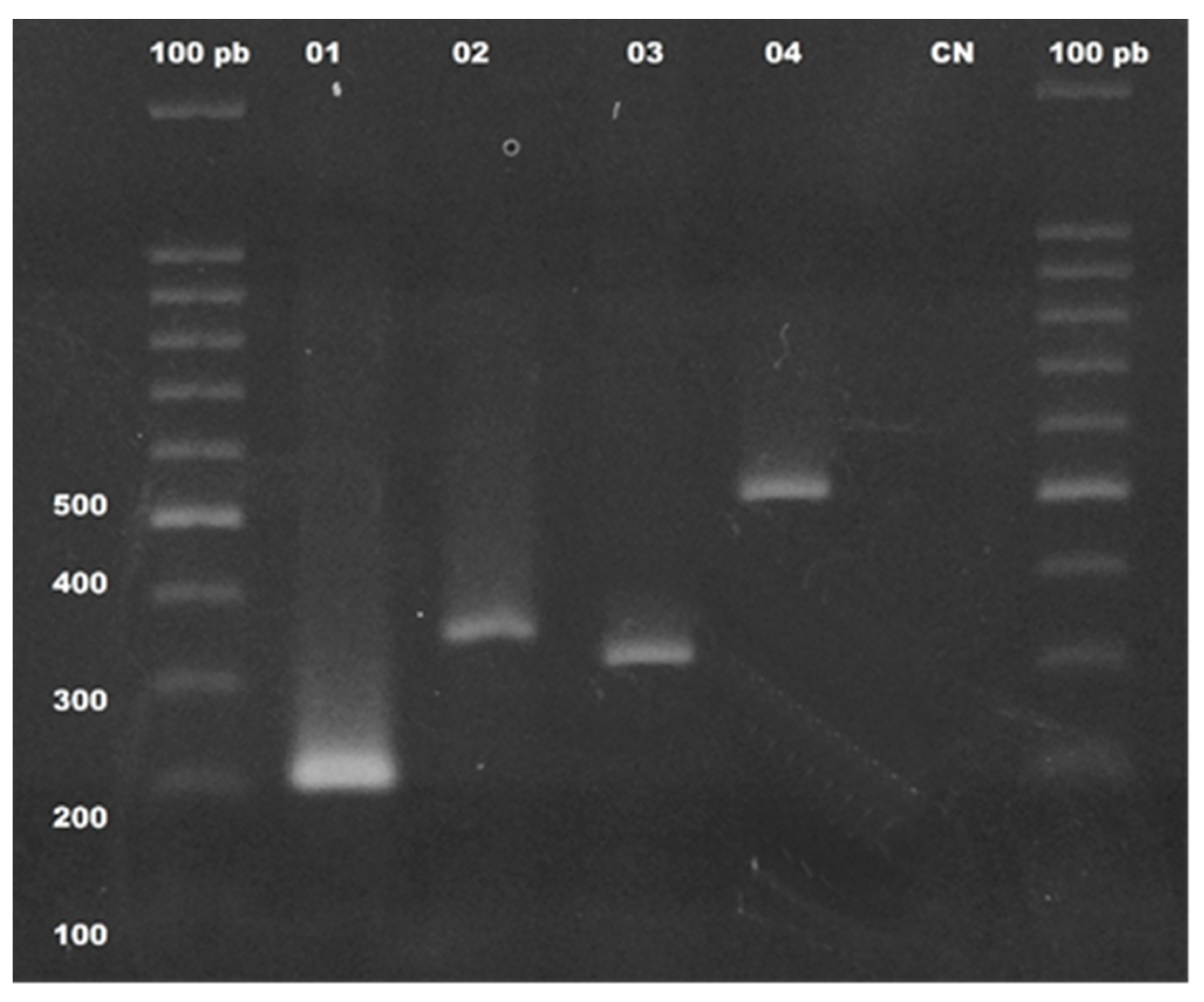
The detection of genes that confer resistance to oxacillin (blaOXA-23, blaOXA-24, blaOXA-51, and blaOXA-58), polymyxin E (mcr-1), and class C of cephalosporinase (AmpC-β-lactamases) were also investigated in all 99 strains of P. aeruginosa (illustrated in Figure 2). A high frequency of ampC (97.9%) was observed in this study, and the blaOXA51 gene was found in three isolates. It is interesting to note that one of these isolates (SLZ183) also presented the specific fragment for the blaOXA23 gene. The mcr-1 gene, encoding the colistin resistance protein (polymyxin E), was detected in one of the isolates (24LCM) recovered from a urine sample. This isolate showed resistance to at least one antibiotic of each tested class, except for carbapenems, imipenem, and meropenem, to which it was sensitive.

3.4. Sequencing Analysis of blaOXA-23, blaOXA-51, and mcr-1 Genes and the 16S Ribosomal Region

Due to the novelty of data regarding the occurrence of blaOXA-23, blaOXA-51, and mcr-1 in P. aeruginosa, the PCR products of the 16S ribosomal region for four isolates positive for these genes were sequenced. The phenograms obtained for each isolate were aligned by the Mega v.6.0 program using sequences of the same genes present in Genbank for the elimination of ambiguities. The consensus sequences were submitted to the Blastn program of the BLAST 2.0 package (Basic Alignment Search Tool—http://www.ncbi.nlm.nih.gov/BLAST (accessed on 23 May 2020)). It obtained nucleotide similarity indexes ranging from 96.38 to 100% for the 16 S ribosomal region and from 99.13 to 100% for the mcr-1, blaOXA-23, and blaOXA-51 genes. All these sequences were deposited at Genbank and are available for access. For sequences from the 16 S ribosomal region, the access numbers are MT876550 to MT876553, while for the other genes, the access numbers are MT880235 to MT880239.

3.5. Clonal Profile of Pseudomonas aeruginosa Isolates by ERIC-PCR

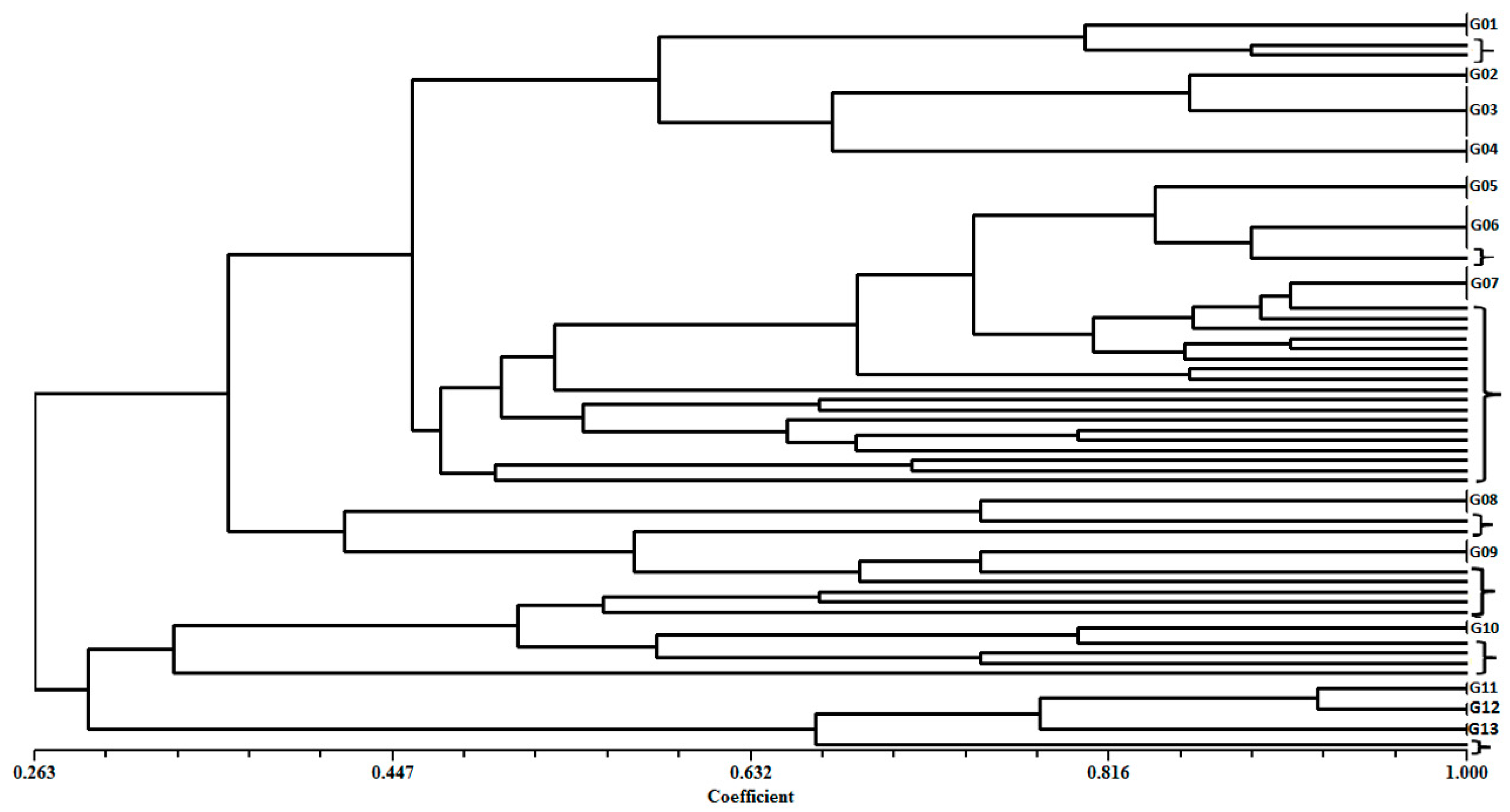
The electrophoretic analysis of the molecular profiles of the fragments generated by ERIC-PCR and submitted at multivariate analysis by NTSYS showed an average number of three to seven fragments in the range of 100 to 3000 bp. ERIC-PCR analysis allowed for grouping of most of the isolates (66.7%) into thirteen distinct clusters (G01 to G13). In contrast, the other 33 isolates exhibited unique genotypic profiles and they were identified in the dendrogram using square brackets (Table 7 and Figure 3).

4. Discussion

Infections caused by P. aeruginosa are difficult to treat since this bacterium has several mechanisms of resistance and virulence, such as the acquisition of plasmids or integrons. This results in acquisition genes related to (i) production of enzymes that inactivate or modify the drug targets, (ii) overexpression of efflux pumps, (iii) production of toxic proteins, (iv) biofilm formation, and (v) decreased expression of pore-forming proteins [31]. This panorama denotes the relevance of epidemiological studies employing molecular and phenotypic approaches to characterize the pathogenic profile of the strains in circulation in the hospitals. In this study, the presence of genes related to virulence and antimicrobial resistance was investigated in 99 strains obtained from patients of different hospitals located in São Luís (Maranhão, Brazil).
Overall, the strains exhibited high levels of antimicrobial resistance for the drugs used for the treatment of Gram-negative infections. It was possible to observe one strain with resistance to colistin, while no resistance was detected towards polymyxin B. These results are important since polymyxins are the last therapeutic options for the treatment of multi-resistant Gram-negative bacterial infections [32,33].
In fact, dissemination of the plasmid that confers resistance to colistin (mcr-1) is a recent phenomenon in the literature [34]. Some genetic and structural modifications have been related to the occurrence of colistin resistance in P. aeruginosa such as changes in Lipid A; in mutations on cupC1, phoQ, oprQ, rlpA, hpaC, lpxC, and arnB, and in other genes related to transport and energy production [34,35].
Several drugs were shown to be ineffective against most of the evaluated strains (piperacillin/tazobactam, ciprofloxacin, ceftazidime, and cefepime), besides those for which P. aeruginosa has intrinsic resistance. This resistance may be related to the expression of several drug efflux pumps, the low permeability of its outer membrane and the natural occurrence of chromosomal β-lactamase or ampC [3,36].
The multiple resistance profiles observed for these isolates are in concordance with the literature data. For example, several studies have shown high rates of resistance towards the following classes of antimicrobials: cephalosporins (ceftazidime 93% and cefepime 89%) [37]; to carbapenems (Imipenem 41%, meropenem 88%) [38]; aminoglycosides (Amikacin and gentamycin, 91% and 98%, respectively); fluoroquinolone (ciprofloxacin, 98%) [39].
Similarly, the values of detection of P. aeruginosa with MDR profiles have varied among studies from different countries: in Malaysia, the value was found to be 19.6% [40]; while studies performed in Nepal [41] and Iran [42] detected rates of 89.4% and 100%, respectively. It is well-known that large-scale use of different classes of antimicrobial agents such as Monobactams, Penicillins, synthetic derivatives of penicillin, aminoglycosides, cephalosporins, fluoroquinolones, carbapenems, and polymyxins in the treatment of bacterial infections has led to the emergence of MDR, XDR, and even pan-drug-resistant (PDR) strains [43,44].
Given the high level of drug resistance in the strains evaluated in this study, the occurrence of genes related to antimicrobial resistance was investigated. It was observed that almost all strains carried the ampC gene (97.9%), related to cephalosporins resistance. This alarming incidence of ampC (or its overexpression) in clinical isolates of P. aeruginosa was also reported by other authors [45,46]. Studies have shown that the rates of molecular detection of ampC has varied among clinical isolates of P. aeruginosa. These differences between detection rates may be related to mutations affecting the AmpC gene or specificity of the different primers or methodologies that have been used for their detection in P. aeruginosa [47,48,49]. On the other hand, three isolates carrying the gene blaOXA51 were detected, and in one of these isolates, the blaOXA23 gene was also detected. The mcr-1 gene encoding the colistin resistance protein (polymyxin E) was detected in one of the isolates evaluated in this study.
The presence of the blaOXA23 and blaOXA51 genes in P. aeruginosa isolates is an unprecedented finding for the literature. These genes encoding Ambler class D oxacillinase (OXA)-like carbapenemase are often detected in Acinetobacter baumannii and are related to resistance to oxacilicins, cephalosporins, and carbapenems [50,51,52]. Similarly, since its discovery in Escherichia coli [34], the mcr-1 gene has been described in other microorganisms, including K. pneumoniae [53], Enterobacter aerogenes [54] and Enterobacter cloacae [55]. In Italy, several bacterial species were detected in hospital surfaces with colistin resistance driven by mcr-1 gene, including in P. aeruginosa and P. putida [56].
The detection of several genes that confer virulence to P. aeruginosa has also been investigated in this study. We observed that the most prevalent genes were oprI and oprL, which encode outer membrane lipoprotein I (maintenance of cell membrane integrity) and peptidoglycan-associated lipoprotein OprL (maintains the shape of the bacterium), respectively [57]. These results agree with the findings observed for P. aeruginosa isolates recovered from burn, pulmonary tract and blood infections [58].
In relation to the high detection rate found for the lasB gene, similar results were observed by other researchers [59,60]. Several proteases have been considered an important virulence factor of P. aeruginosa. The elastase that is encoded by the lasB gene is a metalloprotease that degrades elastin and tissue collagen, breaks down fibrin, and destroys other structural proteins. In this sense, this protein has been implicated in the process of invasion and permanence of that bacterium in tissues [61].
High rates of strains carrying the genes exoY, exoT, and exoS were observed in the strains used in this study. The results obtained for exoY (ExoY exotoxin) were higher than those described in recent literature, which report detection of this gene ranging from 14.5% to 72.5% [33,62,63]. Regarding the rate of 82.8% that was observed for exoT, this result was higher than those obtained by Fazeli and Momtz (2014) (36.27%) [1] and Mahdavi et al. (2017) (76.32%) [64]. On the other hand, the result of 45.4% found for the exoS gene was higher than observed Mahdavi et al. (2017) [64] that found it in 30.9%. However, higher rates of exoS were obtained by Fazeli and Momtz (2014) [1] (67.6%) and Yousefi-Avarvand et al. (2015) [62] (65.4%).
Another important virulence factor found in most of the isolates (80.8%) was the toxA gene, which encodes the P. aeruginosa exotoxin A. This protein is a cytotoxic agent that, similar to diphtheria toxin, inhibits protein biosynthesis, causing extensive damage to tissues and organs [65]. The result found for the toxA gene is in agreement with the findings obtained by Yousefi-Avarvand et al. (2015) [62]. However, Fazeli and Momtz (2014) [1] found a rate of 35.29% in P. aeruginosa strains recovered from burn infections.
Other virulence genes with high detection rates were algD (75.7%), plcN (75.7%), and phzI (70.7%). The prevalence observed for algD was superior to that detected by Benie et al. (2017) [7] in P. aeruginosa samples isolated from beef and fish. The product of algD (GDP-mannose 6-dehydrogenase) is involved in the synthesis of alginate, a polysaccharide that is the main extracellular homopolymer substance and the main component of P. aeruginosa biofilm [11,66]. Alginate synthesis is often associated with chronic lung infections in patients with cystic fibrosis [67]. It is interesting to note that, although the algD gene is considered intrinsic to Pseudomonas aeruginosa, this gene was detected only in 75.7% of the isolates. However, other studies have also shown variation in the detection rate of the algD gene, which probably may be related to the specificity of the primers used and or the origin of the bacterial isolate [68,69,70,71].
The presence of the gene that codifies nonhemolytic (plcN) and hemolytic (plcH) phospholipases C in the clinical isolates evaluated here was verified in 75.7% and 51.5%, respectively. It is interesting to note that Benie et al. (2017) [7] found a rate of 72.1% for plcH in P. aeruginosa isolated from animals. On the other hand, in the isolates of the present study, the prevalence of plcN was higher than that observed for plcH. These two proteins act on the breakdown of phospholipids, and they contribute to bacterial invasion through their cytotoxic effects on neutrophils, playing an important role in the establishment of bacterial infection [1,72].
The phzI gene belonging to the phenazine operon was detected in 70.7% of P. aeruginosa isolates. This result was lower than the 90% rate found by Ertugrul et al. (2017) [73]. The product encoded by the phzI gene acts with other virulence factors (phzH, phzM, and phzS) to destroy host tissue components [74]. The pilB gene, which encoded fimbrial-type IV protein, was found in 27 (27.3%) of the isolates. This result was lower than that found in the literature. Relatively varied rates have been found for this gene, such as 35%[75], 73.2% [59] and 75.0% [70], respectively.
The presence of the gene encoding an alkaline metalloprotease (apr) was detected in 22.2% of the isolates. This result was superior to that found by Fazeli and Momtz (2014) [74], who detected this gene in 12.7% of its isolates. In contrast, Nahar et al. (2017) [76] in an in silico study analyzing 18 Genbank sequences found the prevalence of apr in 66.6%. Similarly, Prot IV was detected in 20.2% of the samples. This gene encodes an alkaline protease that acts directly on the cytoplasm of the host cell, being responsible for the destruction of structural proteins and helping to establish infection [33,36].
The results also showed a low prevalence for the nan-1 gene that codifies a neuraminidase. However, this value was superior to that found by Benie et al. (2017) [7], who did not obtain any positive isolates for this gene. However, Habibi and Honarmand (2015) [70] and Heidary et al. (2016) [59] detected rates of 12.5% and 36.61%, respectively.
The aprA gene was detected in 6.0% of the isolates analyzed, which is a rate below the values founds in other studies such as those performed by Mitov, Strateva, and Markova (2010) [68] (91%) and Badamchi et al. (2017) [6] (81.2%). On the other hand, Ertugrul et al. (2017) [73] did not detect the presence of this gene in any tested strains. The product of aprA is an elastase that target lymphocytes, neutrophils, and other cells [74].
Analysis of the ERIC-PCR profiles allows for the study of genetic polymorphism among bacterial isolates, determining different ones and grouping similar profiles. In the present study, the phenotypic profile of 99 isolates identified genetically as being of the species P. aeruginosa was characterized as heterogeneous, showing 13 different clusters. Thus, a wide genetic diversity among the isolates was observed, although they belonged to the same species, denoting that different P. aeruginosa clones circulated in the hospitals from São Luis-MA. Since the ERIC-PCR methodology is fast, sensitive, and reproducible, it has been widely used for the molecular epidemiological analysis of different bacterial species [77,78].

5. Conclusions

In the present study, a combination of molecular and phenotypic assays revealed a high level of antimicrobial resistance in 99 P. aeruginosa isolates, including a high frequency of strains with MDR and XDR profiles. In addition, the occurrence of blaOXA-23, blaOXA-51, and mcr-1 genes in P. aeruginosa isolates was detected. Furthermore, these strains carry multiple genes related to important virulence pathways in P. aeruginosa, including those related to the impermeability of the outer membrane, tissue damage, exotoxin release, and biofilm formation. These strains also exhibited a wide genetic diversity, denoting that different clones are present in the studied area. Taken together, these data reveal the need for constant investigation of the occurrence of strains with multidrug resistance and with high pathogenic potential in order to guide the implantation of effective approaches to reduce their spread and to then improve the quality of health care facilities.

Author Contributions

F.N., B.O.d.M., S.G.M., A.D.S.J., and R.d.J.G.T. conducted the literature research and contributed to writing of the manuscript. T.A.F.F., P.C.R.C., H.J.C.M., A.Z., and A.d.S.M. performed statistical analysis and formatting and helped in writing the paper. L.C.N.d.S., V.M.-N., and M.R.Q.B. acquired, analyzed, and interpreted of data for this work and provided editorial input and revision for the final version. All authors have read and agreed to the published version of the manuscript.

Funding

This research did not receive any specific grant from funding agencies in the public, commercial, or not-for-profit sectors.

Institutional Review Board Statement

The study was conducted according to the guidelines of the Declaration of Helsinki, and approved by the Research Ethics Committee of the University of Ceuma (No. 813.293/2014).

Informed Consent Statement

Patient consent was waived because deidentified data were used.

Data Availability Statement

The data presented in this study are available on request from the corresponding author.

Acknowledgments

We thank the laboratories participating in the research, and Laboratory Cedro (clinical analysis sector) and Laboratory of Clinical Analysis of Maranhão LTDA (LACMAR) for technical assistance.

Conflicts of Interest

The authors declare to have no conflict of interest.

References

  1. Fazeli, N.; Momtaz, H. Virulence Gene Profiles of Multidrug-Resistant Pseudomonas aeruginosa Isolated from Iranian Hospital Infections. Iran. Red Crescent Med. J. 2014, 16, e15722. [Google Scholar] [CrossRef] [Green Version]
  2. Gomila, M.; Pena, A.; Mulet, M.; Lalucat, J.; Garcia-Valdes, E. Phylogenomics and systematics in Pseudomona. Front. Microbiol. 2015, 6, 214. [Google Scholar] [CrossRef] [Green Version]
  3. Fujitani, S.; Moffett, K.S.; Yu, V. Pseudomonas aeruginosa. Antimicrobe: Infectious Disease & Antimicrobial Agents. Available online: http://www.antimicrobe.org/new/b112 (accessed on 1 March 2017).
  4. Vale de Macedo, G.H.R.; Costa, G.D.E.; Oliveira, E.R.; Damasceno, G.V.; Mendonca, J.S.P.; Silva, L.D.S.; Chagas, V.L.; Bazan, J.M.N.; Alianca, A.; Miranda, R.C.M.; et al. Interplay between ESKAPE Pathogens and Immunity in Skin Infections: An Overview of the Major Determinants of Virulence and Antibiotic Resistance. Pathogens 2021, 10, 148. [Google Scholar] [CrossRef]
  5. Gellatly, S.L.; Hancock, R.E. Pseudomonas aeruginosa: New insights into pathogenesis and host defenses. Pathog. Dis. 2013, 67, 159–173. [Google Scholar] [CrossRef] [Green Version]
  6. Badamchi, A.; Masoumi, H.; Javadinia, S.; Asgarian, R.; Tabatabaee, A. Molecular detection of six virulence genes in Pseudomonas aeruginosa isolates detected in children with urinary tract infection. Microb. Pathog. 2017, 107, 44–47. [Google Scholar] [CrossRef]
  7. Benie, C.K.; Dadie, A.; Guessennd, N.; N’Gbesso-Kouadio, N.A.; Kouame, N.D.; N’Golo, D.C.; Aka, S.; Dako, E.; Dje, K.M.; Dosso, M. Characterization of Virulence Potential of Pseudomonas aeruginosa Isolated from Bovine Meat, Fresh Fish, and Smoked Fish. Eur. J. Microbiol. Immunol. 2017, 7, 55–64. [Google Scholar] [CrossRef] [Green Version]
  8. Schiavano, G.F.; Carloni, E.; Andreoni, F.; Magi, S.; Chironna, M.; Brandi, G.; Amagliani, G. Prevalence and antibiotic resistance of Pseudomonas aeruginosa in water samples in central Italy and molecular characterization of oprD in imipenem resistant isolates. PLOS ONE 2017, 12, e0189172. [Google Scholar] [CrossRef] [PubMed] [Green Version]
  9. Ruffin, M.; Brochiero, E. Repair Process Impairment by Pseudomonas aeruginosa in Epithelial Tissues: Major Features and Potential Therapeutic Avenues. Front Cell Infect. Microbiol. 2019, 9, 182. [Google Scholar] [CrossRef] [PubMed] [Green Version]
  10. Kerr, K.G.; Snelling, A.M. Pseudomonas aeruginosa: A formidable and ever-present adversary. J. Hosp. Infect. 2009, 73, 338–344. [Google Scholar] [CrossRef]
  11. Mittal, R.; Aggarwal, S.; Sharma, S.; Chhibber, S.; Harjai, K. Urinary tract infections caused by Pseudomonas aeruginosa: A minireview. J. Infect. Public Health 2009, 2, 101–111. [Google Scholar] [CrossRef] [PubMed] [Green Version]
  12. Moore, N.M.; Flaws, M.L. Antimicrobial resistance mechanisms in Pseudomonas aeruginosa. Am. Soc. Clin. Lab. Sci. 2011, 24, 47–51. [Google Scholar] [CrossRef]
  13. Magiorakos, A.-P.; Srinivasan, A.; Carey, R.; Carmeli, Y.; Falagas, M.; Giske, C.; Harbarth, S.; Hindler, J.; Kahlmeter, G.; Olsson-Liljequist, B. Multidrug-resistant, extensively drug-resistant and pandrug-resistant bacteria: An international expert proposal for interim standard definitions for acquired resistance. Clin. Microbiol. Infect. 2012, 18, 268–281. [Google Scholar] [CrossRef] [PubMed] [Green Version]
  14. Loureiro, R.J.; Roque, F.; Rodrigues, A.T.; Herdeiro, M.T.; Ramalheira, E. O uso de antibióticos e as resistências bacterianas: Breves notas sobre a sua evolução. Rev. Port. Saúde Pública 2016, 34, 77–84. [Google Scholar] [CrossRef] [Green Version]
  15. Nóbrega, M.d.S.; do Carmo Filho, J.R.; Pereira, M.S. Drug resistance evolution of Pseudomonas aeruginosa and Acinetobacter baumannii in intensive care units. Rev. Eletrônica Enferm. 2013, 15, 696–703. [Google Scholar]
  16. Kritsotakis, E.I.; Kontopidou, F.; Astrinaki, E.; Roumbelaki, M.; Ioannidou, E.; Gikas, A. Prevalence, incidence burden, and clinical impact of healthcare-associated infections and antimicrobial resistance: A national prevalent cohort study in acute care hospitals in Greece. Infect. Drug Resist. 2017, 10, 317–328. [Google Scholar] [CrossRef] [Green Version]
  17. Matos, E.C.O.d.; Matos, H.J.d.; Conceição, M.L.; Rodrigues, Y.C.; Carneiro, I.C.d.R.S.; Lima, K.V.B. Clinical and microbiological features of infections caused by Pseudomonas aeruginosa in patients hospitalized in intensive care units. Rev. Soc. Bras. Med. Trop. 2016, 49, 305–311. [Google Scholar] [CrossRef] [PubMed]
  18. Clinical and Laboratory Standards Institute. Performance Standards for Antimicrobial Susceptibility Testing: 20th Informational Supplement. CLSI Document M100-S20; Clinical and Laboratory Standards Institute: Wayne, PA, USA, 2010. [Google Scholar]
  19. Lanotte, P.; Watt, S.; Mereghetti, L.; Dartiguelongue, N.; Rastegar-Lari, A.; Goudeau, A.; Quentin, R. Genetic features of Pseudomonas aeruginosa isolates from cystic fibrosis patients compared with those of isolates from other origins. J. Med. Microbiol. 2004, 53, 73–81. [Google Scholar] [CrossRef] [PubMed]
  20. Wolfgang, M.C.; Kulasekara, B.R.; Liang, X.; Boyd, D.; Wu, K.; Yang, Q.; Miyada, C.G.; Lory, S. Conservation of genome content and virulence determinants among clinical and environmental isolates of Pseudomonas aeruginosa. Proc. Natl. Acad. Sci. USA 2003, 100, 8484–8489. [Google Scholar] [CrossRef] [Green Version]
  21. De Vos, D.; Lim, A., Jr.; Pirnay, J.P.; Struelens, M.; Vandenvelde, C.; Duinslaeger, L.; Vanderkelen, A.; Cornelis, P. Direct detection and identification of Pseudomonas aeruginosa in clinical samples such as skin biopsy specimens and expectorations by multiplex PCR based on two outer membrane lipoprotein genes, oprI and oprL. J. Clin. Microbiol. 1997, 35, 1295–1299. [Google Scholar] [CrossRef] [Green Version]
  22. Wozniak, D.J.; Ohman, D. Transcriptional analysis of the Pseudomonas aeruginosa genes algR, algB, and algD reveals a hierarchy of alginate gene expression which is modulated by algT. J. Bacteriol. 1994, 176, 6007–6014. [Google Scholar] [CrossRef] [PubMed] [Green Version]
  23. Sabharwal, N.; Dhall, S.; Chhibber, S.; Harjai, K. Molecular detection of virulence genes as markers in Pseudomonas aeruginosa isolated from urinary tract infections. Int. J. Mol. Epidemiol. Genet. 2014, 5, 125–134. [Google Scholar]
  24. Khan, A.A.; Cerniglia, C.E. Detection of Pseudomonas aeruginosa from clinical and environmental samples by amplification of the exotoxin A gene using PCR. Appl. Environ. Microbiol. 1994, 60, 3739–3745. [Google Scholar] [CrossRef] [Green Version]
  25. Smith, L.; Rose, B.; Tingpej, P.; Zhu, H.; Conibear, T.; Manos, J.; Bye, P.; Elkins, M.; Willcox, M.; Bell, S.; et al. Protease IV production in Pseudomonas aeruginosa from the lungs of adults with cystic fibrosis. J. Med. Microbiol. 2006, 55, 1641–1644. [Google Scholar] [CrossRef] [PubMed] [Green Version]
  26. Paulyn, A.T.; Umeh, E.; Nna, E. Detection of virulence genes in urinary Pseudomonas aeruginosa from pregnant women attending Antenatal clinic in Makurdi, Central Nigeria. Sci. Res. J. 2016, 4, 9. [Google Scholar]
  27. Woodford, N.; Ellington, M.J.; Coelho, J.M.; Turton, J.F.; Ward, M.E.; Brown, S.; Amyes, S.G.; Livermore, D.M. Multiplex PCR for genes encoding prevalent OXA carbapenemases in Acinetobacter spp. Int. J. Antimicrob. Agents 2006, 27, 351–353. [Google Scholar] [CrossRef] [PubMed]
  28. Monstein, H.J.; Ostholm-Balkhed, A.; Nilsson, M.V.; Nilsson, M.; Dornbusch, K.; Nilsson, L.E. Multiplex PCR amplification assay for the detection of blaSHV, blaTEM and blaCTX-M genes in Enterobacteriaceae. APMIS 2007, 115, 1400–1408. [Google Scholar] [CrossRef]
  29. Devereux, R.; Willis, S.G. Amplification of ribosomal RNA sequences. In Molecular Microbial Ecology Manual; Akkermans, A.D.L., Van Elsas, J.D., De Bruijn, F.J., Eds.; Springer: Berlin, Germany, 1995; pp. 277–287. [Google Scholar]
  30. Versalovic, J.; Koeuth, T.; Lupski, R. Distribution of repetitive DNA sequences in eubacteria and application to finerpriting of bacterial enomes. Nucleic Acids Res. 1991, 19, 6823–6831. [Google Scholar] [CrossRef]
  31. Mesaros, N.; Nordmann, P.; Plésiat, P.; Roussel-Delvallez, M.; Van Eldere, J.; Glupczynski, Y.; Van Laethem, Y.; Jacobs, F.; Lebecque, P.; Malfroot, A.; et al. Pseudomonas aeruginosa: Resistance and therapeutic options at the turn of the new millennium. Clin. Microbiol. Infect 2007, 13, 560–578. [Google Scholar] [CrossRef] [PubMed] [Green Version]
  32. Haidar, G.; Philips, N.J.; Shields, R.K.; Snyder, D.; Cheng, S.; Potoski, B.A.; Hao, B.; Press, E.G.; Cooper, V.S.; Clancy, C.J. Ceftolozane-tazobactam for the treatment of multidrug-resistant Pseudomonas aeruginosa infections: Clinical effectiveness and evolution of resistance. Clin. Infect. Dis. 2017, 65, 110–120. [Google Scholar] [CrossRef] [PubMed]
  33. Azimi, A.; Peymani, A.; Pour, P.K. Phenotypic and molecular detection of metallo-β-lactamase-producing Pseudomonas aeruginosa isolates from patients with burns in Tehran, Iran. Rev. Soc. Bras. Med. Trop. 2018, 51, 610–615. [Google Scholar] [CrossRef]
  34. Liu, Y.-Y.; Wang, Y.; Walsh, T.R.; Yi, L.-X.; Zhang, R.; Spencer, J.; Doi, Y.; Tian, G.; Dong, B.; Huang, X. Emergence of plasmid-mediated colistin resistance mechanism MCR-1 in animals and human beings in China: A microbiological and molecular biological study. Lancet Infect. Dis. 2016, 16, 161–168. [Google Scholar] [CrossRef]
  35. Chung, E.S.; Lee, J.Y.; Rhee, J.Y.; Ko, K.S. Colistin resistance in Pseudomonas aeruginosa that is not linked to arnB. J. Med. Microbiol. 2017, 66, 833–841. [Google Scholar] [CrossRef] [PubMed]
  36. Pachori, P.; Gothalwal, R.; Gandhi, P. Emergence of antibiotic resistance Pseudomonas aeruginosa in intensive care unit; a critical review. Genes Dis. 2019, 6, 109–119. [Google Scholar] [CrossRef] [PubMed]
  37. Cabot, G.; Ocampo-Sosa, A.A.; Domínguez, M.A.; Gago, J.F.; Juan, C.; Tubau, F.; Rodríguez, C.; Moyà, B.; Peña, C.; Martínez-Martínez, L. Genetic markers of widespread extensively drug-resistant Pseudomonas aeruginosa high-risk clones. Antimicrob. Agents Chemother. 2012, 56, 6349–6357. [Google Scholar] [CrossRef] [PubMed] [Green Version]
  38. Paluchowska, P.; Skałkowska, M.; Spelak, A.; Budak, A. Occurrence of alert pathogens in hospital environment. Part II. Multidrug-resistant non-fermenting bacilli. Med. Dośw. I Mikrobiol. 2012, 64, 45–53. [Google Scholar]
  39. Michalska, A.D.; Sacha, P.T.; Ojdana, D.; Wieczorek, A.; Tryniszewska, E. Prevalence of resistance to aminoglycosides and fluoroquinolones among Pseudomonas aeruginosa strains in a University Hospital in Northeastern Poland. Braz. J. Microbiol. 2014, 45, 1455–1458. [Google Scholar] [CrossRef] [PubMed] [Green Version]
  40. Pathmanathan, S.G.; Samat, N.A.; Mohamed, R. Antimicrobial susceptibility of clinical isolates of Pseudomonas aeruginosa from a Malaysian Hospital. Malays. J. Med. Sci. 2009, 16, 27–32. [Google Scholar] [PubMed]
  41. Bhandari, S.; Banjara, M.R.; Lekhak, B.; Bhatta, D.R.; Regmi, S.R. Multi-drug and pan-drug resistant Pseudomonas aeruginosa: A challenge in post-antibiotic era. Nepal J. Sci. Technol. 2013, 13, 197–202. [Google Scholar] [CrossRef] [Green Version]
  42. Moazami, G.S.; Eftekhar, F. Assessment of carbapenem susceptibility and multidrug-resistance in Pseudomonas aeruginosa burn isolates in Tehran. Jundishapur J. Microbiol. 2013, 6, 162–165. [Google Scholar]
  43. Saderi, H.; Owlia, P. Detection of multidrug resistant (MDR) and extremely drug resistant (XDR) P. aeruginosa isolated from patients in Tehran, Iran. Iran. J. Pathol. 2015, 10, 265–271. [Google Scholar]
  44. Regenbogen, B.; Willmann, M.; Steglich, M.; Bunk, B.; Nübel, U.; Peter, S.; Neher, R.A. Rapid and consistent evolution of colistin resistance in XDR Pseudomonas aeruginosa during morbidostat culture. Antimicrob. Agents Chemother. 2017, 61, e00043-17. [Google Scholar] [CrossRef] [Green Version]
  45. Dantas, R.C.C.; Silva, R.T.E.; Ferreira, M.L.; Goncalves, I.R.; Araujo, B.F.; Campos, P.A.; Royer, S.; Batistao, D.; Gontijo-Filho, P.P.; Ribas, R.M. Molecular epidemiological survey of bacteremia by multidrug resistant Pseudomonas aeruginosa: The relevance of intrinsic resistance mechanisms. PLOS ONE 2017, 12, e0176774. [Google Scholar] [CrossRef] [PubMed]
  46. Atkin, S.D.; Abid, S.; Foster, M.; Bose, M.; Keller, A.; Hollaway, R.; Sader, H.S.; Greenberg, D.E.; Finklea, J.D.; Castanheira, M.; et al. Multidrug-resistant Pseudomonas aeruginosa from sputum of patients with cystic fibrosis demonstrates a high rate of susceptibility to ceftazidime-avibactam. Infect. Drug Resist 2018, 11, 1499–1510. [Google Scholar] [CrossRef] [Green Version]
  47. Rafiee, R.; Eftekhar, F.; Tabatabaei, S.A.; Minaee Tehrani, D. Prevalence of Extended-Spectrum and Metallo beta-Lactamase Production in AmpC beta-Lactamase Producing Pseudomonas aeruginosa Isolates From Burns. Jundishapur J. Microbiol. 2014, 7, e16436. [Google Scholar] [CrossRef] [PubMed] [Green Version]
  48. Berrazeg, M.; Jeannot, K.; Ntsogo Enguene, V.Y.; Broutin, I.; Loeffert, S.; Fournier, D.; Plesiat, P. Mutations in beta-Lactamase AmpC Increase Resistance of Pseudomonas aeruginosa Isolates to Antipseudomonal Cephalosporins. Antimicrob. Agents Chemother. 2015, 59, 6248–6255. [Google Scholar] [CrossRef] [PubMed] [Green Version]
  49. Treepong, P.; Kos, V.N.; Guyeux, C.; Blanc, D.S.; Bertrand, X.; Valot, B.; Hocquet, D. Global emergence of the widespread Pseudomonas aeruginosa ST235 clone. Clin. Microbiol. Infect. 2018, 24, 258–266. [Google Scholar] [CrossRef] [PubMed] [Green Version]
  50. Xiao, S.Z.; Chu, H.Q.; Han, L.Z.; Zhang, Z.M.; Li, B.; Zhao, L.; Xu, L. Resistant mechanisms and molecular epidemiology of imipenem-resistant Acinetobacter baumannii. Mol. Med. Rep. 2016, 14, 2483–2488. [Google Scholar] [CrossRef] [PubMed] [Green Version]
  51. Uwingabiye, J.; Lemnouer, A.; Roca, I.; Alouane, T.; Frikh, M.; Belefquih, B.; Bssaibis, F.; Maleb, A.; Benlahlou, Y.; Kassouati, J.; et al. Clonal diversity and detection of carbapenem resistance encoding genes among multidrug-resistant Acinetobacter baumannii isolates recovered from patients and environment in two intensive care units in a Moroccan hospital. Antimicrob. Resist. Infect. Control. 2017, 6, 99. [Google Scholar] [CrossRef] [Green Version]
  52. Castilho, S.R.A.; Godoy, C.S.M.; Guilarde, A.O.; Cardoso, J.L.; Andre, M.C.P.; Junqueira-Kipnis, A.P.; Kipnis, A. Acinetobacter baumannii strains isolated from patients in intensive care units in Goiania, Brazil: Molecular and drug susceptibility profiles. PLOS ONE 2017, 12, e0176790. [Google Scholar] [CrossRef]
  53. Teo, J.W.; Chew, K.L.; Lin, R.T. Transmissible colistin resistance encoded by mcr-1 detected in clinical Enterobacteriaceae isolates in Singapore. Emerg. Microbes Infect. 2016, 5, e87. [Google Scholar] [CrossRef] [PubMed]
  54. Zeng, K.J.; Doi, Y.; Patil, S.; Huang, X.; Tian, G.B. Emergence of the Plasmid-Mediated mcr-1 Gene in Colistin-Resistant Enterobacter aerogenes and Enterobacter cloacae. Antimicrob Agents Chemother 2016, 60, 3862–3863. [Google Scholar] [CrossRef] [PubMed] [Green Version]
  55. Baron, S.; Bardet, L.; Dubourg, G.; Fichaux, M.; Rolain, J.M. mcr-1 plasmid-mediated colistin resistance gene detection in an Enterobacter cloacae clinical isolate in France. J. Glob. Antimicrob. Resist. 2017, 10, 35–36. [Google Scholar] [CrossRef] [PubMed]
  56. Caselli, E.; D’Accolti, M.; Soffritti, I.; Piffanelli, M.; Mazzacane, S. Spread of mcr-1-Driven Colistin Resistance on Hospital Surfaces, Italy. Emerg Infect Dis 2018, 24, 1752–1753. [Google Scholar] [CrossRef] [PubMed] [Green Version]
  57. Lin, Y.M.; Wu, S.J.; Chang, T.W.; Wang, C.F.; Suen, C.S.; Hwang, M.J.; Chang, M.D.; Chen, Y.T.; Liao, Y.D. Outer membrane protein I of Pseudomonas aeruginosa is a target of cationic antimicrobial peptide/protein. J. Biol. Chem. 2010, 285, 8985–8994. [Google Scholar] [CrossRef] [PubMed] [Green Version]
  58. Khattab, M.; Nour, M.; ElSheshtawy, N. Genetic identification of Pseudomonas aeruginosa virulence genes among different isolates. J. Microb. Biochem. Technol. 2015, 7, 274–277. [Google Scholar]
  59. Heidary, Z.; Bandani, E.; Eftekhary, M.; Jafari, A.A. Virulence genes profile of multidrug resistant Pseudomonas aeruginosa isolated from Iranian children with UTIs. Acta Med. Iran. 2016, 54, 201–210. [Google Scholar] [PubMed]
  60. Al-Dahmoshi, H.O.; Al-Khafaji, N.S.; Jeyad, A.A.; Shareef, H.K.; Al-Jebori, R.F. Molecular detection of some virulence traits among Pseudomonas aeruginosa isolates, Hilla-Iraq. Biomed. Pharmacol. J. 2018, 11, 835–842. [Google Scholar] [CrossRef]
  61. Galdino, A.C.M.; Branquinha, M.H.; Santos, A.L.; Viganor, L. Pseudomonas aeruginosa and its arsenal of proteases: Weapons to battle the host. In Pathophysiological Aspects of Proteases; Chakraborti, S., Dhalla, N., Eds.; Springer: Singapore, 2017; pp. 381–397. [Google Scholar]
  62. Yousefi-Avarvand, A.; Khashei, R.; Ebrahim-Saraie, H.S.; Emami, A.; Zomorodian, K.; Motamedifar, M. The frequency of exotoxin A and exoenzymes S and U genes among clinical isolates of Pseudomonas aeruginosa in Shiraz, Iran. Int. J. Mol. Cell. Med. 2015, 4, 167–173. [Google Scholar]
  63. Adwan, G. Detection of Type III secretion toxins encoding-genes of Pseudomonas aeruginosa isolates in the West Bank-Palestine. J. Adv. Biol. Biotechnol. 2017, 11, 1–10. [Google Scholar] [CrossRef] [Green Version]
  64. Mahdavi, M.; Zahraei Salehi, T.; Amini, K.; Mobasseri, P. Frequency of exoY, exoS, exoT and exoU genes among Pseudomonas aeruginosa Isolated from patients in Tehran hospitals by Multiplex PCR. Iran. J. Med. Microbiol. 2017, 11, 9–17. [Google Scholar]
  65. Dong, D.; Zou, D.; Liu, H.; Yang, Z.; Huang, S.; Liu, N.; He, X.; Liu, W.; Huang, L. Rapid detection of Pseudomonas aeruginosa targeting the toxA gene in intensive care unit patients from Beijing, China. Front. Microbiol. 2015, 6, 1100. [Google Scholar] [CrossRef] [PubMed] [Green Version]
  66. Evans, L.R.; Linker, A. Production and characterization of the slime polysaccharide of Pseudomonas aeruginosa. J. Bacteriol. 1973, 116, 915–924. [Google Scholar] [CrossRef] [PubMed] [Green Version]
  67. May, T.B.; Shinabarger, D.; Maharaj, R.; Kato, J.; Chu, L.; DeVault, J.D.; Roychoudhury, S.; Zielinski, N.A.; Berry, A.; Rothmel, R.K.; et al. Alginate synthesis by Pseudomonas aeruginosa: A key pathogenic factor in chronic pulmonary infections of cystic fibrosis patients. Clin. Microbiol. Rev. 1991, 4, 191–206. [Google Scholar] [CrossRef]
  68. Mitov, I.; Strateva, T.; Markova, B. Prevalence of virulence genes among bulgarian nosocomial and cystic fibrosis isolates of Pseudomonas aeruginosa. Braz. J. Microbiol. 2010, 41, 588–595. [Google Scholar] [CrossRef] [PubMed]
  69. Rashno Taee, S.; Khansarinejad, B.; Abtahi, H.; Najafimosleh, M.; Ghaznavi-Rad, E. Detection of algD, oprL and exoA Genes by New Specific Primers as an Efficient, Rapid and Accurate Procedure for Direct Diagnosis of Pseudomonas aeruginosa Strains in Clinical Samples. Jundishapur J. Microbiol. 2014, 7, e13583. [Google Scholar] [CrossRef] [PubMed] [Green Version]
  70. Habibi, A.; Honarmand, R. Profile of Virulence Factors in the Multi-Drug Resistant Pseudomonas aeruginosa Strains of Human Urinary Tract Infections (UTI). Iran. Red Crescent Med. J. 2015, 17, e26095. [Google Scholar] [CrossRef] [PubMed] [Green Version]
  71. Elmaraghy, N.; Abbadi, S.; Elhadidi, G.; Hashem, A.; Yousef, A. Virulence genes in Pseudomonas aeruginosa strains isolated at Suez Canal University Hospitals with respect to the site of infection and antimicrobial resistance. Int. J. Clin. Microbiol. Biochem. Technol. 2019, 2, 8–19. [Google Scholar]
  72. Cotar, A.I.; Chifiriuc, M.C.; Dinu, S.; Bucur, M.; Iordache, C.; Banu, O.; Dracea, O.; Larion, C.; Lazar, V. Screening of molecular virulence markers in Staphylococcus aureus and Pseudomonas aeruginosa strains isolated from clinical infections. Int. J. Mol. Sci. 2010, 11, 5273–5291. [Google Scholar] [CrossRef] [Green Version]
  73. Ertugrul, B.M.; Oryasin, E.; Lipsky, B.A.; Willke, A.; Bozdogan, B. Virulence genes fliC, toxA and phzS are common among Pseudomonas aeruginosa isolates from diabetic foot infections. Infect. Dis. 2017, 50, 273–279. [Google Scholar] [CrossRef]
  74. Fadhil, L.; Al-Marzoqi, A.H.; Al Taee, Z.M.; Shalan, A.A. Molecular and phenotypic study of virulence genes in a pathogenic strain of Pseudomonas aeruginosa isolated from various clinical origins by PCR: Profiles of genes and toxins. Res. J. Pharm. Biol. Chem. Sci. 2016, 7, 590–598. [Google Scholar]
  75. Ra’oof, W.A.M. Distribution of algD, lasB, pilB and nan1 genes among MDR clinical isolates of Pseudomonas aeruginosa in respect to site of infection. Tikrit Med. J. 2011, 17, 148–160. [Google Scholar]
  76. Nahar, N.; Asad, S.; Ahmed, T.; Setu, N.I.; Kayser, M.S.; Islam, M.S.; Kamrul, M.; Islam, M.M.R.; Al Aman, D.A.; Rashid, R.B. In silico assessment of the genotypic distribution of virulence and antibiotic resistance genes in Pseudomonas aeruginosa. J. Appl. Pharm. Sci. 2017, 7, 55–61. [Google Scholar]
  77. Ahmed, M.U.; Dunn, L.; Ivanova, E.P. Evaluation of current molecular approaches for genotyping of Campylobacter jejuni strains. Foodborne Pathog. Dis. 2012, 9, 375–385. [Google Scholar] [CrossRef] [PubMed] [Green Version]
  78. Ranjbar, R.; Tabatabaee, A.; Behzadi, P.; Kheiri, R. Enterobacterial repetitive intergenic consensus polymerase chain reaction (ERIC-PCR) genotyping of Escherichia coli strains isolated from different animal stool specimens. Iran. J. Pathol. 2017, 12, 25–34. [Google Scholar] [CrossRef] [PubMed]
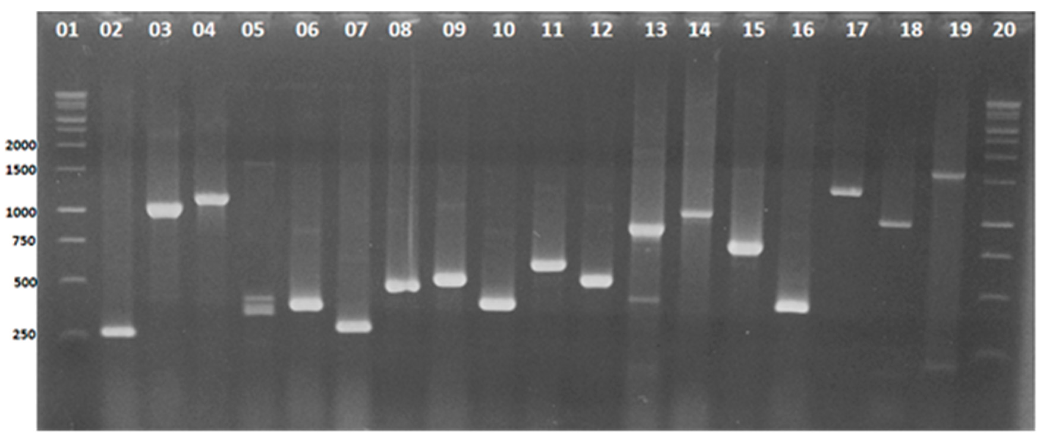
Figure 1. Electrophoretic profiles of the virulence genes investigated in the 99 isolates of Pseudomonas aeruginosa: 1.5% agarose gel stained with ethidium bromide. Lines 1 and 20: 1 Kb DNA ladder (Promega, USA). Lines 2 to 19 profiles of the isolates carrying the genes: oprI (249 bp), exoY (1035 bp), exoT (1.159 bp), lasB (284 bp), toxA (396 bp), algD (300 bp), plcN (481 bp), oprL (504 bp), phzl (392 bp), plcH (608 bp), exoS (504 bp), phzM (875 bp), apr (1.017 bp), Prot IV (752 bp), pilB (408 bp), nan-1 (1.316 bp), aprA (993 bp), and pilA (1.675 bp).
Microorganisms 09 00786 g001
Figure 2. Molecular detection of genes that confer resistance to antimicrobials cephalosporin (ampC), oxacillins, and colistin (polymyxin E) in clinical isolates of Pseudomonas aeruginosa: 1.5% agarose gel, stained with ethidium bromide, showing the amplification profiles obtained for the ampC genes (lines 1–218 bp), mcr-1 gene (lines 2–400 bp), blaOXA-51 gene (lines 3–353 bp), and blaOXA-23 gene (lines 4–501 bp). Molecular marker, 100 bp DNA ladder (Promega, USA) and CN = negative control of the reagents used in the PCR amplification assays.
Microorganisms 09 00786 g002
Figure 3. Dendrogram of genetic similarity derived from the DICE similarity coefficient showing the relationships between Pseudomonas aeruginosa isolates. Construction made using the NTSYS program with the Unweighted Pair Group Method with Arithmetic Mean (UPGMA) algorithm.
Microorganisms 09 00786 g003
Table 1. Primers used for the detection of different virulence genes by mPCR in Pseudomonas aeruginosa isolates evaluated in this study.
Groups (for mPCR)Target Gene Primers Sequence (5′-3′)Amplicon Size (pb)Virulence Fator (Gene Product)References
Group IexoSCTTGAAGGGACTCGACAAGG (F)
TTCAGGTCCGCGTAGTGAAT (R)
504Exotoxin S[19]
exoTCAATCATCTCAGCAGAACCC (F)
TGTCGTAGAGGATCTCCTG
1159Exotoxin T[20]
phzICATCAGCTTAGCAATCCC (F)
CGGAGAAACTTTTCCCTC (R)
392Phenazine
prodution
[20]
phzMATGGAGAGCGGGATCGACAG (F)
ATGCGGGTTTCCATCGGCAG (R)
875Phenazine prodution[20]
aprTGTCCAGCAATTCTCTTGC (F)
CGTTTTCCACGGTGACC (R)
1017Protease[20]
pilAACAGCATCCAACTGAGCG (F)
TTGACTTCCTCCAGGCTG (R)
1675Type IV fimbria[20]
Group IIoprIATGAACAACGTTCTGAAATTCTCTGCT (F)
CTTGCGGCTGGCTTTTTCCAG (R)
248Outer membrane lipoprotein I[21]
oprLATGGAAATGCTGAAATTCGGC (F)
CTTCTTCAGCTCGACGCGACG (R)
504Peptidoglycan-associated lipoprotein[21]
pilBTCGAACTGATGATCGTGG (F)
CTTTCGGAGTGAACATCG (R)
408Fimbrial protein[20]
nanAGGATGAATACTTATTTTGAT (F)
TCACTAAATCCATCTCTGACCCGATA (R)
1316Putative neuraminidase[19]
algDAAGGCGGAAATGCCATCTCC (F)
AGGGAAGTTCCGGGCGTTTG (R)
300GDP-mannose 6-dehydrogenase (alginate production)[22]
plcHGCACGTGGTCATCCTGATGC (F)
TCCGTAGGCGTCGACGTAC (R)
608Hemolytic phospholipase C[23]
exoYTATCGACGGTCATCGTCAGGT (F)
TTGATGCACTCGACCAGCAAG (R)
1035Adenylate cyclase[1]
Group IIItoxAGACAACGCCCTCAGCATCACCAGC (F)
CGCTGGCCCATTCGCTCCAGCGCT (R)
396Exotoxin A[24]
lasBGAATGAACGAAGCGTTCTCCGAC (F)
TGGCGTCGACGAACACCTCG (R)
284Protease[1]
aprAGTCGACCAGGCGGCGGAGCAGATA (F)
GCCGAGGCCGCCGTAGAGGATGTC (R)
993Alkaline protease[23]
Prot IVTATTTCGCCGACTCCCTGTA (F)
GAATAGACGCCGCTGAAATC (R)
752Protease type IV[25]
plcNTCCGTTATCGCAACCAGCCCTACG (F)
TCGCTGTCGAGCAGGTCGAAC (R)
481Non-hemolytic phospholipase C[26]
Table 2. Primers for the detection of different resistance genes in Pseudomonas aeruginosa isolates evaluated in this study.
Groups (for mPCR)Target GenePrimer Sequence (5′-3)Amplicon Size (pb)Gene ProductReference
Group IVblaOxa23GATCGGATTGGAGAACCAGA (F)
ATTTCTGACCGCATTTCCAT (R)
501Oxacilinase[27]
blaOxa24GGTTAGTTGGCCCCCTTAAA (F)
AGTTGAGCGAAAAGGGGATT (R)
246Oxacilinase[27]
blaOxa51TAATGCTTTGATCGGCCTTG (F)
TGGATTGCACTTCATCTTGG (R)
353Oxacilinase[27]
blaOxa58AAGTATTGGGGCTTGTGCTG (F)
CCCCTCTGCGCTCTACATAC (R)
599Oxacilinase[27]
Group VampCCGGCTCGGTGAGCAAGACCTTC (F)
AGTCGCGGATCTGTGCCTGGTC (R)
218Cephalosporinase[28]
mcr-1CAGTATAATTGCCGTAATTATCCCACCGT (F)
GTCTCGGCTTGGTCGGTCTGTAG (R)
400LPS-modifying enzymeThis study
Table 3. Distribution of Pseudomonas aeruginosa isolates according to gender, age group, and number of isolates from the type of clinical specimen.
GenderTissue Fragment (n = 11)/%Blood Culture (n = 40)/%Tracheal Secretion (n = 37)/%Urine (n = 11)/%p Value
Male436.418452156.8872.7ns
Female763.622551643.2327.3ns
Age group (years) p < 0.05
up to 11 0-717.5----
12 to 190-12.525.419.1
20 to 60327.3922.51129.7327.3
over 60 (years)872.72357.52464.9763.6
Caption: ns = no significant. p value < 0.05: statistical significance.
Table 4. Antibiotic resistance profile of Pseudomonas aeruginosa isolates evaluated in this study as determined by the Vitek 2 Compact system.
AntimicrobialMIC (mg/mL)Number of Isolates% Resistance
Cefoxitin≥6499100.0
Ceftazidime≥647070.7
Cefepime≥646161.6
Ampicilin/sulbactam≥32/1699100.0
Piperacilin/tazobactam≥128/47474.7
Aztreonam≥323939.4
Ciprofloxacin≥46666.7
Gentamicin≥165858.6
Amikacin≥644444.4
Imipenem≥165757.6
Meropenem≥165656.6
Colistin ≥811.0
Polymyxin B≤0.500.0
Tigecycline≥1699100.0
Table 5. Antimicrobial resistance profile of Pseudomonas aeruginosa isolates by infection site.
Fragment of TissueBlood CultureTracheal SecretionUrine
No%No%No%No%
Amikacin545.51640.01745.9654.5ns
Gentamycin654.51742.52567.61090.9<0.05
Cefepime1090.91947.52567.6763.6<0.05
Cefotaxime11100.04010037100.011100.0<0.05
Cefoxitin11100.04010037100.011100.0<0.05
Ceftazidime11100.01947.53183.8981.8<0.05
Imipenem1090.9922.53081.1872.7<0.05
Meropenem1090.9922.52978.4872.7<0.05
Ampicillin/Sulbactam11100.04010037100.011100.0ns
Piperacillin/Tazobactam981.82562.53081.11090.9ns
Aztreonam981.8717.51745.9654.5<0.05
Ciprofloxacin11100.02255.02567.6872.7<0.05
Polymyxin B00.000.000.000.0ns
Polymyxin E00.000.000.011.0
Tigecycline11100.040100.037100.011100.0ns
Caption: No = number; ns = not significant. p-value < 0.05: statistical significance.
Table 6. Distribution of virulence genes detected in Pseudomonas aeruginosa isolates according to the type of clinical specimen.
Clinical SpecimenGenes Detected in Clinical Isolates of Pseudomonas aeruginosa
oprloprLlasBexoYtoxAexoTalgDplcNphzlplcHpilAExoSphzMpilBaprProt IVnan-1aprA
Blood culture404034323130233227111151357565
Tracheal secretion373735353433322629256191313101050
Urine11119968986725231231
Fragment of tissue1111111110111198856664300
Total genes99998987818275757051144534272220146
Table 7. Profiles of groups of fragments and number of isolates in each clade formed by multivariate analysis using NTSYS.
CladesBase Pair Fragments (bp)Number of Isolates in Each Group
G01350–500–10008
G02350–650–10005
G03150–350-650–10007
G04150–350-40013
G05250–350–7502
G06250–350–750–10007
G07250–350–750–1000–25005
G08250–350–750–1000–1300–25003
G09150–250–350–4005
G10350–650–850–20002
G11350–400–550–700–800–1100–20002
G12350–400–700–800–1100–20002
G13350–550–800–1100–20005
Isolated in bracketsIsolates with heterogeneous profiles with different base pair sizes33
Total 99
Publisher’s Note: MDPI stays neutral with regard to jurisdictional claims in published maps and institutional affiliations.