Article

Motility, Adhesion and c-di-GMP Influence the Endophytic Colonization of Rice by Azoarcus sp. CIB

by
Helga Fernández-Llamosas
,
Eduardo Díaz
and
Manuel Carmona
*
Microbial and Plant Biotechnology Department, Centro de Investigaciones Biológicas Margarita Salas—CSIC, Ramiro de Maeztu 9, 28040 Madrid, Spain
*
Author to whom correspondence should be addressed.
Microorganisms 2021, 9(3), 554; https://doi.org/10.3390/microorganisms9030554
Submission received: 2 February 2021 / Revised: 18 February 2021 / Accepted: 5 March 2021 / Published: 8 March 2021
(This article belongs to the Section Environmental Microbiology)

Abstract

:
Proficient crop production is needed to ensure the feeding of a growing global population. The association of bacteria with plants plays an important role in the health state of the plants contributing to the increase of agricultural production. Endophytic bacteria are ubiquitous in most plant species providing, in most cases, plant promotion properties. However, the knowledge on the genetic determinants involved in the colonization of plants by endophytic bacteria is still poorly understood. In this work we have used a genetic approach based on the construction of fliM, pilX and eps knockout mutants to show that the motility mediated by a functional flagellum and the pili type IV, and the adhesion modulated by exopolysaccarides are required for the efficient colonization of rice roots by the endophyte Azoarcus sp. CIB. Moreover, we have demonstrated that expression of an exogenous diguanylate cyclase or phophodiesterase, which causes either an increase or decrease of the intracellular levels of the second messenger cyclic di-GMP (c-di-GMP), respectively, leads to a reduction of the ability of Azoarcus sp. CIB to colonize rice plants. Here we present results demonstrating the unprecedented role of the universal second messenger cyclic-di-GMP in plant colonization by an endophytic bacterium, Azoarcus sp. CIB. These studies pave the way to further strategies to modulate the interaction of endophytes with their target plant hosts.

1. Introduction

Plant associated bacteria play a fundamental role in the healthy state of the plant by contributing to its nutrition, development and defense [1,2,3]. A good number of the plant-associated bacteria colonize the plant tissues as endophytes being the roots the major target for endophytic colonization [2]. The endophytes are microorganisms that spend most of their life cycle inside plant tissues without causing symptoms of plant damage [4], but some endophytes are able to live outside of the plant without losing the capacity to colonize seedlings [5]. In addition, some endophytic bacteria exhibit beneficial effects on the host plant, such as plant growth promotion, the induction of increased resistance to pathogens, as well as the supply of fixed nitrogen to the host plant [6]. The effective root colonization by endophytic bacteria involves the coordinated expression of a number of gene factors, many of which are still poorly understood [7,8,9,10]. To analyze the genes involved in plant colonization several genomes from endophytic bacteria were compared [2,11,12,13,14]. Moreover, to establish the determinants that differentiate endophytes from non-endophytic bacteria able to interact with plants, genomes from endophytic bacteria were compared with those from bacterial plant pathogens, bacteria that colonize the rhizosphere and bacteria from the rhizobia group [12]. Some properties such as motility, chemotaxis and adherence were considered crucial for initiating endophytic colonization. Bacterial motility plays an important role in plant colonization. Since endophytes are attracted to the root by plant exudates [2], most of them have flagella [14]. Chemotaxis towards the plant varies greatly between the different species of endophytes according to the rank of their preferred host [11]. For example, while in Gluconacetobacter diazotrophicus only 12 genes related to transmembrane receptors, and two-component response regulators are involved in the signal transduction for chemotaxis, the genome of Azoarcus sp. BH72 has 51 such genes, a number similar to that of Burkholderia phytofirmans PsJN [14].
The flagellum is considered one of the first bacterial factors that come into contact with the plant, activating the induced systemic resistance [15]. It has been reported that the Azoarcus sp. BH72 flagellum has an active role during the effective endophytic colonization of rice roots [16]. Once the bacteria contacted with plant roots, adherence and motility of the twitching type mediated by the pili type IV becomes especially important [11,17]. Thus, the presence of functional pili seems to be also essential for the colonization of rice by Azoarcus sp. BH72 [7,18]. After the adhesion of the bacteria to the roots microcolonies are formed involving the essential participation of exopolysaccharides and lipopolysaccharides [19,20,21], as it has been reported in the colonization of rice roots by Herbaspirillum seropedicae [22] and G. diazotrophicus [23]. In fact, bacterial exopolysaccharide is related with a multitude of vital functions for the cell, supporting the cohesion and adhesion to biotic and abiotic surfaces through cell aggregation and participating in biofilm formation [24]. In fact, some reports have suggested the participation of exopolysaccharide in the establishment of symbiotic and plant-bacterial interactions between rhizobia and legumes [25], and in the rhizosphere interactions of some strains of P. fluorescens [26] or endophytes such G. diazotrophicus [23].
Cyclic di-GMP (c-di-GMP) is a second universal messenger in prokaryotes that controls many bacterial processes such as the transition from a planktonic to a sessile state favoring the establishment of biofilms and modulating bacterial virulence [27,28]. In fact, it has been reported that the levels of c-di-GMP in bacteria control the synthesis of exopolysaccharides, adhesins and the formation of biofilms [29,30,31,32,33], hence regulating, among others, colony morphology [34,35], quorum sensing processes [36], cell motility [37,38,39,40], nodulation [33,41] and virulence [42,43]. However, reports on the role of c-di-GMP in plant-bacteria interactions are scare. The few available studies have mainly focused on the regulation of virulence in plant pathogenic bacteria [44] such as Pseudomonas syringae [45,46], P. savastanoi [45], Xanthomonas campestris [47,48,49] or X. oryzae [50,51], and in the study of the interaction between plants and symbiotic bacteria [45,52]. The results obtained from these studies show disparity. Increasing c-di-GMP levels by the overexpression of the diguanylate cyclase (DGC) PleD in pathogenic strains P. syringae pv. tomato and P. syringae pv. phaseolicola reduces the expression of the type III secretion system (associated with pathogenesis) without affecting virulence. However, the overexpression of PleD in P. savastanoi does not affect the expression of the secretion system but delays the appearance of necrosis [45]. In general, high concentrations of cyclic di-GMP promote biofilm formation, aggregation, attachment, and reduced motility of pathogens such as Vibrio cholerae [53], Xanthomonas campestris pv. campestris [54] or Yersinia pestis [55]. In plant associated bacteria, the study of how c-di-GMP levels affect interactions, for example between Rhizobium etli and Phaseolus vulgaris or between R. leguminosarum and Vicia sativa, have shown that, in both cases, high levels of c-di-GMP favor adhesion to the roots of the plant, associated with a greater formation of biofilm, but negatively affect later stages, reducing nitrogen fixation and, consequently, plant growth [45]. Nevertheless, and as far as we know, there are no reports on the role of c-di-GMP in plant colonization by endophytes.
Azoarcus sp. CIB is a beta-proteobacterium, facultative anaerobic (denitrifying) strain able to colonize rice roots as an endophyte [56] that display plant growth promoting traits such as the ability to fix nitrogen, solubilize inorganic phosphate, produce the plant growth hormone indoleacetic acid [56] and promote the rice plant growth under certain environmental stress conditions [57]. Some genetic determinants likely involved in the endophytic interaction, such as the presence of flagella and pili type IV, can be inferred from the genome sequence of strain CIB [58]. In addition, strain CIB owns some other interesting biotechnological properties such as the ability to degrade toxic aromatic compounds, e.g., toluene/xylene, under aerobic and anaerobic conditions [59,60], and tolerate high concentrations of certain metals and metalloids for their conversion into metal nanoparticles of industrial value [61,62]. All these features make strain CIB significantly different to the typical Azoarcus endophytic strains, and more closely related to members of the new genus Aromatoleum [63].
The aim of this work is to explore the role of the flagellum, pili type IV and exopolysaccarides on the colonization of rice roots by Azoarcus sp. CIB. Since the second messenger c-di-GMP is connected with motility and adhesion, in this work we investigated if the levels of c-di-GMP also control the ability of the bacterium to colonize plant, an aspect scarcely explored in endophytes.

2. Materials and Methods

2.1. Strains, Seeds and Plasmids Used

The bacterial strains and plasmids used in this work are listed in Table 1. Azoarcus sp. strain CIB was deposited in the Spanish Type Culture Collection (CECT #5669). Seeds from Oryza sativa L. GLEVA were kindly provided by Castells Seeds Co. (Tarragona, Spain).

2.2. Bacterial Growth Conditions

Azoarcus strains were grown on MC medium (MA basal medium plus trace elements and vitamins). MA basal medium is detailed in Table 2 [59]. As carbon source, 0.2% (w/v) pyruvate was added. When needed, bacterial strains were also grown on a variant of the VM-ethanol rich medium [70] with the composition detailed in Table 2. E. coli strains were grown in lysogeny broth (LB) medium [71] at 37 °C. When required, kanamycin (Km) or gentamicin (Gm) was added at 50 μg mL−1 or 10 μg mL−1, respectively. The growth of the cultures was monitored by measuring the absorbance at 600 nm (A600) using a Shimadzu UV-260 spectrophotometer or by counting viable cells. Cell morphology was analyzed with a Nikon OPTIPHOT-2 phase contrast microscope.

2.3. Molecular Biology Techniques

Standard molecular biology techniques were performed as previously described [72]. DNA fragments were purified with Gene-Turbo (BIO101 Systems). Plasmids and PCR products were purified with a High Pure Plasmid and PCR Product Purifications kits (Roche), respectively. Oligonucleotides were supplied by Sigma Co and they are detailed in Supplementary Materials Table S2. All cloned inserts and DNA fragments were confirmed by DNA sequencing with fluorescently labeled dideoxynucleotide terminators [73] and AmpliTaq FS DNA polymerase (Applied Biosystems) in an ABI Prism 377 automated DNA sequencer (Applied Biosystems, Foster City, CA, USA). Transformations of E. coli were carried out by using the RbCl method or by electroporation (Gene Pulser, Bio-Rad, Hercules, CA, USA) [72]. Transformation of Azoarcus sp. CIB was done by biparental conjugation using the strain E. coli S17-1λpir as donor, following a protocol previously established [64] with slight modifications: donor cells were grown to A600 of 5 and the receptor CIB strain was grown on MC medium supplemented with pyruvate 0.2%, and concentrated to reach an A600 of 35. The transconjugants were selected on MC medium supplemented with 10 mM glutarate plus the corresponding antibiotic.

2.4. Construction of Azoarcus sp. CIBdpilX, Azoarcus sp. CIBdfliM and Azoarcus sp. CIBdepsF Mutant Strains

For insertional disruption of pilX, fliM and epsF genes in the genome of Azoarcus sp. CIB we used single homologous recombination, that usually generates polar effects downstream of the the mutated gene, with PCR-amplified DNA fragments obtained with the primer pairs 5′pilX/3′pilX; 5′fliM/3′fliM and 5′epsF/3′epsF, respectively (Table S2). The obtained fragments were double-digested with the appropriate restriction enzymes, generating the pK18mobpilX, pK18mobfliM and pK18mobepsF recombinant plasmids (Table 1). These plasmids were transferred from E. coli S17-1λpir (donor strain) to Azoarcus sp. CIB (recipient strain) by biparental filter mating [72], and exconjugants strains Azoarcus sp. CIBdpilX, Azoarcus sp. CIBdfliM and Azoarcus sp. CIBdepsF were isolated on kanamycin-containing MC agar plates harboring 10 mM glutarate as the sole carbon source for counterselection of donor cells. The mutant strains were verified by PCR amplification using a standard oligonucleotide (F24 or R24) flanking the multiple cloning site of the plasmid and a second specific oligonucleotide external to the fragment used to perform the homologous recombination (Table S2). RT-PCRs were used to ensure that the mutated genes were not expressed in their corresponding host mutant strains (Figure S1).

2.5. RNA Extraction and Quantitative Reverse Transcription-PCR (qRT-PCR) Experiments

RNA was purified from bacterial cells grown up to the middle of the exponential phase and resuspended in a solution containing TE buffer (Tris-HCl 10 mM pH 8.0, EDTA 1 mM) and lysozyme 50 mg mL−1 (Sigma). Total RNA was obtained using High Pure RNA Isolation Kit (Roche). The DNA was removed with DNAse and Removal Treatment Kit (Ambion). The concentration and purity of RNA was spectrophotometrically determined at A260 and calculating the A260/A280 ratio, respectively. The cDNA was obtained by using the Transcriptor First Strand cDNA Synthesis kit (Roche). Each RT reaction (20 μL) contained 1 μg RNA, 10 U reverse transcriptase, RNAse inhibitor 20 U, dNTPS 1 mM and 60 μM random hexamer primers. The standard procedure of cDNA production includes a 10 min incubation at 25 °C followed by 1 cycle of 30 min at 55 °C and another incubation of 5 min at 85 °C using the Mastercycler Gradient equipment (Eppendorf). Then, 1 μL of the obtained cDNA was used as template for the PCR. The analysis was performed in three technical replicates from three biological samples. Reactions (20 μL) contained 1 μL of cDNA, 0.25 μM of each of the three target-specific primers, and 10 μL of SYBR Green I Master Mix (Roche). Oligonucleotides 5′RTpilY1 and 3′RTpilY1 (Table S2) were used to amplify transcripts from the pilY1 gene. Oligonucleotides 5′RTfliC and 3′RTfliC (Table S2) were used to amplify transcripts from the fliC gene. Oligonucleotides 5′RTepsF and 3′RTepsF (Table S2) were used to amplify transcripts from the epsF gene. Oligonucleotides 5′POLIIIHK and 3′POLIIIHK (Table S2) were used to amplify transcripts from the dnaE gene (αDNApol III subunit gene) used as an internal control to normalize the sample data as reported before [74]. PCR amplifications were carried out with one denaturation cycle (95 °C for 5 min), followed by 30 cycles of amplification (95 °C for 10 s, 60 °C for 10 s, and 72 °C for 10 s). After amplification, 7 melting curves were generated to confirm amplification of a single product. For relative quantification of the fluorescence values, a calibration curve was constructed by seven-fold serial dilutions of Azoarcus sp. CIB genomic DNA sample ranging from 1 ng to 0.5 × 10⁻⁷ ng. This curve was then used as a reference standard for extrapolating the relative abundance of the cDNA target within the linear range of the curve. The results are shown as relative quantification using the ΔΔCt method [75]

2.6. Inoculation of Rice Seedlings with Bacteria

Dehulled rice seeds (O. sativa L. GLEVA) were surface sterilized by shaking them at 25 °C for 30 min in 30 mL 1% (v/v) sodium hypochlorite. After rinsing them three times for 10 min in sterile water, the seeds were incubated in VM-ethanol for 48 h. The germ-free seedlings were selected for inoculation. The germination of the seeds continued on humidified filter papers for 24 h prior to inoculation with bacterial cells. Bacteria were obtained as described previously [56]. After growth up to mid-exponential phase, bacterial cells were collected by centrifugation, washed with sterile 0.9% NaCl (w/v) solution, resuspended in 1 mL of sterile distilled water, and the cell suspension was inoculated onto the surface of each seedling in aseptic conditions. After inoculation, the seedlings were grown at 25 °C under natural daylight conditions (10 h of light and 14 h of darkness) for 5–10 days.

2.7. Recovery and Quantification of Endophytes

Rice seedlings were collected 5 days after inoculation with bacterial strains. The roots were weighed, proceeding to its surface disinfection by immersion for 3 min in a 1% (v/v) solution of sodium hypochlorite and three washes with sterile water. The roots were then homogenized by adding 1 mL of saline (0.85% NaCl) solution to a Potter-Elvehjem [76]. Once the extract was obtained, serial dilutions were made and plated in VM medium plates (1.5% agar) supplemented with kanamycin in order to determine the number of Colony Forming Units (CFUs). Each experiment was performed by triplicate (n = 3) and the total number of roots collected for each replica was 20.

2.8. Motility Tests

Swimming-type motility assays were performed on semi-solid VM-agar 0.3% medium plates using cultures grown in VM-ethanol liquid medium up to an A600 of 0.6. For swarming motility assays, plates of MC medium with 0.2% pyruvate (w/v) and semi-solid 0.4% agar were used employing cultures grown in liquid MC medium with 0.2% pyruvate (w/v) up to an A600 of 0.6. The plates were incubated for 7 days at 30 °C and the diameter of the growth halos of the different Azoarcus sp. CIB strains was measured.

2.9. Colony Morphology

The morphological study of the colonies of Azoarcus sp. CIB strains was performed on VM medium plates supplemented with 0.5 g L−1 of Congo Red dye (Sigma-Aldrich, San Louis, MO, USA). The bacteria were grown in VM-ethanol liquid medium up to an A600 of 0.6. Subsequently, serial dilutions were made in saline solution (0.85% NaCl) and plated on VM-Congo Red plates. The plates were incubated for 7 days at 30 °C and the morphology of isolated colonies was analyzed with a Leica MZ16FA stereomicroscope.

2.10. Sequence Data Analyses

Nucleotide sequence analyses were done at the National Center for Biotechnology Information (NCBI) server (https://www.ncbi.nlm.nih.gov/, accessed on 7 March 2021).

2.11. Statistical Analysis

The data were analyzed with the GraphPad software package (San Diego, CA, USA) using paired or unpaired t-test. Statistical differences were presented as p < 0.01 (**) or p < 0.05 (*).

3. Results and Discussion

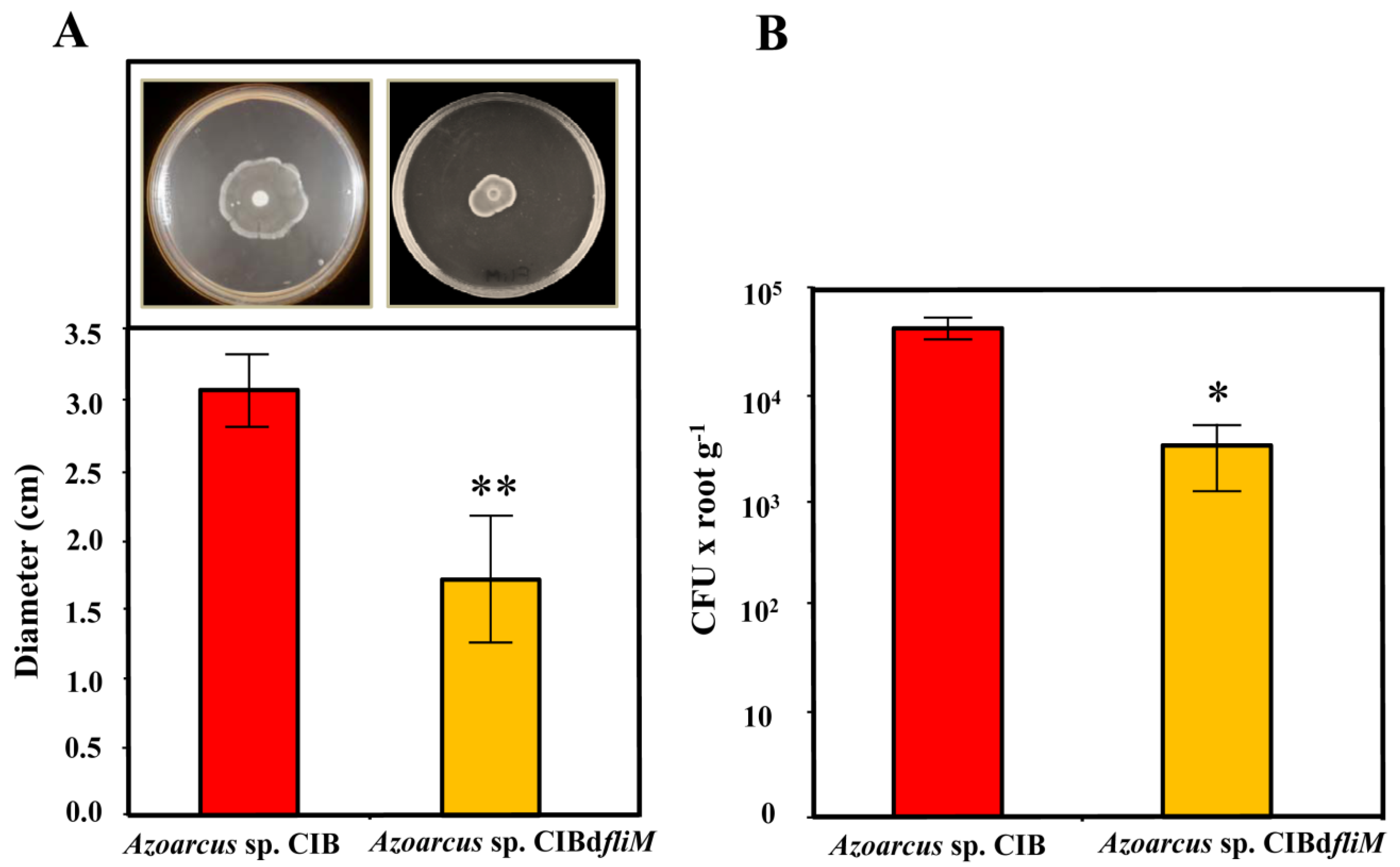
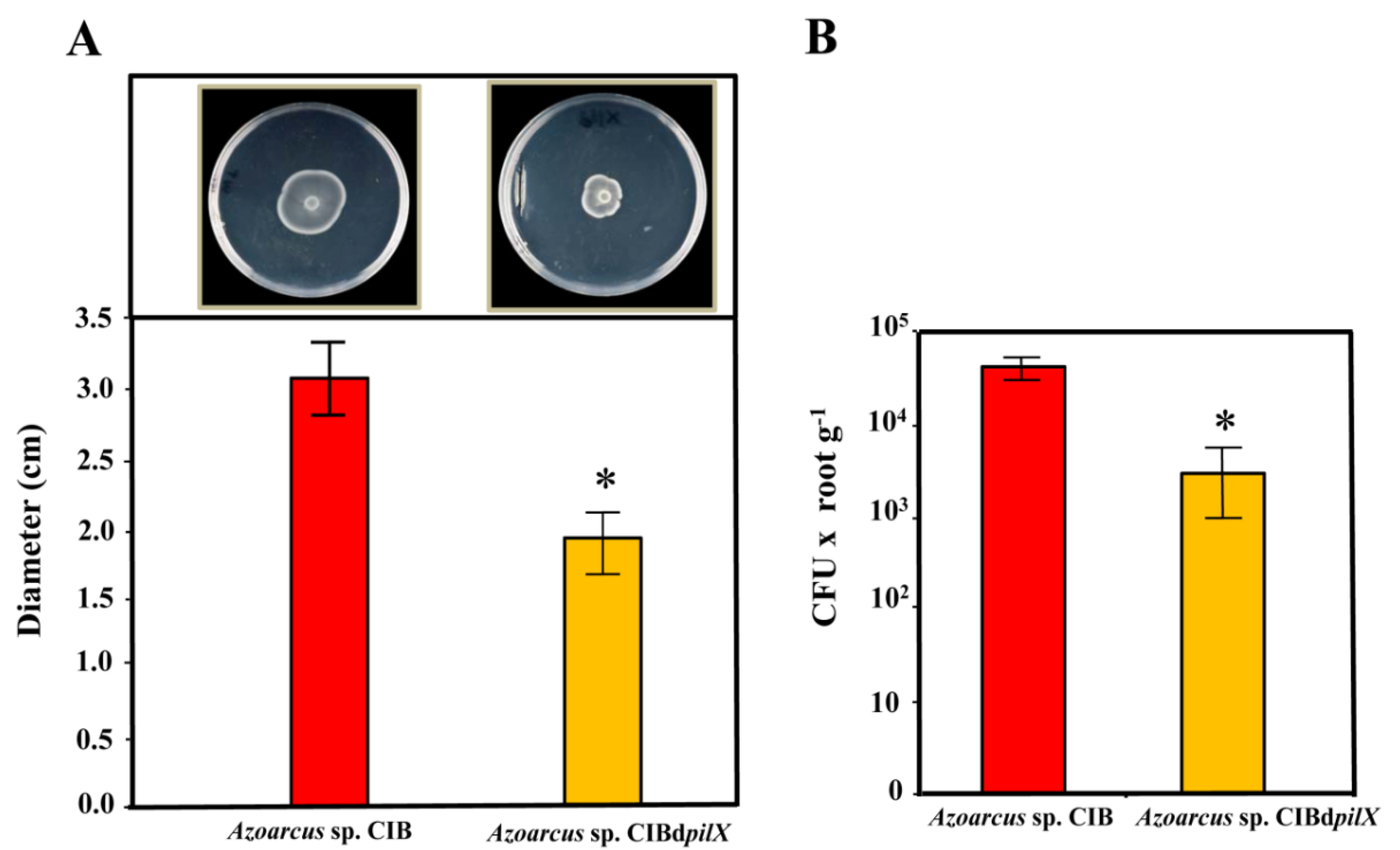
3.1. A Functional Flagellum and Pili Type IV Are Required for Rice Colonization of Azoarcus sp. CIB

As mentioned in the Introduction, bacterial motility plays an important role in plant colonization. The flagellum has traditionally been regarded as the main responsible for bacterial motility. Three gene clusters predicted to control the synthesis and regulation of the flagellum have been identified at three different positions of the genome of Azoarcus sp. CIB ([58], Table S1). We then checked whether a mutation that abolish flagellum functionality may have an effect on the endophytic colonization of rice by strain CIB.
To try to generate a motility minus phenotype in Azoarcus sp. CIB, we constructed a mutant strain in the fli operon. The Azoarcus sp. CIBdfliM strain (Table 1) harbors a disruptional insertion of the fliM (AzCIB_0962) gene, encoding the FliM protein of the C ring which has been shown to be directly involved in flagellum movement and the flagellar export apparatus [77,78,79,80]. To check whether the fliM mutant strain showed an altered motility phenotype, we performed bacterial swimming assays. As shown in Figure 1A, the movement of the mutant cells was significantly diminished (around 50%) with respect to that of the parental strain, hence suggesting an important loss of functionality of the flagellum in Azoarcus sp. CIBdfliM strain.
We then checked the ability of the mutant strain to colonize rice seedlings. Interestingly, the number of endophytes recovered after inoculation of rice seedlings with Azoarcus sp. CIBdfliM strain was around an order of magnitude lower (3.4 × 10³ per g of roots) than those recovered from seedlings inoculated with the wild type CIB strain (4.5 × 10⁴ per g of roots) (Figure 1B). These results indicate an important role of the flagellum at some stage of the endophytic colonization of rice by Azoarcus sp. CIB. These results are in agreement with previous reports showing that deletion of the fliC3 gene, encoding the main flagellin (FliC) of the flagellum, in the endophyte Azoarcus sp. BH72 strain leads to a significant decrease of around 80% in the plant colonizing capacity on competition assays between the wild type and the mutant strain [16], hence supporting that the flagellum is involved in the Azoarcus-plant interaction.
Different studies conducted on Azoarcus spp. strains have concluded that pili type IV are needed for plant colonization since they are responsible of adhesion and movement to the plant roots [7,16,18,81]. The genes predicted to be responsible for the synthesis of pili type IV in Azoarcus sp. CIB are organized in several gene clusters distributed along the genome ([58], Table S1). In order to analyze if the pil genes from Azoarcus sp. CIB are involved in the colonization of rice root, we constructed an Azoarcus sp. CIBdpilX mutant strain containing a disruption of the pilX gene (AzCIB_3117) (Table 1). The motility (swarming pattern) of the pilX mutant strain was reduced by 40% compared to that observed with the wild-type strain (Figure 2A), which is in agreement with the already observed lower motility in pil mutant bacterial strains [82,83] and confirms that the pil genes indeed encode proteins that participate in the motility apparatus in Azoarcus sp. CIB.
Interestingly, the plant colonization ability of the Azoarcus sp. CIBdpilX strain was more than one order of magnitude lower than that of the parental CIB strain (Figure 2B). Therefore, these results allowed us to conclude that pili type IV are involved in the interaction of the CIB strain with rice. Our results in strain CIB are in agreement with those reported in Azoarcus sp. BH72 where a deletion of pilX did not inhibit the synthesis of the pili but affected its functionality [81,82,83,84,85] and decreased by 80% the plant colonizing capacity of the mutant strain [7,18].
In summary, all these results taken together indicate that both, flagellum and pili type IV are needed for the interaction of Azoarcus sp. CIB with rice.

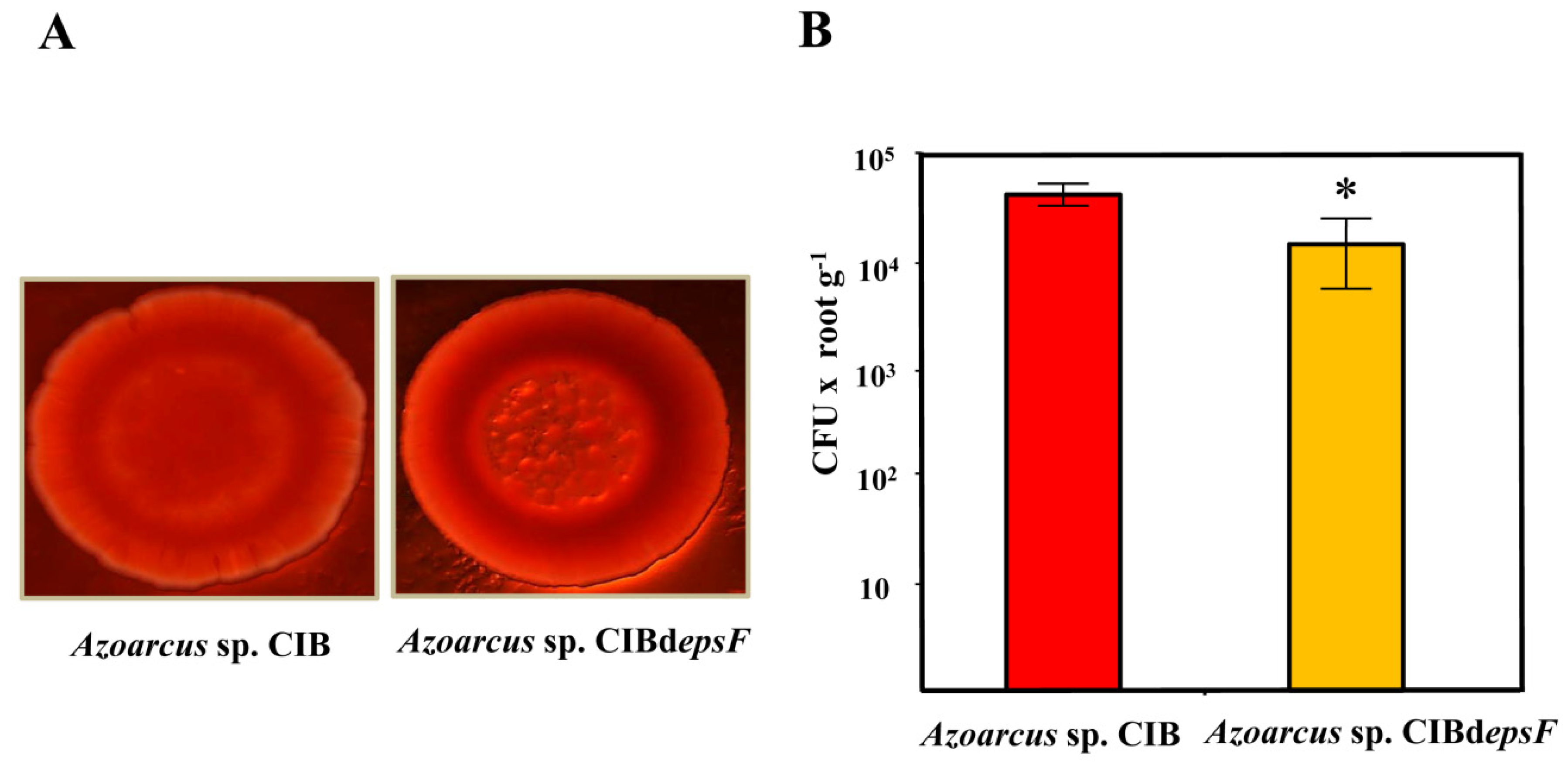
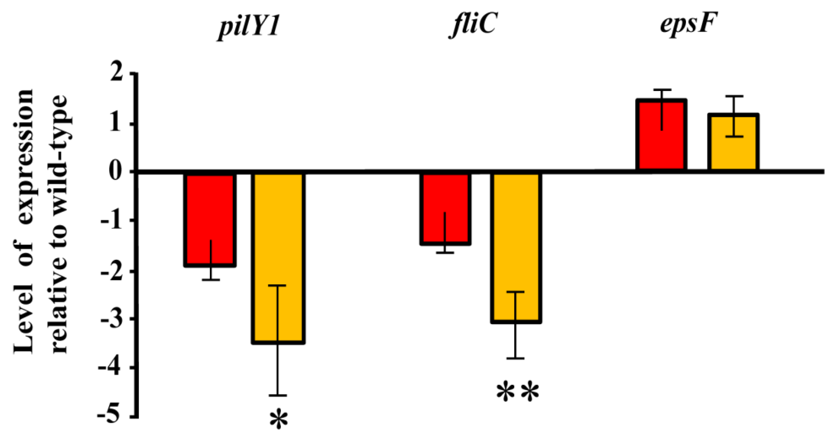
3.2. Role of the Genes Involved in Exopolysaccharide Modification in the Interaction between Azoarcus sp. CIB and Rice

As mentioned in the Introduction, exopolysaccharides have an important role in plant-bacteria interactions [23,25,26]. In the genome of Azoarcus sp. CIB we have identified three gene clusters putatively involved in the synthesis and modification of exopolysaccharides ([56], Table S1). Previous results have shown that the gene cluster AzCIB_0813-0833 was strongly induced at the transcriptional level in the presence of rice extracts [57]. To analyze the potential role of this gene cluster in the colonization of rice roots by the CIB strain, it was inactivated by insertional disruption at the AzCIB_0818 (epsF) gene. The surface of the colonies of the Azoarcus sp. CIBdepsF mutant strain grown on a solid culture medium supplemented with Congo Red dye [86] showed a rugose phenotype in contrast to the smooth phenotype shown by colonies of the wild-type strain (Figure 3A). These changes in colony morphology strongly suggest the existence of significant alterations in the exopolysaccharide of the epsF mutant strain [87], hence indicating that the eps genes are indeed involved in exopolysaccharide synthesis/modification. The next step was to study the ability of the strain Azoarcus sp. CIBdepsF to colonize rice roots as an endophyte. The plant colonization results showed that the number of Azoarcus sp. CIBdepsF endophytes recovered from rice roots (1.65 × 10⁴ bacteria per g of roots) was 36% lower than that obtained from roots inoculated with the parental strain Azoarcus sp. CIB (4.57 × 10⁴ bacteria per g of roots) (Figure 3B). Therefore, the results obtained reveal that the inactivation of the eps cluster reduces the ability of Azoarcus sp. CIB to interact with the rice plant. These results provide strong experimental support on the role of the exopolysaccharide(s) promoting the endophytic lifestyle in members of the Azoarcus/Aromatoleum group.

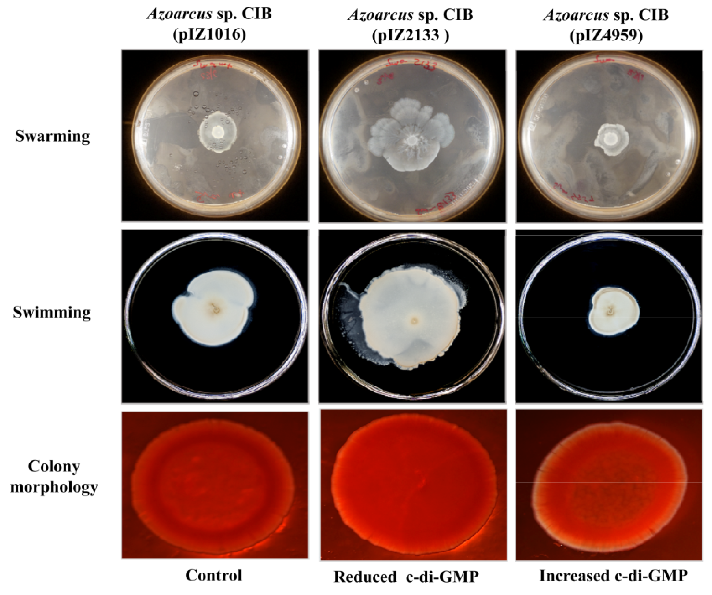
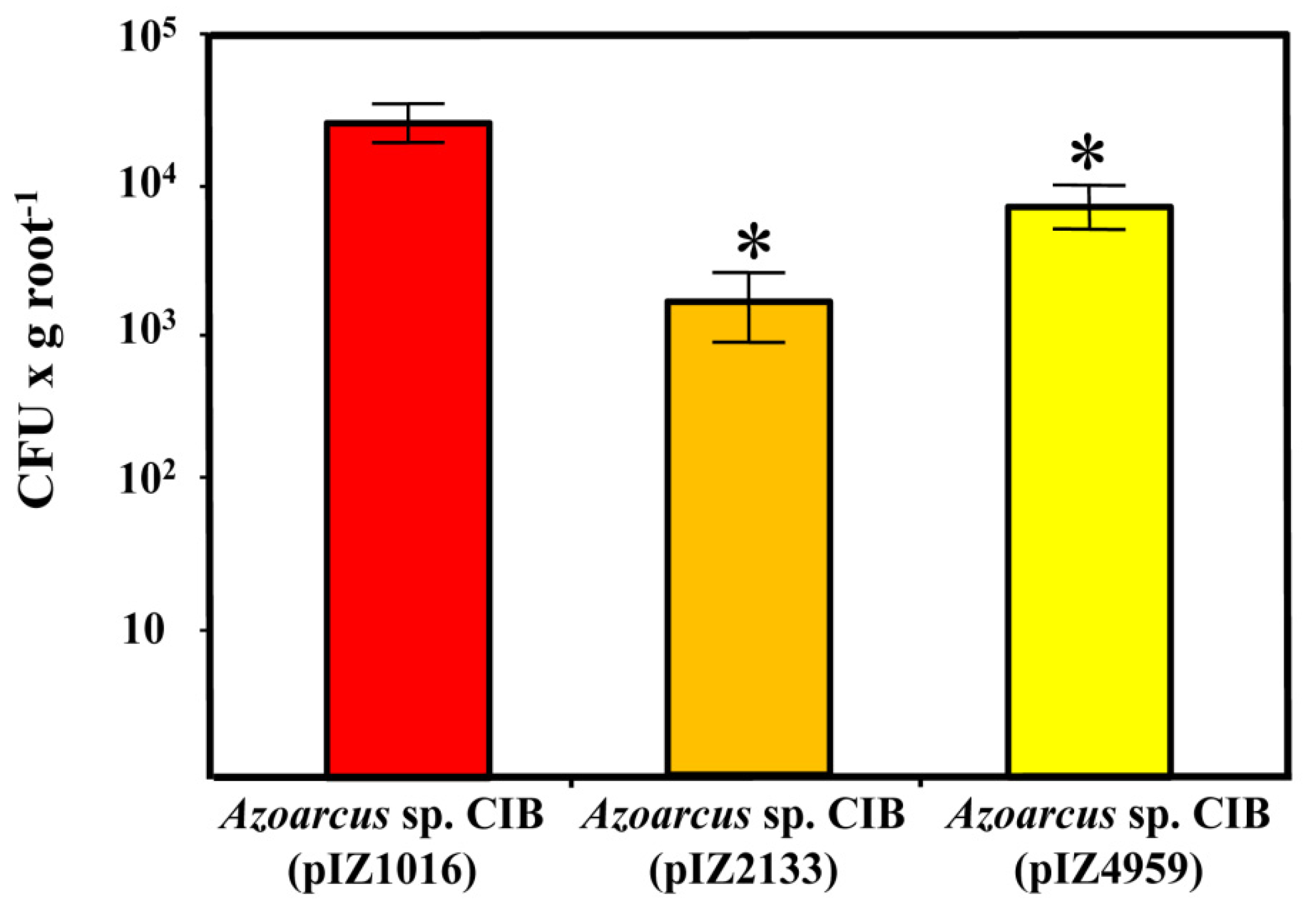
3.3. c-di-GMP Levels Influence the Endophytic Lifestyle of Azoarcus sp. CIB

As mentioned in the Introduction, c-di-GMP is a second messenger involved in the regulation of many bacterial functions. However, its role in beneficial plant-bacteria interactions and, more specifically, in their endophytic relationships, is poorly studied and far to be understood. Since the production and/or functionality of pili type IV, flagellum and exopolysaccharide (all of which were shown above to be involved in the Azoarcus sp. CIB-rice interactions) have been related with the intracellular levels of c-di-GMP in bacteria [88,89,90], it was tempting to speculate that c-di-GMP could be also regulating the endophytic lifestyle of strain CIB. To study further the proposed new function of c-di-GMP in bacterial endophytes, we checked recombinant Azoarcus sp. CIB strains containing different intracellular levels of c-di-GMP. To this end, we used Azoarcus sp. CIB (pIZ2133), expressing the PA2133 c-di-GMP phosphodiesterase from P. aeruginosa PAO1 [26], and Azoarcus sp. CIB (pIZ4959), expressing the PP4959 diguanilate cyclase from P. putida KT2440 [91], two strains described for their reduced or increased levels of c-di-GMP, respectively, with respect to those of the parental strain Azoarcus sp. CIB (pIZ1016) ([68], Table 1). To confirm that the two recombinant strains showed the phenotypes anticipated according to the variations in c-di-GMP levels [28], we analyzed first their swarming and swimming motility. As expected, whereas Azoarcus sp. CIB (pIZ2133) (reduced c-di-GMP levels) showed an increased swarming and swimming motility, the strain Azoarcus sp. CIB (pIZ4959) (increased c-di-GMP levels) showed a reduced swarming and swimming motility with respect to that of the control strain (Figure 4). The growth rates of the CIB control strain and its derivative strains CIB (pIZ2133) and CIB (pIZ4959) were similar (data not shown), so the observed different motility cannot be attributed to differences in their cell growth. The morphology of the colonies was later analyzed and whereas Azoarcus sp. CIB (pIZ2133) generated smoother and more plain colonies than the control strain, Azoarcus sp. CIB (pIZ4959) showed rough colonies (Figure 4), which is consistent with a higher exopolysaccharide synthesis [28].
To analyze whether the altered levels of c-di-GMP in Azoarcus sp. CIB (pIZ2133) and Azoarcus sp. CIB (pIZ4959) strains might influence their plant colonization abilities, rice seedlings colonization tests were performed. As shown in Figure 5, the number of endophytes recovered from seedlings inoculated with Azoarcus sp. CIB (pIZ2133) cells (1.46 × 10³ bacteria per g of root), was more than one order of magnitude lower than the number of endophytes recovered after inoculation with the CIB control strain (2.5 × 10⁴ bacteria per g of root). On the other hand, inoculation with the strain Azoarcus sp. CIB (pIZ4959) led to 7.63 × 10³ endophytes per g of root, which represents a loss of efficiency in colonization of 70% with respect to the control strain. Therefore, these data suggest that either the artificial decrease or increase of c-di-GMP levels in Azoarcus sp. CIB affect negatively the endophytic colonization of rice by this bacterium. These results are in agreement with previous observations that any modification in the homeostasis of c-di-GMP negatively affects the interactions between the plant and pathogenic or symbiont bacteria [41,42,43]. These results can be explained by taken into account that both motility and adhesion are needed for successful plant colonization [2]. Thus, an increase in c-di-GMP levels leads to greater adhesion but, at the same time, to a decrease in bacterial motility; conversely, a decrease of c-di-GMP levels enhance bacterial motility but are detrimental for adhesion and formation of microcolonies needed for the entrance of the bacteria to the internal tissues of the plant [2,92].
To try to determine if the presumed modification of c-di-GMP levels in Azoarcus sp. CIB affected the transcription of genes involved in bacterial motility and/or exopolysaccharide formation, an expression analysis of the pilY1 gene (encodes a protein for the synthesis and stabilization of pili type IV), fliC gene (encodes a structural protein of the flagellum) and the epsF gene (encodes a protein for exopolysaccharide modification) was performed. The results obtained revealed that the expression of the epsF gene was not significantly altered in any of the two strains that have modified levels of c-di-GMP, i.e., Azoarcus sp. CIB (pIZ2133) and Azoarcus sp. CIB (pIZ4959) (Figure 6). These results suggest that c-di-GMP does not control the exopolysaccharide genes at the transcriptional level, pointing to a post-transcriptional control, as already described for exopolysaccharide genes in other bacteria [93], as a more likely regulatory mechanism. Similarly, the expression of the pilY1 and fliC genes was not significantly altered in Azoarcus sp. CIB (pIZ2133) (reduced c-di-GMP levels) when compared with their expression in the control strain (Figure 6), suggesting that the increased motility observed in the former strain might be controlled by a c-di-GMP dependent post-transcriptional regulation as already shown in P. aeruginosa PAO1 [94]. In contrast, the expression of pilY1 and fliC genes was 3–3.5 times lower in the strain Azoarcus sp. CIB (pIZ4959) than in the control strain (Figure 6), suggesting that the increase of c-di-GMP levels leads to a transcriptional repression of such genes. The repression of the pil and fli genes in response to elevated cellular levels of c-di-GMP was previously observed in other bacteria such as V. cholerae [95,96], and is in agreement with the reduced motility observed in swarming and swimming assays (Figure 4). It is known that the c-di-GMP mediated transcriptional regulation of motility genes takes place through the FleQ regulator in P. aeruginosa and P. putida [37,97,98], or through alterations in chemotactic machinery affecting receptors with PilZ domains [99,100]. Since Azoarcus sp. CIB does not have a FleQ ortholog and only a protein with a putative PilZ domain has been identified by genome search, it is currently difficult to predict the signaling cascade involved in the transcriptional control mediated by c-di-GMP on the pil and fli genes in the CIB strain.
In summary, all the results presented here suggested for the first time how the modification of intracellular levels of c-di-GMP in an endophytic bacterium affects its ability to colonize the host plant, and they reveal an unnoticed role of c-di-GMP in bacteria controlling the endophytic lifestyle. It is tempting to speculate that, as described in the endophyte bacterium Azoarcus sp. CIB, the modification of c-di-GMP levels may modulate pathogenesis of plant pathogenic bacteria. In this sense, there have been described some examples where changes in the levels of c-di-GMP control pathogenesis, e.g., modulating flagellar swimming motility and plant disease progression in Erwinia amylovora [101] or Pseudomonas syringae pv. tomato DC3000 [102]. However, these findings could not be generalized to all pathogenic bacteria. Thus, increasing c-di-GMP levels in P. savastanoi pv. savastanoi might reduce the necrosis observed on tomato or bean but the development of other disease symptoms did not seem significantly affected by high c-di-GMP [45]. More work needs to be done to elucidate the signaling cascade that responds to the levels of c-di-GMP in Azoarcus sp. CIB.

4. Conclusions

The results of the present study describe the participation of the motility mediated by the flagellum and pili type IV, the adhesion modulated by exopolysaccharides and, remarkably, the unnoticed participation of the intracellular levels of c-di-GMP in the colonization of rice roots by Azoarcus sp. CIB. More experiments need to be developed to understand the complete landscape of molecular determinants involved in the signaling cascade that responds to the levels of c-di-GMP in Azoarcus sp. The results of these experiments might be used to set strategies to modulate the interaction of endophytes with their target plant hosts.

Supplementary Materials

The following are available online at https://www.mdpi.com/2076-2607/9/3/554/s1, Figure S1: Expression of the pilX, fliM and epsF genes in the wild-type and mutant strains Azoarcus sp. CIBdpilX, Azoarcus sp. CIBdfliM and Azoarcus sp. CIBdepsF. Table S1: Azoarcus sp. CIB genes putatively involved in type IV pili, flagellum and exopolysaccharide production, Table S2: Oligonucleotides used in this study.

Author Contributions

H.F.-L. performed most experiments. H.F.-L., E.D. and M.C. designed and analyzed data. H.F.-L., E.D. and M.C. initiated and coordinated the study and contributed to the experimental design and data interpretation. M.C., E.D. mainly wrote the manuscript. All authors have read and agreed to the published version of the manuscript.

Funding

This research was funded by grants BIO2016-79736-R, PCIN-2014-113, and PCI2019-111833-2 from the Ministry of Economy and Competitiveness of Spain; by Grant CSIC 2017 2 0I 015; and by European Union H2020 Grant 760994.

Institutional Review Board Statement

Not applicable.

Informed Consent Statement

Not applicable.

Data Availability Statement

The data presented in this study are available within this article.

Acknowledgments

We thank A. Valencia for technical assistance and Secugen S.L. for DNA sequencing.

Conflicts of Interest

The authors declare no conflict of interest.

References

  1. Bulgarelli, D.; Schlaeppi, K.; Spaepen, S.; van Themaat, E.V.L.; Schulze-Lefert, P. Structure and functions of the bacterial microbiota of plants. Annu. Rev. Plant Biol. 2013, 64, 807–838. [Google Scholar] [CrossRef] [Green Version]
  2. Reinhold-Hurek, B.; Bünger, W.; Burbano, C.S.; Sabale, M.; Hurek, T. Roots shaping their microbiome: Global hotspots for microbial activity. Annu. Rev. Phytopathol. 2015, 53, 403–424. [Google Scholar] [CrossRef] [PubMed]
  3. Gopal, M.; Gupta, A. Microbiome selection could spur next-generation plant breeding strategies. Front. Microbiol. 2016, 7, 1971. [Google Scholar] [CrossRef] [Green Version]
  4. Reinhold-Hurek, B.; Hurek, T. Life in the grasses: Diazotrophic endophytes. Trends Microbiol. 1998, 6, 139–144. [Google Scholar] [CrossRef]
  5. Rosenblueth, M.; Martínez-Romero, E. Bacterial endophytes and their interactions with hosts. Mol. Plant Microbe Interact. 2006, 19, 827–837. [Google Scholar] [CrossRef] [Green Version]
  6. Mano, H.; Morisaki, H. Endophytic bacteria in the rice plant. Microbes Environ. 2008, 23, 109–117. [Google Scholar] [CrossRef] [PubMed] [Green Version]
  7. Shidore, T.; Dinse, T.; Öhrlein, J.; Becker, A.; Reinhold-Hurek, B. Transcriptomic analysis of responses to exudates reveal genes required for rhizosphere competence of the endophyte Azoarcus sp. strain BH72. Environ. Microbiol. 2012, 14, 2775–2787. [Google Scholar] [CrossRef]
  8. Alquéres, S.; Meneses, C.; Rouws, L.; Rothballer, M.; Baldani, I.; Schmid, M.; Hartmann, A. The bacterial superoxide dismutase and glutathione reductase are crucial for endophytic colonization of rice roots by Gluconacetobacter diazotrophicus PAL5. Mol. Plant Microbe Interact. 2013, 26, 937–945. [Google Scholar] [CrossRef] [Green Version]
  9. Drogue, B.; Sanguin, H.; Borland, S.; Prigent-Combaret, C.; Wisniewski-Dyé, F. Genome wide profiling of Azospirillum lipoferum 4B gene expression during interaction with rice roots. FEMS Microbiol. Ecol. 2014, 87, 543–555. [Google Scholar] [CrossRef] [PubMed] [Green Version]
  10. Kaul, S.; Sharma, T.; Dhar, M.K. “Omics” tools for better understanding the plant-endophyte interactions. Front. Plant Sci. 2016, 7, 955. [Google Scholar] [CrossRef] [Green Version]
  11. Hardoim, P.R.; van Overbeek, L.S.; van Elsas, J.D. Properties of bacterial endophytes and their proposed role in plant growth. Trends Microbiol. 2008, 16, 463–471. [Google Scholar] [CrossRef]
  12. Hardoim, P.R.; van Overbeek, L.S.; Berg, G.; Pirttilä, A.M.; Compant, S.; Campisano, A.; Döring, M.; Sessitsch, A. The hidden world within plants: Ecological and evolutionary considerations for defining functioning of microbial endophytes. Microbiol. Mol. Biol. Rev. 2015, 79, 293–320. [Google Scholar] [CrossRef] [PubMed] [Green Version]
  13. Reinhold-Hurek, B.; Hurek, T. Living inside plants: Bacterial endophytes. Curr. Opin. Plant Biol. 2011, 14, 435–443. [Google Scholar] [CrossRef]
  14. Mitter, B.; Petric, A.; Shin, M.W.; Chain, P.S.G.; Hauberg-Lotte, L.; Reinhold-Hurek, B.; Nowak, J.; Sessitsch, A. Comparative genome analysis of Burkholderia phytofirmans PsJN reveals a wide spectrum of endophytic lifestyles based on interaction strategies with host plants. Front. Plant Sci. 2013, 4, 120. [Google Scholar] [CrossRef] [PubMed] [Green Version]
  15. Newman, M.A.; Sundelin, T.; Nielsen, J.T.; Erbs, G. MAMP (microbe-associated molecular pattern) triggered immunity in plants. Front. Plant Sci. 2013, 4, 139. [Google Scholar] [CrossRef] [PubMed] [Green Version]
  16. Buschart, A.; Sachs, S.; Chen, X.; Herglotz, J.; Krause, A.; Reinhold-Hurek, B. Flagella mediate endophytic competence rather than act as MAMPS in rice-Azoarcus sp. strain BH72 interactions. Mol. Plant Microbe Interact. 2012, 25, 191–199. [Google Scholar] [CrossRef] [PubMed] [Green Version]
  17. Sheibani-Tezerji, R.; Rattei, T.; Sessitsch, A.; Trognitz, F.; Mitter, B. Transcriptome profiling of the endophyte Burkholderia phytofirmans PsJN indicates sensing of the plant environment and drought stress. MBio 2015, 6, 1–11. [Google Scholar] [CrossRef] [PubMed] [Green Version]
  18. Böhm, M.; Hurek, T.; Reinhold-Hurek, B. Twitching motility is essential for endophytic rice colonization by the N2-fixing endophyte Azoarcus sp. strain BH72. Mol. Plant Microbe Interact. 2007, 20, 526–533. [Google Scholar] [CrossRef] [PubMed] [Green Version]
  19. James, E.K.; Gyaneshwar, P.; Mathan, N.; Barraquio, W.L.; Reddy, P.M.; Iannetta, P.P.M.; Olivares, F.L.; Ladha, J.K. Infection and colonization of rice seedlings by the plant growth-promoting bacterium Herbaspirillum seropedicae Z67. Mol. Plant Microbe Interact. 2002, 15, 894–906. [Google Scholar] [CrossRef] [Green Version]
  20. Danhorn, T.; Fuqua, C. Biofilm formation by plant-associated bacteria. Annu. Rev. Microbiol. 2007, 61, 401–422. [Google Scholar] [CrossRef]
  21. Compant, S.; Kaplan, H.; Sessitsch, A.; Nowak, J.; Ait Barka, E.; Clément, C. Endophytic colonization of Vitis vinifera L. by Burkholderia phytofirmans strain PsJN: From the rhizosphere to inflorescence tissues. FEMS Microbiol. Ecol. 2008, 63, 84–93. [Google Scholar] [CrossRef] [Green Version]
  22. Balsanelli, E.; Tuleski, T.R.; de Baura, V.A.; Yates, M.G.; Chubatsu, L.S.; de Oliveira Pedrosa, F.; de Souza, E.M.; Monteiro, R.A. Maize root lectins mediate the interaction with Herbaspirillum seropedicae via N-acetyl glucosamine residues of lipopolysaccharides. PLoS ONE 2013, 8, e77001. [Google Scholar] [CrossRef] [PubMed]
  23. Meneses, C.H.S.G.; Rouws, L.F.M.; Simoes-Araujo, J.L.; Vidal, M.S.; Baldani, J.I. Exopolysaccharide production is required for biofilm formation and plant colonization by the nitrogen-fixing endophyte Gluconacetobacter diazotrophicus. Mol. Plant Microbe Interact. 2011, 24, 1448–1458. [Google Scholar] [CrossRef] [PubMed] [Green Version]
  24. Schmid, J.; Sieber, V.; Rehm, B. Bacterial exopolysaccharides: Biosynthesis pathways and engineering strategies. Front. Microbiol. 2015, 6, 496. [Google Scholar] [CrossRef] [PubMed] [Green Version]
  25. Janczarek, M.; Rachwał, K.; Cieśla, J.; Ginalska, G.; Bieganowski, A. Production of exopolysaccharide by Rhizobium leguminosarum bv. trifolii and its role in bacterial attachment and surface properties. Plant Soil 2015, 388, 211–227. [Google Scholar] [CrossRef] [Green Version]
  26. Nian, H.J.; Zhang, J.; Liu, S.; Song, F.P.; Huang, D.F. Effect of bacteriaphage and exopolysaccharide on root colonization and rhizosphere competence by Pseudomonas fluorescens. Ann. Microbiol. 2010, 60, 369–372. [Google Scholar] [CrossRef]
  27. Römling, U.; Galperin, M.Y.; Gomelsky, M. Cyclic di-GMP: The first 25 years of a universal bacterial second messenger. Microbiol. Mol. Biol. Rev. 2013, 77, 1–52. [Google Scholar] [CrossRef] [Green Version]
  28. Jenal, U.; Reinders, A.; Lori, C. Cyclic di-GMP: Second messenger extraordinaire. Nat. Rev. Microbiol. 2017, 15, 271–284. [Google Scholar] [CrossRef] [Green Version]
  29. Hickman, J.W.; Tifrea, D.F.; Harwood, C.S. A chemosensory system that regulates biofilm formation through modulation of cyclic diguanylate levels. Proc. Natl. Acad. Sci. USA 2005, 102, 14422–14427. [Google Scholar] [CrossRef] [Green Version]
  30. Lee, V.T.; Matewish, J.M.; Kessler, J.L.; Hyodo, M.; Hayakawa, Y.; Lory, S. A cyclic-di-GMP receptor required for bacterial exopolysaccharide production. Mol. Microbiol. 2007, 65, 1474–1484. [Google Scholar] [CrossRef] [PubMed] [Green Version]
  31. Borlee, B.R.; Goldman, A.D.; Murakami, K.; Samudrala, R.; Wozniak, D.J.; Parsek, M.R. Pseudomonas aeruginosa uses a cyclic di-GMP-regulated adhesin to reinforce the biofilm extracellular matrix. Mol. Microbiol. 2010, 75, 827–842. [Google Scholar] [CrossRef] [PubMed] [Green Version]
  32. Liang, Z.X. The expanding roles of c-di-GMP in the biosynthesis of exopolysaccharides and secondary metabolites. Nat. Prod. Rep. 2015, 32, 663–683. [Google Scholar] [CrossRef] [PubMed] [Green Version]
  33. Schäper, S.; Krol, E.; Skotnicka, D.; Kaever, V.; Hilker, R.; Sǿgarrd-Andersen, L.; Becker, A. Cyclic di-GMP regulates multiple cellular functions in the symbiotic alphaproteobacterium Sinorhizobium meliloti. J. Bacteriol. 2015, 198, 521–535. [Google Scholar] [CrossRef] [Green Version]
  34. Simm, R.; Morr, M.; Kader, A.; Nimtz, M.; Römling, U. GGDEF and EAL domains inversely regulate cyclic di-GMP levels and transition from sessility to motility. Mol. Microbiol. 2004, 53, 1123–1134. [Google Scholar] [CrossRef]
  35. Starkey, M.; Hickman, J.H.; Ma, L.; Zhang, N.; De Long, S.; Hinz, A.; Palacios, S.; Manoil, C.; Kirisits, M.J.; Starner, T.D.; et al. Pseudomonas aeruginosa rugose small-colony variants have adaptations that likely promote persistence in the cystic fibrosis lung. J. Bacteriol. 2009, 191, 3492–3503. [Google Scholar] [CrossRef] [Green Version]
  36. Waters, C.M.; Lu, W.; Rabinowitz, J.D.; Bassler, B.L. Quorum sensing controls biofilm formation in Vibrio cholerae through modulation of cyclic di-GMP levels and repression of vpsT. J. Bacteriol. 2008, 190, 2527–2536. [Google Scholar] [CrossRef] [PubMed] [Green Version]
  37. Kazmierczak, B.I.; Lebron, M.B.; Murray, T.S. Analysis of FimX, a phosphodiesterase that governs twitching motility in Pseudomonas aeruginosa. Mol. Microbiol. 2006, 60, 1026–1043. [Google Scholar] [CrossRef] [Green Version]
  38. Wolfe, A.J.; Visick, K.L. Get the message out: Cyclic-di-GMP regulates multiple levels of flagellum-based motility. J. Bacteriol. 2008, 190, 463–475. [Google Scholar] [CrossRef] [Green Version]
  39. McCarter, L.L.; Gomelsky, M. Fifty ways to inhibit motility via cyclic di-GMP: The emerging Pseudomonas aeruginosa swarming story. J. Bacteriol. 2015, 197, 406–409. [Google Scholar] [CrossRef] [Green Version]
  40. Skotnicka, D.; Petters, T.; Heering, J.; Hoppert, M.; Kaever, V.; Sogaard-Andersen, L. c-di-GMP regulates type IV pili-dependent-motility in Myxococcus xanthus. J. Bacteriol. 2015, 198, 77–90. [Google Scholar] [CrossRef] [Green Version]
  41. Sun, Y.; Xie, Z.; Sui, F.; Liu, X.; Cheng, W. Identification of Cbp1, a c-di-GMP binding chemoreceptor in Azorhizobium caulinodans ORS571 involved in chemotaxis and nodulation of the host plant. Front. Microbiol. 2019, 10, 638. [Google Scholar] [CrossRef]
  42. Tischler, A.D.; Camilli, A. Cyclic diguanylate regulates Vibrio cholerae virulence gene expression. Infect. Immun. 2005, 73, 5873–5882. [Google Scholar] [CrossRef] [Green Version]
  43. Ryan, R.P. Cyclic di-GMP signalling and the regulation of bacterial virulence. Microbiology 2013, 159, 1286–1297. [Google Scholar] [CrossRef]
  44. Martinez-Gil, M.; Ramos, C. Role of cyclic di-GMP in the bacterial virulence and evasion of the plant immunity. Curr. Issues Mol. Biol. 2018, 25, 199–222. [Google Scholar] [CrossRef]
  45. Pérez-Mendoza, D.; Aragón, I.M.; Prada-Ramírez, H.A.; Romero-Jiménez, L.; Ramos, C.; Gallegos, M.T.; Sanjuán, J. Responses to elevated c-di-GMP levels in mutualistic and pathogenic plant-interacting bacteria. PLoS ONE 2014, 9, e91645. [Google Scholar] [CrossRef] [Green Version]
  46. Aragón, I.M.; Pérez-Mendoza, D.; Gallegos, M.T.; Ramos, C. The c-di-GMP phosphodiesterase BifA is involved in the virulence of bacteria from the Pseudomonas syringae complex. Mol. Plant Pathol. 2015, 16, 604–615. [Google Scholar] [CrossRef] [PubMed]
  47. McCarthy, Y.; Ryan, R.P.; O’Donovan, K.; He, Y.Q.; Jiang, B.L.; Feng, J.X.; Tang, J.L.; Dow, J.M. The role of PilZ domain proteins in the virulence of Xanthomonas campestris pv. campestris. Mol. Plant Pathol. 2008, 9, 819–824. [Google Scholar] [CrossRef]
  48. Ryan, R.P.; McCarthy, Y.; Andrade, M.; Farah, C.S.; Armitage, J.P.; Dow, J.M. Cell-cell signal-dependent dynamic interactions between HD-GYP and GGDEF domain proteins mediate virulence in Xanthomonas campestris. Proc. Natl. Acad. Sci. USA 2010, 107, 5989–5994. [Google Scholar] [CrossRef] [PubMed] [Green Version]
  49. An, S.Q.; Allan, J.H.; McCarthy, Y.; Febrer, M.; Dow, J.M.; Ryan, R.P. The PAS domain-containing histidine kinase RpfS is a second sensor for the diffusible signal factor of Xanthomonas campestris. Mol. Microbiol. 2014, 92, 586–597. [Google Scholar] [CrossRef] [Green Version]
  50. Zhang, Y.; Wei, C.; Jiang, W.; Wang, L.; Li, C.; Wang, Y.; Dow, J.M.; Sun, W. The HD-GYP domain protein RpfG of Xanthomonas oryzae pv. oryzicola regulates synthesis of extracellular polysaccharides that contribute to biofilm formation and virulence on rice. PLoS ONE 2013, 8, e59428. [Google Scholar] [CrossRef] [PubMed] [Green Version]
  51. Yang, F.; Tian, F.; Li, X.; Fan, S.; Chen, H.; Wu, M.; Yang, C.H.; He, C. The degenerate EAL-GGDEF domain protein filp functions as a cyclic di-GMP receptor and specifically interacts with the PilZ-domain protein PXO_02715 to regulate virulence in Xanthomonas oryzae pv. oryzae. Mol. Plant Microbe Interact. 2014, 27, 578–589. [Google Scholar] [CrossRef] [Green Version]
  52. Gao, S.; Romdhane, S.B.; Beullens, S.; Kaever, V.; Lambrichts, I.; Fauvart, M.; Michiels, J. Genomic analysis of cyclic-di-GMP-related genes in rhizobial type strains and functional analysis in Rhizobium etli. Appl. Microbiol. Biotechnol. 2014, 98, 4589–4602. [Google Scholar] [CrossRef] [PubMed]
  53. Lim, B.; Beyhan, S.; Meir, J.; Yildiz, F.H. Cyclic-diGMP signal transduction systems in Vibrio cholerae: Modulation of rugosity and biofilm formation. Mol. Microbiol. 2006, 60, 331–348. [Google Scholar] [CrossRef]
  54. Ryan, R.P.; Fouhy, Y.; Lucey, J.F.; Dow, J.M. Cyclic di-GMP signaling in bacteria: Recent advances and new puzzles. J. Bacteriol. 2006, 188, 8327–8334. [Google Scholar] [CrossRef] [Green Version]
  55. Bobrov, A.G.; Kirillina, O.; Perry, R.D. The phosphodiesterase activity of the HmsP EAL domain is required for negative regulation of biofilm formation in Yersinia pestis. FEMS Microbiol. Lett. 2005, 247, 123–130. [Google Scholar] [CrossRef] [Green Version]
  56. Fernández-Llamosas, H.; Prandoni, N.; Fernández-Pascual, M.; Fajardo, S.; Morcillo, C.; Díaz, E.; Carmona, M. Azoarcus sp. CIB, an anaerobic biodegrader of aromatic compounds shows an endophytic lifestyle. PLoS ONE 2014, 9, e110771. [Google Scholar]
  57. Fernández-Llamosas, H.; Ibero, J.; Thijs, S.; Imperato, V.; Vangronsveld, J.; Díaz, E.; Carmona, M. Enhancing the rice seedlings growth promotion abilities of Azoarcus sp. CIB by heterologous expression of ACC deaminase to improve performance of plants exposed to cadmium stress. Microorganisms 2020, 8, 1453. [Google Scholar] [CrossRef]
  58. Martín-Moldes, Z.; Zamarro, M.T.; del Cerro, C.; Valencia, A.; Gómez, M.J.; Arcas, A.; Udaondo, Z.; García, J.L.; Nogales, J.; Carmona, M.; et al. Whole-genome analysis of Azoarcus sp. strain CIB provides genetic insights to its different lifestyles and predicts novel metabolic features. Syst. Appl. Microbiol. 2015, 38, 462–471. [Google Scholar] [CrossRef] [Green Version]
  59. López-Barragán, M.J.; Carmona, M.; Zamarro, M.T.; Thiele, M.; Boll, M.; Fuchs, G.; García, J.L.; Díaz, E. The bzd gene cluster, coding for anaerobic benzoate catabolism in Azoarcus sp. CIB. J. Bacteriol. 2004, 186, 5462–5474. [Google Scholar]
  60. Blázquez, B.; Carmona, M.; Díaz, E. Transcriptional regulation of the peripheral pathway for the anaerobic catabolism of toluene and m-xylene in Azoarcus sp. CIB. Front. Microbiol. 2018, 9, 506. [Google Scholar] [CrossRef] [PubMed] [Green Version]
  61. Fernández-Llamosas, H.; Castro, L.; Blázquez, M.L.; Díaz, E.; Carmona, M. Biosynthesis of selenium nanoparticles by Azoarcus sp. CIB. Microb. Cell. Factories 2017, 15, 109. [Google Scholar] [CrossRef] [PubMed]
  62. Durante-Rodríguez, G.; Fernández-Llamosas, H.; Alonso-Fernandes, E.; Fernández-Muñiz, M.N.; Muñoz-Olivas, R.; Díaz, E.; Carmona, M. ArxA from Azoarcus sp. CIB, an anaerobic arsenite oxidase from an obligate heterotrophic and mesophilic bacterium. Front. Microbiol. 2019, 10, 1699. [Google Scholar]
  63. Rabus, R.; Wöhlbrand, L.; Thies, D.; Meyer, M.; Reinhold-Hurek, B.; Kämpfer, P. Aromatoleum gen. nov., a novel genus accommodating the phylogenetic lineage including Azoarcus evansii and related species, and proposal of Aromatoleum aromaticum sp. nov., Aromatoleum petrolei sp. nov., Aromatoleum bremense sp. nov., Aromatoleum toluolicum sp. nov. and Aromatoleum diolicum sp. nov. Int. J. Syst. Evol. Microbiol. 2019, 69, 982–997. [Google Scholar]
  64. De Lorenzo, V.; Timmis, K.N. Analysis and construction of stable phenotypes in Gram-negative bacteria with Tn5- and Tn10-derived minitransposons. Methods Enzymol. 1994, 235, 386–405. [Google Scholar] [PubMed]
  65. Manoil, C.; Beckwith, J. TnphoA: A transposon probe for protein export signals. Proc. Natl. Acad. Sci. USA 1985, 82, 8129–8133. [Google Scholar] [CrossRef] [Green Version]
  66. Silva-Rocha, R.; Martínez-García, E.; Calles, B.; Chavarría, M.; Arce-Rodríguez, A.; de las Heras, A.; Páez-Espino, A.D.; Durante-Rodríguez, G.; Kim, J.; Nikel, P.I.; et al. The standard european vector architecture (SEVA): A coherent platform for the analysis and development of complex prokaryotic phenotypes. Nucleic Acids Res. 2013, 41, D666–D675. [Google Scholar] [CrossRef]
  67. Moreno-Ruiz, E.; Hernáez, M.J.; Martínez-Pérez, O.; Santero, E. Identification and functional characterization of Sphingomonas macrogolitabida strain TFA genes involved in the first two steps of the tetralin catabolic pathway. J. Bacteriol. 2003, 185, 2026–2030. [Google Scholar] [CrossRef] [Green Version]
  68. Martín-Moldes, Z.; Blázquez, B.; Baraquet, C.; Harwood, C.S.; Zamarro, M.T.; Díaz, E. Degradation of cyclic diguanosine monophosphate by a hybrid two-component protein protects Azoarcus sp. strain CIB from toluene toxicity. Proc. Natl. Acad. Sci. USA 2016, 113, 13174–13179. [Google Scholar] [CrossRef] [Green Version]
  69. Schäfer, A.; Tauch, A.; Jäger, W.; Kalinowski, J.; Thierbach, G.; Pühler, A. Small mobilizable multi-purpose cloning vectors derived from the Escherichia coli plasmids pK18 and pK19: Selection of defined deletions in the chromosome of Corynebacterium glutamicum. Gene 1994, 145, 69–73. [Google Scholar] [CrossRef]
  70. Reinhold-Hurek, B.; Hurek, T.; Gillis, M.; Hoste, B.; Vancanneyt, M.; Kersters, K.; De Ley, J. Azoarcus gen. nov., nitrogen-fixing proteobacteria associated with roots of Kallar grass (Leptochloa fusca (L.) Kunth), and description of two species, Azoarcus indigens sp. nov. and Azoarcus communis sp. nov. Int. J. Syst. Bacteriol. 1993, 43, 574–584. [Google Scholar] [CrossRef]
  71. Miller, J.H. Experiments in Molecular Genetics; Cold Spring Harbor Laboratory Press: Cold Spring Harbor, NY, USA, 1972. [Google Scholar]
  72. Sambrook, J.; Rusell, D. Molecular Cloning: A Laboratory Manual; Cold Spring Harbor Laboratory Press: Cold Spring Harbor, NY, USA, 2011. [Google Scholar]
  73. Sanger, F.; Nicklen, S.; Coulson, A.R. DNA sequencing with chain-terminating inhibitors. Proc. Natl. Acad. Sci. USA 1977, 74, 5463–5467. [Google Scholar] [CrossRef] [Green Version]
  74. Valderrama, J.A.; Durante-Rodríguez, G.; Blázquez, B.; García, J.L.; Carmona, M.; Díaz, E. Bacterial degradation of benzoate: Cross-regulation between aerobic and anaerobic pathways. J. Biol. Chem. 2012, 287, 10494–10508. [Google Scholar] [CrossRef] [Green Version]
  75. Livak, K.J.; Schmittgen, T.D. Analysis of relative gene expression data using real-time quantitative PCR and the 2−ΔΔCT. Method. Methods 2001, 25, 402–408. [Google Scholar] [CrossRef]
  76. Gyaneshwar, P.; James, E.K.; Mathan, N.; Reddy, P.M.; Reinhold-Hurek, B.; Ladha, J.K. Endophytic colonization of rice by a diazotrophic strain of Serratia marcescens. J. Bacteriol. 2001, 183, 2634–2645. [Google Scholar] [CrossRef] [Green Version]
  77. Marykwas, D.L.; Berg, H.C. A mutational analysis of the interaction between FliG and FliM, two components of the flagellar motor of Escherichia coli. J. Bacteriol. 1996, 178, 1289–1294. [Google Scholar] [CrossRef] [Green Version]
  78. Paul, K.; Nieto, V.; Carlquist, W.C.; Blair, D.F.; Harshey, R.M. The c-di-GMP binding protein YcgR controls flagellar motor direction and speed to affect chemotaxis by a “backstop brake” mechanism. Mol. Cell. 2010, 38, 128–139. [Google Scholar] [CrossRef] [PubMed] [Green Version]
  79. Morimoto, Y.; Minamino, T. Structure and function of the bi-directional bacterial flagellar motor. Biomolecules 2014, 4, 217–234. [Google Scholar] [CrossRef] [PubMed] [Green Version]
  80. Kojima, S.; Blair, D.F. The bacterial flagellar motor: Structure and function of a complex molecular machine. Int. Rev. Cytol. 2004, 233, 93–134. [Google Scholar] [PubMed]
  81. Anyan, M.E.; Amiri, A.; Harvey, C.W.; Tierra, G.; Morales-Soto, N.; Driscoll, C.M.; Alber, M.S.; Shrout, J.D. Type IV pili interactions promote intercellular association and moderate swarming of Pseudomonas aeruginosa. Proc. Natl. Acad. Sci. USA 2014, 111, 18013–18018. [Google Scholar] [CrossRef] [PubMed] [Green Version]
  82. Köhler, T.; Curty, L.K.; Barja, F.; van Delden, C.; Pechère, J.C. Swarming of Pseudomonas aeruginosa is dependent on cell-to-cell signaling and requires flagella and pili. J. Bacteriol. 2000, 182, 5990–5996. [Google Scholar] [CrossRef] [Green Version]
  83. Kuchma, S.L.; Griffin, E.F.; O’Toole, G.A. Minor pilins of the type IV pilus system participate in the negative regulation of swarming motility. J. Bacteriol. 2012, 194, 5388–5403. [Google Scholar] [CrossRef] [Green Version]
  84. Dörr, J.; Hurek, T.; Reinhold-Hurek, B. Type IV pili are involved in plant-microbe and fungus-microbe interactions. Mol. Microbiol. 1998, 30, 7–17. [Google Scholar] [CrossRef] [PubMed]
  85. Giltner, C.L.; Nguyen, Y.; Burrows, L.L. Type IV pilin proteins: Versatile molecular modules. Microbiol. Mol. Biol. Rev. 2012, 76, 740–772. [Google Scholar] [CrossRef] [PubMed] [Green Version]
  86. Chung, J.W.; Altman, E.; Beveridge, T.J.; Speert, D.P. Colonial morphology of Burkholderia cepacia complex genomovar III: Implications in exopolysaccharide production, pilus expression, and persistence in the mouse. Infect. Immun. 2003, 71, 904–909. [Google Scholar] [CrossRef] [PubMed] [Green Version]
  87. Sousa, A.M.; Machado, I.; Nicolau, A.; Pereira, M.O. Improvements on colony morphology identification towards bacterial profiling. J. Microbiol. Methods 2013, 95, 327–335. [Google Scholar] [CrossRef] [Green Version]
  88. Bordeleau, E.; Purcell, E.B.; Lafontaine, D.A.; Fortier, L.C.; Tamayo, R.; Burrus, V. Cyclic di-GMP riboswitch-regulated type IV pili contribute to aggregation of Clostridium difficile. J. Bacteriol. 2015, 197, 819–832. [Google Scholar] [CrossRef] [Green Version]
  89. Schmid, J.; Rühmann, B.; Sieber, V.; Romero-Jiménez, L.; Sanjuán, J.; Pérez-Mendoza, D. Screening of c-di-GMP-regulated exopolysaccharides in host interacting bacteria. In Methods in Molecular Biology; Medina, C., López-Baena, F., Eds.; Humana Press: New York, NY, USA, 2018; Volume 1734, pp. 263–275. [Google Scholar]
  90. Baker, A.E.; Webster, S.S.; Diepold, A.; Kuchma, S.L.; Bordeleau, E.; Armitage, J.P.; O’Toole, G.A. Flagellar stators stimulate c-di-GMP production by Pseudomonas aeruginosa. J. Bacteriol. 2019, 201, e00741-18. [Google Scholar] [CrossRef] [PubMed] [Green Version]
  91. Matilla, M.A.; Travieso, M.L.; Ramos, J.L.; Ramos-González, M.I. Cyclic diguanylate turnover mediated by the sole GGDEF/EAL response regulator in Pseudomonas putida: Its role in the rhizosphere and an analysis of its target processes. Environ. Microbiol. 2011, 13, 1745–1766. [Google Scholar] [CrossRef]
  92. Kandel, S.L.; Joubert, P.M.; Doty, S.L. Bacterial endophyte colonization and distribution within plants. Microorganisms 2017, 5, 77. [Google Scholar] [CrossRef] [Green Version]
  93. Pérez-Mendoza, D.; Sanjuán, J. Exploiting the commons: Cyclic diguanylate regulation of bacterial exopolysaccharide production. Curr. Opin. Microbiol. 2016, 30, 36–43. [Google Scholar] [CrossRef] [PubMed]
  94. Lin Chua, S.; Liu, Y.; Li, Y.; Jun Ting, H.; Kohli, G.S.; Cai, Z.; Suwanchaikasem, P.; Kit Go, K.K.; Pin Ng, S.; Tolker-Nielsen, T.; et al. Reduced intracellular c-di-GMP content increases expression of quorum sensing-regulated genes in Pseudomonas aeruginosa. Front. Cell Infect. Microbiol. 2017, 7, 451. [Google Scholar] [CrossRef]
  95. Beyhan, S.; Tischler, A.D.; Camilli, A.; Yildiz, F.H. Transcriptome and phenotypic responses of Vibrio cholerae to increased cyclic di-GMP level. J. Bacteriol. 2006, 188, 3600–3613. [Google Scholar] [CrossRef] [PubMed] [Green Version]
  96. Srivastava, D.; Harris, R.C.; Waters, C.M. Integration of cyclic di-GMP and quorum sensing in the control of vpsT and aphA in Vibrio cholerae. J. Bacteriol. 2011, 193, 6331–6341. [Google Scholar] [CrossRef] [PubMed] [Green Version]
  97. Hickman, J.W.; Harwood, C.S. Identification of FleQ from Pseudomonas aeruginosa as a c-di-GMP-responsive transcription factor. Mol. Microbiol. 2008, 69, 376–389. [Google Scholar] [CrossRef] [PubMed] [Green Version]
  98. Molina-Henares, M.A.; Ramos-González, M.I.; Daddaoua, A.; Fernández-Escamilla, A.M.; Espinosa-Urgel, M. FleQ of Pseudomonas putida KT2440 is a multimeric cyclic diguanylate binding protein that differentially regulates expression of biofilm matrix components. Res. Microbiol. 2017, 168, 36–45. [Google Scholar] [CrossRef]
  99. Pitzer, J.E.; Sultan, S.Z.; Hayakawa, Y.; Hobbs, G.; Miller, M.R.; Motaleb, M.A. Analysis of the Borrelia burgdorferi cyclic-di-GMP-binding protein PlzA reveals a role in motility and virulence. Infect. Immun. 2011, 79, 1815–1825. [Google Scholar] [CrossRef] [Green Version]
  100. Russell, M.H.; Bible, A.N.; Fang, X.; Gooding, J.R.; Campagna, S.R.; Gomelsky, M.; Alexandre, G. Integration of the second messenger c-di-GMP into the chemotactic signaling pathway. MBio 2013, 4, e00001-13. [Google Scholar] [CrossRef]
  101. Edmunds, A.C.; Castiblanco, L.F.; Sundin, G.W.; Waters, C.M. Cyclic di-GMP modulates the disease progression of Erwinia amylovora. J. Bacteriol. 2013, 195, 2155–2165. [Google Scholar] [CrossRef] [Green Version]
  102. Pfeilmeier, S.; Saur, I.M.L.; Rathjen, J.P.; Zipfel, C.; Malone, J.G. High levels of cyclic-di-GMP in plant-associated Pseudomonas correlate with evasion of plant immunity. Mol. Plant. Pathol. 2016, 17, 521–531. [Google Scholar] [CrossRef] [Green Version]
Figure 1. Swimming and rice colonization phenotypes of the Azoarcus sp. CIB and Azoarcus sp. CIBdfliM strains. (A) Swimming motility of the Azoarcus sp. CIB (carrying the control plasmid pSEVA237) and Azoarcus sp. CIBdfliM strains, and quantification of the diameter of the halos (n = 3); the error bars indicate the standard deviation. Two asterisks mean a very significant difference (p = 0.0076), according to paired t-test. (B) Quantification of the number of endophytes recovered from rice roots after inoculation with Azoarcus sp. CIB (pSEVA237) and Azoarcus sp. CIBdfliM strains. Graph shows the CFU values per gram of root (fresh weight) from three independent experiments ± standard deviation. An asterisk means a significant difference (p = 0.0319), according to paired t-test.
Microorganisms 09 00554 g001
Figure 2. Motility and rice colonization phenotypes of the Azoarcus sp. CIB and Azoarcus sp. CIBdpilX strains. (A) Swarming motility of Azoarcus sp. CIB (carrying the control plasmid pSEVA237) (left) and Azoarcus sp. CIBdpilX (right), and quantification of the diameter of the halos (n = 3); the error bars indicate the standard deviation. An asterisk means a significant difference (p = 0.0377), according to paired t-test. (B) Quantification of the number of endophytes recovered from rice roots after inoculation of rice seedlings with the Azoarcus sp. CIB (pSEVA237) and Azoarcus sp. CIBdpilX. The graph shows the CFU values per gram of root (fresh weight) of three independent experiments; error bars indicate standard deviation. An asterisk means a significant difference (p = 0.0219), according to paired t-test.
Microorganisms 09 00554 g002
Figure 3. Colony morphology and rice colonization capacity of the Azoarcus sp. CIB and Azoarcus sp. CIBdepsF strains. (A) Morphology of the colonies of Azoarcus sp. CIB (pSEVA237) and Azoarcus sp. CIBdepsF strain grown in VM solid medium supplemented with Congo red (0.5 g L−1). The colonies were visualized with a Leica MZ16FA stereomicroscope. (B) Quantification of the number of endophytes recovered from rice roots after inoculation with Azoarcus sp. CIB (pSEVA237) and Azoarcus sp. CIBdepsF strains. Graph shows the CFU values per gram of root (fresh weight) of three independent experiments ± standard deviation. An asterisk means a significant difference (p = 0.0375), according to paired t-test.
Microorganisms 09 00554 g003
Figure 4. Motility and morphology of the colonies of Azoarcus sp. CIB strains expressing genes involved in the metabolism of c-di-GMP. The plates were inoculated with Azoarcus sp. CIB (pIZ1016) (empty plasmid), Azoarcus sp. CIB (pIZ2133) (expressing the PA2133 phosphodiesterase) and Azoarcus sp. CIB (pIZ4959) (expressing the PP4959 diguanylate cyclase).
Microorganisms 09 00554 g004
Figure 5. Rice roots colonization by Azoarcus sp. CIB strains producing different levels of c-di-GMP. Rice seedlings were inoculated with Azoarcus sp. CIB (pIZ1016) (empty plasmid), Azoarcus sp. CIB (pIZ2133) (expressing the PA2133 phosphodiesterase) and Azoarcus sp. CIB (pIZ4959) (expressing the PP4959 diguanylate cyclase). Plants were grown at 25 °C for 5 days and bacteria present inside the roots were determined as described in Methods. The graph shows the CFU values per gram of root (fresh weight) of three independent experiments; error bars represent the standard deviation. Values of Azoarcus sp. CIB (pIZ1016) versus Azoarcus sp. CIB (pIZ2133) (p = 0.0142) and Azoarcus sp. CIB (pIZ4959) (p = 0.0154) were significantly different from each other and are indicated with one asterisk (paired t-test analysis).
Microorganisms 09 00554 g005
Figure 6. Expression of the pilY1, fliC and epsF genes in Azoarcus sp. CIB strains producing different levels of c-di-GMP. The transcript levels of the pilY1, fliC and epsF genes in the Azoarcus sp. CIB (pIZ2133) (red columns) and Azoarcus sp. CIB (pIZ4959) (yellow columns) strains grown in VM-ethanol medium for 24 h, were determined by quantitative RT-PCR using dnaE gene as internal control, as detailed in Methods. The expression of the genes in each strain is shown relative to their expression in the control strain Azoarcus sp. CIB (pIZ1016). The mean value and the standard deviation corresponding to the results of three independent experiments are indicated. Values of expression of pilY gene (p = 0.0174) in Azoarcus sp. CIB (pIZ2133) and Azoarcus sp. CIB (pIZ4959), were significantly different (p = 0.0174; one asterisk). Values of expression of fliC gene in Azoarcus sp. CIB (pIZ2133) and Azoarcus sp. CIB (pIZ4959), were very significantly different (p = 0.0095; two asterisks). Values of expression of epsF gene in Azoarcus sp. CIB (pIZ2133) and Azoarcus sp. CIB (pIZ4959) were not significantly different (p = 0.0516). All the statistic data were obtained according to unpaired t-test.
Microorganisms 09 00554 g006
Table 1. Bacterial strains and plasmids used in this study.
Strain or PlasmidRelevant Genotype and Characteristic(s)Reference or Source
E. coli strains
DH10BF’, mcrA, Δ(mrr hsdRMS-mcrBC), Φ80lacZΔM15,ΔlacX74, deoR, recA1, araD139, Δ(ara-leu)7697, galU, galK, rpsL (Smr), endA1, nupG,Life Technologies
S17-1λpirTpr Smr recA thi hsdRM+ RP4::2-Tc::Mu::Km, λpir phage lysogen[64]
CC118Δ(ara-leu), araD, ΔlacX7, galE, galK, phoA20, rpoB, thi-1, rpsE, (Spr), (Rfr), argE, (Am), recA1[65]
Azoarcus strains
CIBWild type strain[59]
CIBdpilXKmR, CIB mutant by insertion in the pilX geneThis work
CIBdfliMKmR, CIB mutant by insertion in the fliM geneThis work
CIBdepsFKmR, CIB mutant by insertion in the epsF geneThis work
Plasmids
pSEVA237Kmr, ori pBBR1, harbors the gfp gene under the control of the PlexA promoter[66]
pIZ1016Gmr, ori pBBR1MCS-5 derivative vector for cloning and expression harboring the Ptac promoter and the lacI gene[67]
pIZ2133GmR, pIZ1016 derivative containing the gene PA2133 from P. aeruginosa PAO1 under the control of the Ptac promoter[68]
pIZ4959GmR, pIZ1016 derivative containing the gene PP4959 from P. putida KT2440 under the control of the Ptac promoter[68]
pK18mobKmR, oriColE1, Mob+, lacZα, suicide vector for the construction of insertion mutants by homologous recombination[69]
pK18mobpilXKmR, pK18mob derivative that includes a 455 bp pilX HindIII/BamHI internal fragmentThis work
pK18mobfliMKmR, pK18mob derivative that includes a 512 bp fliM HindIII/BamHI internal fragmentThis work
pK18mobepsFKmR, pK18mob derivative that includes a 635 bp epsF HindIII/BamHI internal fragmentThis work
Kmr: kanamycin resistant; Gmr: gentamicin resistant; Smr: streptomycin resistant.
Table 2. Composition of growth media used in this study.
MediumQuantity per Litre of Distillated Water
MA (pH 7.5)
KH2PO40.33 g
Na2HPO41.20 g
NH4Cl0.11 g
MgSO4 × 7H2O0.10 g
CaCl20.04 g
Trace elements (stock solution 100×) (pH 6.5)
Nitrilotriacetic acid1.50 g
MgSO4 × 7H2O3.00 g
MnSO4 × 2H2O0.50 g
NaCl1.00 g
FeSO4 × 7H2O0.10 g
CoSO4 × 7H2O0.18 g
CaCl2 × 2H2O0.10 g
ZnSO4 × 7H2O0.18 g
CuSO4 × 5H2O0.01 g
KAl(SO4)2 × 12H2O0.02 g
H3BO30.01 g
Na2MoO × 2H2O0.01 g
NiCl2 × 6H2O0.025 g
Na2ScO3 × 5H2O0.30 mg
Vitamin solution (stock 1000×)
Biotin20 mg
Folic acid20 mg
Pyridoxine-HCl10 mg
Thiamine-HCl × 2H2O50 mg
Riboflavin50 mg
Nicotinic acid50 mg
Calcium D-pantothenic acid50 mg
Vitamin B1250 mg
p-aminobenzoic acid50 mg
Modified VM-ethanol rich medium (pH 6.8)
KH2PO40.40 g
K2HPO40.60 g
NaCl1.10 g
NH4Cl0.50 g
MgSO4 × 7H2O0.20 g
CaCl226 mg
MnSO410 mg
Na2MoO42 mg
Fe(III)-EDTA66 mg
Yeast extract1.00 g
Bactopeptone3.00 g
Ethanol6.00 mL
Publisher’s Note: MDPI stays neutral with regard to jurisdictional claims in published maps and institutional affiliations.

