Article

Comparative Phenotypic, Proteomic, and Phosphoproteomic Analysis Reveals Different Roles of Serine/Threonine Phosphatase and Kinase in the Growth, Cell Division, and Pathogenicity of Streptococcus suis

1
State Key Laboratory of Agricultural Microbiology, College of Veterinary Medicine, Huazhong Agricultural University, Wuhan 430070, China
2
Institute of Animal Husbandry and Veterinary Science, Jiangxi Academy of Agricultural Sciences, Nanchang 330200, China
3
Cooperative Innovation Center of Sustainable Pig Production, Wuhan 430070, China
4
International Research Center for Animal Disease, Ministry of Science and Technology of China, Wuhan 430070, China
5
School of Pharmacy and Biomedical Sciences, University of Portsmouth, Portsmouth PO1 2UP, UK
*
Authors to whom correspondence should be addressed.
Microorganisms 2021, 9(12), 2442; https://doi.org/10.3390/microorganisms9122442
Submission received: 1 November 2021 / Revised: 22 November 2021 / Accepted: 22 November 2021 / Published: 26 November 2021
(This article belongs to the Section Veterinary Microbiology)

Abstract

:
Eukaryote-like serine/threonine kinases (STKs) and cognate phosphatases (STPs) comprise an important regulatory system in many bacterial pathogens. The complexity of this regulatory system has not been fully understood due to the presence of multiple STKs/STPs in many bacteria and their multiple substrates involved in many different physiological and pathogenetic processes. Streptococci are the best materials for the study due to a single copy of the gene encoding STK and its cognate STP. Although several studies have been done to investigate the roles of STK and STP in zoonotic Streptococcus suis, respectively, few studies were performed on the coordinated regulatory roles of this system. In this study, we carried out a systemic study on STK/STP in S. suis by using a comparative phenotypic, proteomic, and phosphoproteomic analysis. Mouse infection assays revealed that STK played a much more important role in S. suis pathogenesis than STP. The ∆stk and ∆stpstk strains, but not ∆stp, showed severe growth retardation. Moreover, both ∆stp and ∆stk strains displayed defects in cell division, but they were abnormal in different ways. The comparative proteomics and phosphoproteomics revealed that deletion of stk or stp had a significant influence on protein expression. Interestingly, more virulence factors were found to be downregulated in ∆stk than ∆stp. In ∆stk strain, a substantial number of the proteins with a reduced phosphorylation level were involved in cell division, energy metabolism, and protein translation. However, only a few proteins showed increased phosphorylation in ∆stp, which also included some proteins related to cell division. Collectively, our results show that both STP and STK are critical regulatory proteins for S. suis and that STK seems to play more important roles in growth, cell division, and pathogenesis.

1. Introduction

Reversible protein phosphorylation is a key mechanism that regulates many cellular processes in both prokaryotes and eukaryotes [1]. In bacteria, protein phosphorylation, which is mainly mediated by histidine kinases (HKs), eukaryote-like serine/threonine kinases (STKs), and tyrosine kinases, plays an important role in a variety of biological processes [2,3,4,5]. Recently, eukaryote-like serine/threonine kinases (STKs) and cognate phosphatases (STPs), as functional pairs [6,7], have emerged as important regulators with parallel or overlapping signaling networks for the regulation of many different physiological and pathogenetic processes in bacteria [3,8].
STK plays an important role in many aspects, including growth, metabolism, biofilm formation, stress response, adhesion, and pathogenesis in many pathogenic bacteria, such as Mycobacteria tuberculosis [9,10], Staphylococcus aureus [11,12], Streptococcus pneumoniae [13,14], Streptococcus pyogenes [15,16], and Streptococcus suis [17,18,19,20,21]. STK itself acts as an important component of the cell division machinery. In S. pneumoniae, STK localizes to division sites, resulting in elongated morphologies when it is deleted [22]. The extracellular PASTA repeats of STK are required for regulating its autophosphorylation and septal cell wall synthesis [23]. In addition, STK influences a wide range of physiological processes through its substrate proteins via modulating their phosphorylation states. The verified or suspected substrates of STK include DivIVA [24,25], MapZ [26], GpsB [27], FtsA, FtsZ [28], MacP [29], GlmM [7,30], and MurC [31].
STP is a negative regulator of kinase activity and a global serine/threonine phosphatase [32]. Unlike STK, the physiological roles of STP are less well understood. Previous studies have suggested that STPs are involved in the regulation of bacterial cell wall synthesis [33], cell division [32,34], and virulence [35]. However, STP is not essential for the growth or survival of the encapsulated S. pneumoniae D39 strain [36]. Proteomic analysis has revealed that cell division-related proteins, including DivIVA, MapZ, GpsB, MreC, MltG, GlmM, MacP, and Jag could be its potential substrates [8], but further studies and direct evidence are needed to reveal more substrates of bacterial STPs.
The complexity of the STK/STP regulatory system is not yet fully understood, largely due to multiple STKs and STPs existing in many bacteria, and their multiple substrates involved in many different physiological and pathogenetic processes. For example, M. tuberculosis has 11 STKs [37], and Corynebacterium glutamicum has 4 STKs [38]. Streptococci including S. pneumoniae and S. suis, may be the best model bacteria for studying the functions of STK and STP because they each have a single STK and its cognate STP. It should be noted that although STK and STP are cognate pairs involved in reversible protein phosphorylation, they do not function in completely overlapped processes, but have some specific functions [3,8,39]. Moreover, most previous studies have focused on only each individual protein instead of providing a holistic understanding of the functions of the kinase-phosphatase pair. For the zoonotic S. suis, although several studies including ours have been done to investigate the roles of STK [17,18,19,20,21,40] and STP [41,42], respectively, only one report has attempted to study the virulence-related phenotypes and possible underlying mechanisms of STK and STP by comparing the ∆stk and ∆stp with the wild type strain [21]. This report confirmed the previous finding that STK and STP reversibly modified the phosphorylation status of the cell division protein DivIVA [24], and demonstrated that stk deletion reduced capsule formation and survival in macrophages while stp deletion did the opposite, and both ∆stk and ∆stp displayed decreased survival in whole blood and virulence in mice. In this study, we systemically compared the phenotypes of three mutant strains ∆stk, ∆stp, and ∆stkstp with the wild-type strain of S. suis, and tried to reveal the mode of the coordinated action of this regulatory system by comparative proteomic and phosphoproteomic analysis.

2. Materials and Methods

2.1. Bacterial Strains, Media, and Growth Conditions

All strains used in this study are listed in Table S1. S. suis was isolated from a sick pig during an epidemic outbreak in Sichuan Province, China in 2005 [43]. S. suis and its derivatives were cultured in tryptic soy broth (TSB) (Becton Dickinson, Sparks, NV, USA) supplemented with 10% inactivated newborn bovine serum (NBS) (Sijiqing, Hangzhou, China) at 37 °C. Erythromycin was used at a final concentration of 90 μg/mL. Chemically defined minimal medium (CDM) supplemented with 1% glucose was prepared as described previously [44].

2.2. Construction of Plasmids, Mutants, and Complemented Strains

All primers used in this study are listed in Table S2. Because stp overlaps with stk, to avoid affecting stk when deleting stp, the coding sequence of the erythromycin resistance gene was used to replace the CDS of stp [36]. Briefly, upstream and downstream 1000 bp fragments flanking stp and/or stk were amplified from the S. suis genome using primers Pup_F/R/Pdown_F/R, Kup_F/R/Kdown_F/R, and pKup_F/R/pKdown_F/R, and the erm cassette was amplified from plasmid pAT18 [45] using primers Erm_F/R. The fragments were cloned into plasmid pSET4s [46] by seamless cloning using the ClonExpress MultiS One Step Cloning Kit (Vazyme, Nanjing, China), generating the plasmids of pSET4s-P/K/PK. S. suis competent cells were transformed with pSET4s derived plasmids, and the single exchanged and mutant strains were screened as previously described [47]. Considering the variation in plasmid copy numbers, the complement strains were constructed by integrating the target gene into the chromosome [48]. The stp, stk, and stp/stk flanked with the respective upstream and downstream fragments were cloned into pSET4s, and the complement strains were constructed as described above. The expression of STP or STK in each strain was detected by real time quantitative PCR (RT-qPCR) and Western blotting.

2.3. RT-qPCR

To test the expression of stp or stk, total RNA was extracted from WT, ∆stp, and ∆stk strains. Briefly, RNA was isolated using the SV Total RNA Isolation System (Promega, Madison, WI, USA) according to the manufacturer’s instructions. HiScript Q Select RT SuperMix (+gDNA wiper) (Vazyme, Nanjing, China) was used to remove residual genomic DNA and synthesize cDNA according to the manufacturer’s instructions. cDNA was used as the template for RT-qPCR using the TB Green™ Premix Ex Taq™ II kit (Takara) according to the manufacturer’s instructions with an ABI 7500 HT Sequence Detection System (Applied Biosystems, Waltham, MA, USA). The expression level of the tested gene was calculated using the 2−ΔΔCt method and normalized to the housekeeping gene gapdh of S. suis [49].

2.4. Western Blotting

To further confirm the mutant and complemented strains, we performed Western blot analysis to detect the expression of STP and STK in each strain as previously described [50]. Cells at the mid-log phase of each strain were collected, washed with PBS, and lysed by sonication. The amount of total protein loaded was normalized using a Micro BCA protein assay kit (Cwbiotech, Beijing, China), followed by Western blot analysis using anti-STP, anti-STK, or anti-GidA serum, respectively, which were produced in mice as previously described [51]. Antibody-tagged protein bands were detected by using the Western ECL Substrate Kit (Cat# 1705060, Bio-Rad, Hercules, CA, USA).

2.5. Animal Infection Experiments

All animal experiments were approved by the Laboratory Animal Monitoring Committee of Huazhong Agricultural University (HZAUMO-2019-074) and performed according to the recommendations in the Guide for the Care and Use of Laboratory Animals of Hubei Province, China. Six- to seven-week-old C57BL/6 mice with similar body weights were used for all animal infection experiments. Mice were randomly divided into different groups and injected intraperitoneally. The survival rate of the mice was recorded every 24 h post-infection for seven days. For the bacterial load assays, mice were euthanized at each indicated time point, and the organs were collected, weighed, homogenized in sterile saline, and plated onto TSA for cell counting. A competitive infection assay [52], an accurate and sensitive approach to determine relative virulence, was used to compare the pathogenicity of the three mutants, in which mice were infected intraperitoneally with a 1:1 mixture of the Δstp and Δstk (2.85 × 10⁷ CFU in total), Δstp and ΔstpΔstk (2.8 × 10⁷ CFU in total), Δstk and ΔstpΔstk (3.05 × 10⁷ CFU in total), followed by bacterial load enumeration as described above. The competitive index (CI) was calculated by output (log10CFU1/log10CFU2)/Input (log10CFU1/log10CFU2). Δstp, which was inframe substituted with the Ermr coding sequence, has erythromycin resistance, which can be differentiated by plating on an erythromycin plate. The numbers of the Δstk and ΔstpΔstk strains in the recovered colonies were determined by using PCR.

2.6. Growth Assay

The growth assay was measured in a Bioscreen C system (Lab Systems Helsinki, Vantaa, Finland). To detect bacterial growth in TSB and CDM, overnight grown cell culture was diluted in TSB and CDM medium in a Bioscreen plate to an initial OD600 nm of 0.1. The OD600 nm was automatically monitored every 30 min at 37 °C with shaking. The assay was performed in triplicate.

2.7. Morphological Analysis

Light microscopy, scanning electron microscopy (SEM), and dual-color structured illumination microscopy (SIM) were used to analyze bacterial morphology. Bacteria were cultured in TSB containing 10% inactivated NBS overnight at 37 °C, diluted 1: 100 into fresh medium, grown to the mid-log phase, collected, and washed three times with PBS, stained with Gram staining reagents according to established procedures, and observed under a light microscope. Scanning electron microscopy (SEM) was performed as previously described [53], with some modifications. Bacteria at the mid-log phase were washed three times with PBS, spotted onto glass coverslips, fixed with 2.5% glutaraldehyde overnight at 4 °C, dehydrated with increasing concentrations of ethanol of 30% (15 min), 50% (15 min), 70% (15 min), 90% (15 min), and 100% (15 min, twice), air-dried, covered with a 10 nm gold/platinum layer (JSM-6390LV, JEOL, Japan), and observed with SEM (JFC-1600, JEOL, Tokyo, Japan). FDL, which is a kind of fluorescent D-amino acid dye (FDAA) [54], was used to label the newly synthesized peptidoglycan. Alexa Fluor™ 647 NHS Ester (AF-647) (Thermo Scientific, Waltham, MA, USA) was used to label the outline of S. suis. Bacteria were washed three times with PBS, mixed with FDL at a final concentration of 200 μM, incubated at 37 °C for 10 min, and washed three times with PBS. The resuspension was mixed with AF-647 at a final concentration of 20 nM, incubated at 28 °C for 30 min, washed three times with PBS, and imaged with dual-color structured illumination microscopy (SIM) (Nikon Instruments, Inc., Tokyo, Japan) (FDL, excitation at 490 nm and emission at 525 nm; AF-647, excitation at 651 nm and emission at 672 nm).

2.8. Trypsin Digestion

Triplicate cultures were harvested at the mid-log phase, resuspended in lysis buffer, and ultrasonicated (PTM Bio, Zhejiang, China). The protein was precipitated with precooled 20% trichloroacetic acid (TCA) for 2 h at −20 °C and redissolved in 8 M urea. The protein was digested overnight with trypsin (1: 50 trypsin: protein mass ratio).

2.9. TMT Labeling

The peptides were desalted using a Strata X C18 SPE column (Phenomenex), vacuum-dried, and reconstituted with 0.5 M TEAB according to the manufacturer’s protocol (Thermo Fisher Scientific, Waltham, MA, USA). Each sample was labeled with different tags, incubated for 2 h at room temperature, pooled, desalted, and dried by vacuum centrifugation.

2.10. HPLC Fractionation and Enrichment of Phosphorylated Peptides

Peptide fractions were acquired on a Thermo Betasil C18 column (5 μm particles, 10 mm ID, 250 mm length) through high pH reversed-phase HPLC. For enrichment of phosphomodified peptides, tryptic peptide mixtures were mixed with IMAC microspheres in loading buffer with gentle vibration. After centrifugation, the supernatant was removed, and IMAC microspheres with bound phosphopeptides were acquired. These IMAC microspheres were washed with 50% acetonitrile/6% trifluoroacetic acid and 30% acetonitrile/0.1% trifluoroacetic acid continuously to remove nonspecifically adsorbed peptides. To elute the enriched phosphopeptides from the IMAC microspheres, elution buffer containing 10% NH4OH was added, and the enriched phosphopeptides were eluted with vibration. Finally, the peptides were lyophilized for LC-MS/MS analysis.

2.11. LC-MS/MS Analysis

After the peptides were dissolved using 0.1% formic acid and separated with an EASY-nLC 1000 ultrahigh-performance liquid phase system, they were subjected to the NSI source followed by Orbitrap FusionTM system analysis. The NCE (set to 28) and Orbitrap (resolution = 17,500) were used to select peptides for MS/MS and detect the fragments.

2.12. Data Analysis

In this study, both proteomics and phosphoproteomics assays were conducted on the same cohort with the same batch of samples. The relative quantitative value of the phosphopeptide is divided by the relative quantitative value of corresponding proteins to remove the influence from protein expression. The MaxQuant search engine (v.1.5.2.8) was used to analyze the MS/MS data. Parameter setting: UniProt_Streptococcus_suis_strain_SC84 (2104 sequences). Image Pro Plus 6.0 was used for chain-length measurement. ImageJ was used to measure cell length and width.

2.13. Statistical Analysis

GraphPad Prism (version 8) software was used for statistical analysis. The Student’s t-test was used to analyze the differences between the two groups. Survival rates between different groups in the animal infection assay were analyzed by the log-rank (Mantel-Cox) test [55].

3. Results

3.1. Construction of S. suis Δstp, Δstk, and ΔstpΔstk Mutants

To investigate the functions of protein phosphorylation and dephosphorylation mediated by STK and STP in S. suis, we constructed three mutant strains, ∆stp, ∆stk, and ∆stpstk of S. suis, as well as their complement strains C∆stp, C∆stk, and C∆stpstk. As previously reported that stp and stk were cotranscribed [20], it was critical to ensure that deletion of stp did not affect the expression of stk. Therefore, the coding sequence (CDS) of the erythromycin resistance gene was used to replace the CDS of stp to avoid downstream polar effects (Figure 1A), and the expression of stp and stk in each strain was analyzed by RT-qPCR and Western blot. Figure 1B,C show that the expression level of STK was comparable in the ∆stp and WT strains at both the mRNA and protein levels. The complemented strains also showed restored expression of STP and STK, respectively. These results suggested that the mutant strains and their complement strains were constructed successfully.

3.2. The Role of STP, STK, and STP/STK in Bacterial Pathogenicity in Mice

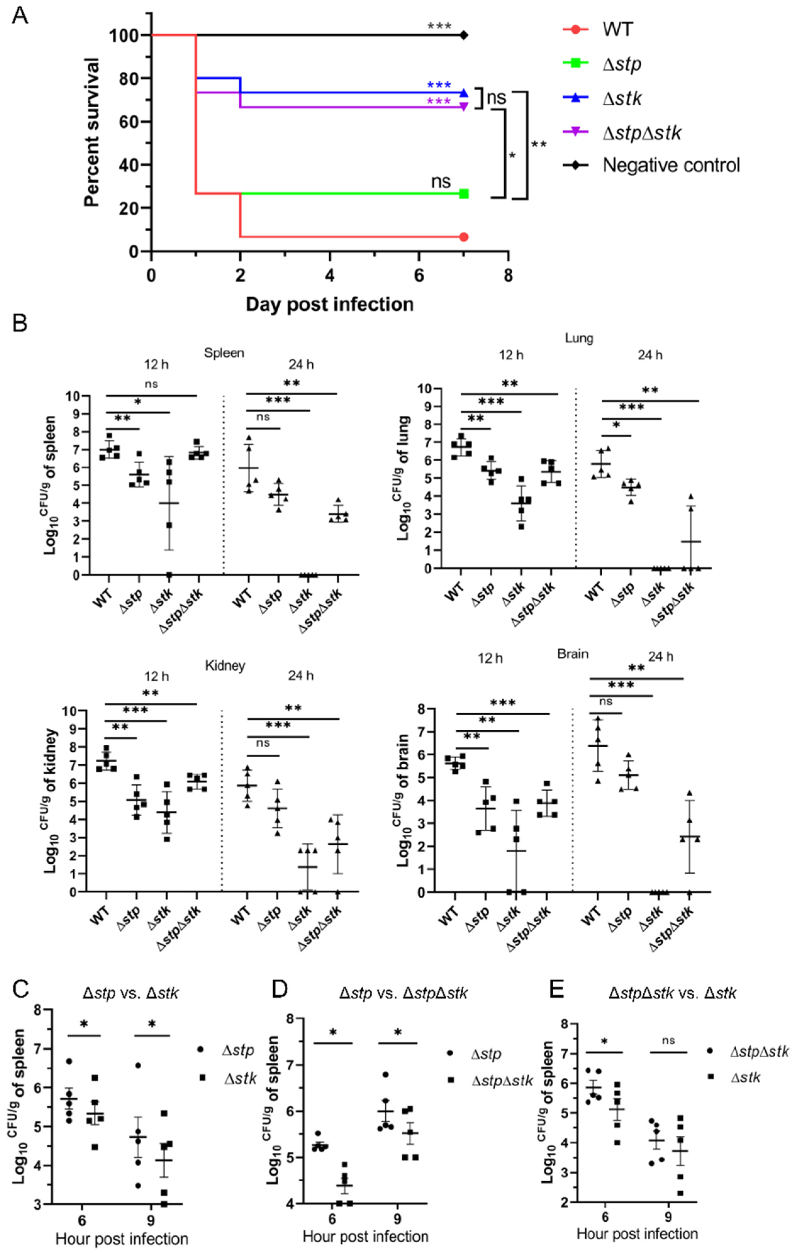
STP and STK are believed to be cognate pairs responsible for reversible protein phosphorylation, and they have both been shown to be involved in bacterial pathogenesis [34,35,56]. However, the exact role of each protein in S. suis pathogenesis remains to be further investigated. Therefore, we used a mouse infection model to compare the virulence of Δstp, Δstk, and ΔstpΔstk strains with that of the WT strain of S. suis. In the mouse survival assay, only 7% (1/15) of the mice intraperitoneally injected with 8 × 10⁸ CFU of the WT strain survived. In comparison, the survival rates for the Δstp, Δstk, and ΔstpΔstk strains were 27% (4/15), 73% (11/15), and 67% (10/15), respectively, indicating that stk played a more important role than stp in regulating the virulence of S. suis (Figure 2A). We further performed an in vivo colonization assay in mice with the WT strain and three mutants. The results showed that from 12 h post-infection (hpi) to 24 hpi, the bacterial load of the WT strain in each organ slowly decreased, indicating a strong ability to colonize the host. In contrast, the bacterial loads of the mutant strains showed significantly faster clearance in each organ. In particular, at 24 hpi, the Δstk strain was almost completely cleared in the lung, spleen, and brain (Figure 2B), which was consistent with the results of the mouse survival assay. In contrast, the Δstp and ΔstpΔstk strains showed a significantly higher ability to colonize mice than the Δstk strain. To more accurately compare the virulence of the mutants, we performed a series of competitive infection assays. As shown in Table 1 and Figure 2C–E, Δstp significantly outcompeted Δstk (CI6h = 1.16746, p < 0.05; CI9h = 1.24600, p < 0.05) and ΔstpΔstk (CI6h = 1.54531, p < 0.05; CI9h = 1.39062, p < 0.05). ΔstpΔstk showed a significantly higher bacterial load than Δstk at 6 hpi (CI6h = 1.26691, p < 0.05), but no significant difference in bacterial load was observed at 9 hpi (CI9h = 1.24696, p > 0.05). Collectively, our data demonstrated that the deletion of stk significantly attenuated the virulence of S. suis, which was consistent with the observations in our previous reports [17]. In comparison, stp deletion resulted in reduced impact compared to the stk deletion on impairing the virulence of S. suis.

3.3. The Role of STP and STK in Growth and Cell Division

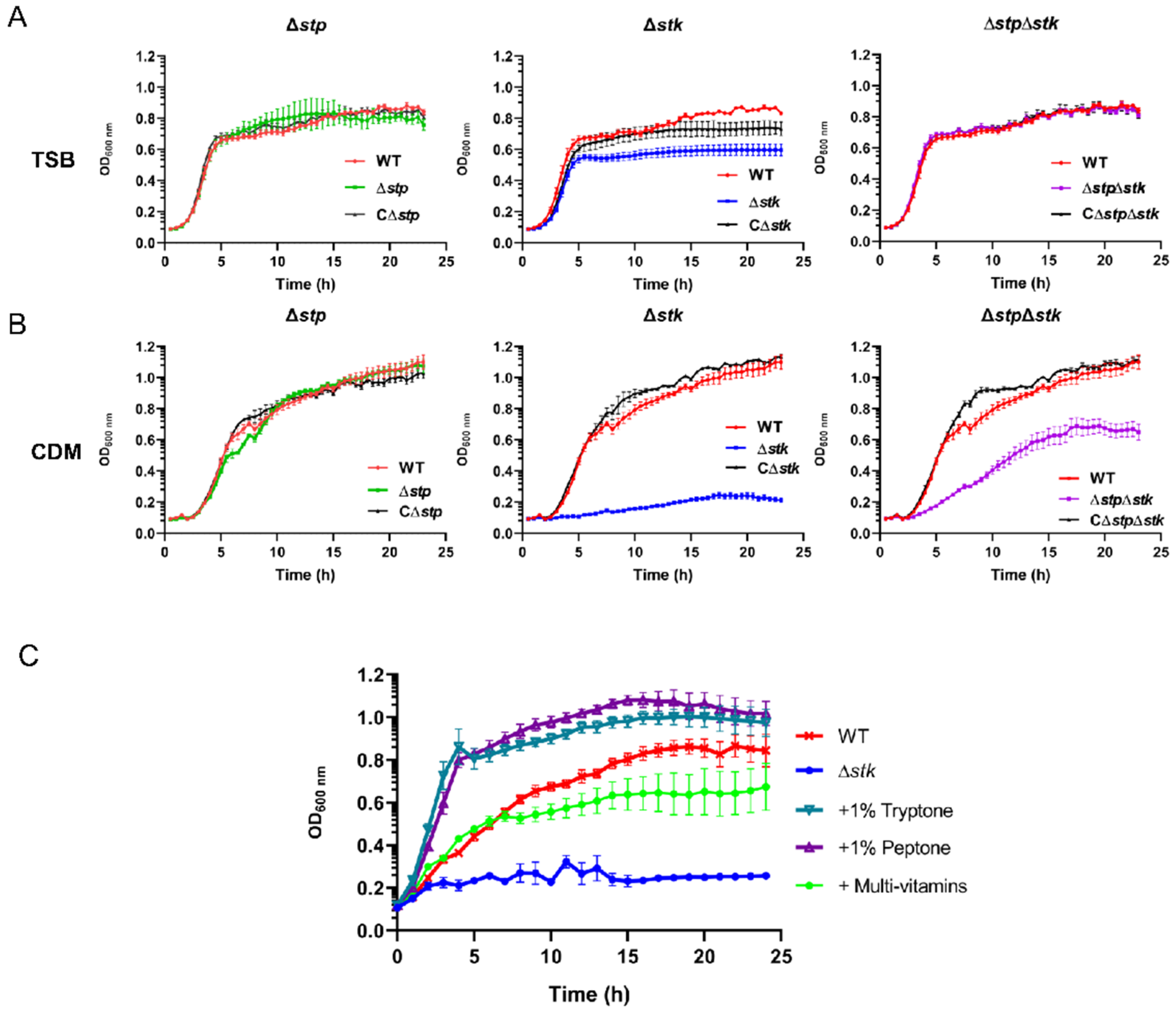
We further tested other important phenotypes, including growth and cell division of the Δstp, Δstk, and ΔstpΔstk strains. The growth of each mutant and WT strain in TSB medium or chemically defined minimal medium (CDM) was tested. As shown in Figure 3A, the growth of WT, Δstp, and ΔstpΔstk was comparable in TSB, whereas Δstk presented retarded growth and reached a lower OD600 nm in the stationary phase (Figure 3A). Surprisingly, the Δstk strain could barely grow in CDM (Figure 3B). However, the growth of Δstp was similar to that of the WT strain, and the growth of ΔstpΔstk was much slower than that of WT but better than that of Δstk (Figure 3B). To determine the cause of such growth defects, CDM supplemented with different carbon sources, casamino acids (CAAs), vitamins, purines, or complex nutrients (tryptone or peptone) was used to test the growth of the Δstk strain (Figure 3C and Figure S1). As shown in Figure 3C, in addition to the observation that the growth of Δstk was greatly increased in CDM supplemented with tryptone or peptone, it was interesting to see that its growth in CDM supplemented with multivitamins was also significantly recovered. These results suggested that STK was more important than STP for the growth of S. suis.
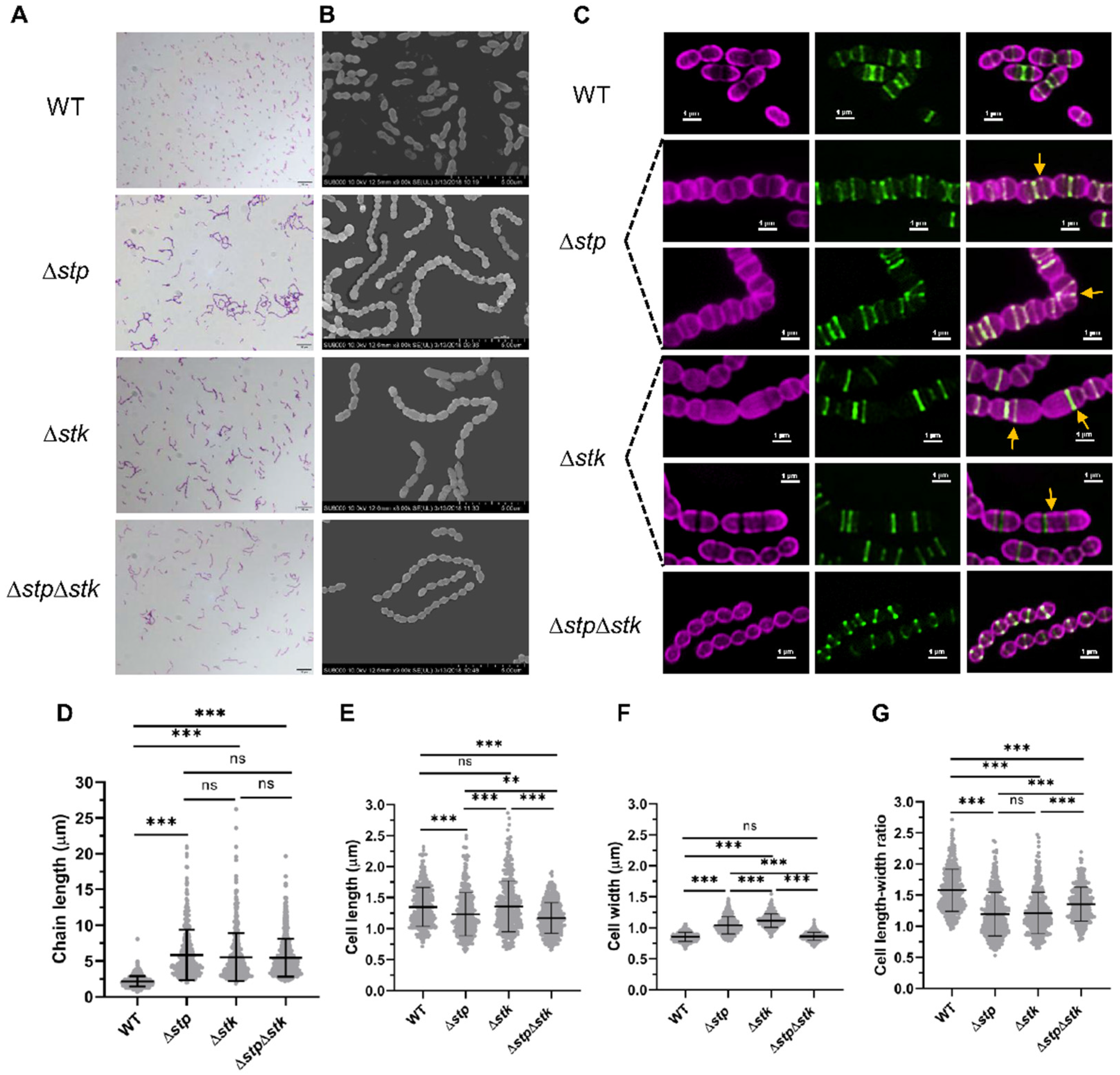
STK has been reported to play important roles in cell division in Streptococcus [23,29], while limited information about the role of STP in cell division has been reported. Therefore, we subsequently analyzed the morphology, cell division, and nascent peptidoglycan synthesis of WT and mutant strains using light microscopy (LM), scanning electron microscopy (SEM), and dual-color structured illumination microscopy (SIM). One of the most significant morphological changes among the mutants was the increase in the length of bacterial chains. All three mutants formed long chains, with each chain comprising over ten cells compared with the WT strain, which showed a normal chain length with each chain harboring three to five cells (Figure 4A–D). The morphology of all the complemented strains was restored (Figure S2). These results revealed the presence of defects in cell division or daughter cell separation. Further analysis with SIM revealed abnormal cell division in both Δstp and Δstk strains. By staining with Alexa Fluor 647 dye (AF-647), which specifically labels the cytoplasmic membrane, and fluorescein-D-lysine (FDL), which labels nascent peptidoglycan (PG), followed by visualization using SIM, the cell morphology and division septum could be clearly observed (Figure 4C). Compared with the WT cells that displayed a standard ellipsoidal shape, 37.3% (142/381) of Δstp cells showed a rounded morphology due to a decreased length-width ratio (Figure 4E–G). Moreover, in 9.4% (36/381) of Δstp cells, the Z-ring was not localized to the mid-cell, and the angles of the Z-rings with respect to the cell long axis were incorrect, leading to the formation of twisted cell chains (Figure 4C, marked by yellow arrows). The Δstk cells also showed abnormal cell division; however, they displayed a distinctly aberrant morphology. Approximately 6.8% (26/385) of the Δstk cells underwent disordered division, leading to the formation of asymmetric daughter cells, and 10.4% (40/385) formed elongated cells containing multiple, unconstricted cell division rings (Figure 4C, marked with yellow arrows). In contrast, the ΔstpΔstk strain showed relatively normal cell division. These results demonstrated that STP and STK both played an essential role in cell division.

3.4. Comparative Proteomic Analysis Revealing the Influence of stp and stk on Protein Expression

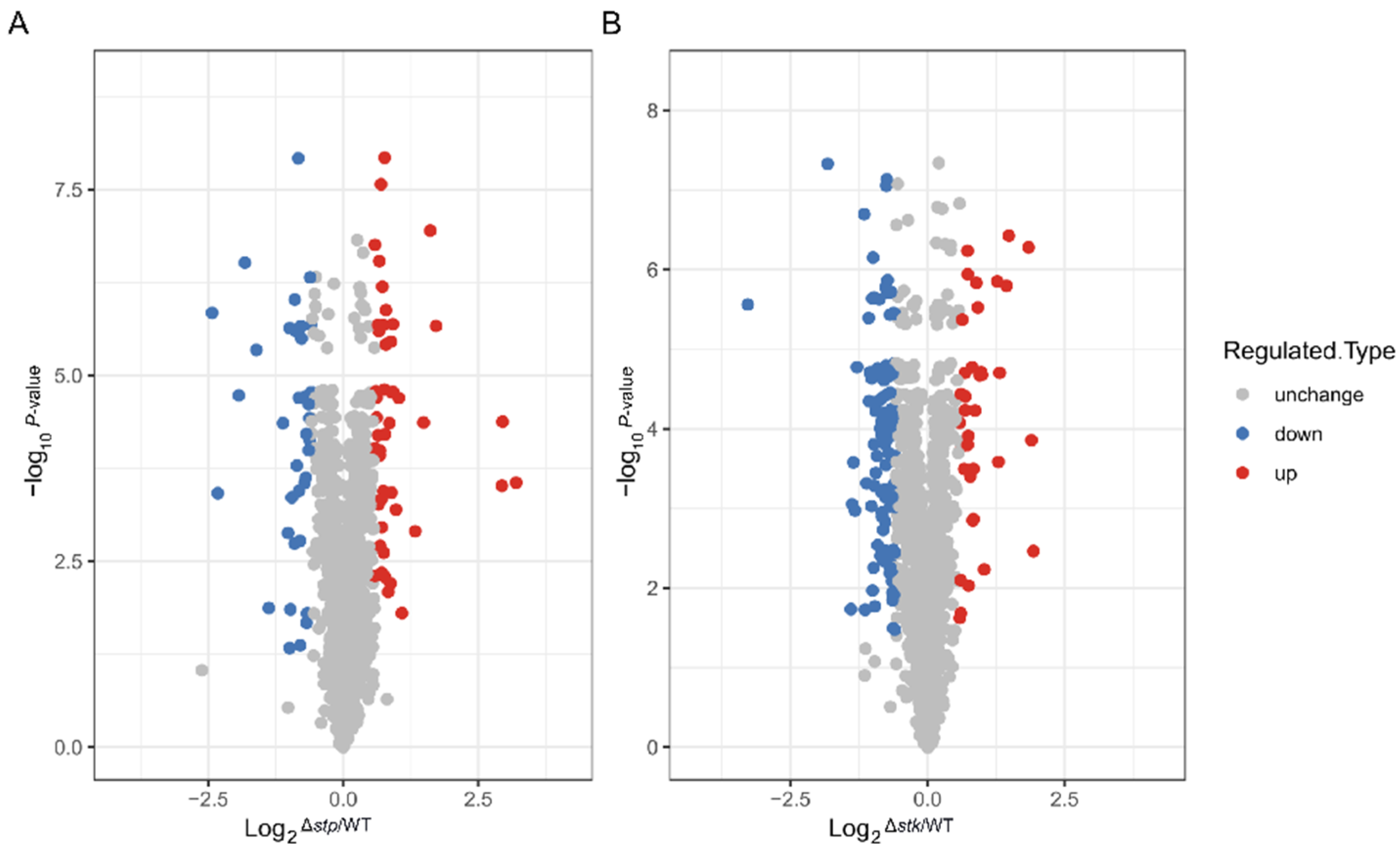
To investigate the influence of STP and STK on protein expression in S. suis, we performed a comparative proteomics analysis with the WT, Δstp, and Δstk strains using liquid chromatography mass spectrometry/mass spectrometry (LC-MS/MS), as illustrated in Figure S3A. A total of 1427 proteins were identified, constituting 72% of the proteins encoded by the S. suis SC19 genome. Among these identified proteins, the abundance of 1379 proteins was quantitatively compared between the WT and each mutant strain (Figure S3B). As shown in Figure 5A,B, compared with the WT strain, the Δstk strain possessed the most dramatic change in protein abundance, in which 139 genes were differentially expressed, with 37 proteins upregulated and 102 proteins downregulated. For the Δstp strain, 82 genes were differentially expressed compared with the WT strain, including 47 upregulated and 35 downregulated genes (fold change > 1.5, p < 0.05).
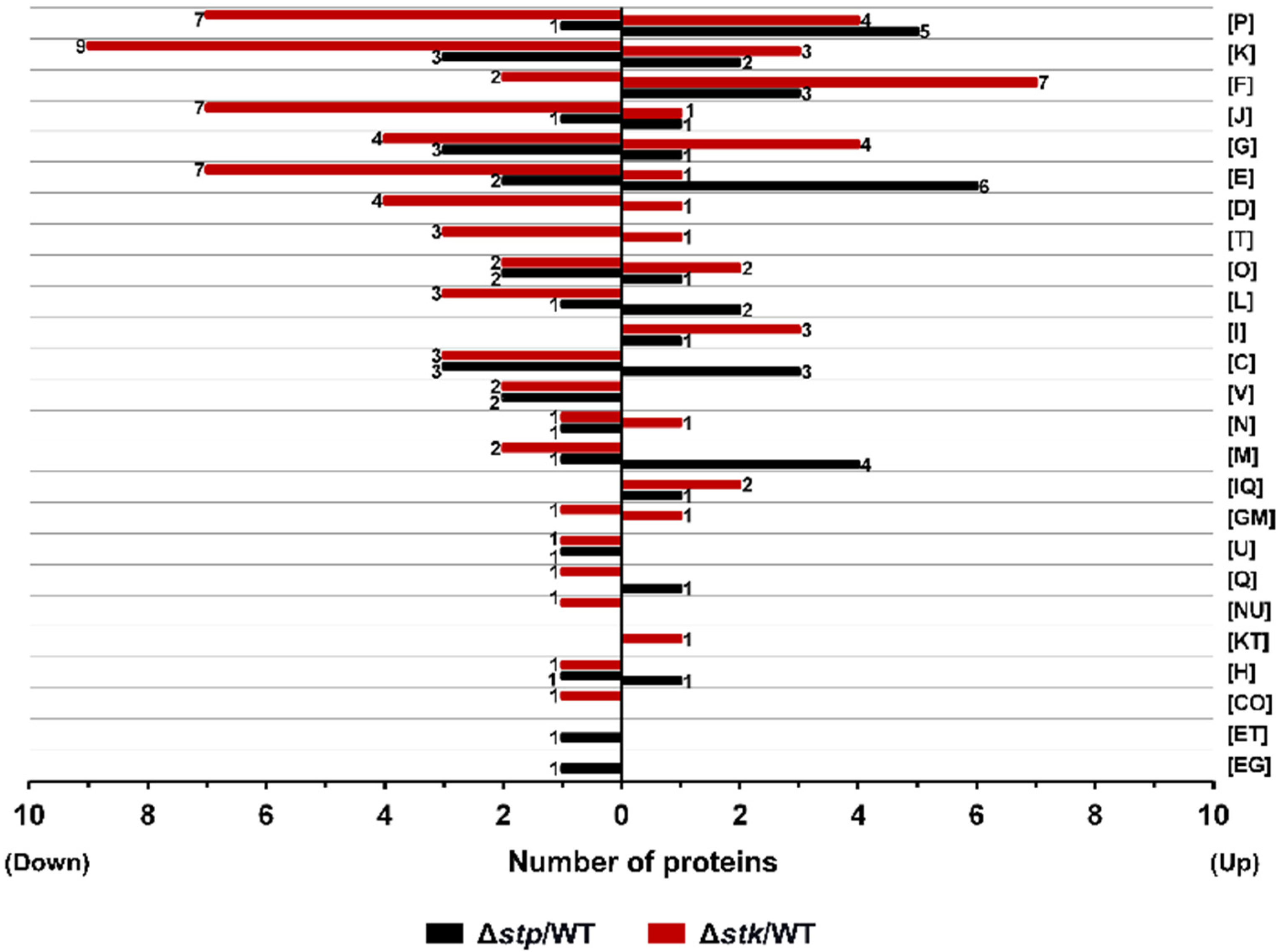
The EggNOG v5.0 database was used to analyze the functions of the differentially expressed proteins [57]. As shown in Figure 6, apart from the proteins with unknown functions, the differentially expressed proteins between the WT strain and each mutant were found to be distributed in almost all functional groups. Δstk harbored the most downregulated proteins, in which the differentially expressed proteins most frequently fell in categories including K (Transcription), P (Inorganic ion transport and metabolism), J (Translation, ribosomal structure, and biogenesis), and G (Carbohydrate transport and metabolism). In contrast, Δstp presented fewer downregulated proteins but had more proteins with increased abundance in which the differentially expressed proteins most frequently fell in categories including E (Amino acid transport and metabolism), P (Inorganic ion transport and metabolism), K (Transcription), and C (Energy production and conversion) (Figure 6). The data suggested that both STP and STK were global regulators of protein expression. As Δstk showed retarded growth in CDM but the growth was restored in CDM supplemented with multivitamins, by analyzing the differentially expressed genes in the mutants, we found that a coA-binding protein (NZ_CP020863.1_prot_WP_012027318.1_1268) was significantly downregulated in Δstk but was not differentially expressed in Δstp (Table S3).
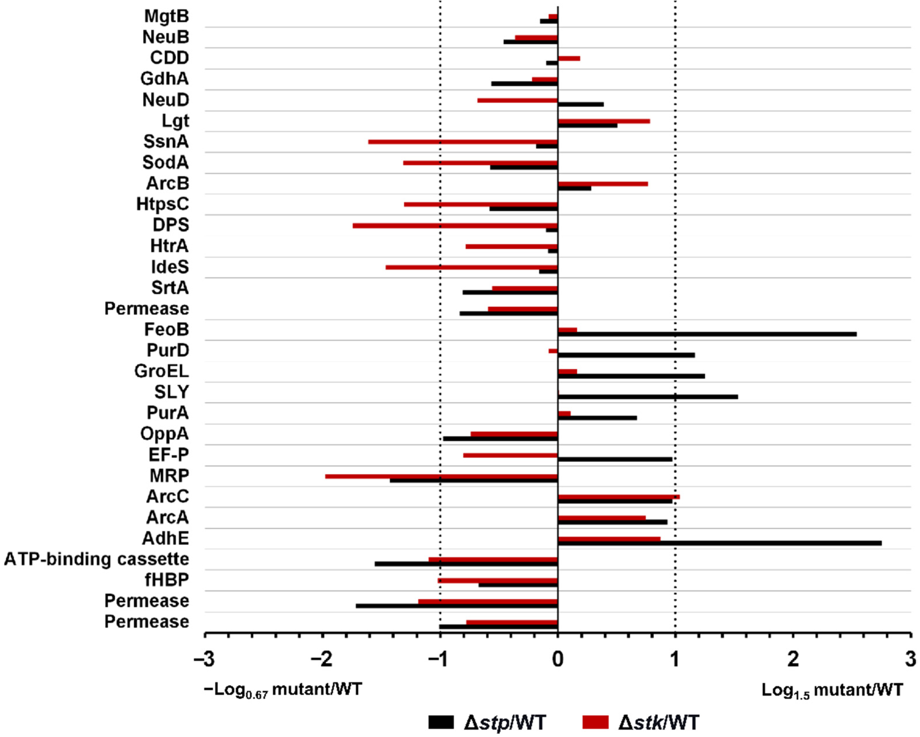
As there was a difference in virulence between Δstp and Δstk strains, the abundance of several known virulence factors (VFs) in the mutant strains was compared with that in the WT strain [58]. As shown in Figure 7, among the 30 VFs, nine were significantly downregulated, including SsnA (B9H01_09460), SodA (B9H01_07435), HtpsC (B9H01_07610), DPS (B9H01_08150), IdeS (B9H01_02750), MRP (B9H01_03725), ATP-binding cassette (B9H01_06780), fHBP (B9H01_01430), and Permease (B9H01_06785), and only one, ArcC (B9H01_03140), was upregulated in Δstk. In contrast, in Δstp, only three VFs were significantly downregulated, including MRP (B9H01_03725), ATP-binding cassette (B9H01_06780), and Permease (B9H01_06785), and five VFs were upregulated, including FeoB (B9H01_06805), PurD (B9H01_00260), GroEL (B9H01_00900), SLY (B9H01_06770), and AdhE (B9H01_01465), compared with those in the WT strain. These results corresponded with the virulence phenotype associated with the disruption of STK with significantly attenuated virulence of S. suis, while deletion of STP showed a gently reduced virulence.

3.5. Comparative Phosphoproteomic Analysis

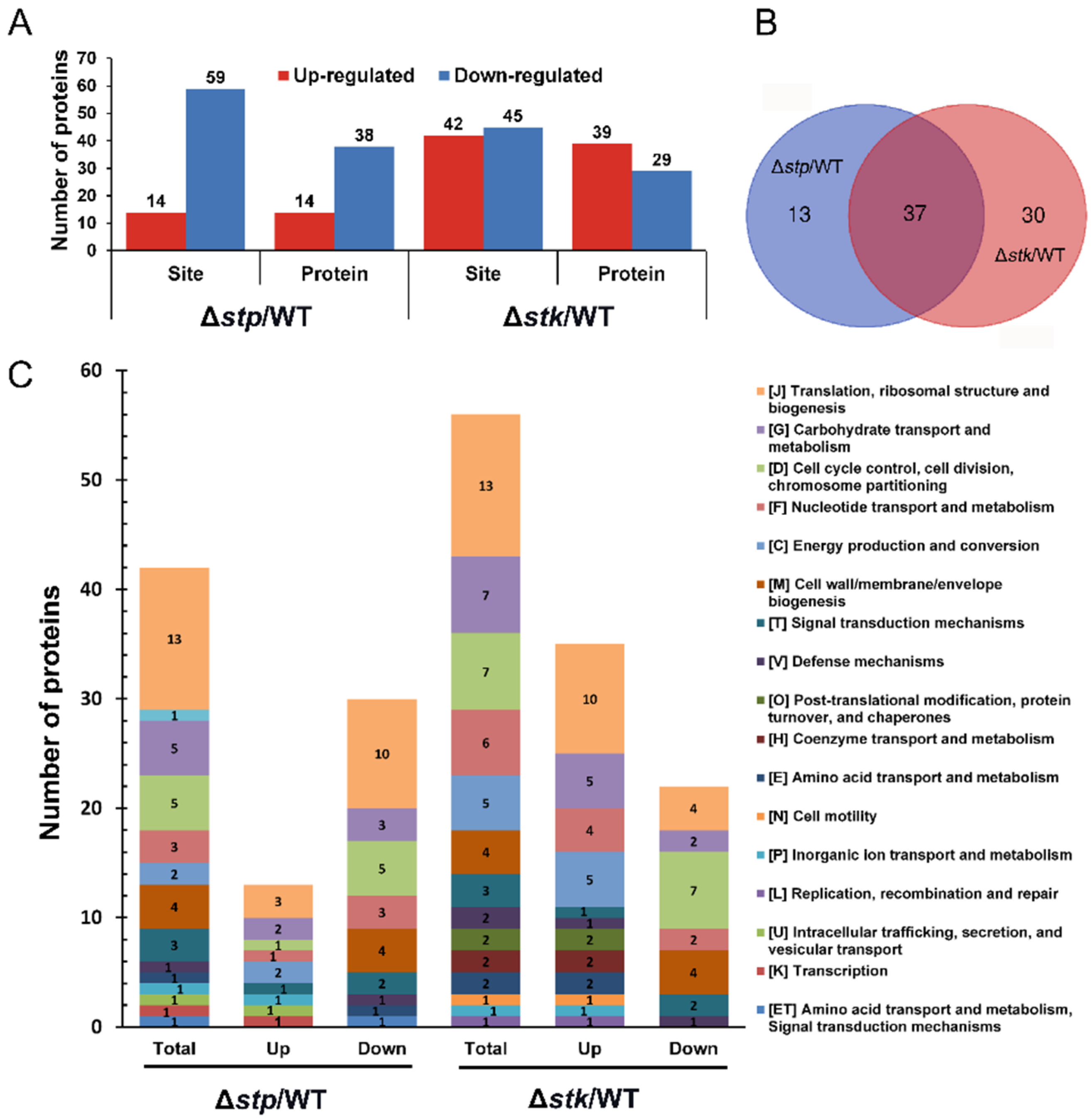
STK and STP are serine/threonine kinases and phosphatases, respectively. Therefore, we next analyzed the changes in the abundance of serine/threonine phosphorylated proteins in Δstp and Δstk compared with the WT strain. In Δstp, a total of 73 unique phosphosites involving 50 proteins showed a significantly altered abundance (fold change > 1.5, p < 0.05), among which 14 proteins were less abundant and 38 proteins were more abundant compared with those in the WT strain. Between the Δstk and WT strains, 87 unique phosphosites involving 67 proteins were identified with a changed abundance (fold change > 1.5, p < 0.05), among which 29 proteins were less abundant and 39 proteins were more abundant (Figure 8A and Table S4). A Venn diagram analysis showed that a total of 37 proteins were identified with changed abundance in both the Δstp and Δstk strains, suggesting potential overlapping functions of STP and STK (Figure 8B).
By COG analysis, it was shown that the phosphoproteins with changed abundance in both Δstp and Δstk strains were mainly related to “Translation, ribosomal structure and biogenesis”, “Carbohydrate transport and metabolism” and “Cell cycle control, cell division, chromosome partitioning”. In most of the COG categories, Δstk presented more phosphoproteins with altered abundance than Δstp (Figure 8C and Table S3), which coincided with the above observations that deletion of stk caused more severe phenotypic changes than stp deletion, including the impact on growth, metabolism, and virulence of S. suis.
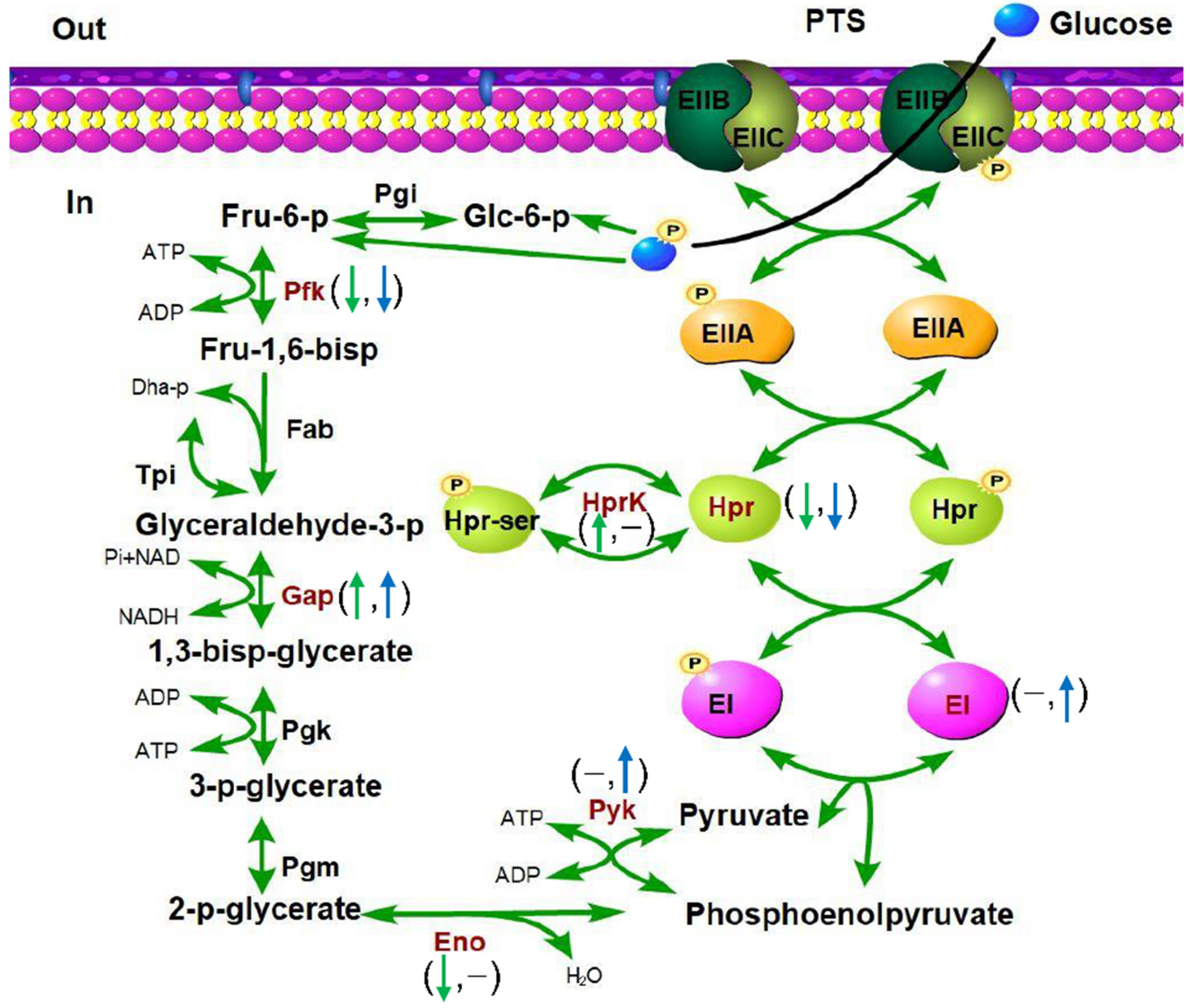
Specifically, as shown in Table S4, several important proteins involved in central energy metabolism were identified to contain phosphosites with altered abundance in both Δstp and Δstk strains. HprK is a metabolic regulator that mediates the phosphorylation of HPr to control the metabolic rate [59]. The phosphorylation at S/T residues of Hpr was downregulated in Δstp and Δstk, while that of an S residue of HprK was upregulated only in Δstp. The phosphorylation levels of GAPDH and GlmM were increased in both mutant strains compared with the WT strain, while the phosphorylation level of PfkA was decreased in both mutant strains compared with the WT strain. The phosphosite abundances of PtsEI and Pyk were increased in Δstk and did not change in Δstp. However, Eno exhibited an opposite situation (Figure 9).
Next, we analyzed the proteins with a reduced phosphorylation level in the Δstk strain and increased phosphorylation level in the Δstp strain, which might be their potential direct substrate proteins. Among the 29 proteins that showed a reduced abundance after enrichment of Ser/Thr phosphomodified peptides in Δstk, a large proportion were cell division-related proteins, including MapZ, GpsB, MapZ, FtsZ, DivIVA, SepF, FtsW, MltG, Jag, and GlmS. The remaining proteins were mainly involved in metabolism, such as Hpr, NeuB, PfKA, ADK, GT, PhoH, and LmrC, and protein translation, such as InfB, EF-P, EF-G, and BipA (Table 2). In contrast, among the fourteen proteins that showed increased abundance after Ser/Thr phosphomodified peptide enrichment in the Δstp strain, only four proteins, including DivIVA, STK, MltG, and GlmM, were involved in cell division, and the others were involved in metabolism, such as NADP, PrfA, GAPDH, OppF, Hprk, ArgS, SecA, and IDH (Table 2). These results suggested that STP and STK might affect the cell division, energy metabolism, and protein translation of S. suis in a manner dependent on Ser/Thr protein phosphorylation.

4. Discussion

The eukaryote-like Ser/Thr protein kinases (STKs) and phosphatases (STPs) in bacteria have recently attracted extensive research interest due to their important roles in cell division, metabolism, and pathogenesis in many important bacterial pathogens [22,35,56,60,61,62]. STKs and STPs comprise a complex regulatory system, of which the mechanism of regulation has not been fully understood. Many studies have been performed to reveal the functions of STKs and STPs, however, most of them focus on STK or STP individually. To provide a holistic understanding of this complex regulatory system, systemic investigations are needed. Therefore, we performed a comparative study in S. suis, an important zoonotic bacterial pathogen where only one copy of gene encoding STK and its cognate STP is present, with ∆stk, ∆stp, ∆stkstp, and the wild-type strains. Comprehensive phenotypic analysis combined with comparative proteomics and phosphoproteomics analysis were carried out to reveal the STK/STP mediated regulation network.
In the animal infection assay, we showed that STK was essential for the virulence of S. suis, which is consistent with previous studies [17,20]. Some previous studies also showed that deletion of stp led to decreased virulence [42,63]. Similarly, we presented detailed data showing that stp deletion also compromised the virulence of S. suis, however, the degree in virulence attenuation was much lower than that of stk deletion. The comparative proteomic results also revealed that more VFs showed downregulated expression in the Δstk strain than in the Δstp strain, which could potentially explain the difference in virulence between the two strains.
In the growth assay, it is interesting to see that the growth of the Δstk strain was almost abolished in CDM, and the addition of multivitamins largely restored this growth defect. Among the differentially expressed proteins in Δstk, we found that coA-binding protein was significantly downregulated (Table S3) and was not differentially expressed in Δstp, which might explain the partial recovering effect of multivitamins on Δstk.
It was noticed that in the results of both virulence evaluation and growth assays found that the double deletion mutant did not display a more severe attenuation than the single deletion mutant Δstk. One possible explanation could be that potential phosphorylation systems in addition to the Ser/Thr kinase-phosphatase system may exist that have common targets. Indeed, crosstalk between different phosphorylation systems has been reported [64,65]. Under this circumstance, when STK and STP are both deleted, the substrate proteins may be phosphorylated by other phosphorylation systems, therefore giving phenotypes that are less severe than each of the single deletion mutants.
Bacterial cell division is a vital process requiring sophisticated spatial and temporal regulation to maintain accurate proliferation and proper morphology [66,67]. Our results showed that both Δstp and Δstk displayed a chained morphology, which was consistent with previous studies of that impaired in cell division [22,62] and peptidoglycan synthesis [23,33,68]. Comparative phosphoproteomic data in this study revealed that 31% of the proteins with a decreased phosphorylation level in the Δstk strain were cell division related proteins, including MapZ, GpsB, FtsZ, DivIVA, SepF, FtsW, MltG [69], Jag [70], and GlmS [71]. Among these proteins, MapZ [26], GpsB [72], FtsZ [28], DivIVA [24], and Jag [73] have been demonstrated to be direct substrates of STK, while whether SepF, FtsW, MltG, and GlmS are STK substrates needs further confirmation. In the Δstp strain, DivIVA, STK, MltG, and GlmM also showed increased phosphorylation levels, suggesting that these proteins may be common targets of STP and STK. The high prevalence of cell division-related proteins among the proteins with changed phosphorylation states in Δstp and Δstk further suggests the important roles of STP and STK in S. suis cell division.
Other than cell division-related proteins, it was observed that in Δstk, several translation-related proteins also showed a downregulated phosphorylation level, which is consistent with previous studies [27,37]. Although other phosphatases also exist in S. suis, our phosphoproteomic data showed that the phosphorylation level of a variety of proteins was significantly changed in Δstp, indicating an important role of STP. We also performed a phosphatase activity assay using the method previously described [74] with the WT, Δstp, and CΔstp strains, which showed that the total phosphatase activity of the Δstp stain was indeed significantly lower than that of the WT strain (Figure S4). It is worth noting that Table 2 shows the proteins with downregulated phosphorylation in the Δstk strain and upregulated phosphorylation in the Δstp strain, which are more likely to be direct target proteins. As shown in Table S4, other proteins had upregulated phosphorylation levels in the Δstk strain and downregulated phosphorylation levels in the Δstp strain. The influence of STP and STK on the phosphorylation of these proteins could be an indirect effect. When comparing the proteins with altered phosphorylation in the Δstk strain and Δstp strain, there were some overlapping proteins, such as DivIVA and MltG. However, many more of the proteins were found specifically in each individual mutant strain. These data suggest that STP and STK functionally overlap but may also have specific functions, which has also been reported in previous studies. For example, in S. pneumonia, protein including Pgm, ComB, and RsMF are found to have changed phosphorylation status in Δstp but not in Δstk by the phosphoproteomics analysis, indicating the presence of potential STP-specific substrates [8]. In S. pyogenes, STP is shown to be secreted to the host cells to potentially activate proapoptotic signaling cascades; however, its cognate STK is unlikely to interfere with the host cell signaling [39]
In this study, comparative proteomics and phosphoproteomics analysis were used to reveal how STK and STP affect the growth, cell division, and virulence of S. suis. However, the omics data can just provide some clues. More experiments are needed to further reveal the underlying mechanisms. For example, questions, such as how STK affects the cell division of S. suis and which substrate proteins are responsible for the formation of the aberrant morphology and mislocalized Z-ing, and why STK deletion causes different growth defects in different growth media need further elucidation. Moreover, in the phosphoproteomics analysis, a variety of proteins showed downregulated phosphorylation in Δstk and upregulated phosphorylation in Δstp. These could be their potential substrate proteins. However, more studies are needed to verify them.

5. Conclusions

Although the STK and cognate STP are involved in the reversible protein phosphorylation, this study suggests that STK may play a much more important role in S. suis growth and pathogenesis than STP. Both ∆stp and ∆stk were defective in cell division, where they displayed an aberrant cell morphology. The ∆stk strain presented many more differentially expressed proteins than the ∆stp strain when compared with the wild-type strain. Many more known virulence factors were downregulated in ∆stk than in ∆stp. In ∆stk a substantial number of the proteins with reduced abundance after phosphopeptide enrichment were involved in cell division, followed by energy metabolism and protein translation. However, only a few proteins showed an increased abundance after phosphopeptide enrichment, which was also related to cell division. Collectively, both STP and STK are critical regulatory proteins in S. suis, and STK may play a more important role than STP in growth, cell division, and pathogenesis.

Supplementary Materials

The following are available online at https://www.mdpi.com/article/10.3390/microorganisms9122442/s1. Figure S1: Growth analysis of ∆stk strains. (A) The growth of ∆stk strain in CDM or CDM supplemented with each indicated nutrient. Growth was monitored with an automatic plate reader with shaking at 37 °C. Data are presented as means ± SD of triplicate. Figure S2: Morphological analysis of C∆stp, C∆stk, and C∆stp∆stk strains. Gram staining (A) and scanning electron microscopy (SEM) (B) analysis of C∆stp, C∆stk, and C∆stp∆stk strains. Figure S3: Comparative proteomic analysis between the WT and Δstp or Δstk strains. (A) The systematic workflow of the quantitative profiling of the global mass spectrum; (B) The basic statistics of mass spectrum data. Figure S4: Phosphatase activity assay. Cells at the mid-log phase of each indicated strain were collected, washed three times with buffer1 [20 mM Tris-HCl (pH 8.0) and 200 mM NaCl2], resuspended in lyse buffer [50 mM Tris-HCl (pH 8.0), 150 mM NaCl2, 100 U/mL mutanolysin (Sigma), and 1 mM PMSF], and lysed by sonication. The amount of total protein loaded was normalized using a Micro BCA protein assay kit (Cwbiotech, China). Samples were mixed with buffer2 [50 mM Tris-HCl (pH 8.0), 2 mM MnCl2, and 20 mM p-nitrophenyl phosphate (p-NPP)], and incubated at 37 °C for 10 min. The absorbance at 405 nm was then measured. Purified recombinant STP protein was used as the positive control, the reaction buffer2 used as the blank. Data are presented as means ± SD of triplicate. Statistical significance was determined by two-tailed, unpaired Student’s t-tests (ns, p value > 0.05; *, p value < 0.05). Table S1: Strains and plasmids used in this study. Table S2: oligonucleotides used in this study. Table S3: The differentially expressed proteins in ∆stp and ∆stk compared with WT. Table S4: Proteins identified with changed abundance after phosphopeptide enrichment in ∆stp and ∆stk compared with WT.

Author Contributions

R.Z. and Q.H. (Qi Huang): Funding acquisition, Conceptualization, Project administration, Writing—review & editing. Q.H. (Qiao Hu): Investigation, Project administration, Visualization, Writing—original draft. L.Y., X.L., L.-S.Z., H.-T.L., T.-T.L., Q.-G.J., M.-F.T. and L.L.: Performed the statistical analysis, and contributed with reagents/materials/analysis tools. R.R.D.: Writing—review & editing. All authors have read and agreed to the published version of the manuscript.

Funding

This work was funded by the National Key Research and Development Plans (2018YFE0101600 & 2017YFD0500201).

Institutional Review Board Statement

This study was approved by the Laboratory Animal Ethics Committee of Huazhong Agriculture University (HZAUMO-2019-074), and implemented in accordance with the animal care and ethics guidelines. This article does not contain any studies with human participants performed by any of the authors.

Informed Consent Statement

Not applicable.

Data Availability Statement

The mass spectrometry proteomics data have been deposited to the ProteomeXchange Consortium via the PRIDE partner repository with the dataset identifier PXD029994.

Acknowledgments

We thank Zhe Hu from the State Key Laboratory of Agricultural Microbiology, Huazhong Agricultural University for the support in microscopy analysis. We are also grateful to Yosuke Murakami for his pSET plasmids.

Conflicts of Interest

The authors declare no conflict of interest.

References

  1. Hunter, T. Protein Kinases and Phosphatases: The Yin and Yang of Protein Phosphorylation and Signaling. Cell 1995, 80, 225–236. [Google Scholar] [CrossRef] [Green Version]
  2. Goulian, M. Two-Component Signaling Circuit Structure and Properties. Curr. Opin. Microbiol. 2010, 13, 184–189. [Google Scholar] [CrossRef] [PubMed] [Green Version]
  3. Pereira, S.F.F.; Goss, L.; Dworkin, J. Eukaryote-Like Serine/Threonine Kinases and Phosphatases in Bacteria. Microbiol. Mol. Biol. Rev. 2011, 75, 192–212. [Google Scholar] [CrossRef] [Green Version]
  4. Shi, L.; Ji, B.; Kolar-Znika, L.; Boskovic, A.; Jadeau, F.; Combet, C.; Grangeasse, C.; Franjević, D.; Talla, E.; Mijakovic, I. Evolution of Bacterial Protein-Tyrosine Kinases and Their Relaxed Specificity Toward Substrates. Genome Biol. Evol. 2014, 6, 800–817. [Google Scholar] [CrossRef] [PubMed]
  5. Shi, L.; Pigeonneau, N.; Ravikumar, V.; Dobrinic, P.; Macek, B.; Franjević, D.; Noirot-Gros, M.-F.; Mijakovic, I. Cross-Phosphorylation of Bacterial Serine/Threonine and Tyrosine Protein Kinases on Key Regulatory Residues. Front. Microbiol. 2014, 5, 495. [Google Scholar] [CrossRef] [PubMed] [Green Version]
  6. Osaki, M.; Arcondéguy, T.; Bastide, A.; Touriol, C.; Prats, H.; Trombe, M.-C. The StkP/PhpP Signaling Couple in Streptococcus pneumoniae: Cellular Organization and Physiological Characterization. J. Bacteriol. 2009, 191, 4943–4950. [Google Scholar] [CrossRef] [PubMed] [Green Version]
  7. Nováková, L.; Sasková, L.; Pallová, P.; Janeček, J.; Novotná, J.; Ulrych, A.; Echenique, J.; Trombe, M.-C.; Branny, P. Characterization of a Eukaryotic Type Serine/Threonine Protein Kinase and Protein Phosphatase of Streptococcus pneumoniae and Identification of Kinase Substrates. FEBS J. 2005, 272, 1243–1254. [Google Scholar] [CrossRef] [PubMed]
  8. Hirschfeld, C.; Mejia, A.G.; Bartel, J.; Hentschker, C.; Rohde, M.; Maaß, S.; Hammerschmidt, S.; Becher, D. Proteomic Investigation Uncovers Potential Targets and Target Sites of Pneumococcal Serine-Threonine Kinase StkP and Phosphatase PhpP. Front. Microbiol. 2019, 10, 3101. [Google Scholar] [CrossRef] [PubMed] [Green Version]
  9. Refaya, A.K.; Sharma, D.; Kumar, V.; Bisht, D.; Narayanan, S. A Serine/threonine kinase PknL is involved in the Adaptive Response of Mycobacterium tuberculosis. Microbiol. Res. 2016, 190, 1–11. [Google Scholar] [CrossRef]
  10. Malhotra, V.; Arteaga-Cortés, L.T.; Clay, G.; Clark-Curtiss, J.E. Mycobacterium tuberculosis protein kinase K Confers Survival Advantage During Early Infection in Mice and Regulates Growth in Culture and during Persistent Infection: Implications for Immune Modulation. Microbiology 2010, 156, 2829–2841. [Google Scholar] [CrossRef] [PubMed] [Green Version]
  11. Débarbouillé, M.; Dramsi, S.; Dussurget, O.; Nahori, M.-A.; Vaganay, E.; Jouvion, G.; Cozzone, A.; Msadek, T.; Duclos, B. Characterization of a Serine/Threonine Kinase Involved in Virulence of Staphylococcus aureus. J. Bacteriol. 2009, 191, 4070–4081. [Google Scholar] [CrossRef] [Green Version]
  12. Liu, Q.; Fan, J.; Niu, C.; Wang, D.; Wang, J.; Wang, X.; Villaruz, A.E.; Li, M.; Otto, M.; Gao, Q. The Eukaryotic-Type Serine/Threonine Protein Kinase Stk is Required for Biofilm Formation and Virulence in Staphylococcus epidermidis. PLoS ONE 2011, 6, e25380. [Google Scholar] [CrossRef] [PubMed]
  13. Echenique, J.; Kadioglu, A.; Romao, S.; Andrew, P.W.; Trombe, M.-C. Protein Serine/Threonine Kinase StkP Positively Controls Virulence and Competence in Streptococcus pneumoniae. Infect. Immun. 2004, 72, 2434–2437. [Google Scholar] [CrossRef] [Green Version]
  14. Herbert, J.; Mitchell, A.; Mitchell, T. A Serine-Threonine Kinase (StkP) Regulates Expression of the Pneumococcal Pilus and Modulates Bacterial Adherence to Human Epithelial and Endothelial Cells in Vitro. PLoS ONE 2015, 10, e0127212. [Google Scholar] [CrossRef] [Green Version]
  15. Bugrysheva, J.; Froehlich, B.J.; Freiberg, J.A.; Scott, J.R. Serine/Threonine Protein Kinase Stk is Required for Virulence, Stress Response, and Penicillin Tolerance in Streptococcus pyogenes. Infect. Immun. 2011, 79, 4201–4209. [Google Scholar] [CrossRef] [Green Version]
  16. Jin, H.; Pancholi, V. Identification and Biochemical Characterization of a Eukaryotic-type Serine/Threonine Kinase and its Cognate Phosphatase in Streptococcus pyogenes: Their Biological Functions and Substrate Identification. J. Mol. Biol. 2006, 357, 1351–1372. [Google Scholar] [CrossRef]
  17. Zhang, C.; Sun, W.; Tan, M.; Dong, M.; Liu, W.; Gao, T.; Li, L.; Xu, Z.; Zhou, R. The Eukaryote-Like Serine/Threonine Kinase STK Regulates the Growth and Metabolism of Zoonotic Streptococcus suis. Front. Cell. Infect. Microbiol. 2017, 7, 66. [Google Scholar] [CrossRef] [PubMed] [Green Version]
  18. Rui, L.; Weiyi, L.; Yu, M.; Hong, Z.; Jiao, Y.; Zhe, M.; Hongjie, F. The Serine/Threonine Protein Kinase of Streptococcus Suis Serotype 2 Affects the Ability of the Pathogen to Penetrate the Blood-Brain Barrier. Cell Microbiol. 2018, 20, e12862. [Google Scholar] [CrossRef] [PubMed]
  19. Zhu, H.; Zhou, J.; Wang, D.; Yu, Z.; Li, B.; Ni, Y.; He, K. Quantitative Proteomic Analysis reveals that Serine/Threonine Kinase is involved in Streptococcus suis Virulence and Adaption to Stress Conditions. Arch. Microbiol. 2021, 4715–4726. [Google Scholar] [CrossRef] [PubMed]
  20. Zhu, H.; Zhou, J.; Ni, Y.; Yu, Z.; Mao, A.; Hu, Y.; Wang, W.; Zhang, X.; Wen, L.; Li, B.; et al. Contribution of Eukaryotic-Type Serine/Threonine Kinase to Stress Response and Virulence of Streptococcus suis. PLoS ONE 2014, 9, e91971. [Google Scholar] [CrossRef] [PubMed] [Green Version]
  21. Liu, H.; Ye, C.; Fu, H.; Yue, M.; Li, X.; Fang, W. Stk and Stp1 participate in Streptococcus suis serotype 2 Pathogenesis by Regulating Capsule Thickness and Translocation of Certain Virulence Factors. Microb. Pathog. 2021, 152, 104607. [Google Scholar] [CrossRef]
  22. Beilharz, K.; Nováková, L.; Fadda, D.; Branny, P.; Massidda, O.; Veening, J.-W. Control of Cell Division in Streptococcus pneumoniae by the Conserved Ser/Thr Protein Kinase StkP. Proc. Natl. Acad. Sci. USA 2012, 109, E905–E913. [Google Scholar] [CrossRef] [Green Version]
  23. Zucchini, L.; Mercy, C.; Garcia, P.S.; Cluzel, C.; Gueguen-Chaignon, V.; Galisson, F.; Freton, C.; Guiral, S.; Brochier-Armanet, C.; Gouet, P.; et al. PASTA repeats of the Protein Kinase StkP Interconnect Cell Constriction and Separation of Streptococcus pneumoniae. Nat. Microbiol. 2018, 3, 197–209. [Google Scholar] [CrossRef]
  24. Ni, H.; Fan, W.; Li, C.; Wu, Q.; Hou, H.; Hu, D.; Zheng, F.; Zhu, X.; Wang, C.; Cao, X.; et al. Streptococcus suis DivIVA Protein Is a Substrate of Ser/Thr Kinase STK and Involved in Cell Division Regulation. Front. Cell. Infect. Microbiol. 2018, 8, 85. [Google Scholar] [CrossRef] [Green Version]
  25. Kang, C.M.; Nyayapathy, S.; Lee, J.Y.; Suh, J.W.; Husson, R.N. Wag31, a Homologue of the Cell Division Protein DivIVA, Regulates Growth, Morphology and Polar Cell Wall Synthesis in Mycobacteria. Microbiology 2008, 154, 725–735. [Google Scholar] [CrossRef] [PubMed] [Green Version]
  26. Fleurie, A.; Lesterlin, C.; Manuse, S.; Zhao, C.; Cluzel, C.; Lavergne, J.-P.; Franz-Wachtel, M.; Macek, B.; Combet, C.; Kuru, E.; et al. MapZ Marks the Division Sites and Positions FtsZ rings in Streptococcus pneumoniae. Nature 2014, 516, 259–262. [Google Scholar] [CrossRef] [PubMed] [Green Version]
  27. Pompeo, F.; Foulquier, E.; Serrano, B.; Grangeasse, C.; Galinier, A. Phosphorylation of the Cell Division Protein GpsB regulates PrkC Kinase Activity through a Negative Feedback Loop in Bacillus subtilis. Mol. Microbiol. 2015, 97, 139–150. [Google Scholar] [CrossRef] [PubMed]
  28. Maurya, G.K.; Modi, K.; Banerjee, M.; Chaudhary, R.; Rajpurohit, Y.S.; Misra, H.S. Phosphorylation of FtsZ and FtsA by a DNA Damage-Responsive Ser/Thr Protein Kinase Affects their Functional Interactions in Deinococcus radiodurans. mSphere 2018, 3, e00325-18. [Google Scholar] [CrossRef] [Green Version]
  29. Fenton, A.K.; Manuse, S.; Flores-Kim, J.; Garcia, P.S.; Mercy, C.; Grangeasse, C.; Bernhardt, T.G.; Rudner, D.Z. Phosphorylation-Dependent Activation of the Cell Wall Synthase PBP2a in Streptococcus Pneumoniae by MacP. Proc. Natl. Acad. Sci. USA 2018, 115, 2812–2817. [Google Scholar] [CrossRef] [PubMed] [Green Version]
  30. Li, W.; Yin, Y.; Meng, Y.; Ma, Z.; Lin, H.; Fan, H. The Phosphorylation of Phosphoglucosamine Mutase GlmM by Ser/Thr Kinase STK Mediates Cell Wall Synthesis and Virulence in Streptococcus suis serotype 2. Veter. Microbiol. 2021, 258, 109102. [Google Scholar] [CrossRef] [PubMed]
  31. Falk, S.P.; Weisblum, B. Phosphorylation of the Streptococcus pneumoniae Cell Wall Biosynthesis Enzyme MurC by a Eukaryotic-Like Ser/Thr kinase. FEMS Microbiol. Lett. 2013, 340, 19–23. [Google Scholar] [CrossRef] [PubMed] [Green Version]
  32. Iswahyudi; Mukamolova, G.V.; Straatman-Iwanowska, A.A.; Allcock, N.; Ajuh, P.; Turapov, O.; O’Hare, H.M. Mycobacterial phosphatase PstP regulates Global Serine Threonine Phosphorylation and Cell Division. Sci. Rep. 2019, 9, 8337. [Google Scholar] [CrossRef] [Green Version]
  33. Jarick, M.; Bertsche, U.; Stahl, M.; Schultz, D.; Methling, K.; Lalk, M.; Stigloher, C.; Steger, M.; Schlosser, A.; Ohlsen, K. The Serine/Threonine Kinase Stk and the Phosphatase Stp Regulate Cell Wall Synthesis in Staphylococcus aureus. Sci. Rep. 2018, 8, 13693. [Google Scholar] [CrossRef] [PubMed] [Green Version]
  34. Sharma, A.K.; Arora, D.; Singh, L.K.; Gangwal, A.; Sajid, A.; Molle, V.; Singh, Y.; Nandicoori, V.K. Serine/Threonine Protein Phosphatase PstP of Mycobacterium tuberculosis is Necessary for Accurate Cell Division and Survival of Pathogen. J. Biol. Chem. 2016, 291, 24215–24230. [Google Scholar] [CrossRef] [PubMed] [Green Version]
  35. Sajid, A.; Arora, G.; Singhal, A.; Kalia, V.C.; Singh, Y. Protein Phosphatases of Pathogenic Bacteria: Role in Physiology and Virulence. Annu. Rev. Microbiol. 2015, 69, 527–547. [Google Scholar] [CrossRef]
  36. Agarwal, S.; Pancholi, P.; Pancholi, V. Strain-Specific Regulatory Role of Eukaryote-Like Serine/Threonine Phosphatase in Pneumococcal Adherence. Infect. Immun. 2012, 80, 1361–1372. [Google Scholar] [CrossRef] [PubMed] [Green Version]
  37. Wright, D.P.; Ulijasz, A.T. Regulation of Transcription by Eukaryotic-Like Serine-Threonine Kinases and Phosphatases in Gram-Positive Bacterial Pathogens. Virulence 2014, 5, 863–885. [Google Scholar] [CrossRef] [PubMed] [Green Version]
  38. Schultz, C.; Niebisch, A.; Schwaiger, A.; Viets, U.; Metzger, S.; Bramkamp, M.; Bott, M. Genetic and Biochemical Analysis of the Serine/Threonine Protein Kinases PknA, PknB, PknG and PknL of Corynebacterium glutamicum: Evidence for Non-Essentiality and for Phosphorylation of OdhI and FtsZ by Multiple Kinases. Mol. Microbiol. 2009, 74, 724–741. [Google Scholar] [CrossRef] [PubMed] [Green Version]
  39. Agarwal, S.; Agarwal, S.; Jin, H.; Pancholi, P.; Pancholi, V. Serine/Threonine Phosphatase (SP-STP), Cecreted from Streptococcus pyogenes, Is a Pro-apoptotic Protein. J. Biol. Chem. 2012, 287, 9147–9167. [Google Scholar] [CrossRef] [PubMed] [Green Version]
  40. Segura, M.; Calzas, C.; Grenier, D.; Gottschalk, M. Initial Steps of the Pathogenesis of the Infection caused by Streptococcus suis: Fighting against Nonspecific Defenses. FEBS Lett. 2016, 590, 3772–3799. [Google Scholar] [CrossRef] [Green Version]
  41. Zhu, H.; Huang, D.; Zhang, W.; Wu, Z.; Lu, Y.; Jia, H.; Wang, M.; Lu, C. The Novel Virulence-Related Gene stp of Streptococcus suis serotype 9 strain Contributes to a Significant Reduction in Mouse Mortality. Microb. Pathog. 2011, 51, 442–453. [Google Scholar] [CrossRef]
  42. Fang, L.; Zhou, J.; Fan, P.; Yang, Y.; Shen, H.; Fang, W. A Serine/Threonine Phosphatase 1 of Streptococcus suis type 2 is an Important Virulence Factor. J. Veter. Sci. 2017, 18, 439–447. [Google Scholar] [CrossRef] [PubMed] [Green Version]
  43. Li, W.; Liu, L.; Chen, H.; Zhou, R. Identification of Streptococcus suis Genes Preferentially Expressed under Iron Starvation by Selective Capture of Transcribed Sequences. FEMS Microbiol. Lett. 2009, 292, 123–133. [Google Scholar] [CrossRef] [PubMed] [Green Version]
  44. van de Rijn, I.; Kessler, R.E. Growth Characteristics of Group A Streptococci in a New Chemically Defined Medium. Infect. Immun. 1980, 27, 444–448. [Google Scholar] [CrossRef] [Green Version]
  45. Trieu-Cuot, P.; Carlier, C.; Poyart-Salmeron, C.; Courvalin, P. Shuttle Vectors Containing a Multiple Cloning Site and a lacZ Alpha Gene for Conjugal Transfer of DNA from Escherichia coli to Gram-Positive Bacteria. Gene 1991, 102, 99–104. [Google Scholar] [CrossRef]
  46. Takamatsu, D.; Osaki, M.; Sekizaki, T. Thermosensitive Suicide Vectors for Gene Replacement in Streptococcus suis. Plasmid 2001, 46, 140–148. [Google Scholar] [CrossRef] [PubMed]
  47. Zhang, T.; Ding, Y.; Li, T.; Wan, Y.; Li, W.; Chen, H.; Zhou, R. A Fur-like protein PerR regulates Two Oxidative Stress Response Related Operons dpr and metQIN in Streptococcus suis. BMC Microbiol. 2012, 12, 85. [Google Scholar] [CrossRef] [PubMed] [Green Version]
  48. Dortet, L.; Mostowy, S.; Samba-Louaka, A.; Gouin, E.; Nahori, M.A.; Wiemer, E.A.; Dussurget, O.; Cossart, P. Recruitment of the Major Vault Protein by InlK: A Listeria Monocytogenes Strategy to Avoid Autophagy. PLoS Pathog. 2011, 7, e1002168. [Google Scholar] [CrossRef]
  49. Liu, W.; Tan, M.; Zhang, C.; Xu, Z.; Li, L.; Zhou, R. Functional Characterization of murB-potABCD Operon for Polyamine Uptake and Peptidoglycan Synthesis in Streptococcus suis. Microbiol. Res. 2018, 207, 177–187. [Google Scholar] [CrossRef] [PubMed]
  50. Tan, M.-F.; Gao, T.; Liu, W.-Q.; Zhang, C.-Y.; Yang, X.; Zhu, J.-W.; Teng, M.-Y.; Li, L.; Zhou, R. MsmK, an ATPase, Contributes to Utilization of Multiple Carbohydrates and Host Colonization of Streptococcus suis. PLoS ONE 2015, 10, e0130792. [Google Scholar] [CrossRef] [PubMed] [Green Version]
  51. Li, W.; Hu, X.; Liu, L.; Chen, H.; Zhou, R. Induction of Protective Immune Response against Streptococcus suis serotype 2 infection by the Surface Antigen HP0245. FEMS Microbiol. Lett. 2011, 316, 115–122. [Google Scholar] [CrossRef] [PubMed]
  52. Zheng, C.; Xu, J.; Li, J.; Hu, L.; Xia, J.; Fan, J.; Guo, W.; Chen, H.; Bei, W. Two Spx Regulators Modulate Stress Tolerance and Virulence in Streptococcus suis Serotype 2. PLoS ONE 2014, 9, e108197. [Google Scholar] [CrossRef]
  53. Wang, Y.; Zhang, W.; Wu, Z.; Lu, C. Reduced Virulence is an Important Characteristic of Biofilm Infection of Streptococcus suis. FEMS Microbiol. Lett. 2011, 316, 36–43. [Google Scholar] [CrossRef] [PubMed]
  54. Kuru, E.; Tekkam, S.; Hall, E.K.; Brun, Y.V.; Van Nieuwenhze, M.S. Synthesis of Fluorescent D-amino Acids and their use for Probing Peptidoglycan Synthesis and Bacterial Growth in Situ. Nat. Protoc. 2015, 10, 33–52. [Google Scholar] [CrossRef] [Green Version]
  55. Lin, L.; Xu, L.; Lv, W.; Han, L.; Xiang, Y.; Fu, L.; Jin, M.; Zhou, R.; Chen, H.; Zhang, A. An NLRP3 Inflammasome-Triggered Cytokine Storm Contributes to Streptococcal Toxic Shock-Like Syndrome (STSLS). PLoS Pathog. 2019, 15, e1007795. [Google Scholar] [CrossRef] [PubMed]
  56. Pensinger, D.A.; Schaenzer, A.J.; Sauer, J.-D. Do Shoot the Messenger: PASTA Kinases as Virulence Determinants and Antibiotic Targets. Trends Microbiol. 2018, 26, 56–69. [Google Scholar] [CrossRef] [PubMed]
  57. Huerta-Cepas, J.; Szklarczyk, D.; Heller, D.; Hernández-Plaza, A.; Forslund, S.K.; Cook, H.V.; Mende, D.R.; Letunic, I.; Rattei, T.; Jensen, L.J.; et al. eggNOG 5.0: A Hierarchical, Functionally and Phylogenetically Annotated Orthology Resource based on 5090 Organisms and 2502 Viruses. Nucleic Acids Res. 2019, 47, D309–D314. [Google Scholar] [CrossRef] [PubMed] [Green Version]
  58. Zheng, F.; Ji, H.; Cao, M.; Wang, C.; Feng, Y.; Li, M.; Pan, X.; Wang, J.; Qin, Y.; Hu, F.; et al. Contribution of the Rgg Transcription Regulator to Metabolism and Virulence of Streptococcus suis Serotype 2. Infect. Immun. 2011, 79, 1319–1328. [Google Scholar] [CrossRef] [PubMed] [Green Version]
  59. Deutscher, J.; Aké, F.M.D.; Derkaoui, M.; Zébré, A.C.; Cao, T.N.; Bouraoui, H.; Kentache, T.; Mokhtari, A.; Milohanic, E.; Joyet, P. The Bacterial Phosphoenolpyruvate: Carbohydrate Phosphotransferase System: Regulation by Protein Phosphorylation and Phosphorylation-Dependent Protein-Protein Interactions. Microbiol. Mol. Biol. Rev. 2014, 78, 231–256. [Google Scholar] [CrossRef] [Green Version]
  60. Macek, B.; Forchhammer, K.; Hardouin, J.; Weber-Ban, E.; Grangeasse, C.; Mijakovic, I. Protein Post-Translational Modifications in Bacteria. Nat. Rev. Microbiol. 2019, 17, 651–664. [Google Scholar] [CrossRef] [PubMed]
  61. Grangeasse, C. Rewiring the Pneumococcal Cell Cycle with Serine/Threonine- and Tyrosine-kinases. Trends Microbiol. 2016, 24, 713–724. [Google Scholar] [CrossRef]
  62. Fleurie, A.; Manuse, S.; Zhao, C.; Campo, N.; Cluzel, C.; Lavergne, J.-P.; Freton, C.; Combet, C.; Guiral, S.; Soufi, B.; et al. Interplay of the Serine/Threonine-Kinase StkP and the Paralogs DivIVA and GpsB in Pneumococcal Cell Elongation and Division. PLoS Genet. 2014, 10, e1004275. [Google Scholar] [CrossRef] [PubMed] [Green Version]
  63. Agarwal, S.; Pancholi, P.; Pancholi, V. Role of Serine/Threonine Phosphatase (SP-STP) in Streptococcus pyogenes Physiology and Virulence*. J. Biol. Chem. 2011, 286, 41368–41380. [Google Scholar] [CrossRef] [PubMed] [Green Version]
  64. Piñas, G.E.; Vizcaino, N.R.; Barahona, N.Y.Y.; Cortes, P.R.; Durán, R.; Badapanda, C.; Rathore, A.; Bichara, D.R.; Cian, M.B.; Olivero, N.B.; et al. Crosstalk between the Serine/Threonine Kinase StkP and the Response Regulator ComE controls the Stress Response and Intracellular Survival of Streptococcus pneumoniae. PLoS Pathog. 2018, 14, e1007118. [Google Scholar] [CrossRef] [Green Version]
  65. Libby, E.A.; Reuveni, S.; Dworkin, J. Multisite Phosphorylation Drives Phenotypic Variation in (p)ppGpp Synthetase-Dependent Antibiotic Tolerance. Nat. Commun. 2019, 10, 5133. [Google Scholar] [CrossRef] [Green Version]
  66. Monteiro, J.M.; Pereira, A.R.; Reichmann, N.T.; Saraiva, B.M.; Fernandes, P.B.; Veiga, H.; Tavares, A.C.; Santos, M.; Ferreira, M.T.; Macario, V.; et al. Peptidoglycan Synthesis Drives an FtsZ-Treadmilling-Independent Step of Cytokinesis. Nature 2018, 554, 528–532. [Google Scholar] [CrossRef]
  67. Lutkenhaus, J. Assembly Dynamics of the Bacterial MinCDE System and Spatial Regulation of the Z Ring. Annu. Rev. Biochem. 2007, 76, 539–562. [Google Scholar] [CrossRef] [Green Version]
  68. Sacco, E.; Cortes, M.; Josseaume, N.; Rice, L.B.; Mainardi, J.-L.; Arthur, M. Serine/Threonine Protein Phosphatase-Mediated Control of the Peptidoglycan Cross-Linking l, d-Transpeptidase Pathway in Enterococcus faecium. mBio 2014, 5, e01446-14. [Google Scholar] [CrossRef] [PubMed] [Green Version]
  69. Hsu, P.-C.; Chen, C.-S.; Wang, S.; Hashimoto, M.; Huang, W.-C.; Teng, C.-H. Identification of MltG as a Prc Protease Substrate Whose Dysregulation Contributes to the Conditional Growth Defect of Prc-Deficient Escherichia coli. Front. Microbiol. 2020, 11, 2000. [Google Scholar] [CrossRef] [PubMed]
  70. Zheng, J.J.; Perez, A.J.; Tsui, H.-C.T.; Massidda, O.; Winkler, M.E. Absence of the KhpA and KhpB (JAG/EloR) RNA-binding Proteins Suppresses the Requirement for PBP2b by Overproduction of FtsA in Streptococcus pneumoniae D39. Mol. Microbiol. 2017, 106, 793–814. [Google Scholar] [CrossRef] [Green Version]
  71. Patel, V.; Wu, Q.; Chandrangsu, P.; Helmann, J.D. A Metabolic Checkpoint Protein GlmR is important for Diverting Carbon into Peptidoglycan biosynthesis in Bacillus subtilis. PLoS Genet. 2018, 14, e1007689. [Google Scholar] [CrossRef] [PubMed]
  72. Rued, B.E.; Zheng, J.J.; Mura, A.; Tsui, H.T.; Boersma, M.J.; Mazny, J.L.; Corona, F.; Perez, A.J.; Fadda, D.; Doubravova, L.; et al. Suppression and Synthetic-Lethal Genetic Relationships of DeltagpsB mutations indicate that GpsB Mediates Protein Phosphorylation and Penicillin-Binding Protein Interactions in Streptococcus pneumoniae D39. Mol. Microbiol. 2017, 103, 931–957. [Google Scholar] [CrossRef] [PubMed] [Green Version]
  73. Ulrych, A.; Holečková, N.; Goldová, J.; Doubravová, L.; Benada, O.; Kofroňová, O.; Halada, P.; Branny, P. Characterization of Pneumococcal Ser/Thr Protein Phosphatase phpP Mutant and Identification of a Novel PhpP Substrate, Putative RNA Binding Protein Jag. BMC Microbiol. 2016, 16, 247. [Google Scholar] [CrossRef] [PubMed] [Green Version]
  74. McAvoy, T.; Nairn, A.C. Serine/Threonine Protein Phosphatase Assays. Curr. Protoc. Mol. Biol. 2010, 92, 18. [Google Scholar] [CrossRef] [Green Version]
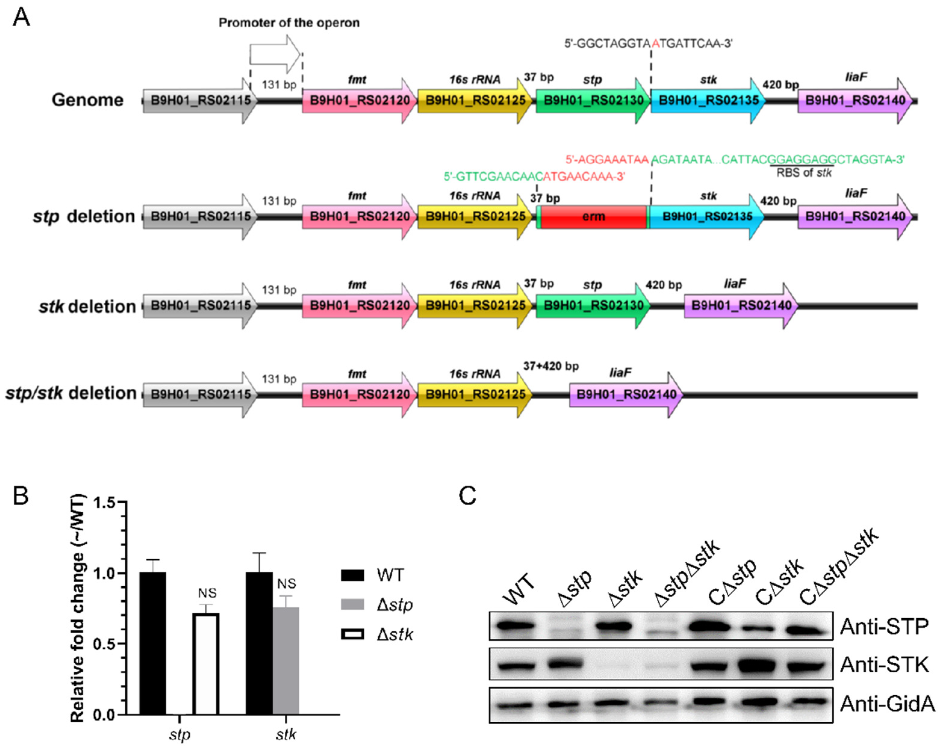
Figure 1. Construction and verification of Δstp, Δstk, and ΔstpΔstk. (A) Schematic representation of the genome organization of the stp/stk operon in wild-type and mutant strains of S. suis. (B) Transcription levels of the stp and stk genes in the WT, ∆stp, and ∆stk strains. Total RNA was extracted from the WT, ∆stp, and ∆stk strains, respectively, and reverse-transcribed to cDNA, which was used as the template for RT-qPCR. The gapdh gene was used as the internal reference for normalization. Data are representative of three independent experiments with the mean ± standard deviation, and fold changes were calculated relative to wild-type expression levels. Fold change ≥ 1.5 and p value ≤ 0.05 were considered to represent a differential expression. (C) Detection of STP and STK expression by Western blot. Cells of each indicated strain at the mid-log phase were collected, washed with PBS, and lysed by sonication, followed by immunoblot analysis using STP, STK, and GidA polyclonal antibodies. Protein samples were normalized before loading. The GidA antibody was used as a control. C∆stp, C∆stk, and C∆stpstk indicate the corresponding complemented strains.
Microorganisms 09 02442 g001
Figure 2. Virulence evaluation of the Δstp, Δstk, and ΔstpΔstk strains. (A) Mouse survival assay. Mice were intraperitoneally injected with 8 × 10⁸ CFU of WT, Δstp, Δstk, or ΔstpΔstk, respectively, and PBS was used as a negative control. Significant differences in survival rates between different groups were analyzed by the log-rank (Mantel-Cox) test. Fifteen mice were used in each group. (B) Bacterial colonization assay. Mice were intraperitoneally injected with 9 × 10⁷ CFU of WT, Δstp, Δstk, or ΔstpΔstk, respectively. Mice were euthanized at each indicated time point. The spleen, lung, kidney, and brain were collected, resuspended in PBS, homogenized, and plated on TSA plates for colony enumeration. Five mice were used in each group. Data are presented as the mean ± standard deviations. Statistical significance is determined by two-tailed, unpaired Student’s t-tests (ns, p value > 0.05; *, p value < 0.05; **, p value < 0.01; ***, p value < 0.001). (CE) Competitive infection assay. Mice were infected with a 1:1 mixture of Δstp and Δstk (C), Δstp and ΔstpΔstk (D), or ΔstpΔstk and Δstk (E). Spleens were collected, resuspended in PBS, homogenized, and plated on TSA plates for colony enumeration. Five mice were used in each group. Statistical significance was determined by two-tailed, unpaired Student’s t-tests (ns, p value > 0.05; *, p value < 0.05).
Microorganisms 09 02442 g002
Figure 3. Growth analysis. Cells of the WT strain and each mutant grown overnight were inoculated in TSB medium (A) or CDM (B) with a similar initial OD600 nm value. (C) The growth of WT strain in CDM and that of the Δstk strain in CDM or CDM supplemented with each indicated nutrient. Growth was monitored with an automatic plate reader with shaking at 37 °C. Data are presented as means ± SD of triplicate.
Microorganisms 09 02442 g003
Figure 4. Morphological analysis of the Δstp, Δstk, and ΔstpΔstk strains. (A) Gram staining images of the mid-log phase cells of WT, ∆stp, ∆stk, and ∆stpstk strains. The scale bars are 10 µm. (B) Scanning electron microscope analysis of the WT, ∆stp, ∆stk, and ∆stpstk strains. The scale bars are 5 µm. (C) Structured illumination microscopy (SIM) analysis of WT, ∆stp, ∆stk, and ∆stpstk strains. The mid-log phase cells were stained with AF-647 and FDL dyes, followed by imaging with SIM. The scale bars are 1 µm. (D) Chain length of WT, ∆stp, ∆stk, ∆stpstk strains. The results were obtained by measuring at least 400 chains per sample. (EG) Cell size parameters of WT, ∆stp, ∆stk, and ∆stpstk strains. nWT = 370, nstp = 381, nstk = 385, nstpstk = 447. Data are presented as the mean ± standard deviations. Statistical significance was determined by two-tailed, unpaired Student’s t-tests (ns, p value > 0.05; **, p value < 0.01; ***, p value < 0.001).
Microorganisms 09 02442 g004
Figure 5. Comparative proteomic analysis between the WT and Δstp or Δstk strains. Volcano plots showing the differentially expressed proteins in ∆stp (A) or ∆stk (B) compared to WT strain. Fold change > 1.5 and p < 0.05 is regarded as significant change. Blue dots represent down-regulated proteins, red dots represent up-regulated proteins, and grey dots represent proteins with no significant change.
Microorganisms 09 02442 g005
Figure 6. COG enrichment of the differentially expressed proteins in Δstp and Δstk strains. The differentially expressed proteins (fold change > 1.5, p value < 0.05) were analyzed with the EggNOG v5.0 database (http://eggnog5.embl.de/#/app/home (accessed on 25 June 2021)). [P] Inorganic ion transport and metabolism; [K] Transcription; [F] Nucleotide transport and metabolism; [J] Translation, ribosomal structure and biogenesis; [G] Carbohydrate transport and metabolism; [E] Amino acid transport and metabolism; [D] Cell cycle control, cell division, chromosome partitioning; [T] Signal transduction mechanisms; [O] Posttranslational modification, protein turnover, and chaperones; [L] Replication, recombination and repair; [I] Lipid transport and metabolism; [C] Energy production and conversion; [V] Defense mechanisms; [N] Cell motility; [M] Cell wall/membrane/envelope biogenesis; [IQ] Lipid transport and metabolism, Secondary metabolites biosynthesis, transport, and catabolism; [GM] Carbohydrate transport and metabolism, Cell wall/membrane/envelope biogenesis; [U] Intracellular trafficking, secretion, and vesicular transport; [Q] Secondary metabolites biosynthesis, transport, and catabolism; [NU] Cell motility, Intracellular trafficking, secretion, and vesicular transport; [KT] Transcription, Signal transduction mechanisms; [H] Coenzyme transport and metabolism; [CO] Energy production and conversion, Posttranslational modification, protein turnover, and chaperones; [ET] Amino acid transport and metabolism, Signal transduction mechanisms; [EG] Amino acid transport and metabolism, Carbohydrate transport and metabolism.
Microorganisms 09 02442 g006
Figure 7. Expression of the virulence factors in Δstp and Δstk strains. The abundance of each indicated virulence factor in Δstp or Δstk strain was compared with that in WT, respectively. The proteins listed on the Y-axis are virulence factors of S. suis, and the numbers on the X-axis indicate the relative expression level compared with the WT strain. Fold change > 1.5 and p < 0.05 was regarded as significantly different.
Microorganisms 09 02442 g007
Figure 8. Comparative phosphoproteomic analysis between the WT and Δstp or Δstk strains. (A) Proteins identified with changed abundance after phosphopeptide enrichment between the WT and Δstp or Δstk strains. (B) Venn diagrams showing the proteins with changed phosphorylation levels in ∆stp and ∆stk compared with the WT strain. (C) COG enrichment of proteins with changed phosphorylation levels in the ∆stp and ∆stk compared with the WT strain. The COG enrichment analysis was performed using the EggNOG v5.0 database (http://eggnog5.embl.de/#/app/home (accessed on 25 June 2021)).
Microorganisms 09 02442 g008
Figure 9. Proteins involved in bacterial glycolysis and carbohydrate transport with altered phosphorylation levels in Δstp and Δstk. The names of the proteins with altered phosphorylation levels are shown in dark red. In the brackets, the green arrow on the left indicates the change in phosphorylation level in the ∆stp strain compared with WT (p < 0.05), and the blue arrow on the right indicates the change in the ∆stk strain compared with WT (p < 0.05). The hyphen indicates no significant change (p > 0.05). Pgi, phosphoglucose isomerase; Pfk, phosphofructokinase; Fba, fructose-1,6-bisphosphate aldolase; Tpi, triosephosphate isomerase; Gap, glyceraldehyde-3-phosphate dehydrogenase; Pgk, phosphoglycerate kinase; Pgm, phosphoglycerate mutase; Eno, enolase; Pyk, pyruvate kinase; EI, PTS-enzyme I; Hpr, phosphocarrier protein; HprK, HPr kinase/phosphorylase.
Microorganisms 09 02442 g009
Table 1. Competitive index (n = 5).
StrainsMean CIp ValueSignificance
6 h9 h6 h9 h6 h9 h
stp vs. ∆stk1.167461.246000.011460.03982**
stp vs. ∆stpstk1.545311.390620.011700.02246**
stpstk vs. ∆stk1.266911.246960.019640.12846*NS
Note: n is the number of animals in each group. CI = Output (log10CFU1/log10CFU2)/Input (log10CFU1/log10CFU2). * indicates p value < 0.05; NS indicates no statistical significance.
Table 2. Proteins with down- and up-regulated abundance after phospho-peptide enrichment in Δstk and Δstp compared with WT.
No.Protein NameProtein DescriptionAmino AcidPositionRegulated TypeRatiop ValueFunction
Δstk/WT
1GpsBcell division regulator GpsBS73Down0.0840.0008Cell division
T86Down0.2460.0298
T66Down0.4990.0257
2MapZMidcell-anchored protein ZT66Down0.0754 × 10−5
T26Down0.1074 × 10−6
3FtsZcell division protein FtsZT356Down0.4830.0026
4DivIVADivIVA domain-containing proteinT211Down0.2890.0006
T199Down0.468 × 10−5
5SepFcell division protein SepFS179Down0.3970.0033
6FtsWFtsW/RodA/SpoVE family cell cycle proteinT402Down0.1284 × 10−5
7Jagprotein jagT87Down0.3210.0002
T129Down0.4131 × 10−6
S103Down0.5551 × 10−6
S84Down0.6150.0385
8MltGendolytic transglycosylase MltGT211Down0.1070.0137
T10Down0.1090.0005
T64Down0.1470.0009
T197Down0.2038 × 10−5
T122Down0.2396× 10−5
9GlmSglutamine-fructose-6-phosphate transaminase (isomerizing)T235Down0.128 × 10−9
10Hprphosphocarrier protein HPrS31Down0.6214 × 10−5Metabolism
S27Down0.6240.0013
T34Down0.6650.0007
11NeuBN-acetylneuraminate synthaseT69Down0.3390.006
12PfKAATP-dependent 6-phosphofructokinaseT240Down0.5960.0002
13ADKadenylate kinaseT137Down0.5474 × 10−5
14GTglycosyltransferaseT441Down0.0541 × 10−7
15-phosphotransferaseT5Down0.140.0013
16PhoHphosphate starvation-inducible protein PhoHT328Down0.311 × 10−4
17LmrCABC transporter ATP-binding proteinT324Down0.2088 × 10−8
18InfBtranslation initiation factor IF-2T326Down0.5340.0043Translation
19EF-Pelongation factor PT144Down0.1880.0162
20EF-Gelongation factor GT43Down0.3122 × 10−5
21BipAtranslational GTPase TypAT559Down0.260.0007
22-PASTA domain-containing proteinT25Down0.2630.0024Other
S30Down0.3740.0071
23-nucleoid-associated proteinT244Down0.6050.0016
24-IreB family regulatory phosphoproteinT7Down0.352 × 10−5
25-50S ribosomal protein L7/L12T16Down0.1822 × 10−5
26-Putative exported proteinT72Down0.0262 × 10−7
Putative exported proteinT38Down0.4334 × 10−7
27-Putative membrane proteinT4Down0.0933 × 10−6
28-Putative exported proteinT80Down0.0750.0392
T114Down0.5590.0079
29-hypothetical proteinT48Down0.0448 × 10−5
Δstp/WT
1DivIVADivIVA domain-containing proteinT199Up2.6742 × 10−5Cell division
2STKStk1 family PASTA domain-containing Ser/Thr kinaseT50Up7.9810.0002
3MltGendolytic transglycosylase MltGS115Up1.560.0141
4GlmMphosphoglucosamine mutaseS101Up1.5250.0009
5NADPNADP-dependent isocitrate dehydrogenaseS102Up2.7050.0034Metabolism
6PrfApeptide chain release factor 1S297Up2.3290.0042
7GAPDHtype I glyceraldehyde-3-phosphate dehydrogenaseT212Up2.2530.0004
8OppFABC transporter ATP-binding proteinS298Up2.1070.0013
9HprkHPr kinase/phosphorylaseS300Up1.70.0008
10ArgSarginine--tRNA ligaseS187Up1.6262 × 10−5
11SecApreprotein translocase subunit SecAS808Up1.6048 × 10−5
12IDHL-lactate dehydrogenaseS225Up1.5168 × 10−5
13-CsbD family proteinS2Up1.8950.0185Other
14-30S ribosomal protein S3S168Up1.5960.0249
Note: The assay was performed with triplicate and the ratio was presented as the average protein abundance. The p value indicates the statistical difference of each protein between the mutant strain and the WT strain by using the Student’s t-test.
Publisher’s Note: MDPI stays neutral with regard to jurisdictional claims in published maps and institutional affiliations.