Article

Development and Evaluation of Alternative Methods to Identify the Three Most Common Serotypes of Salmonella enterica Causing Clinical Infections in Kazakhstan

by
Sabyrkhan M. Barmak
1,2,*,
Yuriy A. Sinyavskiy
2,
Aidar B. Berdygaliev
2,
Turegeldy Sh. Sharmanov
2,
Irina S. Savitskaya
1,
Gulmira T. Sultankulova
3 and
Elena V. Zholdybayeva
4
1
Faculty of Biology and Biotechnology, Al-Farabi Kazakh National University, Almaty 050040, Kazakhstan
2
Biotechnology and Biologically Active Substances Laboratory, Kazakh Academy of Nutrition, Almaty 050008, Kazakhstan
3
Department of Pediatric Surgery, Asfendiyarov Kazakh National Medical University, Almaty 050012, Kazakhstan
4
National Center for Biotechnology, National Scientific Shared Laboratory of Biotechnology, Nur-Sultan 010000, Kazakhstan
*
Author to whom correspondence should be addressed.
Microorganisms 2021, 9(11), 2319; https://doi.org/10.3390/microorganisms9112319
Submission received: 20 September 2021 / Revised: 29 October 2021 / Accepted: 3 November 2021 / Published: 9 November 2021
(This article belongs to the Special Issue Salmonella and Salmonellosis)

Abstract

:
In this study, we aimed to compare the performance of conventional PCR and real-time PCR assays as screening methods for identification of three frequent, clinically significant Salmonella serovars in Kazakhstan. We determined the diagnostic efficacy of three molecular methods for detection of S. enterica subsp. enterica and typing S. Typhimurium, S. Enteritidis, and S. Virchow. A total of 137 clinical samples and 883 food samples were obtained in Almaty in 2018–2019. All tests showed high analytical specificity for detecting S. enterica and its corresponding serovariants (100%). The sensitivity of real-time PCR for each of the tested targets was 1–10 microbial cells and in conventional PCR 10–100 microbial cells. The trials with conventional PCR and real-time PCR had a diagnostic efficacy (DE) of 100% and 99.71%, respectively. The DE of real-time PCR and conventional PCR for detecting S. Enteritidis and S. Typhimurium was 99.90%, while the DE of conventional PCR and real-time PCR for detecting S. Virchow was 99.31% and 99.80%, respectively. The RAPD-PCR analysis of the genomic DNA of Salmonella enterica showed the genetic kinship of S. Enteritidis isolates, and the genetic heterogeneity of S. Typhimurium and S. Virchow isolates. Thus, the developed methods can be considered as alternatives to classical serotyping using antisera.

1. Introduction

The sickness rate of salmonellosis remains one of the most pressing health problems in many countries of the world. Salmonellosis occurs in both developed and developing countries [1,2,3]. In the etiology of bacterial intestinal infections in humans, Salmonella enterica subsp. enterica takes the leading place. To date, over 2500 Salmonella enterica serovars have been registered [4,5]. Many serotypes of S. enterica are pathogenic to both animals and humans. Serovars S. Typhimurium and S. Enteritidis are the most common causes of human salmonellosis in many countries of the world [6,7], including Kazakhstan [8]. However, in some regions, other serovars are more important [9].
Outbreaks of salmonellosis are associated with the consumption of contaminated food, which often leads to serious illnesses that may require hospitalization or even death. Salmonella contamination of food can occur at any stage of production and distribution and therefore requires continuous and effective monitoring of Salmonella in the production chain using rapid, effective, and proven methods. In addition, timely identification and typing of Salmonella is critical for the epidemiological surveillance of Salmonellosis and tracing the source of the outbreak [10].
Until now, the gold standard for the diagnosis of salmonellosis is the microbiological method (isolation of Salmonella spp. from feces, blood, vomit, urine, bile, blood). However, the long waiting period for results due to the need to quickly conduct appropriate antibiotic therapy is undoubtedly a disadvantage of this method. The situation is compounded by the fact that many patients are treated with antibiotics before symptoms of sepsis develop. In such cases, blood cultures are very difficult to perform since they contain antibiotics that inhibit the growth of microorganisms. Therefore, the detection of microbial nucleic acids is promising for efficient, accurate, and rapid diagnosis of salmonellosis. In addition, the sensitivity of molecular methods is higher than the sensitivity of the culture method, and the preliminary use of antibiotics does not affect the test results. Thus, there is a need to develop, validate, and implement alternative methods for the detection of Salmonella.
This study aimed to develop alternative molecular methods (PCR, RT-PCR, RAPD PCR) for the detection and typing of Salmonella in clinical samples and food products and their assessment on clinical samples from sick patients and food products collected in Almaty in 2018–2019.

2. Materials and Methods

2.1. Microorganisms Strains

Reference strains of Salmonella and other bacteria were used to determine the specificity of the reaction: S. Enteritidis (S.e-0071), S. Typhimurium TA 98 (reference strain), S. Typhimurium (S.t-0072), S. Virchow (reference strain), S. Infantis (S.i-0073), S. Abortusovis 37, S. Gallinarum 65, S. Abortus equi 17, S. Cholera-suis 51, S. Dublin 31, Pasteurella multocida subsp. multocida (ATCC 10544), Clostridium perfringens Strain S 107 (ATCC 13124), Clostridium sporogenes NCTC 532 (ATCC-19404), Escherichia coli (ATCC 25922), Bacillus cereus (ATCC 11778), Bacillus subtilis (ATCC-6633), Staphylococcus aureus (ATCC 25923), Staphylococcus aureus (ATCC-6538P), Pseudomonas aeruginosa Strain Boston 41501 (ATCC 27853), Pseudomonas aeruginosa (ATCC-9027), Candida albicans 3147 (ATCC-10231), Mycoplasma hyorhinis BTS-7 (ATCC-17981), Mycoplasma gallisepticum (ATCC-19610), Mycoplasma synoviae WVU 1853 [NCTC 10124] (ATCC-25204), Klebsiella pneumoniae (ATCC 13883), Aspergillus brasiliensis (ATCC-9642) formerly identified as A. niger.

2.2. Salmonella Isolates Used for PCR Method Validation

The validation of the PCR method included the analysis of 1020 samples (883 samples from food products and 137 clinical samples) collected in Almaty (Table 1). Samples of various food products were randomly collected from retail markets in Almaty between May 2018 and April 2019. In large shopping centers, sufficient attention is paid to food safety; therefore, the bulk of the food products were purchased from various markets in Almaty for research purposes. Samples were collected and prepared according to the recommendations [11,12].
The collection of clinical material (stool) of a patient suffering from an acute intestinal infection was carried out by doctors at the Children’s City Clinical Infectious Disease Hospital in Almaty in 2018–2019. Clinical samples were selected and transported following the requirements noted in the sanitary rules [13]. A total of 137 clinical samples were collected from patients for research.
Each sample was labeled, placed in a sterile plastic sample bag, transported to the laboratory on ice, and processed immediately.

2.3. Isolation and Identification of Salmonella

Isolation and identification of Salmonella were performed using standard methods described in regulatory documents [14,15,16].

2.4. DNA Extraction

Bacterial genomic DNA was extracted using the DNeasy Blood & Tissue Kit (Qiagen, Hilden, Germany). The concentration and purity of the extracted genomic DNA were measured on a MaestroNano® spectrophotometer (Maestrogen, Las Vegas, NV, USA).

2.5. Specific Primers and Probes

Specific PCR primers and a probe for typing S. Enteritidis have been developed for the gene encoding the protein Prot6e of fimbrial biosynthesis located at a specific site of the 60-kb plasmid of virulence of S. Enteritidis (U66901.1) [17].
The mdh gene encoding the S. Typhimurium malic acid dehydrogenase enzyme is conservative and was chosen for the development of specific primers and a real-time PCR probe (X61029.1) [18].
Specific PCR primers and a probe for typing S. Virchow were designed for the CRISPR gene located in the conservative region of the 100–1400 bp CRISPR gene (KF931137.1) [19].
The primers were designed using the Vector NTI Suite 9 software (Invitrogen, Carlsbad, CA, USA) and tested using BLAST to confirm their specificity (Table 2).

2.6. Real-Time Polymerase Chain Reaction

Real-time PCR was performed by TaqMan technology using a Rotor-Gene Q thermal cycler (Qiagen, Hilden, Germany). When performing real-time PCR, Platinum SuperFi PCR Master Mix (Invitrogen, Waltham, MA, USA) was amplified with primers and probes specific for Salmonella enterica bacteria and its types: S. Enteritidis, S. Typhimurium, and S. Virchow (Table 1).
The RT-PCR mixture consisted of 2.5 µL of 10× AccuPrime PCR Buffer II; 1 μL of each (10 pmol) primers; 1 μL of (5 pmol) Probe-FAM; 0.5 μL of AccuPrime Taq DNA Polymerase; 1 μL of DNA; up to 25 μL of water. The thermocycling protocol was 95 °C for 3 min; 94 °C for 20 s, 57 °C (for S. enterica); 60 °C (for S. Enteritidis); 62 °C (for S. Typhimurium); 59 °C (for S. Virchow) for 30 s for a total 45 cycles.

2.7. PCR

The conventional PCR was performed with Platinum SuperFi PCR Master Mix (Invitrogen, Waltham, MA, USA) and specific primers for S. enterica and its types: S. Enteritidis, S. Typhimurium, and S. Virchow (Table 1). The production of specific DNA regions of Salmonella bacterium was carried out in a Mastercycler Pro thermal cycler (Eppendorf, Hamburg, Germany). The following is the reaction composition for amplification of bacterial DNA fragment: 2.5 µL of 10× buffer; 1 μL of dNTPs; 2 μL of MgCl2; 1 μL of each (20 pmol) primers; 0.5 μL of Taq DNA Polymerase; 3 μL of DNA; up to 25 μL of water. The thermal cycling conditions were 94 °C for 5 min; 95 °C for 30 s, 55 °C (for S. enterica); 59 °C (for S. Enteritidis); 59 °C (for S. Typhimurium); 60 °C (for S. Virchow) for 30 s, 72 °C for 1 min for a total of 35 cycles; 72 °C for 7 min; stored at 4 °C.

2.8. Randomly Amplified Polymorphic DNA (RAPD) PCR

RAPD PCR mixtures contained the following components: 2.5 µL of 10× buffer; 1 μL of dNTPs; 1 μL of MgCl2; 2 μL of primer; 0.5 μL of Taq DNA polymerase; 5 μL of DNA; up to 25 μL of water. The primer RAPD-A 5′GCG GGA ATG CTG AAG ATA AG3′ was used to amplify DNA (Table 1). PCR conditions were as follows: 94 °C for 5 min; 94 °C for 45 s, 35 °C for 5 s, 72 °C for 1.20 min for a total of 40 cycles; 72 °C for 10 min.

2.9. Electrophoretic Analysis of DNA Amplification Products

All RAPD PCR products were separated by horizontal electrophoresis (BioRad, Munich, Germany) on a 1.5% agarose solution in Tris-acetate-EDTA buffer. The results are documented by photographing gels in the BioRad gel documenting the system.

2.10. Statistical Analysis

When determining the performance indicators of laboratory tests, true positive (TP), true negative (TN), false positive (FP), and false negative (FN) research results were used.
The calculations are based on the following formulas: sensitivity (SN) = (TP/TP + FN), specificity (SP) = (TN/TN + FP), positive predictive value PPV = (TP/TP + FP), negative predictive value (NPV) = (TN/TN + FN), diagnostic efficacy (DE) = (TP + TN/TP + FP + FN + TN) [20]. Ninety-five percent confidence interval (95% CI) was evaluated by Wilson’s calculation method [21].

3. Results

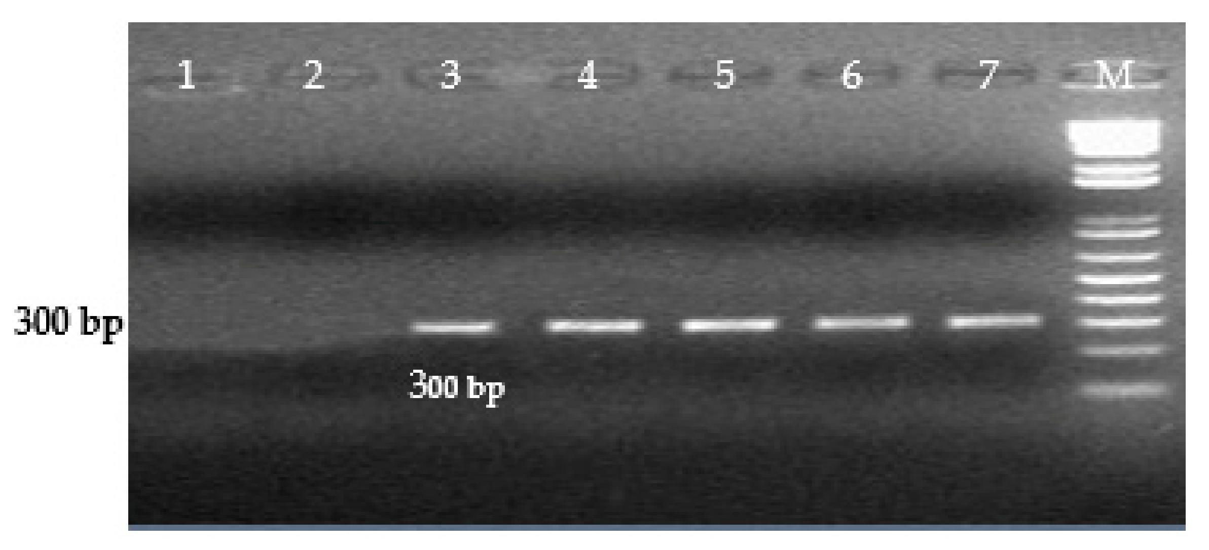
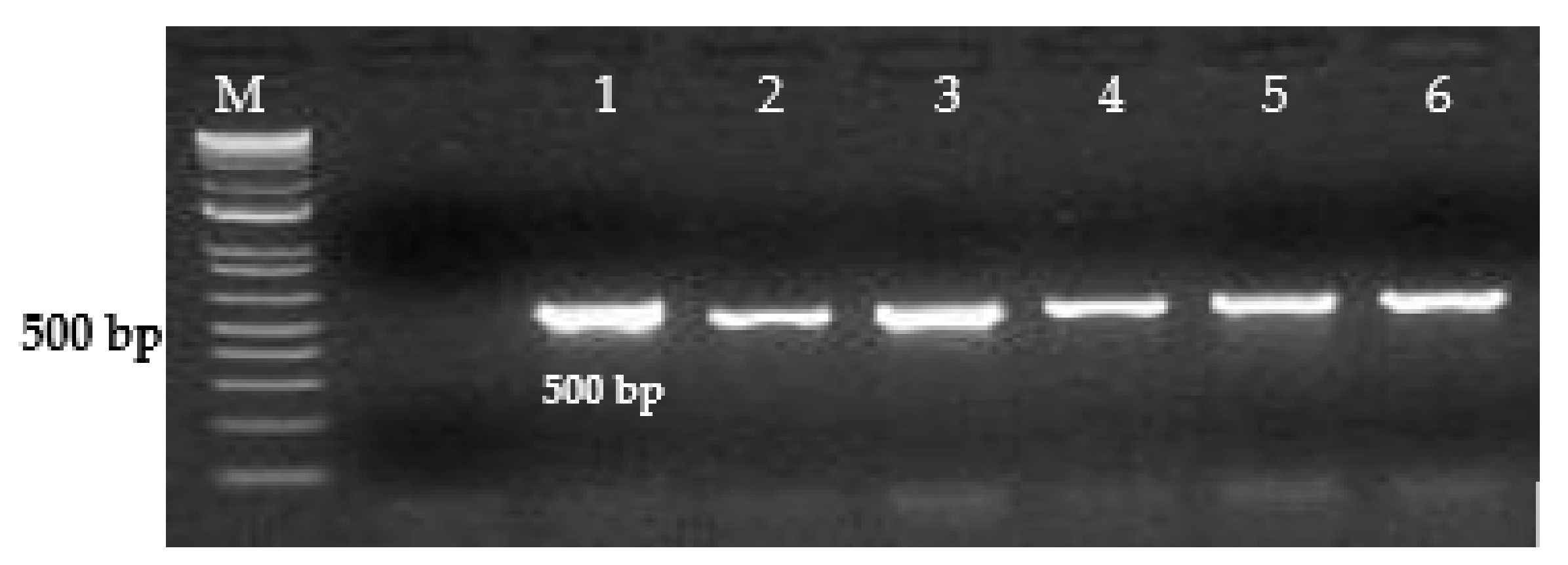
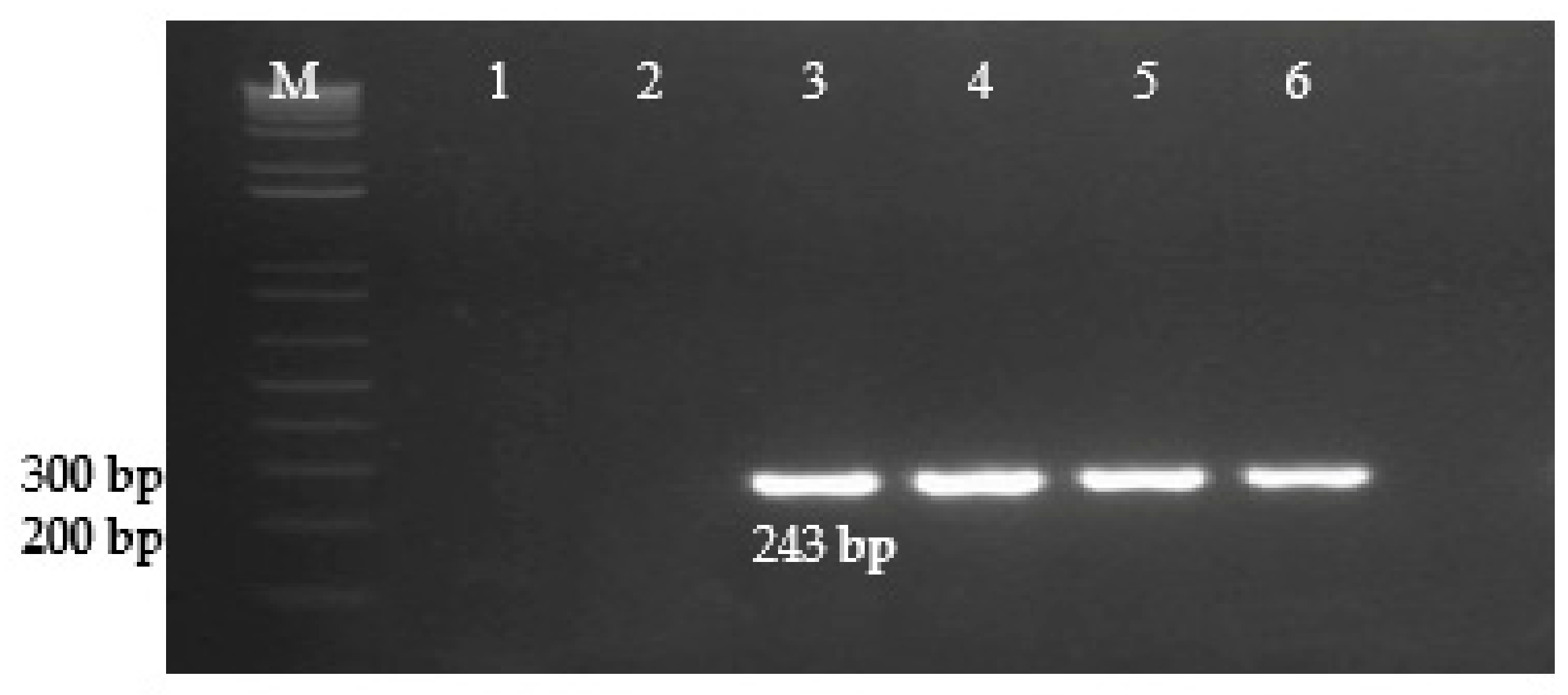
3.1. Specificity, Sensitivity, and Efficacy of Real-Time PCR and Conventional PCR

The diagnostic primers developed in this study for the Inv gene of S. enterica amplified a 500 bp DNA fragment. (Figure 1), for the Prot6e gene, S. Enteritidis amplified a DNA fragment of 300 bp. (Figure 2), for the mdh gene, S. Typhimurium amplified a DNA fragment of 243 bp. (Figure 3), for the CRISPR gene, S. Virchow amplified a 269 bp DNA fragment (Figure 4).
The specificity of conventional PCR and real-time PCR was confirmed by testing the S. enterica bacterium, its serotypes, and non-salmonella microorganisms (Table 3). The specificity of the oligonucleotides and probes used was first confirmed by testing on a panel of 10 Salmonella control organisms, and then expanded to testing a panel of 34 Salmonella isolates isolated from food samples and 65 Salmonella isolates isolated from clinical samples. Both tests showed high analytical specificity in detecting S. enterica and its serotypes. No cross-reaction was observed when determining the affiliation of strains and isolates to S. Enteritidis, S. Typhimurium, and S. Virchow in both conventional and real-time PCR.
The limit of detection (LOD) was calculated by amplifying a series of 10-fold dilutions of Salmonella bacterium DNA extracts. The LOD for detection at the threshold of real-time PCR results was 100 copies/mL of target sequence. The linear measurement range was 100–10,000,000 copies/mL of the target sequence. The analytical sensitivity of classical PCR was 10–100 microbial cells.
To estimate the diagnostic efficacy of real-time PCR and conventional PCR methods in detecting S. enterica and its serotypes, 1020 biological samples (883 samples from food products and 137 samples of clinical sample) were analyzed (Table 4 and Table 5).
Of the 1020 samples obtained from clinical samples, food raw materials, and food products, S. enterica were detected in 99 (9.70%) by real-time PCR. The same results were obtained using cultivation methods. Conventional PCR detected S. enterica in 96 (9.41 %) samples. The diagnostic efficacy of real-time PCR in the detection of S. enterica was 100, while for the conventional PCR was 99.71%.
Real-time PCR detected S. Enteritidis in 20 (1.96%) samples, S. Typhimurium in 42 (4.12%), and S. Virchow in 24 (2.35%) out of 1020 samples in parallel with the conventional PCR, in which S. Enteritidis was detected in 20 (1.96%) samples, S. Typhimurium in 42 (4.12%), and S. Virchow in 19 (1.86%). The diagnostic efficacy of real-time PCR and conventional PCR for the detection of S. Enteritidis and S. Typhimurium was 99.90%. The diagnostic efficacy of real-time PCR for detection of S. Virchow was 99.80%, and conventional PCR was 99.31%.

3.2. Typing of Salmonella enterica Strains by RAPD PCR

To identify the genetic diversity of S. Enteritidis strains (13 isolates from clinical sample and 8 isolates from food), S. Typhimurium (29 isolates from the clinical samples and 14 isolates from food), and S. Virchow (23 isolates from clinical sample and 3 isolates from food), a PCR analysis was performed using a RAPD primer. The PCR analysis of the genomic DNA of Salmonella enterica bacteria using RPC primers showed the heterogeneity of the studied strains (Table 6).
The same specific set of DNA fragments were obtained with the RAPD primer for all the studied S. Enteritidis isolates. For all isolates extracted from both clinical samples and food products (group A), 6 amplicons were identified with a length of approximately 250, 350, 650, 1000, 1250, and 3000 bp, which indicates their genetic relationship.
When genotyping S. Typhimurium strains, 4 amplicons (200, 350, 1000, 1250 bp) were detected in 12 isolates extracted from food products and in all 29 studied isolates extracted from clinical sample (group B), and 3 amplicons (200, 350, 1000 bp) were obtained for 2 isolates extracted from food products (group C).
The results of S. Virchow strain DNA amplification with RAPD-primer showed that for all isolates extracted from food products, 3 amplicons were identified, with lengths of approximately 200, 650, and 1200 bp (group D), which indicates their genetic relationship.
In the study of S. Virchow isolates extracted from clinical samples, three groups with different numbers of amplicons were identified. In group I, 19 isolates were identified (3 amplicons with a length of approximately 300, 650, 1200 bp), in group F, 2 isolates (2 amplicons with a size of 400, 650 bp), and in group G2 isolates (4 amplicons with a size of 300, 500, 650, 1200 bp). The results show that S. Virchow isolates obtained from the clinical sample are genetically diverse.

4. Discussion

Rapid and effective diagnosis of food pathogens continues to be a serious public health problem. Monitoring the presence of foodborne pathogens is a key condition for identifying potential problems in the production, processing, and preparation of food products or the process of sales. The classical methods of detecting Salmonella used to date are time-consuming and take 3 to 5 days to complete. Alternative methods based on the detection of nucleic acids are still not applicable for wide usage. One of the reasons is that the developed methods have not passed validation tests.
All clinical patients in Kazakhstan are tested in regard to the current surveillance program for the presence of Salmonella with the use of standard methods based on the isolation of cultures with the following identification by biochemical and serological methods. Molecular identification methods are rarely used. It should also be noted that there are few reports of Salmonella contamination of food products in Kazakhstan. This indicates the lack of studies of food products for Salmonella contamination. Therefore, Salmonella contamination of food products in the retail trade should be solved by constant monitoring and control using modern methods.
This study shows the results of the improvement of conventional PCR and real-time PCR for detecting S. enterica and its serotypes. There are quite a few methods applied to indicate and identify Salmonella bacteria [22,23]. Various genes are known to act as genetic markers of Salmonella. Several reports have been published on the use of the PCR method to detect S. enterica targeting the invA gene [22,23,24,25]. In our studies, primers targeting the invA gene were designed for both tests to detect S. enterica. Previously, the PCR targeting of the InvA gene for S. enterica showed 100% specificity when testing 94 Salmonella strains (inclusiveness) and 32 non-Salmonella strains (exclusivity) [26]. The conventional PCR and real-time PCR conducted with our primers on the invA gene allowed us to obtain similar results. Both tests showed a fairly high specificity for S. enterica. The sensitivity of real-time PCR for each of the tested targets was 1–10 microbial cells, and in classical PCR, 10-100 microbial cells. The sensitivity of real-time PCR for each of the tested targets was 1-10 microbial cells, while in conventional PCR, it was 10–100 microbial cells.
The choice of specific target genes is crucial for Salmonella serotyping. Despite the high homology among serovariants, it was found that some genes are associated with specific serovars. Thus, it was established that the genes Prot6e [17], mdh [18], CRISPR [19], spvA [27], rfb [28], Sdf-1 [23] fliC [28,29], SefA [30], invA [31], fimA [32], and ipaJ [33], are suitable for the specific detection and serotyping of Salmonella in various clinical samples. As noted in Section 2.5, the site of the Prot6e gene was selected for the detection of S. Enteritidis, the site of the mdh gene for the detection of S. Typhimurium, and the site of the CRISPR gene for the detection of S. Virchow. In silico analysis revealed no mismatch between primers and probes with the available bacterial genome in GenBank. Both tests showed a fairly high specificity of S. enterica serotypes. All S. Enteritidis isolates tested in this study were positive with Prot6e reagents, S. Typhimurium with mdh reagents, and S. Virchow with CRISPR reagents, as expected.
The performance of the developed PCR methods was verified on clinical samples and food samples collected in 2018–2019 in Almaty. The results of real-time PCR in the study of clinical samples and food samples demonstrated an excellent correlation with the cultivation method. Therefore, examination of clinical samples showed that in 65 (47.7%) out of 137 samples were positive results in real PCR; at the same time, out of 883 studied food samples, only 34 (3.85%) were PCR-positive. Similar results were obtained using the classical method (cultivation). The high proportion of PCR-positive results among the studied clinical samples may be due to the fact that clinical samples were taken only from patients hospitalized with acute intestinal infections. In the study of clinical and food samples, in all cases except real-time PCR on S. enterica, from 1 to 3 false negative results were obtained. Perhaps this is due to the fact that some Salmonella strains have natural mutations in the loci used in PCR as targets, which can lead to false negative results [34,35]. To prove this hypothesis, it is necessary to sequence target loci in these strains. The diagnostic efficacy of real-time PCR and PCR for the detection of S. enterica was 100% and 99.71%, respectively. The diagnostic efficacy of real-time PCR and PCR for the detection of S. Enteritidis and S. Typhimurium was the same 99.90%, while the diagnostic efficacy of PCR and real-time PCR for the detection of S. Virchow were 99.31% and 99.80%, respectively.
The information about the origin of new species of S. enterica in various countries can help in the identification and tracking of new emerging pathogens. It is possible now to determine the serotypes of Salmonella that prevail in Kazakhstan with the help of the developed PCR methods. As a result of the study of 1020 biological samples (883 samples from food products and 137 clinical samples) collected in 2018-19 in Almaty, 99 isolates were identified by the developed PCR methods and isolated by the cultivation method. Of these, 21 (21.2%) isolates are classified as S. Enteritidis, 43 (43.4%) isolates as S. Typhimurium, and 26 (26.3%) isolates as S. Virchow. Earlier, it was shown that two serotypes prevail in Kazakhstan: S. Typhimurium and S. Enteritidis [8]. One of the focuses of this study was to show that S. Virchow is also a serotype that is common to Kazakhstan. To confirm this, it is necessary to conduct additional studies on a larger number of clinical samples and food products.
The performed molecular analysis using RAPD PCR to identify the genetic diversity of S. enterica bacterial isolates obtained from the food chain and clinical samples showed the genetic relationship of S. Enteritidis isolates and the genetic heterogeneity of S. Typhimurium and S. Virchow isolates.

5. Conclusions

The research results demonstrate that the PCR testing platforms and methods are sensitive and specific, which makes these methods valuable tools for detecting S. Enteritidis, S. Typhimurium, and S. Virchow directly in food samples and clinical material. The developed PCR methods meet the requirements of diagnostic PCR, and after further interlaboratory validation studies, can become standardized methods for rapid detection of Salmonella in diagnostic laboratories.

Author Contributions

Conceptualization, S.M.B.; methodology, S.M.B., A.B.B., Y.A.S., G.T.S., and I.S.S.; investigation, S.M.B., A.B.B., and I.S.S.; writing original draft preparation, S.M.B.; writing—review and editing, S.M.B., Y.A.S., and E.V.Z.; supervision, T.S.S. All authors have read and agreed to the published version of the manuscript.

Funding

This research was financially supported by grants from the Science Committee of the Ministry of Education and Science of the Republic of Kazakhstan (AP05131147).

Institutional Review Board Statement

Not applicable.

Informed Consent Statement

Not applicable.

Data Availability Statement

The data presented in this study are available on request from the corresponding author.

Acknowledgments

The authors would like to thank the staff of Laboratory Microbiology of the Al-Farabi Kazakh National University for technical and consulting support.

Conflicts of Interest

The authors declare that they have no conflict of interest related to this study.

References

  1. Yan, S.S.; Pendrak, M.L.; Abela-Ridder, B.; Punderson, J.W.; Fedorko, D.P.; Foley, S.L. An overview of Salmonella typing: public health perspectives. Clin. Appl. Immunol. Rev. 2003, 4, 189–204. [Google Scholar] [CrossRef]
  2. Zhao, S.; McDermott, P.F.; Friedman, S.; Abbott, J.; Ayers, S.; Glenn, A.; Hall-Robinson, E.; Hubert, S.K.; Harbottle, H.; Walker, R.D.; et al. Antimicrobial resistance and genetic relatedness among Salmonella from retail foods of animal origin: NARMS retail meat surveillance. Foodborne Pathog Dis. 2006, 3, 106–117. [Google Scholar] [CrossRef]
  3. Messens, W.; Grijspeerdt, K.; De Reu, K.; De Ketelaere, B.; Mertens, K.; Bamelis, F.; Kemps, B.; De Baerdemaeker, J.; Decuypere, E.; Herman, L. Eggshell penetration of various types of hens’ eggs by Salmonella enterica serovar Enteritidis. J. Food Prot. 2007, 70, 623–628. [Google Scholar] [CrossRef] [PubMed]
  4. Grimont, P.A.D.; Weill, F.X. Antigenic Formulae of the Salmonella Serovars. 9th Edition, World Health Organization Collaborating Center for Reference and Research on Salmonella. Institut. Pasteur. Paris 2007, 9, 3. [Google Scholar]
  5. Guibourdenche, M.; Roggentin, P.; Mikoleit, M.; Fields, P.I.; Bockemuhl, J.; Grimont, P.A.; Weill, F.X. Supplement 2003–2007 (No. 47) to the White-Kauffmann-Le Minor scheme. Res. Microbiol. 2010, 161, 26–29. [Google Scholar] [CrossRef] [PubMed]
  6. Hendriksen, R.S.; Vieira, A.R.; Karlsmose, S.; Lo Fo Wong, D.M.; Jensen, A.B.; Wegener, H.C.; Aarestrup, F.M. Global monitoring of Salmonella serovar distribution from the world health organization global foodborne infections network country data bank: results of quality assured laboratories from 2001 to 2007. Foodborne Pathog. Dis. 2011, 8, 887–900. [Google Scholar] [CrossRef] [PubMed] [Green Version]
  7. Herikstad, H.; Motarjemi, Y.; Tauxe, R.V. Salmonella surveillance: a global survey of public health serotyping. Epidemiol. Infect. 2002, 129, 1–8. [Google Scholar] [CrossRef] [PubMed]
  8. Smagulova, M.K.; Sataeva, A.M. Analysis of the incidence of intestinal infections in the Republic of Kazakhstan for 2019, tasks for 2020. Environ. Public Health 2020, 1, 24–32. [Google Scholar]
  9. Yasin, R.M.; Tiew, C.C.; Jegathesan, M. Human salmonellosis in Malaysia for the period 1989–July 1994. Southeast Asian J. Trop. Med. Public Health 1995, 26, 457–460. [Google Scholar]
  10. Turki, Y.; Mehri, I.; Fhoula, I.; Hassen, A.; Ouzari, H. Comparison of five molecular subtyping methods for differentiation of Salmonella Kentucky isolates in Tunisia. World J. Microbiol. Biotechnol. 2014, 30, 87–98. [Google Scholar] [CrossRef] [PubMed]
  11. Interstate standard GOST 31904-2012 Food products. Sampling methods for microbiological analyses. Available online: https://internet-law.ru/gosts/gost/53156/ (accessed on 1 July 2013).
  12. U.S. Food and Drug Administration. Food Sampling and Preparation of Sample Homogenate, Chap. 1. In Bacteriological Analytical Manual. Available online: http://www.fda.gov/Food/FoodScienceResearch/LaboratoryMethods/ucm063335.htm (accessed on 15 June 2016).
  13. Sanitary rules “Sanitary and epidemiological requirements for laboratories using potentially hazardous chemical and biologi-cal substances”. Order of the Minister of Health of the Republic of Kazakhstan. Available online: https://adilet.zan.kz/rus/docs/V1700015990 (accessed on 15 October 2021).
  14. Laboratory Diagnostics of Salmonella, Detection of Salmonella in Food and Environmental Objects: Guidelines—M.: Federal Center for Hygiene and Epidemiology of Rospotrebnadzor. 2011. 111p. Available online: https://files.stroyinf.ru/Data2/1/4293790/4293790005.pdf (accessed on 2 February 2010).
  15. Interstate Standard GOST 31659-2012 (ISO 6579: 2002) Food Products. Method for the Detection of Bacteria of the Genus Salmonella (ISO 6579: 2002, MOD). Available online: https://docs.cntd.ru/document/1200098239 (accessed on 1 July 2013).
  16. U.S. Food and Drug Administration. Bacteriological Analytical Manual (BAM) Chapter 5: Salmonella. Available online: http://www.fda.gov/Food/FoodScienceResearch/LaboratoryMethods/ucm070149.htm (accessed on 15 December 2015).
  17. Clavijo, R.I.; Loui, C.; Andersen, G.L.; Riley, L.W.; Lu, S. Identification of genes associated with survival of Salmonella enterica serovar Enteritidis in chicken egg albumen. Appl. Environ. Microbiol. 2006, 72, 1055–1064. [Google Scholar] [CrossRef] [PubMed] [Green Version]
  18. Lin, J.S.; Tsen, H.Y. Development and Use of Polymerase Chain Reaction for the Specific Detection of Salmonella Typhimurium in Stool and Food Samples. J. Food Protection. 1999, 62, 1103–1110. [Google Scholar] [CrossRef] [PubMed]
  19. Bachmann, N.L.; Petty, N.K.; Zakour, N.L.B.; Szubert, J.M.; Savill, J.; Beatson, S.A. Genome analysis and CRISPR typing of Salmonella enterica serovar Virchow. BMC Genomics 2014, 15, 389. [Google Scholar] [CrossRef] [PubMed] [Green Version]
  20. Van Engelsdorp, D.; Lengerich, E.; Spleen, A.; Dainat, B.; Cresswell, J.; Baylis, K.; Nguyen, B.K.; Soroker, V.; Underwood, R.; Human, H. Standard epidemiological methods to understand and improve Apis mellifera health. J. Apic. Res. 2013, 52, 1–16. [Google Scholar] [CrossRef] [Green Version]
  21. Wilson, E.B. Probable inference, the law of succession, and statistical inference. J. Am. Stat. Assoc. 1927, 22, 209–212. [Google Scholar] [CrossRef]
  22. Malorny, B.; Bunge, C.; Hoorfar, J.; Helmuth, R. Multicenter validation of the analytical accuracy of Salmonella PCR: towards an international standard. Appl. Environ. Microb. 2003, 69, 290–296. [Google Scholar] [CrossRef] [Green Version]
  23. Kasturi, K.N.; Drgon, T. Real-Time PCR Method for Detection of Salmonella spp. in Environmental Samples. Appl. Environ. Microbiol. 2017, 83, e00644-17. [Google Scholar] [CrossRef] [PubMed] [Green Version]
  24. Hein, I.; Flekna, G.; Krassnig, M.; Wagner, M. Real-time PCR for the detection of Salmonella spp. in food: An alternative approach to a conventional PCR system suggested by the FOOD-PCR project. J. Microbiol. Methods 2006, 66, 538–547. [Google Scholar] [CrossRef]
  25. Malorny, B.; Hoorfar, J.; Hugas, M.; Heuvelink, A.; Fach, P.; Ellerbroek, L.; Bunge, C.; Dorn, C.; Helmuth, R. Inter-laboratory diagnostic accuracy of a Salmonella specific PCR-based method. Int. J. Food Microbiol. 2003, 89, 241–249. [Google Scholar] [CrossRef]
  26. Siala, M.; Barbana, A.; Smaoui, S.; Hachicha, S.; Marouane, C.; Kammoun, S.; Gdoura, R.; Messadi-Akrout, F. Screening and Detecting Salmonella in Different Food Matrices in Southern Tunisia Using a Combined Enrichment/Real-Time PCR Method: Correlation with Conventional Culture Method. Front. Microbiol. 2017, 8, 2416. [Google Scholar] [CrossRef] [PubMed]
  27. Lampel, K.A.; Keasler, S.P.; Hanes, D.E. Specific detection of Salmonella enterica serotype Enteritidis using the polymerase chain reaction. Epidemiol. Infect. 1996, 116, 137–145. [Google Scholar] [CrossRef] [PubMed] [Green Version]
  28. Hirose, K.; Itoh, K.; Nakajima, H.; Kurazono, T.; Yamaguchi, M.; Moriya, K.; Ezaki, T.; Kawamura, Y.; Tamura, K.; Watanabe, H. Selective Amplification of Tyv (Rfbe), Prt (Rfbs), Viab, and fliC Genes by Multiplex PCR for Identification of Salmonella enterica Serovars Typhi and Paratyphi A. J. Clin. Microbiol. 2002, 40, 633–636. [Google Scholar] [CrossRef] [PubMed]
  29. Hong, Y.; Liu, T.; Lee, M.D.; Hofacre, C.L.; Maier, M.; White, D.G.; Ayers, S.; Wang, L.; Berghaus, R.; Maurer, J.J. Rapid screening of Salmonella enterica serovars Enteritidis, Hadar, Heidelberg and Typhimurium using a serologically-correlative allelotyping PCR targeting the O and H antigen alleles. BMC Microbiol. 2008, 8, 1–8. [Google Scholar] [CrossRef] [PubMed] [Green Version]
  30. Mirmomeni, M.H.; Sisakhtnezhad, S.; Sharifi, A. Rapid Detection of Salmonella Enteritidis by PCR Amplification of The SefA Gene and It’s Cloning. Pak. J. Biol. Sci. 2008, 11, 428–432. [Google Scholar] [CrossRef] [PubMed] [Green Version]
  31. Rahn, K.; De Grandis, S.A.; Clarke, R.C.; McEwen, S.A.; Galán, J.E.; Ginocchio, C.; Curtiss, R., III; Gyles, C.L. Amplification of an invA gene sequence of Salmonella Typhimurium by polymerase chain reaction as a specific method of detection of Salmonella. Mol. Cell. Probes. 1992, 271–279. [Google Scholar] [CrossRef]
  32. Cohen, H.J.; Mechanda, S.M.; Lin, W. PCR amplification of the fimA gene sequence of Salmonella Typhimurium, a specific method for detection of Salmonella spp. Appl Environ. Microbiol. 1996, 62, 4303–4308. [Google Scholar] [CrossRef] [PubMed] [Green Version]
  33. Xu, L.; Liu, Z.; Li, Y.; Yin, C.; Hu, Y.; Xie, X.; Li, Q.; Jiao, X. A Rapid method to identify Salmonella enterica serovar Gallinarum biovar pullorum using a specific target gene ipaJ. Avian Pathol. 2018, 47, 238–244. [Google Scholar] [CrossRef] [PubMed] [Green Version]
  34. Ginocchio, C.C.; Rahn, K.; Clarke, R.C.; Galan, J.E. Naturally occurring deletions in the centisome 63 pathogenicity island of environmental isolates of Salmonella spp. Infect. Immun. 1997, 65, 1267–1272. [Google Scholar] [CrossRef] [Green Version]
  35. Malorny, B.; Paccassoni, E.; Fach, P.; Bunge, C.; Martin, A.; Helmuth, R. Diagnostic real-time PCR for detection of Salmonella in food. Appl. Environ. Microbiol. 2004, 70, 7046–7052. [Google Scholar] [CrossRef] [Green Version]
Figure 1. Detection of S. enterica using PCR primers SE Inv-1F and SE Inv-1R (500 bp). Lane M—1kb ladder (Invitrogen). Lane 1—PCR products amplified from S. Enteritidis (S. e-0071), lane 2—PCR products amplified from S. Typhimurium TA 98, reference strain, lane 3—PCR products amplified from S. Virchow, reference strain, lanes 4, 5, 6—clinical samples, positive for S. enterica.
Microorganisms 09 02319 g001
Figure 2. Detection of S. Enteritidis using PCR primers SE Prot6e-1F and SE Prot6e-1R (300 bp). Lane M—1kb ladder (Invitrogen). Lane 1—S. Typhimurium (S.t-0072), lane 2—S. Virchow (reference strain), lane 3—PCR products amplified from S. Enteritidis (S.e-0071) (positive control), lanes 4, 5, 6, 7—clinical samples, positive for S. Enteritidis.
Microorganisms 09 02319 g002
Figure 3. Detection of S. Typhimurium using PCR primers ST mdh-1F and ST mdh-1R (243 bp). Lane M—1kb ladder (Invitrogen). Lane 1—PCR products amplified from S. Enteritidis (S.e-0071), lane 2—S. Virchow (reference strain), lane 3—PCR products amplified from S. Typhimurium (S.t-0072) (positive control), lanes 4, 5, 6—clinical samples, positive for S. Typhimurium.
Microorganisms 09 02319 g003
Figure 4. Detection of S. Virchow using PCR primers SV CRISPR–1F and SV CRISPR–1R (269 bp). Lane M—50 bp ladder (Invitrogen). Lane 1—PCR products amplified from S. Virchow, reference strain (positive control). Lanes 2, 3, 4, 5, 6—clinical samples, positive for S. Virchow, lane 7—S. Enteritidis (S.e-0071), lane 8—S. Typhimurium (S.t-0072).
Microorganisms 09 02319 g004
Table 1. List of biological samples collected in 2018–2019 in Almaty for the isolation of Salmonella.
nNameNumber of Tested Samples Number of Isolated Salmonella Isolates
201820192018 2019
1Clinical samples 1370650
2Meat and meat products635571
3Fish and fish products383214
4Vegetables673510
5Berries231700
6Bird353482
7Milk and dairy products946531
8Mushrooms271600
9Salads201300
10Dried fruits362500
11Fruits242700
12Confectionery232100
13Eggs504351
Total637383909
Table 2. Oligonucleotide primers and probes for identification of Salmonella enterica species and their types in real-time PCR and PCR.
TypePrimer or ProbeSequencePCR Product Size
Salmonella entericaSE-FAGGTGACGCTATTGCCGGCAT155
SE-RATGCGGGGATCTGGGCGA
SE-ProbeFAM-ATTTCGGTGGGGATGACTCGCCAT-BHQ-1
S. EnteritidisSEE-FCGTCGTTGCTGCTTCCGGGA176
SEE-RGCTACAGAGAGTCACACTAA
SEE-ProbeFAM- TGCTGTAGATGCAAGGGTGCCTAA-BHQ-1
S. TyphimuriumSET-FGAAGTTGAAGTGCCGGTGAT251
SET-RCATTCCACCACGCCCTTCT
SET-ProbeFAM- CAGATTCCAGGCGTAAGTTTTA-BHQ-1
S. VirchowSEV-FACACCAGTACGACGATCTGCG105
SEV-RATAAACCGGGCAACTGGG
SEV-ProbeFAM-GGAACACATAAACAGCGCCCAGAT-BHQ-1
Salmonella entericaSE Inv-1FGTGAAATTATCGCCACGTTCGG500
SE Inv-1RATCGCCATTTACGCGGGTCA
S. EnteritidisSE Prot6e-1FTAACCGGAGAGGCGCTCATC300
SE Prot6e-1R AACCATGCTCAGCTGCTCCA
S. TyphimuriumST mdh-1FGTGCCGGTGATTGGCGGGCA243
ST mdh-1RCGCATTCCACCACGCCCTTC
S. VirchowSV CRISPR–1FGATCTGCGCGAACAATATCA269
SV CRISPR–1RCCGTTGTACTGATCATCTTC
S. Enteritidis
S. Typhimurium
S. Virchow
RAPD-AGCGGGAATGCTGAAGATAAG
Note: All oligonucleotides have been developed in the framework of this research.
Table 3. Diagnostic specificity of conventional PCR and real-time PCR.
Control OrganismReal-Time PCRConventional PCR
S. entericaS. EnteritidisS. TyphimuriumS. VirchowS. entericaS. EnteritidisS. TyphimuriumS. Virchow
S. Enteritidis (S.e-0071)PosPosNegNegPosPosNegNeg
S. Typhimurium TA 98 (reference strain)PosNegPosNegPosNegPosNeg
S. Typhimurium (S.t-0072)PosNegPosNegPosNegPosNeg
S. Virchow (reference strain)PosNegNegPosPosNegNegPos
S. Infantis (S.i-0073)PosNegNegNegPosNegNegNeg
S. Abortusovis 37PosNegNegNegPosNegNegNeg
S. Gallinarum 65PosNegNegNegPosNegNegNeg
S. Abortus equi 17PosNegNegNegPosNegNegNeg
S. Cholera suis 51PosNegNegNegPosNegNegNeg
S. Dublin 31PosNegNegNegPosNegNegNeg
Pasterella multocida subsp. multocida (ATCC-10544)NegNegNegNegNegNegNegNeg
Clostridium perfringens Strain S 107 (ATCC-13124)NegNegNegNegNegNegNegNeg
Clostridium sporogenes NCTC 532 (ATCC-19404)NegNegNegNegNegNegNegNeg
Escherichia coli (ATCC-25922)NegNegNegNegNegNegNegNeg
Bacillus cereus (ATCC-11778)NegNegNegNegNegNegNegNeg
Bacillus subtilis subsp. spizizenii (ATCC-6633)NegNegNegNegNegNegNegNeg
Staphylococcus aureus (ATCC-25923)NegNegNegNegNegNegNegNeg
Staphylococcus aureus subsp. aureus (ATCC-6538P)NegNegNegNegNegNegNegNeg
Pseudomonas aeruginosa Strain Boston 41501 (ATCC-27853)NegNegNegNegNegNegNegNeg
Pseudomonas aeruginosa (ATCC-9027)NegNegNegNegNegNegNegNeg
Candida albicans; 3147 (ATCC-10231)NegNegNegNegNegNegNegNeg
Mycoplasma hyorhinis; BTS-7 (ATCC-17981)NegNegNegNegNegNegNegNeg
Mycoplasma gallisepticum (ATCC-19610)NegNegNegNegNegNegNegNeg
Mycoplasma synoviae; WVU 1853 [NCTC 10124] (ATCC-25204)NegNegNegNegNegNegNegNeg
Klebsiella pneumoniae (ATCC-13883)NegNegNegNegNegNegNegNeg
Aspergillus brasiliensis; formerly A. niger (ATCC-9642)NegNegNegNegNegNegNegNeg
Pos, positive. Neg, negative. All laboratory isolates were sequenced using 16S RNA.
Table 4. Identification of Salmonella enterica and its types S. Enteritidis, S. Typhimurium, and S. Virchow in samples using various tests.
ResultTrue Positive
(TP)
False Positive
(FP)
False Negative
(FN)
True Negative
(TN)
Total
Test
Real-time PCR S. enterica99 (9.71%)00921 (90.29%)1020 (100%)
Real-time PCR S. Enteritidis20 (1.96%)01 (0.10%)999 (97.94%)1020 (100%)
Real-time PCR S. Typhimurium 42 (4.12%)01 (0.10%)977 (95.78%)1020 (100%)
Real-time PCR S. Virchow24 (2.35%)1 (0.10%)1 (0.10%)994 (97.45%)1020 (100%)
PCR
S. enterica
96 (9.41%)2 (0.20%)1 (0.10%)921 (90.29%)1020 (100%)
PCR
S. Enteritidis
20 (1.96%)01 (0.10%)999 (97.94%)1020 (100%)
PCR
S. Typhimurium
42 (4.12%)01 (0.10%)977 (95.78%)1020 (100%)
PCR
S. Virchow
19 (1.86%)4 (0.40%)3 (0.29%)994 (97.45%)1020 (100%)
Cultivating
S. enterica
99 (9.70%)00921 (90.30%)1020 (100%)
Table 5. Comparison of various tests for the detection of Salmonella enterica and its types S. Enteritidis, S. Typhimurium, and S. Virchow.
ResultSN,
at 95% CI
SP,
at 95% CI
PPV,
at 95% CI
NPV,
at 95% CI
Diagnostic Efficacy
Test
Real-time PCR
S. enterica
100100100100100
Real-time PCR
S. Enteritidis
95.23
(93.93–96.53)
10010099.90
(99.71–100)
99.90
Real-time PCR
S. Typhimurium
97.67
(96.77–98.57)
10010099.90
(99.71–100)
99.90
Real-time PCR
S. Virchow
96.00
(94.8–97.2)
99.90
(99.71–100)
96.00
(94.8–97.2)
99.90
(99.71–100)
99.80
PCR
S. enterica
98.97
(98.35–99.59)
99.78
(99.50–100)
97.95
(97.15–98.75)
99.89
(99.69–100)
99.71
PCR
S. Enteritidis
95.24
(93.94–96.54)
10010099.90
(99.71–100)
99.90
PCR
S. Typhimurium
97.67
(96.77–98.57)
10010099.89
(99.69–100)
99.90
PCR
S. Virchow
86.36
(84.26–88.46)
99.60 (99.30–99.90)82.61
(80.31–84.91)
99.70
(99.40–100)
99.31
Cultivating S. enterica100100100100100
SN—Sensitivity; SP—Specificity; PPV—Positive Predictive Value; NPV—Negative Predictive Value; 95% CI—95% confidence interval.
Table 6. Typing of Salmonella enterica strains by RAPD PCR.
Serovar Food ProductClinical Sample
GroupNumber of IsolatesNumber of AmpliconsSize of Amplicons, bpGroupNumber of IsolatesNumber of AmpliconsSize of Amplicons, bp
S. EnteritidisA86250, 350, 650, 1000, 1250, 3000A136250, 350, 650, 1000, 1250, 3000
S. TyphimuriumB124250, 350, 1000, 1250B294250, 350, 1000, 1250
C23250, 350, 1000
S. VirchowD33200, 650, 1200I193300, 650, 1200
F22400, 650
G24300, 500, 650, 1200
Publisher’s Note: MDPI stays neutral with regard to jurisdictional claims in published maps and institutional affiliations.

