Article

Comparative Secretomics Analysis Reveals the Major Components of Penicillium oxalicum 16 and Trichoderma reesei RUT-C30

1
College of Life Science, Jiangxi Normal University, Nanchang 330022, China
2
Department of Computational Medicine and Bioinformatics, University of Michigan, Ann Arbor, MI 48109, USA
3
Nanchang Key Laboratory of Microbial Resources Exploitation & Utilization from Poyang Lake Wetland, Jiangxi Normal University, Nanchang 330022, China
*
Author to whom correspondence should be addressed.
These authors contributed equally to this work.
Microorganisms 2021, 9(10), 2042; https://doi.org/10.3390/microorganisms9102042
Submission received: 24 July 2021 / Revised: 16 September 2021 / Accepted: 24 September 2021 / Published: 27 September 2021

Abstract

:
In this study, the major secretome components of Penicillium oxalicum 16 and Trichoderma reesei RUT-C30 under wheat bran (WB) and rice straw (RS) solid-state fermentation were systematically analyzed. The activities of the major components, e.g., cellulase, hemicellulase, and amylase, were consistent with their abundance in the secretomes. P. oxalicum 16 secreted more abundant glycoside hydrolases than T. reesei RUT-C30. The main up-regulated proteins from the induction of WB, compared with that from RS, were amylase, pectinase, and protease, whereas the main down-regulated enzymes were cellulase, hemicellulase, swollenin, and lytic polysaccharide monooxygenase (LPMO). Specifically, WB induced more β-1,4-glucosidases, namely, S8B0F3 (UniProt ID), and A0A024RWA5 than RS, but RS induced more β-1,4-exoglucanases and β-1,4-endoglucanases, namely, A0A024RXP8, A024SH76, S7B6D6, S7ZP52, A024SH20, A024S2H5, S8BGM3, S7ZX22, and S8AIJ2. The P. oxalicum 16 xylanases S8AH74 and S7ZA57 were the major components responsible for degrading soluble xylan, and S8BDN2 probably acted on solid-state hemicellulose instead of soluble xylan. The main hemicellulase component of T. reesei RUT-C30 in RS was the xyloglucanase A0A024S9Z6 with an abundance of 16%, but T. reesei RUT-C30 lacked the hemicellulase mannanase and had a small amount of the hemicellulase xylanase. P. oxalicum 16 produced more amylase than T. reesei RUT-C30, and the results suggest amylase S7Z6T2 may degrade soluble starch. The percentage of the glucoamylase S8B6D7 did not significantly change, and reached an average abundance of 5.5%. The major auxiliary degradation enzymes of P. oxalicum 16 were LPMOs S7Z716 and S7ZPW1, whereas those of T. reesei RUT-C30 were swollenin and LPMOs A0A024SM10, A0A024SFJ2, and A0A024RZP7.

1. Introduction

Carbohydrates are the most widely distributed, abundant renewable biomass resource on Earth [1,2]. Improper treatment such as in situ incineration of large amounts of idle carbohydrates causes environmental pollution and resource waste [2]. In addition, non-renewable resources, such as fossil energy and lithium for batteries, which are extremely popular at present, will eventually be exhausted [3,4,5]. Therefore, it is of great value to transform superfluous carbohydrates into useful biofuels and chemicals [6].
The most abundant carbohydrates include cellulose, which constitutes 40–60% of the total biomass on Earth [7], hemicellulose which accounts for 20–35% of the renewable resources [8], and the widely distributed starch. The corresponding enzymes that hydrolyze these three abundant carbohydrates are cellulase, hemicellulase, and amylase, respectively. Cellulase is further classified into three classes, i.e., β-1,4-endoglucanases (EG; EC 3.2.1.4), cellobiohydrolases (CBH; EC 3.2.1.91), and β-1,4-glucosidases (BGL; EC 3.2.1.21) [9]. Hemicellulase includes xylanase (EC 3.2.1.8), mannanase (EC 3.2.1.78) and xyloglucanase (EC 3.2.1.151) [10]. Amylase is composed of α-amylase (EC 3.2.1.1), β-amylase (EC 3.2.1.2), and glucoamylase (EC 3.2.1.3) [11]. In addition to these enzymes, lytic polysaccharide monooxygenase (LPMO) participates in oxidative degradation of solid carbohydrates (cellulose, starch, hemicellulose, chitin, etc.) [12,13,14] and swollenin accelerates the expansion and fracture of solid cellulose [15].
In previous studies, we screened a new, fast-growing wild fungus, Penicillium oxalicum 16, which secretes amylase, cellulase, and hemicellulase [2,16], and the cellulase combination from P. oxalicum 16 and Trichoderma reesei RUT-C30 can synergistically degrade cellulose [2]. Although some studies have sequenced and analyzed the genomes and secretomes of P. oxalicum and T. reesei [10,17,18,19,20,21], and concluded that P. oxalicum has more diverse lignocellulolytic enzymes according to genomics information, particularly for cellulose binding domain-containing proteins and hemicellulases compared to the widely used cellulase producer T. reesei [20], the main components have not been systematically and completely summarized. Furthermore, we found that, compared to T. reesei RUT-C30, the yield of cellulase in P. oxalicum 16 is not sufficiently high [2,16]. Therefore, it is important to quantitate the secretome information of P. oxalicum 16 before engineering this useful strain.
WB and RS are the main agricultural wastes in China. In our previous study, we found that wheat bran (WB) and rice straw (RS) compositions are very different: WB is made of 36% cellulose, 28% hemicellulose, 8% lignin, 5% ash, 12% starch, and 11% other components, including pectin and protein, and RS comprises about 30% cellulose, 25% hemicellulose, 19% lignin, 18% ash, and 8% other constituents [2].
Although the enzymes and BGL of P. oxalicum 16 were studied by our group [2,4,5,22], its secretomics information remains unknown. To address this, here we investigated the main components by comparative secretomics analysis of P. oxalicum 16 and T. reesei RUT-C30 through WB or RS solid-state fermentation. The results of the analysis provide different conclusions from previous reports, such as showing lower hemicellulase of P. oxalicum 16 compared to other studies [10,20,21], in addition to revealing amylase information, among other novel findings.

2. Materials and Methods

2.1. Materials

P. oxalicum 16 was deposited in the China Center for Type Culture Collection (CCTCC, Wuhan, China) with the accession number AF2015017, and T. reesei RUT-C30 was obtained from the New World Institute of Biotechnology.
Carboxymethylcellulose sodium salt (CMC), 4-nitrophenyl-β-D-cellobioside (pNPC), xylan, NH4HCO3, dithiothreitol, iodoacetamide, and salicin were purchased from Sigma-Aldrich (St. Louis, MO, USA). Soluble starch and microcrystalline cellulose (MCC) were purchased from Sinopharm Chemical Reagent Co., Ltd. (Beijing, China). Trypsin, formic acid, and acetonitrile were purchased from Promega (Madison, WI, USA), Sigma-Aldrich Fluka (St. Louis, MO, USA), and Fisher Chemical (Fair Lawn, NJ, USA), respectively.

2.2. Enzyme Production and Extraction

Approximately, 2 × 10⁶ spores of T. reesei RUT-C30 and P. oxalicum 16 were incubated in 250 mL Erlenmeyer flasks with the solid-state medium containing either 5 g of WB or RS, 0.09 g KH2PO4, 0.09 g (NH4)2SO4, 0.015 g CaCl2, 0.015 g urea, 0.015 g MgSO4·7H2O, and 200 μL Mandels mineral salt solution [2,23]. The solid-state mediums inoculated with the two strains were cultured at 75% humidity, an initial pH of 5, and were kept at 28 °C for 5 days.
To extract enzymes, the suspension containing 1 g of dry solid-state medium and 3 mL of acetate buffer (50 mM, pH 5) was shaken at 180 rpm at room temperature for 1 h, the supernatant was collected by centrifugation for 10 min at 10,000× g at 4 °C, and the precipitate was resuspended with 3 mL of acetate buffer twice.

2.3. Determination of Enzyme Activity and Protein Content

The enzyme activities of EG, BGL, xylanase, and amylase were assayed using the dinitrosalicylic acid (DNS) method [2,24]. Specifically, 50 μL diluted culture supernatants were mixed with 450 μL of 1% of the corresponding substrates CMC, salicin, xylan, and soluble starch (50 mM acetate buffer, pH 5) at 50 °C for 30 min, and the reaction was stopped by adding 500 μL DNS. Then the mixture was boiled for 10 min and cooled on ice to stabilize the color. One enzyme activity unit was defined as the amount of enzyme that produced 1 μmol reducing sugar (i.e., glucose or xylose) within a minute at the given experimental conditions.
CBH’s activity was determined by releasing 4-nitrophenol measured at 420 nm after adding 150 μL of 10% Na2CO3 to stop the reaction. A quantity of 100 μL diluted culture supernatants was incubated with 50 μL of 1 mg/mL pNPC (50 mM acetate buffer, pH 5) at 50 °C for 30 min [16,25]. One CBH activity unit was defined as the amount of enzyme which released 1 μmol 4-nitrophenol per minute.
Protein was quantified by the Bradford method using bovine serum albumin as a standard [26].

2.4. Pretreatment of Corncob Powder (Pr-CP), RS (Pr-RS), and MCC (Pr-MCC)

RS was cut to about 3 cm, and milled by machine (Huangcheng 800, Yongkang, China) for 5 min, which was not filtered using any mesh sieve. CP and milled RS were pretreated using a laboratory autoclave (Boxun 18 L, Shanghai, China) at 121 °C for 1 h, and then washed with deionized water until the washed water was sugar-free and the pH was 7. All Pr-CP and Pr-RS were dried at 55 °C to a constant weight and subsequently milled to 100 mesh particle size for further use.
To prepare Pr-MCC, 3 g cellufloc-200 cellulose, 20 glass balls with 3 mm diameters, and 60 mL deionized water were added to a 250 mL Erlenmeyer flask, put into a laboratory autoclave (Boxun 18 L, Shanghai, China) at 121 °C for 30 min, and then shaken at 180 rpm for 48 h. Pr-MCC was washed 3 times and dried at 55 °C to a constant weight for further use.

2.5. Enzymatic Hydrolysis of Pr-CP, Pr-RS, and Pr-MCC

A quantity of 25 mg of Pr-CP, Pr-RS, or Pr-MCC was incubated with 100 μg of the cultured supernatant enzymes from T. reesei RUT-C30 or P. oxalicum 16 with a total water-insoluble solid loading of 2.5% (w/v) (the final volume was 1 mL). The hydrolytic reactions were carried out in 50 mM acetate buffer (pH 5) at 50 °C and 180 rpm for 96 h; the total sugar produced in the reactions was determined by the anthrone colorimetric method at 600 nm [27]. All reactions were carried out in a 1.5 mL EP tube.

2.6. SDS-PAGE of Secretome, and In-Gel Digestion

To obtain 50 μg of protein in the separating gel of SDS-PAGE, electrophoresis was carried out at 120 V for 40 min, and the gel stained by Coomassie blue was decolorized. Gel pieces that could not be decolorized were cut from the decolorized gel for further use.
We sent gel pieces to PTM BioLab Inc. (Hongzhou City, China), for identification of the secretomes. For in-gel tryptic digestion, gel pieces were destained in the solution containing 50 mM NH4HCO3 and 50% acetonitrile (v/v) until clear [28]. Gel pieces were dehydrated with 100 μL of 100% acetonitrile for 5 min. Then the liquid was removed, and the gel pieces were rehydrated in 10 mM dithiothreitol and incubated at 56 °C for 60 min. Following this, the gel pieces were again dehydrated in 100% acetonitrile; after the liquid was removed, the gel pieces were rehydrated with 55 mM iodoacetamide. Samples were incubated at room temperature in the dark for 45 min. Gel pieces were washed with 50 mM NH4HCO3 and dehydrated with 100% acetonitrile. Gel pieces were rehydrated with 10 ng/μL trypsin resuspended in 50 mM NH4HCO3 on ice for 1 h. Excess liquid was removed, and gel pieces were digested with trypsin at 37 °C overnight. Peptides were extracted with 50% acetonitrile/5% formic acid and followed by 100% acetonitrile. Peptides were dried to completion and resuspended in 2% acetonitrile/0.1% formic acid.

2.7. LC-MS/MS Analysis

The tryptic peptides were dissolved in solvent A (0.1% formic acid) and directly loaded onto a reversed-phase analytical column (15 cm × 75 μm) made by PTM BioLab Inc. The gradient for solvent B (0.1% formic acid in 98% acetonitrile) was from 6 to 23% for 16 min, 23 to 35% for 8 min, and climbed to 80% for 3 min, and was finally kept at 80% for 3 min. A constant flow rate of 400 nL/min was used in the EASY-nLC 1000 UPLC system.
The peptides were subjected to NSI source followed by tandem mass spectrometry (MS/MS) in Q ExactiveTM Plus (Thermo, Waltham, MA, USA) coupled online to the UPLC [29]. The electrospray voltage applied was 2.0 kV. The m/z scan range was 350 to 1800 for a full scan, and intact peptides were detected in the Orbitrap at a resolution of 70,000. Peptides were then selected for MS/MS using an NCE set at 28 and the fragments were detected in the Orbitrap at a resolution of 17,500. A data-dependent procedure that alternated between one MS scan followed by 20 MS/MS scans with 15.0 s dynamic exclusion was used. Automatic gain control (AGC) was set at 5E4.

2.8. Data Processing

The resulting MS/MS data were processed using Proteome Discoverer 1.3. Tandem mass spectra were searched against the UniProt P. oxalicum database (9977 sequences) and UniProt T. reesei database (9848 sequences) (https://www.uniprot.org, accessed on 24 March 2017), and the Mycocosm database (https://mycocosm.jgi.doe.gov/ accessed on 4 September 2021). The annotation of substrate of lignocellulase was manually predicted by combination of UniProt, Mycocosm, Baidu Search, and NCBI Blast. Trypsin/P (or other enzymes if any) was specified as a cleavage enzyme allowing up to two missing cleavages. The mass error was set to 10 ppm for precursor ions and 0.02 Da for fragment ions. The carbamidomethyl of Cys was specified as a fixed modification, whereas oxidation of Met was specified as a variable modification. Peptide confidence was set as “high” and the peptide ion score was set to >20.

3. Results

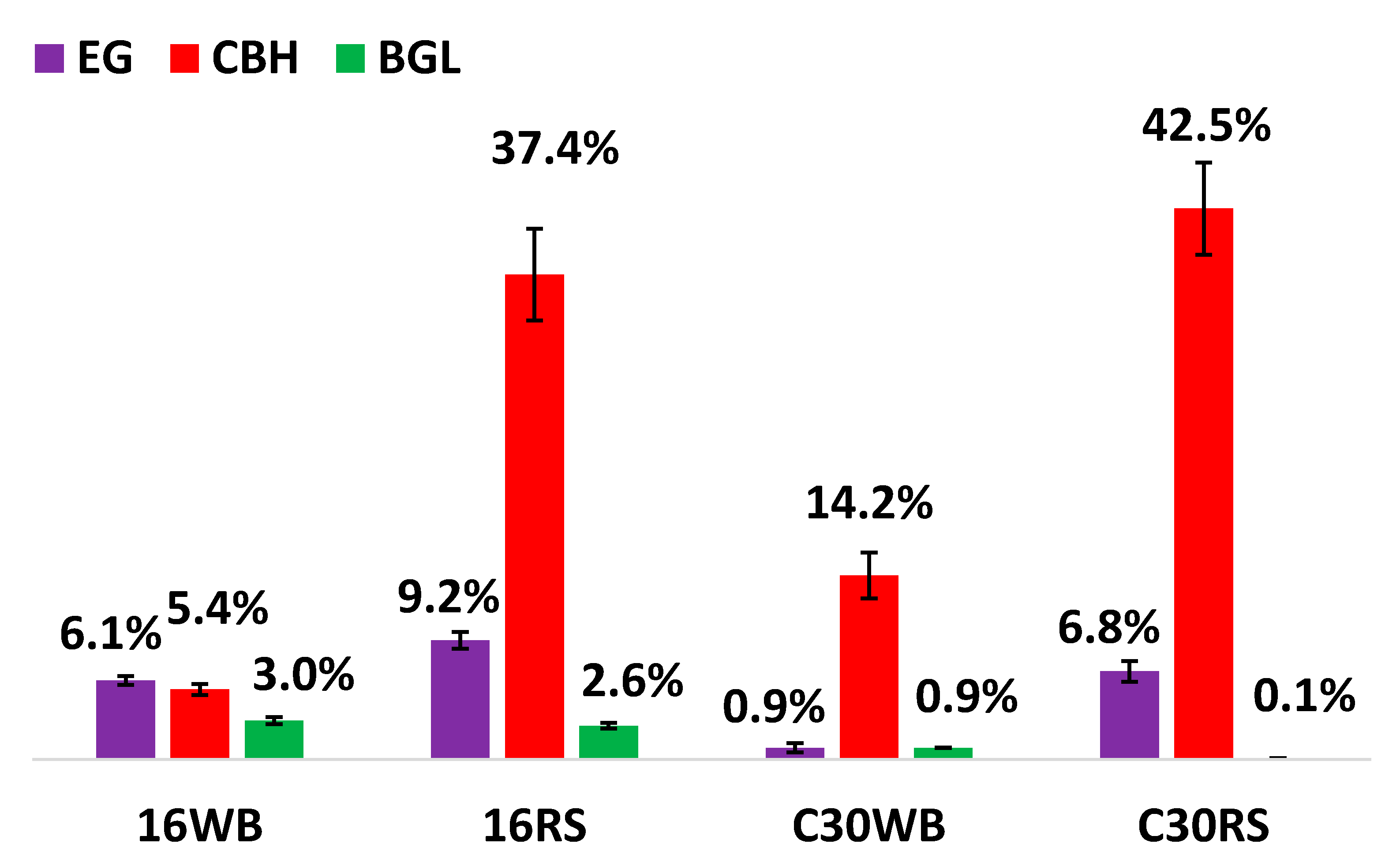
3.1. Enzymatic Activities of Major Glycoside Hydrolases (GHs)

Various GHs of P. oxalicum 16 cultured in the WB solid-state medium (16WB), P. oxalicum 16 cultured in the RS solid-state medium (16RS), T. reesei RUT-C30 cultured in the WB solid-state medium (C30WB), and T. reesei RUT-C30 cultured in the RS solid-state medium (C30RS) were induced, and their enzymatic activities were determined. As shown in Table 1, 16WB achieved the highest activities for a number of enzymes, e.g., 998 IU/gds amylase, 283 IU/gds xylanase, and 42 IU/gds BGL, but it produced the lowest activities for EG and CBH. 16RS attained increased EG and CBH activities of 211 and 0.31 IU/gds, respectively, but its amylase and xylanase activities were only 373 and 150 IU/gds, respectively. C30RS showed the highest activities of EG and CBH, which were approximately three and 21 times higher than those of 16WB. Although C30WB EG activity was slightly higher than that of 16WB and 16RS, its amylase and xylanase activities were about 100 and 3.6 times lower than those of 16WB.

3.2. Hydrolytic Ability of GHs

To evaluate the degradation ability of GHs from 16WB, 16RS, C30WB, and C30RS toward pretreated agricultural waste, we measured the total sugar released from WB, Pr-MCC, Pr-RS, and Pr-CP. The chemical compositions of WB and RS without pretreatment were introduced in our previous study [2]; MCC is pure cellulose, and the composition of CP contains 35.1–35.87% cellulose, 34.1–34.4% xylan, 20.9–21.96% lignin, and 8.1–9.6% other components [30,31]. Thus, Pr-RS and Pr-CP are intermediate biomasses between WB and Pr-MCC. Therefore, these chemical compositions contribute to our understanding of biomass degradation. As shown in Table 2, 16WB produced the highest percentage of total sugar with 10,571 μg/mL when hydrolyzing WB, which was 1.67, 5.43, and 7 times higher than 16RS, C30WB, and C30RS, respectively. The total sugar released from Pr-RS had the lowest percentage compared with that from WB, Pr-MCC, and Pr-RS, indicating that Pr-RS was the most difficult to degrade. Additionally, when considering Pr-MCC and Pr-CP, C30RS produced more total sugar than C30WB, 16RS, and 16WB.

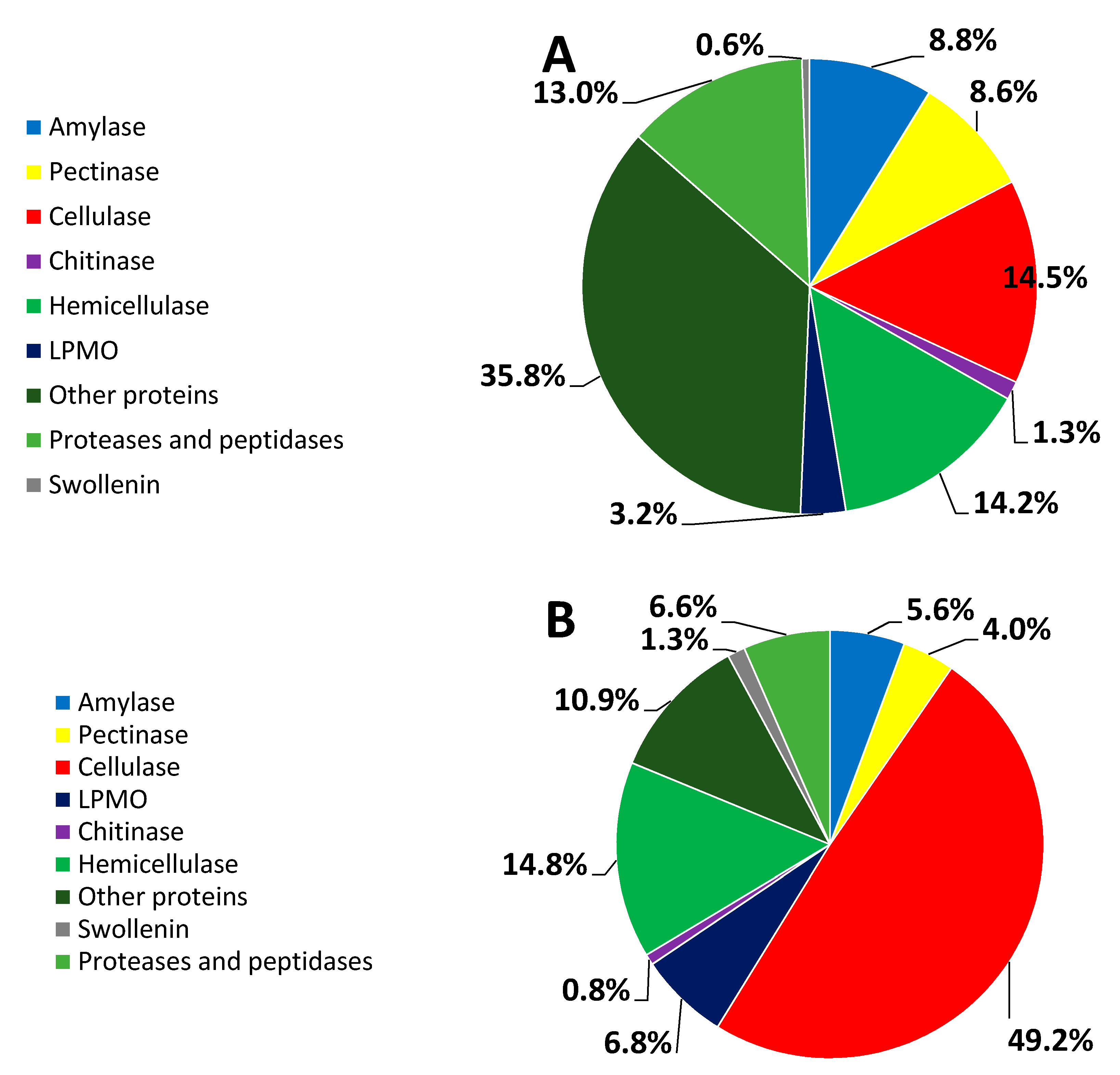
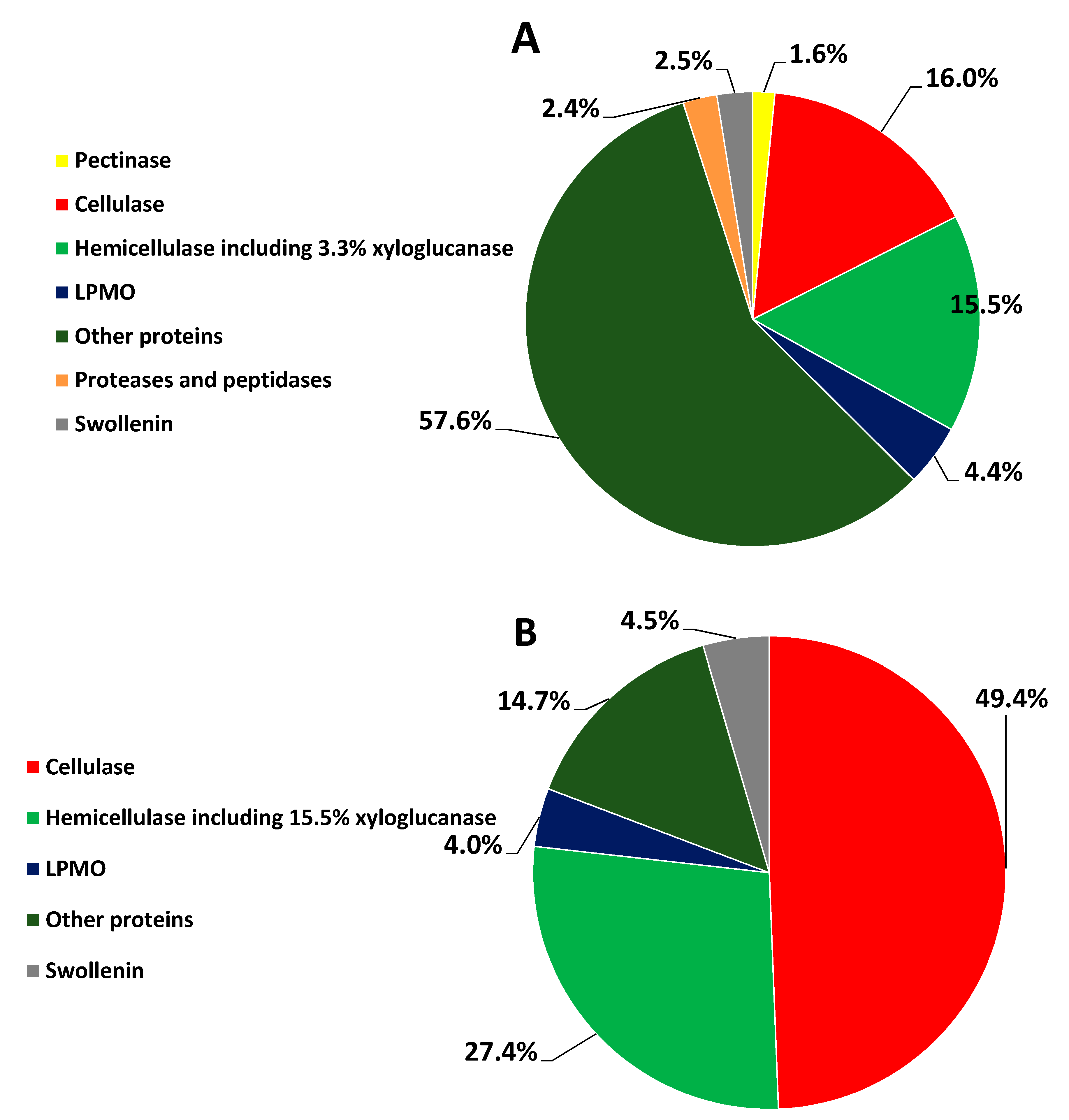
3.3. Percent Abundance of the Identified Proteins

To obtain the GH distribution, it was important to analyze the secretomes of the four enzymatic preparations. The number of identified proteins for 16RS and 16WB reached 291, 181 proteins of which were quantified. In contrast, 532 proteins were identified for C30RS and C30WB, among which 100 proteins were quantified. The total number of GH families (GHFs) from the 16WB secretome reached 88 and was 1.42, 1.76, and 3.14 times higher than that of 16RS, C30WB, and C30RS, respectively, indicating that 16WB produced more abundant and extensive GHs as seen in the Supplementary Materials.
As shown in Figure 1A, 35.8% of the secretome of 16WB was composed of other proteins such as 0.09% laccase which degrades lignin. The 16WB hemicellulase accounted for 14.2% of all the identified proteins. The other enzymes that had a relatively high percentage (e.g., >8%) were protease and peptidase (13%), cellulase (14.5%), amylase (8.8%), and pectinase (8.6%), which includes rhamnogalacturonan proteins, arabinofuranosidase, arabinosidase, arabinanase, endo-polygalacturonase, and pectin lyase. In contrast to the low expression of cellulase in 16WB, the 16RS had a high percentage of cellulase, up to 49.2% (Figure 1B). The percentage of the 16RS hemicellulase was similar to that of the 16WB hemicellulase (14.2% vs. 14.8%). The auxiliary enzymes for degrading cellulose mainly included LPMO and swollenin, which showed a higher percentage in 16RS than in 16WB.
As shown in Figure 2A, C30WB produced 1.6% pectinase (arabinofuranosidase), 57.6% other proteins with 10.5% oxidoreductase, 16% cellulase, and 15.5% hemicellulase including 3.3% xyloglucanase. In addition, C30WB secreted 2.5% swollenin, and 2.4% proteases and peptidases. As shown in Figure 2B, C30RS produced 49.4% cellulase, 27.4% hemicellulase including 15.5% xyloglucanase, and 4.5% swollenin, which was 3.5 times higher than in 16RS.
As shown in Figure 1 and Figure 2, C30RS and 16RS strongly produced cellulase, hemicellulase, swollenin, and LPMO, surpassing C30WB and 16WB. Chitinase and amylase were produced by 16WB and 16RS, but they did not exist in either C30WB or C30RS. The protease and peptidase percentages of 16WB and 16RS was higher than those of C30WB, and C30RS did not possess proteases or peptidase. Moreover, 16RS and 16WB hemicellulases had lower percentages than those of C30WB and C30RS, but the 16RS and 16WB GHs were more balanced than C30WB and C30RS as described above, consistent with a previous study [10].
Figure 3 shows the percentage of CBH, EG, and BGL from 16WB, 16RS, C30RS, and C30WB. 16WB CBH and EG had the lowest percentage, but it obtained the highest percentage of BGL (~3%). The C30RS and 16RS achieved the highest percentages of CBH at 42.5% and 37.4%, respectively. In addition, it was obvious that C30WB and C30RS had a very low percentage of BGL, suggesting that T. reesei RUT-C30 may be a poor expressor of BGL.

3.4. Revealing Up-Regulated and Down-Regulated Proteins

As shown in Table 3 and Table 4, the up-regulated proteins of 16WB compared with 16RS were mainly related to amylase, pectinase, and protease, whereas the down-regulated proteins of that were mainly related to cellulase, hemicellulase, swollenin, and LPMO. Compared with the 16RS secretome, the up-regulated classification of the main 16WB components consisted of 30 proteins, i.e., putative EG (UniProt ID S7ZMB4), α-amylase (S7Z6T2), glucoamylase (S7ZIW0), glycosidase (S8AIA9), α-1,2-mannosidase (S7Z4H1), α-galactosidase (S7ZFY8), β-mannosidase A (S8BFI1), endo-polygalacturonase (S7ZD03), β-xylanase (S7ZA570, S7ZAV8), endo-β-1,4-mannanase F (S7ZDN1), endo-1,3-β-glucanase eglC (S7ZAG7), non-reducing end α-L-arabinofuranosidase (S7ZW00), CBH I (S7ZJL3), arabinogalactan endo-β-1,4-galactanase (S7ZBM4), β-galactosidase (S7Z5H6), putative endo-β-1,4-xylanase (S8AH74), chitinase (S8AWH6), putative α-L-arabinofuranosidase (S7Z4P2, S8B8M7), putative rhamnogalacturonan α-L-rhamnopyranohydrolase (S7ZZQ8), putative β-glucuronidase (S8B0N0), putative exo-β-1,3-galactanase (S7ZUD9), putative β-glucanase (S7ZMU5), putative β-1,3-1,4-glucanase (S7ZCP1), putative endo-β-1,4-xylanase (S8B2H7, S8AH74), putative α-mannosidase (S8B2R2, S8AUX2), and putative endo-β-1,6-galactanase(S8AXM3); the down-regulated classification of the main 16WB components including auxiliary decomposed enzymes of cellulose was comprised of 19 proteins, namely, putative β-1,6-glucanase (S8AMF6), putative endo-β-1,3-glucanase (S7ZAS9), CBH I (S7ZRD6), CBH II (S7ZP52), endo-1,4-β-xylanase (S8AMN0, S8BDN2), glucoamylase (S8B6D7), chitinase (S7Z8G1, S7ZR03, and S8B6N1), BGL (S8B0F3), non-reducing end α-L-arabinofuranosidase (S7Z3I8), endo-β-1,4-mannanase F (S7ZL65), EG (S8BGM3, S7ZX22, and S8AIJ2), EG1 (S8B2B2), putative swollenin (S7ZAB6), and LPMO (S7ZPW1).
Compared with the C30RS secretome, the up-regulated classification of the main C30WB components included 13 proteins, which were β-mannosidase (A0A024RUF8), endo-polygalacturonase (A0A024S1V1), β-1,4-endoxylanase (A0A024S0A7), endo-β-1,6-galactanase (A0A024S0G1), xylanase (A0A024RWW9), β-1,3-endoglucanase (A0A024S1W9), α-galactosidase (A0A024SGF7), α-glucuronidase (A0A024S166), β-xylosidase (A0A024SDM6), GH16 domain-containing protein (A0A024S732), chitinase (A0A024S1T5), α-L-arabinofuranosidase (A0A024S2Y7), and β-1,3-endoglucanase (A0A024SAF4); the down-regulated classification of the main components including auxiliary decomposed enzymes of cellulose from the C30WB secretome was comprised of 15 proteins which were xyloglucanase (A0A024S9Z6), EG (A0A024SNB7, A0A024SH20, A0A024S2H5), CBH I (A0A024RXP8), CBH II (A0A024SH76), non-reducing end α-L-arabinofuranosidase (A0A024SGE7), endo-1,4-β-xylanase (P36217, A0A024SIB3), chitinase (A0A024S0K1), mannan endo-1,4-β-mannosidase (A0A024SIJ3), β-1,4-endoxylanase (A0A024RV01), BGL (A0A024SCX9), LPMO (A0A024SFJ2), and swollenin (A0A024RZP7).

3.5. Ascertaining Identities with a High Percentage

Revealing the identities of the highly expressed GHs is helpful to design GH systems that are suitable for diverse substrates. As shown in Table 5, β-xylanase (S7ZA57) ranked first in 16WB with an abundance of 6.5%, glucoamylase (S8B6D7) ranked second at 5.7%, and CBH I (S7ZRD6) ranked third at 3.7%. EG S8AH74 and hemicellulase endo-β-1,4-mannanase F (S7ZDN1) obtained abundances of 1.1% and 1.7%, respectively, but they decreased sharply in RS. 16RS CBH I (S7B6D6) had the highest abundance at 26%, whereas the second-highest was β-xylanase (S8BDN2) at 14% in 16RS, but the 16RS β-xylanases S7ZA57 and S8AH74 decreased to 0.36% and 0.06%, respectively. 16RS CBH II (S7ZP52) possessed the third-highest percentage at 9.6%. Of particular interest was that glucoamylase (S8B6D7) also achieved an amazing 5.2% in 16RS. In addition, three EGs (S8BGM3, S7ZX22, and S8AIJ2) of 16RS also increased significantly to 3.7%, 3.3%, and 1.2%, respectively. The BGL (S8B0F3) percentage of 16WB and 16RS changed little and accounted for 3 and 2.5%, respectively. According to our secretomics information, we studied P. oxalicum 16 BGL (S8B0F3) [4,5,22], and its properties were improved by directed evolution [4,5]. The main LPMO was S7ZPW1, and reached 5.6% in 16RS and 3.2% in 16WB, but the main LPMO S7Z716 only existed in 16RS at an abundance of 1.2%.
CBH I removes cellobiose from the reducing end of the cellooligosaccharide, whereas CBH II releases cellobiose from the non-reducing end of the cellooligosaccharide. In general, these two enzymes work together to accelerate the degradation of cellulose. As shown in Table 5, the average percentage of CBH I and CBH II from 16RS and 16WB was 2.7:1, but that of CBH I and CBH II from C30RS and C30WB was close to 1:1. Both of the C30RS CBH I (A0A024RXP8) and CBH II (A0A024SH76) percentages were 21% and were 2.68 and 3.39 times higher, respectively, than those from C30WB. C30RS xyloglucanase (A0A024S9Z6) accounted for 16%, which was 4.7 times higher than C30WB. However, xyloglucanase was not found in 16WB and 16RS. There was only one glucoamylase (A0A024SN40) in T. reesei RUT-C30, but we could not find it in C30RS or C30WB. Other hydrolases may have taken the place of the amylase role for hydrolyzing starch, but the degradation efficiency was extremely low. In addition, the percentage of the swollenin (A0A024RZP7) from C30RS was 4.5% and was 1.8 times higher than that from C30WB.

4. Discussion

Natural substrates generate more GHs than pure substrates for fungi [10,32,33,34], so we used WB and RS for induction, rather than MCC, CMC, xylan, or starch. In addition, WB and RS are the main agricultural wastes in the north and south of China, respectively, so it was meaningful to realize utilization of “turning waste into treasure”. WB mainly consists of cellulose, hemicellulose, starch, and a small amount of pectin, whereas RS is mainly composed of cellulose, hemicellulose, and lignin [2]. In our study, WB induced more diverse secretome proteins than RS and is suitable for inducing pectinase, xylanase, and amylase; however, RS generated more cellulase, hemicellulase, LPMO, and swollenin. Some studies have shown that lactose, sophorose, or sophorose analogs were the main inducers of T. reesei RUT-C30 [34], but they are expensive and not suitable for industrial application. However, WB and RS are a good choice as cheap and widely existing carbon sources.
Designing enzyme preparation according to different substrates is beneficial and effective to completely degrade the renewable biomass resources with their complex and stubborn structures [10,19,20]. Therefore, it is important to thoroughly ascertain the secretome information of P. oxalicum 16 and T. reesei RUT-C30, which will lay a foundation for future molecular modification. Through our comparative analysis, we found that the P. oxalicum 16 xylanases S8AH74 and S7ZA57 are probably the main factors for the degradation of soluble xylan. However, the increased percentage of the xylanase S8BDN2 did not improve the hydrolysis of soluble xylan, but on the contrary, its degradation ability of soluble xylan was reduced. Therefore, we believe that it is highly likely that S8BDN2 acts on solid-state hemicellulose instead of soluble xylan. Furthermore, we speculate that the amylase S7Z6T2 is the main degradation factor for soluble starch, because the glucoamylase S8B6D7 did not significantly increase or decrease under the induction of WB or RS. We could not find amylase or glucoamylase in C30WB or C30RS, indicating that T. reesei RUT-C30 is not an amylase producer.
Natural cellulose is the most abundant renewable biomass resource, but it is difficult to degrade [35]. Therefore, how to effectively degrade cellulose is an ongoing key issue, and it has been proven that its degree of decomposition is mainly related to CBHs [36]. In addition, according to comparative secretomics analysis of the two strains, we found that the highest yield of secreted enzymes from the induction of RS was that of CBH. Thus, we focused on the discussion concerning the synergistic degradation of cellulose. The whole enzymatic system of 16RS and C30RS showed a significant difference in the hydrolysis of Pr-MCC and Pr-CP. Although CBH I was strongly induced in 16RS, the total proportion of CBH I S7ZRD6 and CBH II S7ZP52 (about 2.7:1) in 16RS exceeded that of C30RS. Furthermore, the proportion of CBH I A0A024RXP8 and CBH II A0A024SH76 in C30RS was close to 1:1. Therefore, the different proportions of CBH I and CBH II in 16RS and C30RS may have resulted in the different hydrolysis degrees of Pr-MCC and Pr-CP (Table 2), which is consistent with the findings reported by Schülein et al. [36]. Different combinations and proportions of CBH I, CBH II, and EG may or may not produce synergistic degradation against different substrates [36]. It has been demonstrated that the substrates CMC or MCC do not trigger synergistic degradation by CBH I and EG, but amorphous cellulose leads to synergistic degradation [36]. CBH I and EG with a proportion of 1:1 have the greatest synergistic degradation, but CBH II and EG show synergistic degradation against any substrate [34]. CBH I and CBH II, with a ratio of 1:4, exhibits the maximum synergistic degradation of solid cellulose [34].
In contrast to GHs, the auxiliary degradation enzyme LPMO, which is a kind of metal enzyme containing Cu2+ and generates a synergistic degradation effect with cellulase, amylase, hemicellulase, etc., uses cellobiose deoxygenase and ascorbic acid as electron donors, and the peroxide hydrogen and oxygen as co-substrates [12,13]. In the study, LPMO A0A024SM10 was only secreted in C30WB, but not in C30RS. LPMOs S7ZPW1 and A0A024SFJ2 were up-regulated in 16RS and C30RS, respectively. Our inference that the main substrate of A0A024SM10 and A0A024SFJ2 is cellulose is consistent with the report of Corrêa et al. [14], and other substrates of A0A024SM10 may be pectin or hemicellulose. LPMO S7Z716 only existed in 16RS, and could not be detected in 16WB. The relative abundance of S7ZPW1 was increased in 16RS. In addition, we found that RS had more cellulose, whereas WB had more starch, pectin, etc. in our previous work [2]. Therefore, we speculated that S7Z716 and S7ZPW1 may mainly perform oxidative cleavage of cellulose. Although reducing sugar could not be detected in the reaction solution, swollenin can indeed break and expand crystalline cellulose [15]. Interestingly, the LPMO in 16RS showed a higher relative abundance than that in C30RS, but swollenin in T. reesei RUT-C30 had a higher relative abundance than that in P. oxalicum 16 (Table 5). Additionally, C30RS enzymes released more reduced sugar when dealing with Pr-MCC (Table 2). Therefore, we speculate that the C30RS swollenin goes beyond its LPMO and assumes the main responsibility of synergistic degradation against cellulose.
An effective cellulase preparation with the proportion 4:1:1 of CBH II, CBH I, and EG, with the addition of swollenin and LPMO, will be the focus of our future work. According to the above results, P. oxalicum 16 possesses more diverse GHs than T. reesei RUT-C30, so engineering the strain P. oxalicum 16 will be used to generate the proportion with auxiliary degradation enzymes according to its genomics, transcriptomics and secretomics information. By analogy, other effective enzyme mixtures can be also created.

5. Conclusions

The secretome comparison analysis here revealed that WB induced P. oxalicum 16 and T. reesei RUT-C30 to produce more abundant and balanced GHs than RS, but RS induced more cellulase and hemicellulase. Moreover, the study found that P. oxalicum 16 possessed more diverse GHs than T. reesei RUT-C30. In addition, the study characterized the up-regulated, down-regulated, and main component identities. Based on the study, it may be feasible to design combinations from the modified P. oxalicum 16 to decompose specific substrates.

Supplementary Materials

The following are available online at www.mdpi.com/article/10.3390/microorganisms9102042/s1, Excel Table S1: Identified protein information.

Author Contributions

X.Z. conceived the project, acquired funding, designed experiments, prepared original manuscript, and assumed supervision. K.W. and N.Z. did experiments, collected data, and prepared original manuscript. R.P. revised the whole manuscript. S.Y. did experiments. All authors have read and agreed to the published version of the manuscript.

Funding

This work was financially supported by the National Natural Science Foundation of China (22068014, 21666010), and the Doctoral Starting up Foundation of Jiangxi Normal University (5451).

Institutional Review Board Statement

Not applicable.

Informed Consent Statement

Not applicable.

Conflicts of Interest

The authors declare they have no conflict of interest.

References

  1. Watt, G.D. A new future for carbohydrate fuel cells. Renew. Energy 2014, 72, 99–104. [Google Scholar] [CrossRef]
  2. Zhao, X.; Yi, S.; Li, H. The optimized co-cultivation system of Penicillium oxalicum 16 and Trichoderma reesei RUT-C30 achieved a high yield of hydrolase applied in second-generation bioethanol production. Renew. Energy 2019, 136, 1028–1035. [Google Scholar] [CrossRef]
  3. Cheng, Y.; Zhang, L.; Xu, S.; Zhang, H.; Ren, B.; Li, T.; Zhang, S. Ionic liquid functionalized electrospun gel polymer electrolyte for use in a high-performance lithium metal battery. J. Mater. Chem. A 2018, 6, 18479–18487. [Google Scholar] [CrossRef]
  4. Wang, K.; Huang, Q.; Li, H.; Zhao, X. Co-evolution of β-glucosidase activity and product tolerance for increasing cellulosic ethanol yield. Biotechnol. Lett. 2020, 42, 2239–2250. [Google Scholar] [CrossRef]
  5. Huang, Q.; Wang, K.; Li, H.; Yi, S.; Zhao, X. Enhancing cellulosic ethanol production through coevolution of multiple enzymatic characteristics of β-glucosidase from Penicillium oxalicum 16. Appl. Microbiol. Biotechnol. 2020, 104, 8299–8308. [Google Scholar] [CrossRef]
  6. Zhang, J.; Chen, Y.; Wu, C.; Liu, P.; Wang, W.; Wei, D. The transcription factor ACE3 controls cellulase activities and lactose metabolism via two additional regulators in the fungus Trichoderma reesei. J. Biol. Chem. 2019, 294, 18435–18450. [Google Scholar] [CrossRef]
  7. Duchesne, L.C.; Larson, D.W. Cellulose and the evolution of plant life. Bioscience 1989, 39, 238–241. [Google Scholar] [CrossRef]
  8. Saha, B.C. Hemicellulose bioconversion. J. Ind. Microbiol. Biotechnol. 2003, 30, 279–291. [Google Scholar] [CrossRef]
  9. Zhang, Y.H.P.; Himmel, M.E.; Mielenz, J.R. Outlook for cellulase improvement: Screening and selection strategies. Biotechnol. Adv. 2006, 24, 452–481. [Google Scholar] [CrossRef]
  10. Gong, W.; Zhang, H.; Liu, S.; Zhang, L.; Gao, P.; Chen, G.; Wang, L. Comparative secretome analysis of Aspergillus niger, Trichoderma reesei, and Penicillium oxalicum during solid-state fermentation. Appl. Biochem. Biotechnol. 2015, 177, 1252–1271. [Google Scholar] [CrossRef]
  11. Han, X.; Wen, H.; Luo, Y.; Yang, J.; Xiao, W.; Ji, X.; Xie, J. Effects of α-amylase and glucoamylase on the characterization and function of maize porous starches. Food Hydrocoll. 2021, 116, 106661. [Google Scholar] [CrossRef]
  12. Vaaje-Kolstad, G.; Westereng, B.; Horn, S.J.; Liu, Z.; Zhai, H.; Sørlie, M.; Eijsink, V.G.H. An oxidative enzyme boosting the enzymatic conversion of recalcitrant polysaccharides. Science 2010, 330, 219–222. [Google Scholar] [CrossRef]
  13. Limsakul, P.; Phitsuwan, P.; Waeonukul, R.; Pason, P.; Tachaapaikoon, C.; Poomputsa, K.; Kosugi, A.; Sakka, M.; Ratanakhanokchai, K. A novel AA10 from Paenibacillus curdlanolyticus and its synergistic action on crystalline and complex polysaccharides. Appl. Microbiol. Biotechnol. 2020, 104, 7533–7550. [Google Scholar] [CrossRef] [PubMed]
  14. Corrêa, T.L.R.; Dos Santos, L.V.; Pereira, G.A.G. AA9 and AA10: From enigmatic to essential enzymes. Appl. Microbiol. Biotechnol. 2016, 100, 9–16. [Google Scholar] [CrossRef] [PubMed]
  15. Zhang, H.; Wang, Y.; Brunecky, R.; Yao, B.; Xie, X.; Zheng, F.; Luo, H. A Swollenin from Talaromyces leycettanus JCM12802 enhances cellulase hydrolysis toward various substrates. Front. Microbiol. 2021, 12, 715. [Google Scholar]
  16. Zhao, X.H.; Wang, W.; Tong, B.; Zhang, S.P.; Wei, D.Z. A newly isolated Penicillium oxalicum 16 cellulase with high efficient synergism and high tolerance of monosaccharide. Appl. Biochem. Biotechnol. 2016, 178, 173–183. [Google Scholar] [CrossRef]
  17. Liu, P.; Lin, A.; Zhang, G.; Zhang, J.; Chen, Y.; Shen, T.; Zhao, J.; Wei, D.; Wang, W. Enhancement of cellulase production in Trichoderma reesei RUT-C30 by comparative genomic screening. Microb. Cell Fact. 2019, 18, 81. [Google Scholar] [CrossRef] [PubMed]
  18. Herpoël-Gimbert, I.; Margeot, A.; Dolla, A.; Jan, G.; Mollé, D.; Lignon, S.; Mathis, H.; Sigoillot, J.C.; Monot, F.; Asther, M. Comparative secretome analyses of two Trichoderma reesei RUT-C30 and CL847 hypersecretory strains. Biotechnol. Biofuels 2008, 1, 18. [Google Scholar] [CrossRef]
  19. Marx, I.J.; Van Wyk, N.; Smit, S.; Jacobson, D.; Viljoen-Bloom, M.; Volschenk, H. Comparative secretome analysis of Trichoderma asperellum S4F8 and Trichoderma reesei Rut C30 during solid-state fermentation on sugarcane bagasse. Biotechnol. Biofuels 2013, 6, 172. [Google Scholar] [CrossRef] [Green Version]
  20. Liu, G.; Zhang, L.; Wei, X.; Zou, G.; Qin, Y.; Ma, L.; Li, J.; Zheng, H.; Wang, S.; Wang, C.; et al. Genomic and secretomic analyses reveal unique features of the lignocellulolytic enzyme system of Penicillium decumbens. PLoS ONE 2013, 8, e55185. [Google Scholar] [CrossRef] [Green Version]
  21. Song, W.; Han, X.; Qian, Y.; Liu, G.; Yao, G.; Zhong, Y.; Qu, Y. Proteomic analysis of the biomass hydrolytic potentials of Penicillium oxalicum lignocellulolytic enzyme system. Biotechnol. Biofuels 2016, 9, 68. [Google Scholar] [CrossRef] [PubMed] [Green Version]
  22. Mandels, M.; Reese, E.T. Induction of cellulase in fungi by cellobiose. J. Bacteriol. 1960, 79, 816. [Google Scholar] [CrossRef] [Green Version]
  23. Miller, G.L.; Blum, R.; Glennon, W.E.; Burton, A.L. Measurement of carboxymethylcellulase activity. Anal. Biochem. 1960, 1, 127–132. [Google Scholar] [CrossRef]
  24. Deshpande, M.V.; Eriksson, K.E.; Pettersson, L.G. An assay for selective determination of exo-1, 4,-β-glucanases in a mixture of cellulolytic enzymes. Anal. Biochem. 1984, 138, 481–487. [Google Scholar] [CrossRef]
  25. Kruger, N.J. The Bradford method for protein quantitation. In The Protein Protocols Handbook; Humana Press: Totowa, NJ, USA, 2009; pp. 17–24. [Google Scholar]
  26. Dubois, M.; Gilles, K.A.; Hamilton, J.K.; Rebers, P.T.; Smith, F. Colorimetric method for determination of sugars and related substances. Anal. Biochem. 1956, 28, 350–356. [Google Scholar] [CrossRef]
  27. Cao, R.; He, Q.; Zhou, J.; He, Q.; Liu, Z.; Wang, X.; Chen, P.; Xie, J.; Liang, S. High-throughput analysis of rat liver plasma membrane proteome by a nonelectrophoretic in-gel tryptic digestion coupled with mass spectrometry identification. J. Proteome Res. 2008, 7, 535–545. [Google Scholar] [CrossRef]
  28. Wei, J.; Sun, J.; Yu, W.; Jones, A.; Oeller, P.; Keller, M.; Woodnutt, G.; Short, J.M. Global proteome discovery using an online three-dimensional LC-MS/MS. J. Proteome Res. 2005, 4, 801–808. [Google Scholar] [CrossRef]
  29. Brownell, H.H.; Saddler, J.N. Steam pretreatment of lignocellulosic material for enhanced enzymatic hydrolysis. Biotechnol. Bioeng. 1987, 29, 228–235. [Google Scholar] [CrossRef]
  30. Zhang, X.; Yuan, Q.; Cheng, G. Deconstruction of corncob by steam explosion pretreatment: Correlations between sugar conversion and recalcitrant structures. Carbohydr. Polym. 2017, 156, 351–356. [Google Scholar] [CrossRef]
  31. Qi, W.; He, C.; Wang, Q.; Liu, S.; Yu, Q.; Wang, W.; Leksawasdi, N.; Wang, C.; Yuan, Z. Carbon-based solid acid pretreatment in corncob saccharification: Specific xylose production and efficient enzymatic hydrolysis. ACS Sustain. Chem. Eng. 2018, 6, 3640–3648. [Google Scholar] [CrossRef]
  32. Liao, H.; Li, S.; Wei, Z.; Shen, Q.; Xu, Y. Insights into high-efficiency lignocellulolytic enzyme production by Penicillium oxalicum GZ-2 induced by a complex substrate. Biotechnol. Biofuels 2014, 7, 162. [Google Scholar] [CrossRef] [Green Version]
  33. Liu, D.; Li, J.; Zhao, S.; Zhang, R.; Wang, M.; Miao, Y.; Shen, Y.; Shen, Q. Secretome diversity and quantitative analysis of cellulolytic Aspergillus fumigatus Z5 in the presence of different carbon sources. Biotechnol. Biofuels 2013, 6, 149. [Google Scholar] [CrossRef] [Green Version]
  34. Huang, T.T.; Wages, J.M. New-to-nature sophorose analog: A potent inducer for gene expression in Trichoderma reesei. Enzym. Microb. Technol. 2016, 85, 44–50. [Google Scholar] [CrossRef] [PubMed] [Green Version]
  35. Jing, X.; Zhang, X.; Bao, J. Inhibition performance of lignocellulose degradation products on industrial cellulase enzymes during cellulose hydrolysis. Appl. Biochem. Biotechnol. 2009, 159, 696–707. [Google Scholar] [CrossRef] [PubMed]
  36. Henrissat, B.; Driguez, H.; Viet, C.; Schülein, M. Synergism of cellulases from Trichoderma reesei in the degradation of cellulose. Nat. Biotechnol. 1985, 3, 722–726. [Google Scholar] [CrossRef]
Figure 1. Percentage of the proteins identified in P. oxalicum 16 ((A): 16WB; (B): 16RS).
Microorganisms 09 02042 g001
Figure 2. Percentage of the proteins identified in T. reesei Rut-C30 ((A): C30WB; (B): C30RS).
Microorganisms 09 02042 g002
Figure 3. Percentage of EG, CBH, and BGL.
Microorganisms 09 02042 g003
Table 1. Comparation of the main GH activities.
OriginsEG (IU/gds)CBH (IU/gds)BGL (IU/gds)Amylase (IU/gds)Xylanase (IU/gds)
16WB209 ± 20.02 ± 0.042 ± 1.3998 ± 8283 ± 2
16RS211 ± 20.31 ± 0.0132.1 ± 1.2373 ± 1150 ± 3
C30WB259 ± 10.09 ± 0.009.2 ± 1.024.8 ± 1.173 ± 0
C30RS628 ± 40.41 ± 0.0211.0 ± 0.810.2 ± 0.379 ± 6
The experiments were repeated three times.
Table 2. Total soluble sugars released from pretreated agricultural wastes for 96 h.
OriginsPr-RS (μg/mL)WB (μg/mL)Pr-MCC (μg/mL)Pr-CP (μg/mL)
16WB1821 ± 7210,571 ± 4783475 ± 634101 ± 63
16RS3488 ± 816322 ± 4414625 ± 3708278 ± 171
C30WB3209 ± 4801948 ± 4246107 ± 1177978 ± 111
C30RS3650 ± 1531509 ± 437222 ± 738674 ± 189
The experiments were repeated three times.
Table 3. The up-regulated and down-regulated proteins of 16WB compared with 16RS.
UniProt IDProtein DescriptionSubstrateClassification16WB/16RS RatioRegulated Type
S8AHA8Putative β-xylosidasexylooligosaccharideGH3168 ± 8Up
S8BFI1Putative β-mannosidaseβ-mannose-oligosaccharideGH271 ± 7Up
S7ZIW0GlucoamylasedextrinGH1538 ± 1Up
S7Z5H6β-galactosidaseβ-lactoseGH3529 ± 1Up
S7ZA57β-xylanasexylanGH1010 ± 0Up
S8B2R2Putative α-mannosidaseα-mannose-oligosaccharideGH9210 ± 1Up
S8AH74Putative endo-β-1,4-xylanasexylanGH3010 ± 1Up
S7ZW00Putative α-L-arabinofuranosidasearabinogalactan, arabinoglycan, etc.GH629.4 ± 0.4Up
S8AXM3Putative endo-β-1,6-galactanaseβ-1,6-galactanGH309.2 ± 0.8Up
S7ZUD9Putative exo-β-1,3-galactanaseβ-1,3-galactanGH437.3 ± 0.6Up
S7ZBM4Arabinogalactan endo-β-1,4-galactanaseβ-galactosidic linkages in type I arabinogalactansGH537.1 ± 0.4Up
S8B8M7Putative α-L-arabinofuranosidasearabinogalactan, arabinoglycan, etc.GH436.6 ± 0.2Up
S7ZMB4Putative EGcelluloseGH125.1 ± 0.1Up
S8B2H7Putative endo-β-1,4-xylanasexylanGH304.5 ± 0.3Up
S7ZJL3CBH IMCCGH73.0 ± 0.1Up
S8B0N0Putative β-glucuronidaseproteoglycanGH22.8 ± 0.2Up
S7ZMU5Putative β-glucanaseβ-1,3(4)-glucanGH162.4 ± 0.1Up
S7ZFY8α-galactosidaseα-lactoseGH272.2 ± 0.1Up
S8AWH6Putative chitinasechitinGH181.7 ± 0.1Up
S7Z6T2α-amylase Amy13Aα-1,4-starchGH131.7 ± 0.1Up
S7ZZQ8Putative α-L-rhamnopyranohydrolaseR-α-L-rhamnopyranosideGH281.7 ± 0.1Up
S7ZD03Endo-polygalacturonasepectin or pectinic acidGH281.6 ± 0.1Up
S7Z4P2Putative α-L-arabinofuranosidasearabinogalactan, arabinoglycan, etc.GH431.6 ± 0.1Up
S7Z4H1α-1,2-mannosidaseα-1,2-mannose-oligosaccharideGH471.6 ± 0.1Up
S7ZAV8β-xylanasexylanGH101.5 ± 0.0Up
S8B7P9Putative α-L-arabinofuranosidasearabinogalactan, arabinoglycan, etc.GH541.5 ± 0.1Up
S7ZWC7Putative exo-α-L-1,5-arabinanaseα-L-1,5-arabinoglycanGH931.5 ± 0.1Up
S7ZDN1Putative endo-β-1,4-glucanasecelluloseGH51.4 ± 0.1Up
S7ZCP1Putative β-1,3-1,4-glucanaseβ-1,3-1,4-glucanGH161.4 ± 0.1Up
S8AUX2Putative α-mannosidaseα-mannose-oligosaccharideGH921.3 ± 0.1Up
S7ZR03Putative chitinasechitinGH180.76 ± 0.04Down
S8B6D7GlucoamylasedextrinGH150.70 ± 0.03Down
S8B0F3BGLcellooligosaccharideGH30.67 ± 0.03Down
S7Z8G1Putative chitinasechitinGH180.54 ± 0.03Down
S8AMF6Putative β-1,6-glucanaseβ-1,6-glucanGH300.53 ± 0.01Down
S8B6N1Putative chitinasechitinGH180.51 ± 0.01Down
S8AXN0Putative pectate lyasepectinic acidpolysaccharide lyase 1 family0.43 ± 0.00Down
S7Z3I8Putative α-L-arabinofuranosidasearabinogalactan, arabinoglycan, etc.GH620.40 ± 0.02Down
S7ZPW1LPMOpolysaccharideAA90.33 ± 0.00Down
S7ZAS9Putative endo-β-1,3-glucanaseβ-1,3-glucanPectate lyase superfamily0.32 ± 0.00Down
S7ZAB6Putative swolleninsolid celluloseExpansin_EG450.24 ± 0.01Down
S7ZP52CBH IIMCCGH60.09 ± 0.00Down
S8B2B2EG1celluloseGH70.09 ± 0.00Down
S7ZL65Putative β-1,4-mannanasemannanGH50.08 ± 0.00Down
S8AMN0Endo-1,4-β-xylanasexylanGH110.08 ± 0.00Down
S7ZRD6CBH IMCCGH70.08 ± 0.00Down
S8BGM3EGcelluloseGH50.06 ± 0.00Down
S8BDN2β-xylanasexylanGH100.04 ± 0.00Down
S7ZX22EGcelluloseGH50.04 ± 0.00Down
S8AIJ2EGcelluloseGH50.02 ± 0.00Down
The experiments were repeated three times. 16WB/16RS ratio > 1 is defined as up-regulation, and 16WB/16RS ratio < 1 is defined as down-regulation.
Table 4. The up-regulated and down-regulated proteins of C30WB compared with C30RS.
UniProt IDProtein DescriptionSubstrateClassificationC30WB/C30RS RatioRegulated Type
A0A024SGF7α-galactosidaseα-lactoseGH2712 ± 0Up
A0A024S1T5ChitinasechitinGH187.7 ± 0.7Up
A0A024SDM6β-xylosidasexylooligosaccharideGH35.9 ± 0.4Up
A0A024RWW9xylanasexylanGH305.8 ± 0.3Up
A0A024SAF4β-1,3-endoglucanaseβ-1,3-glucanGH174.0 ± 0.1Up
A0A024S166α-glucuronidasexylanGH673.3 ± 0.1Up
A0A024S732β-glucanaseβ-1,3(4)-glucanGH162.5 ± 0.2Up
A0A024S1W9β-1,3-endoglucanaseβ-1,3-glucanGH172.3 ± 0.1Up
A0A024S0A7β-1,4-endoxylanasexylanGH431.9 ± 0.1Up
A0A024S2Y7α-N-arabinofuranosidaseα-L-arabinosideGH541.9 ± 0.1Up
A0A024S1V1Endopolygalacturonasepectin, pectinic acidGH281.8 ± 0.1Up
A0A024RUF8β-mannosidase Aβ-mannose-oligosaccharideGH21.5 ± 0.1Up
A0A024S0G1Endo-β-1,6-galactanaseβ-1,6-galactanGH301.2 ± 0.1Up
A0A024SIJ3β-mannase (Fragment)mannanGH50.62 ± 0.02Down
A0A024RZP7Swolleninsolid celluloseExpansin_EG450.50 ± 0.03Down
A0A024SNB7EGcelluloseGH70.49 ± 0.02Down
P36217Endo-1,4-β-xylanase 2xylanGH110.46 ± 0.01Down
A0A024RXP8CBH IMCCGH70.33 ± 0.01Down
A0A024S0K1ChitnasechitinGH180.33 ± 0.02Down
A0A024SGE7α-L-arabinofuranosidaseα-L-arabinosideGH620.30 ± 0.00Down
A0A024SH76CBH IIMCCGH60.26 ± 0.01Down
A0A024S9Z6XyloglucanasexyloglucanGH740.22 ± 0.00Down
A0A024SFJ2LPMOpolysaccharideAA90.21 ± 0.00Down
A0A024RV01β-1,4-endoxylanasexylanGH300.16 ± 0.01Down
A0A024SH20EGcelluloseGH50.15 ± 0.00Down
A0A024SCX9BGLcellooligosaccharideGH30.10 ± 0.00Down
A0A024SIB3Endo-1,4-β-xylanase 3xylanGH100.09 ± 0.00Down
A0A024S2H5EGcelluloseGH120.05 ± 0.00Down
The experiments were repeated three times. C30WB/C30RS ratio > 1 is defined as up-regulation, and C30WB/C30RS ratio <1 is defined as down-regulation.
Table 5. Identities of the main components.
OriginsUniProt IDDescriptionRelative Abundance (%)SubstrateClassification
16WBS7ZRD6CBH I3.7 ± 0.2MCCGH7
S7ZP52CBH II1.5 ± 0.0MCCGH6
S7ZA57β-xylanase6.5 ± 0.3xylanGH10
S8AH74endo-β-1,4-xylanase1.1 ± 0.0xylanGH30
S8B6D7Glucoamylase5.7 ± 0.3dextrinGH15
S7Z6T2α-amylase my13A1.8 ± 0.1starchGH13
S7ZDN1Endo-β-1,4-mannanase F1.7 ± 0.0mannanGH5
S7ZMB4EG1.5 ± 0.0celluloseGH12
S8B0F3BGL3.0 ± 0.1cellooligosaccharideGH3
S7ZPW1LPMO3.2 ± 0.1polysaccharideAA9
S7ZAB6Swollenin0.6 ± 0.0solid celluloseExpansin_EG45
16RSS7ZRD6CBH I26 ± 3MCCGH7
S7ZP52CBH II9.6 ± 0.6MCCGH6
S8BDN2β-xylanase14 ± 1xylanGH10
S8B6D7Glucoamylase5.2 ± 0.3dextrinGH15
S7Z6T2α-amylase my13A0.7 ± 0.0starchGH13
S8BGM3EG3.7 ± 0.1celluloseGH5
S7ZX22EG3.3 ± 0.1celluloseGH5
S8AIJ2EG1.2 ± 0.0celluloseGH5
S8B0F3BGL2.5 ± 0.1cellooligosaccharideGH3
S7Z716LPMO1.2 ± 0.0polysaccharideAA9
S7ZPW1LPMO5.6 ± 0.2polysaccharideAA9
S7ZAB6Swollenin1.3 ± 0.1solid celluloseExpansin_EG45
C30WBA0A024RXP8CBH I7.8 ± 0.4MCCGH7
A0A024SH76CBH II6.2 ± 0.3MCCGH6
A0A024S9Z6Xyloglucanase3.3 ± 0.0xyloglucanGH74
A0A024RWA5BGL0.5 ± 0.2cellooligosaccharideGH3
A0A024SM10LPMO3.4 ± 0.2polysaccharideAA9
A0A024SFJ2LPMO1.0 ± 0.1polysaccharideAA9
A0A024RZP7Swollenin2.5 ± 0.1solid celluloseExpansin_EG45
C30RSA0A024RXP8CBH I21 ± 2MCCGH7
A0A024SH76CBH II21± 1MCCGH6
A0A024S9Z6Xyloglucanase16 ± 1xyloglucanGH74
A0A024SH20EG5.5 ± 0.4celluloseGH5
A0A024S2H5EG1.2 ± 0.0celluloseGH12
A0A024SFJ2LPMO4.0 ± 0.1polysaccharideAA9
A0A024RZP7Swollenin4.5 ± 0.2solid celluloseExpansin_EG45
The experiments were repeated three times.
Publisher’s Note: MDPI stays neutral with regard to jurisdictional claims in published maps and institutional affiliations.

