Article

Bifidobacterium β-Glucosidase Activity and Fermentation of Dietary Plant Glucosides Is Species and Strain Specific

1
Department of Microbiology, Nutrition and Dietetics, Czech University of Life Sciences Prague, Kamycka 129, 16500 Prague 6, Czech Republic
2
Department of Soil Science and Soil Protection, Czech University of Life Sciences Prague, Kamycka 129, 16500 Prague 6, Czech Republic
*
Author to whom correspondence should be addressed.
Microorganisms 2020, 8(6), 839; https://doi.org/10.3390/microorganisms8060839
Submission received: 13 May 2020 / Revised: 28 May 2020 / Accepted: 1 June 2020 / Published: 3 June 2020
(This article belongs to the Special Issue Bifidobacteria: From Molecular Research to Host Interaction)

Abstract

:
Dietary plant glucosides are phytochemicals whose bioactivity and bioavailability can be modified by glucoside hydrolase activity of intestinal microbiota through the release of acylglycones. Bifidobacteria are gut commensals whose genomic potential indicates host-adaption as they possess a diverse set of glycosyl hydrolases giving access to a variety of dietary glycans. We hypothesized bifidobacteria with β-glucosidase activity could use plant glucosides as fermentation substrate and tested 115 strains assigned to eight different species and from different hosts for their potential to express β-glucosidases and ability to grow in the presence of esculin, amygdalin, and arbutin. Concurrently, the antibacterial activity of arbutin and its acylglycone hydroquinone was investigated. Beta-glucosidase activity of bifidobacteria was species specific and most prevalent in species occurring in human adults and animal hosts. Utilization and fermentation profiles of plant glucosides differed between strains and might provide a competitive benefit enabling the intestinal use of dietary plant glucosides as energy sources. Bifidobacterial β-glucosidase activity can increase the bioactivity of plant glucosides through the release of acylglycone.

Graphical Abstract

1. Introduction

Phytochemicals are found in leaves, fruits, vegetables, grains, and beans. Some glycosidic phytochemicals have been used in traditional medicine for centuries [1]. For example, the phenolic β-glucoside arbutin is a component of Arctostaphylos uva-ursi (bearberry leaf), which has been used in urinary tract infections [2]. Other plant derived β-glucosides include amygdalin (naturally occurring in almonds), esculin (dandelion coffee), fraxin (kiwi), polydatin (grapes), sinigrin (broccoli), and vanillin (vanilla) [3]. The biological effects of many glycosides are not attributed to their glycoside forms but to the corresponding aglycones (Figure 1). Aglycones are bioactive compounds that have lower molecular weight and hydrophilicity. After consumption, plant glucosides can be either taken up in the small intestine and undergo enterohepatic circulation, or they can be hydrolyzed by glycosidic activity of the gut microbiota [2]. Bacterial β-glucosidases, which have been classified within glycoside hydrolases (GH) families GH1, GH3, GH5, GH9, GH30, and GH116, cleave β-D-glucosidic linkages liberating glucose and the corresponding acylglycones. Some acylglycones have been shown to be antimicrobially active [4].
Several taxa of the major gut colonizers Firmicutes, Bacteroidetes and Actinobacteria possess β-glucosidase activity [5]. Bifidobacterium spp. (Actinobacteria) represent an important group of human commensals, being among the first microbial colonizers with considerable relevance for health in later life [6,7]. Intestinal competitiveness of bifidobacteria is attributed to their ability to degrade and metabolize a diversity of carbohydrates, and to carbohydrate resource sharing and cross-feeding [8,9]. Host adaptation seems to be linked to the ability to use dietary or host-derived glycans in a glycan-rich gut environment and differs between species and strains [10,11]. Bifidobacterium spp. frequently possess the potential to encode a wide array of glycosyl hydrolases, including β-glucosidases [12].
In humans, prevalence and diversity of members of the genus Bifidobacterium change in succession during life, with the species Bifidobacterium longum subsp. infantis and Bifidobacterium bifidum representing about 80% of the intestinal microbiota of infants, while Bifidobacterium adolescentis, Bifidobacterium longum subsp. longum, Bifidobacterium breve, Bifidobacterium catenulatum, and Bifidobacterium pseudocatenulatum prevail in adults representing about 1% of gut microbes [13,14,15,16,17,18]. Colonization of B. bifidum and B. breve seems to be limited to the human intestinal tract [12,19], whereas B. adolescentis, B. longum subsp. longum, Bifidobacterium animalis subsp. animalis and lactis, B. catenulatum, and B. pseudocatenulatum are considered multi-host species that were isolated from other mammals such as dogs, primates, and young ruminants on the milk diet [11,20]. Bifidobacterium dentium likely colonizes the oral cavity, and might only transiently pass the intestine [21,22].
Bifidobacterium spp. are common inhabitants of the human intestinal tract throughout life, and intestinal bifidobacterial β-glucosidase activity might modify the bioactivity and bioavailability of dietary plant glucosides. However, as there has been no systematic investigation, we tested 115 Bifidobacterium strains belonging to eight species that were associated with different hosts for β-glucosidase activity, the ability to grow in the presence of the coumarin glucoside esculin, the cyanogenic diglucoside amygdalin and the phenolic β-glucoside arbutin (Figure 1), and investigated the antibacterial activity of arbutin and its acylglycone hydroquinone. We additionally used genomic data of representative strains to screen for the presence of β-glucosidases encoding genes.

2. Materials and Methods

2.1. Bacterial Strains

Bifidobacterial strains (n = 115) of the species B. adolescentis, B. animalis, B. bifidum, B. breve, B. dentium, B. longum, and B. catenutalum, and B. pseudocatenulatum were obtained from the Deutsche Sammlung von Mikroorganismen und Zellkulturen (DSMZ, Germany) or the strain collection of the Department of Microbiology, Nutrition, and Dietetics (CZU, Czechia) (Table 1). Strain identity was confirmed with MALDI-TOF MS (Bruker Daltonik GmbH, Germany) according to Modrackova et al. (2019) [23]. MALDI-TOF MS failed to distinguish B. catenulatum and B. pseudocatenulatum, and to identify subspecies of B. longum. Subspecies of B. animalis were classified in previous studies [11,24,25]. Strains were routinely cultured in Wilkins–Chalgren broth (Oxoid, UK) supplemented with soya peptone (5 g L−1, Oxoid), L-cysteine (0.5 g L−1), and Tween 80 (1 mL L−1, both Sigma-Aldrich, USA) (WSP broth) in an oxygen-free carbon dioxide environment at 37 °C for 24 h. Stock cultures were stored at –80 °C in 30% glycerol and were reactivated in WSP broth for 24 h to obtain working cultures. Purity was routinely confirmed by phase-contrast microscopy.

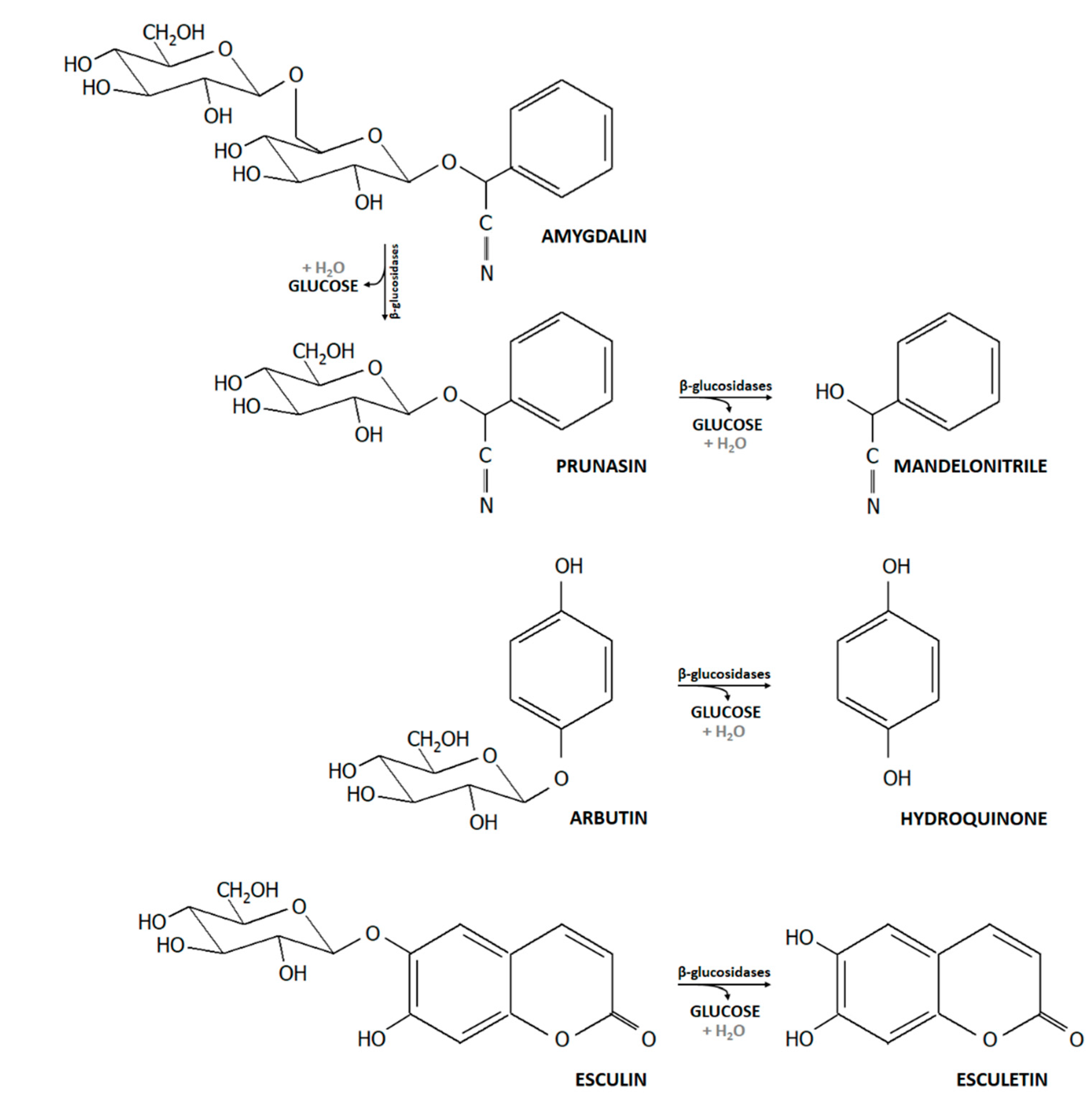
2.2. Utilization of Selected Dietary Plant Glucosides

The ability of bifidobacteria to utilize glucosylated substrates was investigated in sterile 96-well microtiter plates (VWR, USA). Stock solutions of esculin (7-hydroxycoumarin-6-glucoside; Sigma-Aldrich), amygdalin (D-mandelonitrile-β-gentiobioside; Sigma-Aldrich), and arbutin (hydroquinone-β-D-glucopyranoside; Alfa Aesar, USA) were prepared in concentrations corresponding to 28 mM (5 g L−1) glucose (Penta, Czechia) in API 50 CHL medium (BioMérieux, France) with bromocresol purple as a pH indicator, and were filter sterilized. Glucose served as a growth positive control, API medium without added substrate was used to determine background growth.
Overnight cultures were centrifuged and re-suspended in the same volume of API medium. Strains were added (20 µL) to 180 µL API medium with or without added substrate. Plates were incubated under anaerobic conditions (GENbag anaer, bioMérieux, France) at 37 °C for 72 h. The color change from purple to yellow indicated a positive reaction. We measured absorbance at 434 nm and 588 nm using a Tecan Infinite M200 spectrometer (Tecan Group, Männedorf, Switzerland) and calculated the ration 434/588 [26], which was categorized as: no growth (<2); poor growth (2.1–2.4); growth (2.5–3.4); and very good growth (>3.5). Every strain was tested two or three times.

2.3. Metabolite Formation Analysis Using Ion Chromatography with Suppressed Conductivity Detection

Concentration of main fermentation metabolites lactate, acetate, and formate, was determined for selected strains using capillary high-pressure ion-exchange chromatography with suppressed conductivity detection. A Dionex ICS 4000 system equipped with IonPac AS11-HC 4 µm (Thermo Scientific, USA) guard and analytical columns. Eluent composition was as follows: 0–10 min isocratic: 1 mM KOH; 10–20 min linear gradient: 1–60 mM KOH; and 20–25 min again isocratic: 60 mM KOH. The flow rate was set to 0.012 mL min−1. An ACES 300 suppressor (Thermo Scientific, USA) was used to suppress eluent conductivity, while a carbonate Removal Device 200 (Thermo Scientific) was implemented to suppress carbon dioxide baseline shift. Chromatograms were processed with Chromeleon 7.20 (Dionex, USA). Standards were prepared from 1 g L−1 stock solutions (Analytika, Czechia; Inorganic Ventures, USA). Deionized water (conductivity <0.055 µS cm−1; Adrona, Latvia) was used for eluent and standard preparation (0.1–40 mg L−1).

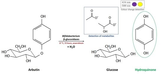
2.4. Determination of Whole Cell β-Glucosidase Activity, and Release of the Acylglycone Esculetin

Beta-glucosidase activity of whole cells was assessed by enzymatic assay with 4-nitrophenyl β-D-glucopyranoside (PNP-G; Sigma-Aldrich, USA) as substrate. All samples were tested at least twice. Overnight cultures (1 mL) were centrifuged, supernatant was discarded and cell pellets were frozen at –20 °C. Frozen cells were re-suspended in 20 µL BifiBuffer (1.2 g L−1 K2HPO4, 0.333 g L−1 KH2PO4; Lachner, Czechia), 1 µL of this suspension was added to 99 µL of PNP-G solution (20 mM in Bifibuffer). Absorbance at 405 nm was measured before and after 4 h of incubation at 37 °C using a Tecan Infinite M200 spectrometer: reactions with a difference of absorbance >0.1 units were considered positive.
For selected strains, β-glucosidase activity was additionally tested using kits RAPID ID 32 A (bioMerieux, France), or ANAERO test 23 (Erba Lachema, Czechia) which employ PNP-G.
The release of esculetin from esculin was tested using ammonium iron citrate (Sigma Aldrich, USA) as scavenger, the reaction of esculetin with ferric ions changes the color from purple to opaque black. Bifidobacteria were inoculated in API medium supplied with 10.2 g L−1 esculin (corresponding to 28 mM solution of glucose) and 1 g L−1 ammonium iron citrate in microtiter plates as described above; and were incubated under anaerobic conditions (GENbag anaer) at 37 °C for 72 h. The color change was assessed visually. Every strain was tested at least twice.

2.5. Antibacterial Activity of Arbutin and Hydroquinone against Selected Bifidobacterium Strains

The antibacterial activity of arbutin and its acylglycone hydroquinone (Sigma-Aldrich, St. Louis, MI, USA) was tested using two-fold broth dilution assay in 96-well sterile microtiter plates. Overnight cultures of selected strains (DSM 20083, DSM 20104, DSM 10140, DSM 20456, DSM 20213, DSM 20211, DSM 20219, and DSM 20088), which represented five of the species tested (Table 2), grown in WSP broth, were centrifuged, and the cell pellet was resuspended in the same volume of API medium supplied with 14 mM glucose. A two-fold dilution series was prepared in microtiter plates using API stock solutions containing 14 mM glucose, and 28 mM arbutin or hydroquinone. Cultures (10%) were added, and plates were incubated under anaerobic conditions (GENbag anaer) at 37 °C for 24 h. Absorbance at 434 and 588 nm was determined using a spectrophotometer and the ratio of 434 nm/588 nm was calculated as described above. The minimal inhibitory concentration (MIC) was defined as the concentration that prevented growth, metabolite formation and thereby color change of the API medium. Every strain was analyzed at least three times.
To test whether the presence of arbutin in API medium impacted growth, we additionally conducted growth kinetics of selected strains that were able or lacked the ability to grow with arbutin in API medium supplied with 28 mM glucose, 14 mM glucose and 14 mM arbutin, or 28 mM arbutin to ensure the availability of the same concentration of glucose. Strains were grown in microtiter plates as described before, and absorbance was measured at 0, 3, 6, 9, 12, 24, 30, 36, and 48 h at 434 and 588 nm, to calculate the ratio of 434 nm/588 nm as described above.

2.6. Identification and Comparison of β-Glucosidases Encoded by Representative Bifidobacterium spp. of the Species Investigated

Homologues of previously characterized β-glucosidases of B. animalis subsp. lactis [27] and B. pseudocatenulatum IPLA 36007 [28] were identified in genomes of B. adolescentis DSM 20083 (AP009256), B. animalis subsp. lactis BB12 (CP001853.1), DSM 10140 (CP001606.1), B. animalis subsp. animalis DSM 20104 (CP002567.1), B. bifidum DSM 20456 (AP012323.1), B. breve DSM 20213 (ACCG02000000), B. longum subsp. longum DSM 20219 (AP010888.1), B. longum subsp. infantis DSM 20088 (CP001095.1), B. longum subsp. suis 20211 (JGZA01000002.1), B. catenulatum DSM 16992 (ABXY01000009.1), B. catenulatum subsp. kashiwanohense DSM 21854 (JGYY01000015.1), B. pseudocatenulatum DSM 20438 (ABXX02000004.1) and B. dentium DSM 20436 (FNSE01000001.1) using blastP. To identify additional β-glucosidases, genomic data were obtained from NCBI and were annotated with RAST using default settings [29]. Glycosyl hydrolases of family 1 and 3 were identified using the dbCAN database based on a search for signature domains of every CAZyme family [30].

3. Results

3.1. Distribution of (Putative) β-Glucosidases Encoding Genes in Genomes of Representative Bifidobacterium spp.

We screened the genomes of selected strains for the presence of GH1 and GH3 encoding genes (Table 3). Homologous proteins related to the four GH3 β-glucosidases characterized in B. pseudocatenulatum IPLA 36007 were present in B. adolescentis DSM 20083, B. breve DSM 20213, B. catenulatum subsp. kashiwanohense DSM 21854, B. catenulatum DSM 16992, and B. pseudocatenulatum DSM 20438. Multiple β-glucosidases (n = 1 GH1, and n = 11 GH3) were encoded by the genome of B. dentium DSM 20438 including homologues to the four β-glucosidases of B. pseudocatenulatum IPLA 36007. Strains of B. animalis harbored a homologue of Bbg572 (GH1) of B. animalis subsp. lactis SH5, and in addition a homologue of r-β-gluE of B. pseudocatenulatum IPLA 36007. The distribution of β-glucosidases in B. longum differed between subspecies, B. longum subsp. longum DSM 20219 and B. longum subsp. suis DSM 20211 possessed homologues of r-β-gluE, which were also highly similar (>96%) to a characterized β-glucosidase of B. longum H1 [31], while r-β-gluD was present in all three subspecies but was truncated in B. longum subsp. longum DSM 20219. B. longum subsp. infantis DSM 20288 additionally possessed a homologue of Bbg572. B. bifidum DSM 20456 harbored only one putative GH1 β-glucosidase with low homology to Bbg572.

3.2. Beta-Glucosidase Activity of Bifidobacterium spp.

Whole cell β-glucosidase activity was investigated by enzymatic assay using PNP-G as substrate. B. adolescentis, B. animalis, B. breve, B. catenulatum/pseudocatenulatum, and B. dentium were consistently β-glucosidase positive (Table 1). In contrast, all tested strains of B. bifidum, with a few exceptions, and most strains of B. longum, were β-glucosidase negative. From B. longum, only four strains that originated from dog feces (10/6b, 32/3na, 33/5nb, and 33/4nc), two isolates from stool of adults (B1, PEG080) and one from pig feces (DSM 20211) showed β-glucosidase activity. For selected strains, β-glucosidase activity was confirmed using RAPID ID 32 A and ANAEROtest 23 (Table 1).

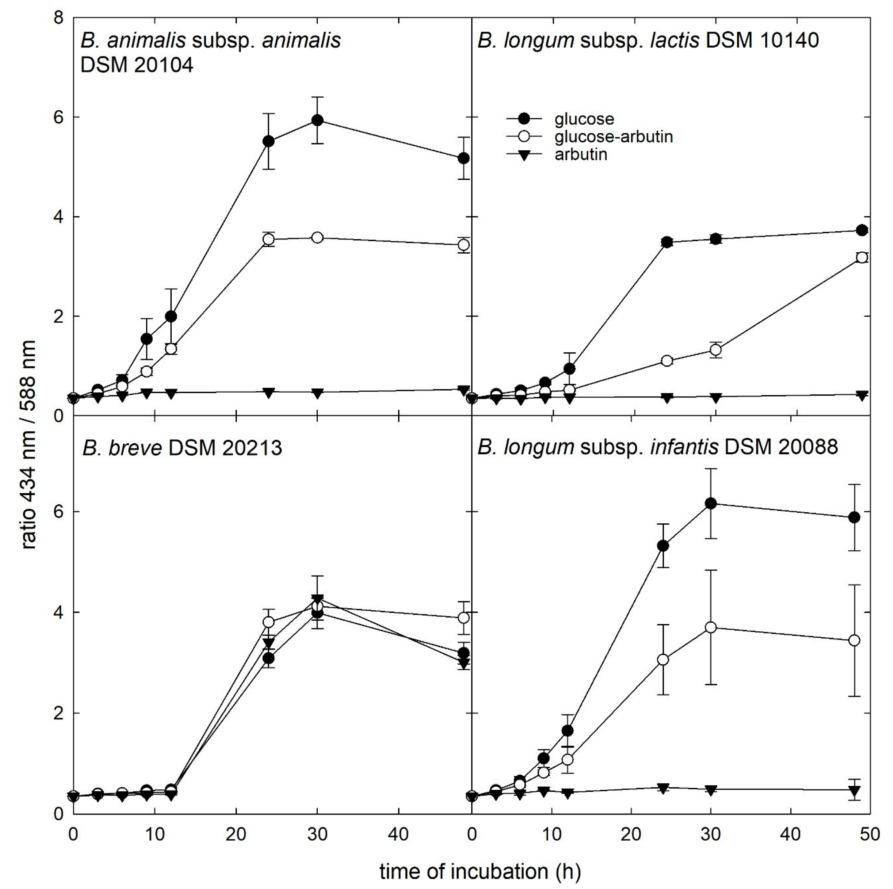
3.3. Growth in the Presence of β-Glucosides

We observed that β-glucosidase activity is a common yet species dependent feature of Bifidobacterium spp. To investigate whether β-glucosidase activity relates to the ability to use plant glucosides, we grew strains with esculin, amygdalin, and arbutin as a sole carbohydrate source in API 50 CHL medium (Table 1). All strains were able to grow in the presence of glucose, verifying the suitability of the assay.
In general, amygdalin was the preferred β-glucosylated substrate used by 54% of strains, followed by esculin (47%), and arbutin (24%).
Strains belonging to B. dentium were most versatile in the utilization of the provided β-glucosides as all strains grew in the presence of amygdalin and esculin. Only three B. dentium strains (DSM 20436, TH1, and VOK II) were not able to use arbutin. All strains of B. breve used amygdalin and esculin (with one exception, B43), while the utilization of arbutin was less frequent (46%). Within B. catenulatum/pseudocatenulatum, 83% strains grew in the presence of amygdalin, while 67% utilized esculin and 38% arbutin. The majority of the B. adolescentis strains (69%) was capable of using amygdalin, strains B35 and B39 grew in presence of arbutin and esculin. For B. animalis, we observed subspecies dependent differences in substrate utilization. The majority of B. animalis subsp. lactis strains, except three isolates from dog feces (P2N1, 43/7nb, and 11/6a), utilized amygdalin (80%), 67% used esculin, and only PEG084 grew in the presence of arbutin. In contrast, B. animalis subsp. animalis strains were not capable to grow with amygdalin and arbutin, while 57% utilized esculin. None of the B. longum strains was able to use esculin and arbutin, amygdalin utilization was detected for B. longum DSM 20211 and PEG080, while B. bifidum strains were not able to utilize any of the provided plant β-glucosides.

3.4. Release of the Acylglycone Esculetin

We qualitatively determined whether β-glucoside hydrolysis of esculin would lead to the release of the acylglycone esculetin using ammonium iron citrate as a scavenger (Table 1). The majority (94%) of the strains that were positive in the PNP-G enzymatic assay released esculetin confirming β-glucosidase activity. Few strains (namely DSM 20211, B1, 32/3na, 33/5nb, and 33/4nc), all from B. longum, were PNP-G positive, while esculetin release was not detected. More than half (66%) of the strains that were able to release esculetin grew when esculin was present as substrate.

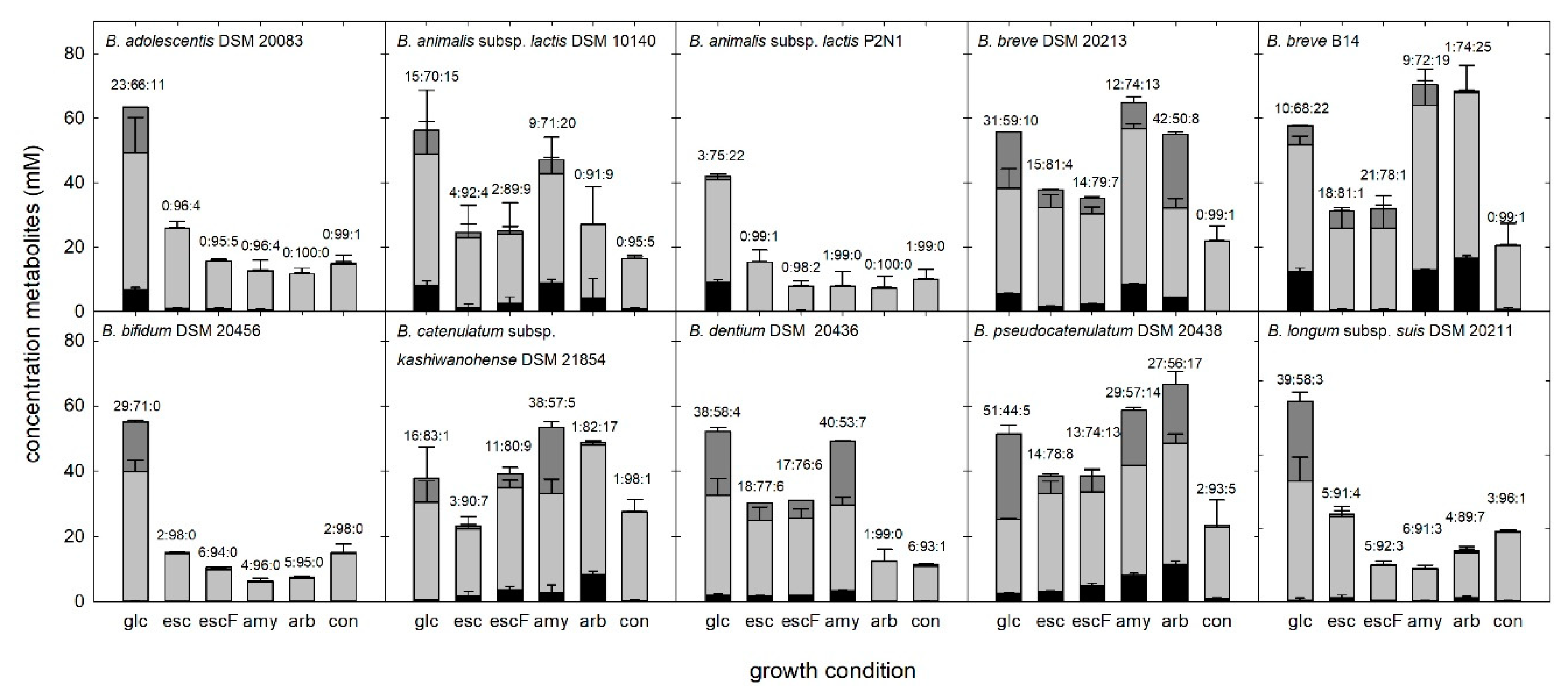
3.5. Metabolite Formation during Growth of Bifidobacteria in the Presence of Plant Glucosides

For representative strains from different species, we investigated metabolite formation as an indicator of fermentation activity. Lactate, acetate, and formate concentrations were measured by capillary ion-exchange chromatography with suppressed conductivity detection (Figure 2). All strains grew in the presence of glucose, producing mainly acetate metabolite (44%–83% of metabolites formed), followed by lactate (3%–51%) and formate (0%–22%). In negative controls (API medium without added glycan source), and samples without visible growth, acetate was formed as the major metabolite, otherwise the acetate:lactate:formate ratios differed between strains, and compared to growth in glucose.

3.6. Antibacterial Activity of Hydroquinone and Arbutin and Impact on Growth Kinetics

We tested the antibacterial and growth affecting activity of arbutin and its acylglycone hydroquinone on representative strains using two-fold dilution assay in microtiter plates and growth kinetics, respectively. Glucose (14 mM) was added to API medium to avoid growth inhibition due to substrate limitation. None of the strains were affected by the presence of arbutin even at the maximum concentration tested (25.5 mM) (Table 2). B. animalis subsp. animalis DSM 20104, B. longum subsp. longum DSM 20019, B. longum subsp. suis DSM 20211, and B. longum subsp. infantis DSM 20088 were most sensitive towards hydroquinone with MIC ≤ 0.05 mM while the MIC of the other strains was 0.1–0.2 mM (Table 2).
Strains B. animalis subsp. animalis DSM 20104 and B. animalis subsp. lactis DSM 10140, B. breve DSM 20213, and B. longum subsp. infantis DSM 20088 were additionally grown in the presence of glucose, glucose and arbutin, or arbutin (Figure 3). The presence of arbutin did not impact the growth of B. breve DSM 20213, while the lag phase of the other strains was delayed in the presence of glucose and arbutin and the final absorbance ratio reached was approximately 50% compared to glucose only.

4. Discussion

Plant derived β-glucosides encompass structurally diverse compounds, which, when ingested, can reach the colon and be enzymatically modified by gut microbes. Beta-glucosidases are widely distributed in gut microbes and play important roles in biological processes. Here, we demonstrate species and strain dependent variability of Bifidobacterium spp. in the utilization of dietary plant glucosides linked to aromatic aglycones.

4.1. Genomic Potential for β-Glucosidase Activity Partly Predicts Activity

Based on genomic data, strains of the phylogenetically closely related species B. adolescentis, B. catenulatum, B. pseudocatenulatum, B. dentium, and the more distant B. breve [10], harbored a core of four β-glucosidases. In agreement, all strains of these species possessed β-glucosidase activity but showed preference for different substrates. Indeed, the purified β-glucosidases of B. pseudocatenulatum IPLA 36007 varied in their ability to release of the aglycones daidzein and genistein from isoflavone glycosides daidzin and genistin, indicating enzyme dependent substrate preference [28].
Strains of B. longum did not show β-glucosidase activity despite the presence of genes encoding β-glucosidases with high homology to a purified β-glucosidases of B. longum H1, which hydrolyzed arbutin. Lack of β-glucosidase activity of whole cells might suggest that enzymes were either not expressed or expressed at concentrations not sufficient to confer activity at test conditions.
B. animalis subsp. lactis and B. animalis subsp. animalis harbored highly similar GH1 and GH3 β-glucosidases and possessed β-glucosidase activity. The similar GH1 β-glucosidase Bgl572 hydrolyzed PNP-G and arbutin when purified [31]. However, growth in the presence of plant glucosides differed between B. animalis subspecies, possibly due different transport mechanism or sensitive towards the released acylglycones. Indeed, B. animalis subsp. animalis DSM 20104 was more sensitive towards hydroquinone than B. animalis subsp. lactis DSM 10140.

4.2. Bifidobacterium β-Glucosidase Increases Bioactivity and Bioavailability of Plant Glucosidases and Acylglycones

Plants are used for antibacterial properties in medical applications, and bacterial β-glucosidase activity leads to the release of bioactive acylglycones. Indeed, we confirmed the release of esculetin from esculin, by almost all strains with β-glucosidase activity, modifying bioactivity and bioavailability. Despite the ability to hydrolyze esculin, not all strains were able to grow when esculin was supplied as substrate, which might be due to the antimicrobial activity of esculetin [33].
Bioactivity of plant glucosides is likely lower than of acylglycones due to larger molecular weight and higher hydrophobicity. In confirmation, arbutin at the concentrations tested did not show inhibition while hydroquinone conferred strong antibacterial activity against the Bifidobacterium strains tested. Hydroquinone MIC values of bifidobacteria ranging from ≤0.05–0.2 mM were lower than reported for Staphylococcus aureus (1–11 mM) [34,35] and various aerobic Gram-positive and -negative strains [4] including Enterococcus faecalis, Escherichia coli, Pseudomonas aeruginosa, and Klebsiella pneumonia (1.5–6 mM).
MIC were up to 10-fold lower than the levels of hydroquinone theoretically released during growth. However, in the MIC assay, a low concentration of bacterial cells is exposed to high concentrations of the antibacterial at the beginning of the growth phase, while in the growth assay, increasing amounts of hydroquinone face an increasing number of bacterial cells. Sensitivity of bifidobacteria to hydroquinone, which, if released in the intestinal tract, could reduce the growth potential of β-glucosidase active strains, but also of neighboring cells.
In addition, β-glucosidase activity of Bifidobacterium spp. increased the bioavailability of acylglycones. Hydroquinone has been linked to anticancer activity in vitro and in vivo [36] and has been shown to confer antimycobacterial and antileishmanial activity in vitro [37].

4.3. Niche Adaption of Bifidobacteria Seems Related to β-Glucosidase Activity

Host adaptation of Bifidobacterium spp. was suggested to be linked to the ability to use dietary or host-derived glycans and differs between species and strains [10,11]. Indeed, strains of B. bifidum and B. longum subsp. infantis, which are among the most abundant microbes during the first months of life, lacked β-glucosidase activity in agreement to previous observations [5], and with the absence of β-glucosidase encoding genes in the genomes. Both species occur in the infant gut when glycan substrates are limited to human breast milk, endogenous mucin, or infant formula. Indeed, a previous cohort of studies observed that fecal β-glucosidase activity was low after birth and gradually increased with the introduction of a more diverse diet [38]. Interestingly, B. animalis subsp. animalis showed only little growth in the presence of plant glucosides despite possessing β-glucosidase activity. Most isolates were obtained from lamb and calf feces early when animals received a milk diet.
B. breve colonizes infant and adults, and similar to B. adolescentis and other adult or multi-host species, possesses multiple β-glucosidases. In the adult gut, Bifidobacterium spp. contribute a minor share of the population, and compete with other β-glucosidase positive taxa for substrate [5]. Beta-glucosidase activity of Bifidobacterium strains colonizing adults might enhance ecological competitiveness.
For the multi-host subspecies of B. animalis subsp. lactis, plant glucoside utilization profiles likewise suggested host adaption in agreement with genetic and phenotypic host-specific differences previously observed [11]. Strains from adult ruminant hosts (Cameroon sheep, Barbary sheep, and an okapi), that naturally receive a plant-based diet, used plant glucosides in contrast to strains isolated from omnivorous dogs.

4.4. Substrate Source Impacted Fermentation Profiles

Bifidobacteria metabolize hexoses via the “bifid shunt” with fructose-6-phosphoketolase as the key enzyme. Glucose (1 mol) theoretically yields 1.5 mol acetate, 1 mol lactate, and 2.5 ATP [39]. Whether the intermediate pyruvate is cleaved to acetyl phosphate and formate, or reduced to lactate, depends on type and supply of the substrate carbohydrate [40], possibly in the presence of oxygen [8] and on different rates in substrate consumption. A previous study reported that with a decrease of consumption rate, relatively more acetic and formic acid and less lactic acid was produced by Bifidobacterium spp. [41]. Indeed, the proportion of lactate was reduced for most strains grown in the presence of esculetin, and to a lesser extent with arbutin, which might indicate that hydrolysis activity reduced the consumption rate.

5. Conclusions

Our data shows that bifidobacterial β-glucosidase activity is preserved among species which might be related to niche adaption. Structural homology of a core set of β-glucosidases of species associated with adult humans could suggest that these enzymes evolved together. Beta-glucosidase activity may provide a competitive advantage in the mammalian gut proving access to energy sources, but they might also have environmental impact due to the release of bioactive antibacterial acylglycones; thus, increasing bioavailability due to the formation of fermentation metabolites.

Author Contributions

Conceptualization, V.N.-B. and C.S.; methodology, V.N.-B. and C.S.; validation, N.M., V.N.-B., and C.S.; formal analysis, N.M.; investigation, N.M., V.T., C.S., and V.N.-B.; resources, V.N.-B.; data curation, C.S.; writing—original draft preparation, N.M., C.S., and V.N.-B.; writing—review and editing, V.N.-B., C.S., and N.M.; visualization, N.M. and C.S.; supervision, V.N.-B. and C.S.; project administration, E.V.; and funding acquisition, E.V. All authors have read and agreed to the published version of the manuscript.

Funding

This research was funded by European Regional Development Fund-Project, “Centre for the investigation of synthesis and transformation of nutritional substances in the food chain in interaction with potentially harmful substances of anthropogenic origin: comprehensive assessment of soil contamination risks for the quality of agricultural products” [No: CZ.02.1.01/0.0/0.0/16_019/0000845], and by METROFOOD-CZ research infrastructure project [MEYS Grant No: LM2018100] including access to its facilities.

Acknowledgments

The authors thank Katerina Kindlova for her valuable assistance in laboratory experiments.

Conflicts of Interest

The authors declare no conflict of interest.

References

  1. Biernat, K.A.; Li, B.; Redinbo, M.R. Microbial unmasking of plant glycosides. MBio 2018, 9, e02433-17. [Google Scholar] [CrossRef] [Green Version]
  2. De Arriba, S.G.; Naser, B.; Nolte, K.-U. Risk assessment of free hydroquinone derived from Arctostaphylos Uva-ursi folium herbal preparations. Int. J. Toxicol. 2013, 32, 442–453. [Google Scholar] [CrossRef]
  3. Theilmann, M.C.; Goh, Y.J.; Nielsen, K.F.; Klaenhammer, T.R.; Barrangou, R.; Abou Hachem, M. Lactobacillus acidophilus metabolizes dietary plant glucosides and externalizes their bioactive phytochemicals. MBio 2017, 8. [Google Scholar] [CrossRef] [Green Version]
  4. Jurica, K.; Gobin, I.; Kremer, D.; Čepo, D.V.; Grubešić, R.J.; Karačonji, I.B.; Kosalec, I. Arbutin and its metabolite hydroquinone as the main factors in the antimicrobial effect of strawberry tree (Arbutus unedo L.) leaves. J. Herb. Med. 2017, 8, 17–23. [Google Scholar] [CrossRef]
  5. Dabek, M.; McCrae, S.I.; Stevens, V.J.; Duncan, S.H.; Louis, P. Distribution of β-glucosidase and β-glucuronidase activity and of β-glucuronidase gene gus in human colonic bacteria. FEMS Microbiol. Ecol. 2008, 66, 487–495. [Google Scholar] [CrossRef] [Green Version]
  6. Ventura, M.; Turroni, F.; Motherway, M.O.; MacSharry, J.; van Sinderen, D. Host-microbe interactions that facilitate gut colonization by commensal bifidobacteria. Trends Microbiol. 2012, 20, 467–476. [Google Scholar] [CrossRef] [PubMed]
  7. Russell, D.A.; Ross, R.P.; Fitzgerald, G.F.; Stanton, C. Metabolic activities and probiotic potential of bifidobacteria. Int. J. Food Microbiol. 2011, 149, 88–105. [Google Scholar] [CrossRef] [PubMed]
  8. Schwab, C.; Ruscheweyh, H.J.; Bunesova, V.; Pham, V.T.; Beerenwinkel, N.; Lacroix, C. Trophic interactions of infant bifidobacteria and Eubacterium hallii during L-fucose and fucosyllactose degradation. Front. Microbiol. 2017, 8. [Google Scholar] [CrossRef] [PubMed]
  9. Turroni, F.; Milani, C.; Duranti, S.; Mahony, J.; van Sinderen, D.; Ventura, M. Glycan utilization and cross-feeding activities by bifidobacteria. Trends Microbiol. 2018, 26, 339–350. [Google Scholar] [CrossRef]
  10. Milani, C.; Turroni, F.; Duranti, S.; Lugli, G.A.; Mancabelli, L.; Ferrario, C.; Van Sinderen, D.; Ventura, M. Genomics of the genus Bifidobacterium reveals species-specific adaptation to the glycan-rich gut environment. Appl. Environ. Microbiol. 2016, 82, 980–991. [Google Scholar] [CrossRef] [Green Version]
  11. Bunesova, V.; Killer, J.; Javurkova, B.; Vlkova, E.; Tejnecky, V.; Musilova, S.; Rada, V. Diversity of the subspecies Bifidobacterium animalis subsp. lactis. Anaerobe 2017, 44, 40–47. [Google Scholar] [CrossRef] [PubMed]
  12. Bottacini, F.; Morrissey, R.; Esteban-Torres, M.; James, K.; Van Breen, J.; Dikareva, E.; Egan, M.; Lambert, J.; Van Limpt, K.; Knol, J.; et al. Comparative genomics and genotype-phenotype associations in Bifidobacterium breve. Sci. Rep. 2018, 8. [Google Scholar] [CrossRef] [PubMed] [Green Version]
  13. Derrien, M.; van Hylckama Vlieg, J.E.T. Fate, activity, and impact of ingested bacteria within the human gut microbiota. Trends Microbiol. 2015, 23, 354–366. [Google Scholar] [CrossRef] [PubMed] [Green Version]
  14. Bunešová, V.; Joch, M.; Musilová, S.; Rada, V. Bifidobacteria, lactobacilli, and short chain fatty acids of vegetarians and omnivores. Sci. Agric. Bohem. 2017, 48, 47–54. [Google Scholar] [CrossRef]
  15. Makino, H.; Kushiro, A.; Ishikawa, E.; Kubota, H.; Gawad, A.; Sakai, T.; Oishi, K.; Martin, R.; Ben-Amor, K.; Knol, J. Mother-to-infant transmission of intestinal bifidobacterial strains has an impact on the early development of vaginally delivered infant’s microbiota. PLoS ONE 2013, 8, e78331. [Google Scholar] [CrossRef]
  16. Nouioui, I.; Carro, L.; García-López, M.; Meier-Kolthoff, J.P.; Woyke, T.; Kyrpides, N.C.; Pukall, R.; Klenk, H.-P.; Goodfellow, M.; Göker, M. Genome-based taxonomic classification of the phylum Actinobacteria. Front. Microbiol. 2018, 9, 2007. [Google Scholar] [CrossRef] [Green Version]
  17. Sharma, V.; Mobeen, F.; Prakash, T. Exploration of survival traits, probiotic determinants, host interactions, and functional evolution of bifidobacterial genomes using comparative genomics. Genes 2018, 9, 477. [Google Scholar] [CrossRef] [Green Version]
  18. Turroni, F.; Peano, C.; Pass, D.A.; Foroni, E.; Severgnini, M.; Claesson, M.J.; Kerr, C.; Hourihane, J.; Murray, D.; Fuligni, F. Diversity of bifidobacteria within the infant gut microbiota. PLoS ONE 2012, 7, e36957. [Google Scholar] [CrossRef] [Green Version]
  19. Duranti, S.; Lugli, G.A.; Milani, C.; James, K.; Mancabelli, L.; Turroni, F.; Alessandri, G.; Mangifesta, M.; Mancino, W.; Ossiprandi, M.C.; et al. Bifidobacterium bifidum and the infant gut microbiota: An intriguing case of microbe-host co-evolution. Environ. Microbiol. 2019, 21, 3683–3695. [Google Scholar] [CrossRef]
  20. Masco, L.; Ventura, M.; Zink, R.; Huys, G.; Swings, J. Polyphasic taxonomic analysis of Bifidobacterium animalis and Bifidobacterium lactis reveals relatedness at the subspecies level: Reclassification of Bifidobacterium animalis as Bifidobacterium animalis subsp. animalis subsp. nov. and Bifidobacterium lactis as Bifidobacterium animalis subsp. lactis subsp. nov. Int. J. Syst. Evol. Microbiol. 2004, 54, 1137–1143. [Google Scholar] [CrossRef] [Green Version]
  21. Henne, K.; Rheinberg, A.; Melzer-Krick, B.; Conrads, G. Aciduric microbial taxa including Scardovia wiggsiae and Bifidobacterium spp. in caries and caries free subjects. Anaerobe 2015, 35, 60–65. [Google Scholar] [CrossRef] [PubMed]
  22. Neves, B.G.; Stipp, R.N.; Bezerra, D.D.S.; Guedes, S.F.D.F.; Rodrigues, L.K.A. Quantitative analysis of biofilm bacteria according to different stages of early childhood caries. Arch. Oral. Biol. 2018, 96, 155–161. [Google Scholar] [CrossRef] [PubMed]
  23. Modrackova, N.; Makovska, M.; Mekadim, C.; Vlkova, E.; Tejnecky, V.; Bolechova, P.; Bunesova, V. Prebiotic potential of natural gums and starch for bifidobacteria of variable origins. Bioact. Carbohydr. Diet. Fibre 2019, 20. [Google Scholar] [CrossRef]
  24. Bunesova, V.; Domig, K.J.; Killer, J.; Vlkova, E.; Kopecny, J.; Mrazek, J.; Rockova, S.; Rada, V. Characterization of bifidobacteria suitable for probiotic use in calves. Anaerobe 2012, 18, 166–168. [Google Scholar] [CrossRef] [PubMed]
  25. Bunesova, V.; Vlkova, E.; Killer, J.; Rada, V.; Rockova, S. Identification of Bifidobacterium strains from faeces of lambs. Small Rumin. Res. 2012, 105, 355–360. [Google Scholar] [CrossRef]
  26. Pei, K.; Xiong, Y.; Li, X.; Jiang, H.; Xiong, Y. Colorimetric ELISA with an acid–base indicator for sensitive detection of ochratoxin A in corn samples. Anal. Methods 2018, 10, 30–36. [Google Scholar] [CrossRef]
  27. Youn, S.Y.; Park, M.S.; Ji, G.E. Identification of the β-glucosidase gene from Bifidobacterium animalis subsp. lactis and its expression in B. bifidum BGN4. J. Microbiol. Biotechnol. 2012, 22, 1714–1723. [Google Scholar] [CrossRef]
  28. Guadamuro, L.; Flórez, A.B.; Alegría, Á.; Vázquez, L.; Mayo, B. Characterization of four β-glucosidases acting on isoflavone-glycosides from Bifidobacterium pseudocatenulatum IPLA 36007. Food Res. Int. 2017, 100, 522–528. [Google Scholar] [CrossRef] [Green Version]
  29. Aziz, R.K.; Bartels, D.; Best, A.; DeJongh, M.; Disz, T.; Edwards, R.A.; Formsma, K.; Gerdes, S.; Glass, E.M.; Kubal, M.; et al. The RAST Server: Rapid annotations using subsystems technology. BMC Genom. 2008, 9. [Google Scholar] [CrossRef] [Green Version]
  30. Yin, Y.; Mao, X.; Yang, J.; Chen, X.; Mao, F.; Xu, Y. DbCAN: A web resource for automated carbohydrate-active enzyme annotation. Nucleic Acids Res. 2012, 40, W445–W451. [Google Scholar] [CrossRef]
  31. Jung, I.H.; Lee, J.H.; Hyun, Y.J.; Kim, D.H. Metabolism of ginsenoside Rb1 by human intestinal microflora and cloning of its metabolizing β-D-glucosidase from Bifidobacterium longum H-1. Biol. Pharm. Bull. 2012, 35, 573–581. [Google Scholar] [CrossRef] [PubMed] [Green Version]
  32. Florindo, R.N.; Souza, V.P.; Manzine, L.R.; Camilo, C.M.; Marana, S.R.; Polikarpov, I.; Nascimento, A.S. Structural and biochemical characterization of a GH3 β-glucosidase from the probiotic bacteria Bifidobacterium adolescentis. Biochimie 2018, 148, 107–115. [Google Scholar] [CrossRef] [PubMed]
  33. Yang, L.; Ding, W.; Xu, Y.; Wu, D.; Li, S.; Chen, J.; Guo, B. New insights into the antibacterial activity of hydroxycoumarins against Ralstonia solanacearum. Molecules 2016, 21, 468. [Google Scholar] [CrossRef] [PubMed]
  34. Rúa, J.; Fernández-Álvarez, L.; De Castro, C.; Del Valle, P.; De Arriaga, D.; García-Armesto, M.R. Antibacterial activity against foodborne Staphylococcus aureus and antioxidant capacity of various pure phenolic compounds. Foodborne Path Dis. 2011, 8, 149–157. [Google Scholar] [CrossRef] [PubMed]
  35. Ma, C.; He, N.; Zhao, Y.; Xia, D.; Wei, J.; Kang, W. Antimicrobial mechanism of hydroquinone. Appl. Biochem. Biotechnol. 2019, 189, 1291–1303. [Google Scholar] [CrossRef]
  36. Byeon, S.E.; Yi, Y.S.; Lee, J.; Yang, W.S.; Kim, J.H.; Kim, J.; Hong, S.; Kim, J.-H.; Cho, J.Y. Hydroquinone exhibits in vitro and in vivo anti-cancer activity in cancer cells and mice. Int. J. Mol. Sci. 2018, 19, 903. [Google Scholar] [CrossRef] [PubMed] [Green Version]
  37. Horn, C.M.; Aucamp, J.; Smit, F.J.; Seldon, R.; Jordaan, A.; Warner, D.F.; N’Da, D.D. Synthesis and in vitro antimycobacterial and antileishmanial activities of hydroquinone-triazole hybrids. Med. Chem. Res. 2020. [Google Scholar] [CrossRef]
  38. Mykkänen, H.; Tikka, J.; Pitkänen, T.; Hänninen, O. Fecal bacterial enzyme activities in infants increase with age and adoption of adult-type diet. J. Pediatr. Gastroenterol. Nutr. 1997, 25, 312–316. [Google Scholar] [CrossRef]
  39. De Vries, W.; Stouthamer, A. Pathway of glucose fermentation in relation to the taxonomy of bifidobacteria. J. Bacteriol. 1967, 93, 574–576. [Google Scholar] [CrossRef] [PubMed] [Green Version]
  40. Palframan, R.J.; Gibson, G.R.; Rastall, R.A. Carbohydrate preferences of Bifidobacterium species isolated from the human gut. Curr. Issues Intest. Microbiol. 2003, 4, 71–75. [Google Scholar]
  41. Van der Meulen, R.; Adriany, T.; Verbrugghe, K.; De Vuyst, L. Kinetic analysis of bifidobacterial metabolism reveals a minor role for succinic acid in the regeneration of NAD+ through its growth-associated production. Appl. Environ. Microbiol. 2006, 72, 5204–5210. [Google Scholar] [CrossRef] [PubMed] [Green Version]
Figure 1. Structures of dietary plant glucosides (amygdalin, arbutin, and esculin) and their products after β-glucosidase hydrolysis.
Microorganisms 08 00839 g001
Figure 2. Metabolites formed during growth in the presence of glucose or plant glucosides. Formation of fermentation metabolites, lactate (dark grey), acetate (light grey), and formate (black), by selected strains grown in API 50 CHL medium supplied with plant glucosides (equivalent to 28 mM glucose) was analyzed after 72 h incubation by capillary ion-exchange chromatography with suppressed conductivity. The proportion of major metabolites lactate, acetate, and formate for each growth condition are shown above the bar. Strains were tested two or three times. glc, glucose; esc, esculin; escF, esculin and ammonium iron citrate; amy, amygdalin; arb, arbutin; and con, negative control.
Microorganisms 08 00839 g002
Figure 3. Impact of arbutin addition on growth. Growth kinetics of selected strains were tested in microtiter plates with API medium supplied with 28 mM glucose, 14 mM glucose and arbutin, or 28 mM arbutin. The plates were spectrophotometrically measured at 0, 3, 6, 9, 12, 24, 30, 36, and 48 h at 434 and 588 nm, to calculate the ratio of 434 nm/588 nm.
Microorganisms 08 00839 g003
Table 1. Bifidobacterial utilization of β-glucosylated substrates during growth, and β-glucosidase activity. The ability to utilize plant glucosides was tested in API 50 CHL media. The color change detection of cultivation media after 72 h of incubation at 37 °C under anaerobic conditions was determined spectrophotometrically (434 nm/588 nm). A ratio of <2 was considered no growth (-); 2.1–2.4: poor growth (+); 2.5–3.4: growth (++); and >3.5: very good growth (+++). Beta-glucosidase activity was tested by enzymatic assay with 4-nitrophenyl β-D-glucopyranoside (β-GLU) as substrate with spectrophotometric measurement at 405 nm; the difference of >0.1 was considered positive (+). The release of the acylglycone from esculin was determined visually using ammonium iron citrate as scavenger (ESN rel.).
Species/SubspeciesStrainOriginGLUESCAMYGARBAPIβ-GLUESN rel.
B. adolescentisDSM 20083Intestine of adult+++----++
B34Stool of infant+++-+++--++
B35Stool of infant+++++++++-++
B36Stool of infant+++----+R+
B38Stool of infant++++---++
B2Stool of adult++-++--+R+
B9Stool of adult++-++--+R+
B30Stool of adult+++-+++--+R+
B39Stool of adult+++++++++++-++
B41Stool of adult+++-+++--++
B56Stool of adult++----++
PEG038Stool of adult+++-+++--++
10/6dDog feces+++-+++--++
B. animalis subsp. animalisDSM 20104Rat feces+++----++
805P4Calf feces++++++---+R+
012II1Calf feces++++---+R+
023IICalf feces+++----+R+
J1 (L1)Lamb feces++----+R+
J5 (L4)Lamb feces++++---+R+
J6 (L3)Lamb feces+++++---++
B. animalis subsp. lactisDSM 10140Yoghurt+++-+++--++
BB12Probiotic product++++++--++
DanProbiotic product++++++--++
NestléInfant nutrition+++++++--++
S7Ovine cheese++++++--++
B22Stool of infant++++++++--++
B25Stool of infant+++++++--++
PEG042Stool of adult+++-+++--++
PEG084Stool of adult++++++++-++
P2N1Dog feces+++----++
43/7nbDog feces+++----++
11/6aDog feces++----++
ZDK1Cameroon sheep feces++++++++--++
ZDK4Barbary sheep feces++++++--++
ZDK7Okapi feces++++++++--++
B. bifidumDSM 20456Stool of infant+++------
DSM 20239Stool of infant+++------
B6Stool of infant+++-----R-
B33Stool of infant+++-----R-
B10Stool of adult+++-----R-
B29Stool of adult+++-----R-
B40Stool of adult+++------
B55Stool of adult+++------
B. breveDSM 20213Intestine of infant+++++++++++-++
BR03Probiotic product+++++++--++
B13Stool of infant+++++++++++-+R+
B14Stool of infant+++++++++++-+R+
B37Stool of infant+++++++++--++
B42Stool of infant+++++++++-++
B43Stool of infant+++-++--++
B50Stool of infant++++++++++++-++
B57Stool of infant+++++++++--++
PEG010Stool of adult+++++++--++
PEG064Stool of adult+++++++++--++
PEG071Stool of adult++++++++--++
PEG074Stool of adult+++++++++++-++
B. catenulatum
B. catenulatum subsp.
kashiwanohense
B. pseudocatenulatum
B. catenulatum/
pseudocatenulatum
DSM 16992Human feces++++++++++++-++
DSM 21854Stool of infant+++-++++-++
DSM 20438Stool of infant++++++++++++-++
B12Stool of infant+++-+++--+R+
B46Stool of infant+++++++++++-++
B48Stool of infant++++++---++
B23Stool of adult++++++++--+R+
B32Stool of adult++++++++--+R+
B51Stool of adult++-+++--++
B52Stool of adult++++-+++-++
B53Stool of adult+++-+++--++
22/4nbDog feces++++++++--++
B. dentiumDSM 20436Dental caries+++++++++--++
FD1Stool of infant+++++++++++-+ A+
TH1Stool of infant+++++++++--+ A+
VOK IIStool of infant++++++++--+ A+
PEG020Stool of adult+++++++++++-++
A1/5AMonkey feces++++++++++++-+ A+
N12Monkey feces++++++++++++-+ A+
N21Monkey feces++++++++++++-+ A+
N23Monkey feces++++++++++++-+ A+
N26Monkey feces++++++++++++-+ A+
N77Monkey feces++++++++++++-+ A+
N79Monkey feces++++++++++++-+ A+
N105Monkey feces++++++++++++-+ A+
N109Monkey feces++++++++++++-+ A+
N110Monkey feces++++++++++++-+ A+
N111Monkey feces++++++++++++-+ A+
N112Monkey feces++++++++++++-+ A+
B. longum subsp. infantis
B. longum subsp. longum
B. longum subsp. suillum
B. longum subsp. suis
B. longum
DSM 20088Stool of infant+++------
DSM 20219Intestine of adult+++------
DSM 28597Feces of piglets+++-----A-
DSM 20211Pig feces+++-+++--+-
5/9Calf feces+++------
INFNUTProbiotic product++------
B3Stool of infant+++-----R-
B4Stool of infant++-----R-
B7Stool of infant+++------
B8Stool of infant+++------
B11Stool of infant+++------
B16Stool of infant+++------
B17Stool of infant+++----- R-
B19Stool of infant+++-----R-
B20Stool of infant++-----R-
B27Stool of infant++------
B28Stool of infant+++------
B44Stool of infant+++-----A-
B49Stool of infant+++------
B1Stool of adult+++----+-
B26Stool of adult+++-----R-
PEG057Stool of adult+++------
PEG059Stool of adult+++------
PEG080Stool of adult++-++--++
PEG104Stool of adult+++------
022IICalf feces+++-----R-
10/6bDog feces+++----++
32/3naDog feces+++----+-
33/5nbDog feces++----+-
33/4ncDog feces++----+-
GLU, glucose; ESC, esculin; AMYG, amygdalin; ARB, arbutin; API, negative control; β-GLU, β-glucosidase activity; ESN rel., esculetin release; superscript letter A, the shown reaction (positive/negative) of β-glucosidase activity is confirmed by ANAEROtest 23; and superscript letter R, the shown reaction of β-glucosidase activity is confirmed by RAPID ID 32 A.
Table 2. Minimal inhibitory concentrations of hydroquinone and arbutin. Minimal inhibitory concentrations (MIC) were determined using a two-fold dilution assay in microtiter plates and API medium supplied with glucose (14 mM), and hydroquinone or arbutin. The MIC was defined as the concentration that completely inhibited growth of strains determined using the absorbance ration 434 nm/588 nm. MIC were tested in 3–5 independent replicates.
Species or SubspeciesStrainMinimal Inhibitory Concentration (mM)
HydroquinoneArbutin
B. adolescentisDSM 200830.05–0.10>25.5
B. animalis subsp. animalisDSM 20104≤0.05>25.5
B. animalis subsp. lactisDSM 101400.10–0.20>25.5
B. bifidumDSM 204560.10–0.20>25.5
B. breveDSM 202130.10–0.20>25.5
B. longum subsp. suisDSM 20211≤0.05>25.5
B. longum subsp. longumDSM 20219≤0.05>25.5
B. longum subsp. infantisDSM 20088≤0.05>25.5
Table 3. Distribution of β-glucosidases in representative strains of Bifidobacterium species. Beta-glucosidases putatively encoded by the genomes were compared to characterized β-glucosidases of B. animalis subsp. lactis SH5 (Bbg572, GH1) or to four GH3 β-glucosidases r-β-gluA, r-β-gluB, r-β-gluD, and r-β-gluE of B. pseudocatenulatum IPLA36007.
SpeciesStrainCharacterized Beta-GlucosidaseNot Yet Characterized β-Glucosidases
GH Family 1GH family 3
Bbg572 (JX274651)
461 AA
r-β-gluE
(AW18_08090, KEF28001.1)
787 AA
r-β-gluB
(AW18_09810, KEF27912.1)
809 AA
r-β-gluD
(AW18_08145, KEF28010.1)
748 AA
r-β-gluA
(AW18_01575, KEF29323.1)
964 AA
B. breveDSM 20213-EFE88733.1A
93% I, 90% P
in 774 AA
EFE90113.1
80% I, 97% P
in 833 AA
EFE88739.1
82% I, 90% P
in 757 AA
EFE90117.1
70% I, 81% P
in 811 AA
B. adolescentisDSM 20083-BAF39978.1
96% I, 98% P
in 780 AA
BAF40379.1
90% I, 94% P
in 811 AA
BAF39975.1 B
85% I, 92% P
in 748 AA
BAF40392.1
97% I, 93% P
in 962 AA
BAF40391.1
B. longum subsp. longumDSM 20019-BAJ67169.1 C
82% I, 89% P
in 776 AA
-BAJ67164.1
78% I, 87% P
in 507 AA D
-
B. longum subsp. suisDSM 20211-KFI73778.1 E
82% I, 90% P
in 775 AA
-KFI73782.1
83% I, 91% P
in 752 AA
-KFI73422.1
B. longum subsp. infantisDSM 20088ACJ52977.1
69% I, 81% P
in 417 AA
--ACJ51732.1
82% I, 90% P
in 756 AA
-
B. animalis subsp. animalisDSM 20104AFI62379.1
96% I, 98% P
in 460 AA
AFI63691.1
73% I, 84% P
in 776 AA
---
B. animalis subsp. lactisDSM 10140ACS47112.1
100% I, 100% P
in 476 AA
ACS48458.1
73% I, 84% P
in 771 AA
---
B. animalis subsp. lactisBB12 ADC85172.1
100% I, 100% P
in 460 AA
ADC84934.1
73% I, 84% P
in 771 AA
---ADC84934.1
B. bifidumDSM 20456BAQ97280.1
47% I, 63% P
in 437 AA
----
B. catenulatum subsp. kashiwanohenseDSM 21854KFI63440.1
71% I, 82% P
in 458 AA
KFI67404.1
97% I, 98% P
in 780 AA
KFI63941.1
95% I, 97% P
in 728 AA
KFI67400.1
98% I, 99% P
in 748 AA
KFI63834.1
92% I, 99% P
in 299 AA*
B. catenulatumDSM 16992-EEB22212.1
96% I, 98% P
in 780 AA
EEB21148.1
95% I, 97% P
in 809 AA
EEB22216.1
98% I, 99% P
in 748 AA
EEB22373.1
93% I, 96% P
in 696 AA
EEB22212.1
B. pseudocatenulatumDSM 20438-EEG71163.1
96% I, 98% P
in 780 AA
EEG71238.1
99% I, 99% P
in 809 AA
EEG71159.1
98% I, 99% P
in 748 AA
EEG70226.1
99% I, 99% P
in 964 AA
B. dentiumDSM 20436SEC02936.1
69% I, 80% P
in 457 AA
SEC47920.1
99% I, 95% P
in 774 AA
SEC11609.1
87% I, 93% P
in 809 AA
SEC48734.1
94% I, 98% P
in 748 AA
SEC11364.1
90% I, 95% P
in 962 AA
SEC11543.1
61% I, 76% P
in 962 AA
SEC18208.1
SEB97687.1 SEB79266.1
SEC47658.1
SEC14043.1
SEB96905.1
A I = Identities, P = Positives; B BaBgl3 was characterized by Florindo et al. (2018) [32]; C 96% I, 98% P in 783 AA to β-glucosidase of B. longum H1 [31]; D truncated protein; E 96% I, 98% P in 787 AA to β-glucosidase of B. longum H1 [31].

