Article

Effects of Halophyte Root Exudates and Their Components on Chemotaxis, Biofilm Formation and Colonization of the Halophilic Bacterium Halomonas Anticariensis FP35T

1
Department of Microbiology, Pharmacy Faculty, Campus de Cartuja s/n, 18071 Granada, Spain
2
Biomedical Research Center (CIBM), Biotechnology Institute, Avda del Conocimiento s/n, 18100 Armilla, Granada, Spain
3
Department of Soil Microbiology and Symbiotic Systems, Estación Experimental del Zaidín, Spanish National Research Council (CSIC), Profesor Albareda 1, 18008 Granada, Spain
4
Xtrem Biotech S.L., European Business Innovation Center, Avenida de la Innovación, 1, 18016 Armilla, Granada, Spain
5
Department of Forestry Science, Bioremediation Laboratory, Faculty of Agricultural and Forestry Science, University of La Frontera, 01145 Temuco, Chile
*
Author to whom correspondence should be addressed.
Microorganisms 2020, 8(4), 575; https://doi.org/10.3390/microorganisms8040575
Submission received: 19 March 2020 / Revised: 13 April 2020 / Accepted: 14 April 2020 / Published: 16 April 2020
(This article belongs to the Special Issue Extremophiles 2.0)

Abstract

:
Increase in soil salinity poses an enormous problem for agriculture and highlights the need for sustainable crop production solutions. Plant growth-promoting bacteria can be used to boost the growth of halophytes in saline soils. Salicornia is considered to be a promising salt-accumulating halophyte for capturing large amounts of carbon from the atmosphere. In addition, colonization and chemotaxis could play an important role in Salicornia-microbe interactions. In this study, the role of chemotaxis in the colonization of the halophilic siredophore-producing bacteria, Halomonas anticariensis FP35T, on Salicornia hispanica plants was investigated. The chemotactic response of FP35T to Salicornia root exudates showed optimum dependence at a salt concentration of 5 % NaCl (w/v). Oleanolic acid, the predominant compound in the exudates detected by HPLC and identified by UPLC-HRMS Q-TOF, acts as a chemoattractant. In vitro experiments demonstrated the enhanced positive effects of wild-type H. anticariensis strain FP35T on root length, shoot length, germination and the vigour index of S. hispanica. Furthermore, these positive effects partially depend on an active chemotaxis system, as the chemotaxis mutant H. anticariensis FP35 ΔcheA showed reduced plant growth promotion for all the parameters tested. Overall, our results suggest that chemotaxis responses to root exudates play an important role in interactions between Salicornia and halophilic bacteria, enhance their colonization and boost plant growth promotion. Preliminary results also indicate that root exudates have a positive impact on H. anticariensis FP35T biofilm formation under saline conditions, an effect which totally depends on the presence of the cheA gene.

1. Introduction

Increase in the salinization of arable soils is commonly found in semi-arid and arid areas. According to global change scenarios, agriculture under saline conditions has to be regarded as an alternative strategy for improving food production. The principal strategies used to improve crop yields in saline soils involve the use of halotolerant plant growth promoting rhizobacteria (PGPR) and/or halophytes [1]. Halophytes play an important role in saline soil reclamation [2] and phytoremediation of contaminated saline soils [3]. The genus Salicornia (Chenopodiaceae) is composed of approximately 25–30 plant species which are extremely salt tolerant and widely dispersed in Eurasia, North America and South Africa [4,5]. Salicornia species have been the subject of considerable research, particularly in relation to their use in food production [6], their medicinal applications [7,8], their use as a forage crop [9] and their salt tolerance mechanisms [10]. Salicornia is also considered to be a promising salt-accumulating halophyte for capturing large amounts of carbon in the atmosphere [11].
While there have been few studies of PGPR bacteria associated with this halophyte [12,13], halophilic bacteria of the genus Halomonas have been reported to colonize Salicornia roots [14]. H. anticariensis FP35T, an important member of this genus, is a moderately halophilic siderophore-producing bacterium which was isolated from a saline wetland in Malaga, Spain [15] and was characterized in order to produce an exopolysaccharide containing quorum-sensing signalling molecules with biotechnological applications [16,17].
Colonization is a major feature of plant-PGPR interactions, while the chemotaxis-mediated responses of PGPR to the root system are required before root colonization can take place [18]. Chemotaxis is based on the concerted action of excitatory and adaptive mechanisms by the sensing of molecular species via membrane chemoreceptors or MCPs (Methyl-Accepting Chemotaxis Proteins) (a.k.a., chemoreceptor sensor proteins) that transduce the signal into an appropriate motor response by the two-component system CheA/CheY [19]. In this pathway, CheA is the central kinase and it has been shown for other bacteria like Listeria monocytogenes or Helicobacter pylori that its inactivation abolishes all chemotaxis responses [20,21]. Chemotaxis to a plant-derived compound, which facilitates the movement of bacteria and their entry through the plant’s openings, plays an important role in plant colonisation [22]. The responses of the plant growth-promoting rhizobacteria Azotobacter chroococcum and Pseudomonas fluorescens to roots and their exudates are also involved in interactions between bacteria and vesicular-arbuscular mycorrhizal roots [18]. Ling et al. [23] have reported that organic acids in watermelon root exudates recruit Paenibacillus polymyxa SQR-21, while Rudrappa et al. have described how malic acid in plant roots chemically attracts Bacillus subtilis FB17 [24]. However, little is known about the responses of halophilic bacteria to chemical and natural compounds or their chemotaxis machinery, with only a few recent studies describing the chemotaxis-mediated responses of H. anticariensis FP35T to environmental pollutants [25] and analysing the genome of Halomonas strains to search for chemosensory genes and the chemosensory pathways within the genus Halomonas [26,27].
On the other hand, the formation of biofilms by beneficial bacteria on plant roots, which is indicative of effective plant colonization [28], protects the host plant against external stresses such as pathogen attacks and salinity [29,30]. Exopolysaccharides (EPSs) secreted by halophilic bacteria of the genus Halomonas, including those produced by H. anticariensis FP35T, have been reported to be involved in biofilm formation [31,32]. To understand the interactions between halophytes and FP35T, it is crucial to study mechanisms such as chemotaxis and biofilm formation in order to develop strategies for optimizing plant colonization.
This study aims to analyse the effects of Salicornia root exudates on the chemotactic responses and biofilm formation of the halophilic bacterium H. anticariensis FP35T and to evaluate their impact on halophyte colonization. We also provide information on halophyte growth conditions that enhance the recruitment of halophilic bacteria in the rhizosphere and boost Salicornia growth which could be effective in combating soil salinity, water scarcity and global climate change behaviour.

2. Materials and Methods

2.1. Bacterial Strains and Culture Conditions

Wild-type Halomonas anticariensis strain FP35T [15], as well as the mutant strains FP35 ΔcheA [25] and FP35 ΔcheA pJN105-cheA (this study), were cultured in MY liquid medium, composed of 3 g malt extract, 3 g yeast extract, 10 g glucose and 5 g peptone per litre [33,34], modified with a balanced mixture of sea salt solution [35] up to a final concentration of 2, 5, 7.5 and 12.5% (w/v) at 32 °C. H. salina F8-11T [36], H. elongata 1H9T [37], Pseudomonas halophila CECT 5286T [38], H. ventosae Al-12T [39], H. rifensis HK31T [40], H. almeriensis M8T [41], H. cerina SP4T [42], H. eurihalina FP6T [43], H. stenophila N12T [44], H. ramblicola RS16 [45] and H. organivorans G16.1T [46] were cultured at 32 °C in MY liquid medium with 5% (w/v) sea salt solution. The antibiotics rifampicin (50 µg mL−1), gentamycin (20 µg mL−1) and kanamycin (50 µg mL−1) were selectively used to grow the mutant strains.

2.2. Swimming Motility Assay

The swimming motility of the bacterial strains was tested using a plate-based assay described by Ha et al. [47]. Briefly, overnight cultures of bacterial strains were diluted to OD600 of 0.2 and, using toothpick bacteria, were point-inoculated onto M8 plates containing 0.3% agar. Motility was observed after 16 h of incubation at 32 °C.

2.3. Identification of Chemosensory Systems in H. anticariensis FP35T

The draft genome sequence of H. anticariensis FP35T was analysed using the RAST Annotation Server [48] and the Integrated Microbial Genomes Database [49]. All the genes found in the H. anticariensis FP35T genome that coded putative chemosensory related proteins were compared to the well described E. coli chemotaxis proteins (CheA, CheW, CheR, CheB, CheY, CheZ and MCPs) using NCBI available sequences.

2.4. Cloning of CheA ORF in a Multicopy Plasmid

The chromosomal cheA gene of H. anticariensis FP35T was disrupted in a single step by Campbell-type integration of the entire plasmid by homologous recombination using the suicide plasmid pVIK112 as previously described by our research group [25]. For the complementation analysis of mutant FP35 ΔcheA, molecular biology experiments were performed according to standard protocols and the manufacturer’s instructions. A DNA fragment flanking a cheA region of the 1922 bp gene was amplified from total DNA previously extracted from H. anticariensis FP35T using the primers mcheA-EcoRI-Fw3 (5′-GGAAAGAATTCGGATGAAGCCTCCTATTCG-3′) and mcheA-XbaI-Rev2 (5′-GCGTCTAGACTAGAACTCACTCCAGTCGTC-3′), cloned into pCR-XL-TOPO (Thermo Fisher Scientific, Waltham, MA, USA) and then sequenced. A correct EcoRI/XbaI insert was isolated and subcloned into the multicopy plasmid pJN105 [50] which was previously digested with the same restriction enzymes. OmniMax E. coli electro-competent cells (Thermo Fisher Scientific, Waltham, MA, USA) were transformed by electroporation according to the manufacturer’s instructions, and the transformants were selected in LB agar plates supplemented with gentamicin (20 µg mL−1). A correct pJN105-cheA clone and its corresponding empty vector control pJN105 were transferred by biparental mating to the mutant strain H. anticariensis FP35 ΔcheA using the E. coli β2163 as donor strain [51] as described elsewhere [52].

2.5. Extraction of Root Exudates from Salicornia Plants

Briefly, six-month-old plant roots were rinsed thoroughly with deionised water and were then placed in a black plastic wrapped beaker containing 200 mL Milli-Q water at 25–20 °C (day-night) during a 10–14 h light-dark period of illumination. After 24 h, the solution containing the released root exudates in the beaker was filtered through a 0.22 µm membrane and then stored at −20 °C [53].

2.6. Analysis of Salicornia Root Exudates Using UPL-HRMS Q-TOF

The root exudates were collected, freeze-dried, dissolved in 10% (w/v) methanol and analyzed using an ultra high-pressure liquid chromatograph (UPLC) (Acquity UPLC®, Waters Corporation, Milford, MA, USA) coupled to a SYNAPT G2 Q-TOF high resolution mass spectrometer (HRMS; Waters Corporation, Milford, MA, USA). Mass spectrometry was carried out in positive ionization electrospray mode (ESI+). The data obtained were processed by MassLynx software (Waters Corp., Milford, MA, USA). The growth of H. anticariensis FP35T in presence or absence of the main component of root exudates was tested using the assay previously described in Section 2.2.

2.7. Chemotaxis Assays

2.7.1. Qualitative Capillary Chemotaxis Assay

The qualitative capillary chemotaxis assay was carried out as described elsewhere [54]. Briefly, the washed cells were suspended in chemotaxis buffer (CB) to reach a final optical density of 0.1 (OD600) and then placed in a chamber containing a U-shaped tube, microscope slide and cover slip. Microcapillary tubes (1 µL) sealed at one end were filled with the Salicornia root exudates extracted as previously described in Section 2.5 and 2% (w/v) low-melting temperature agarose and then inserted into the pool of bacterial cells. Negative controls (CB) were included in all experiments. The chemotactic response at the mouth of the capillary tube was visualized at 0 and 5 min using dark-field microscopy (Olympus IX73) with an Olympus TH4-100 halogen illuminator and was photographed using an Olympus DP73 CCD camera (version 1.8. software). Dark-field illumination was generated using a condenser with a contrast phase ring (Ph2) at an NA of 0.55 with an UPlanFLN 4x NA 0.13 objective. The images were processed by adding contrast and brightness using Adobe Photoshop Lightroom software. The data were normalized to time 0 using the Matlab R2013a program to obtain a heat map of bacterial chemotactic responses.

2.7.2. Quantitative Capillary Assay

A modified version of the capillary assay described by Adler [55] was used to quantify the chemotactic response of bacteria to a chemical gradient. Exponentially grown cells were harvested by centrifugation at 13,000 rpm for 5 min, washed and suspended in chemotaxis buffer (CB) containing 40 mM K2HPO4/KH2PO4, 0.05% (w/v) glycerol, 10 mM EDTA, pH 7.0 at 2, 5, 7.5 and 12.5% (w/v) sea salt solution to reach a final optical density of 0.1 (OD600). Microcapillary tubes (1 µL) sealed at one end were filled with the Salicornia exudates extracted as previously described in Section 2.5 or high purity commercially available oleanolic acid from Sigma Aldrich and immersed in the cell suspension for 1 h. The tubes were then removed and their exterior was rinsed with sterile water. The contents of each tube were transferred to tubes containing CB by centrifugation (13,000 rpm), diluted (10−3 and 10−4) and then spread on appropriate MY agar plates to quantify the chemotactic response (CFU per capillary tube). Negative controls were performed in all experiments. The results were expressed as the increase in CFU for each compound tested and were normalized using CB. Data were expressed as the mean ± SEM of at least three independent experiments, each of which had three technical replicates.

2.8. Biofilm Formation Assay

The biofilm formation of H. anticariensis FP35T and the mutant FP35 ΔcheA in the presence of root exudates was measured as described elsewhere [56]. Briefly, overnight cultures of each bacterium were grown at 32 °C up to OD600 1.0. The cells were then diluted 1/100, and 96-well microtiter plates were filled with 100 µL of the dilution. Root exudates (10% and 25% (v/v)) or oleanolic acid (0.1 and 1 mM) were added to the medium in each well. Each treatment was replicated four times. After static incubation at 32 °C for 48 h, the biomass of biofilm formed by each bacterium was determined by a crystal violet assay at 0.1% (w/v) according to O´Toole et al. [56]. Biofilm formation was quantified by measuring the OD590 for each well using a Tecan Sunrise microplate reader and XFluor V4.51 software.

2.9. Salicornia Seed Bacterization and Germination Assay

Salicornia seeds (provided by Alsa Garden, Niederhaslach, France) were surface-disinfected with 10% w/v NaClO solution for 10 min and with 70% v/v ethanol for a further 5 min. They were then washed three times with sterile distilled water. To check the efficiency of the sterilisation process, the seeds were placed on plates containing MY medium and incubated for 4 days. The seeds were bacterized according to the method described by Jha et al. with slight modifications [57]. Overnight cultures of H. anticariensis FP35T and H. anticariensis FP35 ΔcheA were centrifuged at 10,000 rpm for 5 min, and each pellet was washed with PBS (pH 7.2). The pellets were re-suspended in PBS to reach a final optical density of 0.6 (OD600). For bacterization, the sterilised seeds were immersed for 24 h in bacterial suspension containing 0.1% (w/v) carboxymethyl cellulose. As a control, seeds were suspended in PBS containing 0.1% (w/v) carboxymethyl cellulose without bacterial culture. Germination tests were performed on 30 bacterized seeds in sterilised petri dishes moistened with NaCl solution (0 and 0.25 mol L−1). All treatments were incubated at 27 ± 0.5 °C and 35 ± 1% humidity and carried out in triplicate. The final percentage of germination was determined after 30 days by measuring root and shoot length. The vigour index was calculated according to the formula described by Abdul Baki and Anderson [58]: vigour index = mean root length + mean shoot length × germination (%).
In order to quantify root bacterial colonization, one-month-old plant roots were rinsed thoroughly with deionised water, and 0.1 g of mixed root tips was ground with 1 mL sterilized distilled water using a mortar. The suspensions were diluted, plated on MY medium using 5% sea salt solution (w/v) and then counted after incubating the plates for 2 days at 32 °C.

2.10. Statistical Analyses

The data obtained were subjected to ANOVA analysis, and multiple pair-wise comparisons were made using the Tukey test.

3. Results

3.1. Assessment of the Chemotactic Response of H. anticariensis FP35T to Salicornia Root Exudates

Bacterial motility was assayed in order to select halophilic motile bacteria for further chemotaxis study. Twelve halophilic bacteria: Halomonas anticariensis FP35T, H. rifensis HK31T, H. elongata 1H9T, H. almeriensis M8T, H. cerina SP4T, H. eurihalina FP6T, H. ventosae Al-12T, H. stenophila N12T, H. ramblicola RS16T, H. salina F8-11T, H. organivorans G16.1T and Pseudomonas halophila DSM 3051 were tested for motility. Seven of the bacteria tested showed some degree of swimming motility, and H. anticariensis FP35T, which had the highest motility, was selected (data not shown).
In this study, we have analyzed the draft genome sequence of FP35T and we have found one cluster of chemotaxis-related genes and 21 chemoreceptors. The gene cluster seems to govern the chemotactic behavior of FP35T and it is very similar to the cluster from Escherichia coli (Figure S1). To investigate the chemotactic response of the moderately halophilic bacterium H. anticariensis to Salicornia root exudates, chemotaxis experiments were performed using the wild-type strain and the mutant FP35 ΔcheA. The cheA gene of FP35T coding for MCPs, which plays an important role in the transduction module of the chemotaxis pathway due to its homology with E. coli, was disrupted in a previous study [25]. The chemotactic response of H. anticariensis FP35T and its mutant FP35 ΔcheA to Salicornia root exudates was first qualitatively analyzed in salt concentrations ranging from 2 to 12.5% (w/v) in order to determine the optimal saline concentration for bacterial chemotaxis. Of the different sea salt solution concentrations tested, the results indicate that the optimal concentration is 5% (w/v) for the chemo-attraction of wild-type cells to root exudates (Figure 1). As expected, the FP35 ΔcheA mutant was observed to lack a chemotaxis response under all the experimental conditions analysed. Figure S2 shows the absence of a detectable chemotaxis response of FP35T chemotaxis buffer (negative control) at the optimal 5% (w/v) concentration for bacterial chemoattraction.
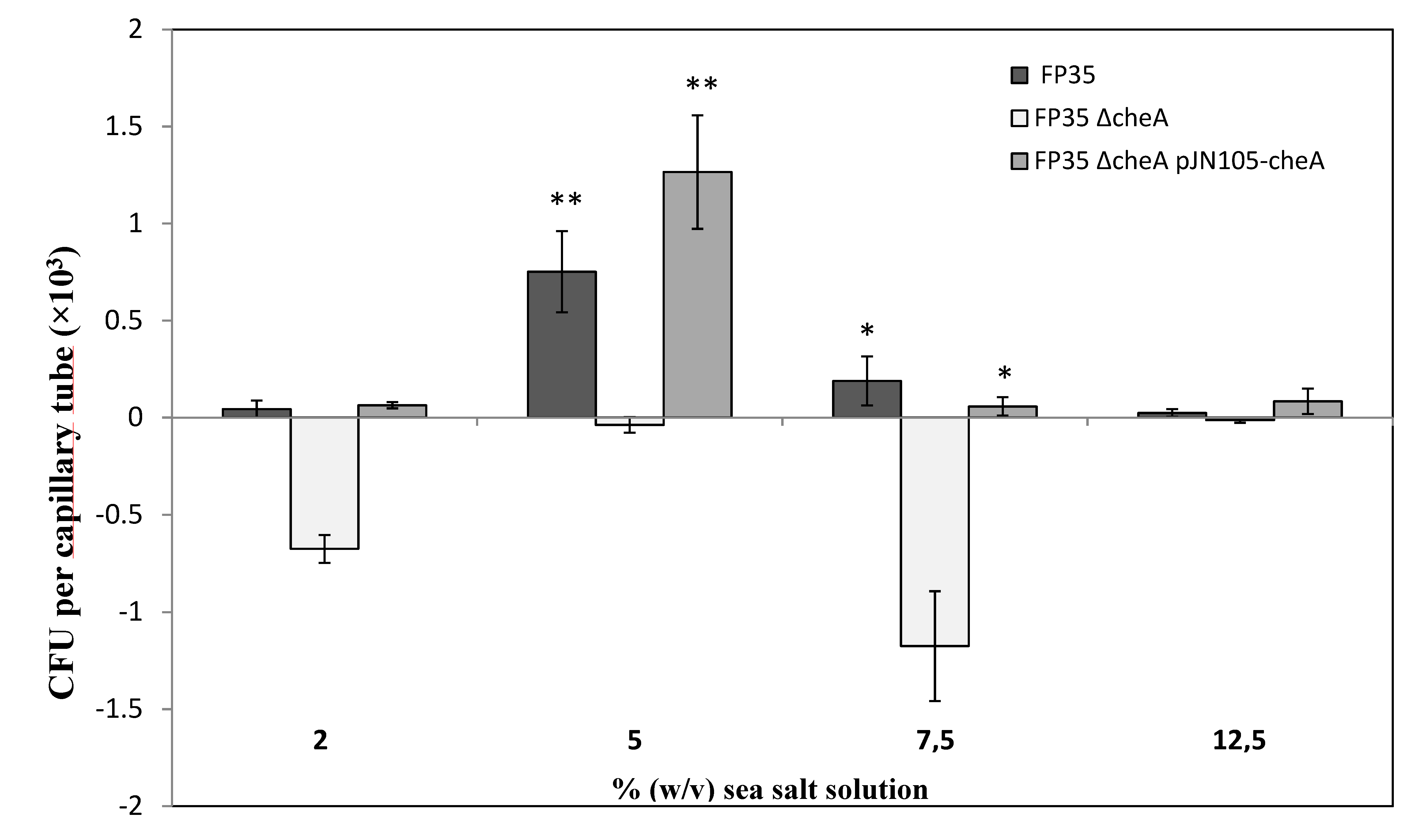
To confirm the role of cheA observed in chemotactic qualitative experiments, quantitative assays were carried out in the same range of salt concentrations (Figure 2). Of the different sea salt solution concentrations tested, the results confirm that the optimal concentration is 5% (w/v) for the accumulation of wild-type cells in capillary tubes containing root exudates (Figure 2). By contrast, the FP35 ΔcheA mutant showed an accumulation of below or close to zero under all the experimental conditions analysed. The negative CFU values at concentrations of 2 and 7.5% (w/v) reflect negative chemotaxis. The cheA required for positive chemotaxis was confirmed by the complementation of the FP35 ΔcheA mutant with a copy of the cheA gene in trans (pJN105-cheA; Figure 2).

3.2. Chemoattraction to Salicornia Root Exudates in a Variety of Halophilic Bacteria

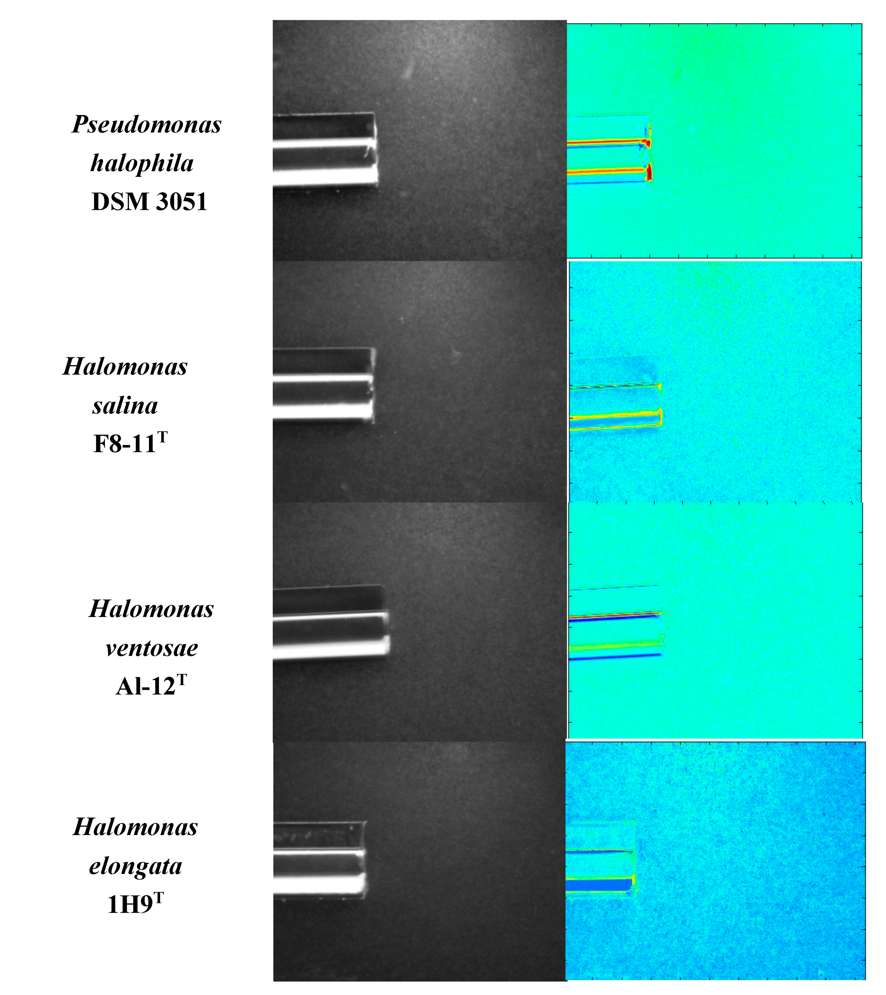
After confirming the positive effect of Salicornia root exudates on the chemotactic response of H. anticariensis FP35T, we hypothesized that these root exudates could also act as chemoattractants to other halophilic bacteria. A qualitative assessment of swimming motility showed an increase in halo size in the other four bacteria, H. ventosae Al-12T, H. elongata 1H9T, H. salina F8-11T and Pseudomonas salina DSM 3051, all grown at an optimal 5% (w/v) salt concentration previously observed for H. anticariensis FP35T. Only two of these bacteria, P. halophila DSM 3051 and H. salina F8-11T, exhibited chemoattraction to Salicornia root exudates (Figure 3). Nevertheless, Salicornia exudates elicited a lower chemotaxis response to these two strains as compared to that to H. anticariensis FP35T.

3.3. Identification of Salicornia Exudate Composition Using UPLC - HRMS Q-TOF

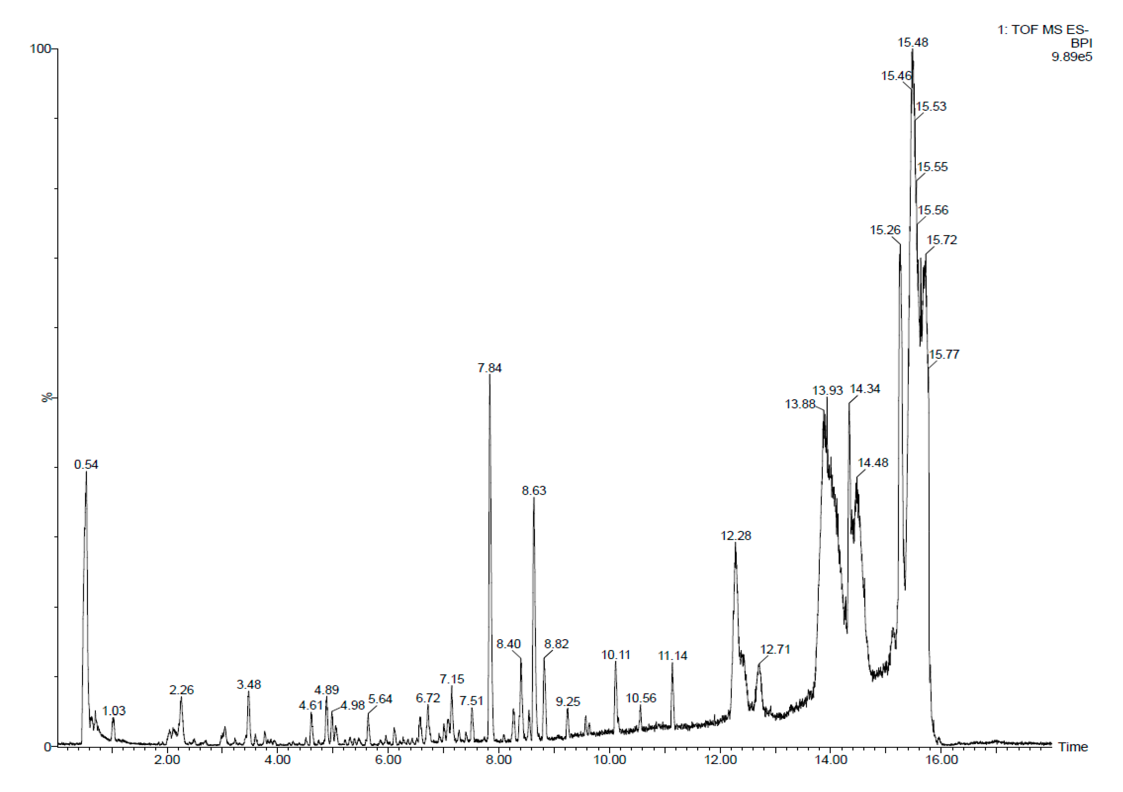
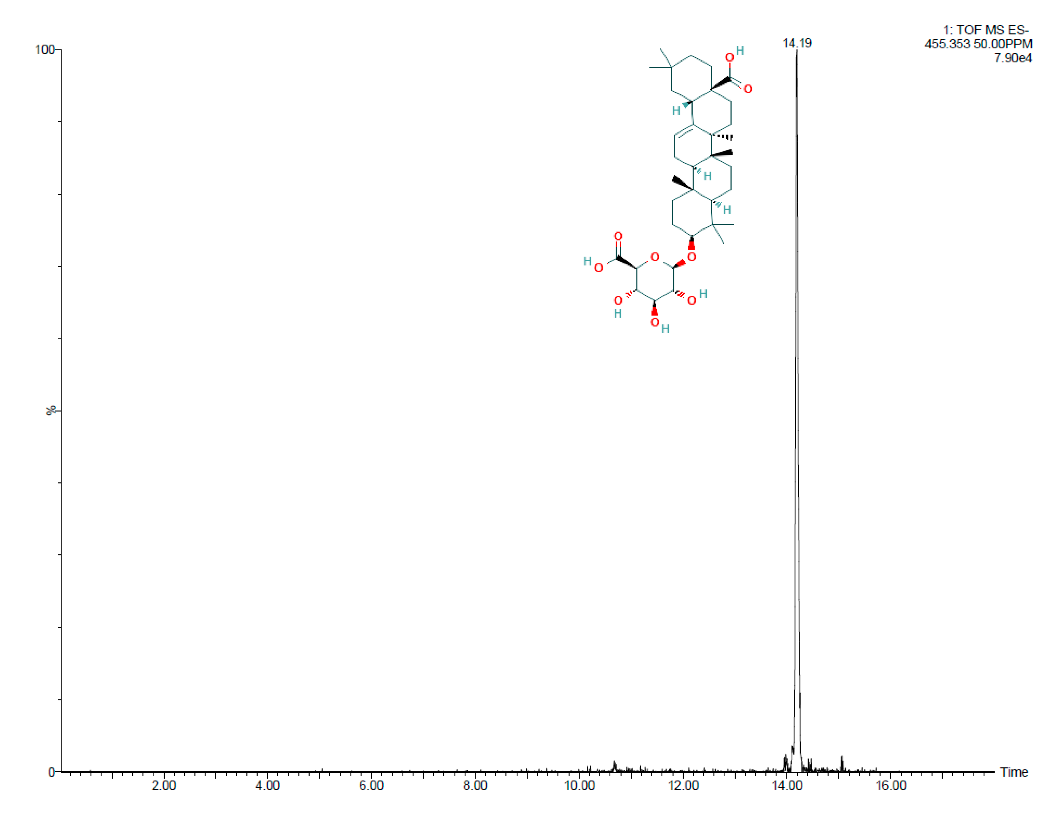
Electrospray quadrupole time-of-flight mass spectrometry (Q-TOFS MS) was used to identify metabolites produced by the exudates tested. Figure 4 shows the total ion chromatogram (TIC) spectrum of the Salicornia root exudates. Profiles of the root exudates from Salicornia plants demonstrate the presence of different peaks, the most abundant of which according to area was detected at a retention time of 14.16 min. This peak, corresponding to [M + H]+ at m/z 455.3525, afforded the molecular formula C30H48O3 and was identified by LC-HRMS to correspond to oleanolic acid (Figure 5).
The concentration of this compound in the Salicornia root exudates sampled was 90 µM. An optimum growth of the bacterium FP35T in presence of oleanolic acid (at concentration of 0.1 mM) was observed (Figure S3). The bacterium growth pattern after 14 h revealed similar values in absence or in presence of this compound (OD600 control: 1.006 ± 0.09 vs. OD600 oleanolic acid, 0.1 mM: 1.16 ± 0.14).

3.4. Assessment of Chemotactic Response of H. anticariensis FP35T to Oleanolic Acid

We evaluated the chemotactic responses of FP35T to the predominant compound, oleanolic acid (Sigma-Aldrich, Missouri, USA), in Salicornia exudates. We carried out experiments at a 5% (w/v) concentration of a sea salt solution which generates higher background accumulation of wild-type cells in capillaries containing root exudates. The concentrations of oleanolic acid (0.1 and 1 mM) tested in this study were selected according to the positive chemotaxis response previously observed for Pseudomonas and Bacillus to other compounds detected in root exudates in this range of concentrations [18,59].
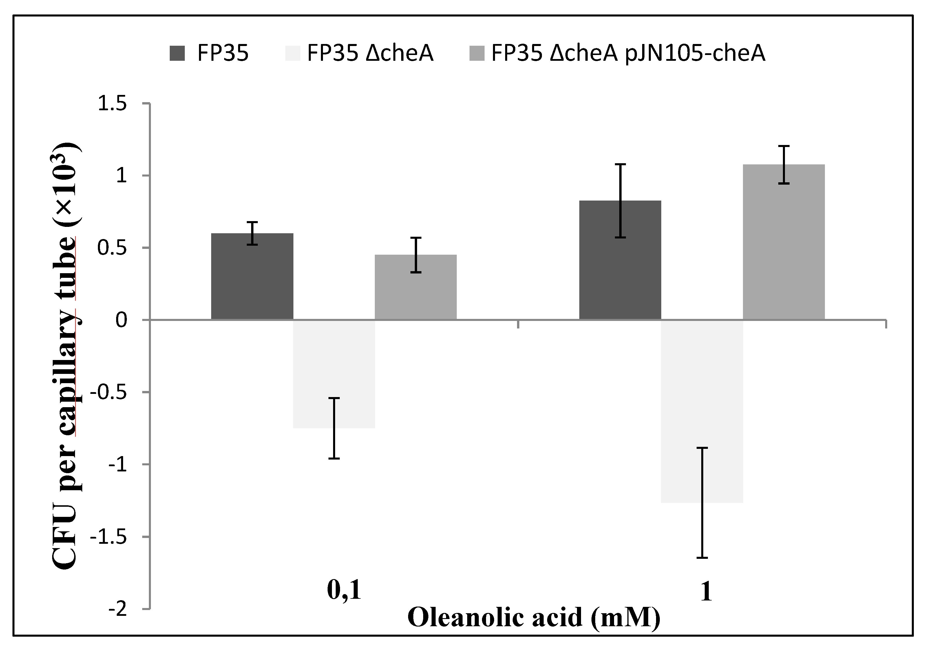
The wild-type strain FP35T showed positive chemotaxis for oleanolic acid tested at concentrations of 0.1 mM and 1 mM (Figure 6), while the FP35 ΔcheA mutant showed negative CFU values, corresponding to negative chemotaxis. As expected, positive responses to oleanolic acid were restored in the complemented mutant FP35 ΔcheA pJN105-cheA (Figure 6). Background accumulation of cells, at close to 1 × 10³, found in capillaries containing 1mM of oleanolic acid was similar to that in the capillaries containing Salicornia exudates. Negative chemotaxis response was observed for ethanol, the solvent used to dissolve oleanolic acid (data not shown).

3.5. Evaluation of H. anticariensis FP35T Biofilm Formation in Response to Salicornia Root Exudates and Oleanolic Acid

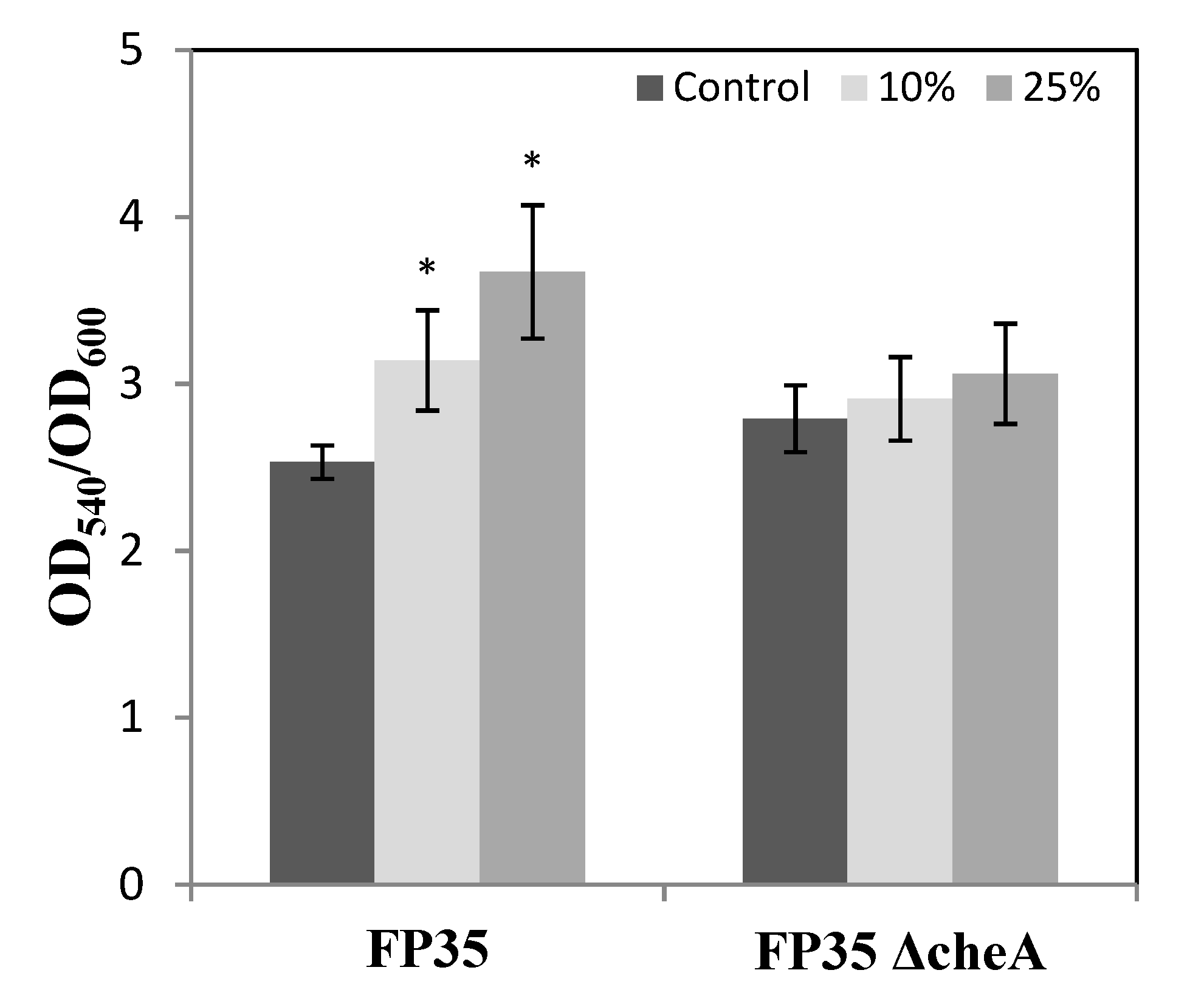
To ascertain their impact on the biofilm formation of H. anticariensis FP35T, 10 and 25% (v/v) concentrations of Salicornia root exudates were added to the cultures grown in 5% (w/v) sea salt solution. The results indicate that both these concentrations of root exudates significantly increased the biofilm biomass of FP35T (Figure 7). However, no statistically significant differences were observed in the chemotaxis mutant FP35 ΔcheA with respect to the two exudate root concentrations tested. This suggests that an active chemotaxis system is required in FP35T order to promote biofilm formation by Salicornia root exudates. Because oleanolic acid is the predominant compound in Salicornia exudates, its impact in the biofilm formation of FP35T was also evaluated. Changes in biofilm in this case were not observed because no significant differences were found between the biofilm formation in the presence of this compound and in the presence of ethanol (the compound used to dissolve this synthetic compound with limited solubility) (OD540/OD600 control ethanol, 0.1 mM or 1 mM: 1.29 ± 0.65; 1.23 ± 0.10 vs. OD540/OD600 oleanolic acid, 0.1 mM or 1 mM: 0.57 ± 0.14; 1.11 ± 0.44).

3.6. Salicornia Plant Reactions to Inoculation with H. anticariensis strain FP35T and its Mutant Strain FP35ΔcheA

The effect of the wild-type H. anticariensis strain FP35T and its mutant H. anticariensis strain FP35 ΔcheA on the root length, shoot length, germination and vigour index of Salicornia was tested. An increase in the shoot and root length of Salicornia seeds was observed following inoculation with H. anticariensis strain FP35T as compared to the non-inoculated control (Table 1). The bacterization of seeds by the halophilic bacteria also significantly increased the percentage of germination. In stark contrast, seeds bacterized by H. anticariensis strain FP35 ΔcheA only boosted root growth, while shoot length and the percentage of germination were similar to those for the non-inoculated control (Table 1). Furthermore, the 153% increase in the vigour index caused by inoculation with the wild-type strain exceeded the 79% increase in that for the chemotaxis mutant. These results highlight the important role of chemotaxis in the PGPR activity of H. anticariensis strain FP35T in Salicornia. Root colonization of the bacterial strains under seed bacterization conditions was tested, which showed a 2.8-fold increase in the H. anticariensis FP35T colonization of Salicornia roots (6.3 × 10⁵ ± 0.42) CFUs g−1 dry root) as compared to colonization by the chemotaxis mutant (2.3 × 10⁵ ± 0.29) CFUs g−1 dry root).

4. Discussion

Increase in soil salinity, which constitutes an enormous challenge for agriculture, needs to be tackled in order to come up with solutions to achieve sustainable crop production. One of the principal strategies for improving crop yields in saline soils is the use of PGPR bacteria for the promotion of halophyte growth in this type of soil. Rhizosphere colonization by PGPR bacteria is associated with root exudates [60] which play an important role in the rhizosphere and act as signals that mediate root-microorganism interactions [61]. Chemotaxis is a key player in initiating crosstalk between plant roots and PGPR bacteria [62]. Though extensively studied in model organisms such as Escherichia coli and Pseudomonas spp. [19], little is known about these chemotactic bacterial systems in halophilic bacteria such as those of the genus Halomonas [25,26,27], which have, however, been studied in relation to Salicornia root colonization [63]. We assayed the motility of bacteria belonging to the genus Halomonas that were isolated from the halophyte rhizosphere. H. anticariensis strain FP35T, which had the highest motility of all the bacteria tested, was selected.
The halophilic bacterium H. anticariensis FP35T has plant growth-promoting properties such as siderophore production, one of the principal advantages of this type of bacterium, which excludes other microorganisms and reduces competition for constituents of root exudates [64]. Strain FP35T produces 2,3-butanediol during glucose fermentation, as well as nitrogenase and nitrate and nitrite reductase, and hydrolyses gelatine [15]. FP35T also produces sulphated exopolysaccharides with many biotechnological applications [17], as well as quorum-sensing signalling molecules [16].
In a previous study, an identification of the draft genome sequence of H. anticariensis FP35T was performed [65]. Analysing this genome sequence, we have found one cluster of chemotaxis-related genes and twenty-one chemoreceptors. This gene cluster seems to govern the chemotactic behavior of FP35T and it is very similar to the cluster from Escherichia coli including the cheA gene. Similar results were previously described for this strain and other species from the genus Halomonas [27]. Given its importance in the chemotaxis pathway of other bacteria, the cheA gene, through its disruption in strain FP35T, was found to play a crucial role in chemotactic responses to environmental hydrocarbon pollutants [25]. We carried out chemotaxis and biofilm formation experiments to investigate how Salicornia root exudates mediate the enhanced root colonization of H. anticariensis strain FP35T. The chemotactic response of the halophilic bacterium FP35T to Salicornia root exudates was analysed using qualitative and quantitative chemotaxis experiments. In addition, a complemented chemotaxis mutant strain FP35 ΔcheA pJN105-cheA was constructed in order to corroborate the chemotaxis results. Salicornia root exudates were observed to act as chemoattractants for strain FP35T grown in concentrations of sea salt solution ranging from 2 to 12.5% (w/v), with the strongest chemotactic response found at 5% (w/v). These results were confirmed by the cheA mutant’s lack of chemotaxis response and the chemoattraction of the complemented mutant strain FP35ΔcheA pJN105-cheA. Interestingly, in a previous study, strain FP35T showed the strongest chemoattraction to environmental pollutant compounds such as phenol and naphthalene when grown in a sea salt solution concentration of 7.5% (w/v) [25]. These findings point to the significance of salt concentrations in the chemotaxis system and of the chemotaxis responses of H. anticariensis FP35T to different substances found in the soil. To the best of our knowledge, only few studies analyse the chemotactic behaviour of Halomonas strains using swimming assays with times of incubation higher than 24 h [27,66].
The chemotaxis response of FP35T to root exudates found differed from that observed in this study for other species of the genus Halomonas spp. This indicates that the genus Halomonas spp. is comprised of bacteria with chemosensory signalling mechanisms of varying complexity, a conclusion previously reached in relation to species of the genus Pseudomonas spp. For example, although tetrachloroethylene acts as a chemoattractant for P. putida F1 and P. stutzeri OX1 [67,68], it has been found to be a chemorepellent for P. aeruginosa PAO1 [69,70].
Chemoattraction and biofilm formation by root exudates are associated with the colonization process [71]. Salicornia exudates act as chemoattractants for FP35T and as stimulators of biofilm formation at sea salt concentrations of 5% (w/v), the optimum concentration for chemoattraction. Previous studies have indicated that tomato and Arabidopsis root exudates increase biofilm formation in different species of the genus Bacillus [24,72]. Furthermore, a variety of exudate components such as citric acid, malic acid and fumaric acid have been reported to stimulate the biofilm formation of bacteria [59,71]. However, to our knowledge, little is known about the impact of halophyte exudates on Halomonas biofilm. As previous studies of other bacteria of the genus Bacillus have suggested [59,72], exudates could act as a carbon source and the growth of FP35T on Salicornia exudates may alter the metabolism of this bacterium in a manner favourable to biofilm formation.
On the other hand, we characterized Salicornia exudates in order to determine which constituent is related to FP35T chemoattraction. Plant root exudates are highly complex compound mixtures and vary according to the plant development stage and environmental conditions [73], although some of the constituents of these compound mixtures are unique to certain plants species [74]. Profiles of Salicornia exudates showed a high-intensity peak due to oleanolic acid (OA), a pentacyclic triterpenoid compound widely distributed in the plant kingdom. OA has a variety of biological functions in nature, with several promising pharmacological applications [75], plays an important role in regulating the virulence pathway of pathogens such as Ralstonia solanacearum [76], appears to be the principal component of Salicornia root exudates and acts as a chemoattractant for FP35T. The levels of bacterial cell accumulation caused by OA were similar to those caused by chemotaxis to exudates, indicating that this organic acid may be the principal mediator of Salicornia plant-bacterium interactions.
Chemotactic reactions to OA were found to be positive at concentrations of 0.1 mM and 1 mM. At these concentrations, a positive chemotaxis response has also been observed to other compounds detected in root exudates such as the gamma-aminobutyric acid (GABA) chemoreceptor in Pseudomonas putida strain KT2440 [77], as well as oxalic and malic acids in Bacillus amyloliquefaciens SQR9 [59] and Pseudomonas fluorescens [18]. An unexpected chemorepellent response of the chemotaxis mutant FP35 ΔcheA to root exudates (at sea salt concentrations of 2 and 7.5%) and OA was observed. Considering that CheA is an essential protein in the chemotaxis signalling cascade in E. coli (bacteria model of chemotaxis signal transduction), the cheA mutant should be non-chemotactic at all. A previous study indicated that a cytoplasmic chemoreceptor, TlpD, in Helicobacter pylori form an autonomous signaling unit and mediate a repellent chemotaxis response to conditions that promote oxidative stress [78]. TlpD is able to localize to the pole and recruits CheW, CheA, and at least two CheV proteins to this location operating independently of other chemoreceptors to organize a chemotaxis signaling complex that mediate this chemorepellent response. The inactivation of cheA abolishes chemotaxis responses but could benefit, under certain conditions, the operating action of cytoplasmic chemoreceptors that could change the role of the principal kinases in the chemotaxis system. Future studies should be performed to increase the knowledge of cytoplasmic chemoreceptors in representatives of the genus Halomonas that could alter the function and the role of chemoreceptor sensor proteins like CheA.
In this study it was difficult to determine the biofilm formation of FP35T caused by oleanolic acid. The levels of biofilm formation in the presence of this compound were similar to those caused by ethanol (used to dissolve this synthetic compound with limited solubility). It has been previously demonstrated that low concentrations of ethanol stimulate biofilm formation in other bacteria such as Pseudomonas aeruginosa [79] and this effect can be a problem when measuring the real biofilm formed by FP35T.
The positive relationship between the PGPR H. anticariensis strain FP35T and Salicornia root exudates could contribute to preferential colonization and stimulation of seed germination. In vitro experiments were carried out to examine the growth-promoting effects of strain FP35T on Salicornia seeds under sterile conditions. Seeds bacterized with the halophilic bacterium FP35T showed an increase in germination percentages, as well as shoot and root length, as compared to the non-inoculated control. Similar results were obtained by the inoculation of Salicornia seeds with halotolerant bacteria such as Brachybacterium saurashtrense and Pseudomonas sp. strain JG10 [53]. However, seeds bacterized with the chemotaxis mutant FP35 ΔcheA only showed an increase in root length as compared to the non-inoculated control. Deletion of the gene mcpG coding for a GABA chemoreceptor or the gene cheA3 coding for a histidine kinase in P. putida KT2440 reduced root colonization that requires chemotaxis [80,81].
This study indicates that the cheA gene coding for MCPs could play an important role in root colonization by FP35T. Furthermore, the increase in biofilm formation, previously observed only in the wild-type strain in the presence of Salicornia exudates, could enable the bacterium to colonize the roots more effectively. Our results suggest that there is a connection between the absence of chemotaxis and the reduction in the vigour index and colonization of halophyte seeds.

5. Conclusions

This study demonstrates that Salicornia exudates act as chemoattractants for the halophilic bacterium H. anticariensis FP35T. Our results also indicate that salt concentration influences chemotactic responses, with optimum levels of chemoattraction observed at a sea salt concentration of 5% (w/v). Oleanolic acid, the principal constituent of Salicornia exudates, appears to chemotactically mediate plant-bacterium interactions. The chemotactic responses and increased biofilm formation observed in this study suggest that halophyte exudates have the potential to expand the colonization of these salt-tolerant plants by halophilic bacteria. The enhancement of bacterial recruitment could be a potentially effective strategy for promoting halophyte growth and lead to major benefits for agriculture under saline conditions.

Supplementary Materials

The following are available online at https://www.mdpi.com/2076-2607/8/4/575/s1, Figure S1: Chemosensory cluster identified in the draft genome sequence of H. anticariensis FP35T. Figure S2: Qualitative capillary chemotaxis assays comparing responses of wild-type H. anticariensis FP35T and the mutant strain FP35 ΔcheA towards chemotaxis buffer grown at a concentration of 5% (w/v) of sea salt solution. Figure S3: H. anticariensis FP35T growth in M8 plates containing 0.3% agar in absence or presence of oleanolic acid (A and B, respectively).

Author Contributions

I.S. and I.L. conceived and supervised the study; I.S. and I.L. designed the experiments; I.S., D.P.-M., L.T., E.P., C.A. and I.L. performed the experiments; I.S., L.T. and E.P. analyzed the data; I.S. prepared the figures and wrote the manuscript; I.S., D.P.-M., L.T., E.P., C.A. and I.L. edited the manuscript and reviewed the literature. All authors have read and agreed to the published version of the manuscript.

Funding

The study was funded by the Ramón y Cajal MINECO program (RYC-2014-15532) in Spain.

Acknowledgments

I.S. wishes to thank MINECO for her Ramón and Cajal contract. Michael O´Shea corrected the English version of the manuscript.

Conflicts of Interest

The authors declare no conflict of interest. The funders played no role in the design of the study; the collection, analysis or interpretation of data; in the writing of the manuscript or in the decision to publish the results.

References

  1. Mayak, S.; Tirosh, T.; Glick, B.R. Plant growth-promoting bacteria confer resistance in tomato plants to salt stress. Plant Physiol. Biochem. PPB 2004, 42, 565–572. [Google Scholar] [CrossRef] [PubMed]
  2. Ravindran, K.C.; Venkatesan, K.; Balakrishnan, V.; Chellappan, K.P.; Balasubramanian, T. Restoration of saline land by halophytes for Indian soils. Soil Biol. Biochem. 2007, 39, 2661–2664. [Google Scholar] [CrossRef]
  3. Manousaki, E.; Kalogerakis, N. Halophytes—An emerging trend in phytoremediation. Int. J. Phytoremediat. 2011, 13, 959–969. [Google Scholar] [CrossRef] [PubMed]
  4. Kadereit, G.; Ball, P.; Beer, S.; Mucina, L.; Sokoloff, D.; Teege, P.; Yaprak, A.E.; Freitag, H. A taxonomic nightmare comes true: Phylogeny and biogeography of glassworts (Salicornia L. Chenopodiaceae). Taxon 2007, 56, 1143–1170. [Google Scholar] [CrossRef] [Green Version]
  5. Ventura, Y.; Wuddineh, W.A.; Ephrath, Y.; Shpigel, M.; Sagi, M. Molybdenum as an essential element for improving total yield in seawater-grown Salicornia europaea (L.). Sci. Hortic. 2010, 126, 395–401. [Google Scholar] [CrossRef]
  6. Ventura, Y.; Sagi, M. Halophyte crop cultivation: The case for salicornia and sarcocornia. Environ. Exp. Bot. 2013, 92, 144–153. [Google Scholar] [CrossRef]
  7. Rhee, M.H.; Park, H.J.; Cho, J.Y. Salicornia herbacea: Botanical, chemical and pharmacological review of halophyte marsh plant. J. Med. Plants Res. 2009, 3, 548–555. [Google Scholar]
  8. Karthivashan, G.; Park, S.Y.; Kweon, M.H.; Kim, J.; Haque, M.E.; Cho, D.Y.; Kim, I.S.; Cho, E.A.; Ganesan, P.; Choi, D.K. Ameliorative potential of desalted Salicornia europaea L. extract in multifaceted Alzheimer’s-like scopolamine-induced amnesic mice model. Sci. Rep. 2018, 8, 7174. [Google Scholar] [CrossRef]
  9. Abdal, M.S. Salicornia production in Kuwait. World Appl. Sci. J. 2009, 6, 1033–1038. [Google Scholar]
  10. Moghaieb, R.E.A.; Saneoka, H.; Fujita, K. Effect of salinity on osmotic adjustment, glycinebetaine accumulation and the betaine aldehyde dehydrogenase gene expression in two halophytic plants, Salicornia europaea and Suaeda maritima. Plant Sci. 2004, 166, 1345–1349. [Google Scholar] [CrossRef]
  11. Glenn, E.P.; Pitelka, L.F.; Olsen, M.W. The use of halophytes to sequester carbon. Water Air Soil Pollut. 1992, 64, 251–263. [Google Scholar] [CrossRef]
  12. Ozawa, T.; Wu, J.; Fujii, S. Effect of inoculation with a strain of Pseudomonas pseudoalcaligenes isolated from the endorhizosphere of Salicornia europea on salt tolerance of the glasswort. Soil Sci. Plant Nutr. 2007, 53, 12–16. [Google Scholar] [CrossRef] [Green Version]
  13. Argandona, M.; Fernandez-Carazo, R.; Llamas, I.; Martinez-Checa, F.; Caba, J.M.; Quesada, E.; del Moral, A. The moderately halophilic bacterium Halomonas maura is a free-living diazotroph. FEMS Microbiol. Lett. 2005, 244, 69–74. [Google Scholar] [CrossRef] [PubMed]
  14. Mapelli, F.; Marasco, R.; Rolli, E.; Barbato, M.; Cherif, H.; Guesmi, A.; Ouzari, I.; Daffonchio, D.; Borin, S. Potential for plant growth promotion of rhizobacteria associated with Salicornia growing in Tunisian hypersaline soils. BioMed Res. Int. 2013, 2013, 248078. [Google Scholar] [CrossRef] [PubMed] [Green Version]
  15. Martinez-Canovas, M.J.; Bejar, V.; Martinez-Checa, F.; Quesada, E. Halomonas anticariensis sp. nov., from Fuente de Piedra, a saline-wetland wildfowl reserve in Malaga, southern Spain. Int. J. Syst. Evol. Microbiol. 2004, 54, 1329–1332. [Google Scholar] [CrossRef] [PubMed] [Green Version]
  16. Llamas, I.; Quesada, E.; Martinez-Canovas, M.J.; Gronquist, M.; Eberhard, A.; Gonzalez, J.E. Quorum sensing in halophilic bacteria: Detection of N-acyl-homoserine lactones in the exopolysaccharide-producing species of Halomonas. Extrem. Life Extrem. Cond. 2005, 9, 333–341. [Google Scholar] [CrossRef] [PubMed]
  17. Mata, J.A.; Bejar, V.; Llamas, I.; Arias, S.; Bressollier, P.; Tallon, R.; Urdaci, M.C.; Quesada, E. Exopolysaccharides produced by the recently described halophilic bacteria Halomonas ventosae and Halomonas anticariensis. Res. Microbiol. 2006, 157, 827–835. [Google Scholar] [CrossRef]
  18. Sood, G. Chemotactic response of plant-growth-promoting bacteria towards roots of vesicular-arbuscular mycorrhizal tomato plants. FEMS Microbiol. Ecol. 2003, 45, 219–227. [Google Scholar] [CrossRef]
  19. Sampedro, I.; Parales, R.E.; Krell, T.; Hill, J.E. Pseudomonas chemotaxis. FEMS Microbiol. Rev. 2015, 39, 17–46. [Google Scholar]
  20. Flanary, P.L.; Allen, R.D.; Dons, L.; Kathariou, S. Insertional inactivation of the Listeria monocytogenes cheYA operon abolishes response to oxygen gradients and reduces the number of flagella. Can. J. Microbiol. 1999, 45, 646–652. [Google Scholar] [CrossRef]
  21. Foynes, S.; Dorrell, N.; Ward, S.J.; Stabler, R.A.; McColm, A.A.; Rycroft, A.N.; Wren, B.W. Helicobacter pylori possesses two CheY response regulators and a histidine kinase sensor, CheA, which are essential for chemotaxis and colonization of the gastric mucosa. Infect. Immun. 2000, 68, 2016–2023. [Google Scholar] [CrossRef] [PubMed] [Green Version]
  22. De Weert, S.; Vermeiren, H.; Mulders, I.H.; Kuiper, I.; Hendrickx, N.; Bloemberg, G.V.; Vanderleyden, J.; De Mot, R.; Lugtenberg, B.J. Flagella-driven chemotaxis towards exudate components is an important trait for tomato root colonization by Pseudomonas fluorescens. Mol. Plant-Microbe Interact. MPMI 2002, 15, 1173–1180. [Google Scholar] [CrossRef] [PubMed] [Green Version]
  23. Ling, N.; Zhang, W.; Wang, D.; Mao, J.; Huang, Q.; Guo, S.; Shen, Q. Root Exudates from Grafted-Root Watermelon Showed a Certain Contribution in Inhibiting Fusarium oxysporum f. sp. niveum. PLoS ONE 2013, 8, e63383. [Google Scholar] [CrossRef] [PubMed]
  24. Rudrappa, T.; Czymmek, K.J.; Pare, P.W.; Bais, H.P. Root-secreted malic acid recruits beneficial soil bacteria. Plant Physiol. 2008, 148, 1547–1556. [Google Scholar] [CrossRef] [Green Version]
  25. Tena-Garitaonaindia, M.; Llamas, I.; Toral, L.; Sampedro, I. Chemotaxis of halophilic bacterium Halomonas anticariensis FP35 towards the environmental pollutants phenol and naphthalene. Sci. Total Environ. 2019, 669, 631–636. [Google Scholar] [CrossRef]
  26. Gasperotti, A.F.; Studdert, C.A.; Revale, S.; Herrera Seitz, M.K. Draft genome sequence of Halomonas sp. KHS3, a polyaromatic hydrocarbon-chemotactic strain. Genome Announc. 2015, 3, e00020-15. [Google Scholar] [CrossRef] [Green Version]
  27. Gasperotti, A.F.; Revuelta, M.V.; Studdert, C.A.; Herrera Seitz, M.K. Identification of two different chemosensory pathways in representatives of the genus Halomonas. BMC Genom. 2018, 19, 266. [Google Scholar]
  28. Li, S.; Zhang, N.; Zhang, Z.; Luo, J.; Shen, B.; Zhang, R.; Shen, Q. Antagonist Bacillus subtilis HJ5 controls Verticillium wilt of cotton by root colonization and biofilm formation. Biol. Fertil. Soils 2013, 49, 295–303. [Google Scholar] [CrossRef]
  29. Xu, Z.; Shao, J.; Li, B.; Yan, X.; Shen, Q.; Zhang, R. Contribution of bacillomycin D in Bacillus amyloliquefaciens SQR9 to antifungal activity and biofilm formation. Appl. Environ. Microbiol. 2013, 79, 808–815. [Google Scholar] [CrossRef] [Green Version]
  30. Kasim, W.A.; Gaafar, R.M.; Abou-Ali, R.M.; Omar, M.N.; Hewait, H.M. Effect of biofilm forming plant growth promoting rhizobacteria on salinity tolerance in barley. Ann. Agric. Sci. 2016, 61, 217–227. [Google Scholar] [CrossRef] [Green Version]
  31. Llamas, I.; del Moral, A.; Martinez-Checa, F.; Arco, Y.; Arias, S.; Quesada, E. Halomonas maura is a physiologically versatile bacterium of both ecological and biotechnological interest. Antonie Van Leeuwenhoek 2006, 89, 395–403. [Google Scholar] [CrossRef]
  32. Qurashi, A.W.; Sabri, A.N. Bacterial exopolysaccharide and biofilm formation stimulate chikpea growth and soil aggregation under salt stress. Braz. J. Microbiol. 2012, 43, 1183–1191. [Google Scholar] [CrossRef] [PubMed] [Green Version]
  33. Haynes, W.C.; Wicherham, L.J.; Hesseltine, C.W. Maintenance of cultures of industrially important microorganisms. Appl. Microbiol. 1955, 3, 361–368. [Google Scholar] [CrossRef] [PubMed] [Green Version]
  34. Moraine, R.A.; Rogovin, P. Kinetics of polysaccharide B-1459 fermentation. Biotechnol. Bioeng. 1966, 8, 511–524. [Google Scholar] [CrossRef]
  35. Rodriguez-Valera, F.; Ruiz-Berraquero, F.; Ramos-Cormenzana, A. Characteristics of the heterotrophic bacterial populations in hypersaline environments of different salt concentrations. Microb. Ecol. 1981, 7, 235–243. [Google Scholar] [CrossRef] [PubMed]
  36. Valderrama, M.J.; Prado, B.; del Moral, A.; Rios, R.; Ramos-Cormenzana, A.; Campos, V. Numerical taxonomy of moderately halophilic gram-positive cocci isolated from the Salar de Atacama (Chile). Microbiologia 1991, 7, 35–41. [Google Scholar] [PubMed]
  37. Vreeland, R.H.; Litchfield, C.D.; Martin, E.L.; Elliot, E. Halomonas elongata, a New Genus and Species of Extremely Salt-Tolerant Bacteria. Int. J. Syst. Evol. Microbiol. 1980, 30, 485–495. [Google Scholar] [CrossRef] [Green Version]
  38. Fendrich, C. Halovibrio variabilis gen. nov. sp. nov., Pseudomonas halophila sp. nov. and a new halophilic aerobic coccoid Eubacterium from Great Salt Lake, Utah, USA. Syst. Appl. Microbiol 1988, 11, 36–43. [Google Scholar] [CrossRef]
  39. Martinez-Canovas, M.J.; Quesada, E.; Llamas, I.; Bejar, V. Halomonas ventosae sp. nov., a moderately halophilic, denitrifying, exopolysaccharide-producing bacterium. Int. J. Syst. Evol. Microbiol. 2004, 54, 733–737. [Google Scholar] [CrossRef] [Green Version]
  40. Amjres, H.; Bejar, V.; Quesada, E.; Abrini, J.; Llamas, I. Halomonas rifensis sp. nov., an exopolysaccharide-producing, halophilic bacterium isolated from a solar saltern. Int. J. Syst. Evol. Microbiol. 2011, 61, 2600–2605. [Google Scholar] [CrossRef]
  41. Martinez-Checa, F.; Bejar, V.; Martinez-Canovas, M.J.; Llamas, I.; Quesada, E. Halomonas almeriensis sp. nov., a moderately halophilic, exopolysaccharide-producing bacterium from Cabo de Gata, Almería, south-east Spain. Int. J. Syst. Evol. Microbiol. 2005, 55, 2007–2011. [Google Scholar] [CrossRef] [PubMed] [Green Version]
  42. González-Domenech, C.M.; Martinez-Checa, F.; Quesada, E.; Bejar, V. Halomonas cerina sp. nov., a moderately halophilic, denitrifying, exopolysaccharide-producing bacterium. Int. J. Syst. Evol. Microbiol. 2008, 58, 803–809. [Google Scholar]
  43. Calvo, C.; Martínez-Checa, F.; Toledo, F.; Porcel, J.; Quesada, E. Characteristics of bioemulsifiers synthesised in crude oil media by Halomonas eurihalina and their effectiveness in the isolation of bacteria able to grow in the presence of hydrocarbons. Appl. Microbiol. Biotechnol. 2002, 60, 347–351. [Google Scholar] [PubMed]
  44. Llamas, I.; Bejar, V.; Martinez-Checa, F.; Martinez-Canovas, M.J.; Molina, I.; Quesada, E. Halomonas stenophila sp. nov., a halophilic bacterium that produces sulphate exopolysaccharides with biological activity. Int. J. Syst. Evol. Microbiol. 2011, 61, 2508–2514. [Google Scholar] [CrossRef] [PubMed]
  45. Luque, R.; Bejar, V.; Quesada, E.; Martinez-Checa, F.; Llamas, I. Halomonas ramblicola sp. nov., a moderately halophilic bacterium from Rambla Salada, a Mediterranean hypersaline rambla. Int. J. Syst. Evol. Microbiol. 2012, 62, 2903–2909. [Google Scholar] [CrossRef] [PubMed]
  46. García, T.; Mellado, E.; Ostos, J.C.; Ventosa, A. Halomonas organivorans sp. nov., a moderate halophile able to degrade aromatic compounds. Int. J. Syst. Evol. Microbiol. 2004, 54, 1723–1728. [Google Scholar]
  47. Ha, D.G.; Kuchma, S.L.; O’Toole, G. Plate-based assay for swimming motility in Pseudomonas aeruginosa. Methods Mol. Biol. 2014, 1149, 59–65. [Google Scholar]
  48. Aziz, R.K.; Bartels, D.; Best, A.A.; DeJongh, M.; Disz, T.; Edwards, R.A.; Formsma, K.; Gerdes, S.; Glass, E.M.; Kubal, M.; et al. The RAST server: Rapid annotations using subsystems technology. BMC Genom. 2008, 9, 75. [Google Scholar] [CrossRef] [Green Version]
  49. Markowitz, V.M.; Chen, I.M.; Palaniappan, K.; Chu, K.; Szeto, E.; Grechkin, Y.; Ratner, A.; Jacob, B.; Huang, J.; Williams, P.; et al. IMG: The integrated microbial genomes database and comparative analysis system. Nucleic Acids Res. 2012, 40, D115–D122. [Google Scholar] [CrossRef] [Green Version]
  50. Newman, J.R.; Fuqua, C. Broad-host-range expression vectors that carry the L-arabinose-inducible Escherichia coli araBAD promoter and the araC regulator. Gene 1999, 227, 197–203. [Google Scholar] [CrossRef]
  51. Demarre, G.; Guerout, A.M.; Matsumoto-Mashimo, C.; Rowe-Magnus, D.A.; Marliere, P.; Mazel, D. A new family of mobilizable suicide plasmids based on broad host range R388 plasmid (IncW) and RP4 plasmid (IncPalpha) conjugative machineries and their cognate Escherichia coli host strains. Res. Microbiol. 2005, 156, 245–255. [Google Scholar] [CrossRef] [PubMed]
  52. Schmid, J.; Ruhmann, B.; Sieber, V.; Romero-Jimenez, L.; Sanjuan, J.; Perez-Mendoza, D. Screening of c-di-GMP-Regulated Exopolysaccharides in Host Interacting Bacteria. Methods Mol. Biol. 2018, 1734, 263–275. [Google Scholar] [PubMed]
  53. Zhao, F.J.; Hamon, R.E.; McLaughlin, M.J. Root exudates of the hyperaccumulator Thlaspi caerulescens do not enhance metal mobilization. New Phytol. 2001, 151, 613–620. [Google Scholar] [CrossRef] [Green Version]
  54. Parales, R.E.; Harwood, C.S. Bacterial chemotaxis to pollutants and plant-derived aromatic molecules. Curr. Opin. Microbiol. 2002, 5, 266–273. [Google Scholar] [CrossRef]
  55. Adler, J. Method for Measuring Chemotaxis and Use of Method to Determine Optimum Conditions for Chemotaxis by Escherichia coli. J. Gen. Microbiol. 1973, 74, 77–91. [Google Scholar] [CrossRef] [Green Version]
  56. O’Toole, G.A. Microtiter dish biofilm formation assay. J. Vis. Exp. JoVE 2011, 47, 2437. [Google Scholar] [CrossRef]
  57. Jha, B.; Gontia, I.; Hartmann, A. The roots of the halophyte Salicornia brachiata are a source of new halotolerant diazotrophic bacteria with plant growth-promoting potential. Plant Soil 2012, 356, 265–277. [Google Scholar] [CrossRef]
  58. Abdul Baki, A.A.; Anderson, J.D. Vigour determination in soya bean by multiple criteria. Crop Sci. 1973, 13, 630–633. [Google Scholar] [CrossRef]
  59. Martins, S.J.; Medeiros, F.H.V.; Lakshmanan, V.; Bais, H.P. Impact of Seed Exudates on Growth and Biofilm Formation of Bacillus amyloliquefaciens ALB629 in Common Bean. Front. Microbiol. 2017, 8, 2631. [Google Scholar] [CrossRef] [Green Version]
  60. Lugtenberg, B.; Kamilova, F. Plant-growth-promoting rhizobacteria. Annu. Rev. Microbiol. 2009, 63, 541–556. [Google Scholar] [CrossRef] [Green Version]
  61. Ling, N.; Raza, W.; Ma, J.; Huang, Q.; Shen, Q. Identification and role of organic acids in watermelon root exudates for recruiting Paenibacillus polymyxa SQR-21 in the rhizosphere. Eur. J. Soil Biol. 2011, 47, 374–379. [Google Scholar] [CrossRef]
  62. Bais, H.P.; Park, S.W.; Weir, T.L.; Callaway, R.M.; Vivanco, J.M. How plants communicate using the underground information superhighway. Trends Plant Sci. 2004, 9, 26–32. [Google Scholar] [CrossRef]
  63. Marasco, R.; Mapelli, F.; Rolli, E.; Mosqueira, M.J.; Fusi, M.; Bariselli, P.; Reddy, M.; Cherif, A.; Tsiamis, G.; Borin, S.; et al. Salicornia strobilacea (Synonym of Halocnemum strobilaceum) grown under different tidal regimes selects rhizosphere bacteria capable of promoting plant growth. Front. Microbiol. 2016, 7, 1286. [Google Scholar] [CrossRef] [Green Version]
  64. Haas, D.; Defago, G. Biological control of soil-borne pathogens by fluorescent pseudomonads. Nat. Rev. Microbiol. 2005, 3, 307–319. [Google Scholar] [CrossRef]
  65. Tahrioui, A.; Quesada, E.; Llamas, I. Genetic and phenotypic analysis of the GacS/GacA system in the moderate halophile Halomonas anticariensis. Microbiology 2013, 159, 462–474. [Google Scholar] [CrossRef] [Green Version]
  66. D’Ippólito, S.; de Castro, R.E.; Herrera Seitz, M.K. Chemotactic response to gas oil of Halomonas spp. strains isolated from saline environments in Argentina. Rev. Argent. Microbiol. 2011, 43, 107–110. [Google Scholar]
  67. Parales, R.E.; Ditty, J.L.; Harwood, C.S. Toluene-degrading bacteria are chemotactic towards the environmental pollutants benzene, toluene, and trichloroethylene. Appl. Environ. Microbiol. 2000, 66, 4098–4104. [Google Scholar] [CrossRef] [Green Version]
  68. Vardar, G.; Barbieri, P.; Wood, T.K. Chemotaxis of Pseudomonas stutzeri OX1 and Burkholderia cepacia G4 toward chlorinated ethenes. Appl. Microbiol. Biotechnol. 2005, 66, 696–701. [Google Scholar] [CrossRef]
  69. Kato, J.; Shitashiro, M.; Yamamoto, M.; Kuroda, A.; Ikeda, T.; Takiguchi, N.; Ohtake, H. Chemotaxis by Pseudomonas aeruginosa toward 2,4-dichlorophenoxyacetate and volatile chlorinated aliphatic compounds. Abstr. Gen. Meet. Am. Soc. Microbiol. 2001, 101, 627. [Google Scholar]
  70. Shitashiro, M.; Kato, J.; Fukumura, T.; Kuroda, A.; Ikeda, T.; Takiguchi, N.; Ohtake, H. Evaluation of bacterial aerotaxis for its potential use in detecting the toxicity of chemicals to microorganisms. J. Biotechnol. 2003, 101, 11–18. [Google Scholar] [CrossRef]
  71. Zhang, N.; Wang, D.; Liu, Y.; Li, S.; Shen, Q.; Zhang, R. Effects of different plant root exudates and their organic acid components on chemotaxis, biofilm formation and colonization by beneficial rhizosphere-associated bacterial strains. Plant Soil 2014, 374, 689–700. [Google Scholar] [CrossRef]
  72. Chen, Y.; Cao, S.; Chai, Y.; Clardy, J.; Kolter, R.; Guo, J.H.; Losick, R. A Bacillus subtilis sensor kinase involved in triggering biofilm formation on the roots of tomato plants. Mol. Microbiol. 2012, 85, 418–430. [Google Scholar] [CrossRef] [PubMed]
  73. Petriacq, P.; Williams, A.; Cotton, A.; McFarlane, A.E.; Rolfe, S.A.; Ton, J. Metabolite profiling of non-sterile rhizosphere soil. Plant J. Cell Mol. Biol. 2017, 92, 147–162. [Google Scholar] [CrossRef] [PubMed]
  74. Mandal, S.M.; Chakraborty, D.; Dey, S. Phenolic acids act as signaling molecules in plant-microbe symbioses. Plant Signal. Behav. 2010, 5, 359–368. [Google Scholar] [CrossRef] [Green Version]
  75. Pollier, J.; Goossens, A. Oleanolic acid. Phytochemistry 2012, 77, 10–15. [Google Scholar] [CrossRef]
  76. Wu, D.; Ding, W.; Zhang, Y.; Liu, X.; Yang, L. Oleanolic Acid Induces the Type III Secretion System of Ralstonia solanacearum. Front. Microbiol. 2015, 6, 1466. [Google Scholar] [CrossRef] [Green Version]
  77. Reyes-Darias, J.A.; Yang, Y.; Sourjik, V.; Krell, T. Correlation between signal input and output in PctA and PctB amino acid chemoreceptor of Pseudomonas aeruginosa. Mol. Microbiol. 2015, 96, 513–525. [Google Scholar] [CrossRef]
  78. Collins, K.D.; Andermann, T.M.; Draper, J.; Sanders, L.; Williams, S.M.; Araghi, C.; Ottemann, K.M. The Helicobacter pylori CZB cytoplasmic chemoreceptor TlpD forms an autonomous polar chemotaxis signalling complex that mediates a tactic response to oxidative stress. J. Bacteriol. 2016, 198, 1563–1575. [Google Scholar] [CrossRef] [Green Version]
  79. Tashiro, Y.; Inagaki1a, A.; Ono, K.; Inaba, T.; Yawata, Y.; Uchiyama, H.; Nomura, N. Low concentrations of ethanol stimulate biofilm and pellicle formation in Pseudomonas aeruginosa. Biosci. Biotechnol. Biochem. 2014, 78, 178–181. [Google Scholar] [CrossRef]
  80. Reyes-Darias, J.A.; Garcia, V.; Rico-Jimenez, M.; Corral-Lugo, A.; Lesouhaitier, O.; Juarez-Hernandez, D.; Yang, Y.; Bi, S.; Feuilloley, M.; Munoz-Rojas, J.; et al. Specific gamma-aminobutyrate chemotaxis in pseudomonads with different lifestyle. Mol. Microbiol. 2015, 97, 488–501. [Google Scholar] [CrossRef]
  81. Matilla, M.A.; Espinosa-Urgel, M.; Rodriguez-Herva, J.J.; Ramos, J.L.; Ramos-Gonzalez, M.I. Genomic analysis reveals the major driving forces of bacterial life in the rhizosphere. Genome Biol. 2007, 8, R179. [Google Scholar] [CrossRef] [Green Version]
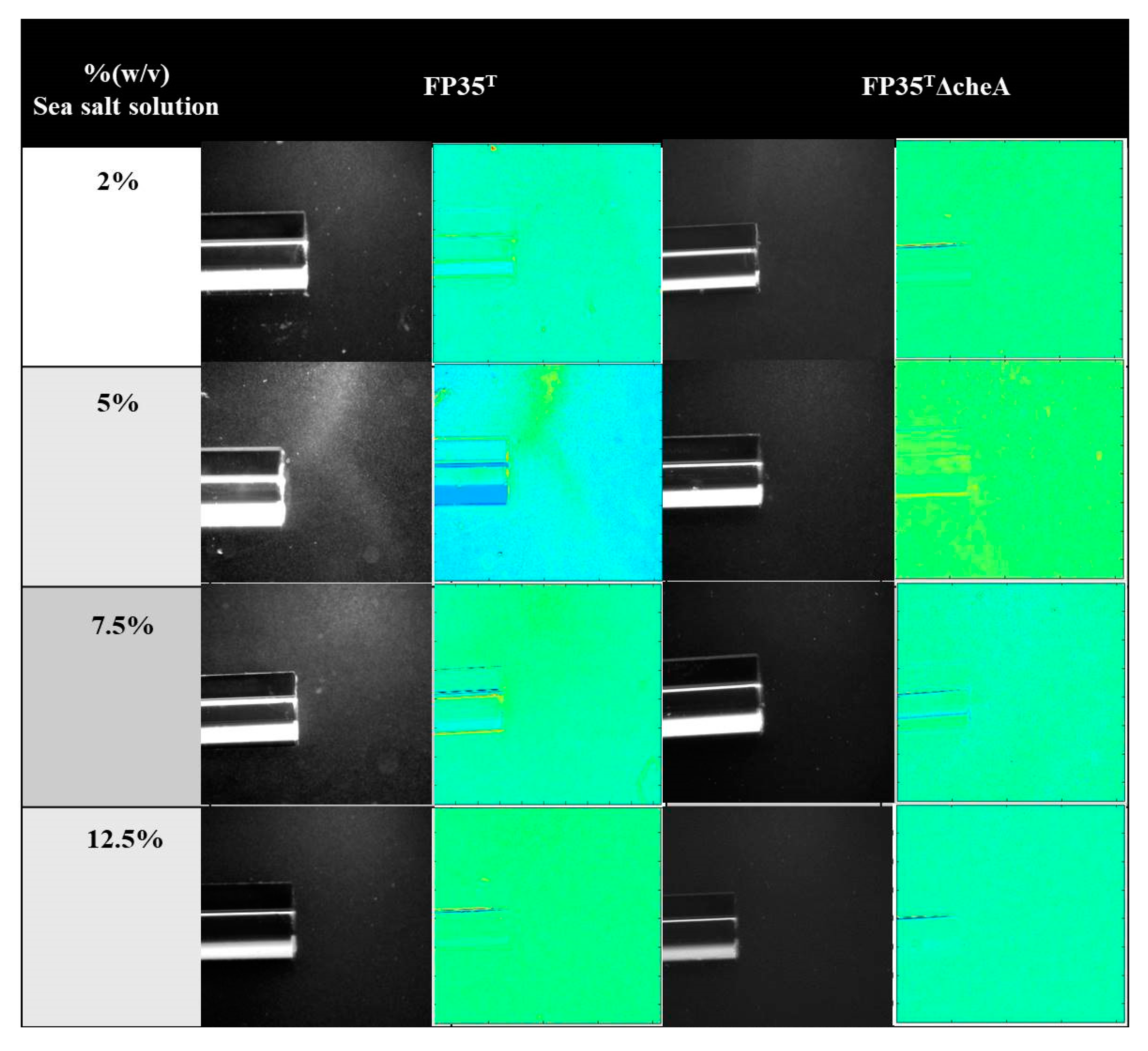
Figure 1. Qualitative capillary chemotaxis assays comparing responses of wild-type H. anticariensis FP35T and the mutant strain FP35 ΔcheA to root exudates grown at different sea salt solution concentrations. All photographs were taken after 10 min. First and third columns: dark-field images of cells gathered at the mouth of capillary tubes containing attractants; second and fourth columns: heat map of normalized images (Matlab R2013a).
Microorganisms 08 00575 g001
Figure 2. Quantitative capillary chemotaxis assays comparing responses of wild-type H. anticariensis FP35T and mutant strains FP35 ΔcheA and FP35 ΔcheA pJN105-cheA to root exudates grown at different % (w/v) sea salt solution concentrations. The results represent the mean of three independent biological samples performed in triplicate, with error bars indicating standard errors. The graph shows the increase in CFU in capillaries normalized by the negative control (CB). The negative CFU values in the capillaries reflect negative chemotaxis. Comparison of different strains tested using the Tukey test (* p ≤ 0.05; ** p < 0.01).
Microorganisms 08 00575 g002
Figure 3. Qualitative capillary chemotaxis assays comparing responses of four halophilic bacteria to root exudates grown at a 5% (w/v) concentration of sea salt solution. All photographs were taken after 10 min. First column: dark-field images of cells gathered at the mouth of capillaries containing exudates; second column: heat map of normalized images (Matlab R2013a).
Microorganisms 08 00575 g003
Figure 4. TIC spectrum obtained from Salicornia exudates.
Microorganisms 08 00575 g004
Figure 5. UPLC-HRMS Q-TOF spectra obtained from Salicornia exudates.
Microorganisms 08 00575 g005
Figure 6. Quantitative capillary chemotaxis assays comparing responses of wild-type H. anticariensis FP35T and its mutant strains FP35 ΔcheA and FP35 ΔcheA pJN105-cheA, grown in 5% (w/v) sea salt solution, to oleanolic acid. The results represent the mean of three independent biological samples performed in triplicate, with error bars indicating standard errors. The graph shows the increase in CFU in capillaries normalized by the negative control (CB). The negative values for CFUs in the capillaries reflect negative chemotaxis.
Microorganisms 08 00575 g006
Figure 7. Biofilm formation of wild-type H. anticariensis strain FP35T and the FP35 ΔcheA mutant grown in 5% (w/v) sea salt solution induced by 10% and 25% (w/w) root exudate concentrations of Salicornia. The results represent the mean of three independent biological samples performed in quadruplicate, with error bars indicating standard deviations. Comparisons with respect to the control for each strain tested were made using the Tukey test (* p ≤ 0.05).
Microorganisms 08 00575 g007
Table 1. Effect of wild-type H. anticariensis strain FP35T and its mutant H. anticariensis FP35 ΔcheA on shoot length, root length, germination and vigour index of Salicornia.
Bacterial
Inoculation
Shoot
(cm)
% IncreaseRoot
(cm)
% IncreaseGermination
(%)
Vigour Index% Increase
Control0.159 ± 0.06a 0.236 ± 0.11a 2912.52
H. anticariensis FP35T0.179 ± 0.08ab12,5%0.679 ± 0.19b287%3731.7153%
H. anticariensis FP35 ΔcheA0.152 ± 0.05a0%0.548 ± 0.13b232%3222.479%
Data expressed as mean ± standard deviation. Data in columns with different letters differ significantly according to the Duncan test (p < 0.05).