Article

Genes on the Move: In Vitro Transduction of Antimicrobial Resistance Genes between Human and Canine Staphylococcal Pathogens

1
Department of Clinical Science and Services, Royal Veterinary College, Hawkshead Lane, North Mymms, Hatfield, Hertfordshire AL9 7TA, UK
2
MRC Centre for Molecular Bacteriology and Infection, Imperial College London, London SW7 2AZ, UK
3
Centre for Infection Medicine, Department of Veterinary Medicine, Institute of Microbiology and Epizootics, Freie Universität Berlin, 14195 Berlin, Germany
4
Institute of Infection and Immunity, St George’s, University of London, Cranmer Terrace, London SW17 0RE, UK
*
Author to whom correspondence should be addressed.
Microorganisms 2020, 8(12), 2031; https://doi.org/10.3390/microorganisms8122031
Submission received: 18 November 2020 / Revised: 11 December 2020 / Accepted: 17 December 2020 / Published: 18 December 2020
(This article belongs to the Section Medical Microbiology)

Abstract

:
Transmission of methicillin-resistant Staphylococcus aureus (MRSA) and methicillin-resistant Staphylococcus pseudintermedius (MRSP) between people and pets, and their co-carriage, are well-described. Potential exchange of antimicrobial resistance (AMR) genes amongst these staphylococci was investigated in vitro through endogenous bacteriophage-mediated transduction. Bacteriophages were UV-induced from seven donor isolates of canine (MRSP) and human (MRSA) origin, containing tet(M), tet(K), fusB or fusC, and lysates filtered. Twenty-seven tetracycline- and fusidic acid- (FA-) susceptible recipients were used in 122 donor-recipient combinations (22 tetracycline, 100 FA) across 415 assays (115 tetracycline, 300 FA). Bacteriophage lysates were incubated with recipients and presumed transductants quantified on antimicrobial-supplemented agar plates. Tetracycline resistance transduction from MRSP and MRSA to methicillin-susceptible S. pseudintermedius (MSSP) was confirmed by PCR in 15/115 assays. No FA-resistance transfer occurred, confirmed by negative fusB/fusC PCR, but colonies resulting from FA assays had high MICs (≥32 mg/L) and showed mutations in fusA, two at a novel position (F88L), nine at H457[Y/N/L]. Horizontal gene transfer of tetracycline-resistance confirms that resistance genes can be shared between coagulase-positive staphylococci from different hosts. Cross-species AMR transmission highlights the importance of good antimicrobial stewardship across humans and veterinary species to support One Health.

1. Introduction

Transmission of multidrug-resistant (MDR) bacterial pathogens between humans and pets contributes to the spread of antimicrobial resistance (AMR) and is facilitated by frequent close contact and advanced veterinary care [1]. While the transfer of MDR bacteria between hosts can be mitigated through screening and hygiene measures, transfer of resistance determinants between co-colonising bacteria will follow microbial rules of gene exchange.
Methicillin-resistant Staphylococcus aureus (MRSA) presents a significant burden to human healthcare through poorer clinical outcomes and higher treatment costs compared with methicillin-susceptible S. aureus (MSSA) [2,3]. MRSA is occasionally isolated from infections in pets, typically after reverse zoonotic transmission [4]. More recently, though, its “canine counterpart”, methicillin-resistant S. pseudintermedius (MRSP), has emerged as a highly drug-resistant, zoonotic pathogen in veterinary clinics [5,6]. Although MRSP is primarily adapted to dogs, it shares many microbiological, clinical and epidemiological characteristics with MRSA. Both are coagulase-positive opportunistic pathogens with the ability to colonise mucosae and skin asymptomatically. Simultaneous co-carriage of and infection with S. aureus and S. pseudintermedius have been documented in humans and dogs [7,8,9].
The acquisition or loss of mobile genetic elements (MGEs) carrying AMR genes, including plasmids, transposons and staphylococcal cassette chromosome mec (SCCmec) elements, can lead to phenotypic changes in AMR profiles of staphylococci [10]. Horizontal gene transfer (HGT) of MGEs can occur between individual bacteria by transformation, conjugation, or transduction [11]. In S. aureus, this is thought to be primarily by bacteriophage-mediated generalised transduction [12]. Comparatively little information exists for S. pseudintermedius [13], but transduction seems the most likely mechanism for HGT amongst co-colonising isolates. Integrated bacteriophages (prophages) have been identified in S. pseudintermedius chromosomes while the tra gene complex, required for conjugation, was not found in 15 sequenced isolates [14,15]. Transformation, which does not require cell-to-cell contact, appears to occur rarely in S. aureus under natural conditions [12].
Bacteriophage-mediated generalised transduction relies on the presence of bacteriophage receptors in recipient bacteria and is further dependent on the ability of MGEs to replicate or integrate into the new host’s genome. HGT is controlled by Restriction-Modification (RM) systems and, more rarely in staphylococci, by clusters of randomly interspersed short palindromic repeats (CRISPR) systems, which protect bacteria from acquiring foreign DNA [12]. The distribution of RM variants is lineage-associated in both S. aureus [16] and S. pseudintermedius [15], resulting in different MGEs circulating within distinct S. aureus or S. pseudintermedius lineages.
Evidence for endogenous inter-species HGT of resistance determinants in staphylococci is currently limited to transfer from coagulase-negative species (CoNS) or enterococci to S. aureus [17,18]. Phenotypic resistance to gentamicin, tetracycline and erythromycin has previously been transferred in vitro and on mouse skin from S. hominis and S. epidermidis into S. aureus (both human- and canine-derived) [17]. Moreover, the large MGE SCCmec, responsible for broad β-lactam resistance in MRSA and MRSP, is thought to have been transferred from CoNS [19,20]. The rapid accumulation of multiple resistance genes in MRSP suggests a less restrained acquisition of genetic material. In vivo, unexpectedly high transfer rates of MGEs, containing genes related to host-adaptation, have been observed in co-colonising S. aureus [21]. This is thought to be resulting from stress-linked generalised transduction [21].
Almost all clinically relevant antimicrobial classes in human medicine are also authorised and used globally in small animal veterinary practice [22,23]. One of the antimicrobial agents reserved for the treatment of serious infections caused by MRSA in humans is fusidic acid (FA) which is also widely used topically in dogs for the treatment of ear, eye and skin infections [24]. “Low-level” (Minimum Inhibitory Concentration [MIC] 4–16 mg/L) FA resistance in both S. aureus and S. pseudintermedius has been associated with fusB or fusC [25,26]. These genes have been primarily described on transposon- or SCCmec-like elements, found within plasmids, staphylococcal pathogenicity islands (SaPIs), or being chromosomally integrated [25,26]. Whether these MGEs can transfer between S. aureus and S. pseudintermedius remains to be answered. High-level resistance to FA (MIC ≥ 64 mg/L) has been linked to chromosomal mutations (in fusA and/or fusE in small colony variants) [25]. Another antimicrobial agent of importance in human and veterinary medicine is tetracycline, a broad-spectrum agent classified by the WHO as “highly important” for humans [22] and widely used for the treatment of respiratory tract infections in animals [27]. However, the wide distribution of tetracycline resistance genes, and their location on transposons (e.g., Tn916) and plasmids [28], suggests a propensity for HGT, evidence for which has yet to be shown.
In this study, we demonstrate HGT of resistance genes between isolates of S. aureus and S. pseudintermedius using assays to detect transduction mediated by induction of natural bacteriophages.

2. Materials and Methods

2.1. Bacterial Isolates

A total of seven donor and 27 recipient bacterial isolates were used from a frozen archive (−20 °C in brain heart infusion broth (BHIB; Oxoid, Basingstoke, UK) and 20% glycerol (Fisher Scientific, Loughborough, UK) (Table 1). Selection criteria were their tetracycline and FA resistance phenotypes (disk diffusion for tetracycline, MICs for FA), genotypes, and their isolation sites, to span human, canine, infection and carriage origins and a range of sequence types (STs). All S. pseudintermedius isolates were collected from clinical submissions, representing the circulating lineages at the time (2007 [n = 1], 2010–2016 [n = 20]). MRSA isolates (CC8 and CC22) represented two clonal complexes found worldwide [29]. Species and respective resistances were confirmed by PCR following previously described methods [30,31] for species-specific thermonuclease (nuc), methicillin-resistance (mecA), and presence or absence of tet(M), tet(K), fusB, and fusC.
Donor isolates for tetracycline assays comprised one well-characterised MRSA of human infection origin (COL), carrying tet(K) on plasmid pT181, and one fully sequenced, prophage-positive canine infection MRSP (1726) with tet(M) on Tn916 [15]. Donor isolates for FA experiments included two fusB-positive and three fusC-positive MRSP, with resistance genes most likely on transposon-like elements in plasmids (fusB) or integrated into the chromosomal DNA in a SCCmec-like cassette (fusC). Selection of FA-resistant donors was limited by the infrequent description of these genes in this species [30]; FA-resistant S. aureus donors were not available for inclusion at the time. Recipient bacteria representing different origins and STs were chosen; all were screened on brain heart infusion agar (BHIA; Oxoid) containing either 30 mg/L tetracycline or 16 mg/L FA to confirm phenotypic susceptibility. Two RM-deficient S. aureus laboratory strains were included as hyper-receptive recipient isolates [18].
To investigate the acquisition of tetracycline resistance, 22 different combinations of two donors and 14 recipients, including the combination of MRSA COL and RM-deficient S. aureus RN4220 were used; for FA assays, 100 combinations of five donors and 20 recipients were performed (Table 1). Initially, all transduction assays were performed in triplicate, but for successful combinations (confirmed by PCR for resistance gene in putative transductants), a further seven experiments (total ten replicates) were performed.

2.2. Induction of Bacteriophage

Overnight colonies from pure culture were grown in BHIB at 37 °C with shaking for 3 h; 1 mL aliquots were centrifuged (3000 × g, 3 min), and supernatant discarded. Cell pellets were resuspended in 7 mL bacteriophage buffer (0.1% 1M MgSO4, 0.4% CaCl2, 5% 1M Tris-HCl pH 7.8, 0.59% NaCl, 0.1% gelatin; Sigma-Aldrich Ltd., Gillingham, UK) and transferred to Petri dishes. The open Petri dish was exposed to UV light (302 nm, UVP Dual-Intensity Transilluminator TM-20) for 20 s to induce prophages [32,33]. Dish contents were added to 7 mL BHIB, incubated for 10 min at room temperature, then for 2 h at 32 °C with gentle agitation, and finally overnight at room temperature to allow cell lysis. Lysates were filtered (0.22 µm filter) and kept at 4 °C before being used for replicate experiments.

2.3. Bacteriophage Count

Recipient RN4220 colonies were incubated in 20 mL BHIB at 37 °C with shaking for 3 h. Bacteriophage lysate was diluted in bacteriophage buffer (10−1 and 10−2); 100 μL of each was added to 400 μL recipient cell broth and 30 μL 1M CaCl2 and incubated at room temperature for 15 min. Dilutions were mixed with 7 mL bacteriophage top agar (bacteriophage buffer containing 2 mg/L agar), poured over bacteriophage bottom agar plates (10 mg/L agar) and incubated at 32 °C for 24 h. Number of lysis plaques within the bacterial lawn were counted, with one plaque representing one phage particle.

2.4. Bacteriophage Transduction

Recipient bacteria were incubated in 20 mL LK broth (LKB; Luria broth with KCl instead of NaCl; 1% tryptone, 0.5% yeast extract, 0.7% KCl; Sigma-Aldrich Ltd., Gillingham, UK) at 37 °C overnight with shaking. Broth was centrifuged (4000× g, 10 min), supernatant discarded, and cell pellets resuspended in 1 mL LKB. In total, 100 µL of the recipient cell suspension, 100 µL bacteriophage lysate, and 200 µL LKB along with 2 µL CaCl2 (Sigma-Aldrich Ltd., Gillingham, UK; to a final concentration of 8 mM) were incubated at 37 °C for 45 min with shaking. Subsequently, 200 µL ice-cold 0.02 M sodium citrate was added (Honeywell International Inc., Bucharest, Romania) to chelate calcium and prevent further phage binding and cell lysis. Cell suspensions were centrifuged (3000× g, 3 min), supernatant discarded, the pellet resuspended in 200 µL ice-cold sodium citrate, and left for 2 h on ice [33].
The 200 µL solutions were spread using hockey-stick spreaders onto the surface of an LK bottom agar plate (10 g/L agar) containing sub-inhibitory antimicrobial concentrations to induce resistance gene expression (0.3 mg/L tetracycline or 0.03 mg/L FA) and incubated at 37 °C for 45 min. Four-to-five mL of LK top agar (2 g/L agar) containing inhibitory antimicrobial concentrations (30 mg/L tetracycline in total or 16 mg/L FA in total) were overlaid, plates incubated upright for 48 h at 37 °C, and colonies counted.
A negative control with 100 µL LKB in place of bacteriophage lysate was included for every combination and growth compared to transduction plates. Colony numbers at least twice those seen on the corresponding negative control were deemed significant growth, indicative of resistance transfer.

2.5. Confirmation of Suspected Transductants

From each assay with significant growth, 2–9 putative transductant colonies were subcultured onto BHIA containing either 30 mg/L tetracycline or 16 mg/L FA to confirm phenotypic susceptibility; expected species and the presence/absence of respective resistance genes were again investigated [30,31]. For isolates grown on FA-supplemented agar but negative for fusB and fusC, MICs were determined for at least two colonies, as well as for their respective donor and recipient [30]. In 1 to 3 representative fusB/fusC negative post-transduction colonies from each recipient with MICs ≥ 32 mg/L, fusA was amplified and sequenced alongside that of their original recipient following a previously described method [30].

2.6. Statistical Analyses

In IBM SPSS Statistics version 26 (significance p < 0.05), transduction rates (transductants/mL) and frequencies were compared by Kruskal-Wallis tests with the Dunn-Bonferroni post hoc method.

3. Results

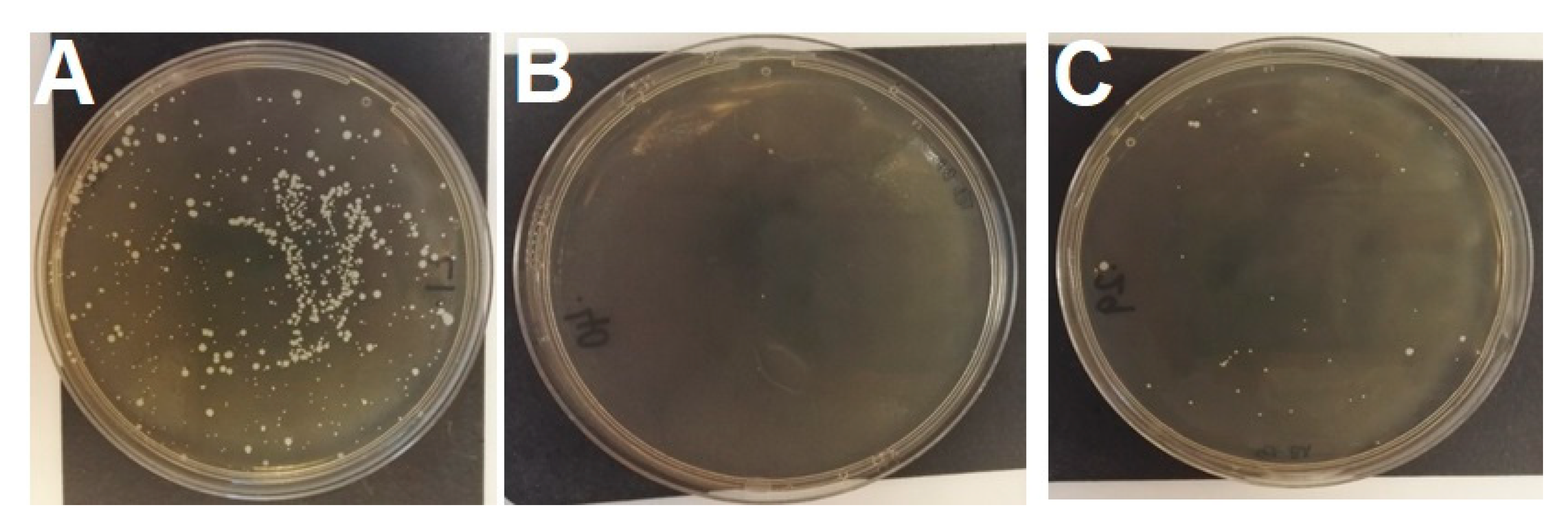
3.1. Bacteriophage Count

Bacteriophage count could not be established as the RN4220 bacterial lawn did not show any lytic plaques for phage lysate from any donor; transducing phage counts have been shown previously not to correlate with lytic phage counts [33].

3.2. Transduction of Tetracycline Resistance

To study HGT of tetracycline resistance, bacteriophage lysates from one tet(M)-positive and one tet(K)-positive donor were cultured with 14 tetracycline-susceptible recipients. Phenotypically tetracycline-resistant colonies grew from seven of the 22 different donor/recipient combinations (initially done in triplicate) (Table 1); expected nuc and acquisition of tet(M) or tet(K) were confirmed in all. Transfer occurred from MRSA COL into control MSSA RN4220 and three methicillin-susceptible S. pseudintermedius (MSSP) recipients, and from MRSP 1726 into three MSSP recipients (Figure 1). In contrast, no transduction of phenotypic tetracycline resistance was seen from MRSP into S. aureus (including both RM-deficient recipients). Including the subsequent additional seven replicates from successful pairings (115 assays in total), transduction occurred in 15/115 assays, confirmed by PCR in all 38 tested colonies. Reproducibility was low in most replicate experiments with a maximum of 4/10 positive repeats found from MRSA COL into RN4220 and from MRSP into an MSSP. Growth of <10 colonies per plate (Figure 1) was seen on 9/23 negative controls, representing seven recipients (6 MSSP, 1 MSSA). There was no difference (P = 0.994) between colony counts/mL for transduction between MRSP-MSSP, MRSA-MSSP or MRSA-RM-deficient MSSA (Table 2).

3.3. Transduction of FA Resistance

For FA resistance, bacteriophage lysate from two fusB- and three fusC-positive MRSP donors was combined with 20 FA-susceptible recipients (Table 1). Of the 300 transduction plates in total, 18 showed significant growth. Growth of up to 50 colonies was seen on 24/35 negative control plates from all but three (P1361, V1273, B021) recipients. Neither fusB nor fusC were detected in the 59 colonies tested post-transduction. Significant growth was seen more frequently on transduction assays for MRSA recipients (13/120 plates) than for MSSP recipients (2/120 plates; P = 0.032); the frequency of growth was similar for the 60 MSSA assays.
All tested colonies from FA transduction assays (two from each plate) had MICs higher than their donor (donors 4 mg/L–16 mg/L; putative transductants 32 mg/L–>64 mg/L) and their recipient isolates (0.03 mg/L–0.06 mg/L). Sequencing of fusA in 11/11 post-transduction assay colonies identified mutations in one of two amino acid positions (Table 3). The most common mutation (9/11 colonies) was in amino acid 457 (H457Y, H457N, H457L); two colonies had the mutation F88L, located in domain I of fusA.

4. Discussion

For the first time, our results provide phenotypic and molecular evidence for AMR transfer between different coagulase-positive staphylococcal species from human and canine origin, mediated by endogenous bacteriophages.
This cross-species spread of AMR, from the human pathogen S. aureus into the canine pathogen S. pseudintermedius, is of particular relevance to the often-close contact settings between pet owners and their pets, with S. aureus acting as a potential reservoir of resistance genes for S. pseudintermedius. It draws new attention to a potential risk to pets from contact with humans. This adds to a wealth of information focusing on the irrefutable priority direction of pet-to-human transfer [34]. Dogs and humans may be at least transient carriers (and co-carriers) of staphylococcal species adapted to the respective “other” primary host [4,5]. Our results add an extra layer of complexity to the potential clinical implications of close companionship with our pets, should HGT occur from S. aureus to S. pseudintermedius in vivo. Why no transduction of tetracycline resistance genes occurred from MRSP into S. aureus (including RN4220) remains unclear, but may include more efficient RM-systems, CRISPRs (although rarely described in staphylococci), a lack of bacteriophage receptors in S. aureus, plasmid incompatibility, or non-compatible RM systems (which may or may not be lineage specific) [12,14,16]. Similar unilateral transfer preferences were previously noted in an earlier study using exogenous bacteriophages in other staphylococcal species [35], although HGT was observed bidirectionally between S. aureus and S. pseudintermedius.
The low reproducibility of transduction of tetracycline resistance genes in successful pairings was surprising. It may have been due to low concentrations of endogenous transducing bacteriophages in lysates, or low copy number of tet(M)/tet(K) within induced bacteriophages. While this may suggest that cross-species gene exchange represents only a minor contribution to the overall spread of AMR, our findings prove a new concept in the evolution of MDR pathogens, in an area directly impacting on human health. Furthermore, transduction rates are difficult to compare as the number of successful replicates are rarely stated (instead described as variation (mean ± SD)). Our transduction rates in successful replicates (number of transductant cells/mL) were similar to those described previously using UV-light induction of bacteriophages (approximately 10–350 cfu/mL previously c.f. 25–1535 in this study; Table 2) [33]. Two MSSP recipients (221833 and 287735) had particularly high transductant cell counts, suggesting they may have weaker transfer barriers or greater phage receptor expression, allowing a higher transduction rate. This is also indicated by the acceptance of DNA by recipients 221833 and 289595 in more replicate experiments, from both MRSA and MRSP donors. It is possible that transfer of resistance genes via transformation of DNA present in lysates could occur, however S. aureus competence genes are poorly expressed by mutated sigma factors and post-transcriptional control, resulting in extremely rare transfer frequency [12,36]. The reasons why the phage lysate did not form plaques in the RN4220 bacterial lawn are unclear. Potentially this could be due to missing or modified phage receptors in this strain, or the induction of a novel transducing phage. It is possible that despite being RM-deficient to our current knowledge, RN4220 may contain other undiscovered types of phage immunity. This non-plaque-forming phenomenon with RN4220 is not uncommon to see when plating transducing phages induced from clinical S. aureus isolates (unpublished data), and it has been previously demonstrated that the presence of lytic phages does not correlate with transducing phage [33]. However, it cannot be discounted that the apparent absence of FA resistance gene transduction could be the result of a lack of transducing phage.
The risk of interspecies HGT may be greater in vivo than in the laboratory, as has been demonstrated previously for other MGEs [17,21]. Plasmid-borne gentamicin resistance transfer from the coagulase-negative S. epidermidis into S. aureus was 10-100-fold greater on mouse skin than in broth filter experiments [17] and similarly, HGT of host-adaptation determinants on pig skin was substantially higher compared to the same isolates co-incubated in vitro [21]. However, the reasons underpinning why transfer is observed at a higher rate in vivo than in vitro are still largely unknown. It is thought that this may relate to environmental conditions that are not replicated in vitro, which may trigger staphylococcal isolates to selectively amplify HGT.
The lack of transfer of fusB and fusC in this study is encouraging with regard to the preservation of FA clinical efficacy in human and veterinary medicine. However, the finding needs to be interpreted with caution. Firstly, the development of high-level resistance likely due to fusA mutations following exposure to relatively low concentrations of FA is of concern, although fusA mutations are rarely documented in clinical isolates [25,30]. This low prevalence of FA resistance, despite FA being widely used in veterinary and human medicine for over 50 years, suggests that its use is not causing a ‘crisis’ of resistance. Indeed, in veterinary medicine FA is used as topical therapy where it exceeds typical MICs for staphylococci by a significant order of magnitude [36]. Thus, it seems prudent to suggest that proactive surveillance of resistance in both human- and veterinary-derived staphylococci would suffice to monitor this situation. However, it does not indicate a current need to restrict the use of this antimicrobial to humans only at this time. In this study, transduction may have also been hampered by a lack of prophage in our donors, as reported for a small number of S. pseudintermedius lineages [14]. The mutations observed in fusA of S. pseudintermedius occurred in the same position as described in S. aureus (amino acid 457), confirming the importance of this mutation in resistance development [25]. The role of the novel mutation (F88L) in conferring tolerance to FA should be further investigated.
In conclusion, the description of MGE transfer between S. aureus and S. pseudintermedius illustrates ongoing genetic evolution amongst major zoonotic staphylococcal pathogens. Selective pressures in one host may thus contribute to the evolution of more drug-resistant isolates adapted to another host. Whilst the wider context of direction of transfer and the prioritisation of human over animal health remain important considerations, there is clearly a need for response to the dissemination of AMR within shared bacterial populations. Despite previous significant attention on the use of antimicrobial agents in livestock, companion animal medicine is in some ways left lagging behind. Efforts to develop and disseminate responsible antimicrobial use guidelines for companion animal medicine need to continue, also to align interests in the sense of One Health. At present, though, the well-documented benefit from pet ownership on human health likely markedly outweighs the risk from zoonotic transmission and HGT in methicillin-resistant staphylococci [37].

Author Contributions

Conceptualization and methodology, S.M.F., R.B., A.J.M., J.A.L., A.L.; investigation, S.M.F., J.A.L., A.L., C.F., S.S.; writing—original draft preparation, S.M.F., R.B., A.J.M., J.A.L., A.L.; writing—review and editing, all authors; funding acquisition, R.B., A.L. All authors have read and agreed to the published version of the manuscript.

Funding

S.M.F. and this study were funded by a Biotechnology and Biological Sciences Research Council (Swindon, U.K.; BBSRC) industrial CASE scholarship in partnership with Dechra Veterinary Products Limited (Shropshire, U.K.; grant number BB/K011952/1). The work of C.F. and S.S. was funded by the German Federal Ministry of Education and Research (BMBF) within the JPIAMR project PET-Risk (grant no. FKZ 01KI1707 (PET-Risk)).

Acknowledgments

The authors thank Vanessa Schmidt, Dorina Timofte, Thomas Grönthal, and Merja Rantala for the donation of some isolates (canine-derived S. pseudintermedius). Preliminary results from this study were presented as a poster abstract at the International Conference on One Health Antimicrobial Resistance, Utrecht, Netherlands, April 2019. The authors gratefully acknowledge the generous donation that allowed for the creation of the Stella Bacterial Archive Collection, where the isolates used in this study are archived.

Conflicts of Interest

The authors’ group (Royal Veterinary College) has received funding from Dechra Veterinary Products Limited (Shropshire, U.K.) in support of laboratory research and clinical teaching of undergraduate and postgraduate students. The funders had no role in the design of the study; collection, analysis, and interpretation of data; or manuscript writing.

References

  1. Schwarz, S.; Loeffler, A.; Kadlec, K. Bacterial resistance to antimicrobial agents and its impact on veterinary and human medicine. Adv. Vet. Dermatol. 2017. [Google Scholar] [CrossRef] [Green Version]
  2. Zhen, X.; Lundborg, C.S.; Sun, X.; Hu, X.; Dong, X. Economic burden of antibiotic resistance in ESKAPE organisms: A systematic review. Antimicrob. Resist. Infect. Control 2019, 8, 137. [Google Scholar] [CrossRef] [PubMed] [Green Version]
  3. Van Hal, S.J.; Jensen, S.O.; Vaska, V.L.; Espedido, B.A.; Paterson, D.L.; Gosbell, I.B. Predictors of mortality in Staphylococcus aureus bacteremia. Clin. Microbiol. Rev. 2012, 25, 362–386. [Google Scholar] [CrossRef] [PubMed] [Green Version]
  4. Loeffler, A.; Lloyd, D.H. Companion animals: A reservoir for methicillin-resistant Staphylococcus aureus in the community. Epidemiol. Infect. 2010, 138, 595–605. [Google Scholar] [CrossRef]
  5. Loeffler, A.; Linek, M.; Moodley, A.; Guardabassi, L.; Sung, J.M.L.; Winkler, M.; Weiss, R.; Lloyd, D.H. First report of multiresistant, mecA-positive Staphylococcus intermedius in Europe: 12 cases from a veterinary dermatology referral clinic in Germany. Vet. Dermatol. 2007, 18, 412–421. [Google Scholar] [CrossRef]
  6. van Hoovels, L.; Vankeerberghen, A.; Boel, A.; van Vaerenbergh, K.; de Beenhouwer, H. First case of Staphylococcus pseudintermedius infection in a human. J. Clin. Microbiol. 2006, 44, 4609–4612. [Google Scholar] [CrossRef] [Green Version]
  7. Loeffler, A.; Boag, A.K.; Sung, J.; Lindsay, J.A.; Guardabassi, L.; Dalsgaard, A.; Smith, H.; Stevens, K.B.; Lloyd, D.H. Prevalence of methicillin-resistant Staphylococcus aureus among staff and pets in a small animal referral hospital in the UK. J. Antimicrob. Chemother. 2005, 56, 692–697. [Google Scholar] [CrossRef]
  8. Börjesson, S.; Gómez-Sanz, E.; Ekström, K.; Torres, C.; Grönlund, U. Staphylococcus pseudintermedius can be misdiagnosed as Staphylococcus aureus in humans with dog bite wounds. Eur. J. Clin. Microbiol. Infect. Dis. 2015, 34, 839–844. [Google Scholar] [CrossRef]
  9. Walther, B.; Hermes, J.; Cuny, C.; Wieler, L.H.; Vincze, S.; Elnaga, Y.A.; Stamm, I.; Kopp, P.A.; Kohn, B.; Witte, W.; et al. Sharing more than friendship—Nasal colonization with coagulase-positive staphylococci (CPS) and co-habitation aspects of dogs and their owners. PLoS ONE 2012, 7, e35197. [Google Scholar] [CrossRef]
  10. Partridge, S.R.; Kwong, S.M.; Firth, N.; Jensen, S.O. Mobile genetic elements associated with antimicrobial resistance. Clin. Microbiol. Rev. 2018, 31, e00088-17. [Google Scholar] [CrossRef] [Green Version]
  11. von Wintersdorff, C.J.H.; Penders, J.; van Niekerk, J.M.; Mills, N.D.; Majumder, S.; van Alphen, L.B.; Savelkoul, P.H.M.; Wolffs, P.F.G. Dissemination of antimicrobial resistance in microbial ecosystems through horizontal gene transfer. Front. Microbiol. 2016, 7, 173. [Google Scholar] [CrossRef] [PubMed] [Green Version]
  12. Lindsay, J.A. Staphylococcus aureus genomics and the impact of horizontal gene transfer. Int. J. Med. Microbiol. 2014, 304, 103–109. [Google Scholar] [CrossRef] [PubMed]
  13. Bannoehr, J.; Guardabassi, L. Staphylococcus pseudintermedius in the dog: Taxonomy, diagnostics, ecology, epidemiology and pathogenicity. Vet. Dermatol. 2012, 23, 253–266. [Google Scholar] [CrossRef] [PubMed]
  14. Brooks, M.R.; Padilla-Vélez, L.; Khan, T.A.; Qureshi, A.A.; Pieper, J.B.; Maddox, C.W.; Alam, M.D. Prophage-mediated disruption of genetic competence in Staphylococcus pseudintermedius. mSystems 2020, 5, e00684-19. [Google Scholar] [CrossRef] [PubMed] [Green Version]
  15. McCarthy, A.J.; Harrison, E.M.; Stanczak-Mrozek, K.; Leggett, B.; Waller, A.; Holmes, M.A.; Lloyd, D.H.; Lindsay, J.A.; Loeffler, A. Genomic insights into the rapid emergence and evolution of MDR in Staphylococcus pseudintermedius. J. Antimicrob. Chemother. 2015, 70, 997–1007. [Google Scholar] [CrossRef] [Green Version]
  16. McCarthy, A.J.; Witney, A.A.; Lindsay, J.A. Staphylococcus aureus temperate bacteriophage: Carriage and horizontal gene transfer is lineage associated. Front. Cell. Infect. Microbiol. 2012, 2, 1–10. [Google Scholar] [CrossRef] [Green Version]
  17. Naidoo, J.; Lloyd, D.H. Transmission of genes between staphylococci on skin. In Antimicrobials and Agriculture; Woodbine, M., Ed.; Butterworths: London, UK, 1984; pp. 282–295. [Google Scholar]
  18. Sung, J.M.L.; Lindsay, J.A. Staphylococcus aureus strains that are hypersusceptible to resistance gene transfer from enterococci. Antimicrob. Agents Chemother. 2007, 51, 2189–2191. [Google Scholar] [CrossRef] [Green Version]
  19. Couto, I.; de Lencastre, H.; Severina, E.; Kloos, W.; Webster, J.A.; Hubner, R.J.; Sanches, I.S.; Tomasz, A. Ubiquitous presence of a mecA homologue in natural isolates of Staphylococcus Sciuri. Microb. Drug Resist. 1996, 2, 377–391. [Google Scholar] [CrossRef]
  20. Rolo, J.; Worning, P.; Nielsen, J.B.; Sobral, R.; Bowden, R.; Bouchami, O.; Damborg, P.; Guardabassi, L.; Perreten, V.; Tomasz, A.; et al. Evidence for the evolutionary steps leading to mecA-mediated β-lactam resistance in staphylococci. PLoS Genet. 2017, 13, e1006674. [Google Scholar] [CrossRef]
  21. McCarthy, A.J.; Loeffler, A.; Witney, A.A.; Gould, K.A.; Lloyd, D.H.; Lindsay, J.A. Extensive horizontal gene transfer during Staphylococcus aureus co-colonization in vivo. Genome Biol. Evol. 2014, 6, 2697–2708. [Google Scholar] [CrossRef]
  22. Critically Important Antimicrobials for Human Medicine, 6th ed.; Licence: CC BY-NC-SA 3.0 IGO; World Health Organization: Geneva, Switzerland, 2019.
  23. Hur, B.A.; Hardefeldt, L.Y.; Verspoor, K.M.; Baldwin, T.; Gilkerson, J.R. Describing the antimicrobial usage patterns of companion animal veterinary practices; free text analysis of more than 4.4 million consultation records. PLoS ONE 2020, 15, e0230049. [Google Scholar] [CrossRef] [Green Version]
  24. Mateus, A.; Brodbelt, D.C.; Barber, N.; Stärk, K.D. Antimicrobial usage in dogs and cats in first opinion veterinary practices in the UK. J. Small Anim. Pract. 2011, 52, 515–521. [Google Scholar] [CrossRef] [PubMed]
  25. Farrell, D.J.; Castanheira, M.; Chopra, I. Characterization of global patterns and the genetics of fusidic acid resistance. Clin. Infect. Dis. 2011, 52, S487–S492. [Google Scholar] [CrossRef] [PubMed]
  26. O’Neill, A.J.; McLaws, F.; Kahlmeter, G.; Henriksen, A.S.; Chopra, I. Genetic basis of resistance to fusidic acid in staphylococci. Antimicrob. Agents Chemother. 2007, 51, 1737–1740. [Google Scholar] [CrossRef] [Green Version]
  27. De Briyne, N.; Atkinson, J.; Pokludová, L.; Borriello, S.P. Antibiotics used most commonly to treat animals in Europe. Vet. Rec. 2014, 175, 325. [Google Scholar] [CrossRef] [PubMed] [Green Version]
  28. McCarthy, A.J.; Lindsay, J.A. The distribution of plasmids that carry virulence and resistance genes in Staphylococcus aureus is lineage associated. BMC Microbiol. 2012, 12, 104. [Google Scholar]
  29. Toleman, M.S.; Reuter, S.; Jamrozy, D.; Wilson, H.J.; Blane, B.; Harrison, E.M.; Coll, F.; Hope, R.J.; Kearns, A.; Parkhill, J.; et al. Prospective genomic surveillance of methicillin-resistant Staphylococcus aureus (MRSA) associated with bloodstream infection, England, 1 October 2012 to 30 September 2013. Eurosurveillance 2019, 24, 1800215. [Google Scholar] [CrossRef] [Green Version]
  30. Frosini, S.M.; Bond, R.; Rantala, M.; Grönthal, T.; Rankin, S.C.; O’Shea, K.; Timofte, D.; Schmidt, V.; Lindsay, J.; Loeffler, A. Genetic resistance determinants to fusidic acid and chlorhexidine in variably susceptible staphylococci from dogs. BMC Microbiol. 2019, 19, 81. [Google Scholar] [CrossRef]
  31. Strommenger, B.; Kettlitz, C.; Werner, G.; Witte, W. Multiplex PCR assay for simultaneous detection of nine clinically relevant antibiotic resistance genes in Staphylococcus aureus. J. Clin. Microbiol. 2003, 41, 4089–4094. [Google Scholar] [CrossRef] [Green Version]
  32. Behzadi, E.; Behzadi, P. An in vitro study on the apoptosis inducing effects of ultraviolet B light in Staphylococcus aureus. Maedica 2012, 7, 54–57. [Google Scholar]
  33. Stanczak-Mrozek, K.I.; Laing, K.G.; Lindsay, J.A. Resistance gene transfer: Induction of transducing phage by sub-inhibitory concentrations of antimicrobials is not correlated to induction of lytic phage. J. Antimicrob. Chemother. 2017, 72, 1624–1631. [Google Scholar] [CrossRef] [PubMed] [Green Version]
  34. Damborg, P.; Broens, E.M.; Chomel, B.B.; Guenther, S.; Pasmans, F.; Wagenaar, J.A.; Weese, J.S.; Wieler, L.H.; Windahl, U.; Vanrompay, D.; et al. Bacterial zoonoses transmitted by household pets: State-of-the-art and future perspectives for targeted research and policy actions. J. Comp. Pathol. 2016, 155, S27–S40. [Google Scholar] [CrossRef] [PubMed] [Green Version]
  35. Uchiyama, J.; Takemura-Uchiyama, I.; Sakaguchi, Y.; Gamoh, K.; Kato, S.; Daibata, M.; Ujihara, T.; Misawa, N.; Matsuzaki, S. Intragenus generalized transduction in Staphylococcus spp. by a novel giant phage. ISME J. 2014, 8, 1949–1952. [Google Scholar] [CrossRef] [PubMed] [Green Version]
  36. Morikawa, K.; Takemura, A.J.; Inose, Y.; Tsai, M.; Nguyen Thi, L.T.; Ohta, T.; Msadek, T. Expression of a cryptic secondary Sigma Factor gene unveils natural competence for DNA transformation in Staphylococcus aureus. PLoS Pathog. 2012, 8, e1003003. [Google Scholar] [CrossRef] [PubMed] [Green Version]
  37. Friedmann, E.; Son, H. The human-companion animal bond: How humans benefit. Vet. Clin. N. Am. Small Anim. Pract. 2009, 39, 293–326. [Google Scholar] [CrossRef] [PubMed]
Figure 1. Recipient MSSP 287735 growth after transduction assays (A) on agar containing 30 mg/L tetracycline with phage lysate from MRSP 1726 (tet(M) donor); (B) on agar containing 30 mg/L tetracycline control with no phage lysate; (C) on agar containing 16 mg/L fusidic acid with no phage lysate. Note the breakthrough growth on plate C (colony count n = 41). Smaller colonies are those embedded in the agar.
Microorganisms 08 02031 g001
Table 1. Results from transduction assays using two tetracycline- and five fusidic acid-resistant bacterial donors (MRSA and MRSP), and 27 MR- and MS- S. aureus and S. pseudintermedius recipients. Numbers represent transduction assays with the growth of more than two-fold higher bacterial colonies than negative control plates, compared to the number of replicate attempts. For tetracycline resistance, confirmation of successful transduction was made by PCR. For fusidic acid assays, all putative transductant colonies were subsequently shown not to carry fusB or fusC; mutations in fusA were identified by sequencing.
Donor
tet(K)tet(M)fusBfusC
MRSA (Human Hospital Environment)MRSP
(Canine Infection)
MRSP
(Canine Infection)
MRSP
(Canine Infection)
COL1726P0983P1067V1061V1100P1248
CC8 (ST250)ST261ST621ST1090ST668ST668ST305
RecipientMSSP
(Canine Infection)
221833ST2631/103/10Not Done
287735ST820/31/10
289869ST540/30/3
289595ST19031/104/10
289589ST19070/30/3
289418ST19051/100/3
289385ST19060/30/3
V1273ST1085Not Done0/30/30/30/30/3
V0451ST10910/30/30/31/30/3
V0806ST540/30/30/30/30/3
P1361ST10860/30/30/30/30/3
P1351ST210/30/30/30/30/3
P1356ST10920/30/30/30/30/3
251648ST710/30/31/30/30/3
70361ST10870/30/30/30/30/3
MSSA
(Canine Infection)
B019CC15 (ST15)Not done0/31/30/31/30/31/3
B021CC15 (ST15)0/30/30/30/30/30/3
B027CC15 (ST15)0/30/30/30/30/30/3
Restriction-deficient MSSA
(Laboratory Strain)
RN4220CC8 (ST8)4/100/30/30/30/30/30/3
Restriction-deficient MRSA
(Laboratory Strain)
NE667 (hsdR mutant of JE2)CC8 (ST8)Not done0/30/30/30/30/30/3
MRSA
(Human Infection)
JE2CC8 (ST8)0/30/31/30/31/31/3
J220CC8 (ST239)Not done0/30/31/32/30/3
J225CC8 (ST239)1/30/31/30/30/3
FPR3757CC8 (ST8)2/30/30/30/30/3
MRSA
(Human Carriage)
19BCC220/30/30/30/30/30/3
TW20CC8 (ST239)Not done0/30/32/31/30/3
MRSA
(Human Hospital Environment)
COLCC8 (ST250)0/30/30/30/30/3
Total number transduction assays per antimicrobial115300
Total plates with increased growth
/total number of transduction assays
7/528/634/601/606/605/602/60
MRSA: methicillin-resistant S. aureus; MRSP: methicillin-resistant S. pseudintermedius; MSSP: methicillin-susceptible S. pseudintermedius; MSSA: methicillin-susceptible S. aureus.
Table 2. Number of transductant cells/mL following successful transduction assays for tet(M) and tet(K). Cell numbers are derived from colony counts following transduction assays incubated at 37 °C for 48 h on LK agar containing 30 mg/L tetracycline.
DonorRecipientNumber of Successful Transduction Assay ReplicatesMedian (Range) Transductant Cells/mL
Bacterial TypeIsolateTetracycline Resistance GeneBacterial TypeIsolate
MRSP1726tet(M)MSSP2218333/101105 (250–1510)
2877351/101535
2895954/1092.5 (25–160)
MRSACOLtet(K)MSSP2218331/101475
2895951/1065
2594181/1040
RM-def MSSARN42204/1062.5 (25–995)
MRSA: methicillin-resistant S. aureus; MRSP: methicillin-resistant S. pseudintermedius; MSSP: methicillin-susceptible S. pseudintermedius; RM-def MSSA: restriction-modification system deficient methicillin-susceptible S. aureus.
Table 3. Fusidic acid minimum inhibitory concentrations (MIC) and mutations in fusA (including the novel position F88L) after exposure of MRSA, MSSA, and MSSP to subinhibitory concentrations of fusidic acid. PCR confirmed species and methicillin-resistance as the same as the original recipient isolate.
StaphylococciOriginal Recipient (Recipient MIC [mg/L])Mutant MIC (mg/L)Amino Acid SubstitutionNucleotide Substitution
MSSP251648 (0.06)32H457YCAC → TAC
MSSAB019 (0.06)32H457YCAC → TAC
32F88LTTC → CTC
MRSATW20 (0.06)32F88LTTC → TTA
J220 (0.06)>64H457NCAC → AAC
32H457YCAC → TAC
J225 (0.06)32H457YCAC → TAC
64H457NCAC → AAC
64H457NCAC → AAC
FPR3757 (0.06)64H457LCAC → CTC
JE2 (0.06)32H457YCAC → TAC
MSSP: methicillin-susceptible S. pseudintermedius; MSSA: methicillin-susceptible S. aureus; MRSA: methicillin-resistant S. aureus.
Publisher’s Note: MDPI stays neutral with regard to jurisdictional claims in published maps and institutional affiliations.

