Review

Streptococcus gordonii: Pathogenesis and Host Response to Its Cell Wall Components

1
Department of Oral Microbiology and Immunology, School of Dentistry, Dental Research Institute, Seoul National University, Seoul 08826, Korea
2
Department of Agricultural Biotechnology and Research Institute of Agriculture and Life Sciences, Seoul National University, Seoul 08826, Korea
3
Institute of Green Bio Science Technology, Seoul National University, Pyeongchang 25354, Korea
*
Author to whom correspondence should be addressed.
These authors contributed equally to this article.
Microorganisms 2020, 8(12), 1852; https://doi.org/10.3390/microorganisms8121852
Submission received: 11 November 2020 / Revised: 23 November 2020 / Accepted: 23 November 2020 / Published: 24 November 2020
(This article belongs to the Special Issue Innate Immunity against Bacterial Infections)

Abstract

:
Streptococcus gordonii, a Gram-positive bacterium, is a commensal bacterium that is commonly found in the skin, oral cavity, and intestine. It is also known as an opportunistic pathogen that can cause local or systemic diseases, such as apical periodontitis and infective endocarditis. S. gordonii, an early colonizer, easily attaches to host tissues, including tooth surfaces and heart valves, forming biofilms. S. gordonii penetrates into root canals and blood streams, subsequently interacting with various host immune and non-immune cells. The cell wall components of S. gordonii, which include lipoteichoic acids, lipoproteins, serine-rich repeat adhesins, peptidoglycans, and cell wall proteins, are recognizable by individual host receptors. They are involved in virulence and immunoregulatory processes causing host inflammatory responses. Therefore, S. gordonii cell wall components act as virulence factors that often progressively develop diseases through overwhelming host responses. This review provides an overview of S. gordonii, and how its cell wall components could contribute to the pathogenesis and development of therapeutic strategies.

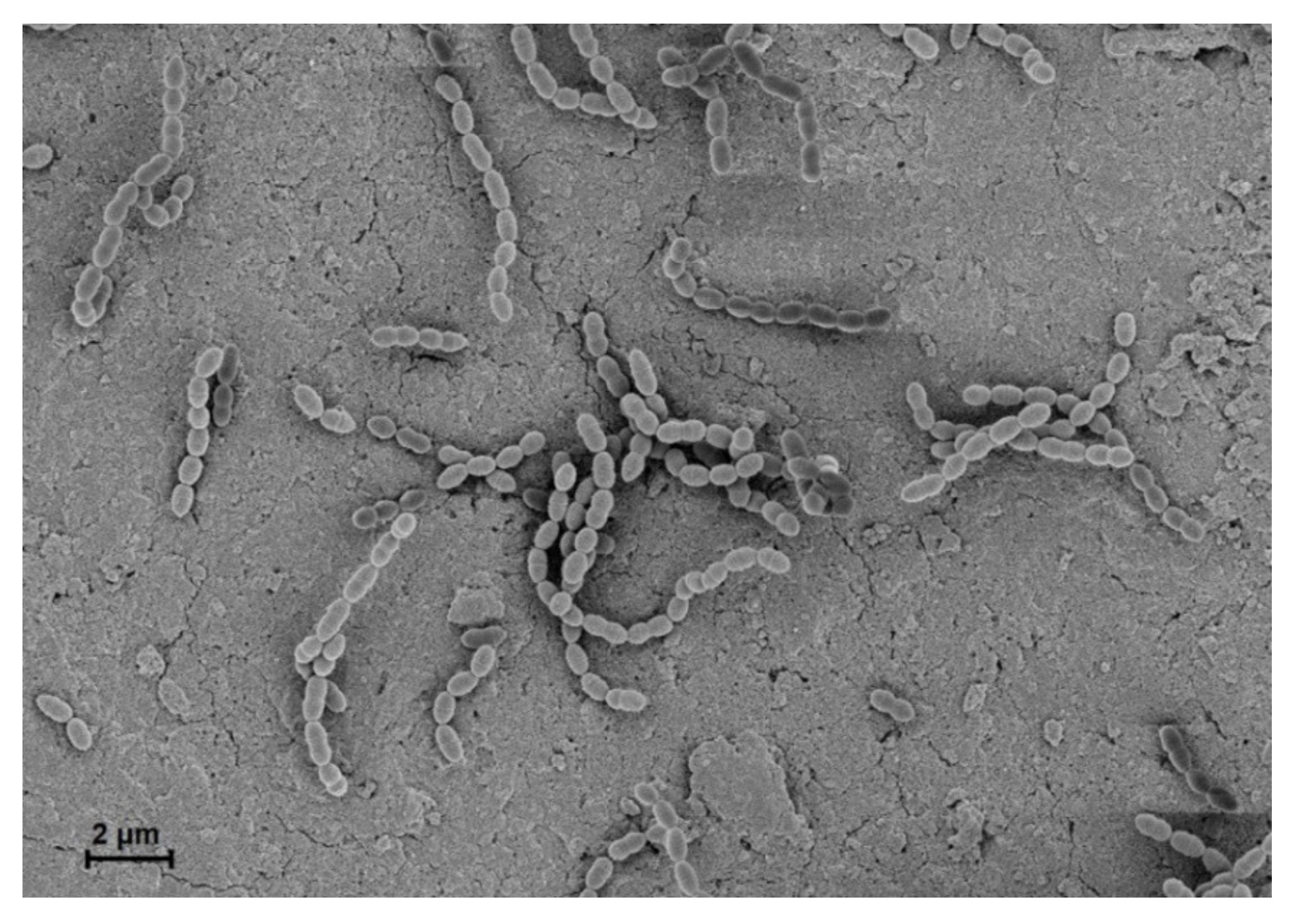
1. Overview of Streptococcus gordonii

In 1884, Rosenbach first termed the Streptococcus group from examining a man with suppurative lesions [1]. This genus is classified as Gram-positive, cocci or spherical, and clustered pairs or chains (Figure 1) [2]. They are homofermentative and facultative anaerobes, exhibiting negative catalase activity and forming no spores [1]. Streptococci are divided into three groups based on hemolysis patterns on blood agar plates: β-hemolysis (complete hemolysis), α-hemolysis (incomplete hemolysis), and γ-hemolysis (no hemolysis) [3]. Recently, phylogenetic approaches ultimately subdivided Streptococcus into eight groups consisting of mitis, sanguinis, anginosus, salivarius, pyogenic, mutans, downei, and bovis [3].
Streptococcus gordonii is commensal, non-pathogenic bacterium that is present in the human body, including the skin, oral cavity, upper respiratory tract, and intestine. It mainly resides on mucosal surfaces, such as the oral cavity, but also live in water, soil, plants, and food [4,5]. S. gordonii, a part of the α-hemolytic (viridans) sanguinis group, primarily inhabits the oral cavity of humans and animals [6]. However, it is also an opportunistic pathogen and can cause a variety of infectious diseases (Figure 2). Recently, metagenomic next-generation sequencing analysis showed that S. gordonii exists in patients with apical periodontitis or caries and the heart valves of patients with infective endocarditis [7,8,9]. In fact, S. gordonii, an initial colonizer on the tooth surface, can co-aggregate with several other oral microorganisms, contributing to the development of periodontal disease and caries [10]. S. gordonii can enter the blood stream by oral bleeding, leading to endocarditis [6]. Furthermore, S. gordonii binds to the cell surface of various host cells, contributing to the initiation of diseases through the inflammatory responses [11,12].
The bacterial cell wall plays a crucial role in the survival and growth of bacteria [13]. In Gram-positive bacteria, the cell wall is composed of thick peptidoglycan (PGN) and various components including lipoteichoic acid (LTA), wall teichoic acid (WTA), cell wall-anchoring glycoproteins, and lipoproteins [14]. Cell wall components can be recognized by host receptors such as pattern recognition receptors (PRRs), which initiate host innate immune responses [14]. S. gordonii expresses cell wall proteins, including Streptococcal surface protein (Ssp) A, SspB, collagen-binding domain protein (CbdA), and serine-rich repeat (SRR) glycoproteins, such as gordonii surface protein B (GspB) and Hs antigen (Hsa) [15,16]. The cell wall proteins of S. gordonii easily adhere to platelets, erythrocytes, monocytes, and dendritic cells (DCs) that could lead to acute immune responses in humans [16,17]. Therefore, understanding how cell wall components of S. gordonii interact with host cells is required not only to determine its entire pathogenesis but also to apply for the treatment and prevention of S. gordonii-mediated diseases.

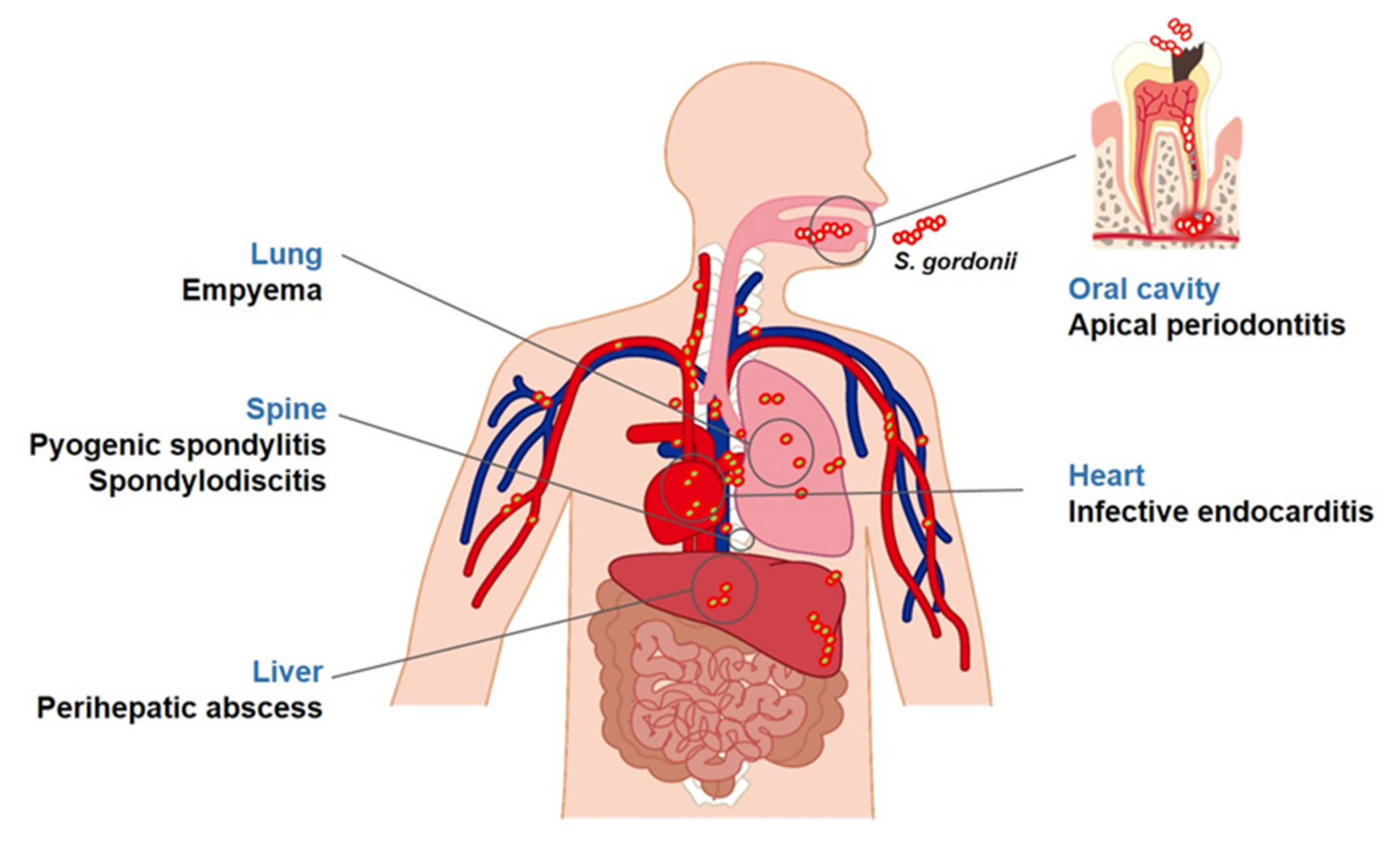
2. Diseases Associated with S. gordonii

2.1. Apical Periodontitis

Apical periodontitis is an inflammatory disease that occurs within periapical tissues [18]. A study in the U.S. has revealed that 4.1% of randomly sampled teeth have apical periodontal disease, and this rate increases up to 31.3% from teeth receiving endodontic treatment [19]. Interestingly, S. gordonii was predominantly isolated from 34 of 100 patients with apical periodontitis [10]. Apical periodontitis is mainly caused by bacterial invasion into the dental pulp and endodontic lesions, and the attachment of bacteria on dentin surfaces is a critical step for the development of apical periodontitis [18]. S. gordonii expresses numerous cell wall proteins that facilitate attachment on dentin surfaces [20]. After successful attachment, S. gordonii forms biofilm matrix by deposition of extracellular polysaccharide, which is an essential structural component of biofilm [21]. S. gordonii found in biofilms is more resistant to antibacterial agents than in the planktonic state, making it harder to eliminate S. gordonii [22,23]. Furthermore, the biofilm formation of S. gordonii on dentin surfaces facilitates invasion into the dental pulp through dentinal tubules [24]. In addition, S. gordonii can interact with host cells followed by its penetration into root canals or dentinal tubules [18].
The interaction subsequently induces inflammatory conditions on periapical lesions. For instance, S. gordonii induces the expression and secretion of chemotactic cytokines called interleukin (IL)-8 by stimulating toll-like receptor (TLR) 2 of human periodontal ligament cells (PDLs) [25]. In fact, S. gordonii lipoprotein triggers the secretion of IL-8, monocyte chemoattractant protein (MCP)-1, cyclooxygenase-2, and prostaglandin E2 in the human dental pulp cells (Figure 3) [26]. Moreover, several studies also demonstrated that S. gordonii induces the expression of IL-8 in human endothelial cells [27]. Consequently, S. gordonii can promote the infiltration of neutrophils and monocytes, causing acute inflammation on penetrated periapical lesions coincident with the activation of other host cells [28,29]. The presence of pro-inflammatory cytokines has been reported in apical periodontitis lesions [30,31]. Moreover, S. gordonii contributes to alveolar bone destruction, a common feature of apical periodontitis, through the upregulation of osteoclast differentiation or the downregulation of osteoblast differentiation [32]. S. gordonii induces pro-inflammatory cytokines such as IL-1β, IL-6, and IL-8, and these cytokines potently activate the differentiation of macrophages into osteoclasts [15,32]. In addition, it has been reported that S. gordonii directly activates the bone resorbing ability of osteoclasts and inhibits the bone forming activity of osteoblasts [29]. Therefore, S. gordonii is considered as an etiological agent that contributes to the development of apical periodontitis and alveolar bone resorption through the induction of acute inflammatory conditions.

2.2. Infective Endocarditis

S. gordonii, released from oral biofilms by tooth brushing, tooth extraction, or oral trauma, can disperse into the circulatory system through blood vessels, leading to systemic infections [22,33]. Infective endocarditis is a life-threatening disease caused by oral streptococci where the hospital mortality rate from this disease counts approximately 20% [34,35]. A nationwide analysis reported that infective endocarditis incidence in the U.S. recorded 47,134 cases [36]. Moreover, S. gordonii showed the highest prevalence rate for Streptococcal infective endocarditis in Denmark, which was 44.2% [37]. The persistent exposure of S. gordonii by low-grade bacteremia can cause infective endocarditis [38]. Therefore, an understanding of the pathogenesis of S. gordonii is necessary for the treatment and prevention of infective endocarditis. Interaction between S. gordonii and host cells in the bloodstream is considered as an important initial step in the pathogenesis of infective endocarditis [39]. When S. gordonii enters the bloodstream, S. gordonii preferentially binds to platelets or erythrocytes using their numerous cell surface proteins and then hematogenously spreads to damaged heart valves [40]. S. gordonii potently binds to human vascular endothelial cells and accumulates on heart valves to form biofilms [22,41], causing the aggregation of platelets that develops into bacterium-platelet-fibrin complexes and further exacerbates the inflammatory responses. In addition, S. gordonii activates human valve interstitial cells to induce IL-6 and IL-8, leading to the infiltration of immune cells through nuclear factor-kappa B (NF-κB) signaling pathway [42]. Immune cells in heart lesions recruited by chemokines are directly activated by S. gordonii. For instance, S. gordonii induces nitric oxide production on macrophages through TLR2 signaling pathway (Figure 3) [43]. Human monocytes stimulated with S. gordonii enhance pro-inflammatory cytokine production and express more cell surface markers, including cluster of differentiation (CD)40, CD54, and CD80 [44]. Moreover, S. gordonii stimulates DCs to cause the induction of pro-inflammatory cytokines, such as IL-6, IL-12, and tumor necrosis factor-α (TNF-α) and co-stimulatory receptors [16,45]. Taken together, S. gordonii originating from oral biofilms enters into the blood stream and induces platelet aggregation and excessive inflammatory conditions by stimulating various host cells.

2.3. Other Diseases

Once S. gordonii enters the bloodstream, it can translocate into other organs through the blood circulation system [46]. Therefore, S. gordonii can cause not only infective endocarditis but also other systemic diseases. For instance, when S. gordonii translocates into the joint, it can rapidly induce excessive inflammatory condition. The inflammatory responses induced by S. gordonii infections directly cause septic arthritis, leading to joint destruction and bone loss [33]. In addition, S. gordonii can cause empyema in the lungs, which is defined as an accumulation of pus in the pleural cavity. S. gordonii has been found in the pleural fluid of empyema patients [47]. S. gordonii has also been isolated in the perihepatic collection fluid originating from perihepatic abscesses that form around the liver [48]. Moreover, S. gordonii translocated into spinal bones induces inflammation, further developing into pyogenic spondylitis or spondylodiscitis [49,50].

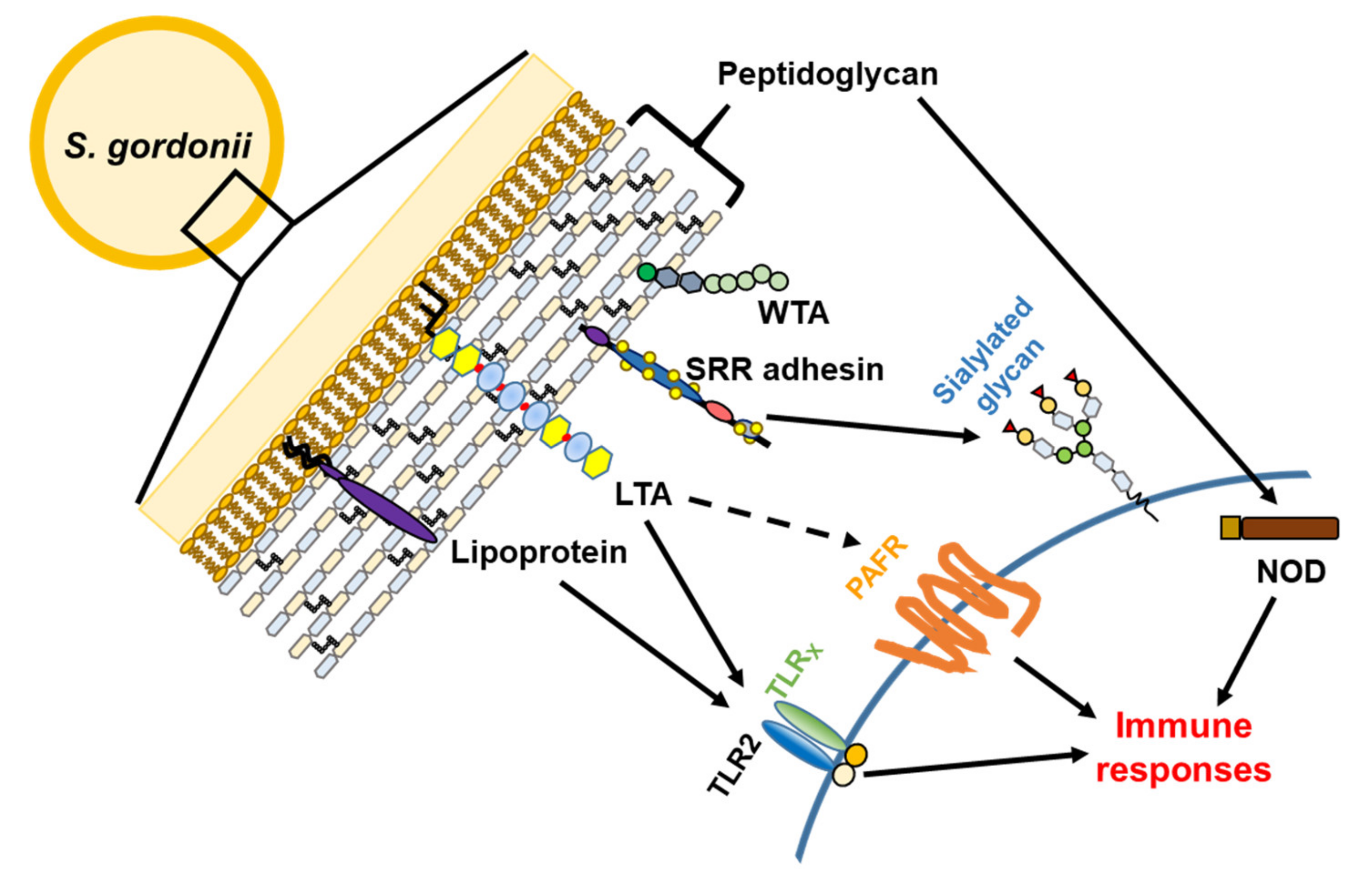
3. Virulence Factors

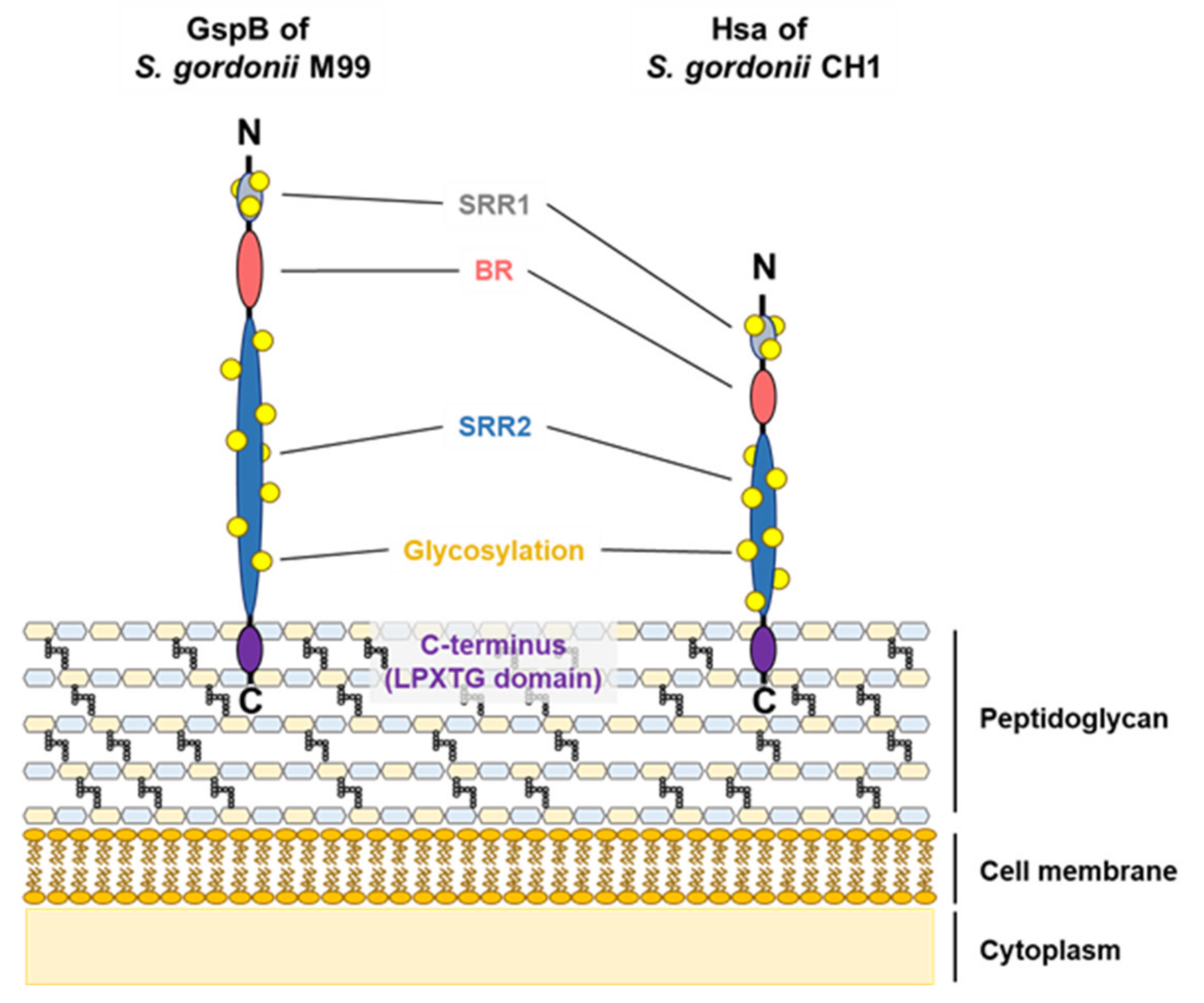
3.1. Serine-Rich Repeat Adhesins

SRR adhesins are Gram-positive bacterial cell wall-anchored glycoproteins [51]. Among the SRR groups, Hsa and GspB are well-characterized homologous adhesins in S. gordonii strains CH1 and M99, respectively [52,53]. SRR adhesins include an N-terminal signal peptide, a short SRR1 region, a ligand-binding basic region (BR), a long SRR2 region, and a LPXTG motif at a C-terminal for cell wall anchoring [54,55,56]. Both Hsa (203 kDa) and GspB (286 kDa) include the basic SRR adhesion domains: an N-terminus, two SRRs, a BR, and a C-terminus (Figure 4) [52,53]. When S. gordonii expresses SRR adhesins, the accessory Sec system, which includes SecA2 and SecY2, mediates the exporting of SRR adhesins from bacterial cytoplasm to the cell wall [57]. Despite the fact that SRR adhesins contain conserved domains, the ligand-binding BR exhibits various structural changes in amino acid sequences, contributing to the binding specificity to each respective ligand [17,55]. For instance, while both Hsa and GspB bind to sialyl-T antigen, only Hsa can bind to 3′-sialyllactose due to the amino acid differences in the BR domains [58,59].
The binding of S. gordonii to host cells or tissues using various adhesins is an important step for the initiation of infection [46,60,61,62]. Among the various adhesins, SRR adhesins are considered as the major molecules for the development of S. gordonii pathogenesis (Table 1). There have been several studies on the attachment of S. gordonii to various host cells using SRR adhesins. For instance, S. gordonii binds to sialylated carbohydrate moieties of glycoprotein Ibα on platelet membranes through the BR domains of Hsa and GspB [17,40,57], and S. gordonii lacking GspB reduces binding potentials to rat and human platelets by approximately 65% [46,58]. In addition, Deng et al. reported that S. gordonii preferentially interacts with sialoglycans in platelets through the BR domain of SRR adhesins in whole blood [40]. Moreover, SRR adhesins of S. gordonii also mediate binding to α2-3-linked sialic acid on glycophorin A of erythrocyte membranes. Two arginine residues (Arg340 and Arg365) of Hsa are major sites of interaction with glycoproteins on erythrocyte membranes [59,63]. In catheterized animal models infected with S. gordonii, bacterial vegetation density challenged with wild type strain was higher than infected with Hsa- or GspB-deficient strain [39,46]. These accumulating studies indicate that SRR adhesins are responsible for the translocation of S. gordonii to the endocardium by attaching to circulating platelets or erythrocytes in the bloodstream causing the pathogenesis of infective endocarditis [39,40].
The adherence of S. gordonii by SRR adhesins to immune cells, such as polymorphonuclear leukocytes (PMNs), macrophages, DCs, or monocytes, evokes the promotion and activation of the host inflammatory condition [16,66,67,68]. Hsa specifically interacts with sialoglycoproteins on monocyte membranes such as CD11b, CD43, and CD50. In addition, mucin-like domains of glycoproteins on PMN surfaces are major sites of Hsa adhesion [66,68]. The interaction of HL-60 cell-derived monocytes with S. gordonii rapidly induces the expression of DC markers including CD1a, CD83, CD86, and IL-12 while Hsa-deficient S. gordonii fails to differentiate monocytes into DCs [67]. Moreover, compared to the wild type treatment, SRR adhesin-deficient S. gordonii poorly induces pro-inflammatory cytokines, including TNF-α, IL-6, and IL-12, in human monocyte-derived DCs coincident with attenuating the proliferation and activation of co-cultured T cells. These results indicate that SRR adhesins have the potential of modulating innate and adaptive immune responses [16].
Accumulating studies demonstrate that SRR adhesins are closely involved in the biofilm formation of some Gram-positive bacteria [69,70,71]. SRR adhesins of S. gordonii also play important roles in biofilm formation. Kim et al. reported that GspB-deficient S. gordonii forms less biofilm than the wild type strain on human dentin surfaces, suggesting that GspB is crucial for the development of dental biofilm formation [64]. In addition, Hsa-deficient S. gordonii exhibits lower biofilm formation than the wild type strain on plates coated with saliva, fetuin, or mucin [65].

3.2. Cell Wall Proteins

S. gordonii has a variety of virulence factors on their cell wall, such as antigen-related proteins, collagen-binding proteins, fibronectin-binding proteins (Fbps), platelet adherence proteins, and α-amylase-binding proteins. As summarized in Table 2, they are well-known to promote the species-specific binding of S. gordonii to various receptors, contributing to the development of dental caries, periodontal diseases, and endodontic diseases caused by S. gordonii [72].
The cell wall proteins, SspA and SspB, are antigen I/II family polypeptides of S. gordonii that mediate the attachment to various hosts and other bacterial cells [72]. These proteins accelerate the infection of S. gordonii into root dentinal tubules by binding type I collagen or β1 integrin, mediating the aggregation and adherence of cells by binding to salivary agglutinin glycoprotein (gp340), and facilitating biofilm formation by interacting with other oral bacterial species, such as Porphyromonas gingivalis [24,72,73]. S. gordonii also has antigen-related polypeptides, CshA and CshB. These antigen-related proteins are responsible for binding to host fibronectins or other oral microorganisms and facilitating invasion into endothelial cells [74,75].
CbdA is a protein of S. gordonii similar to Enterococcus faecalis collagen-binding protein, Ace, in the structure and function [77]. An in silico analysis of S. gordonii Cbd locus showed that CbdA contains a signal sequence at the amino terminus and LPXTG, a PGN anchor motif at the carboxyl terminus [77]. This protein promotes the attachment of S. gordonii to host type I collagen, suggesting that S. gordonii persists in its survival in instrumented root canals by CbdA [77].
FbpA is one of the bacterial cell wall proteins of S. gordonii. Fibronectin is a eukaryotic glycoprotein that exists in the plasma or on host cell surface and engages in cellular adhesion, migration, and differentiation. Fibronectin can also be a target molecule for bacterial attachment [60]. For example, FbpA affects the binding of S. gordonii to eukaryotic fibronectin through the regulation of CshA expression [60].
Platelet adherence protein A (PadA) is a protein that mediates interactions between the major platelet receptor GPIIb/IIIa and S. gordonii [79]. PadA activates platelets and promotes biofilm formation by cooperating with Hsa [78]. PadA and Hsa are involved in each other’s active presentation on the cell wall, indicating that they cooperatively mediate the activation of platelets and promotion of biofilm formation [78].
S. gordonii produces α-amylase-binding protein A and B (AbpA and AbpB) that contribute to biofilm formation and colonization on teeth. AbpA and AbpB bind to α-amylases secreted from animals, which may facilitate the binding of bacteria to the salivary pellicle [80]. In addition, they provide S. gordonii with nutritional benefits by capturing host enzymatic activity to compete with other oral microbial species [80].
Collectively, the cell wall proteins of S. gordonii are important for the interaction with host proteins (Figure 5). Therefore, further studies are necessary to fully understand the sophisticated functions of S. gordonii cell wall proteins and to control various S. gordonii-mediated infectious diseases.

3.3. Lipoproteins

Bacterial lipoproteins are located on the extracellular surface of the cytoplasmic membrane of bacteria. In general, Gram-positive and Gram-negative bacteria contain diacylated- or triacylated- lipoproteins, respectively [81]. Lipoproteins play various physiological functions, such as nutrient acquisition, adherence, adaptation to environmental changes, protein maturation, bacterial growth, and pathogenesis [82,83,84]. Therefore, the deletion of lipoprotein diacylglycerol transferases (lgt) gene, associated with the maturation of lipoproteins [85], causes changes in bacterial physiological properties of Streptococci. For example, the Streptococcus pneumoniae lgt mutant exhibits reduced growth in cation-depleted medium and reduced intracellular concentrations of several cations, such as Fe2+, Zn2+, and Mn2+ [86]. Moreover, mutation in lgt results in impaired growth and attenuated virulence in Streptococcus sanguinis [87]. On the other hand, there are no bacterial morphological, size, and growth pattern differences between lipoprotein-deficient S. gordonii and its wild type [88] (Table 3).
TLRs are one of the PRRs that can recognize bacterial pathogen-associated molecular patterns and are located on the surface or intracellular compartments of host cells [91]. S. gordonii possesses diacylated-lipoproteins, which are considered as more potent stimulators of TLR2 than LTA in S. gordonii [43,88]. Through TLR2 activation, lipoproteins can induce the secretion of pro-inflammatory cytokines and chemokines (Figure 3 and Table 3). For example, the recognition of diacylated- or triacylated lipoproteins, which are agonists of TLR2/TLR6 or TLR2/TLR1, respectively, triggers the MyD88-dependent signaling pathway [92]. In addition, it has been reported that the lipoproteins of S. gordonii induce the production of nitric oxide by activating the NF-κB pathway, STAT1 phosphorylation, and interferon (IFN)-β expression in RAW 264.7 cells [43]. Moreover, the lipoprotein-deficient mutant of S. gordonii fails to induce pro-inflammatory cytokines, such as TNF-α, IL-8, and IL-1β at both mRNA and protein levels in the human monocytic cell line THP-1, and mouse bone-marrow derived macrophages [88]. Lipoproteins of S. gordonii also induce TNF-α, IL-6, IL-12p70, and IL-10, and can upregulate the expression of the DC surface marker CD80 but not CD86 on bone-marrow DCs [89]. Wild type strain, but not lipoprotein-deficient strains, reduces the frequency of CD4+, CD25+, and Foxp3+ regulatory T cells in murine acute phase infection models [90]. Furthermore, the S. gordonii lgt mutant strain is cleared more rapidly in blood and organs such as the spleen and liver of mice than the wild type. Wild type S. gordonii strongly adheres to human umbilical vein endothelial cells compared to the S. gordonii lgt mutant strain [41]. Human PDLs exposed to purified lipoproteins of S. gordonii induce IL-8 production through the TLR2-mediated mitogen-activated protein kinase pathway [25]. Likewise, human dental pulp cells express pro-inflammatory mediators like IL-8 and MCP-1 when stimulated by S. gordonii lipoproteins [26]. Some research have revealed that Pam2CSK4, which mimics Gram-positive bacterial lipoproteins, induces increased expression of pro-inflammatory cytokines and immunosuppressive cytokines in human odontoblast-like cells [93]. Therefore, lipoproteins are involved in the virulence and immunoregulatory process, in which targeting the biosynthesis of lipoproteins could be a potential vaccine strategy [94].

3.4. Teichoic Acids

Teichoic acid (TA) is a predominant cell wall component of Gram-positive bacteria that accounts for approximately 50% of cell wall dry weight [95], and is distinctly classified by structure and location. TA is attached to the cell membrane (LTA) or to the cell walls (WTA). WTA is a PGN-linked polymer with 40 to 60 polymer repeats [96]. LTA is classified by polymeric chain diversity (types I to V) [97]. Among them, the structure of type I LTA is well-characterized. Type I LTA contains an unbranched 1–3 linked glycerol phosphate backbone structure anchored by glycolipids [97]. S. gordonii has type I LTA containing glycerol phosphate and terminal glucose-glycerol phosphate repeat units (Figure 6) [98].
LTA plays a critical role in cell division, growth, and biofilm formation. Lima et al. reported that LTA-deficient S. gordonii strain grows more slowly than wild type S. gordonii on solid media and exhibits decreased biofilm formation on saliva-coated hydroxyapatite disks [98]. In addition, they have analyzed the protein profiles of cell wall fractions from the wild type and LTA-deficient S. gordonii strains using mass spectroscopy. Acetate kinase, phosphoglycerate kinase, serine protease, SspA, and SspB are more abundant in LTA-deficient S. gordonii than the wild type strain. On the other hand, the cell division transport system ATP-binding protein, FtsE, and penicillin-binding protein 2a were more abundant in the wild type strain. These results suggest that LTA affects cell division, cell wall biosynthesis, and the biofilm formation of S. gordonii.
Accumulating reports suggest that LTA induces inflammatory responses through TLR2. Lactobacillus plantarum LTA induces nitric oxide production in the presence of IFN-γ in macrophages [99]. In addition, LTA of Enterococcus faecalis or Streptococcus mutans significantly increases inflammatory cytokines such as TNF-α and nitric oxide through TLR2 activation [100,101]. However, the induction of IL-8 or nitric oxide in human PDLs and murine macrophages by LTA-deficient S. gordonii is similar to the level expressed by wild type S. gordonii [25,43]. This finding indicates that, unlike LTA purified from other bacteria, S. gordonii LTA alone may not induce host immune responses, in other words, has less immunostimulating potential. In addition, immunomodulatory effects of LTA during inflammation have been reported. L. plantarum LTA efficiently inhibits Poly I:C-induced IL-8 production in porcine intestinal epithelial cells and lipopolysaccharide (LPS)-induced endotoxin shock [102,103]. Staphylococcus aureus LTA attenuates LPS-induced B cell proliferation [104]. Therefore, based on these results, future studies are needed to identify the immunomodulatory effects of S. gordonii LTA.
Like LTA, WTA affects various bacterial functions such as adhesion, growth, autolytic activity, and antibiotic resistance [96,105]. WTA also has been shown to modulate the maturation and activation of DCs [106]. DCs treated with WTA-deficient S. aureus exhibit lower DC maturation and activation than wild type S. aureus. However, to the best of our knowledge, no study has reported the effects of S. gordonii WTA in bacterial function and immunemodulation.

3.5. Peptidoglycan

PGN is one of the most abundant microbial cell wall components. It is a polymer of cross-linking linear chains composed of alternating N-acetyl glucosamine and N-acetyl muramic acid (NAM). Each NAM has a short peptide consisting of alternating L- and D-amino acids that involve in the cross-linking of linear sugar chains [107]. These peptides provide significant diversity of PGNs, which vary from species to species [107]. Although species-specific structures of some bacterial PGNs, such as Mycobacterium have been discovered [108], little is known about S. gordonii PGN. Due to the diversity of glycan strands and peptide moieties [109,110], it is expected that PGN structures vary from bacteria to bacteria. Therefore, identifying host responses triggered by S. gordonii-specific PGN would further elucidate the properties of S. gordonii.
Nucleotide oligomerization domains (NODs), NOD1 and NOD2, are mammalian intracellular proteins that engage in sensing bacterial PGN fragments [111]. It has been demonstrated that they recognize distinct bacterial PGN motifs. NOD1 specifically binds to Tri-diaminopimelic acid motifs of Gram-negative bacteria [111]. In contrast, NOD2 detects muramyl dipeptide motifs, suggesting its binding affinity for both Gram-positive and Gram-negative bacterial PGNs [112]. The detection of bacterial PGNs through NOD1 and NOD2 triggers pro-inflammatory responses by activating the NF-κB pathway, which is essential for bacterial clearance from infected host cells [113]. For example, a previous report has demonstrated the synergistic effects of TLR2 and NOD2 signaling pathways on IL-8 production in human PDLs [114]. Another report has revealed that cell wall components, LTA, lipoprotein, and PGN from S. gordonii induces the secretion of pro-inflammatory cytokines such as IL-6 and TNF-α in DCs through the TLR2 signaling pathway [89]. However, specific mechanisms or roles of S. gordonii PGN are poorly studied, and thus further mechanistic studies are necessary.

4. Therapeutic Strategies against S. gordonii Infections

Nowadays, a large number of therapeutics are available to avoid S. gordonii-induced oral and systemic diseases. For example, several antibacterial agents, such as fluorides, chlorohexidine (CHX), and antibiotics, have been widely used to regulate bacterial growth and infections [115,116,117,118]. However, those conventional antibacterial agents can affect bacterial viability or growth, imposing higher selective pressure that adversely increases the possibility of resistance development [119]. In addition, once bacterial biofilm is formed, the therapies are difficult to function properly due to reduced bacterial sensitivity [120]. Therefore, novel therapeutic strategies that target major virulence factors, virulence-mediated pathways, and biofilm formation could be a promising alternative to conventional therapies such as antibiotics.
As mentioned previously, it is well-known that S. gordonii binds to human platelets through the surface proteins Hsa and PadA, which is considered as a crucial step for infective endocarditis [121,122]. Based on this, one study revealed that antibodies targeting Hsa and PadA could delay or even inhibit the platelet aggregation of S. gordonii [123]. Several authors suggest that using these antibodies is likely to be a novel S. gordonii therapeutic strategy [123]. However, no follow-up studies have been made towards the development of S. gordonii-specific vaccines or treatments. Therefore, more research should be performed in various ways, as suggested in the following.

4.1. Regulation of Biofilm

There are many conventional methods to regulate biofilm. Especially for oral biofilm, physical removalby tooth brushing and scaling, is one of the most effective strategies to disrupt the biofilm. Several studies have demonstrated that tooth brushing results in an attenuation of plaque levels [124,125]. In addition, many other studies have revealed that various techniques of scaling, such as hand and ultrasonic scaling, laser scaling, and chemical scaling, have been used to control plaques [126,127].
Another traditional method to control biofilm is through the use of various chemicals, such as CHX, sodium chloride, and calcium chloride. CHX is a potential antimicrobial agent, which participates in disrupting biofilms by binding to negatively charged bacterial cell walls [128,129,130]. Salts, such as sodium chloride and calcium chloride, may weaken electrostatic interactions and, therefore, can attenuate the biofilm matrix [131].
S. gordonii and other oral Streptococci are well-known initial colonizers on tooth surfaces since they generate abundant cell wall adhesins interacting with various types of cells and tissues [132,133,134]. Therefore, targeting such surface colonization by disrupting the adherence of S. gordonii could likely be an effective therapeutic strategy against S. gordonii infections. The target candidates for these therapies can be SRRs and cell wall proteins because they are the major binding factors of S. gordonii. For example, the attachment and internalization of S. gordonii to epithelial cells occurs with SspA and SspB (antigen I/II family) [135]. The SRR adhesins of S. gordonii such as GspB are important for biofilm formation and bacterial aggregation on the surfaces [64]. Additionally, CshA, a multifunctional fibrillary adhesin, binds to host fibronectin and mediates the colonization of S. gordonii [11]. Therefore, the development of agents that can block these proteins can be an effective way to inhibit bacterial adherence and subsequent infections.
Using small molecules that suppress quorum sensing-mediated bacterial communication is in the spotlight as a novel therapeutic strategy for regulating biofilm. By using quorum sensing inhibitors, it is expected biofilm maturation can be easily regulated and bacterial self-protection attenuated from antimicrobial agents furnished by a mature biofilm [136,137]. Multiple approaches could be taken for inhibiting quorum sensing. The most promising approach to regulate quorum sensing involves targeting the S-ribosylhomocysteine lyase (LuxS)/autoinducer-2 (AI-2) quorum sensing system, one of the major bacterial communication systems of S. gordonii [138,139]. Therefore, using quorum-sensing inhibitors associated with LuxS/AI-2 could be an effective alternative to conventional therapies.
Promoting biofilm dispersion is another attractive approach for specific therapies against biofilm-mediated diseases, which can contribute to overcoming bacterial resistance mechanisms [140]. Several studies have revealed that bacterial LTA has an anti-biofilm effect, such as inhibiting biofilm formation, disrupting preformed biofilm, and consequently increasing susceptibility to antibiotics [141,142,143]. However, conversely, planktonic bacteria after biofilm disruption will likely disperse into the entire body and may cause issues in other sites of the host. Therefore, using conventional antibiotics together with LTA would be an effective way for the complete removal of infected microbes [140].
During bacterial infection, host cells recognize the invaded bacteria through PRRs and produce inflammatory mediators, such as antimicrobial peptides, immunoglobulin (Ig), and nitric oxide [14]. Some studies have revealed that inflammatory mediators can directly inhibit biofilm formation. For instance, human β-defensin, which is an antimicrobial peptide, decreases not only single species bacteria biofilm but also multi-species bacteria biofilm through the promotion of bacterial cell death [144,145]. In addition, among the Ig family, only secretory IgA, which is abundant in the oral cavity, effectively inhibits bacterial biofilm formation [146]. On the other hand, nitric oxide also inhibits biofilm formation by triggering bacterial dispersion [147]. Therefore, the activation of host cells to produce inflammatory mediators could be one of the alternative strategies for removing bacterial biofilm.

4.2. Inhibition of Bacterial Cell Wall Components

4.2.1. Lipoprotein

Since lipoprotein-deficient S. gordonii remarkably reduces the induction of inflammatory responses [90], targeting lipoproteins can be one of the therapeutics used against S. gordonii infections. Lipoprotein structures react sensitively to changes in the environment. For example, S. aureus SitC lipoproteins usually exist in the diacyl form under acidic conditions [148]. In cases of S. pneumonia, lipoprotein-based vaccines and antibodies are being developed [149]. Pneumococcal lipoproteins such as pneumococcal surface adhesion A (PsaA), histidine triad protein (Pht) D, PhtE, PhtA, and PhtB are identified as the antigenic regions within Pneumococcal proteins. PsaA, in particular, has been shown to be immunogenic [150]. It has been reported that anti-PsaA monoclonal antibodies efficiently protect against S. pneumonia infections [151]. Therefore, lipoproteins of S. gordonii also can be a potential vaccine candidate.

4.2.2. Lipoteichoic Acid/Wall Teichoic Acid

In general, it is well known that LTA is critical for cell division and growth. The binding of LTA to TLR2 triggers the host innate immune response. Notably, the platelet-activating factor receptor (PAFR) is essential for LTA-induced mucin gene expression in epithelial cells [152]. The inhibition of PAFR attenuates the LTA-induced expression of pro-inflammatory cytokines and chemokines but does not affect LPS-induced pro-inflammatory cytokines and chemokines [153,154]. These reports suggest that PAFR can be one of the potential targets against Gram-positive bacterial infections. Anti-LTA monoclonal antibodies have been reported to improve mice survival rates in methicillin- or vancomycin-resistant S. aureus-induced sepsis models [155]. Human serum anti-WTA IgG induces the opsonophagocytosis of S. aureus by PMNs [156]. In addition, Congo Red is a selective inhibitor for the S. aureus LTA synthase of S. aureus, which is thought to be a potential drug target against Staphylococcal infections [157]. However, because Congo Red is highly toxic [158], it would be desirable to use its molecular action mechanisms for the development of potential therapeutics. A positively charged molecule such as antimicrobial peptides like β-defensins may mask the immunostimulating effect of LTA by binding to LTA’s negative charge. Likewise, it is expected that specific molecule targeting LTAs or WTAs of S. gordonii can be developed as a therapeutic agent.

4.2.3. Peptidoglycan

Since PGN is predominantly found in the bacterial cell wall, β-lactam antibiotics, such as penicillin, are used to induce bacterial cell death through blocking the linkage of PGNs [159]. In addition, vancomycin, an actinobacteria-derived glycopeptide, also inhibits the synthesis of PGNs by preventing transpeptidase and transglycosylase activities [160]. However, these traditional antibiotics can cause the emergence of antibiotic-resistant bacteria. Therefore, the discovery of new cell wall inhibition methods without promoting bacterial resistance has been investigated in recent studies. For example, Ling et al. reported that teixobactin, a new antibiotic, inhibits Gram-positive bacterial cell wall synthesis by binding to precursors of PGN or WTA [161]. Autolysins can be used as antimicrobial agents for breaking PGN bonds [162]. Furthermore, the use of bacteriophages instead of antibiotics is a renewed alternative strategy for the clearance of bacteria on lesions. Bacteriophages have endolysins or lysins that specifically target bacteria and break down the PGN bonds [163]. It has been reported that infective endocarditis was treated with lysins applied to the lesions [164]. Interestingly, PGN fragments can be released through the process of PGN breakdown and then be recognized by host cells through their NOD1 or NOD2 receptors [112]. The recognition of PGNs by NOD1 or NOD2 receptors triggers immune responses [113]. Therefore, PGN-specific receptors can be used as molecular targets for the clearance of S. gordonii through the activation of host immune cells.

5. Conclusions

S. gordonii, a commensal and opportunistic Gram-positive bacterium, plays an important role in the development of apical periodontitis and infective endocarditis. The attachment of S. gordonii to host cells using its adhesion proteins is an essential initiation step for disease development. Once attached, the cell aggregation of S. gordonii leads to biofilm formation on the apical lesions and heart valves. They also continuously interact with host cells, and the molecular interaction led by its cell wall components induces the immune responses. Therefore, S. gordonii may contribute to the progression of diseases due to its attachment, aggregation, and immunostimulatory effects. The bacterial cell wall is involved in important functions of bacteria, such as maintaining bacterial cell shape integrity, and contributing to bacterial growth, reproduction, metabolism, and movement. Therefore, by specifically inhibiting bacterial cell wall components, we could potentially alleviate or even prevent the bacterial infections. The development of novel anti-bacterial molecules, which is involved in targeting specific virulence factors of biofilms, is becoming more important in the prevention and treatment of various bacterial infections including S. gordonii-mediated diseases.

Author Contributions

Conceptualization and methodology, S.H.H.; investigation, validation and data curation, O.-J.P., Y.K.; writing—original draft, O.-J.P., Y.K., C.P., Y.J.S., T.H.P., S.J.; writing—review and editing, O.-J.P., J.I., S.H.H., C.-H.Y.; supervision, S.H.H. All authors have read and agreed to the published version of the manuscript.

Funding

This work was supported by grants from the National Research Foundation of Korea, which is funded by the Korean government (NRF-2018R1A5A2024418, NRF-2019R1A2C2007041, and NRF-2019R1I1A1A01060952), Republic of Korea.

Acknowledgments

We appreciate Ho Seong Seo for his help in carefully reviewing this work. We also thank Yerry Han for her kind help in drawing the illustrations.

Conflicts of Interest

The authors declare no conflict of interest.

References

  1. Hardie, J.M.; Whiley, R.A. Classification and overview of the genera Streptococcus and Enterococcus. J. Appl. Microbiol. 1997, 83, 1S–11S. [Google Scholar] [CrossRef] [PubMed]
  2. Garnier, F.; Gerbaud, G.; Courvalin, P.; Galimand, M. Identification of clinically relevant viridans group streptococci to the species level by PCR. J. Clin. Microbiol. 1997, 35, 2337–2341. [Google Scholar] [CrossRef] [PubMed] [Green Version]
  3. Abranches, J.; Zeng, L.; Kajfasz, J.K.; Palmer, S.R.; Chakraborty, B.; Wen, Z.T.; Richards, V.P.; Brady, L.J.; Lemos, J.A. Biology of oral Streptococci. Microbiol. Spectr. 2018, 6. [Google Scholar] [CrossRef] [PubMed]
  4. Mundt, J.O. The ecology of the streptococci. Microb. Ecol. 1982, 8, 355–369. [Google Scholar] [CrossRef]
  5. Pinto, B.; Pierotti, R.; Canale, G.; Reali, D. Characterization of ‘faecal streptococci’ as indicators of faecal pollution and distribution in the environment. Lett. Appl. Microbiol. 1999, 29, 258–263. [Google Scholar] [CrossRef] [Green Version]
  6. Mosailova, N.; Truong, J.; Dietrich, T.; Ashurst, J. Streptococcus gordonii: A rare cause of infective endocarditis. Case Rep. Infect. Dis. 2019, 2019, 7127848. [Google Scholar] [CrossRef] [Green Version]
  7. Cheng, J.; Hu, H.; Fang, W.; Shi, D.; Liang, C.; Sun, Y.; Gao, G.; Wang, H.; Zhang, Q.; Wang, L.; et al. Detection of pathogens from resected heart valves of patients with infective endocarditis by next-generation sequencing. Int. J. Infect. Dis. 2019, 83, 148–153. [Google Scholar] [CrossRef] [Green Version]
  8. Gilbert, K.; Joseph, R.; Vo, A.; Patel, T.; Chaudhry, S.; Nguyen, U.; Trevor, A.; Robinson, E.; Campbell, M.; McLennan, J.; et al. Children with severe early childhood caries: Streptococci genetic strains within carious and white spot lesions. J. Oral. Microbiol. 2014, 6. [Google Scholar] [CrossRef] [Green Version]
  9. Zandi, H.; Kristoffersen, A.K.; Orstavik, D.; Rocas, I.N.; Siqueira, J.F., Jr.; Enersen, M. Microbial analysis of endodontic infections in root-filled teeth with apical periodontitis before and after irrigation using pyrosequencing. J. Endod. 2018, 44, 372–378. [Google Scholar] [CrossRef]
  10. Chavez de Paz, L.; Svensater, G.; Dahlen, G.; Bergenholtz, G. Streptococci from root canals in teeth with apical periodontitis receiving endodontic treatment. Oral Surg. Oral Med. Oral Pathol. Oral Radiol. Endod. 2005, 100, 232–241. [Google Scholar] [CrossRef]
  11. Back, C.R.; Sztukowska, M.N.; Till, M.; Lamont, R.J.; Jenkinson, H.F.; Nobbs, A.H.; Race, P.R. The Streptococcus gordonii adhesin CshA protein binds host fibronectin via a catch-clamp mechanism. J. Biol. Chem. 2017, 292, 1538–1549. [Google Scholar] [CrossRef] [PubMed] [Green Version]
  12. Loo, C.Y.; Corliss, D.A.; Ganeshkumar, N. Streptococcus gordonii biofilm formation: Identification of genes that code for biofilm phenotypes. J. Bacteriol. 2000, 182, 1374–1382. [Google Scholar] [CrossRef] [PubMed] [Green Version]
  13. Yadav, A.K.; Espaillat, A.; Cava, F. Bacterial strategies to preserve cell wall integrity against environmental threats. Front. Microbiol. 2018, 9, 2064. [Google Scholar] [CrossRef] [PubMed]
  14. Chandler, C.E.; Ernst, R.K. Bacterial lipids: Powerful modifiers of the innate immune response. F1000Res 2017, 6. [Google Scholar] [CrossRef] [Green Version]
  15. Andrian, E.; Qi, G.; Wang, J.; Halperin, S.A.; Lee, S.F. Role of surface proteins SspA and SspB of Streptococcus gordonii in innate immunity. Microbiology (Reading) 2012, 158, 2099–2106. [Google Scholar] [CrossRef]
  16. Ko, E.B.; Kim, S.K.; Seo, H.S.; Yun, C.H.; Han, S.H. Serine-rich repeat adhesins contribute to Streptococcus gordonii-induced maturation of human dendritic cells. Front. Microbiol. 2017, 8, 523. [Google Scholar] [CrossRef]
  17. Takamatsu, D.; Bensing, B.A.; Cheng, H.; Jarvis, G.A.; Siboo, I.R.; Lopez, J.A.; Griffiss, J.M.; Sullam, P.M. Binding of the Streptococcus gordonii surface glycoproteins GspB and Hsa to specific carbohydrate structures on platelet membrane glycoprotein Ibalpha. Mol. Microbiol. 2005, 58, 380–392. [Google Scholar] [CrossRef]
  18. Nair, P.N. Pathogenesis of apical periodontitis and the causes of endodontic failures. Crit Rev. Oral Biol Med. 2004, 15, 348–381. [Google Scholar] [CrossRef] [Green Version]
  19. Buckley, M.; Spangberg, L.S. The prevalence and technical quality of endodontic treatment in an American subpopulation. Oral Surg. Oral Med. Oral Pathol. Oral Radiol. Endod. 1995, 79, 92–100. [Google Scholar] [CrossRef]
  20. Stauffacher, S.; Lussi, A.; Nietzsche, S.; Neuhaus, K.W.; Eick, S. Bacterial invasion into radicular dentine-an in vitro study. Clin. Oral Investig. 2017, 21, 1743–1752. [Google Scholar] [CrossRef]
  21. Zheng, L.; Chen, Z.; Itzek, A.; Herzberg, M.C.; Kreth, J. CcpA regulates biofilm formation and competence in Streptococcus gordonii. Mol. Oral Microbiol. 2012, 27, 83–94. [Google Scholar] [CrossRef] [PubMed]
  22. Jung, C.J.; Yeh, C.Y.; Shun, C.T.; Hsu, R.B.; Cheng, H.W.; Lin, C.S.; Chia, J.S. Platelets enhance biofilm formation and resistance of endocarditis-inducing streptococci on the injured heart valve. J. Infect. Dis. 2012, 205, 1066–1075. [Google Scholar] [CrossRef] [PubMed] [Green Version]
  23. Kanwar, I.; Sah, A.K.; Suresh, P.K. Biofilm-mediated antibiotic-resistant oral bacterial infections: Mechanism and combat strategies. Curr. Pharm. Des. 2017, 23, 2084–2095. [Google Scholar] [CrossRef] [PubMed]
  24. Love, R.M. Bacterial adhesins--their role in tubule invasion and endodontic disease. Aust Endod. J. 2002, 28, 25–28. [Google Scholar] [CrossRef]
  25. Kim, A.R.; Ahn, K.B.; Kim, H.Y.; Seo, H.S.; Kum, K.Y.; Yun, C.H.; Han, S.H. Streptococcus gordonii lipoproteins induce IL-8 in human periodontal ligament cells. Mol. Immunol. 2017, 91, 218–224. [Google Scholar] [CrossRef]
  26. Yoo, Y.J.; Perinpanayagam, H.; Lee, J.Y.; Oh, S.; Gu, Y.; Kim, A.R.; Chang, S.W.; Baek, S.H.; Kum, K.Y. Synthetic human beta defensin-3-C15 peptide in endodontics: Potential therapeutic agent in Streptococcus gordonii lipoprotein-stimulated human dental pulp-derived cells. Int. J. Mol. Sci. 2019, 21. [Google Scholar] [CrossRef] [Green Version]
  27. Vernier, A.; Diab, M.; Soell, M.; Haan-Archipoff, G.; Beretz, A.; Wachsmann, D.; Klein, J.P. Cytokine production by human epithelial and endothelial cells following exposure to oral viridans streptococci involves lectin interactions between bacteria and cell surface receptors. Infect. Immun. 1996, 64, 3016–3022. [Google Scholar] [CrossRef] [Green Version]
  28. Henkels, K.M.; Frondorf, K.; Gonzalez-Mejia, M.E.; Doseff, A.L.; Gomez-Cambronero, J. IL-8-induced neutrophil chemotaxis is mediated by Janus kinase 3 (JAK3). FEBS Lett. 2011, 585, 159–166. [Google Scholar] [CrossRef] [Green Version]
  29. Park, O.J.; Kim, J.; Kim, H.Y.; Kwon, Y.; Yun, C.H.; Han, S.H. Streptococcus gordonii induces bone resorption by increasing osteoclast differentiation and reducing osteoblast differentiation. Microb. Pathog. 2019, 126, 218–223. [Google Scholar] [CrossRef]
  30. Ataoglu, T.; Ungor, M.; Serpek, B.; Haliloglu, S.; Ataoglu, H.; Ari, H. Interleukin-1beta and tumour necrosis factor-alpha levels in periapical exudates. Int. Endod. J. 2002, 35, 181–185. [Google Scholar] [CrossRef]
  31. Safavi, K.E.; Rossomando, E.F. Tumor necrosis factor identified in periapical tissue exudates of teeth with apical periodontitis. J. Endod. 1991, 17, 12–14. [Google Scholar] [CrossRef]
  32. Graunaite, I.; Lodiene, G.; Maciulskiene, V. Pathogenesis of apical periodontitis: A literature review. J. Oral Maxillofac. Res. 2012, 2, e1. [Google Scholar] [CrossRef] [PubMed]
  33. Yombi, J.; Belkhir, L.; Jonckheere, S.; Wilmes, D.; Cornu, O.; Vandercam, B.; Rodriguez-Villalobos, H. Streptococcus gordonii septic arthritis: Two cases and review of literature. BMC Infect. Dis. 2012, 12, 215. [Google Scholar] [CrossRef] [PubMed] [Green Version]
  34. Bannay, A.; Hoen, B.; Duval, X.; Obadia, J.F.; Selton-Suty, C.; Le Moing, V.; Tattevin, P.; Iung, B.; Delahaye, F.; Alla, F.; et al. The impact of valve surgery on short- and long-term mortality in left-sided infective endocarditis: Do differences in methodological approaches explain previous conflicting results? Eur. Heart J. 2011, 32, 2003–2015. [Google Scholar] [CrossRef] [Green Version]
  35. Murdoch, D.R.; Corey, G.R.; Hoen, B.; Miro, J.M.; Fowler, V.G.; Bayer, A.S.; Karchmer, A.W.; Olaison, L.; Pappas, P.A.; Moreillon, P.; et al. Clinical presentation, etiology, and outcome of infective endocarditis in the 21st century: The International Collaboration on Endocarditis-Prospective Cohort Study. Arch. Intern. Med. 2009, 169, 463–473. [Google Scholar] [CrossRef] [Green Version]
  36. Pant, S.; Patel, N.J.; Deshmukh, A.; Golwala, H.; Patel, N.; Badheka, A.; Hirsch, G.A.; Mehta, J.L. Trends in infective endocarditis incidence, microbiology, and valve replacement in the United States from 2000 to 2011. J. Am. Coll. Cardiol. 2015, 65, 2070–2076. [Google Scholar] [CrossRef] [Green Version]
  37. Chamat-Hedemand, S.; Dahl, A.; Ostergaard, L.; Arpi, M.; Fosbol, E.; Boel, J.; Oestergaard, L.B.; Lauridsen, T.K.; Gislason, G.; Torp-Pedersen, C.; et al. Prevalence of infective endocarditis in Streptococcal bloodstream infections is dependent on Streptococcal species. Circulation 2020, 142, 720–730. [Google Scholar] [CrossRef]
  38. Veloso, T.R.; Amiguet, M.; Rousson, V.; Giddey, M.; Vouillamoz, J.; Moreillon, P.; Entenza, J.M. Induction of experimental endocarditis by continuous low-grade bacteremia mimicking spontaneous bacteremia in humans. Infect. Immun. 2011, 79, 2006–2011. [Google Scholar] [CrossRef] [Green Version]
  39. Takahashi, Y.; Takashima, E.; Shimazu, K.; Yagishita, H.; Aoba, T.; Konishi, K. Contribution of sialic acid-binding adhesin to pathogenesis of experimental endocarditis caused by Streptococcus gordonii DL1. Infect. Immun. 2006, 74, 740–743. [Google Scholar] [CrossRef] [Green Version]
  40. Deng, L.; Bensing, B.A.; Thamadilok, S.; Yu, H.; Lau, K.; Chen, X.; Ruhl, S.; Sullam, P.M.; Varki, A. Oral streptococci utilize a Siglec-like domain of serine-rich repeat adhesins to preferentially target platelet sialoglycans in human blood. PLoS Pathog. 2014, 10, e1004540. [Google Scholar] [CrossRef]
  41. Segawa, T.; Saeki, A.; Hasebe, A.; Arimoto, T.; Kataoka, H.; Yokoyama, A.; Kawanami, M.; Shibata, K. Differences in recognition of wild-type and lipoprotein-deficient strains of oral Streptococci in vitro and in vivo. Pathog. Dis. 2013, 68, 65–77. [Google Scholar] [CrossRef] [PubMed] [Green Version]
  42. Shun, C.T.; Yeh, C.Y.; Chang, C.J.; Wu, S.H.; Lien, H.T.; Chen, J.Y.; Wang, S.S.; Chia, J.S. Activation of human valve interstitial cells by a viridians streptococci modulin induces chemotaxis of mononuclear cells. J. Infect. Dis. 2009, 199, 1488–1496. [Google Scholar] [CrossRef] [PubMed] [Green Version]
  43. Kim, H.Y.; Baik, J.E.; Ahn, K.B.; Seo, H.S.; Yun, C.H.; Han, S.H. Streptococcus gordonii induces nitric oxide production through its lipoproteins stimulating Toll-like receptor 2 in murine macrophages. Mol. Immunol. 2017, 82, 75–83. [Google Scholar] [CrossRef] [PubMed]
  44. Ciabattini, A.; Cuppone, A.M.; Pulimeno, R.; Iannelli, F.; Pozzi, G.; Medaglini, D. Stimulation of human monocytes with the gram-positive vaccine vector Streptococcus gordonii. Clin. Vaccine Immunol. 2006, 13, 1037–1043. [Google Scholar] [CrossRef] [Green Version]
  45. Corinti, S.; Medaglini, D.; Cavani, A.; Rescigno, M.; Pozzi, G.; Ricciardi-Castagnoli, P.; Girolomoni, G. Human dendritic cells very efficiently present a heterologous antigen expressed on the surface of recombinant gram-positive bacteria to CD4+ T lymphocytes. J. Immunol. 1999, 163, 3029–3036. [Google Scholar]
  46. Xiong, Y.Q.; Bensing, B.A.; Bayer, A.S.; Chambers, H.F.; Sullam, P.M. Role of the serine-rich surface glycoprotein GspB of Streptococcus gordonii in the pathogenesis of infective endocarditis. Microb. Pathog. 2008, 45, 297–301. [Google Scholar] [CrossRef] [Green Version]
  47. Krantz, A.M.; Ratnaraj, F.; Velagapudi, M.; Krishnan, M.; Gujjula, N.R.; Foral, P.A.; Preheim, L. Streptococcus Gordonii Empyema: A Case Report and Review of Empyema. Cureus 2017, 9, e1159. [Google Scholar] [CrossRef] [Green Version]
  48. Wu, P.; Chung, E.; Marzella, N.; Preis, J. Streptococcus gordonii Perihepatic Abscess: A Case Report. Am. J. Clin. Microbiol. Antimicrob. 2019, 2, 1038. [Google Scholar]
  49. Dadon, Z.; Cohen, A.; Szterenlicht, Y.M.; Assous, M.V.; Barzilay, Y.; Raveh-Brawer, D.; Yinnon, A.M.; Munter, G. Spondylodiskitis and endocarditis due to Streptococcus gordonii. Ann. Clin. Microbiol. Antimicrob. 2017, 16, 68. [Google Scholar] [CrossRef] [Green Version]
  50. Nakamura, D.; Kondo, R.; Makiuchi, A.; Isobe, K. Empyema and pyogenic spondylitis caused by direct Streptococcus gordonii infection after a compression fracture: A case report. Surg. Case Rep. 2019, 5, 52. [Google Scholar] [CrossRef] [Green Version]
  51. Lizcano, A.; Sanchez, C.J.; Orihuela, C.J. A role for glycosylated serine-rich repeat proteins in Gram-positive bacterial pathogenesis. Mol. Oral Microbiol. 2012, 27, 257–269. [Google Scholar] [CrossRef] [PubMed]
  52. Bensing, B.A.; Sullam, P.M. An accessory sec locus of Streptococcus gordonii is required for export of the surface protein GspB and for normal levels of binding to human platelets. Mol. Microbiol. 2002, 44, 1081–1094. [Google Scholar] [CrossRef] [PubMed] [Green Version]
  53. Takahashi, Y.; Konishi, K.; Cisar, J.O.; Yoshikawa, M. Identification and characterization of hsa, the gene encoding the sialic acid-binding adhesin of Streptococcus gordonii DL1. Infect. Immun. 2002, 70, 1209–1218. [Google Scholar] [CrossRef] [PubMed] [Green Version]
  54. Bensing, B.A.; Khedri, Z.; Deng, L.; Yu, H.; Prakobphol, A.; Fisher, S.J.; Chen, X.; Iverson, T.M.; Varki, A.; Sullam, P.M. Novel aspects of sialoglycan recognition by the Siglec-like domains of streptococcal SRR glycoproteins. Glycobiology 2016, 26, 1222–1234. [Google Scholar] [CrossRef] [PubMed] [Green Version]
  55. Takamatsu, D.; Bensing, B.A.; Prakobphol, A.; Fisher, S.J.; Sullam, P.M. Binding of the streptococcal surface glycoproteins GspB and Hsa to human salivary proteins. Infect. Immun. 2006, 74, 1933–1940. [Google Scholar] [CrossRef] [PubMed] [Green Version]
  56. Wu, H.; Fives-Taylor, P.M. Identification of dipeptide repeats and a cell wall sorting signal in the fimbriae-associated adhesin, Fap1, of Streptococcus parasanguis. Mol. Microbiol. 1999, 34, 1070–1081. [Google Scholar] [CrossRef]
  57. Bensing, B.A.; Lopez, J.A.; Sullam, P.M. The Streptococcus gordonii surface proteins GspB and Hsa mediate binding to sialylated carbohydrate epitopes on the platelet membrane glycoprotein Ibalpha. Infect. Immun. 2004, 72, 6528–6537. [Google Scholar] [CrossRef] [Green Version]
  58. Takamatsu, D.; Bensing, B.A.; Sullam, P.M. Two additional components of the accessory sec system mediating export of the Streptococcus gordonii platelet-binding protein GspB. J. Bacteriol. 2005, 187, 3878–3883. [Google Scholar] [CrossRef] [Green Version]
  59. Urano-Tashiro, Y.; Takahashi, Y.; Oguchi, R.; Konishi, K. Two arginine residues of Streptococcus gordonii sialic acid-binding adhesin Hsa are rssential for interaction to host cell receptors. PLoS ONE 2016, 11, e0154098. [Google Scholar] [CrossRef]
  60. Christie, J.; McNab, R.; Jenkinson, H.F. Expression of fibronectin-binding protein FbpA modulates adhesion in Streptococcus gordonii. Microbiology (Reading) 2002, 148, 1615–1625. [Google Scholar] [CrossRef] [Green Version]
  61. McNab, R.; Tannock, G.W.; Jenkinson, H.F. Characterization of CshA, a high molecular mass adhesin of Streptococcus gordonii. Dev. Biol. Stand. 1995, 85, 371–375. [Google Scholar] [PubMed]
  62. Moschioni, M.; Pansegrau, W.; Barocchi, M.A. Adhesion determinants of the Streptococcus species. Microb. Biotechnol. 2010, 3, 370–388. [Google Scholar] [CrossRef] [PubMed] [Green Version]
  63. Yajima, A.; Urano-Tashiro, Y.; Shimazu, K.; Takashima, E.; Takahashi, Y.; Konishi, K. Hsa, an adhesin of Streptococcus gordonii DL1, binds to alpha2-3-linked sialic acid on glycophorin A of the erythrocyte membrane. Microbiol. Immunol. 2008, 52, 69–77. [Google Scholar] [CrossRef] [PubMed]
  64. Kim, A.R.; Ahn, K.B.; Kim, H.Y.; Seo, H.S.; Yun, C.H.; Han, S.H. Serine-rich repeat adhesin gordonii surface protein B is important for Streptococcus gordonii biofilm formation. J. Endod. 2016, 42, 1767–1772. [Google Scholar] [CrossRef] [PubMed]
  65. Oguchi, R.; Takahashi, Y.; Shimazu, K.; Urano-Tashiro, Y.; Kawarai, T.; Konishi, K.; Karibe, H. Contribution of Streptococcus gordonii Hsa adhesin to biofilm formation. Jpn. J. Infect. Dis. 2017, 70, 399–404. [Google Scholar] [CrossRef] [Green Version]
  66. Ruhl, S.; Cisar, J.O.; Sandberg, A.L. Identification of polymorphonuclear leukocyte and HL-60 cell receptors for adhesins of Streptococcus gordonii and Actinomyces naeslundii. Infect. Immun. 2000, 68, 6346–6354. [Google Scholar] [CrossRef]
  67. Urano-Tashiro, Y.; Yajima, A.; Takahashi, Y.; Konishi, K. Streptococcus gordonii promotes rapid differentiation of monocytes into dendritic cells through interaction with the sialic acid-binding adhesin. Odontology 2012, 100, 144–148. [Google Scholar] [CrossRef]
  68. Urano-Tashiro, Y.; Yajima, A.; Takashima, E.; Takahashi, Y.; Konishi, K. Binding of the Streptococcus gordonii DL1 surface protein Hsa to the host cell membrane glycoproteins CD11b, CD43, and CD50. Infect. Immun. 2008, 76, 4686–4691. [Google Scholar] [CrossRef] [Green Version]
  69. Froeliger, E.H.; Fives-Taylor, P. Streptococcus parasanguis fimbria-associated adhesin fap1 is required for biofilm formation. Infect. Immun. 2001, 69, 2512–2519. [Google Scholar] [CrossRef] [Green Version]
  70. Handley, P.S.; Correia, F.F.; Russell, K.; Rosan, B.; DiRienzo, J.M. Association of a novel high molecular weight, serine-rich protein (SrpA) with fibril-mediated adhesion of the oral biofilm bacterium Streptococcus cristatus. Oral Microbiol. Immunol. 2005, 20, 131–140. [Google Scholar] [CrossRef] [Green Version]
  71. Sanchez, C.J.; Shivshankar, P.; Stol, K.; Trakhtenbroit, S.; Sullam, P.M.; Sauer, K.; Hermans, P.W.; Orihuela, C.J. The pneumococcal serine-rich repeat protein is an intra-species bacterial adhesin that promotes bacterial aggregation in vivo and in biofilms. PLoS Pathog. 2010, 6, e1001044. [Google Scholar] [CrossRef] [PubMed]
  72. Love, R.M.; McMillan, M.D.; Jenkinson, H.F. Invasion of dentinal tubules by oral streptococci is associated with collagen recognition mediated by the antigen I/II family of polypeptides. Infect. Immun. 1997, 65, 5157–5164. [Google Scholar] [CrossRef] [PubMed] [Green Version]
  73. Lamont, R.J.; El-Sabaeny, A.; Park, Y.; Cook, G.S.; Costerton, J.W.; Demuth, D.R. Role of the Streptococcus gordonii SspB protein in the development of Porphyromonas gingivalis biofilms on streptococcal substrates. Microbiology (Reading) 2002, 148, 1627–1636. [Google Scholar] [CrossRef] [PubMed] [Green Version]
  74. McNab, R.; Forbes, H.; Handley, P.S.; Loach, D.M.; Tannock, G.W.; Jenkinson, H.F. Cell wall-anchored CshA polypeptide (259 kilodaltons) in Streptococcus gordonii forms surface fibrils that confer hydrophobic and adhesive properties. J. Bacteriol. 1999, 181, 3087–3095. [Google Scholar] [CrossRef] [PubMed] [Green Version]
  75. McNab, R.; Holmes, A.R.; Clarke, J.M.; Tannock, G.W.; Jenkinson, H.F. Cell surface polypeptide CshA mediates binding of Streptococcus gordonii to other oral bacteria and to immobilized fibronectin. Infect. Immun. 1996, 64, 4204–4210. [Google Scholar] [CrossRef] [PubMed] [Green Version]
  76. Davies, J.R.; Svensater, G.; Herzberg, M.C. Identification of novel LPXTG-linked surface proteins from Streptococcus gordonii. Microbiology (Reading) 2009, 155, 1977–1988. [Google Scholar] [CrossRef] [Green Version]
  77. Moses, P.J.; Power, D.A.; Jesionowski, A.M.; Jenkinson, H.F.; Pantera, E.A., Jr.; Vickerman, M.M. Streptococcus gordonii collagen-binding domain protein CbdA may enhance bacterial survival in instrumented root canals ex vivo. J. Endod. 2013, 39, 39–43. [Google Scholar] [CrossRef] [Green Version]
  78. Haworth, J.A.; Jenkinson, H.F.; Petersen, H.J.; Back, C.R.; Brittan, J.L.; Kerrigan, S.W.; Nobbs, A.H. Concerted functions of Streptococcus gordonii surface proteins PadA and Hsa mediate activation of human platelets and interactions with extracellular matrix. Cell Microbiol. 2017, 19. [Google Scholar] [CrossRef] [Green Version]
  79. Petersen, H.J.; Keane, C.; Jenkinson, H.F.; Vickerman, M.M.; Jesionowski, A.; Waterhouse, J.C.; Cox, D.; Kerrigan, S.W. Human platelets recognize a novel surface protein, PadA, on Streptococcus gordonii through a unique interaction involving fibrinogen receptor GPIIbIIIa. Infect. Immun. 2010, 78, 413–422. [Google Scholar] [CrossRef] [Green Version]
  80. Tanzer, J.M.; Grant, L.; Thompson, A.; Li, L.; Rogers, J.D.; Haase, E.M.; Scannapieco, F.A. Amylase-binding proteins A (AbpA) and B (AbpB) differentially affect colonization of rats’ teeth by Streptococcus gordonii. Microbiology (Reading) 2003, 149, 2653–2660. [Google Scholar] [CrossRef] [Green Version]
  81. Kang, J.Y.; Nan, X.; Jin, M.S.; Youn, S.J.; Ryu, Y.H.; Mah, S.; Han, S.H.; Lee, H.; Paik, S.G.; Lee, J.O. Recognition of lipopeptide patterns by Toll-like receptor 2-Toll-like receptor 6 heterodimer. Immunity 2009, 31, 873–884. [Google Scholar] [CrossRef] [PubMed] [Green Version]
  82. Cho, K.; Arimoto, T.; Igarashi, T.; Yamamoto, M. Involvement of lipoprotein PpiA of Streptococcus gordonii in evasion of phagocytosis by macrophages. Mol. Oral Microbiol. 2013, 28, 379–391. [Google Scholar] [CrossRef] [PubMed]
  83. Hutchings, M.I.; Palmer, T.; Harrington, D.J.; Sutcliffe, I.C. Lipoprotein biogenesis in Gram-positive bacteria: Knowing when to hold ‘em, knowing when to fold ‘em. Trends Microbiol. 2009, 17, 13–21. [Google Scholar] [CrossRef] [PubMed] [Green Version]
  84. Nakayama, H.; Kurokawa, K.; Lee, B.L. Lipoproteins in bacteria: Structures and biosynthetic pathways. FEBS J. 2012, 279, 4247–4268. [Google Scholar] [CrossRef]
  85. Kovacs-Simon, A.; Titball, R.W.; Michell, S.L. Lipoproteins of bacterial pathogens. Infect. Immun. 2011, 79, 548–561. [Google Scholar] [CrossRef] [Green Version]
  86. Chimalapati, S.; Cohen, J.M.; Camberlein, E.; MacDonald, N.; Durmort, C.; Vernet, T.; Hermans, P.W.; Mitchell, T.; Brown, J.S. Effects of deletion of the Streptococcus pneumoniae lipoprotein diacylglyceryl transferase gene lgt on ABC transporter function and on growth in vivo. PLoS ONE 2012, 7, e41393. [Google Scholar] [CrossRef] [Green Version]
  87. Nguyen, M.T.; Gotz, F. Lipoproteins of Gram-positive bacteria: Key players in the immune response and virulence. Microbiol. Mol. Biol. Rev. 2016, 80, 891–903. [Google Scholar] [CrossRef] [Green Version]
  88. Kim, H.Y.; Kim, A.R.; Seo, H.S.; Baik, J.E.; Ahn, K.B.; Yun, C.H.; Han, S.H. Lipoproteins in Streptococcus gordonii are critical in the infection and inflammatory responses. Mol. Immunol. 2018, 101, 574–584. [Google Scholar] [CrossRef]
  89. Mayer, M.L.; Phillips, C.M.; Townsend, R.A.; Halperin, S.A.; Lee, S.F. Differential activation of dendritic cells by Toll-like receptor agonists isolated from the Gram-positive vaccine vector Streptococcus gordonii. Scand. J. Immunol. 2009, 69, 351–356. [Google Scholar] [CrossRef]
  90. Saeki, A.; Segawa, T.; Abe, T.; Sugiyama, M.; Arimoto, T.; Hara, H.; Hasebe, A.; Ohtani, M.; Tanizume, N.; Ohuchi, M.; et al. Toll-like receptor 2-mediated modulation of growth and functions of regulatory T cells by oral streptococci. Mol. Oral Microbiol. 2013, 28, 267–280. [Google Scholar] [CrossRef]
  91. Kawasaki, T.; Kawai, T. Toll-like receptor signaling pathways. Front. Immunol. 2014, 5, 461. [Google Scholar] [CrossRef] [PubMed] [Green Version]
  92. Kim, J.; Yang, J.; Park, O.J.; Kang, S.S.; Kim, W.S.; Kurokawa, K.; Yun, C.H.; Kim, H.H.; Lee, B.L.; Han, S.H. Lipoproteins are an important bacterial component responsible for bone destruction through the induction of osteoclast differentiation and activation. J. Bone Miner. Res. 2013, 28, 2381–2391. [Google Scholar] [CrossRef] [PubMed]
  93. Farges, J.C.; Carrouel, F.; Keller, J.F.; Baudouin, C.; Msika, P.; Bleicher, F.; Staquet, M.J. Cytokine production by human odontoblast-like cells upon Toll-like receptor-2 engagement. Immunobiology 2011, 216, 513–517. [Google Scholar] [CrossRef]
  94. Becker, K.; Sander, P. Mycobacterium tuberculosis lipoproteins in virulence and immunity - fighting with a double-edged sword. FEBS Lett. 2016, 590, 3800–3819. [Google Scholar] [CrossRef] [PubMed]
  95. Delcour, J.; Ferain, T.; Deghorain, M.; Palumbo, E.; Hols, P. The biosynthesis and functionality of the cell-wall of lactic acid bacteria. Antonie Van Leeuwenhoek 1999, 76, 159–184. [Google Scholar] [CrossRef]
  96. Brown, S.; Santa Maria, J.P., Jr.; Walker, S. Wall teichoic acids of Gram-positive bacteria. Annu. Rev. Microbiol. 2013, 67, 313–336. [Google Scholar] [CrossRef] [PubMed] [Green Version]
  97. Kang, S.S.; Sim, J.R.; Yun, C.H.; Han, S.H. Lipoteichoic acids as a major virulence factor causing inflammatory responses via Toll-like receptor 2. Arch. Pharm Res. 2016, 39, 1519–1529. [Google Scholar] [CrossRef]
  98. Lima, B.P.; Kho, K.; Nairn, B.L.; Davies, J.R.; Svensater, G.; Chen, R.; Steffes, A.; Vreeman, G.W.; Meredith, T.C.; Herzberg, M.C. Streptococcus gordonii type I lipoteichoic acid contributes to surface protein biogenesis. mSphere 2019, 4. [Google Scholar] [CrossRef] [Green Version]
  99. Kang, S.S.; Ryu, Y.H.; Baik, J.E.; Yun, C.H.; Lee, K.; Chung, D.K.; Han, S.H. Lipoteichoic acid from Lactobacillus plantarum induces nitric oxide production in the presence of interferon-gamma in murine macrophages. Mol. Immunol. 2011, 48, 2170–2177. [Google Scholar] [CrossRef]
  100. Baik, J.E.; Ryu, Y.H.; Han, J.Y.; Im, J.; Kum, K.Y.; Yun, C.H.; Lee, K.; Han, S.H. Lipoteichoic acid partially contributes to the inflammatory responses to Enterococcus faecalis. J. Endod. 2008, 34, 975–982. [Google Scholar] [CrossRef]
  101. Hong, S.W.; Baik, J.E.; Kang, S.S.; Yun, C.H.; Seo, D.G.; Han, S.H. Lipoteichoic acid of Streptococcus mutans interacts with Toll-like receptor 2 through the lipid moiety for induction of inflammatory mediators in murine macrophages. Mol. Immunol. 2014, 57, 284–291. [Google Scholar] [CrossRef] [PubMed]
  102. Kim, H.G.; Kim, N.R.; Gim, M.G.; Lee, J.M.; Lee, S.Y.; Ko, M.Y.; Kim, J.Y.; Han, S.H.; Chung, D.K. Lipoteichoic acid isolated from Lactobacillus plantarum inhibits lipopolysaccharide-induced TNF-alpha production in THP-1 cells and endotoxin shock in mice. J. Immunol. 2008, 180, 2553–2561. [Google Scholar] [CrossRef] [PubMed] [Green Version]
  103. Kim, K.W.; Kang, S.S.; Woo, S.J.; Park, O.J.; Ahn, K.B.; Song, K.D.; Lee, H.K.; Yun, C.H.; Han, S.H. Lipoteichoic acid of probiotic Lactobacillus plantarum attenuates Poly I:C-induced IL-8 production in porcine intestinal epithelial cells. Front. Microbiol. 2017, 8, 1827. [Google Scholar] [CrossRef] [PubMed]
  104. Kang, S.S.; Kim, S.K.; Baik, J.E.; Ko, E.B.; Ahn, K.B.; Yun, C.H.; Han, S.H. Staphylococcal LTA antagonizes the B cell-mitogenic potential of LPS. Sci. Rep. 2018, 8, 1496. [Google Scholar] [CrossRef] [PubMed] [Green Version]
  105. Biswas, R.; Martinez, R.E.; Gohring, N.; Schlag, M.; Josten, M.; Xia, G.; Hegler, F.; Gekeler, C.; Gleske, A.K.; Gotz, F.; et al. Proton-binding capacity of Staphylococcus aureus wall teichoic acid and its role in controlling autolysin activity. PLoS ONE 2012, 7, e41415. [Google Scholar] [CrossRef] [Green Version]
  106. Hong, S.J.; Kim, S.K.; Ko, E.B.; Yun, C.H.; Han, S.H. Wall teichoic acid is an essential component of Staphylococcus aureus for the induction of human dendritic cell maturation. Mol. Immunol. 2017, 81, 135–142. [Google Scholar] [CrossRef]
  107. Swaminathan, C.P.; Brown, P.H.; Roychowdhury, A.; Wang, Q.; Guan, R.; Silverman, N.; Goldman, W.E.; Boons, G.J.; Mariuzza, R.A. Dual strategies for peptidoglycan discrimination by peptidoglycan recognition proteins (PGRPs). Proc. Natl. Acad. Sci. USA 2006, 103, 684–689. [Google Scholar] [CrossRef] [Green Version]
  108. Alderwick, L.J.; Harrison, J.; Lloyd, G.S.; Birch, H.L. The mycobacterial cell wall--peptidoglycan and arabinogalactan. Cold Spring Harb. Perspect. Med. 2015, 5, a021113. [Google Scholar] [CrossRef] [Green Version]
  109. Vollmer, W. Structural variation in the glycan strands of bacterial peptidoglycan. FEMS Microbiol. Rev. 2008, 32, 287–306. [Google Scholar] [CrossRef]
  110. Vollmer, W.; Blanot, D.; de Pedro, M.A. Peptidoglycan structure and architecture. FEMS Microbiol. Rev. 2008, 32, 149–167. [Google Scholar] [CrossRef] [Green Version]
  111. Girardin, S.E.; Boneca, I.G.; Carneiro, L.A.; Antignac, A.; Jehanno, M.; Viala, J.; Tedin, K.; Taha, M.K.; Labigne, A.; Zahringer, U.; et al. Nod1 detects a unique muropeptide from gram-negative bacterial peptidoglycan. Science 2003, 300, 1584–1587. [Google Scholar] [CrossRef] [PubMed] [Green Version]
  112. Girardin, S.E.; Travassos, L.H.; Herve, M.; Blanot, D.; Boneca, I.G.; Philpott, D.J.; Sansonetti, P.J.; Mengin-Lecreulx, D. Peptidoglycan molecular requirements allowing detection by Nod1 and Nod2. J. Biol. Chem. 2003, 278, 41702–41708. [Google Scholar] [CrossRef] [PubMed] [Green Version]
  113. Kufer, T.A.; Banks, D.J.; Philpott, D.J. Innate immune sensing of microbes by Nod proteins. Ann. N. Y. Acad. Sci. 2006, 1072, 19–27. [Google Scholar] [CrossRef] [PubMed] [Green Version]
  114. Jeon, D.I.; Park, S.R.; Ahn, M.Y.; Ahn, S.G.; Park, J.H.; Yoon, J.H. NOD1 and NOD2 stimulation triggers innate immune responses of human periodontal ligament cells. Int. J. Mol. Med. 2012, 29, 699–703. [Google Scholar] [CrossRef] [PubMed] [Green Version]
  115. Bidault, P.; Chandad, F.; Grenier, D. Risk of bacterial resistance associated with systemic antibiotic therapy in periodontology. J. Can. Dent. Assoc. 2007, 73, 721–725. [Google Scholar]
  116. Ivanova, E.N. [The comparative efficacy of local anticaries agents]. Stomatologiia (Mosk) 1990, 69, 60–61. [Google Scholar] [PubMed]
  117. Maltz, M.; Jardim, J.J.; Alves, L.S. Health promotion and dental caries. Braz. Oral Res. 2010, 24 (Suppl 1.), 18–25. [Google Scholar] [CrossRef] [Green Version]
  118. Van Strydonck, D.A.; Timmerman, M.F.; van der Velden, U.; van der Weijden, G.A. Plaque inhibition of two commercially available chlorhexidine mouthrinses. J. Clin. Periodontol. 2005, 32, 305–309. [Google Scholar] [CrossRef]
  119. Rasko, D.A.; Sperandio, V. Anti-virulence strategies to combat bacteria-mediated disease. Nat. Rev. Drug Discov. 2010, 9, 117–128. [Google Scholar] [CrossRef]
  120. Algburi, A.; Comito, N.; Kashtanov, D.; Dicks, L.M.T.; Chikindas, M.L. Control of biofilm formation: Antibiotics and beyond. Appl. Environ. Microbiol. 2017, 83. [Google Scholar] [CrossRef] [Green Version]
  121. Kerrigan, S.W.; Cox, D. Platelet-bacterial interactions. Cell Mol. Life Sci. 2010, 67, 513–523. [Google Scholar] [CrossRef] [PubMed]
  122. Moreillon, P.; Que, Y.A.; Bayer, A.S. Pathogenesis of streptococcal and staphylococcal endocarditis. Infect. Dis. Clin. North. Am. 2002, 16, 297–318. [Google Scholar] [CrossRef]
  123. Mancini, S.; Menzi, C.; Oechslin, F.; Moreillon, P.; Entenza, J.M. Antibodies targeting Hsa and PadA prevent platelet aggregation and protect rats against experimental endocarditis induced by Streptococcus gordonii. Infect. Immun. 2016, 84, 3557–3563. [Google Scholar] [CrossRef] [PubMed] [Green Version]
  124. Rosema, N.; Slot, D.E.; van Palenstein Helderman, W.H.; Wiggelinkhuizen, L.; Van der Weijden, G.A. The efficacy of powered toothbrushes following a brushing exercise: A systematic review. Int. J. Dent. Hyg. 2016, 14, 29–41. [Google Scholar] [CrossRef] [PubMed]
  125. Slot, D.E.; Wiggelinkhuizen, L.; Rosema, N.A.; Van der Weijden, G.A. The efficacy of manual toothbrushes following a brushing exercise: A systematic review. Int J. Dent. Hyg 2012, 10, 187–197. [Google Scholar] [CrossRef] [PubMed]
  126. Drisko, C.H. Nonsurgical periodontal therapy. Periodontol 2001, 25, 77–88. [Google Scholar] [CrossRef] [PubMed]
  127. Khosravi, M.; Bahrami, Z.S.; Atabaki, M.S.; Shokrgozar, M.A.; Shokri, F. Comparative effectiveness of hand and ultrasonic instrumentations in root surface planing in vitro. J. Clin. Periodontol. 2004, 31, 160–165. [Google Scholar] [CrossRef]
  128. Du, T.; Shi, Q.; Shen, Y.; Cao, Y.; Ma, J.; Lu, X.; Xiong, Z.; Haapasalo, M. Effect of modified nonequilibrium plasma with chlorhexidine digluconate against endodontic biofilms in vitro. J. Endod. 2013, 39, 1438–1443. [Google Scholar] [CrossRef]
  129. Jiao, Y.; Tay, F.R.; Niu, L.N.; Chen, J.H. Advancing antimicrobial strategies for managing oral biofilm infections. Int. J. Oral Sci. 2019, 11, 28. [Google Scholar] [CrossRef] [Green Version]
  130. Parsons, G.J.; Patterson, S.S.; Miller, C.H.; Katz, S.; Kafrawy, A.H.; Newton, C.W. Uptake and release of chlorhexidine by bovine pulp and dentin specimens and their subsequent acquisition of antibacterial properties. Oral Surg. Oral Med. Oral Pathol. 1980, 49, 455–459. [Google Scholar] [CrossRef]
  131. Chen, X.; Stewart, P.S. Biofilm removal caused by chemical treatments. Water Res. 2000, 34, 4229–4233. [Google Scholar] [CrossRef]
  132. Jakubovics, N.S.; Yassin, S.A.; Rickard, A.H. Community interactions of oral streptococci. Adv. Appl. Microbiol. 2014, 87, 43–110. [Google Scholar] [CrossRef] [PubMed]
  133. Kreth, J.; Merritt, J.; Qi, F. Bacterial and host interactions of oral streptococci. DNA Cell Biol. 2009, 28, 397–403. [Google Scholar] [CrossRef] [PubMed]
  134. Nobbs, A.H.; Jenkinson, H.F.; Jakubovics, N.S. Stick to your gums: Mechanisms of oral microbial adherence. J. Dent. Res. 2011, 90, 1271–1278. [Google Scholar] [CrossRef] [PubMed] [Green Version]
  135. Nobbs, A.H.; Shearer, B.H.; Drobni, M.; Jepson, M.A.; Jenkinson, H.F. Adherence and internalization of Streptococcus gordonii by epithelial cells involves beta1 integrin recognition by SspA and SspB (antigen I/II family) polypeptides. Cell Microbiol. 2007, 9, 65–83. [Google Scholar] [CrossRef]
  136. Rampioni, G.; Leoni, L.; Williams, P. The art of antibacterial warfare: Deception through interference with quorum sensing-mediated communication. Bioorg. Chem. 2014, 55, 60–68. [Google Scholar] [CrossRef]
  137. Scutera, S.; Zucca, M.; Savoia, D. Novel approaches for the design and discovery of quorum-sensing inhibitors. Expert Opin. Drug Discov. 2014, 9, 353–366. [Google Scholar] [CrossRef]
  138. Jang, Y.J.; Sim, J.; Jun, H.K.; Choi, B.K. Differential effect of autoinducer 2 of Fusobacterium nucleatum on oral streptococci. Arch. Oral Biol. 2013, 58, 1594–1602. [Google Scholar] [CrossRef]
  139. Wang, X.; Li, X.; Ling, J. Streptococcus gordonii LuxS/autoinducer-2 quorum-sensing system modulates the dual-species biofilm formation with Streptococcus mutans. J. Basic Microbiol. 2017, 57, 605–616. [Google Scholar] [CrossRef]
  140. Taylor, P.K.; Yeung, A.T.; Hancock, R.E. Antibiotic resistance in Pseudomonas aeruginosa biofilms: Towards the development of novel anti-biofilm therapies. J. Biotechnol. 2014, 191, 121–130. [Google Scholar] [CrossRef]
  141. Ahn, K.B.; Baik, J.E.; Yun, C.H.; Han, S.H. Lipoteichoic acid inhibits Staphylococcus aureus biofilm formation. Front. Microbiol. 2018, 9, 327. [Google Scholar] [CrossRef] [PubMed] [Green Version]
  142. Jung, S.; Park, O.J.; Kim, A.R.; Ahn, K.B.; Lee, D.; Kum, K.Y.; Yun, C.H.; Han, S.H. Lipoteichoic acids of lactobacilli inhibit Enterococcus faecalis biofilm formation and disrupt the preformed biofilm. J. Microbiol. 2019, 57, 310–315. [Google Scholar] [CrossRef] [PubMed]
  143. Kim, A.R.; Kang, M.; Yoo, Y.J.; Yun, C.H.; Perinpanayagam, H.; Kum, K.Y.; Han, S.H. Lactobacillus plantarum lipoteichoic acid disrupts mature Enterococcus faecalis biofilm. J. Microbiol. 2020, 58, 314–319. [Google Scholar] [CrossRef] [PubMed]
  144. Lee, J.K.; Chang, S.W.; Perinpanayagam, H.; Lim, S.M.; Park, Y.J.; Han, S.H.; Baek, S.H.; Zhu, Q.; Bae, K.S.; Kum, K.Y. Antibacterial efficacy of a human beta-defensin-3 peptide on multispecies biofilms. J. Endod. 2013, 39, 1625–1629. [Google Scholar] [CrossRef] [PubMed]
  145. Lee, J.K.; Park, Y.J.; Kum, K.Y.; Han, S.H.; Chang, S.W.; Kaufman, B.; Jiang, J.; Zhu, Q.; Safavi, K.; Spangberg, L. Antimicrobial efficacy of a human beta-defensin-3 peptide using an Enterococcus faecalis dentine infection model. Int. Endod. J. 2013, 46, 406–412. [Google Scholar] [CrossRef]
  146. Murthy, A.K.; Chaganty, B.K.; Troutman, T.; Guentzel, M.N.; Yu, J.J.; Ali, S.K.; Lauriano, C.M.; Chambers, J.P.; Klose, K.E.; Arulanandam, B.P. Mannose-containing oligosaccharides of non-specific human secretory immunoglobulin A mediate inhibition of Vibrio cholerae biofilm formation. PLoS ONE 2011, 6, e16847. [Google Scholar] [CrossRef]
  147. Barraud, N.; Schleheck, D.; Klebensberger, J.; Webb, J.S.; Hassett, D.J.; Rice, S.A.; Kjelleberg, S. Nitric oxide signaling in Pseudomonas aeruginosa biofilms mediates phosphodiesterase activity, decreased cyclic di-GMP levels, and enhanced dispersal. J. Bacteriol. 2009, 191, 7333–7342. [Google Scholar] [CrossRef] [Green Version]
  148. Kurokawa, K.; Kim, M.S.; Ichikawa, R.; Ryu, K.H.; Dohmae, N.; Nakayama, H.; Lee, B.L. Environment-mediated accumulation of diacyl lipoproteins over their triacyl counterparts in Staphylococcus aureus. J. Bacteriol. 2012, 194, 3299–3306. [Google Scholar] [CrossRef] [Green Version]
  149. Lagousi, T.; Basdeki, P.; Routsias, J.; Spoulou, V. Novel protein-based Pneumococcal vaccines: Assessing the use of distinct protein fragments instead of full-length proteins as vaccine antigens. Vaccines (Basel) 2019, 7. [Google Scholar] [CrossRef] [Green Version]
  150. Gor, D.O.; Ding, X.; Li, Q.; Sultana, D.; Mambula, S.S.; Bram, R.J.; Greenspan, N.S. Enhanced immunogenicity of pneumococcal surface adhesin A (PsaA) in mice via fusion to recombinant human B lymphocyte stimulator (BLyS). Biol. Direct 2011, 6, 9. [Google Scholar] [CrossRef] [Green Version]
  151. Tai, S.S. Streptococcus pneumoniae protein vaccine candidates: Properties, activities and animal studies. Crit. Rev. Microbiol. 2006, 32, 139–153. [Google Scholar] [CrossRef] [PubMed]
  152. Lemjabbar, H.; Basbaum, C. Platelet-activating factor receptor and ADAM10 mediate responses to Staphylococcus aureus in epithelial cells. Nat. Med. 2002, 8, 41–46. [Google Scholar] [CrossRef] [PubMed]
  153. Han, S.H.; Kim, J.H.; Seo, H.S.; Martin, M.H.; Chung, G.H.; Michalek, S.M.; Nahm, M.H. Lipoteichoic acid-induced nitric oxide production depends on the activation of platelet-activating factor receptor and Jak2. J. Immunol. 2006, 176, 573–579. [Google Scholar] [CrossRef] [PubMed]
  154. Park, O.J.; Han, J.Y.; Baik, J.E.; Jeon, J.H.; Kang, S.S.; Yun, C.H.; Oh, J.W.; Seo, H.S.; Han, S.H. Lipoteichoic acid of Enterococcus faecalis induces the expression of chemokines via TLR2 and PAFR signaling pathways. J. Leukoc. Biol. 2013, 94, 1275–1284. [Google Scholar] [CrossRef] [PubMed]
  155. Ohsawa, H.; Baba, T.; Enami, J.; Hiramatsu, K. Protective activity of anti-lipoteichoic acid monoclonal antibody in single or combination therapies in methicillin-resistant Staphylococcus aureus-induced murine sepsis models. J. Infect. Chemother. 2020, 26, 520–522. [Google Scholar] [CrossRef] [PubMed]
  156. Jung, D.J.; An, J.H.; Kurokawa, K.; Jung, Y.C.; Kim, M.J.; Aoyagi, Y.; Matsushita, M.; Takahashi, S.; Lee, H.S.; Takahashi, K.; et al. Specific serum Ig recognizing staphylococcal wall teichoic acid induces complement-mediated opsonophagocytosis against Staphylococcus aureus. J. Immunol. 2012, 189, 4951–4959. [Google Scholar] [CrossRef] [PubMed] [Green Version]
  157. Vickery, C.R.; Wood, B.M.; Morris, H.G.; Losick, R.; Walker, S. Reconstitution of Staphylococcus aureus lipoteichoic acid synthase activity identifies Congo Red as a selective inhibitor. J. Am. Chem. Soc. 2018, 140, 876–879. [Google Scholar] [CrossRef]
  158. Hernandez-Zamora, M.; Martinez-Jeronimo, F.; Cristiani-Urbina, E.; Canizares-Villanueva, R.O. Congo Red dye affects survival and reproduction in the cladoceran Ceriodaphnia dubia. Effects of direct and dietary exposure. Ecotoxicology 2016, 25, 1832–1840. [Google Scholar] [CrossRef]
  159. Bush, K.; Bradford, P.A. Interplay between beta-lactamases and new beta-lactamase inhibitors. Nat. Rev. Microbiol. 2019, 17, 295–306. [Google Scholar] [CrossRef]
  160. Culp, E.J.; Waglechner, N.; Wang, W.; Fiebig-Comyn, A.A.; Hsu, Y.P.; Koteva, K.; Sychantha, D.; Coombes, B.K.; Van Nieuwenhze, M.S.; Brun, Y.V.; et al. Evolution-guided discovery of antibiotics that inhibit peptidoglycan remodelling. Nature 2020, 578, 582–587. [Google Scholar] [CrossRef]
  161. Ling, L.L.; Schneider, T.; Peoples, A.J.; Spoering, A.L.; Engels, I.; Conlon, B.P.; Mueller, A.; Schaberle, T.F.; Hughes, D.E.; Epstein, S.; et al. Erratum: A new antibiotic kills pathogens without detectable resistance. Nature 2015, 520, 388. [Google Scholar] [CrossRef] [PubMed] [Green Version]
  162. Kohanski, M.A.; Dwyer, D.J.; Collins, J.J. How antibiotics kill bacteria: From targets to networks. Nat. Rev. Microbiol. 2010, 8, 423–435. [Google Scholar] [CrossRef] [PubMed] [Green Version]
  163. Fischetti, V.A. Bacteriophage endolysins: A novel anti-infective to control Gram-positive pathogens. Int. J. Med. Microbiol. 2010, 300, 357–362. [Google Scholar] [CrossRef] [PubMed] [Green Version]
  164. Entenza, J.M.; Loeffler, J.M.; Grandgirard, D.; Fischetti, V.A.; Moreillon, P. Therapeutic effects of bacteriophage Cpl-1 lysin against Streptococcus pneumoniae endocarditis in rats. Antimicrob. Agents Chemother. 2005, 49, 4789–4792. [Google Scholar] [CrossRef] [PubMed] [Green Version]
Figure 1. Scanning electron microscopic view of S. gordonii on a human dentin slice. S. gordonii was grown overnight in Todd Hewitt broth with 0.5% yeast extract at 37 °C and then diluted at 1:100 in the fresh medium. Human dentin slices were prepared from premolars with a single root. S. gordonii was grown on sterile dentin slices at 37 °C for 6 h. S. gordonii was visualized under a scanning electron microscope. S. gordonii exhibited spherical and clustered pairs or chains (10,000×). This experiment was performed under the approval of the Institutional Review Board of Seoul National University Dental Hospital, Seoul, Korea (CRI 17010).
Microorganisms 08 01852 g001
Figure 2. Diseases associated with S. gordonii. S. gordonii, a commensal bacterium, is found in areas of the human body, such as the oral cavity and skin. It is also an opportunistic pathogen associated with several diseases. In the oral cavity, S. gordonii is known to be closely associated with apical periodontitis. In addition, it can smear the blood stream during oral trauma and tooth extraction and disperse into various organs potentially causing systemic diseases including endocarditis, empyema, perihepatic abscesses, pyogenic spondylitis, or spondylodiscitis.
Microorganisms 08 01852 g002
Figure 3. Inflammatory responses mediated through S. gordonii lipoproteins. Lipoproteins, which are the major virulence factor of S. gordonii, are directly recognized by heterodimers, consisting of toll-like receptor (TLR) 2 together with TLR1 or TLR6 on host cells, such as periodontal ligament cells, dental pulp cells, dendritic cells, macrophages, and valve interstitial cells. After activation of TLR2, myeloid differentiation primary response 88 (MyD88), an adaptor molecule of TLR2, mediates the activation of transcription factor nuclear factor-kappa B (NF-κB), leading to production of pro-inflammatory cytokines and chemokines, cell maturation, and infiltration of immune cells into lesions. These processes contribute to development of apical periodontitis or infective endocarditis by inducing inflammatory responses.
Microorganisms 08 01852 g003
Figure 4. Illustration of the S. gordonii SRR structure. The serine-rich repeat (SRR) adhesins include a short SRR1 region (grey), a ligand-binding basic region (BR; red), a long SRR2 region (blue), and an LPXTG motif at a C-terminal (purple) for cell wall anchoring. Two SRR regions are highly glycosylated (yellow).
Microorganisms 08 01852 g004
Figure 5. Major cell wall associated virulence factors of S. gordonii and their recognition receptors in the host. The cell wall of S. gordonii is mainly composed of peptidoglycans, lipoteichoic acid (LTA), wall teichoic acid (WTA), lipoproteins, and SRR adhesins. Lipoproteins and LTA are recognized by dimeric receptors containing TLR2 and TLRx (x can be 1, 2, 6, or 10). Platelet-activating factor receptor (PAFR) can be necessary for LTA recognition. SRR adhesins are important for the binding of S. gordonii to host cells through sialylated glycans. Peptidoglycans are recognized by an intracellular receptor, nucleotide oligomerization domain (NOD). The engagement of these cell-wall associated virulence factors stimulate the host innate immune responses through their specific receptors.
Microorganisms 08 01852 g005
Figure 6. The structure of S. gordonii LTA. S. gordonii has type I LTA with a glycerol phosphate backbone structure anchored by glycolipid and terminal glucose-glycerol phosphate repeating units. The R site in the repeating units can be one of glucose, D-alanine, and hydroxyl groups though most of the R site is occupied with glucose. Fatty acids of S. gordonii LTA are heterogeneous containing C 16:0, C18:0, and C18:1.
Microorganisms 08 01852 g006
Table 1. Serine-rich repeat adhesins of S. gordonii and their common features.
Target SurfacesSRR AdhesinsFunctionsReferences
Dentin slicesGspBFacilitating binding of S. gordonii
Contributing to biofilm formation and colonization on teeth
[64]
Plates coated with salivaHsaFacilitating dental biofilm formation[65]
PlateletsGspB
Hsa
Mediating attachment to host cell membrane
Providing opportunities of translocation to other organs from the oral cavity
Contributing to the aggregating of platelets
[17,40,57]
ErythrocytesHsa[59,63]
Polymorphonuclear leukocytesHsaPromoting adherence to host cell surfaces[66]
MonocytesHsaFacilitating adherence to cluster of differentiation (CD)11b, CD43, and CD50 on the host cell membrane
Inducing differentiation of monocytes into dendritic cells
[67,68]
Dendritic cellsGspB
Hsa
Facilitating binding of S. gordonii
Promoting induction of tumor necrosis factor-α (TNF-α), interleukin (IL)-6, and IL-12 production
Activating maturation of dendritic cells
[16]
Table 2. Cell wall proteins of S. gordonii and their common features.
Cell Wall ProteinHost Binding SiteFunctionsReferences
Antigen-related proteinSspA and SspBType I collagen or β1 integrin
Salivary agglutinin glycoprotein (gp340)
Accelerating infection of S. gordonii into root dentinal tubules
Mediating aggregation and adherence of cells
Facilitating biofilm formation by interacting with other bacteria
[24,72,73]
CshA and CshBFibronectinFacilitating invasion into endothelial cells[74,75]
Collagen-binding proteinCbdAType I collagenPromoting S. gordonii to persist its survival in instrumented root canals[76,77]
Fibronectin-binding proteinFbpAFibronectinControlling the attachment of S. gordonii to fibronectin through the regulation of CshA expression[60]
Platelet adherence proteinPadAPlatelet receptor GPIIb/IIIaActivating platelets by cooperating with Hsa
Promoting biofilm formation by cooperating with Hsa to bind to salivary glycoproteins affecting Hsa active presentation on the cell wall
[78,79]
α-amylase-binding proteinAbpA and AbpBα-amylaseContributing to biofilm formation and colonization on teeth
Facilitating the binding of S. gordonii to the salivary pellicle
Providing nutritional benefit by capturing host enzymatic activity to compete with other oral microbial species
[80]
Table 3. Host immune responses by S. gordonii lipoproteins.
Host CellStimuliReceptor or MechanismsReactionsReferences
Murine macrophageS. gordonii lipoprotein extractToll-like receptor (TLR) 2-mediated
nuclear factor-kappaB (NF-κB) pathway
Increased production of nitric oxide
Reduced production of interferon-β (IFN-β)
[43]
Human and mouse macrophageS. gordonii ΔlgtTLR2-mediated
NF-κB pathway
Failed to induce tumor necrosis factor-α (TNF-α), interleukin (IL)-8, IL-1β
Reduced mortality
[88]
S. gordonii lipoprotein extractIncreased expression of TNF-α, IL-8, and IL-1β
Murine dendritic cellS. gordonii lipoprotein extractTLR2-mediated
MyD88 pathway
Increased TNF, IL-6, IL-12p70, and IL-10 production
Increased cluster of differentiation 80 expression
[89]
Natural regulatory T cells in mouse splenocytesS. gordonii wild type and ΔlgtTLR2-mediated
NF-κB pathway
Only wild type reduced the frequency of natural regulatory T cells[90]
Human embryonic kidney 293 cellHeat-killed
S. gordonii Δlgt
Failed to induce NF-κB activation
Human vascular endothelial cellsS. gordonii ΔlgtTLR2-mediated
NF-κB pathway
Weak adherence to the human umbilical vein endothelial cells[41]
Mouse tissuesRapid clearance from the blood flow, liver, and spleen
Reduced amounts of TNF-α production
Human periodontal ligament cellS. gordonii lipoprotein extractTLR2-mediated
mitogen-activated protein kinase pathway
Increased production of IL-8[25]
Human dental pulp cellS. gordonii lipoprotein extractTLR2-mediated
NF-κB pathway
Increased mRNA expression level of IL-8 and monocyte chemoattractant protein-1[26]
Publisher’s Note: MDPI stays neutral with regard to jurisdictional claims in published maps and institutional affiliations.

