Article

Aeromonas hydrophila RIT668 and Citrobacter portucalensis RIT669—Potential Zoonotic Pathogens Isolated from Spotted Turtles

Thomas H. Gosnell School of Life Sciences, Rochester Institute of Technology, Rochester, NY 14623, USA
*
Author to whom correspondence should be addressed.
Microorganisms 2020, 8(11), 1805; https://doi.org/10.3390/microorganisms8111805
Submission received: 12 October 2020 / Revised: 6 November 2020 / Accepted: 13 November 2020 / Published: 17 November 2020
(This article belongs to the Section Molecular Microbiology and Immunology)

Abstract

:
Antimicrobial resistance (AMR) is one of the biggest challenges of the 21st century, and biofilm formation enables bacteria to resist antibiotic at much higher concentrations than planktonic cells. Earlier, we showed that the Gram-negative Aeromonas hydrophila RIT668 and Citrobacter portucalensis RIT669 (closely related to C. freundii NBRC 12681) from infected spotted turtles (Clemmys guttata), formed biofilms and upregulated toxin expression on plastic surfaces, and were predicted to possess multiple antibiotic resistance genes. Here, we show that they each resist several antibiotics in the planktonic phase, but were susceptible to neomycin, and high concentrations of tetracycline and cotrimoxazole. The susceptibility of their biofilms to neomycin and cotrimoxazole was tested using the Calgary device. For A. hydrophila, the minimum inhibitory concentration (MIC) = 500–1000, and the minimum biofilm eradication concentration (MBEC) > 1000 μg/mL, using cotrimoxazole, and MIC = 32.3–62.5, and MBEC > 1000 μg/mL, using neomycin. For C. freundii MIC = 7.8–15.6, and, MBEC > 1000 μg/mL, using cotrimoxazole, and MIC = 7.8, and MBEC > 1000 μg/mL, using neomycin. Both A. hydrophila and C. portucalensis activated an acyl homoserine lactone (AHL) dependent biosensor, suggesting that quorum sensing could mediate biofilm formation. Their multidrug resistance in the planktonic form, and weak biofilm eradication even with neomycin and cotrimoxazole, indicate that A. hydrophila and C. portucalensis are potential zoonotic pathogens, with risks for patients living with implants.

1. Introduction

Antibiotic resistance is an emerging crisis in bacterial diseases, as the frequency of resistance in clinical as well as environmental settings increases, and the development of new antibiotics slows down [1,2,3]. Aeromonas and Citrobacter are two widely distributed Gram-negative genera containing several pathogenic species of clinical concern. Of these, Aeromonas hydrophila and Citrobacter freundii strains are considered emerging pathogens [4,5], which can both form biofilms [6,7,8,9]. A. hydrophila and C. freundii infect both freshwater and marine turtles, and antibiotic resistance among the turtle strains has been reported [10,11,12]. Turtles have been regarded as “sentinel species” for assessing ecosystem health [13,14] and the illegal trade in reptiles at “wet markets” could pose a significant risk of zoonotic transmission [15,16]. In an earlier study, we reported the biofilm formation and Shiga-toxin expression by A. hydrophila and C. freundii strains isolated from infected spotted turtles (Clemmys guttata) [17]. In this study, we have reclassified our C. freundii strain whose classification was based on the 16S-rDNA gene phylogeny, as a C. portucalensis strain on the basis of whole genome comparison. Bacteria associated with reptiles could infect warm-blooded mammals, since reptile and clinical strains of Aeromonas spp. were identical in some cases [18].
In Aeromonas, acquired resistance increases the level of antibiotic resistance in both environmental and clinical strains [19]. The transfer of extended spectrum β-lactamase genes from enterobacteria to Aeromonas via plasmids is known [20]. Resistance to β-lactams, quinolones, macrolides, tetracyclines, sulfonamides, and chloramphenicol may be conferred by transposons, integrons, and plasmids [21,22]. Although Aeromonas spp. can cause various types of pathologies, they are emerging as an enteric pathogen of public health concern [4].
Citrobacter portucalensis was first reported from aquatic ecosystems [23], but multidrug resistant strains have been reported from poultry [24] and green leafy vegetables [25]. The closely related Citrobacter freundii is increasingly a nosocomial and environmental pathogen which causes pneumonia, diarrhea, urinary tract, bloodstream, and invasive brain infections [5,26,27,28,29,30,31,32,33,34]. Foodborne, hospital and pediatric outbreaks, neonatal infections in preterm infants and hemolysis along with diarrhea, were also reportedly caused by C. freundii strains [26,28,35,36,37,38]. Antibiotic resistance in C. freundii strains is increasing, and extended β-lactamase and plasmid-mediated quinolone resistance have been reported [32,39,40,41,42,43]. C. freundii strains have been isolated from mixed biofilms alongside strains such as A. hydrophila, Enterobacter soli, and Stenotrophomonas maltophilia [6]. The presence of C. freundii worsens existing Pseudomonas aeruginosa infections in murine models and also likely in patients with co-infection [7].
While resistance is typically studied in planktonic cells, multiple research groups have shown that biofilms can exhibit a greater than hundred-fold increase in resistance to antibiotics compared to the same bacteria in a planktonic state [44,45,46]. This could be the reason for the frequent lack of correlation between in vitro antimicrobial studies and therapeutic results [12]. Biofilm-associated cells differ from planktonic cells due to the generation of an extracellular polymeric substance (EPS) matrix, reduction in growth rate, and the altered regulation of genes [47]. These infections routinely colonize in-dwelling medical devices (IMDs) [48,49] such as abdominal [50] and coronary stents [51], and are more persistent and complicated to treat than infections caused by planktonic cells [46,52]. This is thought to be due to the physiological alteration of the bacteria upon attachment to a surface, as well as to the cell specialization that occurs within biofilms [53,54,55]. The enhanced virulence of biofilms, their impacts on IMDs, and their relevance for therapeutic success all point to the importance of assessing antibiotic resistance in the biofilm phase. Furthermore, the density-dependent mechanisms of acyl-homoserine lactone (AHL) regulated expression in the process called quorum sensing (QS), which is known to be associated directly with clinical antibiotics carbapenem and mupirocin and other natural products, bactobolin and enacyloxin, that possess antibiotic activities [56,57,58,59].
Both A. hydrophila and C. freundii have been shown to be susceptible to neomycin and cotrimoxazole in the planktonic phase [60,61,62,63,64]. We have previously predicted using whole genome sequencing that A. hydrophila RIT668 contains resistance genes for six classes of antibiotics, while C. freundii RIT669 contains resistance genes for 19 classes of antibiotics [17]. In the same report, we also showed the growth of biofilms of A. hydrophila RIT668 and C. freundii (now C. portucalensis) RIT669, and the upregulated expression of a Shiga-like toxin during adhesion on the polymers, polyethylene (PE) and polypropylene (PP) [17]. However, experimental verification of the antibiotic resistance in the planktonic and biofilm forms was missing. Therefore, we investigated the antibiotic resistance patterns of A. hydrophila RIT668 and C. portucalensis RIT669, and report resistance against gentamicin, tetracycline, doxycycline, kanamycin, streptomycin, tobramycin, penicillin, erythromycin, and novobiocin, in the planktonic phase. Both bacteria are susceptible to cotrimoxazole (sulfamethoxazole-trimethoprim dosed 5:1) and neomycin in the planktonic phase.
We measured the minimum inhibitory concentration (MIC), and the minimum biofilm eradication concentration (MBEC) values for the antibiotics, cotrimoxazole and neomycin, for these two bacteria. The MBEC can be hundreds or thousands of times higher than the MIC for the same antimicrobial and the same target microorganism [44,46,65,66,67,68,69]. The MBEC assay in the 96-well format is a frequently used high throughput method to test for biofilm eradication, which is based on the Calgary biofilm device [44], which allows convenient testing over a wide concentration range (low microgram to low milligram in our panel).

2. Materials and Methods

2.1. Bacterial Isolates and Background

Microbial samples were isolated from 12 rescued adult spotted turtles (Clemmys guttata). The spotted turtle is a small and semi-aquatic species found in North American and is often illegally captured for the pet trade [70,71]. The animals in this study were rescued by the United States Fish and Wildlife Service during a raid on an illegal reptile trading operation (chain of custody ID number-ST#032797). The animals in this study were infected, lethargic and showed reduced/stopped food intake for a week. Eye inflammation was observed, and a slimy secretion surrounded the eyes, nostrils and feet of the spotted turtles. All the animals were infected by A. hydrophila and C. freundii. Starting from a 50% mortality rate at the beginning of the study, all animals had succumbed by the end of the study (mortality rate 100%). The bacteria in this study were swabbed from the eyes, nostrils, and feet of infected hatchling and adult rescued spotted turtles (Clemmys guttata) under captivity. The samples were swabbed onto LB, Blood agar plates (5% sheep blood), and MacConkey plates as to enrich the growth of multiple types of fastidious strains. The plates were incubated overnight at 37 °C and re-streaked on fresh plates to obtain pure cultures. For calculating the doubling times in liquid media, the cells were grown in tryptic soy broth (TSB) at 37 °C and the absorbance at 600 nm recorded.

2.2. Characterization and Identification: Biochemical Assay and 16S rDNA Amplification

Primary identification via Grams staining, oxidase and catalase tests were performed followed by the five groups of biochemical assays for microbial identification: Group 1—glucose, gas, lysine; Group 2—ornithine, H2S, indole; Group 3—adonitol, lactose, arabinose; Group 4—sorbitol, voges prousker, dulcitol; and Group 5—phenylalanine (PA), urea, citrate. The bacteria, two strains were identified via 16S amplification and used in the current study. For the 16S rDNA amplification, the microbial DNA was isolated using the ‘UltraClean Microbial DNA Isolation Kit’ from MO BIO Laboratories Inc. Colony PCR was performed using the primers 341F = 5′ CCTACGGGNGGCWGCAG 3′ and 805R: 5′ GACTACHVGGGTATCTAATCC 3′ (where N = any nucleotide, W = A or T; H = A, C, or T; V = A, C, or G), for the V3–V4 region using the GoTaq Green Mix containing the GreenTaq polymerase from Genescript Inc. (Piscataway, NJ, USA). The PCR conditions were, denaturation at 95 °C for 5 min, followed by 30 cycles at 94 °C for 1 min, 50 °C for 1 min (annealing), 72 °C for 1 min, and a final extension at 72 °C for 10 min. Amplified products were purified using an EZ-10 Spin Column PCR Products Purification Kit (Bio Basic Canada Inc., Markham, ON, Canada), and sequenced with 341F as the sequencing primer at the Genewiz sequencing facility, NJ, USA. The sequences generated were compared using the Basic Local Alignment Search Tool (blastN) [72] to identify the genera using the default parameters.

2.3. Supplemental Bioinformatics Methods

The assembled genomes of RIT668 and RIT669 were uploaded to the Type Strain Genome Server (TYGS) on 2020.11.04. TYGS generates whole genome-based taxonomies and digital DNA–DNA hybridization values for submitted genome assemblies [73]. Briefly, a list of closely related strains was determined based on the 16S sequence extracted from the query genome via RNAmmer [74]. GBDP distances were calculated for genome pairs with close 16S matches [75]. The shortest GBDP distances were used to determine the 10 closest type strains to the query. Mash (a whole-genome clustering method) then determined closest strains to the query strain from a type-strain database [76]. Based on the pairwise digital DNA–DNA hybridization values, TYGS generated a genome-based phylogenetic tree containing the query genome and closely related prokaryotic genomes using FASTME 2.1.4 [77]. Branch lengths were calculated from 100 bootstrap replicates.
To further establish the taxonomic assignment of RIT669, the FASTA assembly of RIT669 was uploaded to JSpecies, a free, web-based service to calculate average nucleotide identity between RIT669 and type strains of Citrobacter portucalensis and Citrobacter freundii [78]. ANI calculations were made between RIT669, C. portucalensis and C. freundii using both BLAST+ and MUMmer methods [79].

2.4. Antimicrobial Susceptibility/Multidrug Resistance Based on Disc Diffusion Assays

2.4.1. Disc Diffusion with Pre-Loaded Discs

Antibiotic susceptibility was quantified using pre-loaded discs containing different antibiotics as follows: gentamicin (10 μg), tetracycline (30 μg), doxycycline (30 μg), kanamycin (30 μg), streptomycin (10 μg), tobramycin (10 μg), neomycin (30 μg), novobiocin (30 μg), erythromycin (15 μg), and sulfamethoxazole (sulfamethoxazole 23.75 μg + trimethoprim 1.25 μg). In brief, Mueller–Hinton plates were inoculated with 100 μL of overnight cultures of A. hydrophila and C. portucalensis at 37 °C. After 24 h, pre-loaded antibiotic discs from (BD Biosciences, East Rutherford, NJ) were placed on each plate and incubated overnight at 37 °C. After the incubation, the zones of inhibition were evaluated based on the Clinical and Laboratory Standards Institute (CLSI) standards for Enterobacteriaceae [80]. In the absence of CLSI data, the European Committee on Antimicrobial Susceptibility Testing (EUCAST) breakpoint recommendations were used [81].

2.4.2. Disc Diffusion with Stock Antibiotic Concentrations

The high concentration tests were prepared as follows: 40 mL tryptic soy agar (TSA) was poured and inoculated with 0.4 mL of overnight culture at OD595 of 0.5 of A. hydrophila or C. portucalensis. After drying and cooling, 6 mm sterile blank paper discs (BD Biosciences, San Jose, CA, USA) were placed on the agar plates. Concentrations for each antibiotic are indicated in brackets: tetracycline (30 mg/mL), rifamycin (30 mg/mL), cefaclor (cephalosporin; 50 mg/mL), fusidic acid (12.5 mg/mL), kanamycin (50 mg/mL), clindamycin (50 mg/mL), neomycin (30 mg/mL), and cotrimaxazole (sulfamethoxazole:trimethoprim = 1:5; 30 mg/mL). Each disc was spotted with 20 μL of an antibiotic stock solution from those listed above. A control disc with sterile water was used.

2.5. Biofilm Formation and Biofilm Growth Check

Cultures of both A. hydrophila and C. portucalensis at a cell density of 106 CFU/mL were added to two individual trough bases and incubated at 37 °C on a rocking platform. The established biofilms on peg lids were transferred to a series of 96 well plate for testing the MBEC, and MIC, alongside sterile control wells. A serial dilution of 10−1 to 10−7 was prepared by transferring 20 μL to each of the eight rows of the 96-well plate. 20 μL was removed from each well and spot plated onto the TSA plates. The 96-well plate was then incubated on a rotary shaker at 37 °C for 16 h. Biofilm growth check was performed and immediately following incubation, specified pegs were removed from the lids using flame-sterilized pliers and each were placed in a new 96-well plate with recovery media. The plate was sonicated for 30 min to recover the biomass. The cell density was confirmed by serial dilution and spot plating.

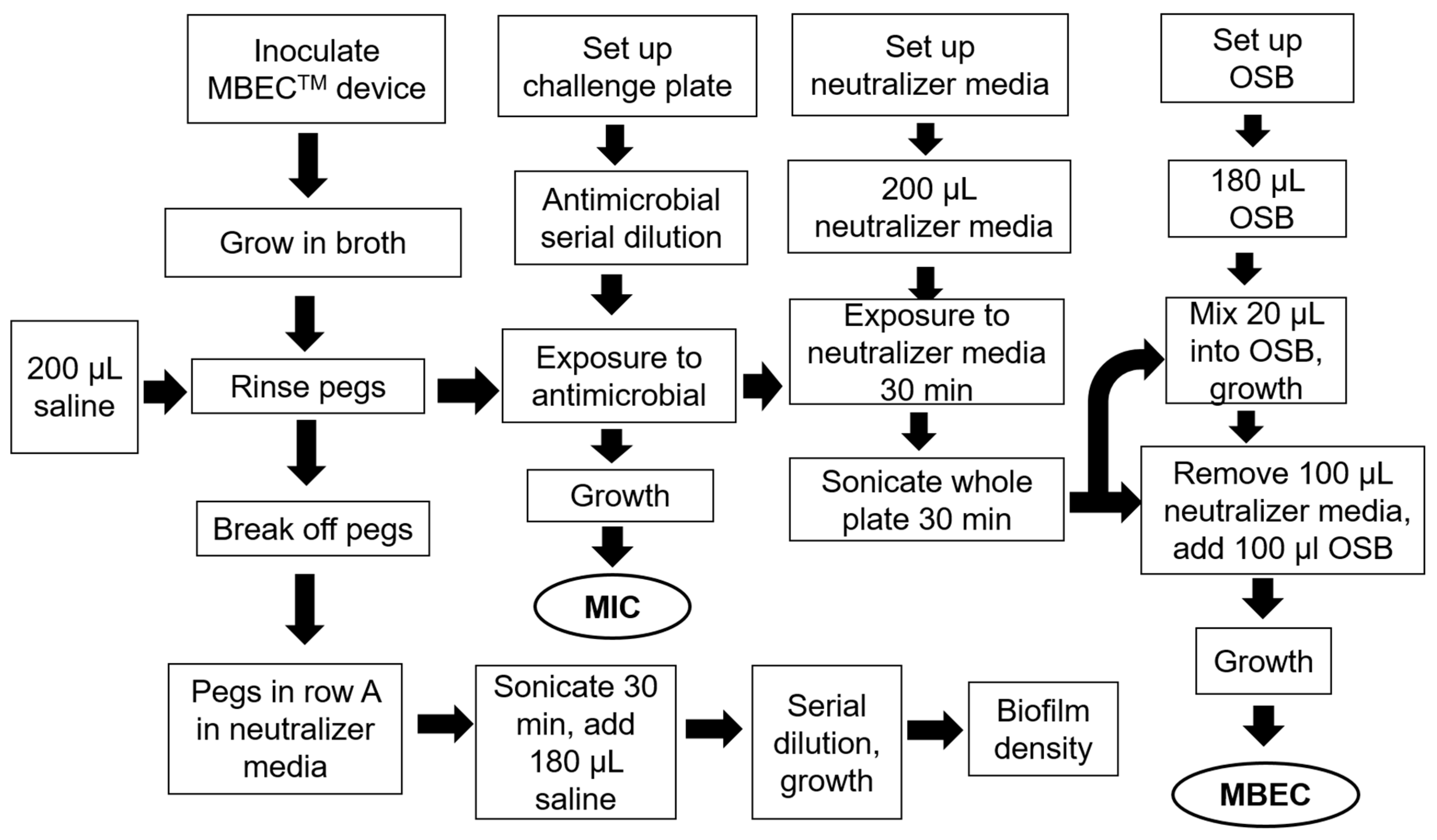
2.6. MIC and MBEC Assay Device

To asses both the MICs of planktonic cells and the efficacy of antimicrobials against biofilm we used the MBEC assay device (Innovotech Inc, Edmonton, AB, Canada). In brief, second sub-cultures of A. hydrophila and C. portucalensis were grown in TSB media were adjusted to a cell density of 106 CFU/mL. The MBEC, MBC, and MIC were analyzed following the standard protocol as per Innovotech’s MBEC assay (Innovotech Inc, Edmonton, AB, Canada), https://www.innovotech.ca/wp-content/uploads/2019/02/MBEC-Procedural-Manual-v2.0.pdf. The assay workflow is presented in Figure 1. The antibiotic concentrations used in each of the eight rows of the 96-well plate are presented in the legends to Figure 1. The challenge plate was prepared by diluting a 1 mg/mL stock solution of neomycin or cotrimoxazole in each row. TSB, sterile neutralizer and water were added to three specific wells designated the sterile, neutralizer, and neutralizer effective control. It was freshly prepared and used within 30 min. The rinse plate was prepared by adding 180 µL of TSB to a new 96-well plate. The peg lid was rinsed by setting the lid into rinse plate for 10 s. The peg lid was transferred to the challenge plate and incubated as per the protocol. The recovery plate was prepared by adding 200 μL of neutralizer media to a new plate and after the appropriate time the peg lid was transferred to the recovery plate. The device was again sonicated to remove the attached biofilm. 100 μL of sterile media was added to each well of recovery plate and incubated at 37 °C for 24 h to determine the MBEC via a micro-titer plate reader at 650 nm. The MBEC value is the minimum concentration of antibiotic that inhibits growth of biofilm as indicated by the control wells with no turbidity. The MBEC protocol used was modified minimally from the manufacturer’s manual neomycin and cotrimoxazole (trimethoprim/sulfamethoxazole) serially diluted with the highest concentration at 1 mg/mL each, were used as antibiotics, with each of the eight rows of the 96-well plate, from A through H, being 1000, 500, 250, 125, 62.5, 31.3, 15.6, and 7.8 µg/mL, respectively.
The MBEC and MIC were analyzed following the standard protocol as per Innovotech’s MBEC assay. The MBEC assay workflow is presented in Figure 1.

2.7. Scanning Electron Microscopy (SEM)

The A. hydrophila and C. portucalensis biofilm-covered pegs treated with cotrimoxazole and neomycin were rinsed with phosphate buffered saline (PBS) and 2% glutaraldehyde. The samples were further processed according to an open source protocol [82]. After sputter-coating for two min with gold-palladium, the SEM was performed at a voltage of 5 kV using a Mira3 Tescan field emission SEM at the Rochester Institute of Technology (RIT) Nanoimaging Lab.

2.8. Identification of Quorum Sensing Signals of the Acyl-Homoserine Lactone (AHL) Class

Complementary formats [83,84] were used to identify then to separate and identify quorum sensing signals of the acyl-homoserine lactone (AHL) class. Plate T-streak and diffusion-disc bioassays are standard protocols in our laboratory and performed as previously described [85,86]. Reverse phase (RP) thin layer chromatogram was performed with silica gel 60 RP-18 plates (20 × 20 cm, EMD Chemical Inc., Gibbstown, NJ, USA). For RP TLC every 2.8 cm on the line, a mark was made to identify the center of each column. Each samples was loaded with 2–3 µL volumes intervals to reach the final sample size. Conditions of TLC development (60% methanol:water), and the preparation and use of whole cell AHL-dependent bacterial biosensors and visualization of AHL signals were as described previously [84,85]. After drying, the position and number of AHLs were detected with a thin overlay of the TraR-dependent Agrobacterium biosensor strain A136 [87]. The biosensor-overlaid plate was left in a sterile hood to solidify and dry for 30 min, then covered and placed in a 28 °C incubator overnight [84,85,86].

2.9. Bioinformatics Analysis of AHL-Related Genes

The genomes of strains A. hydrophila RIT 668 and C. portucalensis RIT669 were scanned for sequences homologous to established quorum sensing genes luxI and luxR. This was performed by first mining each genome for proteins using Prodigal [88], and subsequently using HMMER to scan the predicted protein database using PFAM domains PF03472 and PF0076 as bait for LuxR and LuxI, respectively [89]. The HMM hits were further scrutinized using InterProScan7 [90]. Hits showing domains IPR016032, IPR005143, and IPR000792 were considered to be valid LuxR homologs, while hits showing domains IPR001690 and IPR018311 were considered to be valid LuxR homologs, as described in Gan et al. [91].

2.10. Computational Methods

The doubling times in TSB was calculated using the online tool https://doubling-time.com/compute.php [92], and was reported as an average of the doubling time values at different time intervals after inoculation, such as 35, 85, 115, 130, 145, 165, and 225 min, during the exponential phase. The zones of inhibition were recorded and categorized as—resistant (R), intermediate (I) and susceptible (S). Statistical analysis of the MIC, MBC, and MBEC results were performed using ANOVA with α = 0.05 and Fisher’s (95% confidence intervals).

3. Results

3.1. A. hydrophila

3.1.1. Growth

Doubling time analysis of our A. hydrophila strain when grown in tryptic soy broth at 37 °C showed an average generation time of 94 min. An earlier work on the growth at 35 °C of A. hydrophila strains in Brain Heart Infusion broth buffered with 3-[N-morpholino] propane sulfonic acid (MOPS) and adjusted to pH 7.0, reported generation times of 19.8–23.4 min [93].

3.1.2. Taxonomy

The assembled genomes of RIT668 was placed within taxonomic contexts through a whole-genome approach via the Type Strain Genome Server (TYGS). Strain RIT668 grouped most closely with a type strain of Aeromonas hydrophila ATCC 7966 with a bootstrap support value of 96 on the Genome Blast Distance Phylogeny tree (GBDP), as shown in Figure S1. The phylogenetic tree was created using GBDP distances between RIT668 and 15 closely related genomes in the TYGS database, and had an average branch support of 99.0% with a δ statistic of 0.166, a measure of tree-likeness. The assembly of RIT 668 had a dDDH d4 percentage of 74.9% compared to the genome of Aeromonas hydrophila ATCC 7966 and 72.2% compared to the genome of Aeromonas hydrophila subsp. ranae CIP 107985. The d4 value was chosen to avoid issues caused by an incompletely sequenced genome. The accepted dDDH threshold for two genomes coming from the same species is 70%.

3.1.3. Antimicrobial susceptibility screening with preloaded antibiotic discs

The disc diffusion assay was applied as a preliminary analysis to determine the effect of 11 different antibiotics (Table 1) on the susceptibility of A. hydrophila, under standard conditions, using fixed concentration paper antibiotic discs along with a control. A. hydrophila was resistant to gentamicin (10), tetracycline (30), doxycycline (30), kanamycin (30), streptomycin (10), tobramycin (10), novobiocin (30), and erythromycin (15 µg), respectively. A. hydrophila was susceptible only to neomycin and sulfamethaxazole-trimethoprim.

3.1.4. Antimicrobial Susceptibility Screening Using the Stock Concentration Method

Disc assays using stock concentrations of selected antibiotics did not have an inhibitory effect in most cases on A. hydrophila grown on TSA. Only tetracycline, neomycin and cotrimoxazole had significant inhibition zones (Table 2). The drugs tested belongs to aminoglycoside, tetracycline, aminocoumarin, macrolide, beta-lactam, ansamycin, cephalosporin, fusidane, lincomycin, and sulfonamide classes, out of which our isolates are resistant to the aminoglycoside (except neomycin), fusidane, lincomycin, ansamycin and cephalosporin classes (Table 3). A. hydrophila was susceptible to neomycin, cotrimoxazole, and tetracycline, which target the 30S ribosomal subunit preventing protein synthesis, the enzyme dihydropteroate synthase (DHPS), and the binding of aminoacyl tRNA to the RNA-ribosome complex, respectively.

3.1.5. MIC and MBEC

MIC: The MIC of A. hydrophila when challenged with the antibiotic cotrimoxazole was found to be between 500 and 1000 μg/mL (Table 4); whereas the MIC with Neomycin for this same bacterium was found to be between 32.3 and 62.5 μg/mL (P < 0.05, Table 4). An exact numerical value could not be determined due to the nature of the protocol as the concentrations decreased in each row by a factor of two. Values shown in the MIC ranges were determined as the lowest concentration of the antibiotic where microbial growth was inhibited and the Fisher confidence intervals are shown for cotrimoxazole (Figure 2A) and neomycin (Figure 2B).
MBEC: A trend indicating increased biofilm eradication at higher antibiotic concentrations was observed. Cotrimoxazole and neomycin in the concentration range of 7.8 to 1000 μg/mL, were able to eradicate biofilms of both A. hydrophila (P > 0.05). Between 125 and 250 μg/mL of cotrimoxazole, growth drastically decreased, but biofilms were not eradicated (Figure S2A). Similar results were observed for neomycin (Figure S2B). This could be due to the highest concentration being as low as 1 mg/mL. Overall, the trend showed neomycin treatment with higher concentrations were generally more effective than cotrimoxazole.

3.1.6. Scanning Electron Microscopy (SEM) Analysis of MBEC Samples

SEM imaging of A. hydrophila revealed fewer cells in samples containing highest concentrations of antibiotics, and higher number of cells in samples exposed to lowest concentrations of antibiotics (Figure 3). A. hydrophila treated with cotrimoxazole resulted in biofilms showing multiple layers of cells at the lowest antibiotic concentration (Figure 3A) and a single layer at the highest concentration (Figure 3B), while neomycin-treated samples showed flat biofilms in the presence of both the highest and lowest antibiotic concentrations (Figure 3C,D).

3.1.7. Detection of Quorum Sensing

A. hydrophila produces AHL-type quorum sensing (QS) signals as assessed by the use of two complementary whole-cell biosensors, CV026 and NTL4 (pZLR4) in T-streak, diffuse-disc and thin layer chromatography formats (Figure 4). Using a narrow- and broad-range AHL-detection biosensor, A hydrophila was shown to activate of the reporter genes indicating AHL production. TLC analysis of ethyl acetate extracts from A. hydrophila showed the production of a single detectable AHL that co-migrates with N-(3-hydroxyoctanoyl)-L-homoserine lactone standard.

3.2. C. portucalensis

3.2.1. Growth

The average doubling time at 37 °C in TSB, of our C. portucalensis strain was 103 min. This is much slower than the reported doubling time of 50 min for a clinical strain C. freundii GN346 at the same temperature [84].

3.2.2. Taxonomy

Based on a GBDP tree, RIT669 was most closely related to Citrobacter portucalensis A60, as shown in Figure S3. The GBDP tree containing RIT669 and 13 closely related genomes had an average branch support of 91.7% and a δ statistic of 0.113. The dDDH d4 percentage between the assembly of RIT699 and C. portucalensis A60 was 80.1%, well about the 70% threshold for conspecifics. Additionally, the dDDH d4 value comparing RIT669 and Citrobacter freundii, the next most closely related species, was 58.3%.
To further confirm the placement of RIT669 in the species C. portucalensis, the assembly of RIT669 was uploaded to JSpecies, an online service for calculation of Average Nucleotide Identity values. The ANIb value comparing RIT669 with C. portucalensis strain 4 7 47CFAA was 98.98%, which was above the 95% cutoff for conspecifics. The ANIb value comparing RIT669 with C. freundii strain CAV1321 was 94.13%. The ANIm values also supported assignment of RIT669 to the species C. portucalensis, with a value of 99.27% between RIT669 and C. portucalensis strain 4 7 47CFAA, and a value of 94.67% between RIT669 and C. freundii.

3.2.3. Antimicrobial Susceptibility Screening with Preloaded Antibiotic Discs

C. portucalensis was resistant to gentamicin (10), tetracycline (30), doxycycline (30), kanamycin (30), streptomycin (10), tobramycin (10), novobiocin (30), and erythromycin (15 µg), respectively. The response to sulfamethoxazole-trimethoprim is characterized as intermediate. However, it is susceptible to neomycin (Table 1).

3.2.4. Antimicrobial Susceptibility Screening Using the Stock Concentration Method

Like A. hydrophila, C. portucalensis also resisted all the tested antibiotics at the stock concentrations while growing on TSA, except for tetracycline, neomycin, and cotrimoxazole had significant inhibition zones (Table 2). C. portucalensis was susceptible to neomycin, cotrimoxazole, and tetracycline, all of which target the 30S ribosomal subunit.

3.2.5. MIC and MBEC

MIC: The MIC for C. portucalensis after challenged with cotrimoxazole was determined to be between 7.8 and 15.6 μg/mL (P < 0.05) (Figure 5A). For the neomycin test, the concentration was found to be between 7.8 and 31.3 μg/mL (P < 0.05) (Figure 5B).
MBEC: Neither of the antibiotics were able to significantly eradicate the biofilms of C. portucalensis, at the evaluated concentrations of 7.8–1000 μg/mL (P > 0.05) (Figure S4A,B). Higher concentrations were more effective at biofilm elimination; however, eradication was not observed. Overall, both antibiotics were more effective against the biofilms of C. portucalensis than those of A. hydrophila. The true values of MBEC are probably much higher, since the complete eradication of biofilms was not observed at 1000 µg/mL.

3.2.6. Scanning Electron Microscopy (SEM) Analysis of MBEC Samples

Most samples did not show major morphological changes, except C. portucalensis treated with cotrimoxazole. At the lowest concentrations, biofilm formation can be seen to contain cells with projections (Figure 6A). The cells at the highest concentration occur in a flatter arrangement (Figure 6B), but the cells appear linked by fibers under higher magnification (Figure 6C,D). Additionally, the higher magnification revealed spiky projections in the sample with the highest cotrimaxazole concentration (Figure 6D). In the neomycin treatment of C. portucalensis, there were sparse cells (Figure 7A,B), with some cell elongation observed at the highest concentration (Figure 7B).

3.2.7. Detection of Quorum Sensing

C. portucalensis also produces AHL type QS signals as assessed by the same methods for AHL detection as before (Figure 4). The C. portucalensis strain failed to activate the AHL-dependent response of violacein production in the narrow-range biosensor CV026, but activated the broad-range AHL-TraR-based reporter gene in biosensor NTL4 (pZLR4). The C. portucalensis strain showed a single elongated spot, which upon close inspection, appears as two overlapping joined spots. Upon close comparison with seven different AHL standards, clear co-migrations of either putative AHL spot from C. portucalensis with any AHL standard on the chromatogram was not discernable (Figure 4).

4. Discussion

A. hydrophila has been reported to resist different antibiotic classes such as quinolones, aminoglycosides, beta-lactams, tetracyclines, chloramphenicol, trimethoprim, and sulfonamides [22,95,96]. A. hydrophila strains expressing the AheABC efflux pump were able to export 13 substrates, including nine antibiotics, and the suppression of the efflux process by the inhibitor phenylalanine-arginine-β-naphthylamide, suggested the presence of additional systems leading to intrinsic resistance [97]. The antibiotics moxalactam and cefoxitin were demonstrated to inactivate the metallo-beta-lactamase CphA of an A. hydrophila strain by forming stable adducts via reaction with the active site residues [98]. Apart from harboring metallo-beta-lactamases and efflux pumps, naturally occurring Aeromonas spp. have been shown to be transformable and have a high incidence of horizontal gene transfer [99,100]. Shiga-like toxin production causing bloody diarrhea and hemolytic uremic syndrome is reported from Aeromonas spp. [101], and there is evidence that surface colonization upregulates the expression of the Shiga-like toxin genes [17].
The mortality rate for hospitalized patients with Citrobacter infections is reported to be 6.8% [29], and this figure rises to 18–56% for patients with Citrobacter bacteremia [30,102,103]. C. freundii strains are already known for extended β-lactamase and quinolone resistance [32,39,40,41,42,43]. C. portucalensis strains also are multidrug resistant [24,25], with a poultry isolate considered a “superbug” [24]. Making a rational choice of antimicrobial therapy for Citrobacter infections can be problematic, since they encode chromosomal, inducible ampC β-lactamase genes. High levels of constitutive expression due to mutational changes confer resistance to multiple antibiotics [102,104]. Additional plasmid-mediated determinants of resistance may co-exist [103]. Multidrug resistant C. freundii clinical isolates resist up to 12 antibiotics, including penicillins, cephalosporins, carbapenems, and fluoroquinolones [48]. Extreme drug resistance is also known [49]. Furthermore, the genome of the leafy vegetable strain of C. portucalensis possesses aminoglycoside, beta-lactam, chloramphenicol, sulfonamide, tetracycline, and trimethoprim resistance genes [25], whereas the poultry superbug C. portucalensis encodes 13 antibiotic resistance genes, enabling resistance to eight distinct groups of antibiotics [24]. The mcr-3 gene encoding resistance to the last resort antibiotic colistin, is also disseminated universally in both Enterobacteriaceae (which include Citrobacter spp.) and Aeromonas spp., and the latter is a potential reservoir for this gene [105]. We have used pre-loaded discs and stock concentration tests to assess the resistance at different concentration ranges, primarily to establish upper limits (observe if any effect is seen even at concentrations much higher than allowed for clinical applications). Our results show A. hydrophila and C. portucalensis resist many more classes of antibiotics than predicted by bioinformatics analysis using the Resistance Gene Identifier (RGI) tool [94] (Table 3), suggesting that the database may be incomplete. The existing international criteria for multi-drug resistance (MDR) in the Enterobacteriaceae is the non-susceptibility to at least three classes of antibiotics [106]. Therefore, both the isolates in this study qualify as multidrug resistant. The overexpression of genes encoding for ATP-binding cassette (ABC) transporter membrane proteins or multidrug efflux pumps, which enable the export of drugs out of the cell is a prevalent cause of MDR [107,108,109]. Analysis of the genomes using the RGI server suggest that efflux pumps may be present in both isolates [17]. Future studies should address the mechanisms of MDR in the isolated A. hydrophila and C. portucalensis strains.
Biofilms are directly related to virulence [110]. Sessile cells are resistant to antibiotics and various disinfectants [111]. Frequently, the results of in vitro antimicrobial studies do not correlate with therapeutic results, since most studies assess resistance in the planktonic phase, and biofilms are not taken into account [12]. Biofilm phases of most bacteria are able to resist antibiotics at concentrations hundreds or thousands of times higher than the corresponding planktonic phases [44,46,65,66,67,68,69]. Cotrimoxazole was reported to be effective against A. hydrophila and Citrobacter spp. in the planktonic phase [112]. It has been reported to cure C. freundii infections even when other drugs have failed [113]. Over 70% of Citrobacter isolates in a 11-year single center study were reported to be susceptible to cotrimoxazole [60]. Neomycin has also been reported to clear infections caused by C. freundii [61]. Studies also report most food-associated strains and 50–65% of animal-associated strains of A. hydrophila as being susceptible to neomycin and cotrimoxazole [62,63]. Hence, the susceptibility of our strains to neomycin and cotrimoxazole in the planktonic phase was not surprising.
However, biofilms are expected to be more resistant. In fact, biofilms of C. freundii can be eradicated only by chlorite based disinfectants [114], which are toxic to humans. Therefore, surface colonization by RIT668 and RIT 669 poses a significant risk in case humans do get infected with them, in view of their multidrug resistance and biofilm formation, as well as toxin upregulation in biofilms formed on plastics [17]. Plastics, particularly microplastics, can act as vectors for bacterial pathogens [115] and aquatic animals ingest significant quantities of microplastics [116]. Thus, the environmental transmission of plastic waste from infected aquatic animals may expose humans to antibiotic resistant pathogens. Our strains were obtained from infected turtles, where they caused fatal infections. Additionally, at least one case where reptile and clinical strains of Aeromonas spp. were identical is known [18]. Therefore, the potential for zoonotic transmission of RIT668 and RIT669 must be considered.
SEM can be used to observe the overall shape of microorganisms composing the biofilm and their spatial organization, which can resolve biofilm growth on surfaces containing junctions between materials [117,118]. The optical sectioning of biofilms has provided the insight that biofilm structure depends on the type of bacterium [119]. A. hydrophila biofilms in this study did not show any specialized structures. However, the fibers connecting cells of C. portucalensis reported here are similar to those formed by enteroaggregative Escherichia coli in mixed diarrhea-associated biofilms with C. freundii and whose formation is considered to be mediated by F. pili [8]. Along with the morphological changes of the cells, the SEM analysis of samples in this study also revealed structurally modified cellular products surrounding the biofilm clusters, as reported by other authors [120].
The luxI gene codes for the AHL-synthase enzyme, while the LuxR is the AHL-receptor [121]. The bioinformatics analysis of each genome revealed that A. hydrophila RIT668 possesses a single luxI and three luxR homologs, whereas C. freundii RIT669 encodes for no identifiable luxI homologs but contains a single luxR homolog. However, in our experiments, both A. hydrophila and C. portucalensis produced a dose-dependent response in the AHL-dependent NTL4 (pZLR4) which uses the TraR receptor (Figure 4B). A. hydrophila and A. salmonicida strains have been shown to express homologs of the luxI and luxR, and produce C4 AHL as the major and C6 AHL as the minor quorum sensing signals [122]. Here we show that A. hydrophila RIT668 produces a single AHL, which co-migrates with N-(3-Hydroxyoctanoyl)-L-homoserine lactone. AHL-type QS signals are not produced by certain C. freundii strains [123]. Diketopiperazines (DKPs) are known to be produced in some C. freundii strains, which may activate the AHL-sensing proteins, but no biosensor response was observed in this study with biosensor CV026 (Figure 4A), which is known to cross-activate with DKPs [124]. In contrast, the broad-range TraR-dependent biosensor was activated in a dose-dependent manner (Figure 4B) and TLC analysis showed an elongated spot that upon close examination appear as two but overlapping AHL-like signal spots (Figure 4C). Biofilms of our isolates are not eradicated by even 1000 µg/mL of cotrimoxazole or neomycin. Owing to the well-known hematological toxicity of cotrimoxazole [125], as well as nephrotoxicity and ototoxicity of neomycin [126], such high dosages are precluded from clinical use. Biofilm formation is coordinated by QS mechanisms and since biofilms are harder to treat with antibiotics, the inhibition of QS could be a viable antibacterial strategy [127]. Chestnut honey and vanillin have been reported as having anti-biofilm and anti-QS effects on A. hydrophila [128,129]. N-acetylcysteine (NAC) linked to polymers offers a way to block biofilm formation by both A. hydrophila and C. freundii strains [6]. In view of the multidrug resistance of our isolates, anti-QS and anti-biofilm agents might offer effective alternatives to antibiotics.

5. Conclusions

A. hydrophila RIT668 and C. portucalensis RIT669 are multidrug resistant, and can be controlled only by neomycin and cotrimoxazole in the planktonic phase. However, the eradication of biofilms of either organism may need >> 1000 µg/mL of neomycin or cotrimoxazole, effectively meaning that neomycin or cotrimoxazole administration is not a clinical option, and indirect approaches to suppress biofilms may be necessary. A. hydrophila produces N-(3-Hydroxyoctanoyl)-L-homoserine lactone quorum sensing signals, but their significance for virulence or antibiotic resistance in RIT668 will require further investigations. C. portucalensis RIT669 activated our broad range AHL-dependent whole cell biosensor but further work is need to identify the potential two AHLs identified as well as the biosynthetic pathway used since a luxI homolog was not readily located in our whole genome sequence of this strain. The emergence of multi-drug resistance in Aeromonas spp. strains in water environments [130] and resistance determinants against last resort antibiotics, such as colistin [105,131] make A. hydrophila species increasingly dangerous pathogens. Multidrug resistant C. freundii which produce both carbapenemase and metallo-beta-lactamase have been reported, which resisted all tested antibiotics except tetracycline have been isolated in China [132], while a potentially zoonotic superbug C. portucalensis strain is known [24]. This suggests C. portucalensis and C. freundii strains are also emerging as a significant risk. The capacity for plastic colonization, multidrug resistance, and biofilm formation suggests that our turtle isolates, A. hydrophila RIT668 and C. portucalensis RIT669, are potential zoonotic pathogens, with heightened risks for patients living with polymer implants/IMDs. However, the mechanisms of multidrug resistance in these isolates is unknown and warrant further investigation.

Supplementary Materials

The following are available online at https://www.mdpi.com/2076-2607/8/11/1805/s1, Figure S1: FASTME Phylogenetic tree of 15 strains related to strain RIT668 based on GBDP distances, Figure S2: Fisher individual 95% confidence intervals for the MBEC values from the neutralizer plate of A. hydrophila RIT668, Figure S3: FASTME Phylogenetic tree of 13 strains related to strain RIT669 based on GBDP distances, Figure S4: Fisher individual 95% confidence intervals for the MBEC values from the neutralizer plate of C. portucalensis RIT669.

Author Contributions

The experiments were mainly carried out by S.G.T., M.A. and M.A.G. The experiments were designed by S.G.T. and A.P. in consultation with A.O.H., P.C.W. did the quorum sensing experiments under the direction of M.A.S., P.A.S. provided the infected turtles for the study. S.G.T., A.P. and A.O.H. wrote the initial draft and further improvements were made by C.B.W. and M.A.S. The study was funded by a NIH award to A.O.H. All authors have read and agreed to the published version of the manuscript.

Funding

This research and APC was funded by a National Institutes of Health (NIH) award (R15GM120653) to A.O.H.

Acknowledgments

The authors wish to thank Richard Hailstone for running the SEM samples and Narayan H. Wong for the phylogenetic analysis.

Conflicts of Interest

The authors declare no conflict of interest.

References

  1. Cooper, M.A.; Shlaes, D. Fix the antibiotics pipeline. Nature 2011, 472, 32. [Google Scholar] [CrossRef]
  2. Wright, G.D. The antibiotic resistome. Expert Opin. Drug Discov. 2010, 5, 779–788. [Google Scholar] [CrossRef]
  3. D’Costa, V.M.; King, C.E.; Kalan, L.; Morar, M.; Sung, W.W.L.; Schwarz, C.; Froese, D.G.; Zazula, G.D.; Calmels, F.; Debruyne, R.; et al. Antibiotic resistance is ancient. Nat. Cell Biol. 2011, 477, 457–461. [Google Scholar] [CrossRef]
  4. Igbinosa, I.H.; Igumbor, E.U.; Aghdasi, F.; Tom, M.; Okoh, A.I. EmergingAeromonasSpecies Infections and Their Significance in Public Health. Sci. World J. 2012, 2012, 625023. [Google Scholar] [CrossRef] [PubMed] [Green Version]
  5. Ranjan, K.P.; Ranjan, N. Citrobacter: An emerging health care associated urinary pathogen. Urol. Ann. 2013, 5, 313–314. [Google Scholar] [PubMed]
  6. Kregiel, D.; Rygala, A.; Kolesinska, B.; Nowacka, M.; Herc, A.S.; Kowalewska, A. Antimicrobial and Antibiofilm N-acetyl-L-cysteine Grafted Siloxane Polymers with Potential for Use in Water Systems. Int. J. Mol. Sci. 2019, 20, 2011. [Google Scholar] [CrossRef] [PubMed] [Green Version]
  7. Peng, Q.; Chen, L.; Zhou, S.; Li, H.; Long, J.; Yao, F.; Zhuang, Y.; Zhang, Z.; Huang, Y.; Duan, K. Co-existence of Citrobacter freundii exacerbated Pseudomonas aeruginosa infection in vivo. Int. J. Med. Microbiol. 2020, 310, 151379. [Google Scholar] [CrossRef]
  8. Pereira, A.L.; Silva, T.N.; Gomes, A.C.; Araujo, A.C.G.; Giugliano, L.G. Diarrhea-associated biofilm formed by enteroaggregative Escherichia coli and aggregative Citrobacter freundii: A consortium mediated by putative F pili. BMC Microbiol. 2010, 10, 57. [Google Scholar] [CrossRef] [Green Version]
  9. McCormick, A.; Hoellein, T.J.; Mason, S.A.; Schluep, J.; Kelly, J.J. Microplastic is an Abundant and Distinct Microbial Habitat in an Urban River. Environ. Sci. Technol. 2014, 48, 11863–11871. [Google Scholar] [CrossRef]
  10. Heo, G.-J.; Hossain, S.; Wimalasena, S. Virulence Factors and Antimicrobial Resistance Pattern of Citrobacter freundii Isolated from Healthy Pet Turtles and their Environment. Asian J. Anim. Veter- Adv. 2017, 12, 10–16. [Google Scholar] [CrossRef]
  11. Chung, T.; Yi, S.; Kim, B.; Kim, W.; Shin, G. Identification and antibiotic resistance profiling of bacterial isolates from septicaemic soft-shelled turtles (Pelodiscus sinensis). Vet. Med. 2017, 62, 169–177. [Google Scholar] [CrossRef] [Green Version]
  12. Wimalasena, S.H.M.P.; Shin, G.-W.; Hossain, S.; Heo, G.-J. Potential enterotoxicity and antimicrobial resistance pattern of Aeromonas species isolated from pet turtles and their environment. J. Vet. Med. Sci. 2017, 79, 921–926. [Google Scholar] [CrossRef] [PubMed] [Green Version]
  13. Pace, A.; Dipineto, L.; Fioretti, A.; Hochscheid, S. Loggerhead sea turtles as sentinels in the western Mediterranean: Antibiotic resistance and environment-related modifications of Gram-negative bacteria. Mar. Pollut. Bull. 2019, 149, 110575. [Google Scholar] [CrossRef] [PubMed]
  14. Al-Bahry, S.; Mahmoud, I.; Al-Zadjali, M.; Elshafie, A.; Al-Harthy, A.; Al-Alawi, W. Antibiotic resistant bacteria as bio-indicator of polluted effluent in the green turtles, Chelonia mydas in Oman. Mar. Environ. Res. 2011, 71, 139–144. [Google Scholar] [CrossRef] [PubMed] [Green Version]
  15. Rabinowitz, P.M.; Conti, L. Links among Human Health, Animal Health, and Ecosystem Health. Annu. Rev. Public Health 2013, 34, 189–204. [Google Scholar] [CrossRef] [Green Version]
  16. Rosen, G.E.; Smith, K.F. Summarizing the Evidence on the International Trade in Illegal Wildlife. EcoHealth 2010, 7, 24–32. [Google Scholar] [CrossRef]
  17. Thomas, S.G.; Glover, M.A.; Parthasarathy, A.; Wong, N.H.; Shipman, P.A.; Hudson, A.O. Expression of a Shiga-Like Toxin during Plastic Colonization by Two Multidrug-Resistant Bacteria, Aeromonas hydrophila RIT668 and Citrobacter freundii RIT669, Isolated from Endangered Turtles (Clemmys guttata). Microorganisms 2020, 8, 1172. [Google Scholar] [CrossRef]
  18. McCoy, R.H.; Seidler, R.J. Potential Pathogens in the Environment: Isolation, Enumeration, and Identification of Seven Genera of Intestinal Bacteria Associated with Small Green Pet Turtles1. Appl. Microbiol. 1973, 25, 534–538. [Google Scholar] [CrossRef] [Green Version]
  19. Esteve, C.; Alcaide, E.; Giménez, M.J. Multidrug-resistant (MDR) Aeromonas recovered from the metropolitan area of Valencia (Spain): Diseases spectrum and prevalence in the environment. Eur. J. Clin. Microbiol. Infect. Dis. 2015, 34, 137–145. [Google Scholar] [CrossRef]
  20. Marchandin, H.; Godreuil, S.; Darbas, H.; Jean-Pierre, H.; Jumas-Bilak, E.; Chanal, C.; Bonnet, R. Extended-spectrum beta-lactamase TEM-24 in an Aeromonas clinical strain: Acquisition from the prevalent Enterobacter aerogenes clone in France. Antimicrob. Agents Chemother. 2003, 47, 3994–3995. [Google Scholar] [CrossRef] [Green Version]
  21. Janda, J.M.; Abbott, S.L. The Genus Aeromonas: Taxonomy, Pathogenicity, and Infection. Clin. Microbiol. Rev. 2010, 23, 35–73. [Google Scholar] [CrossRef] [PubMed] [Green Version]
  22. Epiotrowska, M.; Popowska, M. Insight into the mobilome of Aeromonas strains. Front. Microbiol. 2015, 6, 494. [Google Scholar] [CrossRef] [Green Version]
  23. Ribeiro, T.G.; Ribeiro-Gonçalves, B.; Da Silva, M.S.; Novais, Â.; Machado, E.; Carriço, J.A.; Peixe, L. Citrobacter portucalensis sp. nov., isolated from an aquatic sample. Int. J. Syst. Evol. Microbiol. 2017, 67, 3513–3517. [Google Scholar] [CrossRef] [PubMed]
  24. Hasan, S.; Sultana, M.; Hossain, M.A. Complete genome arrangement revealed the emergence of a poultry origin superbug Citrobacter portucalensis strain NR-12. J. Glob. Antimicrob. Resist. 2019, 18, 126–129. [Google Scholar] [CrossRef]
  25. Igbinosa, E.O.; Rathje, J.; Habermann, D.; Brinks, E.; Cho, G.-S.; Franz, C.M. Draft Genome Sequence of Multidrug-Resistant Strain Citrobacter portucalensis MBTC-1222, Isolated from Uziza (Piper guineense ) Leaves in Nigeria. Genome Announc. 2018, 6. [Google Scholar] [CrossRef] [Green Version]
  26. Bai, L.; Xia, S.; Lan, R.; Liu, L.; Ye, C.; Wang, Y.; Jin, D.; Cui, Z.; Jing, H.; Xiong, Y.; et al. Isolation and Characterization of Cytotoxic, Aggregative Citrobacter freundii. PLoS ONE 2012, 7, e33054. [Google Scholar] [CrossRef] [Green Version]
  27. Anderson, M.T.; Mitchell, L.A.; Zhao, L.; Mobley, H.L.T. Citrobacter freundii fitness during bloodstream infection. Sci. Rep. 2018, 8, 11792. [Google Scholar] [CrossRef]
  28. Liu, L.; Chen, D.; Liu, L.; Lan, R.; Hao, S.; Jin, W.; Sun, H.; Wang, Y.; Liang, Y.; Xu, J. Genetic Diversity, Multidrug Resistance, and Virulence of Citrobacter freundii From Diarrheal Patients and Healthy Individuals. Front. Cell. Infect. Microbiol. 2018, 8, 233. [Google Scholar] [CrossRef] [Green Version]
  29. Mohanty, S.; Singhal, R.; Sood, S.; Dhawan, B.; Kapil, A.; Das, B.K. Citrobacter infections in a tertiary care hospital in Northern India. J. Infect. 2007, 54, 58–64. [Google Scholar] [CrossRef]
  30. Samonis, G.; Karageorgopoulos, D.E.; Kofteridis, D.P.; Matthaiou, D.K.; Sidiropoulou, V.; Maraki, S.; Falagas, M.E. Citrobacter infections in a general hospital: Characteristics and outcomes. Eur. J. Clin. Microbiol. Infect. Dis. 2009, 28, 61–68. [Google Scholar] [CrossRef]
  31. Chen, Y.-S.; Wong, W.-W.; Fung, C.-P.; Yu, K.-W.; Liu, C.-Y. Clinical features and antimicrobial susceptibility trends in Citrobacter freundii bacteremia. J. Microbiol. Immunol. Infect. 2002, 35, 109–114. [Google Scholar] [PubMed]
  32. Liu, L.-H.; Wang, N.-Y.; Wu, A.Y.-J.; Lin, C.-C.; Lee, C.-M.; Liu, C.-P. Citrobacter freundii bacteremia: Risk factors of mortality and prevalence of resistance genes. J. Microbiol. Immunol. Infect. 2018, 51, 565–572. [Google Scholar] [CrossRef] [PubMed]
  33. Joaquin, A.; Khan, S.; Russell, N.; Al Fayez, N. Neonatal Meningitis and Bilateral Cerebellar Abscesses due to Citrobacter freundii. Pediatr. Neurosurg. 1991, 17, 23–24. [Google Scholar] [CrossRef] [PubMed]
  34. Samonis, G.; Anaissie, E.; Elting, L.; Bodey, G.P. Review ofCitrobacter bacteremia in cancer patients over a sixteen-year period. Eur. J. Clin. Microbiol. Infect. Dis. 1991, 10, 479–485. [Google Scholar] [CrossRef]
  35. Aminharati, F.; Ehrampoush, M.H.; Dallal, M.M.S.; Yaseri, M.; Tafti, A.A.D.; Rajabi, Z. Citrobacter freundii Foodborne Disease Outbreaks Related to Environmental Conditions in Yazd Province, Iran. Iran J. Public Health 2019, 48, 1099–1105. [Google Scholar]
  36. Tschape, H.; Prager, R.; Streckel, W.; Fruth, A.; Tietze, E.; Böhme, G. Verotoxinogenic Citrobacter freundii associated with severe gastroenteritis and cases of haemolytic uraemic syndrome in a nursery school: Green butter as the infection source. Epidemiol. Infect. 1995, 114, 441–450. [Google Scholar] [CrossRef] [Green Version]
  37. Pardia, S.N.; Verma, I.C.; Deb, M.; Bhujwala, R.A. An outbreak of diarrhea due to citrobacter freundii in a neonatal special care nursery. Indian J. Pediatr. 1980, 47, 81–84. [Google Scholar] [CrossRef]
  38. Chen, Y.; Brook, T.C.; Alcon-Giner, C.; Clarke, P.; Hall, L.J.; Hoyles, L. Draft Genome Sequences of Citrobacter freundii and Citrobacter murliniae Strains Isolated from the Feces of Preterm Infants. Microbiol. Resour. Announc. 2019, 8. [Google Scholar] [CrossRef] [Green Version]
  39. Shao, Y.; Xiong, Z.; Li, X.; Hu, L.; Shen, J.; Li, T.; Hu, F.; Chen, S. Prevalence of plasmid-mediated quinolone resistance determinants in Citrobacter freundii isolates from Anhui province, PR China. J. Med. Microbiol. 2011, 60, 1801–1805. [Google Scholar] [CrossRef] [Green Version]
  40. Park, Y.-J.; Yu, J.K.; Lee, S.; Oh, E.J.; Woo, G.-J. Prevalence and diversity of qnr alleles in AmpC-producing Enterobacter cloacae, Enterobacter aerogenes, Citrobacter freundii and Serratia marcescens: A multicentre study from Korea. J. Antimicrob. Chemother. 2007, 60, 868–871. [Google Scholar] [CrossRef]
  41. Park, Y.-J.; Park, S.Y.; Oh, E.-J.; Park, J.-J.; Lee, K.-Y.; Woo, G.-J.; Lee, K.-W. Occurrence of extended-spectrum β-lactamases among chromosomal AmpC-producing Enterobacter cloacae, Citrobacter freundii, and Serratia marcescens in Korea and investigation of screening criteria. Diagn. Microbiol. Infect. Dis. 2005, 51, 265–269. [Google Scholar] [CrossRef] [PubMed]
  42. Moland, E.S.; Hanson, N.D.; Black, J.A.; Hossain, A.; Song, W.; Thomson, K.S. Prevalence of Newer -Lactamases in Gram-Negative Clinical Isolates Collected in the United States from 2001 to 2002. J. Clin. Microbiol. 2006, 44, 3318–3324. [Google Scholar] [CrossRef] [PubMed] [Green Version]
  43. Choi, S.H.; Lee, J.E.; Park, S.J.; Kim, M.N.; Choo, E.J.; Kwak, Y.G.; Jeong, J.Y.; Woo, J.H.; Kim, N.J.; Kim, Y.S. Prevalence, microbiology, and clinical characteristics of extended-spectrum beta-lactamase-producing Enterobacter spp., Serratia marcescens, Citrobacter freundii, and Morganella morganii in Korea. Eur. J. Clin. Microbiol. Infect. Dis. 2007, 26, 557–561. [Google Scholar] [CrossRef] [PubMed]
  44. Ceri, H.; Olson, M.E.; Stremick, C.; Read, R.R.; Morck, D.; Buret, A. The Calgary Biofilm Device: New Technology for Rapid Determination of Antibiotic Susceptibilities of Bacterial Biofilms. J. Clin. Microbiol. 1999, 37, 1771–1776. [Google Scholar] [CrossRef] [PubMed] [Green Version]
  45. ASTM International. ASTM E2799-17, Standard Method for Testing Disinfectant Efficacy against Pseudomonas Aeruginosa Biofilm Using the MBEC Assay; ASTM International: West Conshohocken, PA, USA, 2017. [Google Scholar] [CrossRef]
  46. Olson, M.E.; Ceri, H.; Morck, D.W.; Buret, A.G.; Read, R.R. Biofilm bacteria: Formation and comparative susceptibility to antibiotics. Can. J. Vet. Res. 2002, 66, 86–92. [Google Scholar]
  47. Donlan, R. Biofilms: Microbial Life on Surfaces. Emerg. Infect. Dis. 2002, 8, 881–890. [Google Scholar] [CrossRef]
  48. Hancock, V.; Ferriã¨res, L.; Klemm, P. Biofilm formation by asymptomatic and virulent urinary tract infectiousEscherichia colistrains. FEMS Microbiol. Lett. 2007, 267, 30–37. [Google Scholar] [CrossRef] [Green Version]
  49. Shunmugaperumal, T. Biofilm Eradication and Prevention: A Pharmaceutical Approach to Medical Device Infections; John Wiley & Sons, Inc.: Hoboken, NJ, USA, 2010. [Google Scholar]
  50. Jaff, M.R. Advances in the management of patients with vascular disease. Expert Rev. Cardiovasc. Ther. 2012, 10, 151–153. [Google Scholar] [CrossRef]
  51. Mani, G.; Feldman, M.D.; Patel, D.; Agrawal, C.M. Coronary stents: A materials perspective. Biomaterials 2007, 28, 1689–1710. [Google Scholar] [CrossRef]
  52. Donlan, R.M.; Costerton, J.W. Biofilms: Survival Mechanisms of Clinically Relevant Microorganisms. Clin. Microbiol. Rev. 2002, 15, 167–193. [Google Scholar] [CrossRef] [Green Version]
  53. De Beer, D.; Stoodley, P.; Roe, F.; Lewandowski, Z. Effects of biofilm structures on oxygen distribution and mass transport. Biotechnol. Bioeng. 1994, 43, 1131–1138. [Google Scholar] [CrossRef] [PubMed]
  54. Keren, I.; Kaldalu, N.; Spoering, A.; Wang, Y.; Lewis, K. Persister cells and tolerance to antimicrobials. FEMS Microbiol. Lett. 2004, 230, 13–18. [Google Scholar] [CrossRef] [Green Version]
  55. Costerton, J.W.; Stewart, P.S.; Greenberg, E.P. Bacterial Biofilms: A Common Cause of Persistent Infections. Science 1999, 284, 1318–1322. [Google Scholar] [CrossRef] [PubMed] [Green Version]
  56. Papp-Wallace, K.M.; Endimiani, A.; Taracila, M.A.; Bonomo, R.A. Carbapenems: Past, Present, and Future. Antimicrob. Agents Chemother. 2011, 55, 4943–4960. [Google Scholar] [CrossRef] [Green Version]
  57. Hudson, I. The efficacy of intranasal mupirocin in the prevention of staphylococcal infections: A review of recent experience. J. Hosp. Infect. 1994, 27, 81–98. [Google Scholar] [CrossRef] [Green Version]
  58. Seyedsayamdost, M.R.; Chandler, J.R.; Blodgett, J.A.V.; Lima, P.S.; Duerkop, B.A.; Oinuma, K.-I.; Greenberg, E.P.; Clardy, J. Quorum-Sensing-Regulated Bactobolin Production byBurkholderia thailandensisE264. Org. Lett. 2010, 12, 716–719. [Google Scholar] [CrossRef]
  59. Mahenthiralingam, E.; Song, L.; Sass, A.; White, J.; Wilmot, C.; Marchbank, A.; Boaisha, O.; Paine, J.; Knight, D.; Challis, G.L. Enacyloxins Are Products of an Unusual Hybrid Modular Polyketide Synthase Encoded by a Cryptic Burkholderia ambifaria Genomic Island. Chem. Biol. 2011, 18, 665–677. [Google Scholar] [CrossRef] [Green Version]
  60. Lee, R.S.; Choi, S.-M.; Jo, S.J.; Lee, J.; Cho, S.-Y.; Kim, S.-H.; Lee, D.-G.; Jeong, H. Clinical Characteristics and Antimicrobial Susceptibility Trends in Citrobacter Bacteremia: An 11-Year Single-Center Experience. Infect. Chemother. 2019, 51, 1–9. [Google Scholar] [CrossRef]
  61. Huttner, B.; Haustein, T.; Uçkay, I.; Renzi, G.; Stewardson, A.J.; Schaerrer, D.; Agostinho, A.; Andremont, A.; Schrenzel, J.; Pittet, D.; et al. Decolonization of intestinal carriage of extended-spectrum -lactamase-producing Enterobacteriaceae with oral colistin and neomycin: A randomized, double-blind, placebo-controlled trial. J. Antimicrob. Chemother. 2013, 68, 2375–2382. [Google Scholar] [CrossRef] [Green Version]
  62. Igbinosa, I.H. Antibiogram profiling and pathogenic status of Aeromonas species recovered from Chicken. Saudi J. Biol. Sci. 2014, 21, 481–485. [Google Scholar] [CrossRef] [Green Version]
  63. Stratev, D.; Odeyemi, O.A. Antimicrobial resistance of Aeromonas hydrophila isolated from different food sources: A mini-review. J. Infect. Public Health 2016, 9, 535–544. [Google Scholar] [CrossRef] [PubMed] [Green Version]
  64. Jones, B.L.; Wilcox, M.H. Aeromonas infections and their treatment. J. Antimicrob. Chemother. 1995, 35, 453–461. [Google Scholar] [CrossRef] [PubMed]
  65. Nishimura, S.; Tsurumoto, T.; Yonekura, A.; Adachi, K.; Shindo, H. Antimicrobial susceptibility of Staphylococcus aureus and Staphylococcus epidermidis biofilms isolated from infected total hip arthroplasty cases. J. Orthop. Sci. 2006, 11, 46–50. [Google Scholar] [CrossRef] [PubMed]
  66. Patel, R. Biofilms and Antimicrobial Resistance. Clin. Orthop. Relat. Res. 2005, 41–47. [Google Scholar] [CrossRef] [PubMed]
  67. Girard, L.; Ceri, H.; Gibb, A.P.; Olson, M.E.; Sepandj, F. MIC versus MBEC to Determine the Antibiotic Sensitivity ofStaphylococcus aureusin Peritoneal Dialysis Peritonitis. Perit. Dial. Int. 2010, 30, 652–656. [Google Scholar] [CrossRef]
  68. Fux, C.A.; Wilson, S.; Stoodley, P. Detachment Characteristics and Oxacillin Resistance of Staphyloccocus aureus Biofilm Emboli in an In Vitro Catheter Infection Model. J. Bacteriol. 2004, 186, 4486–4491. [Google Scholar] [CrossRef] [Green Version]
  69. Evans, R.C.; Holmes, C.J. Effect of vancomycin hydrochloride on Staphylococcus epidermidis biofilm associated with silicone elastomer. Antimicrob. Agents Chemother. 1987, 31, 889–894. [Google Scholar] [CrossRef] [Green Version]
  70. Enneson, J.J.; Litzgus, J.D. Using long-term data and a stage-classified matrix to assess conservation strategies for an endangered turtle (Clemmys guttata). Biol. Conserv. 2008, 141, 1560–1568. [Google Scholar] [CrossRef]
  71. Howell, J.; McKnight, D.; Seigel, R. A novel method of collecting spotted turtles (Clemmys guttata). Herpetol. Rev. 2016, 47, 28–31. [Google Scholar]
  72. Altschul, S.F.; Gish, W.; Miller, W.; Myers, E.W.; Lipman, D.J. Basic local alignment search tool. J. Mol. Biol. 1990, 215, 403–410. [Google Scholar] [CrossRef]
  73. Meier-Kolthoff, J.P.; Göker, M. TYGS is an automated high-throughput platform for state-of-the-art genome-based taxonomy. Nat. Commun. 2019, 10, 2182. [Google Scholar] [CrossRef] [PubMed]
  74. Camacho, C.; Coulouris, G.; Avagyan, V.; Ma, N.; Papadopoulos, J.S.; Bealer, K.; Madden, T.L. BLAST+: Architecture and applications. BMC Bioinform. 2009, 10, 421. [Google Scholar] [CrossRef] [PubMed] [Green Version]
  75. Meier-Kolthoff, J.P.; Auch, A.F.; Klenk, H.-P.; Göker, M. Genome sequence-based species delimitation with confidence intervals and improved distance functions. BMC Bioinform. 2013, 14, 60. [Google Scholar] [CrossRef] [PubMed] [Green Version]
  76. Ondov, B.D.; Treangen, T.J.; Melsted, P.; Mallonee, A.B.; Bergman, N.H.; Koren, S.; Phillippy, A.M. Mash: Fast genome and metagenome distance estimation using MinHash. Genome Biol. 2016, 17, 132. [Google Scholar] [CrossRef] [Green Version]
  77. Lefort, V.; Desper, R.; Gascuel, O. FastME 2.0: A Comprehensive, Accurate, and Fast Distance-Based Phylogeny Inference Program: Table 1. Mol. Biol. Evol. 2015, 32, 2798–2800. [Google Scholar] [CrossRef] [PubMed] [Green Version]
  78. Richter, M.; Rosselló-Móra, R.; Glöckner, F.O.; Peplies, J. JSpeciesWS: A web server for prokaryotic species circumscription based on pairwise genome comparison. Bioinformatics 2016, 32, 929–931. [Google Scholar] [CrossRef]
  79. Holland, B.R.; Huber, K.T.; Dress, A.; Moulton, V. Delta plots: A tool for analyzing phylogenetic distance data. Mol. Biol. Evol. 2002, 19, 2051–2059. [Google Scholar] [CrossRef] [Green Version]
  80. CLSI. Performance Standards for Antimicrobial Susceptibility Testing; Twenty-Third Informational Supplement; CLSI: Wayne, PA, USA, 2013. [Google Scholar]
  81. EUCAST; The European Committee on Antimicrobial Susceptibility Testing. Breakpoint Tables for Interpretation of MICs and Zone Diameters. Version 10.0. 2020. Available online: http://www.eucast.org (accessed on 25 June 2020).
  82. Parthasarathy, A. Scanning Electron Microscopy (SEM) for Microbes—A Simple and Inexpensive Method for Sample Preparation; ResearchGate: Berlin, Germany, 2019. [Google Scholar] [CrossRef]
  83. Steindler, L.; Venturi, V. Detection of quorum-sensingN-acyl homoserine lactone signal molecules by bacterial biosensors. FEMS Microbiol. Lett. 2007, 266, 1–9. [Google Scholar] [CrossRef] [Green Version]
  84. Gan, H.M.; Dailey, L.K.; Halliday, N.; Williams, P.; Hudson, A.O.; Savka, M.A. Genome sequencing-assisted identification and the first functional validation of N-acyl-homoserine-lactone synthases from the Sphingomonadaceae family. PeerJ 2016, 4, e2332. [Google Scholar] [CrossRef] [Green Version]
  85. Scott, R.A.; Weil, J.; Le, P.T.; Williams, P.; Fray, R.G.; Von Bodman, S.B.; Savka, M.A. Long- and Short-Chain Plant-Produced Bacterial N-Acyl-Homoserine Lactones Become Components of Phyllosphere, Rhizosphere, and Soil. Mol. Plant-Microbe Interact. 2006, 19, 227–239. [Google Scholar] [CrossRef] [Green Version]
  86. Gan, H.M.; Buckley, L.; Szegedi, E.; Hudson, A.O.; Savka, M.A. Identification of an rsh Gene from a Novosphingobium sp. Necessary for Quorum-Sensing Signal Accumulation. J. Bacteriol. 2009, 191, 2551–2560. [Google Scholar] [CrossRef] [PubMed] [Green Version]
  87. Bernier, S.P.; Silo-Suh, L.; Woods, D.E.; Ohman, D.E.; Sokol, P.A. Comparative Analysis of Plant and Animal Models for Characterization of Burkholderia cepacia Virulence. Infect. Immun. 2003, 71, 5306–5313. [Google Scholar] [CrossRef] [PubMed] [Green Version]
  88. Hyatt, D.; Chen, G.; Locascio, P.F.; Land, M.; Larimer, F.W.; Hauser, L.J. Prodigal: Prokaryotic gene recognition and translation initiation site identification. BMC Bioinform. 2010, 11, 119. [Google Scholar] [CrossRef] [PubMed] [Green Version]
  89. Eddy, S.R. Multiple alignment using hidden Markov models. Proc. Int. Conf. Intell. Syst. Mol. Boil. 1995, 3, 114–120. [Google Scholar]
  90. Jones, P.; Binns, D.; Chang, H.-Y.; Fraser, M.; Li, W.; McAnulla, C.; McWilliam, H.; Maslen, J.; Mitchell, A.; Nuka, G.; et al. InterProScan 5: Genome-scale protein function classification. Bioinformatics 2014, 30, 1236–1240. [Google Scholar] [CrossRef] [PubMed] [Green Version]
  91. Egan, H.M.; Gan, H.Y.; Ahmad, N.H.; Aziz, N.A.; Hudson, A.O.; Savka, M.A. Whole genome sequencing and analysis reveal insights into the genetic structure, diversity and evolutionary relatedness of luxI and luxR homologs in bacteria belonging to the Sphingomonadaceae family. Front. Cell. Infect. Microbiol. 2014, 4, 188. [Google Scholar] [CrossRef] [Green Version]
  92. Roth, V. Doubling Time Computing. 2006. Available online: http://www.doubling-time.com/compute.php (accessed on 25 June 2020).
  93. Hudson, J.A. Effect of pre-incubation temperature on the lag time of Aeromonas hydrophila. Lett. Appl. Microbiol. 1993, 16, 274–276. [Google Scholar] [CrossRef]
  94. Jia, B.; Raphenya, A.R.; Alcock, B.; Waglechner, N.; Guo, P.; Tsang, K.K.; Lago, B.A.; Dave, B.M.; Pereira, S.; Sharma, A.N.; et al. CARD 2017: Expansion and model-centric curation of the comprehensive antibiotic resistance database. Nucleic Acids Res. 2017, 45, D566–D573. [Google Scholar] [CrossRef]
  95. Del Castillo, C.S.; Hikima, J.-I.; Jang, H.-B.; Nho, S.-W.; Jung, T.S.; Wongtavatchai, J.; Kondo, H.; Hirono, I.; Takeyama, H.; Aoki, T. Comparative Sequence Analysis of a Multidrug-Resistant Plasmid from Aeromonas hydrophila. Antimicrob. Agents Chemother. 2013, 57, 120–129. [Google Scholar] [CrossRef] [Green Version]
  96. Majumdar, T.; Das, B.; Bhadra, R.K.; Dam, B.; Mazumder, S. Complete nucleotide sequence of a quinolone resistance gene (qnrS2) carrying plasmid of Aeromonas hydrophila isolated from fish. Plasmid 2011, 66, 79–84. [Google Scholar] [CrossRef]
  97. Hernould, M.; Gagné, S.; Fournier, M.; Quentin, C.; Arpin, C. Role of the AheABC Efflux Pump in Aeromonas hydrophila Intrinsic Multidrug Resistance. Antimicrob. Agents Chemother. 2008, 52, 1559–1563. [Google Scholar] [CrossRef] [PubMed] [Green Version]
  98. Zervosen, A.; Valladares, M.H.; Devreese, B.; Prosperi-Meys, C.; Adolph, H.-W.; Mercuri, P.S.; Vanhove, M.; Amicosante, G.; Van Beeumen, J.; Frère, J.M.; et al. Inactivation of Aeromonas hydrophila metallo-beta-lactamase by cephamycins and moxalactam. JBIC J. Biol. Inorg. Chem. 2001, 268, 3840–3850. [Google Scholar]
  99. Huddleston, J.R.; Brokaw, J.M.; Zak, J.C.; Jeter, R.M. Natural transformation as a mechanism of horizontal gene transfer among environmental Aeromonas species. Syst. Appl. Microbiol. 2013, 36, 224–234. [Google Scholar] [CrossRef] [PubMed]
  100. Silver, A.C.; Williams, D.; Faucher, J.; Horneman, A.J.; Gogarten, J.P.; Graf, J. Complex Evolutionary History of the Aeromonas veronii Group Revealed by Host Interaction and DNA Sequence Data. PLoS ONE 2011, 6, e16751. [Google Scholar] [CrossRef] [PubMed] [Green Version]
  101. Palma-Martínez, I.; Guerrero-Mandujano, A.; Ruiz-Ruiz, M.J.; Hernández-Cortez, C.; Molina-López, J.; Bocanegra-García, V.; Castro-Escarpulli, G. Active Shiga-Like Toxin Produced by Some Aeromonas spp., Isolated in Mexico City. Front. Microbiol. 2016, 7, 1522. [Google Scholar] [CrossRef] [PubMed] [Green Version]
  102. Lavigne, J.-P.; Defez, C.; Bouziges, N.; Mahamat, A.; Sotto, A. Clinical and molecular epidemiology of multidrug-resistant Citrobacter spp. infections in a French university hospital. Eur. J. Clin. Microbiol. Infect. Dis. 2007, 26, 439–441. [Google Scholar] [CrossRef] [PubMed]
  103. Gupta, N.K.; Yadav, A.; Choudhary, U.; Aropa, D.R. Citrobacter Bacteremia in a Tertiary Care Hospital. Scand. J. Infect. Dis. 2003, 35, 765–768. [Google Scholar] [CrossRef]
  104. Barlow, M.; Hall, B.G. Origin and evolution of the AmpC beta-lactamases of Citrobacter freundii. Antimicrob. Agents Chemother. 2002, 46, 1190–1198. [Google Scholar] [CrossRef] [Green Version]
  105. Shen, Y.; Xu, C.; Sun, Q.; Schwarz, S.; Ou, Y.; Yang, L.; Huang, Z.; Eichhorn, I.; Walsh, T.R.; Wang, Y.; et al. Prevalence and Genetic Analysis of mcr-3-Positive Aeromonas Species from Humans, Retail Meat, and Environmental Water Samples. Antimicrob. Agents Chemother. 2018, 62, AAC.00404–18. [Google Scholar] [CrossRef] [Green Version]
  106. Magiorakos, A.-P.; Srinivasan, A.; Carey, R.; Carmeli, Y.; Falagas, M.; Giske, C.; Harbarth, S.J.; Hindler, J.; Kahlmeter, G.; Olsson-Liljequist, B.; et al. Multidrug-resistant, extensively drug-resistant and pandrug-resistant bacteria: An international expert proposal for interim standard definitions for acquired resistance. Clin. Microbiol. Infect. 2012, 18, 268–281. [Google Scholar] [CrossRef] [Green Version]
  107. Alekshun, M.N.; Levy, S.B. Molecular Mechanisms of Antibacterial Multidrug Resistance. Cell 2007, 128, 1037–1050. [Google Scholar] [CrossRef] [PubMed] [Green Version]
  108. Li, X.-Z.; Nikaido, H. Efflux-Mediated Drug Resistance in Bacteria. Drugs 2009, 69, 1555–1623. [Google Scholar] [CrossRef] [PubMed]
  109. Nikaido, H. Multidrug resistance in bacteria. Annu. Rev. Biochem. 2009, 78, 119–146. [Google Scholar] [CrossRef] [PubMed] [Green Version]
  110. Renier, S.; Hébraud, M.; Desvaux, M. Molecular biology of surface colonization by Listeria monocytogenes: An additional facet of an opportunistic Gram-positive foodborne pathogen. Environ. Microbiol. 2011, 13, 835–850. [Google Scholar] [CrossRef] [PubMed]
  111. Mah, T.-F.C.; O’Toole, G.A. Mechanisms of biofilm resistance to antimicrobial agents. Trends Microbiol. 2001, 9, 34–39. [Google Scholar] [CrossRef]
  112. Wormser, G.P.; Keusch, G.T.; Heel, R.C. Co-trimoxazole (trimethoprim-sulfamethoxazole): An updated review of its antibacterial activity and clinical efficacy. Drugs 1982, 24, 459–518. [Google Scholar] [CrossRef]
  113. Drelichman, V. Bacteremias due to Citrobacter diversus and Citrobacter freundii. Arch. Intern. Med. 1985, 145, 1808–1810. [Google Scholar] [CrossRef]
  114. Cai, L.; Wang, H.; Liang, L.; Wang, G.; Xu, X.; Wang, H. Response of Formed-Biofilm of Enterobacter cloacae, Klebsiella oxytoca, and Citrobacter freundii to Chlorite-Based Disinfectants. J. Food Sci. 2018, 83, 1326–1332. [Google Scholar] [CrossRef]
  115. Zhang, Y.; Lu, J.; Wu, J.; Wang, J.; Luo, Y. Potential risks of microplastics combined with superbugs: Enrichment of antibiotic resistant bacteria on the surface of microplastics in mariculture system. Ecotoxicol. Environ. Saf. 2020, 187, 109852. [Google Scholar] [CrossRef]
  116. Duncan, E.M.; Broderick, A.C.; Fuller, W.J.; Galloway, T.S.; Godfrey, M.H.; Hamann, M.; Limpus, C.J.; Lindeque, P.K.; Mayes, A.G.; Omeyer, L.C.M.; et al. Microplastic ingestion ubiquitous in marine turtles. Glob. Chang. Biol. 2019, 25, 744–752. [Google Scholar] [CrossRef] [Green Version]
  117. Norton, T.; Thompson, R.; Pope, J.; Veltkamp, C.; Banks, B.; Howard, C.V.; Hawkins, S. Using confocal laser scanning microscopy, scanning electron microscopy and phase contrast light microscopy to examine marine biofilms. Aquat. Microb. Ecol. 1998, 16, 199–204. [Google Scholar] [CrossRef] [Green Version]
  118. Hannig, C.; Follo, M.; Hellwig, E.; Al-Ahmad, A. Visualization of adherent micro-organisms using different techniques. J. Med. Microbiol. 2010, 59, 1–7. [Google Scholar] [CrossRef] [PubMed]
  119. Lawrence, J.R.; Korber, D.R.; Hoyle, B.D.; Costerton, J.W.; Caldwell, D.E. Optical sectioning of microbial biofilms. J. Bacteriol. 1991, 173, 6558–6567. [Google Scholar] [CrossRef] [PubMed] [Green Version]
  120. Atshan, S.S.; Shamsudin, M.N.; Karunanidhi, A.; Van Belkum, A.; Lung, L.T.T.; Sekawi, Z.; Nathan, J.J.; Ling, K.H.; Seng, J.S.C.; Ali, A.M.; et al. Quantitative PCR analysis of genes expressed during biofilm development of methicillin resistant Staphylococcus aureus (MRSA). Infect. Genet. Evol. 2013, 18, 106–112. [Google Scholar] [CrossRef]
  121. Li, Z.; Nair, S.K. Quorum sensing: How bacteria can coordinate activity and synchronize their response to external signals? Protein Sci. 2012, 21, 1403–1417. [Google Scholar] [CrossRef]
  122. Swift, S.; Karlyshev, A.V.; Fish, L.; Durant, E.L.; Winson, M.K.; Chhabra, S.R.; Williams, P.; MacIntyre, S.; Stewart, G.S. Quorum sensing in Aeromonas hydrophila and Aeromonas salmonicida: Identification of the LuxRI homologs AhyRI and AsaRI and their cognate N-acylhomoserine lactone signal molecules. J. Bacteriol. 1997, 179, 5271–5281. [Google Scholar] [CrossRef] [Green Version]
  123. Gui, M.; Liu, L.; Wu, R.; Hu, J.; Wang, S.; Li, P. Detection of New Quorum Sensing N-Acyl Homoserine Lactones From Aeromonas veronii. Front. Microbiol. 2018, 9, 1712. [Google Scholar] [CrossRef] [Green Version]
  124. Holden, M.T.; Chhabra, S.R.; De Nys, R.; Stead, P.; Bainton, N.J.; Hill, P.J.; Manefield, M.; Kumar, N.; Labatte, M.; England, D.; et al. Quorum-sensing cross talk: Isolation and chemical characterization of cyclic dipeptides from Pseudomonas aeruginosa and other Gram-negative bacteria. Mol. Microbiol. 1999, 33, 1254–1266. [Google Scholar] [CrossRef]
  125. Heimpel, H.; Raghavachar, A. Hematological side effects of co-trimoxazole. Infection 1987, 15 (Suppl. 5), S248–S253. [Google Scholar] [CrossRef]
  126. National Institute of Diabetes and Digestive and Kidney Diseases LiverTox: Clinical and Research Information on Drug-Induced Liver Injury. 2012. Available online: https://pubmed.ncbi.nlm.nih.gov/31643176/ (accessed on 5 November 2020).
  127. Bjarnsholt, T.; Givskov, M.C. Quorum-sensing blockade as a strategy for enhancing host defences against bacterial pathogens. Philos. Trans. R. Soc. B Biol. Sci. 2007, 362, 1213–1222. [Google Scholar] [CrossRef]
  128. Ponnusamy, K.; Paul, D.; Kweon, J.H. Inhibition of Quorum Sensing Mechanism andAeromonas hydrophilaBiofilm Formation by Vanillin. Environ. Eng. Sci. 2009, 26, 1359–1363. [Google Scholar] [CrossRef]
  129. Truchado, P.; Gil-Izquierdo, A.; Tomás-Barberán, F.; Allende, A. Inhibition by Chestnut Honey ofN-Acyl-l-homoserine Lactones and Biofilm Formation in Erwinia carotovora, Yersinia enterocolitica, and Aeromonas hydrophila. J. Agric. Food Chem. 2009, 57, 11186–11193. [Google Scholar] [CrossRef] [PubMed]
  130. Ranjbar, R.; Salighehzadeh, R.; Sharifiyazdi, H. Antimicrobial Resistance and Incidence of Integrons in Aeromonas Species Isolated from Diseased Freshwater Animals and Water Samples in Iran. Antibiotics 2019, 8, 198. [Google Scholar] [CrossRef] [PubMed] [Green Version]
  131. Eichhorn, I.; Feudi, C.; Wang, Y.; Kaspar, H.; Feßler, A.T.; Lübke-Becker, A.; Michael, G.B.; Shen, J.; Schwarz, S. Identification of novel variants of the colistin resistance gene mcr-3 in Aeromonas spp. from the national resistance monitoring programme GERM-Vet and from diagnostic submissions. J. Antimicrob. Chemother. 2018, 73, 1217–1221. [Google Scholar] [CrossRef]
  132. Yang, L.; Li, P.; Liang, B.; Hu, X.; Li, J.; Xie, J.; Yang, C.; Hao, R.; Wang, L.; Jia, L.; et al. Multidrug-resistant Citrobacter freundii ST139 co-producing NDM-1 and CMY-152 from China. Sci. Rep. 2018, 8, 10653. [Google Scholar] [CrossRef]
Figure 1. Schematic representation of the high throughput in vitro antimicrobial testing model to evaluate efficacy of cotrimoxazole (sulfmethoxazole and trimethoprim) and neomycin, estimated via the MIC and MBEC values against A. hydrophila and C. portucalensis. OSB refers to organism specific broth; in this work trypticase soy broth (TSB) was used.
Microorganisms 08 01805 g001
Figure 2. Fisher individual 95% confidence intervals for the MIC values from the challenge plate of A. hydrophila containing (A) cotrimoxazole; the MIC is determined to be between 1000 and 500 μg/mL (rows A and B) due to lack of growth and a significant difference in the absorbance between rows A and B, but not rows B and C, (B) neomycin; the MIC is determined to be between 62.5 and 32.3 μg/mL (rows E and F) due to lack of growth and a significant difference in the absorbance between rows E and F.
Microorganisms 08 01805 g002
Figure 3. Scanning electron micrographs of A. hydrophila biofilm inoculation pegs at (A) the lowest concentration of cotrimaxazole (7.8 μg/mL; magnified 7070×), (B) the highest concentration of cotrimaxazole (1000 μg/mL; magnified 7120×), (C) the lowest concentration of neomycin (7.8 μg/mL; magnified 6540×), and (D) the highest concentrations of neomycin (1000 μg/mL; magnified 6470×).
Microorganisms 08 01805 g003
Figure 4. Quorum sensing signals of the acyl-homoserine lactone (AHL) class are produced by A. hydrophila RIT668 and C. portucalensis RIT669. Three formats using two complementary AHL-dependent whole cell biosensor strains were used, Chromobacterium violaceum CV026 (panel A) and Agrobacterium tumefaciens NTL4 (pZLR4) (panels (B,C)) [70]. T-streak bioassay using biosensor CV026 (A) C+ strain, DH5a (pT140A) and C− strain, CV026. Disc-diffusion bioassay using increasing volumes of ethyl acetate extract from early stationary stage cultures of A. hydrophila (668) and C. portucalensis (669), C+ N-(3-Oxooctanoyl)-L-homoserine lactone (3OC8), 4 µL 10 nm; C− 30 µL ethyl acetate (B); and Thin layer chromatography separation and detection of AHL signals from ethyl acetate extracts of cultures of A. hydrophila (668) and C. portucalensis (669) (C). AHL standards include: lane 1, N-(3-Oxohexanoyl)-L-homoserine lactone, (3OC6) (4 µL, 100 nM); lane 2, N-(3-Hydroxyhexanoyl)-L-homoserine lactone (3OHC6) (10 µL, 1 mM); lane 3, N-Hexanoyl-L-homoserine lactone (C6) (2 µL, 1 mM); lane 4, N-Butyryl-L-homoserine lactone (C4) (6 µL, 1 mM); lane 5, RIT668 ethyl acetate extract (10 µL of 200× concentrate); lane 6, RIT669 ethyl acetate extract (10 µL, 200× concentrate), lane 7, N-(3-Oxooctanoyl)-L-homoserine lactone (3OC8) (5 µL, 10 nM); lane 8, N-Octanoyl-L-homoserine lactone (C8) (10 µL, 100 mM); lane 9, N-(3-Hydroxyoctanoyl)-L-homoserine lactone (3OHC8) (4 µL, 1 mM). C+ = positive control; C− = negative control; 668 = A. hydrophila RIT668, and 669 = C. portucalensis RIT669.
Microorganisms 08 01805 g004
Figure 5. Fisher individual 95% confidence intervals for the MIC values from the challenge plate of C. portucalensis containing (A) cotrimoxazole; the MIC is determined to be between 15.6 and 7.8 μg/mL (rows G and H) due to effect of antibiotic causing a lack of growth and a significant difference in the absorbance between rows G and H; (B) neomycin; the MIC is determined to be between 31.3 and 7.8 μg/mL (rows F and H) due to a lack of growth. More data is required to determine if the MIC is between 31.3 and 15.6 μg/mL (rows F and G) due to near significant differences in the absorbance between rows F and G.
Microorganisms 08 01805 g005
Figure 6. Scanning electron micrographs of C. portucalensis biofilm inoculation pegs at (A) the lowest (7.8 μg/mL) and (B) the highest (1000 μg/mL) concentrations of cotrimoxazole. The images were magnified 3380× and 4430×, respectively. (C) (18,700×), and (D) (21,300×), show further magnification of cells in (A,B), which received the lowest and highest cotrimoxazole concentrations, respectively.
Microorganisms 08 01805 g006
Figure 7. Scanning electron micrographs of C. portucalensis biofilm inoculation pegs at (A) the lowest (7.8 μg/mL), and (B) the highest (1000 μg/mL) concentrations of neomycin. The images are magnified 3600× and 4880×, respectively.
Microorganisms 08 01805 g007
Table 1. Antibiotic susceptibility tests for A. hydrophila and C. freundii on Mueller-Hinton agar, using BD diffusion discs, pre-loaded with fixed amounts of antibiotics. Sulfamethoxazole * = sulfamethoxazole 23.75 μg + trimethoprim 1.25 μg. The inhibition zone diameters for sensitivity are based on the CLSI recommendations for Enterobacteriaceae [81]. # denotes EUCAST breakpoint for Enterobacterales. * denotes the values given by the following standard, https://microbeonline.com/novobiocin-susceptibility-test-principle-procedure-and-interpretations/. ^ denotes EUCAST breakpoints for Staphylococcus spp. The EUCAST numbers are taken from [81]. The responses are recorded as S = susceptible, R = resistant and I = intermediate.
AntibioticAmount of Antibiotic (μg)Minimum Inhibition Zone Diameter (mm) for SensitivityA. hydrophilaC. portucalensis
Zone of Inhibition (mm)ResponseZone of Inhibition (mm)Response
Gentamicin10150R0R
Tetracycline 30150R0R
Doxycycline 30140R0R
Kanamycin30180R0R
Streptomycin10150R0R
Tobramycin10150R0R
Neomycin3012 #13S16S
Novobiocin3012 *5R0R
Erythromycin1521 ^10R7R
Sulfamethoxazole + Trimethoprim 23.75 + 1.251618S12I
Penicillin10 U156R0R
Sterile disc0N/A0N/A0N/A
Table 2. ‘Stock concentration’ susceptibility tests using blank BD diffusion discs spotted with stock concentrations of antibiotics for A. hydrophila and C. portucalensis grown on tryptic soy agar (TSA). The minimum zone of inhibition diameters are based on the values recommended by the CLSI for Enterobacteriaceae [81]. * denotes recommended for Rifampin by the CLSI for Enterobacter spp. # denotes EUCAST breakpoint for Staphylococcus spp. ^ denotes EUCAST breakpoint for Streptococcus groups. § denotes EUCAST breakpoint for Enterobacterales. Cotrimoxazole = trimethoprim with sulfamethoxazole (ratio 1:5). The EUCAST numbers are taken from [81]. The responses are recorded as S = susceptible, R = resistant and I = intermediate.
Antibiotic (Concentration in mg/mL)Amount on Disc (μg)Minimum Inhibition Zone Diameter for Sensitivity (mm)A. hydrophilaC. portucalensis
Zone of Inhibition (mm)ResponseZone of Inhibition (mm)Response
Tetracycline (30) 6001531S 30.7S
Rifamycin (30)60020 *0R0R
Cefaclor (50)1000180R0R
Fusidic acid (12.5)50024 # 0R0R
Kanamycin (50)1000180R0R
Clindamycin (50)100017 ^8R0R
Neomycin (30)60012 §22.5S23.5S
Cotrimoxazole (30)6001630.5S35S
ControlN/AN/A0N/A0N/A
Table 3. Antibiotics used in this study (Kirby–Bauer disc diffusion) for A. hydrophila and C. portucalensis their drug classes and therapeutic targets. R = resistant, and S = susceptible. Resistance Gene Identifier (RGI) [94] predicts resistance based on the genome information; Yes = resistance predicted; No = resistance not predicted. The predictions are drawn from published data [17].
AntibioticAntibiotic Class/FamilyTherapeutic TargetA. hydrophilaC. portucalensis
ResponseRGI PredictionResponseRGI Prediction
GentamycinAminoglycoside30S ribosomal subunit (protein synthesis)RNoRYes
TetracyclineTetracyclineAminoacyl tRNA binding to RNA-ribosome complex (30S subunit; protein synthesis)R, but S at 600 μgYesR, but S at 600 μgYes
DoxycyclineTetracyclineAminoacyl tRNA binding to RNA-ribosome complex (30S subunit; protein synthesis)RYesRYes
KanamycinAminoglycoside30S ribosomal subunit (protein synthesis)RNoRYes
StreptomycinAminoglycoside30S ribosomal subunit (protein synthesis)RNoRYes
TobramycinAminoglycoside30S ribosomal subunit (protein synthesis)RNoRYes
NeomycinAminoglycoside30S ribosomal subunit (protein synthesis)S NoS Yes (R predicted)
NovobiocinAminocoumarinDNA synthesisRNoRYes
ErythromycinMacrolideGrowthRNoRYes
PenicillinBeta-lactamsFinal stages of cell wall synthesisRNoRYes
RifamycinAnsamycinDNA-dependent RNA polymeraseRNoRYes
CefaclorCephalosporinPeptidoglycan synthesisRYesRYes
Fusidic acidFusidane Translocation of elongation factor G (Protein synthesis)RNoRNo
ClindamycinLincomycin50S ribosomal subunit (protein synthesis)RNoRNo
CotrimoxazoleSulfonamidesDihydropteroate synthase (DHPS)R, but S at 600 μgNo R, but S at 600 μg No
Table 4. Representation of the overall results of the MIC, MBC, and MBEC assay to evaluate the efficacy of cotrimoxazole and neomycin sulfate on the two organisms, A. hydrophila and C. portucalensis. Inoculum = 106 CFU/mL.
AssayA. hydrophila RIT668C. portucalensis RIT669
Cotrimoxazole (μg/mL)Neomycin (μg/mL) Cotrimoxazole (μg/mL)Neomycin (μg/mL)
MIC500–100032.3–62.57.8–15.67.8–31.3
MBEC>1000>1000>1000>1000
Publisher’s Note: MDPI stays neutral with regard to jurisdictional claims in published maps and institutional affiliations.

