Association of Ct Values from Real-Time PCR with Culture in Microbiological Clearance Samples for Shiga Toxin-Producing Escherichia coli (STEC)

 ,
,  , ,
, ,
Abstract
1. Introduction
2. Materials and Methods
2.1. Patient Samples
2.2. Detection and Characterization of STEC
2.3. Statistics
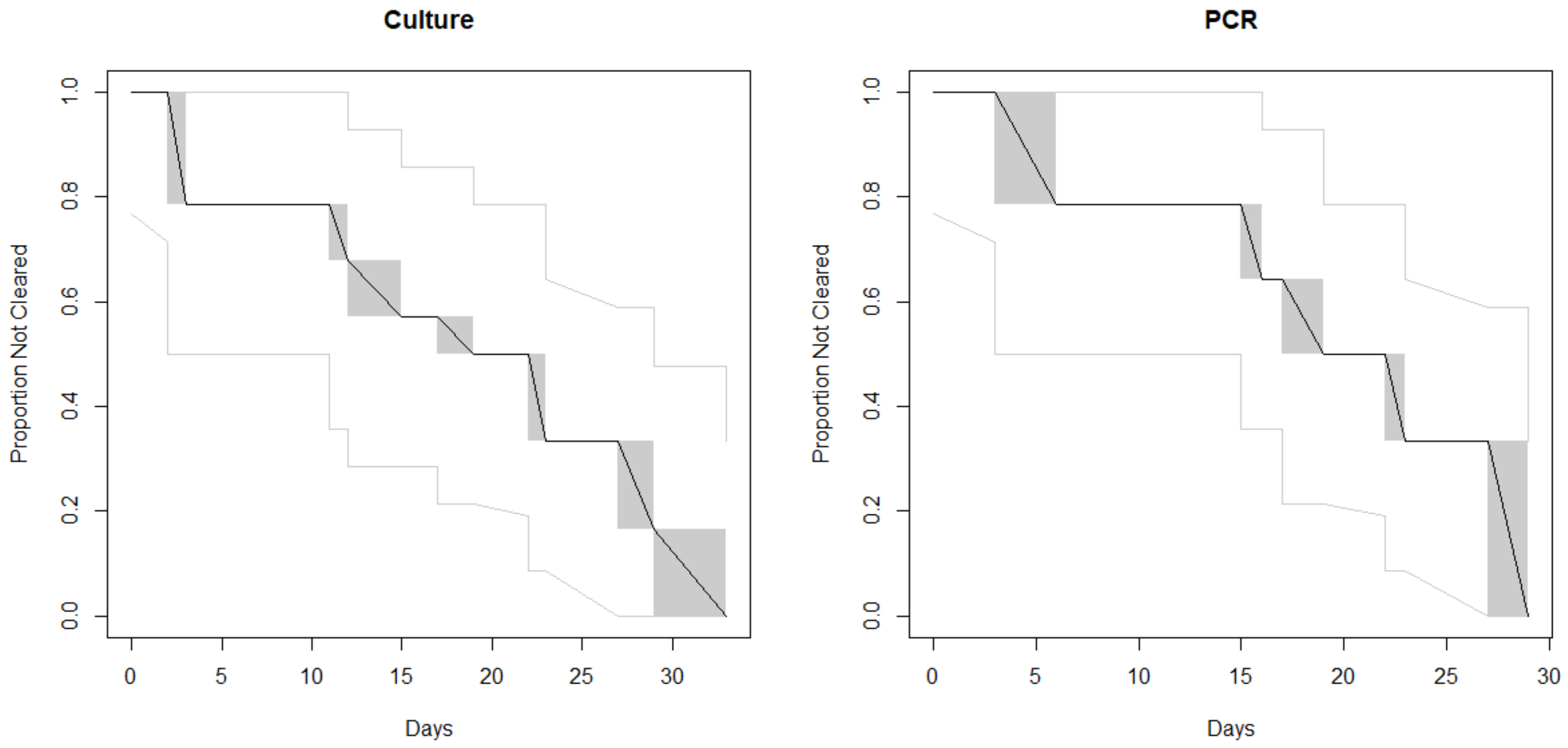
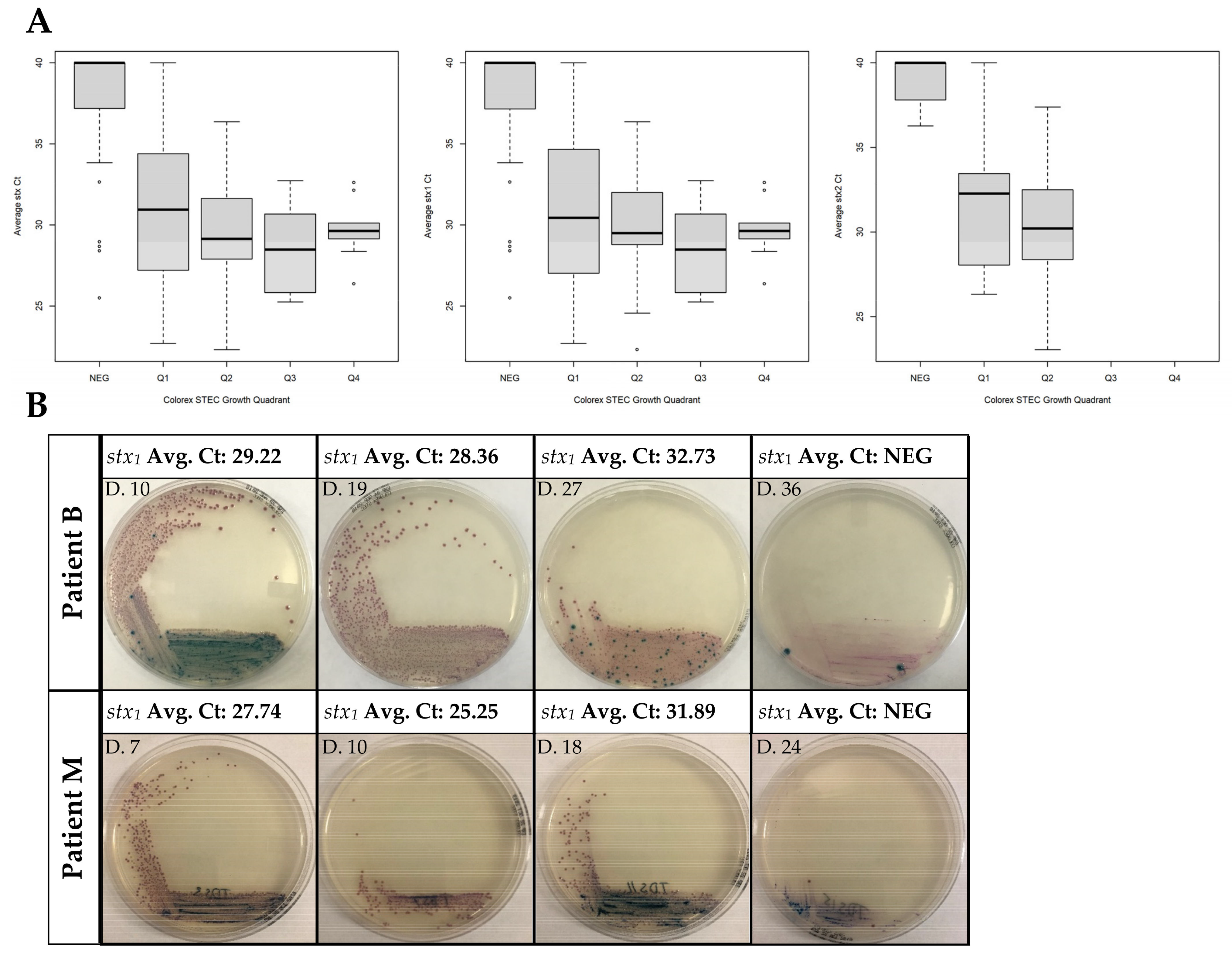
3. Results
4. Discussion
Supplementary Materials
Author Contributions
Funding
Acknowledgments
Conflicts of Interest
References
- Thomas, M.K.; Murray, R.; Flockhart, L.; Pintar, K.; Fazil, A.; Nesbitt, A.; Marshall, B.; Tataryn, J.; Pollari, F. Estimates of foodborne illness–related hospitalizations and deaths in Canada for 30 specified pathogens and unspecified agents. Foodborne Pathog. Dis. 2015, 12, 820–827. [Google Scholar] [CrossRef]
- Thomas, M.K.; Murray, R.; Nesbitt, A.; Pollari, F. The incidence of acute gastrointestinal illness in Canada, foodbook survey 2014–2015. Can. J. Infect. Dis. Med. Microbiol. 2017, 2017, 1–11. [Google Scholar] [CrossRef]
- Henson, S.; Majowicz, S.E.; Masakure, O.; Sockett, P.; MacDougall, L.; Edge, V.L.; Thomas, M.; Fyfe, M.; Kovács, S.; Jones, A. Estimation of the costs of acute gastrointestinal illness in British Columbia, Canada. Int. J. Food Microbiol. 2008, 127, 43–52. [Google Scholar] [CrossRef]
- Majowicz, S.E.; McNab, W.B.; Sockett, P.; Henson, S.; Doré, K.; Edge, V.L.; Buffett, M.C.; Fazil, A.; Read, S.; McEwen, S.; et al. Burden and cost of gastroenteritis in a Canadian community. J. Food Prot. 2006, 69, 651–659. [Google Scholar] [CrossRef] [PubMed]
- Bryan, A.; Youngster, I.; McAdam, A.J. Shiga toxin producing Escherichia coli. Clin. Lab. Med. 2015, 35, 247–272. [Google Scholar] [CrossRef] [PubMed]
- Lim, J.Y.; Yoon, J.; Hovde, C.J. A Brief Overview of Escherichia coli O157:H7 and Its Plasmid O157. J. Microbiol. Biotechnol. 2010, 20, 5–14. [Google Scholar] [CrossRef] [PubMed]
- Zelyas, N.; Poon, A.; Patterson-Fortin, L.; Johnson, R.P.; Lee, W.; Chui, L. Assessment of commercial chromogenic solid media for the detection of non-O157 Shiga toxin-producing Escherichia coli (STEC). Diagn. Microbiol. Infect. Dis. 2016, 85, 302–308. [Google Scholar] [CrossRef]
- Bruyand, M.; Mariani-Kurkdjian, P.; Gouali, M.; De Valk, H.; King, L.A.; Le Hello, S.; Bonacorsi, S.; Loirat, C. Hemolytic uremic syndrome due to Shiga toxin-producing Escherichia coli infection. Med. Mal. Infect. 2018, 48, 167–174. [Google Scholar] [CrossRef] [PubMed]
- Chattopadhyay, S.P.; Borua, P.C.; Rathore, B.S. Value of pilocarpine test in early diagnosis of leprosy. Indian J. Lepr. 1984, 56, 877–883. [Google Scholar]
- Morton, V.; Cheng, J.; Sharma, D.; Kearney, A. An outbreak of Shiga toxin-producing Escherichia coli O121 infections associated with flour—Canada, 2016–2017†. Can. Commun. Dis. Rep. 2017, 43, 154–155. [Google Scholar] [CrossRef]
- Government of Alberta. Escherichia coli Verotoxigenic Infections. May 2018. Available online: https://open.alberta.ca/dataset/2b77e542-cfcb-4f93-b825-dca7d140e024/resource/084c05ea-b5dd-4c07-8561-56da709b2ac3/download/guidelines-escherichia-coli-verotoxigenic-infections-2018-05.pdf (accessed on 7 September 2019).
- McAdam, A.J. Unforeseen consequences: Culture-independent diagnostic tests and epidemiologic tracking of foodborne pathogens. J. Clin. Microbiol. 2017, 55, 1978–1979. [Google Scholar] [CrossRef] [PubMed][Green Version]
- Stokes, W.; Simner, P.J.; Mortensen, J.; Oethinger, M.; Stellrecht, K.; Lockamy, E.; Lay, T.; Bouchy, P.; Pillai, D.R. Multicenter clinical validation of the molecular BD max enteric viral panel for detection of enteric pathogens. J. Clin. Microbiol. 2019, 57. [Google Scholar] [CrossRef] [PubMed]
- Gouali, M.; Ruckly, C.; Carle, I.; Lejay-Collin, M.; Weill, F.-X. Evaluation of CHROMagar STEC and STEC O104 chromogenic agar media for detection of shiga toxin-producing Escherichia coli in stool specimens. J. Clin. Microbiol. 2013, 51, 894–900. [Google Scholar] [CrossRef]
- Delannoy, S.; Chaves, B.D.; Ison, S.A.; Webb, H.E.; Beutin, L.; Delaval, J.; Billet, I.; Fach, P. Revisiting the STEC testing approach: Using espK and espV to make enterohemorrhagic Escherichia coli (EHEC) detection more reliable in beef. Front. Microbiol. 2016, 7, 1. [Google Scholar] [CrossRef] [PubMed]
- Turnbull, B.W. The empirical distribution function with arbitrarily grouped, censored and truncated data. J. R. Stat. Soc. Ser. B Stat. Methodol. 1976, 38, 290–295. [Google Scholar] [CrossRef]
- Fay, M.P.; Shaw, P.A. Exact and asymptotic weighted logrank tests for interval censored data: The interval R Package. J. Stat. Softw. 2010, 36, 1–34. [Google Scholar] [CrossRef] [PubMed]
- Christensen, R.H.B. Ordinal: Regression Models for Ordinal Data. 2019. Available online: https://CRAN.R-project.org/package=ordinal (accessed on 28 October 2020).
- Collins, A.; Fallon, U.B.; Cosgrove, M.; Meagher, G.; Ni Shuileabhan, C. A 10-year analysis of VTEC microbiological clearance times, in the under-six population of the Midlands, Ireland. Epidemiol. Infect. 2017, 145, 1577–1583. [Google Scholar] [CrossRef]
- Alconcher, L.F.; Rivas, M.; Lucarelli, L.I.; Galavotti, J.; Rizzo, M. Shiga toxin-producing Escherichia coli in household members of children with hemolytic uremic syndrome. Eur. J. Clin. Microbiol. Infect. Dis. 2019, 39, 427–432. [Google Scholar] [CrossRef]
- Vonberg, R.P.; Höhle, M.; Aepfelbacher, M.; Bange, F.C.; Campos, C.B.; Claussen, K.; Christner, M.; Cramer, J.P.; Haller, H.; Hornef, M.; et al. Duration of fecal shedding of shiga toxin–producing Escherichia coli O104:H4 in patients infected during the 2011 outbreak in Germany: A multicenter study. Clin. Infect. Dis. 2013, 56, 1132–1140. [Google Scholar] [CrossRef]
- Atmar, R.L.; Opekun, A.R.; Gilger, M.A.; Estes, M.K.; Crawford, S.E.; Neill, F.H.; Graham, D.Y. Norwalk virus shedding after experimental human infection. Emerg. Infect. Dis. 2008, 14, 1553–1557. [Google Scholar] [CrossRef]
- Kirby, A.; Shi, J.; Montes, J.; Lichtenstein, M.; Moe, C.L. Disease course and viral shedding in experimental Norwalk virus and snow mountain virus infection. J. Med. Virol. 2014, 86, 2055–2064. [Google Scholar] [CrossRef] [PubMed]
- Mohawk, K.L.; O’Brien, A.D. Mouse models of Escherichia coli O157:H7 infection and shiga toxin injection. J. Biomed. Biotechnol. 2011, 2011, 1–17. [Google Scholar] [CrossRef] [PubMed]
- Zangari, T.; Melton-Celsa, A.R.; Panda, A.; Boisen, N.; Smith, M.A.; Tatarov, I.; De Tolla, L.J.; Nataro, J.P.; O’Brien, A.D. Virulence of the shiga toxin type 2-expressing Escherichia coli O104:H4 german outbreak isolate in two animal models. Infect. Immun. 2013, 81, 1562–1574. [Google Scholar] [CrossRef] [PubMed][Green Version]
- Verstraete, K.; Van Coillie, E.; Werbrouck, H.; Van Weyenberg, S.; Herman, L.; Del-Favero, J.; De Rijk, P.; De Zutter, L.; Joris, M.-A.; Heyndrickx, M.; et al. A qPCR assay to detect and quantify shiga toxin-producing E. coli (STEC) in cattle and on farms: A potential predictive tool for STEC culture-positive farms. Toxins 2014, 6, 1201–1221. [Google Scholar] [CrossRef]
- Schmidt, H. Shiga-toxin-converting bacteriophages. Res. Microbiol. 2001, 152, 687–695. [Google Scholar] [CrossRef]
- Rhoads, D.D.; Wolcott, R.D.; Sun, Y.; Dowd, S.E. Comparison of culture and molecular identification of bacteria in chronic wounds. Int. J. Mol. Sci. 2012, 13, 2535–2550. [Google Scholar] [CrossRef]


| Reference Gene, Primer/Probe | Sequence 5′-3′ |
|---|---|
| stx1-F | TTT GTY ACT GTS ACA GCW GAA GCY TTA CG |
| stx1-R | CCC CAG TTC ARW GTR AGR TCM ACR TC |
| stx1-P | CTG GAT GAT CTC AGT GGG CGT TCT TAT GTA A |
| stx2-F | TTT GTY ACT GTS ACA GCW GAA GCY TTA CG |
| stx2-R | CCC CAG TTC ARW GTR AGR TCM ACR TC |
| stx2-P | TCG TCA GGC ACT GTC TGA AAC TGC TCC |
| In the sequences: Y is (C, T), S is (C, G), W is (A, T), R is (A, G), M is (A, C) | |
| Patients | A | B | C | D | E | F | G | H | I | J | K | L | M | N |
|---|---|---|---|---|---|---|---|---|---|---|---|---|---|---|
| Number of samples submitted | 4 | 2 | 10 | 4 | 12 | 15 | 6 | 5 | 10 | 7 | 8 | 4 | 19 | 4 |
| Duration of sample submission (days) | 19 | 6 | 49 | 45 | 48 | 32 | 10 | 18 | 20 | 43 | 34 | 10 | 31 | 19 |
| Culture-negative (days) | 15 | 6 | 33 | 38 | 35 | 26 | 3 | 16 | 12 | 38 | 29 | 9 | 23 | 17 |
| Real-time PCR-negative (days) | 19 | 6 | 29 | 38 | 35 | 26 | 6 | 16 | 19 | 38 | 29 | 9 | 23 | 19 |
| Stx status | 2 | 1 | 1 | 1 | 1 | 1 | 1 | 1 | 1 | 1 and 2 | 2 | 1 | 1 and 2 | 1 |
| Serotyped | O121 | O26 | O111 | O26 | O103 | O186 | O118 | O186 | O103 | O157 | O157 | O26 | O157 | O111 |
Publisher’s Note: MDPI stays neutral with regard to jurisdictional claims in published maps and institutional affiliations. |
© 2020 by the authors. Licensee MDPI, Basel, Switzerland. This article is an open access article distributed under the terms and conditions of the Creative Commons Attribution (CC BY) license (http://creativecommons.org/licenses/by/4.0/).
Share and Cite
Bording-Jorgensen, M.; Parsons, B.D.; Tarr, G.A.M.; Shah-Gandhi, B.; Lloyd, C.; Chui, L. Association of Ct Values from Real-Time PCR with Culture in Microbiological Clearance Samples for Shiga Toxin-Producing Escherichia coli (STEC). Microorganisms 2020, 8, 1801. https://doi.org/10.3390/microorganisms8111801
Bording-Jorgensen M, Parsons BD, Tarr GAM, Shah-Gandhi B, Lloyd C, Chui L. Association of Ct Values from Real-Time PCR with Culture in Microbiological Clearance Samples for Shiga Toxin-Producing Escherichia coli (STEC). Microorganisms. 2020; 8(11):1801. https://doi.org/10.3390/microorganisms8111801
Chicago/Turabian StyleBording-Jorgensen, Michael, Brendon D. Parsons, Gillian A.M. Tarr, Binal Shah-Gandhi, Colin Lloyd, and Linda Chui. 2020. "Association of Ct Values from Real-Time PCR with Culture in Microbiological Clearance Samples for Shiga Toxin-Producing Escherichia coli (STEC)" Microorganisms 8, no. 11: 1801. https://doi.org/10.3390/microorganisms8111801
APA StyleBording-Jorgensen, M., Parsons, B. D., Tarr, G. A. M., Shah-Gandhi, B., Lloyd, C., & Chui, L. (2020). Association of Ct Values from Real-Time PCR with Culture in Microbiological Clearance Samples for Shiga Toxin-Producing Escherichia coli (STEC). Microorganisms, 8(11), 1801. https://doi.org/10.3390/microorganisms8111801