Article

ITS rDNA Gene Analysis Versus MALDI-TOF MS For Identification of Neoscytalidium dimidiatum Isolated from Onychomycosis and Dermatomycosis Cases in Medellin (Colombia)

by
Sindy V. Flórez-Muñoz
1,
Juan C. Gómez-Velásquez
2,
Natalia Loaiza-Díaz
2,
Célia Soares
3,
Carla Santos
3,
Nelson Lima
3 and
Ana C. Mesa-Arango
1,*
1
Grupo de Investigación Dermatológica, Instituto de Investigaciones Médicas, Facultad de Medicina, Universidad de Antioquia, Avenida Juan del Corral, 050010 Medellín, Colombia
2
Laboratorio Clínico PROLAB, Miembro del Grupo Synlab, Calle 19 A, 050021 Medellín, Colombia
3
CEB-Centre of Biological Engineering, Micoteca da Universidade do Minho (MUM), University of Minho, Campus de Gualtar, 4710-057 Braga, Portugal
*
Author to whom correspondence should be addressed.
Microorganisms 2019, 7(9), 306; https://doi.org/10.3390/microorganisms7090306
Submission received: 8 August 2019 / Revised: 27 August 2019 / Accepted: 30 August 2019 / Published: 1 September 2019
(This article belongs to the Special Issue Mass Spectrometry: An Undeniable Tool in Current Microbiology)

Abstract

:
Within the Neoscytalidium genus, N. dimidiatum, N. oculus, N. orchidacearum, and N. novaehollandiae have been recognized. Although these species are frequently found in soil, N. dimidiatum has been identified as an etiologic agent of onychomycosis or dermatomycosis, and N. oculus has been identified as an etiologic agent of an ocular lesion. All these species can be cultured in vitro, but their morphological identification by macroscopic and microscopic traits is difficult and imprecise due to their similarity. In this study, 34 isolates of Neoscytalidium spp. from 32 onychomycosis and two dermatomycosis cases in Medellin (Colombia) were identified at the species level using sequencing of the ITS1+5.8S+ITS2 nuclear rDNA region and MALDI-TOF mass spectrometry (MS). Neoscytalidium dimidiatum strain MUM 17.21 was used to construct the reference spectrum in the in-house library to identify the clinical isolates by MALDI-TOF MS. Additionally, N. dimidiatum PPC-216 and PLAB-055 strains were used to validate the in-house constructed reference spectra. Although four groups were observed in the dendrogram obtained from the proteins of each isolate profile, MALDI-TOF MS and sequencing results are in accordance, since all isolates were identified as N. dimidiatum.

1. Introduction

The coelomycete Neoscytalidium (Botryosphaeriaceae) is a dematiaceous fungal genus that is commonly found in the soil of tropical and subtropical zones [1]. The taxonomy of this genus has been problematic and constantly revised. Currently, within this genus, four species are recognized: N. novaehollandiae, N. orchidacearum, N. oculus, and N. dimidiatum [2,3]. The first two species have been reported as phytopathogens, while N. oculus and N. dimidiatum have been associated to ocular or keratinized tissue (skin or nails) infections, which are indistinguishable from dermatophytosis [3,4,5]. Recently, it has been demonstrated that N. oculus can form biofilms and cause hemolysis [3]. In contrast, N. dimidiatum can also cause subcutaneous and deep infections, mainly in immunocompromised patients [6,7,8]. Furthermore, it usually shows low antifungal in vitro susceptibility with an unpredictable clinical response to treatments [9,10].
The identification of this genus has been based on the observation of macroscopic and microscopic characteristics of in vitro cultures [3]. Initially, white colonies are observed in the culture media, but with age, they become black/greenish, and the plate is fully covered with powdery aerial mycelium. Under a microscope, they are observed as being dark brown, thick-walled, with 0–2 septate arthroconidia, and variable in shape [11].
In many clinical laboratories worldwide, fungal identifications are still done by means of morphological methods, with some additional use of physiological and biochemical methods; however, they must be regarded as presumptive identifications and prone to inaccuracy. Phillips et al. [11] consider that it is inappropriate to identify fungal genera or species by only taking into account morphological characteristics, as it is prone to subjectivity. Alternatively, Matrix Assisted Laser Desorption/Ionization Mass Spectrometry with Time-of-Flight detector (MALDI-TOF MS) and gene sequencing, such as the fungal universal barcode nuclear ribosomal internal transcribed spacer (ITS) region [12], can be considered to accomplish a more objective and accurate fungal identification. The former technique has been used successfully in the identification of bacteria and yeasts [13]. However, the identification of some filamentous fungi is still a challenge, but not a restriction, depending more on the protocol and database used than on the equipment [14]. The aim of this study was to compare sequence-based identification using the complete sequences of the ITS1+5.8S+ITS2 rDNA region with MALDI-TOF MS identification of Neoscytalidium spp. clinical isolates.

2. Materials and Methods

2.1. Clinical Isolates

Thirty-four clinical isolates were collected between 2016–2018 from clinical samples of patients with onychomycosis (32) and dermatomycosis (2) in a mycological diagnosis laboratory (PROLAB, grupo Synlab, Medellin—Colombia) (Table 1). The observation of hyaline or pigmented septate hyphae in the direct microscopy examination of clinical samples with Chlorazol Black E (Delasco, IA, USA) was mandatory to consider a positive result and continue to the culture and subsequente analyses. The isolates were grown in Sabouraud dextrose agar with chloramphenicol (Biomerieux®, Paris, France) and incubated at 28 °C in the dark for up to seven days.

2.2. Molecular Identification

2.2.1. Preparation of Mycelium for the Extraction of DNA

Isolates were grown in solid potato dextrose agar (PDA; Oxoid, Basingstoke, Hampshire, England) at 28 °C for 5 days. Then, approximately 0.2 cm2 of the biomass was transferred to a 1.5-mL microtube (KIMA, Piove di Sacco, Padova, Italy) containing 1 mL of yeast malt broth (YMB; yeast extract, 3 g/L; malt extract, 3 g/L; peptone 5 g/L; glucose 10 g/L). The isolates were incubated for 10 days in constant agitation at room temperature. Subsequently, they were centrifuged for 10 min at 14,000 g, the supernatant was discarded, and 1 mL of sterile distilled water was added to remove residues from the culture medium. It was centrifuged again, and the supernatant was removed, allowing the pellet to dry and later be kept at −20 °C for DNA extraction.

2.2.2. DNA Extraction

DNA extraction was performed following the protocol established by Rodrigues et al. [15], with minor modifications: the biomass was transferred into Lysing matrix MP Biomedicals tubes (Santa Ana, CA, USA) containing 500 μL of lysis buffer (200 mM of Tris–HCl pH 8.5; 250 mL of NaCl; 25 mM of EDTA; 0.5% [w/v] SDS) and 300 mg of sterile glass beads of 1.25 to 1.65 mm in diameter (Sigma, St. Louis, MO, USA). Then, samples were mechanically lysed for 40 s in a FastPrep-24TM 5G Instrument (MP Biomedicals Santa Ana, CA, USA); subsequently, they were incubated in a water bath for 30 min at 65 °C, and centrifuged for 10 min at 14,000× g. With the purpose of precipitating the polysaccharides and proteins, 500 μL of cold 3 M NaOAc pH 5.5 were added to each sample, and gently mixed by inversion before incubation at −20 °C for 10 min, and centrifugation at 14,000× g for 10 min. Nucleic acids were precipitated from a mix of 500 μL of the clear supernatant with 500 μL of isopropanol, which was incubated for 90 min, to finally be centrifuged at 14,000× g for 10 min; the precipitate was washed twice with 500 μL of cold 70% ethanol, centrifuged at 6000× g for 7 min, and air dried. DNA was resuspended in 50 μL of ultra-pure sterile water, quantified in a NanoDrop™ 1000 (Thermo Scientific™, Waltham, MA, USA), and stored at −20 °C.

2.2.3. PCR amplification

PCR amplification of the ITS1+5.8S+ITS2 rDNA region was performed with 50 μL of a reaction mixture containing 25 μL of NZYTaq II 2x Green Master Mix (NZTtech, Lisbon, Portugal), 1 μL of primers ITS1 (5′-TCCGTAGGTGAACCTGCGG-3′) and ITS4 (5′-TCCTCCGCTTATTGATATGC-3′) [16], 1 μL of DNA, and 22 μL of sterile ultra-pure water. PCR conditions were as follows: a denaturation step at 94 °C for 3 min; 35 cycles of the annealing step: 1 min at 94 °C, 1 min at 55 °C, and 1 min at 72 °C; and a final elongation step of 5 min at 72 °C. PCR amplifications were conducted in a GeneAmp PCR System 9700 thermocycler (Applied Biosystems, Foster City, CA, USA). The products were analyzed in 1% agarose gel and purified using the NZYgelpure kit from NZYtech. Sequencing of the products was carried out in the STAB VIDA Lda (Caparica, Portugal) using the Sanger/capillary method. Sequences were processed using the FinchTV program, version 1.4.0. Poor-quality end regions were removed. ITS sequences were compared with the National Center for Biotechnology Information (NCBI) database using the Basic Local Alignment Search Tool Nucleotide (BLASTN) to detect non-specific amplicons. Only sequences with 60% coverage and at least 98% identity with Neoscytalidium spp. sequences were retained for further analysis.

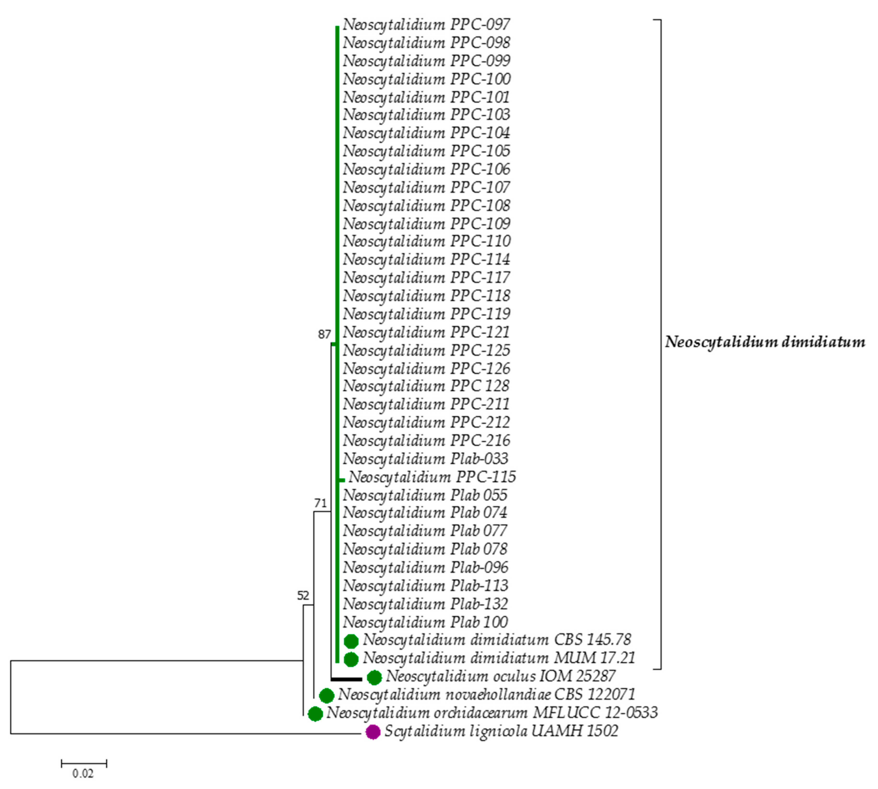
2.2.4. DNA Sequence Processing and Molecular Taxonomic Analysis

All the sequences were oriented according to the direction of the reference sequences and then globally aligned using the nested ClustalW [17] software in MEGA version 7 [18]. The best model was estimated considering the Bayesian information criterion (BIC) and the Akaike information criterion (AIC). The recovered alignments were checked manually to avoid mismatched base pairs. Reconstruction of the maximum likelihood (ML) topology with ITS1 and ITS2 sequences was performed using the Tamura-3-parameter model of nucleotide substitution, with Gamma distribution and 1000 bootstraps. Other sequences extracted from GenBank were included in the analysis, and Scytalidium lignicola was used as an outgroup (Table 1).

2.3. MALDI-TOF MS Identification

2.3.1. Protein Extraction

Protein extraction was performed following the protocol of Packeu et al. [19], with minor modifications: the fungi were cultured in Sabouraud chloramphenicol agar (Biomerieux®, Paris, France) and incubated for 72 h at 28 °C. Mycelia were gently removed with wood sticks and suspended in a 3:1 mixture of ethanol and distilled water in a microtube of 1.5 mL (KIMA,), vortexed and centrifuged for 5 min at 13,000 rpm. The supernatants were discarded, and 50 μL of 70% formic acid was added; after being homogenized, they were left to rest for 15 min. Subsequently, 50 μL of 100% acetonitrile was added, and again, samples were allowed to rest for 15 min. Finally, they were centrifuged at 13,000 rpm for 2 min, and the supernatant was used for fungal identification.

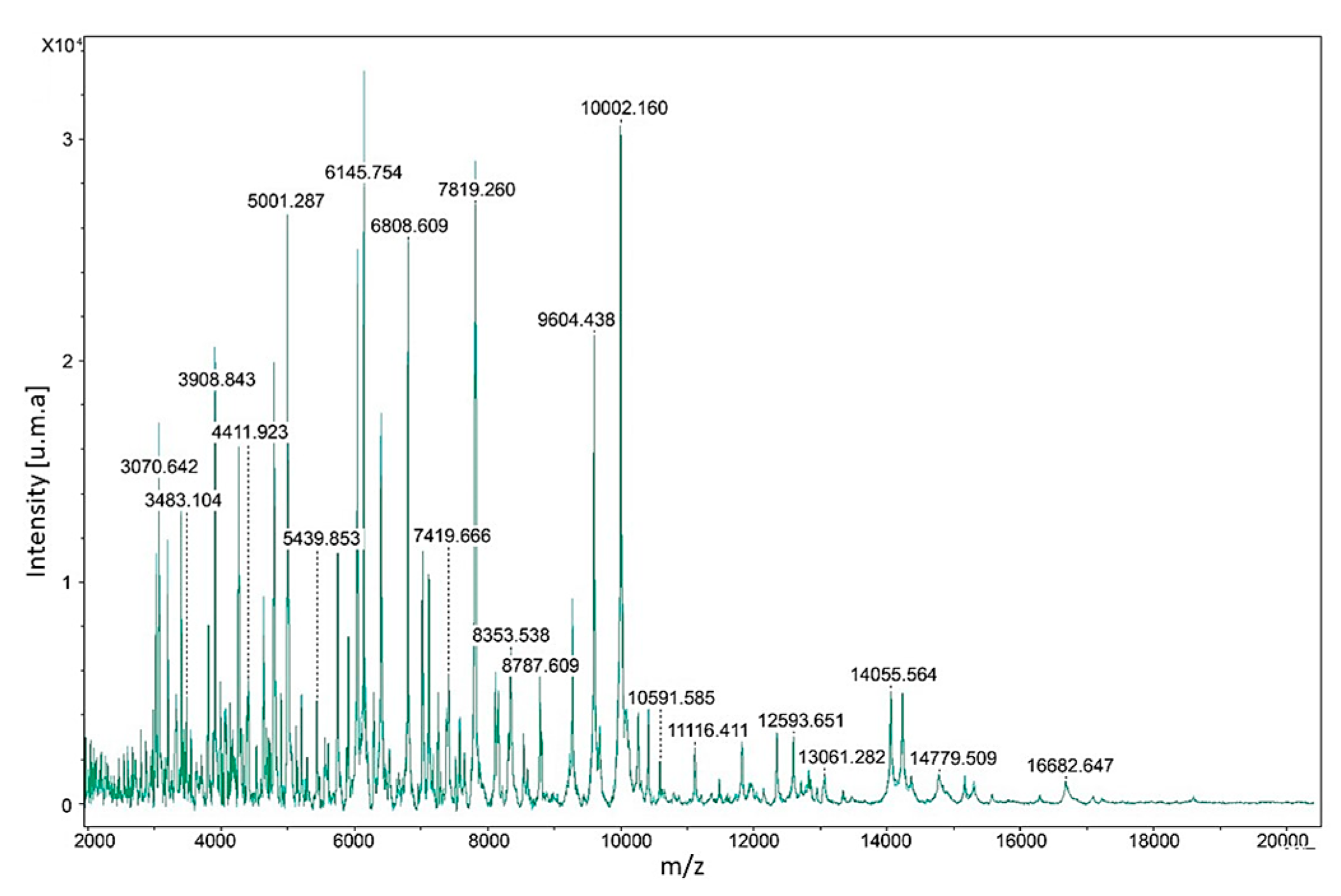
2.3.2. Constructing the Reference Mass Spectra

The reference spectra were constructed from the independent reference strain of N. dimidiatum MUM 17.21, obtained from the public service fungal culture collection Micoteca da Universidade do Minho (MUM, Braga, Portugal), according to the methodology proposed by Cassagne et al. [20]. Four independent cultures of strain MUM 17.21 were incubated at 28 °C for 72 h. Proteins were extracted from each culture (as described above), and 1 μL of each supernatant was transferred to a spot onto the steel target plate (Bruker Daltonics, Bremen, Germany), allowing the samples to dry completely. Then, 1 μL of α-cyano-4-hydroxycinnamic acid matrix (Bruker Daltonics, Bremen, Germany) was added. Eight replicas for each culture were used. The spectra were acquired after at least 240 shots in linear mode using the Microflex LT table mass spectrometer (Bruker Daltonics, Bremen, Germany) in the positive ion mode, with a 337-nm nitrogen laser. A bacterial standard of Escherichia coli extract with RNase and human myoglobin was used as control and for the internal verification of equipment calibration (BTS Bruker Daltonics, Bremen, Germany). Data were acquired automatically using the AutoXecute function of FlexControl software, version 3.3, and then exported to the MALDI Biotyper software, version 3.1 (Bruker Daltonics, Bremen, Germany), to evaluate the quality of the spectrum according to the manufacturer’s recommendations. Two isolates (PPC-216 and PLAB-055), previously identified by sequencing as N. dimidiatum, were used to validate the reference spectrum. Fusarium oxysporum ATCC 48112 and Aspergillus fumigatus ATCC 204305 strains were used as negative controls.

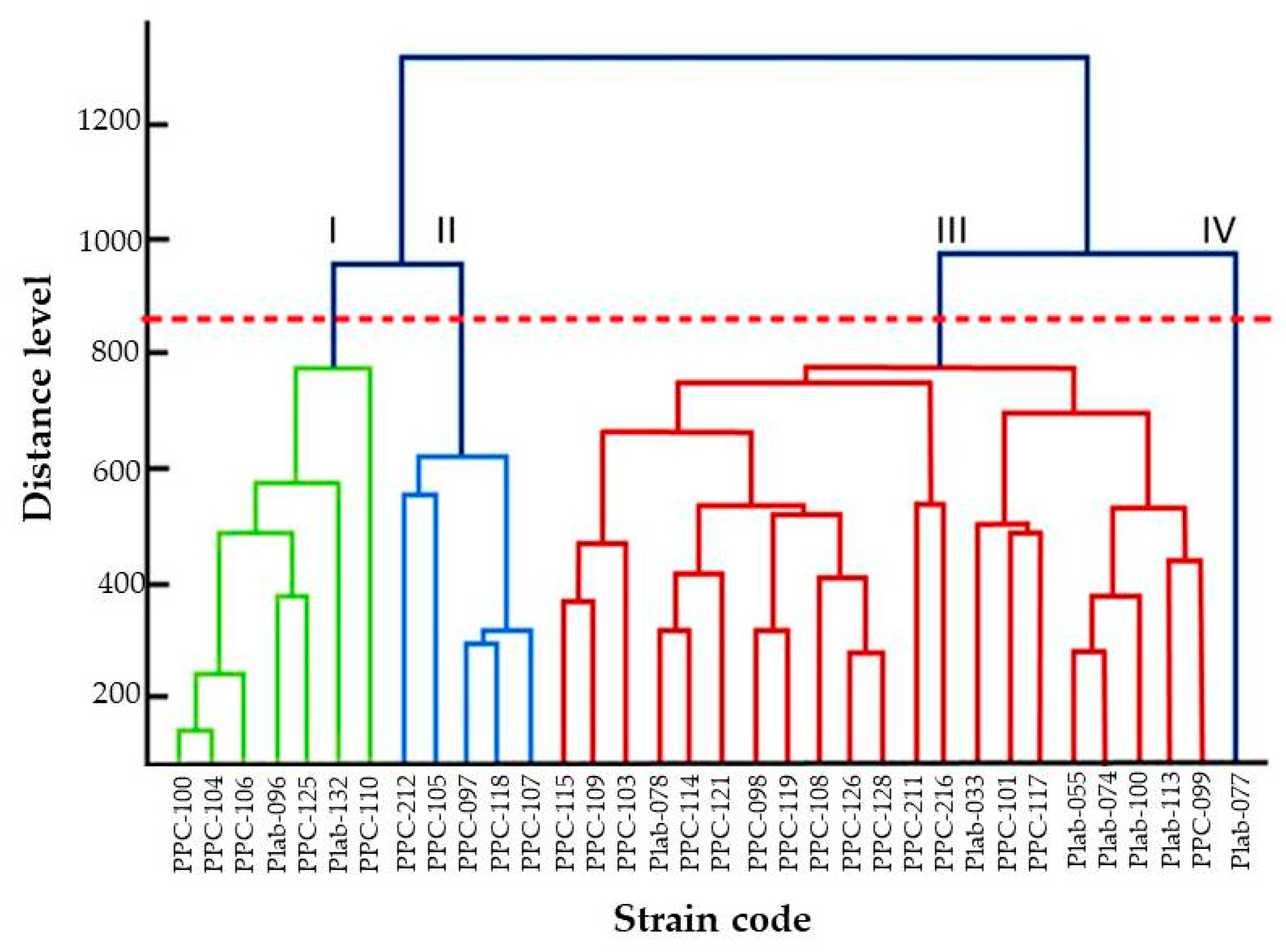
2.3.3. Identification of Clinical Isolates

The protein extraction of clinical isolates was performed as described above. For each isolate, four spectra were obtained and compared to the library constructed in-house using the Maldi Biotyper 3.1 software. Identification was carried out following the manufacturer’s established scores: ≥2.000: species-level identification; 1.700–1.999: genus-level identification; ≤1.699: not reliably identified.

3. Results

Clinical isolates were presumptively identified as N. dimidiatum through classical phenotype methods and also taking into consideration that they were isolated from human patients with onychomycosis or dermatomycosis. In the beginning, all clinical isolates showed white colonies that gradually turned grey, olive green, or black. Under the microscope, mycelia that were branched, septate, hyaline, or dematiaceous with arthroconidia in chains or disarticulated, as well as occasional chlamydoconidia, were observed. In addition, molecular identification was carried out through amplification, sequencing, and phylogenetic analysis using the ITS1+5.8S+ITS2 rDNA region. The sequences of isolates included in this study were deposited in GenBank, and the accession numbers are indicated (Table 1).
The alignment of the non-coding region ITS1+5.8SADNr+ITS2 was carried out with 40 sequences, 34 of them corresponding to clinical isolates from this study, one sequence of the ex-holotype strain of N. dimidiatum, the strain MUM 17.21, and three of the other species of the genus Neoscytalidium, and one of S. lignicola as outgroup (Table 1). Sequences of 407 nucleotides were used for the alignment, and they were distributed as follows: 307 conserved (75.43%); 94 variables (23.10%), of which 87 were singleton (21.38% of the total nucleotides); and six were parsimony-informative (1.47%). All positions contained gaps, and missing data were eliminated from the dataset. All the clinical isolates from this study were grouped together with the type sequence of N. dimidiatum in the same branch, with a bootstrap support of 87% (Figure 1). The presumptive phenotype identifications were totally confirmed by the ITS barcode.
For the identification of clinical isolation by MALDI-TOF MS, four reference spectra were constructed. The range of the masses detected was between 3070.643–16,682.647 Daltons (Figure 2).
The 34 (100%) clinical isolates were identified through the in-house library as N. dimidiatum (score ≥2.00). The dendrogram created with the protein spectra, using the principal component analysis (PCA) method, shows the conformation of four groups (I to IV) above the critical distance (850), as shown in Figure 3. Groups I to III were formed by seven, five, and 21 isolates, respectively. Group IV was formed by the single isolate Plab-077 (MUM 19.71) with an origin in a nail infection.

4. Discussion

The constant change in the taxonomy of fungi that belong to the genus Neoscytalidium has generated confusion; they have been named Dothiorella mangiferae, Exosporina fawcettii, Fusicoccum arbuti, F. eucalypti, F. dimidiatum, Hendersonula agathidia, H. cypria, H. toruloidea, Scytalidium dimidiatum, S. lignicola or Torula dimidiata [21,22].
Traditionally, Neoscytalidium spp. identification has been based only on macroscopic and microscopic observations [23]. Classical phenotypic methods can give inaccurate results in the identification of the species of this genus, because they do not have characteristic structures to distinguish between them [11].
Recently, using sequencing of the ITS1+5.8S+ITS2 rDNA region, or of a fragment of the 28S large subunit ribosomal (LSU), it has been possible to recognize the species N. novaehollandiae, N. orchidaceous, N. dimidiatum, and N. oculus [3,11]. In the present study, the high level of accuracy of using the ITS1+5.8S+ITS2 rDNA region to identify N. dimidiatum reveals that this barcode is fit-for-propose in the clinical field. However, this barcode alone could be unable to clearly discriminate species that exhibited a certain degree of genetic variations. In this latter case, we recommended the use of a multilocus sequence analysis (MSLA). A similar situation was obtained by Pereira et al. [24] for the dermatophyte Trichophyton rubrum, which due to its adaptation to a highly specialized ecological niche, the skin of the human host, combined with exclusively asexual reproduction, can explain a high level of genetic uniformity within the species.
Neoscytalidium dimidiatum has been recognized as an etiologic agent for nail and skin infections [25,26], possibly because it produces keratinases [27]. However, with the increase of patients with immunosuppressive conditions, it has been demonstrated that N. dimidiatum does not only affect external tissues, but also causes eye infections or invades organs such as the lungs or the brain [6,7,8,10].
Some studies in Colombia have reported the isolation of N. dimidiatum or S. dimidiatum, as it has also been named, from skin and nail samples. In general, the identification of this fungus has been based on macroscopic and microscopic observations [5,28,29,30], with exception of the data published by Dionne et al. [7] in a case of pulmonary infection and Bueno et al. [28], who also used ITS1+5.8S+ITS2 nuclear rDNA to identify Scytalidium hyalinum (considered by [8] as an albino variant of N. dimidiatum while others [31] regard them as separate species) and F. dimidiatum as an etiologic agent of onychomycosis. However, the present study is the first one where Neoscytalidium spp. clinical isolates were identified by means of phylogenetic analysis and of MALDI-TOF MS. Phylogenetic analysis showed that all the clinical isolates were N. dimidiatum and grouped in one clade, separated from the other species within the same genus. In addition, it was observed that N. oculus is the closest related species to N. dimidiatum.
The construction of reference spectra was necessary because the commercial library lacks spectra for the identification of Neoscytalidium spp. Although the identification of filamentous fungi using MALDI-TOF MS has limitations, and even more when they are pigmented, the present study achieved both the successful construction of reference spectra and the identification of 34 clinical isolates of N. dimidiatum. The use of young mycelium without pigmentation (72 h of growth) might explain the correct identification by MALDI-TOF MS of all N. dimidiatum clinical isolates in this study. Our results are in contrast to those obtained by Alshawa et al. [32], who only achieved identification of 77.8% of the isolates. In this latter case, the low identification rate may be explained by the incubation time (three weeks), during which pigmentation was developed more intensively in combination with possible cell wall changes, such as becoming thicker and coated by hydrophobins proteins. It is known that the presence of pigments such as melanin can interfere in the identification of some fungi through MALDI-TOF MS, since the desorption/ionization process may be inhibited [33]. Also, in the identification of Neoscytalidium spp. by MALDI-TOF MS carried out by Alshawa et al. [32], the reference spectra of N. dimidiatum had seven mass peaks with an intensity >0.25 × 10⁴ u.m.a., while the ones obtained in the present study showed 34 mass peaks with an intensity >1 × 10⁴ u.m.a. In this case, both the use of young mycelia without pigments and the protein extraction method are the determining factors for adequate identification.
According to the critical distance level of 850 utilized by Chen et al. (2014) [34], the results observed in the dendrogram of Figure 3 suggest the presence of four phenotypes (Group I to IV) which shows the potential of MALDI-TOF MS to group the isolates below the species level [34]. This result contrasts that which has been observed in the consensus phylogenetic tree (Figure 2) where a single homogeneous group is observed. Although ITS1+5.8S+ITS2 rDNA is considered the universal barcoding for fungal identification, it is known that it does not have enough resolution power to detect intraspecific variability as is observed with other housekeeping gene markers, such as the elongation factor, calmodulin, or β-tubulin [12,21,35]. However, for Neoscytalidium spp., sequences of those genes are not available in databases for public use, as is the case of GenBank.

5. Conclusions

In conclusion, the results obtained with mass analysis through dendrogram construction highlights that MALDI-TOF MS can be useful in the accurate identification of strains within N. dimidiatum species, and is a cost-effective technique with the possibility of being used in a mycological diagnosis laboratory.

Author Contributions

The authors S.V.F.M. and J.C.G.V. equally contributed to the experimental work and C.So. and C.Sa supported the molecular biology work and its analysis; the conceptualization was performed mainly by N.L.D., N.L. and A.C.M.A.; all authors equally contributed to the formal analysis, writing, review, and editing; funding acquisition was mainly done by N.L. and A.C.M.A.

Funding

Financial support for this work was provided by the Comité para el Desarrollo de la Investigación, Universidad de Antioquia, Medellín, Colombia. Grant No 2604 and also partially supported by the Portuguese Foundation for Science and Technology (FCT) under the scope of the strategic funding of UID/BIO/04469/2019 unit and BioTecNorte operation (NORTE-01-0145-FEDER-000004) funded by the European Regional Development Fund under the scope of Norte2020—Programa Operacional Regional do Norte.

Acknowledgments

The authors thank to PROLAB, member of the Synlab group, for the continuous support given to this work and the access to the biological resources and MALDI-TOF MS equipment used for the experiments, and they also thank Mario Alberto Ruiz Berganza for his assistance in proofreading this work.

Conflicts of Interest

The authors declare no conflict of interest.

References

  1. Slippers, B.; Boissin, E.; Phillips, A.J.; Groenewald, J.Z.; Lombard, L.; Wingfield, M.J.; Postma, A.; Burgess, T.; Crous, P.W. Phylogenetic lineages in the Botryosphaeriales: A systematic and evolutionary framework. Stud. Mycol. 2013, 76, 31–49. [Google Scholar] [CrossRef]
  2. Huang, S.K.; Tangthirasunun, N.; Phillips, A.J.; Dai, D.Q.; Wanasinghe, D.N.; Wen, T.C.; Bahkali, A.H.; Hyde, K.D.; Kang, J.C. Morphology and Phylogeny of Neoscytalidium orchidacearum sp. nov. (Botryosphaeriaceae). Mycobiology 2016, 44, 79–84. [Google Scholar] [CrossRef]
  3. Calvillo-Medina, R.P.; Martinez-Neria, M.; Mena-Portales, J.; Barba-Escoto, L.; Raymundo, T.; Campos-Guillen, J.; Jones, G.H.; Reyes-Grajeda, J.P.; Gonzalez, Y.M.J.A.; Bautista-de Lucio, V.M. Identification and biofilm development by a new fungal keratitis aetiologic agent. Mycoses 2019, 62, 62–72. [Google Scholar] [CrossRef]
  4. Di Chiacchio, N.; Noriega, L.F.; Gioia Di Chiacchio, N.; Ocampo-Garza, J. Superficial black onychomycosis due to Neoscytalidium dimidiatum. J. Eur. Acad. Derm. Venereol. 2017, 31, e453–e455. [Google Scholar] [CrossRef]
  5. Morales-Cardona, C.A.; Valbuena-Mesa, M.C.; Alvarado, Z.; Solorzano-Amador, A. Non-dermatophyte mould onychomycosis: A clinical and epidemiological study at a dermatology referral centre in Bogota, Colombia. Mycoses 2014, 57, 284–293. [Google Scholar] [CrossRef]
  6. Bakhshizadeh, M.; Hashemian, H.R.; Najafzadeh, M.J.; Dolatabadi, S.; Zarrinfar, H. First report of rhinosinusitis caused by Neoscytalidium dimidiatum in Iran. J. Med. Microbiol. 2014, 63, 1017–1019. [Google Scholar] [CrossRef]
  7. Dionne, B.; Neff, L.; Lee, S.A.; Sutton, D.A.; Wiederhold, N.P.; Lindner, J.; Fan, H.; Jakeman, B. Pulmonary fungal infection caused by Neoscytalidium dimidiatum. J. Clin. Microbiol. 2015, 53, 2381–2384. [Google Scholar] [CrossRef]
  8. Garinet, S.; Tourret, J.; Barete, S.; Arzouk, N.; Meyer, I.; Frances, C.; Datry, A.; Mazier, D.; Barrou, B.; Fekkar, A. Invasive cutaneous Neoscytalidium infections in renal transplant recipients: A series of five cases. Bmc. Infect. Dis. 2015, 15, 535. [Google Scholar] [CrossRef]
  9. Elinav, H.; Izhar, U.; Benenson, S.; Admon, D.; Hidalgo-Grass, C.; Polacheck, I.; Korem, M. Invasive Scytalidium dimidiatum infection in an immunocompetent adult. J. Clin. Microbiol. 2009, 47, 1259–1263. [Google Scholar] [CrossRef]
  10. Yang, S.J.; Ng, C.Y.; Wu, T.S.; Huang, P.Y.; Wu, Y.M.; Sun, P.L. Deep cutaneous Neoscytalidium dimidiatum infection: Successful outcome with amphotericin B therapy. Mycopathologia 2019, 184, 169–176. [Google Scholar] [CrossRef]
  11. Phillips, A.J.; Alves, A.; Abdollahzadeh, J.; Slippers, B.; Wingfield, M.J.; Groenewald, J.Z.; Crous, P.W. The Botryosphaeriaceae: Genera and species known from culture. Stud. Mycol. 2013, 76, 51–67. [Google Scholar] [CrossRef]
  12. Schoch, C.L.; Seifert, K.A.; Huhndorf, S.; Robert, V.; Spouge, J.L.; Levesque, C.A.; Chen, W. Nuclear ribosomal internal transcribed spacer (ITS) region as a universal DNA barcode marker for fungi. Proc. Natl. Acad. Sci. USA 2012, 109, 6241–6246. [Google Scholar] [CrossRef]
  13. Dingle, T.C.; Butler-Wu, S.M. MALDI-TOF mass spectrometry for microorganism identification. Clin. Lab. Med. 2013, 33, 589–609. [Google Scholar] [CrossRef]
  14. SanitáLima, M.; Coutinho de Lucas, R.; Lima, N.; Polizeli Mde, L.M.; Santos, C. Fungal community ecology using MALDI-TOF MS demands curated mass spectral databases. Front. Microbiol. 2019, 10. [Google Scholar] [CrossRef]
  15. Rodrigues, P.; Venâncio, A.; Kozakiewicz, Z.; Lima, N. A polyphasic approach to the identification of aflatoxigenic and non-aflatoxigenic strains of Aspergillus section Flavi isolated from Portuguese almonds. Int. J. Food Microbiol. 2009, 129, 187–193. [Google Scholar] [CrossRef]
  16. White, T.; Bruns, T.; Lee, S.; Taylor, J. Amplification and direct sequencing of fungal ribosomal RNA genes for phylogenetics. In PCR Protocols: A Guide to Methods and Applications; Innis, M., Gelfand, D., Sninsky, J., White, T., Eds.; Academic Press: San Diego, CA, USA, 1990. [Google Scholar]
  17. Thompson, J.D.; Higgins, D.G.; Gibson, T.J. CLUSTAL W: Improving the sensitivity of progressive multiple sequence alignment through sequence weighting, position-specific gap penalties and weight matrix choice. Nucleic Acids Res. 1994, 22, 4673–4680. [Google Scholar] [CrossRef]
  18. Kumar, S.; Stecher, G.; Tamura, K. MEGA7: Molecular evolutionary genetics analysis version 7.0 for bigger datasets. Mol. Biol. Evol. 2016, 33, 1870–1874. [Google Scholar] [CrossRef]
  19. Packeu, A.; Hendrickx, M.; Beguin, H.; Martiny, D.; Vandenberg, O.; Detandt, M. Identification of the Trichophyton mentagrophytes complex species using MALDI-TOF mass spectrometry. Med. Mycol. 2013, 51, 580–585. [Google Scholar] [CrossRef]
  20. Cassagne, C.; Ranque, S.; Normand, A.C.; Fourquet, P.; Thiebault, S.; Planard, C.; Hendrickx, M.; Piarroux, R. Mould routine identification in the clinical laboratory by matrix-assisted laser desorption ionization time-of-flight mass spectrometry. PLoS ONE 2011, 6, e28425. [Google Scholar] [CrossRef]
  21. Schoch, C.L.; Robbertse, B.; Robert, V.; Vu, D.; Cardinali, G.; Irinyi, L.; Meyer, W.; Nilsson, R.H.; Hughes, K.; Miller, A.N.; et al. Finding needles in haystacks: Linking scientific names, reference specimens and molecular data for Fungi. Database (Oxford) 2014, 2014. [Google Scholar] [CrossRef]
  22. Farr, D.F.; Elliott, M.; Rossman, A.Y.; Edmonds, R.L. Fusicoccum arbuti sp. nov. causing cankers on pacific madrone in western North America with notes on Fusicoccum dimidiatum, the correct name for Scytalidium dimidiatum and Nattrassia mangiferae. Mycologia 2005, 97, 730–741. [Google Scholar] [CrossRef] [PubMed]
  23. Nkondjo Minkoumou, S.; Fabrizi, V.; Papini, M. Onychomycosis in Cameroon: A clinical and epidemiological study among dermatological patients. Int. J. Derm. 2012, 51, 1474–1477. [Google Scholar] [CrossRef] [PubMed]
  24. Pereira, L.; Dias, N.; Santos, C.; Lima, N. The use of MALDI-TOF ICMS as an alternative tool for Trichophyton rubrum identification and typing. Enferm. Infecc. Microbiol. Clin. 2014, 32, 11–17. [Google Scholar] [CrossRef] [PubMed]
  25. Simonnet, C.; Berger, F.; Gantier, J.C. Epidemiology of superficial fungal diseases in French Guiana: A three-year retrospective analysis. Med. Mycol. 2011, 49, 608–611. [Google Scholar] [CrossRef] [PubMed]
  26. Lavorato, F.G.; Guimaraes, D.A.; Premazzi, M.G.; Pineiro-Maceira, J.M.; Bernardes-Engemann, A.R.; Orofino-Costa, R. Performance of mycology and histopathology tests for the diagnosis of toenail onychomycosis due to filamentous fungi: Dermatophyte and non-dermatophyte moulds. Mycoses 2017, 60, 587–593. [Google Scholar] [CrossRef] [PubMed]
  27. Oycka, C.A.; Gugnani, H.C. Keratin degradation by Scytalidium species and Fusarium solani. Mycoses 1998, 41, 73–76. [Google Scholar] [CrossRef] [PubMed]
  28. Bueno, J.G.; Martinez, C.; Zapata, B.; Sanclemente, G.; Gallego, M.; Mesa, A.C. In vitro activity of fluconazole, itraconazole, voriconazole and terbinafine against fungi causing onychomycosis. Clin. Exp. Derm. 2010, 35, 658–663. [Google Scholar] [CrossRef]
  29. Zuluaga, A.; Tabares, A.M.; Arango, M. Importancia creciente de los géneros Fusarium y Scytalidium como agentes de onicomicosis. Rev. Asoc. Col. Dermat. 2001, 9, 593–599. [Google Scholar]
  30. Zuluaga, A.; de Bedout, C.; Tabares, A.; Cano, L.; Restrepo, A.; Arango, M. Comportamiento de los agentes etiológicos de las onicomicosis en un laboratorio de micología de referencia (Medellín 1994–2003). Med. Cutan. Iber. Lat. Am. 2005, 33, 251–256. [Google Scholar]
  31. Machouart-Dubach, M.; Lacroix, C.; Vaury, C.; Feuillhade de Chauvin, M.; Bellanné, C.; Derouin, F.; Lorenzo, F. Nucleotide structure of the Scytalidium hyalinum and Scytalidium dimidiatum 18S subunit ribosomal RNA gene: Evidence for the insertion of a group IE intron in the rDNA gene of S. dimidiatum. FEMS Microbiol. Lett. 2001, 208, 187–196. [Google Scholar] [CrossRef] [PubMed]
  32. Alshawa, K.; Beretti, J.L.; Lacroix, C.; Feuilhade, M.; Dauphin, B.; Quesne, G.; Hassouni, N.; Nassif, X.; Bougnoux, M.E. Successful identification of clinical dermatophyte and Neoscytalidium species by matrix-assisted laser desorption ionization-time of flight mass spectrometry. J. Clin. Microbiol. 2012, 50, 2277–2281. [Google Scholar] [CrossRef] [PubMed]
  33. Buskirk, A.D.; Hettick, J.M.; Chipinda, I.; Law, B.F.; Siegel, P.D.; Slaven, J.E.; Green, B.J.; Beezhold, D.H. Fungal pigments inhibit the matrix-assisted laser desorption/ionization time-of-flight mass spectrometry analysis of darkly pigmented fungi. Anal. Biochem. 2011, 411, 122–128. [Google Scholar] [CrossRef] [PubMed]
  34. Chen, P.L.; Lee, T.F.; Wu, C.J.; Teng, S.H.; Teng, L.J.; Ko, W.C.; Hsueh, P.R. Matrix-Assisted Laser Desorption Ionization–Time of Flight Mass Spectrometry can accurately differentiate Aeromonas dhakensis from A. hydrophila, A. caviae, and A. veronii. J. Clin. Microbiol. 2014, 52, 2625–2628. [Google Scholar] [CrossRef] [PubMed]
  35. Xu, J. Fungal DNA barcoding. Genome 2016, 59, 913–932. [Google Scholar] [CrossRef] [PubMed] [Green Version]
Figure 1. Consensus tree constructed using the maximum likelihood method with the ITS1+5.8S+ITS2 rDNA sequences of 34 clinical isolates of N. dimidiatum, five reference sequences of Neoscytalidium species (green circles), and one sequence of S. lignicola as the outgroup (purple circle). Percentage bootstrap values (1000 replicates) are shown next to the respective branches. Scale bar indicates nucleotide substitutions per site.
Microorganisms 07 00306 g001
Figure 2. Reference spectrum of N. dimidiatum. On the X axis, protein mass (Dalton), and on Y, intensity units of atomic mass (u.m.a) are indicated. Numbers on peaks indicate protein mass.
Microorganisms 07 00306 g002
Figure 3. Principal component analysis (PCA) dendrogram generated by MALDI Biotyper mass spectra of 34 clinical isolates of N. dimidiatum. Four groups of populations are observed (I to IV) above the 850 distance level.
Microorganisms 07 00306 g003
Table 1. Clinical and reference fungal strains included in this study.
StrainCollection
Number
Strain
Code
OriginCountryGenBank
ac. Number
N. dimidiatumN/ACBS 145.78 TonychomycosisUKKF531816
N. dimidiatumMUM 17.21UdeA 8023onychomycosisColombiaMN371274
N. dimidiatumMUM 19.44PPC-097onychomycosisColombiaMN257445
N. dimidiatumMUM 19.45PPC-098onychomycosisColombiaMN257446
N. dimidiatumMUM 19.46PPC-099onychomycosisColombiaMN257447
N. dimidiatumMUM 19.47PPC-100dermatomycosisColombiaMN257448
N. dimidiatumMUM 19.48PPC-101onychomycosisColombiaMN257449
N. dimidiatumMUM 19.49PPC-103onychomycosisColombiaMN257450
N. dimidiatumMUM 19.50PPC-104onychomycosisColombiaMN257451
N. dimidiatumMUM 19.51PPC-105onychomycosisColombiaMN257452
N. dimidiatumMUM 19.52PPC-106onychomycosisColombiaMN257453
N. dimidiatumMUM 19.53PPC-107onychomycosisColombiaMN257454
N. dimidiatumMUM 19.54PPC-108onychomycosisColombiaMN257455
N. dimidiatumMUM 19.55PPC-109onychomycosisColombiaMN257456
N. dimidiatumMUM 19.56PPC-110onychomycosisColombiaMN257457
N. dimidiatumMUM 19.57PPC-114onychomycosisColombiaMN257458
N. dimidiatumMUM 19.58PPC-115onychomycosisColombiaMN257459
N. dimidiatumMUM 19.59PPC-117onychomycosisColombiaMN257460
N. dimidiatumMUM 19.60PPC-118onychomycosisColombiaMN257461
N. dimidiatumMUM 19.61PPC-119onychomycosisColombiaMN257462
N. dimidiatumMUM 19.62PPC-121onychomycosisColombiaMN257463
N. dimidiatumMUM 19.63PPC-125onychomycosisColombiaMN257464
N. dimidiatumMUM 19.64PPC-126dermatomycosisColombiaMN257465
N. dimidiatumMUM 19.65PPC-128onychomycosisColombiaMN257466
N. dimidiatumMUM 19.66PPC-211onychomycosisColombiaMN257467
N. dimidiatumMUM 19.67PPC-212onychomycosisColombiaMN257468
N. dimidiatumMUM 19.68PPC-216onychomycosisColombiaMN257469
N. dimidiatumMUM 19.69Plab-033onychomycosisColombiaMN257470
N. dimidiatumMUM 19.70Plab-055onychomycosisColombiaMN257471
N. dimidiatumMUM 19.71Plab-074onychomycosisColombiaMN257472
N. dimidiatumMUM 19.72Plab-077onychomycosisColombiaMN257473
N. dimidiatumMUM 19.73Plab-078onychomycosisColombiaMN257474
N. dimidiatumMUM 19.74Plab-096onychomycosisColombiaMN257475
N. dimidiatumMUM 19.75Plab-100onychomycosisColombiaMN257476
N. dimidiatumMUM 19.76Plab-113onychomycosisColombiaMN257477
N. dimidiatumMUM 19.77Plab-132onychomycosisColombiaMN257478
N. orchidacearumN/AMFLUCC 12-0533environmentalThailandKU179865
N. novaehollandiaeN/ACBS 122071environmentalAustraliaKF766207
N. oculusN/AIOM 25287clinicalMexicoMG764431
S. lignicolaN/AUAMH 1502environmentalItalyNR_121314.1
T = Ex-holotype strain; N/A = Not applicable.

